Dexamethasone-induced immunosuppression: mechanisms and ...
13
RESEARCH ARTICLE Open Access Dexamethasone-induced immunosuppression: mechanisms and implications for immunotherapy Amber J. Giles 1* , Marsha-Kay N. D. Hutchinson 1 , Heather M. Sonnemann 1 , Jinkyu Jung 1 , Peter E. Fecci 2 , Nivedita M. Ratnam 1 , Wei Zhang 1 , Hua Song 1 , Rolanda Bailey 1 , Dionne Davis 1 , Caitlin M. Reid 1 , Deric M. Park 1 and Mark R. Gilbert 1 Abstract Background: Corticosteroids are routinely utilized to alleviate edema in patients with intracranial lesions and are first-line agents to combat immune-related adverse events (irAEs) that arise with immune checkpoint blockade treatment. However, it is not known if or when corticosteroids can be administered without abrogating the efforts of immunotherapy. The purpose of this study was to evaluate the impact of dexamethasone on lymphocyte activation and proliferation during checkpoint blockade to provide guidance for corticosteroid use while immunotherapy is being implemented as a cancer treatment. Methods: Lymphocyte proliferation, differentiation, and cytokine production were evaluated during dexamethasone exposure. Human T cells were stimulated through CD3 ligation and co-stimulated either directly by CD28 ligation or by providing CD80, a shared ligand for CD28 and CTLA-4. CTLA-4 signaling was inhibited by antibody blockade using ipilimumab which has been approved for the treatment of several solid tumors. The in vivo effects of dexamethasone during checkpoint blockade were evaluated using the GL261 syngeneic mouse intracranial model, and immune populations were profiled by flow cytometry. Results: Dexamethasone upregulated CTLA-4 mRNA and protein in CD4 and CD8 T cells and blocked CD28-mediated cell cycle entry and differentiation. Naïve T cells were most sensitive, leading to a decrease of the development of more differentiated subsets. Resistance to dexamethasone was conferred by blocking CTLA-4 or providing strong CD28 co- stimulation prior to dexamethasone exposure. CTLA-4 blockade increased IFNγ expression, but not IL-2, in stimulated human peripheral blood T cells exposed to dexamethasone. Finally, we found that CTLA-4 blockade partially rescued T cell numbers in mice bearing intracranial gliomas. CTLA-4 blockade was associated with increased IFNγ-producing tumor-infiltrating T cells and extended survival of dexamethasone-treated mice. Conclusions: Dexamethasone-mediated T cell suppression diminishes naïve T cell proliferation and differentiation by attenuating the CD28 co-stimulatory pathway. However, CTLA-4, but not PD-1 blockade can partially prevent some of the inhibitory effects of dexamethasone on the immune response. Keywords: Corticosteroids, Immunotherapy, Glioma, Checkpoint blockade, Dexamethasone * Correspondence: [email protected] 1 Neuro-Oncology Branch, CCR, NCI, National Institutes of Health, 37 Convent Dr. Bldg. 37, Rm. 1142B, Bethesda, MD 20892, USA Full list of author information is available at the end of the article © The Author(s). 2018 Open Access This article is distributed under the terms of the Creative Commons Attribution 4.0 International License (http://creativecommons.org/licenses/by/4.0/), which permits unrestricted use, distribution, and reproduction in any medium, provided you give appropriate credit to the original author(s) and the source, provide a link to the Creative Commons license, and indicate if changes were made. The Creative Commons Public Domain Dedication waiver (http://creativecommons.org/publicdomain/zero/1.0/) applies to the data made available in this article, unless otherwise stated. Giles et al. Journal for ImmunoTherapy of Cancer (2018) 6:51 https://doi.org/10.1186/s40425-018-0371-5 on March 12, 2022 by guest. Protected by copyright. http://jitc.bmj.com/ J Immunother Cancer: first published as 10.1186/s40425-018-0371-5 on 11 June 2018. Downloaded from
Transcript of Dexamethasone-induced immunosuppression: mechanisms and ...

RESEARCH ARTICLE Open Access
Dexamethasone-inducedimmunosuppression: mechanisms andimplications for immunotherapyAmber J. Giles1* , Marsha-Kay N. D. Hutchinson1, Heather M. Sonnemann1, Jinkyu Jung1, Peter E. Fecci2,Nivedita M. Ratnam1, Wei Zhang1, Hua Song1, Rolanda Bailey1, Dionne Davis1, Caitlin M. Reid1, Deric M. Park1
and Mark R. Gilbert1
Abstract
Background: Corticosteroids are routinely utilized to alleviate edema in patients with intracranial lesions and arefirst-line agents to combat immune-related adverse events (irAEs) that arise with immune checkpoint blockadetreatment. However, it is not known if or when corticosteroids can be administered without abrogating the effortsof immunotherapy. The purpose of this study was to evaluate the impact of dexamethasone on lymphocyteactivation and proliferation during checkpoint blockade to provide guidance for corticosteroid use whileimmunotherapy is being implemented as a cancer treatment.
Methods: Lymphocyte proliferation, differentiation, and cytokine production were evaluated during dexamethasoneexposure. Human T cells were stimulated through CD3 ligation and co-stimulated either directly by CD28 ligation or byproviding CD80, a shared ligand for CD28 and CTLA-4. CTLA-4 signaling was inhibited by antibody blockade usingipilimumab which has been approved for the treatment of several solid tumors. The in vivo effects of dexamethasoneduring checkpoint blockade were evaluated using the GL261 syngeneic mouse intracranial model, and immunepopulations were profiled by flow cytometry.
Results: Dexamethasone upregulated CTLA-4 mRNA and protein in CD4 and CD8 T cells and blocked CD28-mediatedcell cycle entry and differentiation. Naïve T cells were most sensitive, leading to a decrease of the development of moredifferentiated subsets. Resistance to dexamethasone was conferred by blocking CTLA-4 or providing strong CD28 co-stimulation prior to dexamethasone exposure. CTLA-4 blockade increased IFNγ expression, but not IL-2, in stimulatedhuman peripheral blood T cells exposed to dexamethasone. Finally, we found that CTLA-4 blockade partially rescued Tcell numbers in mice bearing intracranial gliomas. CTLA-4 blockade was associated with increased IFNγ-producingtumor-infiltrating T cells and extended survival of dexamethasone-treated mice.
Conclusions: Dexamethasone-mediated T cell suppression diminishes naïve T cell proliferation and differentiation byattenuating the CD28 co-stimulatory pathway. However, CTLA-4, but not PD-1 blockade can partially prevent some ofthe inhibitory effects of dexamethasone on the immune response.
Keywords: Corticosteroids, Immunotherapy, Glioma, Checkpoint blockade, Dexamethasone
* Correspondence: [email protected] Branch, CCR, NCI, National Institutes of Health, 37 ConventDr. Bldg. 37, Rm. 1142B, Bethesda, MD 20892, USAFull list of author information is available at the end of the article
© The Author(s). 2018 Open Access This article is distributed under the terms of the Creative Commons Attribution 4.0International License (http://creativecommons.org/licenses/by/4.0/), which permits unrestricted use, distribution, andreproduction in any medium, provided you give appropriate credit to the original author(s) and the source, provide a link tothe Creative Commons license, and indicate if changes were made. The Creative Commons Public Domain Dedication waiver(http://creativecommons.org/publicdomain/zero/1.0/) applies to the data made available in this article, unless otherwise stated.
Giles et al. Journal for ImmunoTherapy of Cancer (2018) 6:51 https://doi.org/10.1186/s40425-018-0371-5
on March 12, 2022 by guest. P
rotected by copyright.http://jitc.bm
j.com/
J Imm
unother Cancer: first published as 10.1186/s40425-018-0371-5 on 11 June 2018. D
ownloaded from
BackgroundImmunotherapy is emerging as a promising anti-cancertreatment and is now part of the standard of care forcertain advanced cancers including melanoma andnon-small cell lung carcinoma [1]. Encouraging resultsfrom recent studies suggest that intracranial lesions lo-cated beyond the blood-brain barrier may also be tar-geted by the immune system [2–5]. However, patientswith intracranial lesions are frequently provided cortico-steroids before commencing immunotherapy to combatcerebral edema and reduce symptom burden. Corticoste-roids are also first-line agents against immune-relatedadverse events (irAEs) that may develop during or fol-lowing immunotherapy, particularly checkpoint blockade[6, 7]. To date, it remains unclear how steroids impactadaptive anti-tumor immunity [8, 9], and whether the ef-fects of corticosteroids on immune response differs ifthey are administered prior to initiation of immune ther-apy or after an immune response has been generated.Although a subset of patients receiving corticoste-
roids while undergoing treatment with immuncecheckpoint inhibitors have achieved clinical benefit,there are concerns that they exhibit poorer responseto checkpoint blockade [10–12]. Other studies foundthat corticosteroids do not negatively impact overallsurvival of patients on immunotherapy involvingCTLA-4 blockade [13–18]. However, these studieswere not powered to specifically address the impactof corticosteroids on immunotherapeutic response.Given the varied reports, the immunosuppressive ef-fects of corticosteroids require further interrogation,particularly understanding the impact of this treat-ment when administered at the initiation of check-point blockade therapy, a situation likely to becommon in patients with intracranial malignancies.Exogenous corticosteroids are toxic to immature T
cells, including thymocytes and acute lymphoblasticleukemia blasts [19, 20]. However, less is known aboutthe impact of corticosteroids on mature lymphocytes.Whereas corticosteroids have been shown to suppressIL-2-mediated T cell proliferation and cytokine produc-tion; they can also induce expression of the pro-survivalreceptor IL-7Rα [21, 22]. T cell reactivity against cyto-megalovirus, a potential antigen in glioblastoma [23, 24],was not impaired in patients with glioblastoma activelyreceiving or previously on dexamethasone [25]. Further,pre-operative corticosteroids did not reduce the densityof tumor-infiltrating lymphocytes in patients with brainmetastases [26]. These observations suggest thatantigen-stimulated memory T cells, a critical populationfor patients receiving immunotherapy, may be resistantto corticosteroid exposure.Here, we interrogated the impact of dexamethasone
on T cell proliferation, differentiation, and cytokine
production using human T cells and a murine glioblast-oma model. We demonstrated that dexamethasone atten-uates the CD28 co-stimulatory pathway by upregulatingCTLA-4, thereby severely inhibiting naïve T cell prolifera-tion and differentiation. This inhibition can be overcomein vivo by using a CTLA-4 neutralizing antibody, whichextended survival in a murine syngeneic glioblastomamodel. These findings have important implications forcorticosteroid use with immune checkpoint blockade, par-ticularly in patients with central nervous system tumorswhere corticosteroids are regularly utilized to mitigateperitumoral edema.
MethodsPreparation of Dynabeads®M-450 Tosylactivated beadsDynabeads® M-450 Tosylactivated beads (Invitrogenby Life Technologies) were coupled with 50 μg ofprotein according the manufacturer’s guidelines andas previously shown [27]. Isotype beads were coupledwith 10% Purified NA/LE Mouse IgG2a, k isotypecontrol (BD Bioscience), 30% Ultra-LEAF PurifiedMouse IgG1, k isotype control (Biolegend) and 60%Ultra-LEAF Purified Human IgG1 isotype controlantibody (Biolegend). Stimulatory beads were designedusing 5% Purified NA/LE Mouse anti-human CD3(HIT3a; BD Biosciences), 30% Ultra-LEAF PurifiedHuman IgG1 Isotype control antibody (Biolegend),15% Ultra-LEAF Purified anti-human CD28 antibody(CD28.2; Biolegend) or Recombinant human B7–1/CD80-Fc Chimera Protein, CF (R&D Systems). Beadswere stored in Ca2+ and Mg2+ free PBS with 0.1%BSA and 2 mM EDTA pH 7.4 at 4 °C.
T-cell preparation, culture and treatmentHealthy donor leukapheresis packs were obtainedfrom the NIH blood bank (protocol 99-CC-0168). Tcells were negatively selected using an EasySep Hu-man T cell isolation kit (Stemcell Technologies) andcryopreserved in 90% FBS and 10% DMSO until use.Cells were thawed in a 37°C water bath and culturedovernight in RPMI1640 medium containing 10% fetalbovine serum, 1% penicillin-streptomycin-glutamine,1% MEM non-essential amino acids solution, 15 mMHEPES, 1 mM sodium pyruvate and 55 μM2-mercaptoethanol. Cells were plated at 1*105/200 μlin 96-well round-bottom plates with M-450 Tosylacti-vated beads. Dexamethasone was purchased fromSigma Aldrich (D4902) and dissolved in DMSO.Niviolumab and ipilimumab F(ab’)2 were used toblock PD-1 and CTLA-4, respectively. IpilimumabF(ab’)2 was created using a Pierce F(ab’)2 PreparationKit per the manufacturer’s instructions (ThermoFisherScientific, MA, USA). Cells were incubated at 37 °Cin 20% O2, and 5% CO2 for four days for proliferation
Giles et al. Journal for ImmunoTherapy of Cancer (2018) 6:51 Page 2 of 13
on March 12, 2022 by guest. P
rotected by copyright.http://jitc.bm
j.com/
J Imm
unother Cancer: first published as 10.1186/s40425-018-0371-5 on 11 June 2018. D
ownloaded from

analyses and two days for Western blot and qPCRanalyses.
Flow cytometry analysisαCD152-PE (BNI), αCD8α-Pacific-Blue or Alexa 488(RPA-T8), αCD4-APC (RPA-T4), αCD197-Brilliant Vio-let 605 (G043H7), and αCD45RO-Brilliant Violet785(UCHL1) were purchased from BioLegend.Anti-CD4-Brilliant UV 496 (SK3) and anti-CD279-APC(clone EH12.2.2H7) were purchased from eBioscience.Cells were stained for 30 min at room temperature,rinsed with FACS buffer (1% BSA and 0.01% sodiumazide in PBS), fixed with 4% paraformaldehyde (AlfaAesar, WA, USA), and resuspended in FACS buffer forflow cytometry analysis. Cell Cycle analysis was con-ducted using a Click-iT® Edu Flow Cytometry Assay Kit(Invitrogen, Carlsbad, CA, USA) per the manufacturer’sinstructions. Cell apoptosis was investigated usingAnnexin V-Pacific Blue (Biolegend, San Diego, CA,USA) and propidium iodide (Sigma Aldrich, MO, USA).Data were acquired on a BD LSR Fortessa SORP II orLSR Fortessa X50, analyzed with Flowjo version 9.9.4and SPICE version 5.35.
Western blot analysisIsolated human T cells were collected after 48 h ofstimulation and lysed in RIPA buffer with EDTA- freeprotease inhibitor cocktail set III (EMD Millipore, Bil-lerica, Massachusetts, USA). Pierce BCA protein assaykit (Thermo Fisher Scientific, Rockford, IL, USA) wasused to determine protein concentration. Sampleswere separated by SDS-PAGE (Bio-Rad) and trans-ferred onto 0.2 μm pore size polyvinylidene fluoridemembranes (PVDF) (Invitrogen, Carlsbad, CA, USA).The following antibodies were purchased from CellSignaling: cleaved caspase 3 (5A1E), p27kip (2552 s),cyclin D3 (DCS22), CDK4 (D9G3E). Anti-CTLA-4(EPR1476) was purchased from Abcam. The bandswere detected by Super Signal West Pico chemilumin-escence reagent (Pierce, Rockford, IL, USA). Anti-bodies against β-actin (AG74) or GAPDH (FL-335)were used as internal standards.
RT-PCRTotal RNA was extracted using Trizol reagent (Invitro-gen). Reverse transcription was performed using theSuperscript III First-Strand Synthesis System (Invitro-gen) with oligo dT. Subsequent RT-PCR was performedusing SYBR green reagent from ABI and run on an ABIQuant Studio7. β-actin was used as a control tonormalize gene expression. Primers and conditions usedfor RT-PCR are listed in the table below.
Gene Primer Sequence Tm(°C)
IFNγ F- 5’ TCGGTAACTGACTTGAATGTCCA 3′R- 5’ TCGCTTCCCTGTTTTAGCTGC 3’
60
IL-2 F- 5’ AACTCCTGTCTTGCATTGCAC 3′R- 5’GCTCCAGTTGTAGCTGTGTTT 3’
60
CTLA-4 F- 5’ CATGATGGGGAATGAGTTGACC 3′R- 5’ TCAGTCCTTGGATAGTGAGGTTC 3’
60
β-actin F-5’ CCACACTGTGCCCATCTAC 3′R-5’ CCATCTCTTGCTCGAAGTCC 3’
60
Murine studiesAll experiments were approved by the NCI-BethesdaAnimal Care and Use Committee. Six to 8 week-old fe-male albino C57BL/6 mice were purchased from Jacksonlaboratories (Bar Harbor, ME). For intracranial tumorimplantation, mice were injected with 1*103 GL261 cellsthat were stably transduced with a fireflyluciferase-mCherry lentiviral vector. Water-soluble dexa-methasone (D2915, Sigma Aldrich) was administered at1 mg/kg/day by oral gavage. The non-toxic solubilizer,2-hydroxypropyl-beta-cyclodextrin (H-107, Sigma Al-drich) was dissolved with water and matched in concen-tration to water-soluble dexamethasone for vehiclecontrol. InvivoMab anti-mouse CTLA-4 (BE0131, BioX-Cell) or InVivoMab polyclonal Syrian hamster IgG iso-type antibody (BE0087, BioXCell) were administered byintraperitoneal injection. Tumor was detected by lumi-nescence imaging and analyzed with LivingImageSoftware.
Statistical analysisStatistical Analysis was performed using GraphPadPrism 7.0 software. Data are expressed as the mean +/−standard deviation and statistical significance evaluatedby two-tailed Student’s t-test. P-values < 0.05 were con-sidered significant.
ResultsDexamethasone impairs proliferation of mature T cellsT cells require two signals for an effective proliferativeresponse; the first signal arises when a T cell receptor(TCR) binds its cognate peptide:MHC complex, resultingin an intracellular signaling cascade through CD3. Thesecond, co-stimulatory signal, is received when CD28 onT cells binds either CD80 or CD86 on antigen-presentingcells (APCs) [28]. Here, T cells isolated from healthy donorperipheral blood samples were stimulated with an αCD3antibody for signal 1 and either an αCD28 antibody or re-combinant CD80 protein for co-stimulation [27]. Recom-binant CD80 permitted us to dissect the role ofextracellular molecular interactions between CD80 and itsmultiple binding partners on T cells. These partners
Giles et al. Journal for ImmunoTherapy of Cancer (2018) 6:51 Page 3 of 13
on March 12, 2022 by guest. P
rotected by copyright.http://jitc.bm
j.com/
J Imm
unother Cancer: first published as 10.1186/s40425-018-0371-5 on 11 June 2018. D
ownloaded from
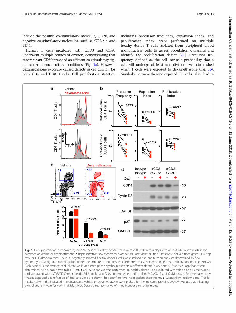
include the positive co-stimulatory molecule, CD28, andnegative co-stimulatory molecules, such as CTLA-4 andPD-1.Human T cells incubated with αCD3 and CD80
underwent multiple rounds of division, demonstrating thatrecombinant CD80 provided an efficient co-stimulatory sig-nal under normal culture conditions (Fig. 1a). However,dexamethasone exposure caused defects in cell division forboth CD4 and CD8 T cells. Cell proliferation statistics,
including precursor frequency, expansion index, andproliferation index, were performed on multipleheathy donor T cells isolated from peripheral bloodmononuclear cells to assess population dynamics andidentify the proliferation defect [29]. Precursor fre-quency, defined as the cell-intrinsic probability that acell will undergo at least one division, was diminishedwhen T cells were exposed to dexamethasone (Fig. 1b).Similarly, dexamethasone-exposed T cells also had a
c
a b
d
Fig. 1 T cell proliferation is impaired by dexamethasone. Healthy donor T cells were cultured for four days with αCD3/CD80 microbeads in thepresence of vehicle or dexamethasone. a Representative flow cytometry plots of CellTrace violet dilution. Plots were derived from gated CD4 (toprow) or CD8 (bottom row) T cells. b Negatively-selected healthy donor T cells were stained and proliferation analyses determined by flowcytometry following four days of culture under the indicated conditions. Precursor Frequency, Expansion Index, and Proliferation Index are shown.Each symbol is the average of duplicate wells, and each paired symbol represents a different donor (n = 5 donors). Statistical significance wasdetermined with a paired two-tailed T test. c Cell cycle analysis was performed on healthy donor T cells cultured with vehicle or dexamethasoneand stimulated with αCD3/CD80 microbeads. EdU uptake and DNA content were used to identify G0/G1, S, and G2/M phases. Representative flowimages (top) and quantification of duplicate wells are shown (bottom) from two independent experiments. d Lysates from healthy donor T cellsincubated with the indicated microbeads and vehicle or dexamethasone were probed for the indicated proteins. GAPDH was used as a loadingcontrol and is shown for each individual blot. Data are representative of three independent experiments
Giles et al. Journal for ImmunoTherapy of Cancer (2018) 6:51 Page 4 of 13
on March 12, 2022 by guest. P
rotected by copyright.http://jitc.bm
j.com/
J Imm
unother Cancer: first published as 10.1186/s40425-018-0371-5 on 11 June 2018. D
ownloaded from

reduced expansion index, a cell-extrinsic statistic used toexpress the fold expansion of the final cell count com-pared to the initial cell count (including undivided cells).Finally, the proliferation index, which represents the num-ber of divisions that cells in the post-mitotic populationhave undergone, was modestly decreased by dexametha-sone. Thus, cell proliferation statistics demonstrate thatdexamethasone impaired the ability of CD80co-stimulated CD4 and CD8 T cells to divide and re-stricted the expansion potential of these populations. Incontrast, dexamethasone imposed only subtle cell divisiondefects under all conditions in which CD28 was exclu-sively ligated with an αCD28 antibody (Additional file 1:Figure S1), indicating that direct ligation of CD28 canconfer resistance to dexamethasone.
Dexamethasone attenuates the CD28 signaling pathwayIncreased apoptosis and cell cycle blockade can eachmanifest as a proliferative defect in vitro.Dexamethasone did not increase the frequency ofapoptotic T cells when cultured with αCD3 and eitherαCD28 or CD80 co-stimulation as demonstrated by flowcytometry and Western analysis of cleaved caspase 3(Additional file 2: Figure S2), demonstrating that celldeath was not responsible for the proliferative defect. In-deed, dexamethasone has been shown to protect T cellsfrom activation-induced cell death [30].Cell cycle progression of CD80-costimulated T cells
was next evaulated in response to dexamethasone expos-ure. Dexamethasone induced, on average, a 13% increasein the G0/G1 phase along with a 67% reduction of cellsin S-phase and a 49% reduction in the G2/M phase (Fig.1c). Dexamethasone-exposed T cells also contained in-creased levels of p27 protein, an inhibitor of cell cycleentry, and concurrent reductions in CDK4 and cyclinD3 protein. Together, these data demonstrate that dexa-methasone blocks cell cycle entry in T cell stimulatedwith αCD3 and CD80.
Co-stimulation protects T cells from the anti-proliferativeeffects of dexamethasoneFollowing stimulation-induced expansion T cells mayadvance to a more terminally differentiated state.However, T cells cultured with dexamethasone had agreater proportion of naïve T cells (TN) and centralmemory T cells (TCM) and fewer effector memory Tcells (TEM) than cells cultured with vehicle controlafter four days of stimulation (Additional file 3: Fig-ure S3). Terminal effector T cells (TTE) were un-changed. These data indicate that dexamethasoneblocked the ability of T cells to differentiate in re-sponse to αCD3/CD80 stimulation. The loss in TEM
numbers was reversed by increasing concentrations ofCD80 (Fig. 2 and Additional file 4: Figure S4), suggesting
that strong co-stimulation may protect this subsetfrom the inhibitory effects of dexamethasone.To directly test the impact of dexamethasone on T cell
differentiation subsets, circulating T cells from healthydonors were sorted by flow cytometry into TN, TCM, andTEM subsets. The proliferative response of each wasassessed in response to stimulation and exposure tovehicle or dexamethasone. Cultures from purified naïveCD4 and CD8 T cells were most severely impaired bydexamethasone, with a diminished precursor frequency,expansion index, and proliferation index (Fig. 3). Incontrast, purified TCM response was comparable tovehicle control. TEM demonstrated reduced precursorfrequency but greater cell number, suggesting thatalthough dexamethasone impaired cell cycle entry, thecells were not lost due to apoptosis. Also, unlike TN,dexamethasone did not impair the expansion orproliferation index of TEM, demonstrating that thissubset was less sensitive to dexamethasone than TN.
CTLA-4 blockade protects T cells from the deleteriouseffects of dexamethasoneCD80 provides a positive co-stimulatory signal to T cellswhen bound by CD28. However, following TCR signaltransduction, CTLA-4 is translocated to the outer mem-brane of T cells where it can outcompete CD28 to bindCD80 and block co-stimulation [31–34]. Using flow cy-tometry, we confirmed that T cell stimulation led to in-creased extracellular CTLA-4 protein levels. In thepresence of dexamethasone, however, stimulation causeda fourfold increase in surface CTLA-4 protein comparedto vehicle treated (Fig. 4a) as well as an increase inCTLA-4 transcription (Fig. 4b). For these experiments,T cells were stimulated with αCD3 and αCD28 anti-bodies because surface CTLA-4 is internalized followingligation by CD80 [35], potentially impairing detection byflow cytometry antibodies. CTLA-4 expression was con-sistently found to be higher in CD4 T cells compared toCD8 T cells during dexamethasone treatment, likely ren-dering CD4 T cells more susceptible to cell cycle inhib-ition by checkpoint molecule.We hypothesized that dexamethasone-induced CTLA-4
upregulation on T cells out-competed CD28 for the sharedCD80 ligand, thereby attenuating the CD28 co-stimulatorypathway and inhibiting cell cycle entry. To test this,CTLA-4 was blocked with ipilimumab, a monoclonalanti-CTLA-4 human antibody, thereby enhancing the abil-ity of CD28 to bind CD80. With ipilimumab treatment,dexamethasone-induced cell proliferation defects were par-tially restored. Precursor frequency and expansion index ofdexamethasone-treated CD4 and CD8 T cells were signifi-cantly increased (Fig. 4c) as well as the proliferation indexof CD4 T cells. In contrast, the proliferation index of CD8T cells co-stimulated with CD80 was not increased with
Giles et al. Journal for ImmunoTherapy of Cancer (2018) 6:51 Page 5 of 13
on March 12, 2022 by guest. P
rotected by copyright.http://jitc.bm
j.com/
J Imm
unother Cancer: first published as 10.1186/s40425-018-0371-5 on 11 June 2018. D
ownloaded from

ipilimumab. T cells exposed to dexamethasone also upregu-lated of PD-1 during in vitro stimulation, but blockingPD-1 with nivolumab did not significantly change T cellprecursor frequency (Additional file 5: Figure S5). Thesedata demonstrate that ipilimumab specifically reversed theproliferation defects caused by dexamethasone, implicatingCTLA-4 as a mechanism for the anti-proliferative activitiesof dexamethasone exposure in T cells.
The relative proportion of TN and TCM were increasedin cultures exposed to dexamethasone compared tovehicle control. CTLA-4 blockade with ipilimumab ledto an expansion of the TCM subset, and this was recapit-ulated in T cells cultured with the combination of ipili-mumab and dexamethasone (Fig. 4d). Blocking CTLA-4with ipilimumab increased IFNγ, but not IL-2 transcrip-tion during dexamethasone exposure, suggesting that
b
a
Fig. 2 Increased co-stimulation ameliorates the inhibitory effects of dexamethasone. Negatively-selected healthy donor T cells were cultured with5 μg/mL αCD3 and increasing concentrations of CD80 in the presence of vehicle or dexamethasone. a-b CD4 T cells cultured with vehicle (a) ordexamethasone (b). Flow cytometry plots showing proliferation of cells cultured with the indicated concentration of CD80 (left) and totalnumbers of naïve (TN), central memory (TCM), effector memory (TEM), and terminal effector (TTE) T cells following four days of culture (right) areshown. Differentiation subsets were assessed by CD45RO and CCR7 staining. Each condition was plated in duplicate, and data are representativeof three independent experiments. Data were analyzed with an unpaired, two-tailed T Test
Fig. 3 Naïve and effector memory T cells show sensitivity to dexamethasone. a Healthy donor T cells were sorted into TN, TCM, and TEM subsetsby flow cytometry. Sorted subsets were cultured with αCD3/CD80 microbeads in the presence of dexamethasone (red) or vehicle control (black).Total cell numbers, Precursor Frequency, Expansion Index, and Proliferation Index of CD4 T cells (top) and CD8 T cells (bottom) are shown. Allsamples were plated in duplicate and analyzed with a paired, two-tailed T test. Data are representative of three independent experiments
Giles et al. Journal for ImmunoTherapy of Cancer (2018) 6:51 Page 6 of 13
on March 12, 2022 by guest. P
rotected by copyright.http://jitc.bm
j.com/
J Imm
unother Cancer: first published as 10.1186/s40425-018-0371-5 on 11 June 2018. D
ownloaded from

CTLA-4 blockade may rescue IFNγ-producing T cellsinhibited by dexamethasone (Fig. 4e).
Checkpoint blockade enhances survival ofdexamethasone-treated tumor-bearing miceTo determine if these findings translated to in vivomodels, the C57Bl/6 syngeneic glioma tumor line GL261was implanted intracranially. Consistent with the humanin vitro data, dexamethasone increased the percentage ofCTLA-4-expressing CD4 T cells of tumor-bearing micein a dose-dependent manner (Fig. 5a). The percent ofCTLA-4-expressing CD8 T cells also significantly in-creased in mice treated with the highest concentrationof dexamethasone (2.5 mg/kg/day). Because dexametha-sone upregulated CTLA-4, we hypothesized thatCTLA-4 blockade would provide a survival advantage to
dexamethasone-treated mice. GL261 tumor cells wereimplanted one week before commencement of dexa-methasone or vehicle treatment to allow for immune sur-veillance and development of differentiated anti-tumor Tcells. CTLA-4 blockade or isotype antibody were adminis-tered on days 13, 16, and 19 to vehicle ordexamethasone-treated mice to emulate the clinical sce-nerios where patients may be off or on corticosteroids be-fore receiving immunotherapy (Fig. 5b). To ensurecomparable tumor burden, mice were randomized intocohorts with equivalent tumor luminescence prior totreatment, and luminescence was measured weekly there-after (Fig. 5c).Single-agent treatment with CTLA-4 blocking anti-
body or dexamethasone did not produce a significantsurvival benefit compared to vehicle-treated animals in
c d
e
a b
Fig. 4 CTLA-4 blockade partially restores T cell proliferation in the presence of dexamethasone. a Flow cytometry analysis of CTLA-4 surfaceexpression on CD4 (left) or CD8 (right) T cells stimulated with αCD3/αCD28 microbeads. Unstimulated (dashed line), stimulated in presence ofvehicle (solid line), and stimulated in presence of dexamethasone (filled red line) are shown (left) and median fluorescence intensity (MFI) ofCTLA-4-expressing T cells is quantified (right). Data are representative of four independent experiments. b Expression of CTLA-4 by qPCR of T cellsstimulated in the presence of vehicle or dexamethasone. Data are representative of four independent experiments. c Healthy donor T cellsstimulated for four days in the presence of vehicle or dexamethasone and with or without ipilimumab F(ab’)2 antibody. Proliferation analysis ofCD4 T cells (top) and CD8 T cells (bottom) was performed, and the ratio of cells stimulated with dexamethasone relative to vehicle control areshown for Precursor Frequency (PF), Expansion Index (EI), and Proliferation Index (PI). All samples were plated in duplicate and the ratios wereanalyzed with an unpaired, two-tailed T test. Data are representative of 7 healthy donors. d Cells were cultured as in (c). The number of T cells ineach differentiation group were quantified by flow cytometry and analyzed by SPICE. e Expression of the indicated cytokines was determined byqPCR. Five healthy donors were assayed for each condition. Each data point represents and average of triplicate wells. Data were analysed with apaired, two-tailed T test
Giles et al. Journal for ImmunoTherapy of Cancer (2018) 6:51 Page 7 of 13
on March 12, 2022 by guest. P
rotected by copyright.http://jitc.bm
j.com/
J Imm
unother Cancer: first published as 10.1186/s40425-018-0371-5 on 11 June 2018. D
ownloaded from

this well-established glioblastoma model. However, micethat received CTLA-4 blockade in addition to dexa-methasone treatment survived significantly longer thancontrol animals, with a median survival of 49 versus39 days, respectively (p = 0.013; Fig. 5d). Here, the im-mune system surveyed the immunogenic GL261luciferase-mCherry tumor for one week before dexa-methasone treatment was initiated. To determine if im-mune priming contributed to dexamethasone resistance,this experiment was repeated to include a cohort of miceexposed to dexamethasone one day prior to tumor im-plantation. In this cohort, CTLA-4 blockade did notprovide a survival benefit (Additional file 6: Figure S6).These data indicate that dexamethasone exposure mayblock a successful anti-tumor immune response if treat-ment occurs before anti-tumor T cells can differentiatefrom the TN pool.The impact of dexamethasone exposure was next
evaluated on lymphocyte populations from tumor-bearingmice, including tumor-infiltrating lymphocytes (TILs) andT cells in the tumor-draining cervical lymph nodes(TDLN). Mice exposed to dexamethasone possessed sig-nificantly reduced numbers of T cells in TDLNs. However,
CTLA-4 blockade increased the total number of CD4 Tcells in TDLN and CD8 TILs (Fig. 6a and b). The numberof regulatory T cells (Treg) in the tumor-bearing brainwere not significantly affected by dexamethasone orCTLA-4 blockade whereas overall numbers were reducedin TDLNs from each dexamethasone-treated cohort (Add-itional file 7: Figure S7A).Dexamethasone exposure reduced the total number
of T cells in the majority of T cell subsets along thedifferentiation spectrum in the tumor-draining lymphnodes, yet as observed in vitro, this was partially res-cued by CTLA-4 blockade (Fig. 6a and b). The num-ber of CD4 TN and TCM TIL subsets were alsosignificantly elevated by CTLA-4 blockade in thedexamethasone-treated group. The relative proportionof TCM TILs was increased by CTLA-4 blockade inboth the vehicle and dexamethasone-treated cohorts(Fig. 6c). GBM has been reported to induce high ex-pression of checkpoint molecules on TILs, resultingin a severe exhaustion signature [36]. Dexamethasonetreatment increased the percentage of CD8 TILs thatexpressed Tim-3 from 2 to 12%, although it did notsignificantly impact other checkpoint molecules (PD-1
dc
a b
Fig. 5 CTLA-4 blockade enhances survival of dexamethasone-treated mice. a CTLA-4 was measured on circulating CD4 (left) and CD8 (right) Tcells 1 h following oral gavage of vehicle or the indicated concentration of dexamethasone. Each cohort contained eight mice with intracranialGL261 tumors. Vehicle and dexamethasone-treated cohorts were statistically analyzed with an unpaired two-tailed student’s T-test. b Schema oftreatment cohorts for (c-d). GL261 ffluc-mCherry glioma cells were orthotopically implanted into C57BL/6 mice one week before treatmentinitiation. Luminescence readings were acquired 6 days following tumor implantation and weekly thereafter. Mice were treated with vehicle ordexamethasone as indicated. CTLA-4 blocking antibody or isotype control were administered on days 13, 16, and 19 following tumorimplantation. c Luminescence of tumor-bearing mice at days 13 and 20 following tumor implantation. d Kaplan Meier survival curves of micereceiving the indicated treatments. n = 7 to 8 mice per cohort. Data are representative of two independent experiments
Giles et al. Journal for ImmunoTherapy of Cancer (2018) 6:51 Page 8 of 13
on March 12, 2022 by guest. P
rotected by copyright.http://jitc.bm
j.com/
J Imm
unother Cancer: first published as 10.1186/s40425-018-0371-5 on 11 June 2018. D
ownloaded from

c
b
a
d
Fig. 6 (See legend on next page.)
Giles et al. Journal for ImmunoTherapy of Cancer (2018) 6:51 Page 9 of 13
on March 12, 2022 by guest. P
rotected by copyright.http://jitc.bm
j.com/
J Imm
unother Cancer: first published as 10.1186/s40425-018-0371-5 on 11 June 2018. D
ownloaded from

or Lag-3) or the frequency of TILs expressing mul-tiple checkpoint molecules (Additional file 7: FigureS7B). Although T cells expressing checkpoint mole-cules were less frequent in TDLN, dexamethasone de-creased the frequency of most checkpoint-expressingT cells with the exception of Tim-3+ CD8 T cells,which increased from 1.5 to 7.5 and 1.3 to 4.7% inisotype and CTLA-4 blockade treated mice, respect-ively. Finally, the number of IFNγ-producing TILs indexamethasone-treated mice was significantly in-creased with CTLA-4 blockade, consistent with invitro data (Fig. 6d). Together, these data demonstratethat CTLA-4 blockade can partially reverse the in-hibitory effects of dexamethasone on T cells in vivo,reduce the proportion of TILs expressing checkpointmolecules, increase IFNγ-expressing TILs and extendsurvival of dexamethasone-treated mice bearing intra-cranial gliomas.
DiscussionCorticosteroids, most commonly dexamethasone, areregularly administered to patients with intracranialtumors to combat cerebral edema and providesymptomatic relief [37]. Additionally, corticosteroidsare used to treat patients who develop irAEs as aresult of immunotherapy. In contrast to the frequentearly use of dexamethasone for tumor-associatededema, corticosteroid use for immunotherapy-relatedtoxicity is always given after treatment has com-menced. Corticosteroids have been established ascausing dose-related immunosuppression, yet themechanisms behind this impaired immune function,particularly in the context of cancer immunotherapy,have not been defined [38]. Importantly, it is notknown if corticosteroids impede the differentiation offreshly stimulated T cells or if they deplete establishedand already differentiated tumor-reactive lymphocytes.In this study, the immunosuppressive effects of
dexamethasone on individual T cell differentiationsubsets were interrogated. TN were identified as beingexceptionally sensitive to dexamethasone-mediated cellcycle blockade. TN are a key source of secondaryanti-tumor immunity mediated by antigen spread in re-sponse to checkpoint blockade [39] and are highly sensi-tive to anergy imposed by expression of CTLA-4 [40].
Dexamethasone exposure strongly upregulated PD-1 andCTLA-4 transcription and protein, consistent with pre-vious in vitro murine studies [41, 42]. The data pre-sented here extend upon these studies to demonstratethat dexamethasone-induced CTLA-4 upregulation ef-fectively blocked TN proliferation and differentiation inboth murine and human T lymphocytes. Intumor-bearing mice, this led to a loss of differentiated Tcell subsets in several lymphoid tissues.In contrast to TN lymphocytes, dexamethasone
exposure had much less impact on memory T cellproliferation following flow cytometry sorting. TEM hadreduced precursor frequency but increased totalnumbers. Our data confirm and expand previous studiesthat demonstrated that the efficacy of TCR transgenic Tcells was not impaired by dexamethasone treatment[43]. Here, endogenously generated anti-tumor immun-ity could be maintained during dexamethasone treat-ment if CTLA-4 blockade was provided. BlockingCTLA-4, but not PD-1, partially rescued T cell prolifera-tion in the presence of dexamethasone in vitro. Thesefindings may reflect that CTLA-4 blockade acts uponless differentiated T cells, which are most sensitive todexamethasone. Systemic immunity elicited by CTLA-4blockade has been previously shown to promoteanti-tumor immunity against melanoma metastaseswithin the central nervous system, indicating thatCTLA-4 blockade functions outside of the CNS [15, 44].The GL261 luciferase-mCherry tumor cells provided animmunogenic intracranial tumor model. By waiting oneweek before dexamethasone treatment, tumor surveil-lance and immune activation were permitted, potentiallyleading to differentiation of tumor-reactive T cells beforedexamethasone exposure. In this model, CTLA-4 block-ade was sufficient to provide a survival advantage todexamethasone-treated mice. In contrast, mice exposedto dexamethasone prior to tumor implantation and anti-gen exposure were unresponsive to CTLA-4 blockade.Collectively, these data suggest that tipping the immuneresponse to more differentiated subsets may lesson theimmune suppression imposed by dexamethasone.These results demonstrate that the timing of
dexamethasone treatment relative to the development ofanti-tumor immunity significantly impacts the efficacy ofimmunotherapy. Previous work has shown that
(See figure on previous page.)Fig. 6 CTLA-4 blockade rescues lymphocyte defects induced by dexamethasone. GL261 ffluc-mCherry tumor-bearing mice were randomized intothe indicated cohorts. Vehicle or dexamethasone treatment was initiated on day 7, and isotype or CTLA-4 blocking antibody were administeredon days 13, 16, and 19 following tumor implantation. Mice were euthanized on day 23 and tissues were harvested for flow cytometry analysis. a-b CD4 (a) and CD8 (b) T cells were quantified along with the indicated differentiation subsets using CD44 and CD62L expression. Brains (n = 8)and cervical lymph nodes (n = 10) were collected. Data are analyzed using a unpaired students T test. c The relative contribution of eachdifferentiation subset is shown for CD4 (top) and CD8 (bottom) TILs. d The total number of IFNγ-producing T cells were quantified from thetumor-bearing hemispheres of mice from the indicated cohorts. Data are analyzed using an unpaired students T test. N = 8 mice/group
Giles et al. Journal for ImmunoTherapy of Cancer (2018) 6:51 Page 10 of 13
on March 12, 2022 by guest. P
rotected by copyright.http://jitc.bm
j.com/
J Imm
unother Cancer: first published as 10.1186/s40425-018-0371-5 on 11 June 2018. D
ownloaded from

corticosteroids provided to alleviate irAEs did not im-pact the overall response rate of patients with melan-oma who received nivolumab [45]. For those patients,corticosteroids were provided after immunotherapywas initiated. In contrast, patients with intracranialtumors are routinely provided high-dose corticoste-roids from the period of initial diagnosis until chemo-radiation completion, a period that can span 8–12 weeks. Corticosteroids are also provided duringsurgical resection, as they have been shown to extendsurvival in this context [46]. Thus, for patients withintracranial tumors, corticosteroids provided beforeinitiation of immunotherapy, a time when the im-mune system is unlikely to be actively proliferating,may blunt the generation of an anti-tumor response.Similarly, dexamethasone provided to immunologically
“cold” tumors or those with insufficient anti-tumor im-munity will likely abrogate new priming and differenti-ation of anti-tumor T cells. However, the concurrent useof CTLA-4 blockade can encourage TN activation,thereby contributing to antigen spread. Once anti-tumorimmunity has been initiated, the negative impact of cor-ticosteroids on immune function is markedly reduced.These results may have important implications in de-signing future immunotherapy strategies helping tooptimize clinical trials for patients with brain cancers aswell as other diseases where corticosteroid use iscommon.Steroid alternatives may need to be considered for
patients with intracranial tumors who wish to enroll onimmunotherapy trials. For example, blockade of vascularendothelial growth factor by bevacizumab reducesedema by normalizing tumor vasculature. Further, it hasbeen shown to promote lymphocyte infiltration into thetumor and increase circulating memory T cell numbers[47]. Such alternative approaches may be needed tomanage symptoms in patients with intracranial tumorswhile preserving the potential for anti-tumor immunity.
ConclusionsHere, we interrogated the impact of dexamethasone onT cell subsets in the setting of immunotherapy.Dexamethasone blocks naïve T cell proliferation anddifferentiation by attenuating CD28 co-stimulation. Be-cause co-stimulation is essential for successful T cell prim-ing and expansion, these data suggest that corticosteroidsimpair response in immunotherapy treatment-naïve pa-tients or those with poorly antigenic tumors. However, Tcells may be partially protected or rescued from the im-munosuppressive effects of dexamethasone with adminis-tration of CTLA-4 blockade. Additionally, negativecorticosteroid effects are diminished after developing asuccessful anti-tumor immune response.
Additional files
Additional file 1: Figure S1. T cell stimulated with αCD3/αCD28microbeads proliferate in the presence of dexamethasone.Healthy donorT cells were cultured for four days with the indicated ratio of αCD3/αCD28 microbeads:total T cells in the presence of vehicle ordexamethasone. A, Representative flow cytometry plots of CellTraceviolet dilution. Plots were derived from gated CD4 (top row) or CD8(bottom row) T cells. B-D, Proliferation analyses of CD4 T cells (top) andCD8 T cells (bottom) performed on the samples shown in (A). PrecursorFrequency (B), Expansion Index (C), and Proliferation Index (D) are shown.Samples were plated in duplicate and analyzed with an unpairedstudents T test. Data are representative of three independentexperiments. (PDF 3563 kb)
Additional file 2: Figure S2. A, Negatively-selected healthy donor Tcells were cultured with the indicated microbeads and vehicle or dexa-methasone. The percent of apoptotic CD4 (top) and CD8 (bottom) T cellswas assessed by Annexin V/PI. Data are representative of four independ-ent experiments. B, Lysates from healthy donor T cells incubated withthe indicated microbeads and vehicle or dexamethasone were probedfor the indicated proteins. GAPDH was used as a loading control. (PDF693 kb)
Additional file 3: Figure S3. T cell differentiation subsets formedduring in vitro stimulation with αCD3/CD80 stimulation. Negatively-selected healthy donor T cells were cultured with 5 μg/mL αCD3 and theindicated concentration of CD80. T cell differentiation subsets were quan-tified following four days of culture. A, Flow plot of gating strategy toidentify the indicated T cell differentiation subsets. B, Flow plots of CD4(top) and CD8 (bottom) T cells cultured under the indicated conditions.(PDF 3995 kb)
Additional file 4: Figure S4. Increased co-stimulation ameliorates theinhibitory effects of dexamethasone. Negatively-selected healthy donor Tcells were cultured with 5 μg/mL αCD3 and increasing concentrations ofCD80 in the presence of vehicle or dexamethasone. A-B. CD8 T cells cul-tured with vehicle (A) or dexamethasone (B). Flow cytometry plots show-ing proliferation of cells cultured with the indicated concentration ofCD80 (left) and total numbers of naïve (TN), central memory (TCM), effectormemory (TEM), and terminal effector (TTE) T cells following four days ofculture (right) are shown. Differentiation subsets were assessed byCD45RO and CCR7 staining. Each condition was plated in duplicate, anddata are representative of three independent experiments. Data were an-alyzed with an unpaired, two-tailed T Test. (PDF 2573 kb)
Additional file 5: Figure S5 PD-1 blockade does not rescuedexamethasone-mediated proliferation defects. A, Flow cytometry ana-lysis of PD-1 surface expression on CD4 (left) or CD8 (right) T cells stimu-lated with αCD3/αCD28 microbeads. Unstimulated (dashed line),stimulated in presence of vehicle (solid line), and stimulated in presenceof dexamethasone (filled red line) are shown. B, Geometric median fluor-escence intensity (gMFI) of PD-1 staining on CD4 or CD8 T cells. Cells cul-tured with vehicle (black bars) and dexamethasone (red bars) are shown.Data are an average of duplicate samples. C, Expression of PD-1 by qPCRof T cells stimulated in the presence of vehicle or dexamethasone. Dataare representative of four independent experiments. D-E. Healthy donorT cells were stimulated for four days in the presence of vehicle or dexa-methasone and nivolumab or ipilimumab F(ab’)2 antibody as indicated.Precursor frequency of CD4 and CD8 T cells was quantified by FlowJo.The ratio of dexamethasone to vehicle for CD4 (C) and CD8 (D) T cells isshown. All samples were plated in duplicate and the ratios were analyzedwith a one-way ANOVA. Data are representative of n = 4 healthy donors.(PDF 2522 kb)
Additional file 6: Figure S6 CTLA-4 blockade does not rescue dexa-methasone pre-treated mice. A, Schema of survival experiment. AlbinoC57Bl/6 mice received intracranial implantation of GL261 ffluc-mCherryglioma cells. Dexamethasone was initiated one day prior to tumor im-plantation (dex (D-1)) or one week following tumor implantation (dex(D7)). CLTA-4 blockade or isotype antibody were injected on days 13, 16,and 19 following tumor implantation. Mice were randomized on day 6following tumor implantation into groups of equivalent tumor
Giles et al. Journal for ImmunoTherapy of Cancer (2018) 6:51 Page 11 of 13
on March 12, 2022 by guest. P
rotected by copyright.http://jitc.bm
j.com/
J Imm
unother Cancer: first published as 10.1186/s40425-018-0371-5 on 11 June 2018. D
ownloaded from

luminescence. B, Kaplan Meier survival curves of mice receiving the indi-cated treatments. n = 8 to 9 mice per cohort. Data are representative oftwo independent experiments. (PDF 1525 kb)
Additional file 7: Figure S7. Quantification of Treg and checkpointmolecules in tumor-bearing mice. GL261 ffluc-mCherry tumor-bearingmice were randomized into the indicated cohorts based on biolumines-cence values from tumor. Vehicle or dexamethasone treatment was initi-ated on day 7, and isotype or CTLA-4 blocking antibody wereadministered on days 13, 16, and 19 following tumor implantation. Micewere euthanized on day 23 and tissues were harvested for flow cytome-try analysis. A, Treg cell number from tumor-bearing brain hemisphere(left; n = 8) or the cervical tumor-draining lymph nodes (right; n = 10). B,The percentage of CD4 (top two plots) or CD8 (bottom two plots) T cellsexpressing the indicated checkpoint molecules. Co-expression of mole-cules was quantified using a Boolean gating strategy. Data were analyzedusing a unpaired students T test. (PDF 1891 kb)
AbbreviationsAPC: Antigen-Presenting Cell; EI: Expansion Index; irAE: immune-relatedAdverse Events; PF: Precursor Frequency; PI: Proliferation Index; Tcm: CentralMemory T cells; Tem: Effector Memory T cells; Tn: naïve T cells
AcknowledgementsThe authors wish to acknowledge Jonathan D. Ashwell, M.D. for insightfulscientific discussions.
FundingThis research was supported in part by the Intramural Research Program ofthe NIH, NCI.
Availability of data and materialsAll data analyzed are included in this article and additional information isavailable upon request.
Authors’ contributionsAJG designed research studies, conducted experiments, acquired andanalyzed data, and wrote the manuscript. MH designed research studies,conducted experiments, acquired and analyzed data. HMS, JJ, NR, CMR, WZ,HS, RB, and DD conducted experiments, acquired and analyzed data. PEFcontributed data and edited the manuscript. DMP designed research studies,provided reagents, and edited the manuscript. MRG designed researchstudies, analyzed data, and helped write the manuscript. All authors read andapproved the final manuscript.
Ethics approval and consent to participateNot applicable; not a clinical trial.
Competing interestsThe authors declare that they have no competing interests.
Publisher’s NoteSpringer Nature remains neutral with regard to jurisdictional claims inpublished maps and institutional affiliations.
Author details1Neuro-Oncology Branch, CCR, NCI, National Institutes of Health, 37 ConventDr. Bldg. 37, Rm. 1142B, Bethesda, MD 20892, USA. 2Department ofNeurosurgery, Duke University Medical Center, Durham, NC, USA.
Received: 20 February 2018 Accepted: 30 May 2018
References1. Hoos A. Development of immuno-oncology drugs - from CTLA4 to PD1 to
the next generations. Nat Rev Drug Discov. 2016;15(4):235–47. https://doi.org/10.1038/nrd.2015.35.
2. Farber SH, Tsvankin V, Narloch JL, Kim GJ, Salama AK, Vlahovic G, et al.Embracing rejection: immunologic trends in brain metastasis.
Oncoimmunology. 2016;5(7):e1172153. https://doi.org/10.1080/2162402X.2016.1172153.
3. Batich KA, Reap EA, Archer GE, Sanchez-Perez L, Nair SK, Schmittling RJ,et al. Long-term survival in glioblastoma with cytomegalovirus pp65-targeted vaccination. Clin Cancer Res. 2017;23(8):1898–909. https://doi.org/10.1158/1078-0432.CCR-16-2057.
4. Reardon DA, Gokhale PC, Klein SR, Ligon KL, Rodig SJ, Ramkissoon SH,et al. Glioblastoma eradication following immune checkpoint blockadein an Orthotopic. Immunocompetent Model Cancer Immunol Res. 2016;4(2):124–35. https://doi.org/10.1158/2326-6066.CIR-15-0151.
5. Fecci PE, Ochiai H, Mitchell DA, Grossi PM, Sweeney AE, Archer GE, etal. Systemic CTLA-4 blockade ameliorates glioma-induced changes tothe CD4+ T cell compartment without affecting regulatory T-cellfunction. Clin Cancer Res. 2007;13(7):2158–67. https://doi.org/10.1158/1078-0432.CCR-06-2070.
6. Larkin J, Hodi FS, Wolchok JD. Combined Nivolumab and Ipilimumab ormonotherapy in untreated melanoma. N Engl J Med. 2015;373(13):1270–1.https://doi.org/10.1056/NEJMc1509660.
7. Weber JS, Kahler KC, Hauschild A. Management of immune-related adverseevents and kinetics of response with ipilimumab. J Clin Oncol. 2012;30(21):2691–7. https://doi.org/10.1200/JCO.2012.41.6750.
8. Michot JM, Bigenwald C, Champiat S, Collins M, Carbonnel F, Postel-Vinay S,et al. Immune-related adverse events with immune checkpoint blockade: acomprehensive review. Eur J Cancer. 2016;54:139–48. https://doi.org/10.1016/j.ejca.2015.11.016.
9. Garant A, Guilbault C, Ekmekjian T, Greenwald Z, Murgoi P, Vuong T.Concomitant use of corticosteroids and immune checkpoint inhibitorsin patients with hematologic or solid neoplasms: a systematic review.Crit Rev Oncol Hematol. 2017;120:86–92. https://doi.org/10.1016/j.critrevonc.2017.10.009.
10. Margolin K, Ernstoff MS, Hamid O, Lawrence D, McDermott D, Puzanov I, etal. Ipilimumab in patients with melanoma and brain metastases: an open-label, phase 2 trial. Lancet Oncol. 2012;13(5):459–65. https://doi.org/10.1016/S1470-2045(12)70090-6.
11. Queirolo P, Spagnolo F, Ascierto PA, Simeone E, Marchetti P, Scoppola A, etal. Efficacy and safety of ipilimumab in patients with advanced melanomaand brain metastases. J Neuro-Oncol. 2014;118(1):109–16. https://doi.org/10.1007/s11060-014-1400-y.
12. Parakh S, Park JJ, Mendis S, Rai R, Xu W, Lo S, et al. Efficacy of anti-PD-1therapy in patients with melanoma brain metastases. Br J Cancer. 2017;116(12):1558–63. https://doi.org/10.1038/bjc.2017.142.
13. Horvat TZ, Adel NG, Dang TO, Momtaz P, Postow MA, Callahan MK, etal. Immune-related adverse events, need for systemicimmunosuppression, and effects on survival and time to treatmentfailure in patients with melanoma treated with Ipilimumab at memorialSloan Kettering Cancer center. J Clin Oncol. 2015;33(28):3193–8. https://doi.org/10.1200/JCO.2015.60.8448.
14. Attia P, Phan GQ, Maker AV, Robinson MR, Quezado MM, Yang JC, et al.Autoimmunity correlates with tumor regression in patients with metastaticmelanoma treated with anti-cytotoxic T-lymphocyte antigen-4. J Clin Oncol.2005;23(25):6043–53. https://doi.org/10.1200/JCO.2005.06.205.
15. Phan GQ, Yang JC, Sherry RM, Hwu P, Topalian SL, Schwartzentruber DJ, etal. Cancer regression and autoimmunity induced by cytotoxic Tlymphocyte-associated antigen 4 blockade in patients with metastaticmelanoma. Proc Natl Acad Sci U S A. 2003;100(14):8372–7. https://doi.org/10.1073/pnas.1533209100.
16. Harmankaya K, Erasim C, Koelblinger C, Ibrahim R, Hoos A, Pehamberger H,et al. Continuous systemic corticosteroids do not affect the ongoingregression of metastatic melanoma for more than two years followingipilimumab therapy. Med Oncol. 2011;28(4):1140–4. https://doi.org/10.1007/s12032-010-9606-0.
17. Beck KE, Blansfield JA, Tran KQ, Feldman AL, Hughes MS, Royal RE, etal. Enterocolitis in patients with cancer after antibody blockade ofcytotoxic T-lymphocyte-associated antigen 4. J Clin Oncol. 2006;24(15):2283–9. https://doi.org/10.1200/JCO.2005.04.5716.
18. Downey SG, Klapper JA, Smith FO, Yang JC, Sherry RM, Royal RE, et al.Prognostic factors related to clinical response in patients with metastaticmelanoma treated by CTL-associated antigen-4 blockade. Clin Cancer Res.2007;13(22 Pt 1):6681–8. https://doi.org/10.1158/1078-0432.CCR-07-0187.
19. Marchetti MC, Di Marco B, Cifone G, Migliorati G, Riccardi C.Dexamethasone-induced apoptosis of thymocytes: role of glucocorticoid
Giles et al. Journal for ImmunoTherapy of Cancer (2018) 6:51 Page 12 of 13
on March 12, 2022 by guest. P
rotected by copyright.http://jitc.bm
j.com/
J Imm
unother Cancer: first published as 10.1186/s40425-018-0371-5 on 11 June 2018. D
ownloaded from

receptor-associated Src kinase and caspase-8 activation. Blood. 2003;101(2):585–93. https://doi.org/10.1182/blood-2002-06-1779.
20. Mitchell CD, Richards SM, Kinsey SE, Lilleyman J, Vora A, Eden TO, et al.Benefit of dexamethasone compared with prednisolone for childhoodacute lymphoblastic leukaemia: results of the UK Medical Research CouncilALL97 randomized trial. Br J Haematol. 2005;129(6):734–45. https://doi.org/10.1111/j.1365-2141.2005.05509.x.
21. Bianchi M, Meng C, Ivashkiv LB. Inhibition of IL-2-induced Jak-STAT signalingby glucocorticoids. Proc Natl Acad Sci U S A. 2000;97(17):9573–8. https://doi.org/10.1073/pnas.160099797.
22. Franchimont D, Galon J, Vacchio MS, Fan S, Visconti R, Frucht DM, et al.Positive effects of glucocorticoids on T cell function by up-regulation of IL-7receptor alpha. J Immunol. 2002;168(5):2212–8.
23. Mitchell DA, Xie W, Schmittling R, Learn C, Friedman A, McLendon RE, et al.Sensitive detection of human cytomegalovirus in tumors and peripheralblood of patients diagnosed with glioblastoma. Neuro-Oncology. 2008;10(1):10–8. https://doi.org/10.1215/15228517-2007-035.
24. Scheurer ME, Bondy ML, Aldape KD, Albrecht T, El-Zein R. Detection ofhuman cytomegalovirus in different histological types of gliomas. ActaNeuropathol. 2008;116(1):79–86. https://doi.org/10.1007/s00401-008-0359-1.
25. Crough T, Beagley L, Smith C, Jones L, Walker DG, Khanna R. Ex vivofunctional analysis, expansion and adoptive transfer of cytomegalovirus-specific T-cells in patients with glioblastoma multiforme. Immunol Cell Biol.2012;90(9):872–80. https://doi.org/10.1038/icb.2012.19.
26. Berghoff AS, Fuchs E, Ricken G, Mlecnik B, Bindea G, Spanberger T, et al.Density of tumor-infiltrating lymphocytes correlates with extent of brainedema and overall survival time in patients with brain metastases.Oncoimmunology. 2016;5(1):e1057388. https://doi.org/10.1080/2162402X.2015.1057388.
27. Broeren CP, Gray GS, Carreno BM, June CH. Costimulation light: activation ofCD4+ T cells with CD80 or CD86 rather than anti-CD28 leads to a Th2cytokine profile. J Immunol. 2000;165(12):6908–14.
28. Lanier LL, O'Fallon S, Somoza C, Phillips JH, Linsley PS, Okumura K, et al.CD80 (B7) and CD86 (B70) provide similar costimulatory signals for T cellproliferation, cytokine production, and generation of CTL. J Immunol. 1995;154(1):97–105.
29. Roederer M. Interpretation of cellular proliferation data: avoid thepanglossian. Cytometry A. 2011;79(2):95–101. https://doi.org/10.1002/cyto.a.21010.
30. Zacharchuk CM, Mercep M, Chakraborti PK, Simons SS Jr, Ashwell JD.Programmed T lymphocyte death. Cell activation- and steroid-inducedpathways are mutually antagonistic. J Immunol. 1990;145(12):4037–45.
31. Linsley PS, Nadler SG, Bajorath J, Peach R, Leung HT, Rogers J, et al. Bindingstoichiometry of the cytotoxic T lymphocyte-associated molecule-4 (CTLA-4). A disulfide-linked homodimer binds two CD86 molecules. J Biol Chem.1995;270(25):15417–24.
32. van der Merwe PA, Bodian DL, Daenke S, Linsley P, Davis SJ. CD80 (B7-1)binds both CD28 and CTLA-4 with a low affinity and very fast kinetics. J ExpMed. 1997;185(3):393–403.
33. Walker LS, Sansom DM. The emerging role of CTLA4 as a cell-extrinsicregulator of T cell responses. Nat Rev Immunol. 2011;11(12):852–63. https://doi.org/10.1038/nri3108.
34. Engelhardt JJ, Sullivan TJ, Allison JP. CTLA-4 overexpression inhibits T cellresponses through a CD28-B7-dependent mechanism. J Immunol. 2006;177(2):1052–61.
35. Qureshi OS, Zheng Y, Nakamura K, Attridge K, Manzotti C, Schmidt EM, et al.Trans-endocytosis of CD80 and CD86: a molecular basis for the cell-extrinsicfunction of CTLA-4. Science. 2011;332(6029):600–3. https://doi.org/10.1126/science.1202947.
36. Woroniecka K, Chongsathidkiet P, Rhodin KE, Kemeny HR, Dechant CA,Farber SH, et al. T Cell Exhaustion Signatures Vary with Tumor Type and areSevere in Glioblastoma. Clin Cancer Res. 2018; https://doi.org/10.1158/1078-0432.CCR-17-1846.
37. Kaal EC, Vecht CJ. The management of brain edema in brain tumors. CurrOpin Oncol. 2004;16(6):593–600.
38. Min L, Hodi FS, Kaiser UB. Corticosteroids and immune checkpoint blockade.Aging (Albany NY). 2015;7(8):521–2. https://doi.org/10.18632/aging.100797.
39. Gulley JL, Madan RA, Pachynski R, Mulders P, Sheikh NA, Trager J, et al. Roleof antigen spread and distinctive characteristics of immunotherapy inCancer treatment. J Natl Cancer Inst. 2017;109(4) https://doi.org/10.1093/jnci/djw261.
40. Greenwald RJ, Boussiotis VA, Lorsbach RB, Abbas AK, Sharpe AH. CTLA-4regulates induction of anergy in vivo. Immunity. 2001;14(2):145–55.
41. Xia M, Gasser J, Feige U. Dexamethasone enhances CTLA-4 expressionduring T cell activation. Cell Mol Life Sci. 1999;55(12):1649–56. https://doi.org/10.1007/s000180050403.
42. Xing K, Gu B, Zhang P, Wu X. Dexamethasone enhances programmed celldeath 1 (PD-1) expression during T cell activation: an insight into theoptimum application of glucocorticoids in anti-cancer therapy. BMCImmunol. 2015;16(39) https://doi.org/10.1186/s12865-015-0103-2.
43. Hinrichs CS, Palmer DC, Rosenberg SA, Restifo NP. Glucocorticoids do notinhibit antitumor activity of activated CD8+ T cells. J Immunother. 2005;28(6):517–24.
44. Schartz NE, Farges C, Madelaine I, Bruzzoni H, Calvo F, Hoos A, et al.Complete regression of a previously untreated melanoma brain metastasiswith ipilimumab. Melanoma Res. 2010;20(3):247–50. https://doi.org/10.1097/CMR.0b013e3283364a37.
45. Weber JS, Hodi FS, Wolchok JD, Topalian SL, Schadendorf D, Larkin J, et al.Safety profile of Nivolumab monotherapy: a pooled analysis of patients withadvanced melanoma. J Clin Oncol. 2017;35(7):785–92. https://doi.org/10.1200/JCO.2015.66.1389.
46. Jelsma R, Bucy PC. The treatment of glioblastoma multiforme of the brain. JNeurosurg. 1967;27(5):388–400. https://doi.org/10.3171/jns.1967.27.5.0388.
47. Hodi FS, Lawrence D, Lezcano C, Wu X, Zhou J, Sasada T, et al. Bevacizumabplus ipilimumab in patients with metastatic melanoma. Cancer ImmunolRes. 2014;2(7):632–42. https://doi.org/10.1158/2326-6066.CIR-14-0053.
Giles et al. Journal for ImmunoTherapy of Cancer (2018) 6:51 Page 13 of 13
on March 12, 2022 by guest. P
rotected by copyright.http://jitc.bm
j.com/
J Imm
unother Cancer: first published as 10.1186/s40425-018-0371-5 on 11 June 2018. D
ownloaded from