Developmental disturbances of jaws and teeth
41
Developmental disturbances of oral and paraoral structures DR.SWAPNIL PAKHALE
-
Upload
swapnil-pakhale -
Category
Health & Medicine
-
view
56 -
download
6
Transcript of Developmental disturbances of jaws and teeth
Developmental disturbances of
oral and paraoral structures
DR.SWAPNIL PAKHALE
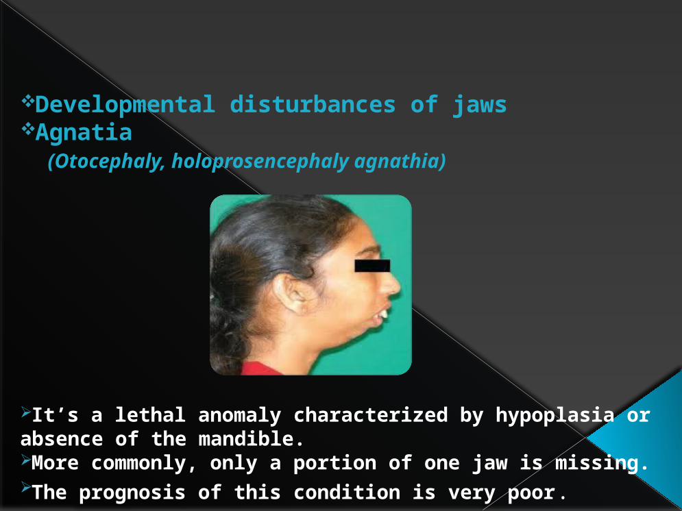
Developmental disturbances of jawsAgnatia (Otocephaly, holoprosencephaly agnathia)
It’s a lethal anomaly characterized by hypoplasia or absence of the mandible.More commonly, only a portion of one jaw is missing.The prognosis of this condition is very poor.

Micrognathia
Micrognathia means a small jaw, and either the maxilla or the mandible may b affected.True micrognathia is classified as either congenital or aquired.Congenital type is associated with congenital heart diseases and the Pierre Robin syndrome.The aquired type is of postnatal origin and results from a disturbances in the area of the temporomandibular joint.

Macrognathia
Macrognathia refers to the condition of abnormally large jaws.
It may be associated with Paget’s disease of bone, Acromegaly, leontiasis ossea, a form of fibrous dysplasia.

Facial Hemihyperatrophy
It is characterized by asymmetric overgrowth of one or more body parts.Hemifacial hyperplasia is the ivolvement of one side of the face.The permanent teeth on the affected side are often enlarged.The tongue is commonly invilved.
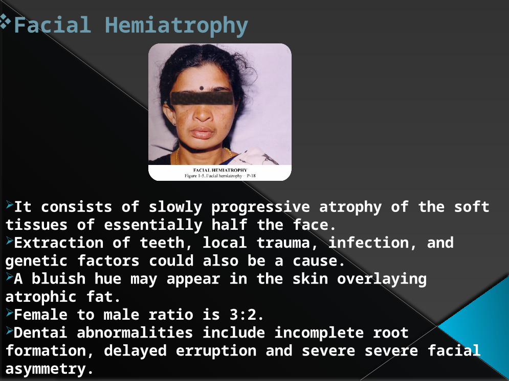
Facial Hemiatrophy
It consists of slowly progressive atrophy of the soft tissues of essentially half the face.Extraction of teeth, local trauma, infection, and genetic factors could also be a cause.A bluish hue may appear in the skin overlaying atrophic fat.Female to male ratio is 3:2.Dentai abnormalities include incomplete root formation, delayed erruption and severe severe facial asymmetry.
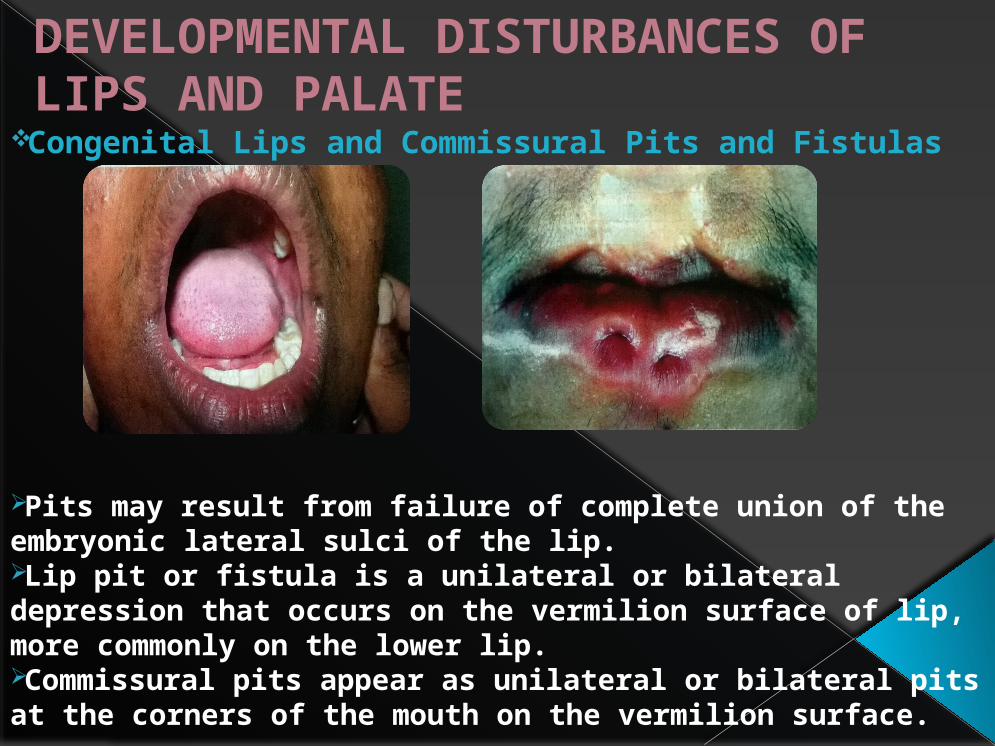
DEVELOPMENTAL DISTURBANCES OF LIPS AND PALATE
Congenital Lips and Commissural Pits and Fistulas
Pits may result from failure of complete union of the embryonic lateral sulci of the lip.Lip pit or fistula is a unilateral or bilateral depression that occurs on the vermilion surface of lip, more commonly on the lower lip.Commissural pits appear as unilateral or bilateral pits at the corners of the mouth on the vermilion surface.

Van der Woude Syndrome(Cleft lip syndrome, lip pit syndrome, dimpled papillae of the lip)
It is an autosomal dominant syndrome. Consisting of a cleft lip or cleft palate and pits of lower lips.Affected individuals may have maxillary hypodontia; missing maxillary incisors or missing premolars.Oral manifestations include syngnathia, narrow, high, arched palate; and ankyloglossia.
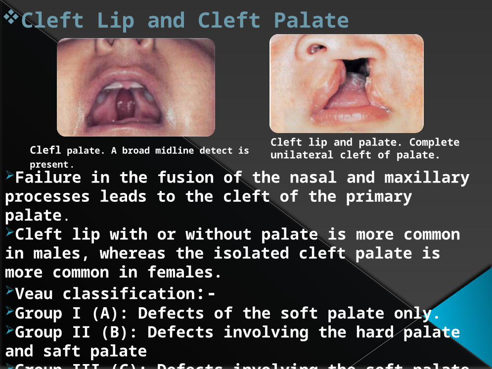
Cleft Lip and Cleft Palate
Failure in the fusion of the nasal and maxillary processes leads to the cleft of the primary palate.Cleft lip with or without palate is more common in males, whereas the isolated cleft palate is more common in females.Veau classification:-Group I (A): Defects of the soft palate only.Group II (B): Defects involving the hard palate and saft palateGroup III (C): Defects involving the soft palate to the alveolus, usually involving the lip.Group IV (D): Complete bilaterl clefts.
Cleft lip and palate. Complete unilateral cleft of palate.Clefl palate. A broad midline detect is
present.

DEVELOPMENTAL DISTURBANCES OF ORAL MUCOSA
Fordyce’s Granules(Fordyce’s disease)
Heterotopic collections of sebaceous glands at various sites in the oral cavity.Appears as small yellow spots, often projecting slightly above the surface of the tissue.The glands are superficial and consist of few or many lobules, all grouped around one or more ducts which open on the surface of the mucosa.Theses ducts may show keratin plugging.
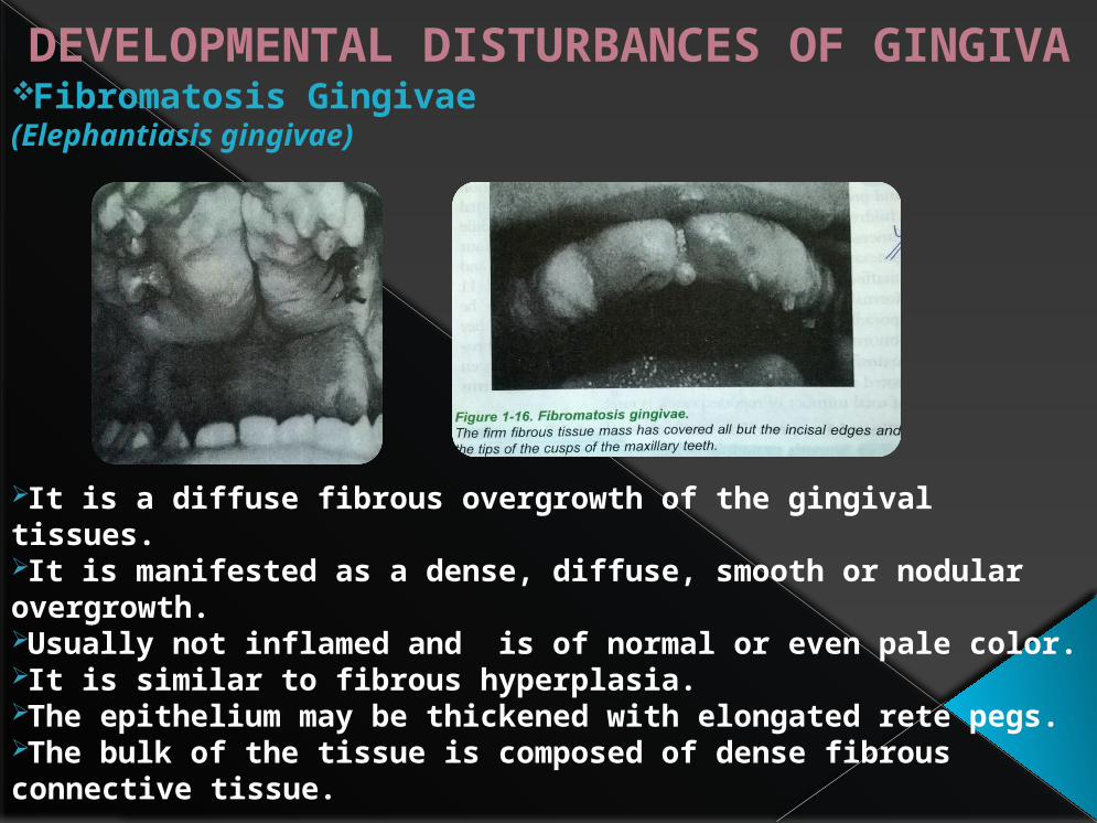
DEVELOPMENTAL DISTURBANCES OF GINGIVAFibromatosis Gingivae(Elephantiasis gingivae)
It is a diffuse fibrous overgrowth of the gingival tissues.It is manifested as a dense, diffuse, smooth or nodular overgrowth.Usually not inflamed and is of normal or even pale color.It is similar to fibrous hyperplasia.The epithelium may be thickened with elongated rete pegs.The bulk of the tissue is composed of dense fibrous connective tissue.
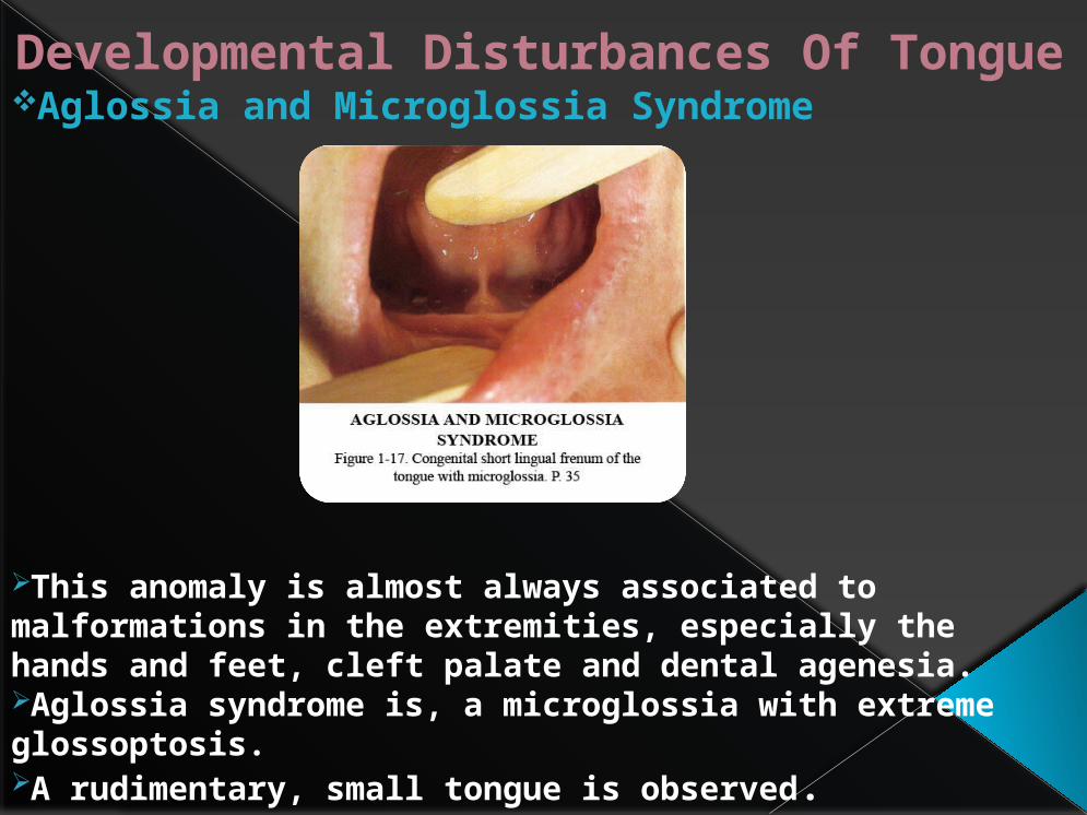
Developmental Disturbances Of TongueAglossia and Microglossia Syndrome
This anomaly is almost always associated to malformations in the extremities, especially the hands and feet, cleft palate and dental agenesia.Aglossia syndrome is, a microglossia with extreme glossoptosis.A rudimentary, small tongue is observed.
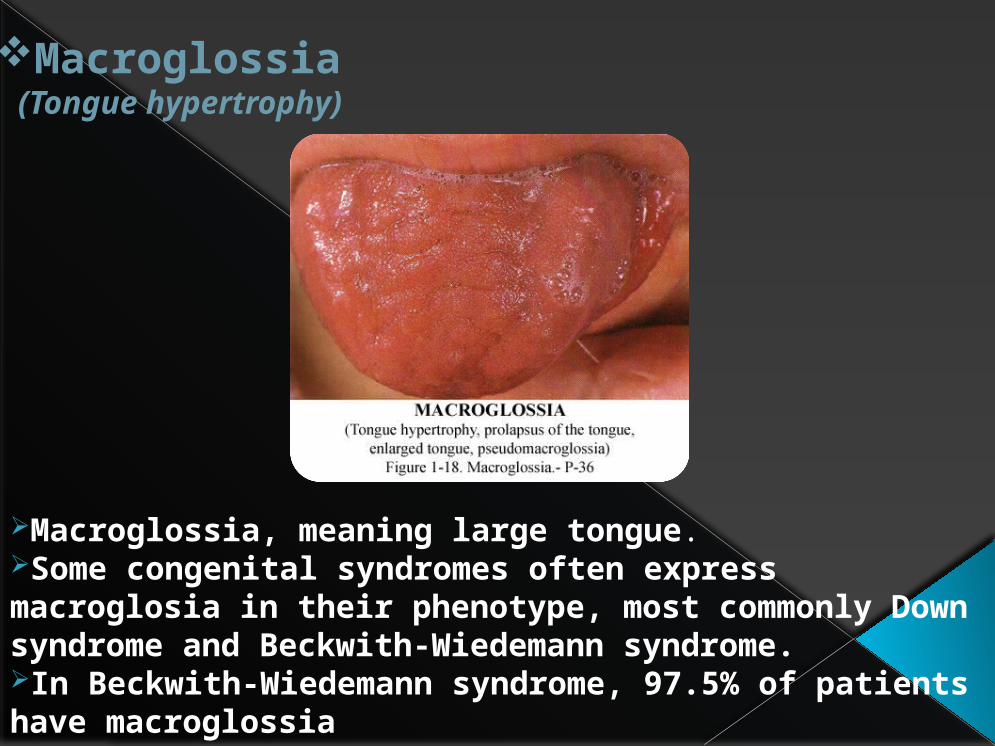
Macroglossia(Tongue hypertrophy)
Macroglossia, meaning large tongue.Some congenital syndromes often express macroglosia in their phenotype, most commonly Down syndrome and Beckwith-Wiedemann syndrome.In Beckwith-Wiedemann syndrome, 97.5% of patients have macroglossia

Ankylogossia or Tongue-tie
It is said to exist when the inferior frenulum attaches to the bottom of the tongue.It restricts free movement of the tongue which causes speech defects.It can cause feeding problems in in infants.
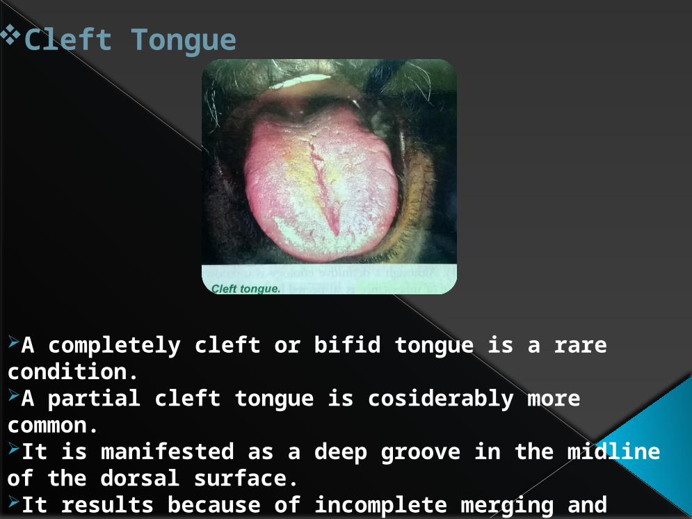
Cleft Tongue
A completely cleft or bifid tongue is a rare condition.A partial cleft tongue is cosiderably more common.It is manifested as a deep groove in the midline of the dorsal surface.It results because of incomplete merging and failure of groove obliteration by underlying mesenchymal proliferation.
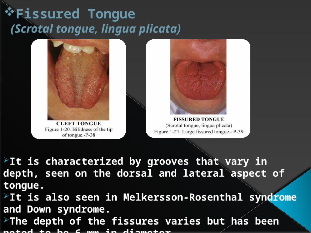
Fissured Tongue(Scrotal tongue, lingua plicata)
It is characterized by grooves that vary in depth, seen on the dorsal and lateral aspect of tongue.It is also seen in Melkersson-Rosenthal syndrome and Down syndrome.The depth of the fissures varies but has been noted to be 6 mm in diameter.

Median Rhomboid Glossitis
It is present in the posterior midline of the dorsum of the tongue, just anterior to the V-shaped grouping of the circumvallate papillae.Filliform papillae are absent.There appears to be a 3:1 male predilection.Lesions are less than 2 cm in dimension.
Median rhomboid glossitis. The typical lozenge-shaped areaof depapillation in the midline of the tongue.
Median rhomboid glossitis. There is hyperplasia withelongated rete processes, hyperparakeratosis and an inflammatoryinfiltrate of lymphocytes and plasma cells in the connective tissue.
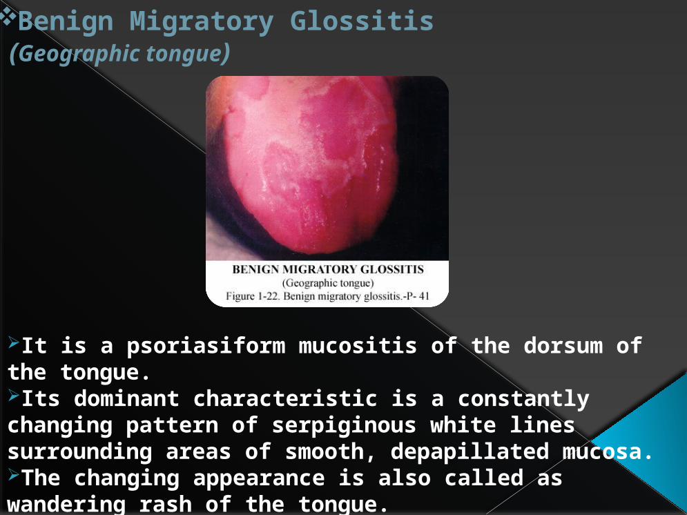
Benign Migratory Glossitis(Geographic tongue)
It is a psoriasiform mucositis of the dorsum of the tongue.Its dominant characteristic is a constantly changing pattern of serpiginous white lines surrounding areas of smooth, depapillated mucosa.The changing appearance is also called as wandering rash of the tongue.
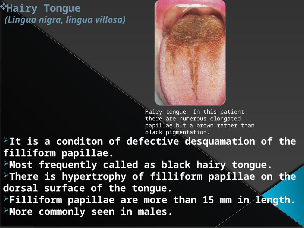
Hairy Tongue(Lingua nigra, lingua villosa)
It is a conditon of defective desquamation of the filliform papillae.Most frequently called as black hairy tongue.There is hypertrophy of filliform papillae on the dorsal surface of the tongue.Filliform papillae are more than 15 mm in length.More commonly seen in males.
Hairy tongue. In this patient there are numerous elongatedpapillae but a brown rather than black pigmentation.

DEVELOPMENTAL DISTURBANCES IN SIZE OF TEETHMicrodontia
This term is used to describe teeth which are smaller than normal.In true generalized microdontia, aal teeth are smaller than normal.In relative generalized microdontia, normal or slightly smaller teeth are present in large jaws.Microdontia involving only a single tooth is a common condition, called as ‘peg lateral’.

Macrodontia
Teeth that are larger than normal.In true generalized macrodontia, all teeth are larger tha normal.In relative generalized macrodontia, there is presence of larger than normal teeth in small jaws.

DEVELOPMENTAL DISTERBANCES IN SHAPE OF TEETH
Gemination
Geminated teeth are arises from an attempt at division of a single tooth germ by an invagination, with resultant incomplete formation of two teeth.
The structure is usually one with two completely or incompletely separated crowns, that have a single root and root canal.
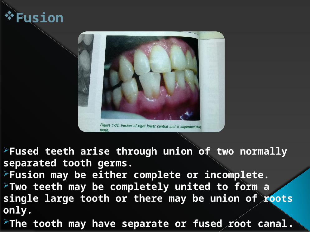
Fusion
Fused teeth arise through union of two normally separated tooth germs.Fusion may be either complete or incomplete.Two teeth may be completely united to form a single large tooth or there may be union of roots only.The tooth may have separate or fused root canal.

Concrescence
It is a form of fusion which occurs after root formation has been completed.Teeth are united by cementum only.It’s a results of traumatic injury or crowding of teeth with resorption of interdental bone.
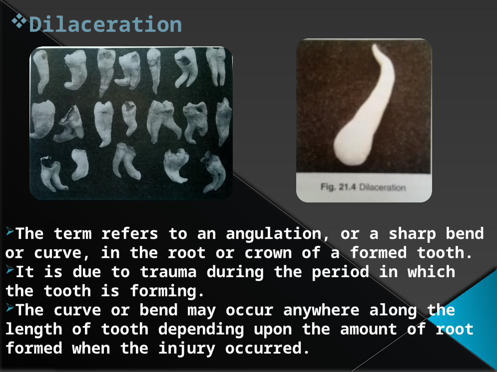
Dilaceration
The term refers to an angulation, or a sharp bend or curve, in the root or crown of a formed tooth.It is due to trauma during the period in which the tooth is forming.The curve or bend may occur anywhere along the length of tooth depending upon the amount of root formed when the injury occurred.
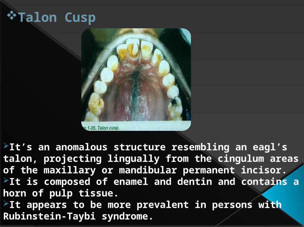
Talon Cusp
It’s an anomalous structure resembling an eagl’s talon, projecting lingually from the cingulum areas of the maxillary or mandibular permanent incisor.It is composed of enamel and dentin and contains a horn of pulp tissue.It appears to be more prevalent in persons with Rubinstein-Taybi syndrome.

Dens in Dente(Dens invaginatus, dilated composite odontome)
It is a developmental variation which arise as a result of an invagination in the surface of tooth crown before calcification has occurred.The permanent maxillary lateral incisors are most commonly involved.It appears to represent simply an accentuation in the development of the lingual pit.

Dens Evaginatus(Leong’s premolar, occlusal enamel pearl)
It appears clinically as an accessory cusp or globule of enamel on the occlusal surface between the buccal and lingual cusps of premolars.
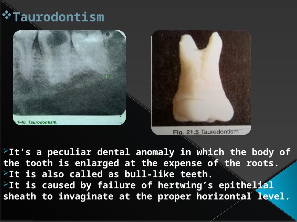
Taurodontism
It’s a peculiar dental anomaly in which the body of the tooth is enlarged at the expense of the roots.It is also called as bull-like teeth.It is caused by failure of hertwing’s epithelial sheath to invaginate at the proper horizontal level.
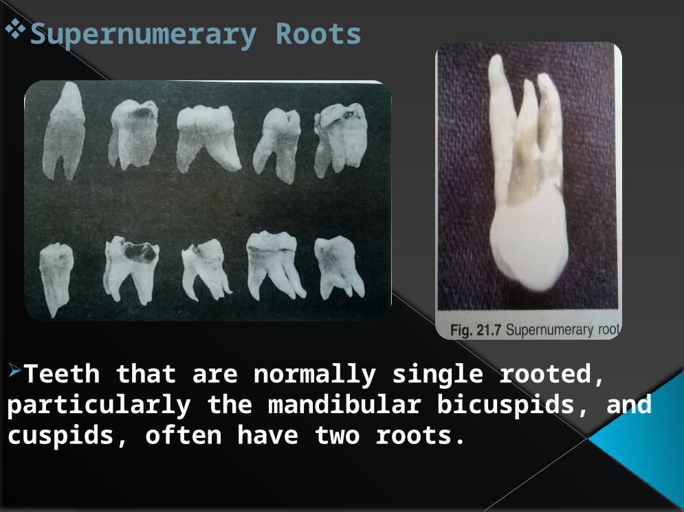
Supernumerary Roots
Teeth that are normally single rooted, particularly the mandibular bicuspids, and cuspids, often have two roots.

DEVELOPMENTAL DISTURBANCES IN NUMBER OF TEETH
Anodontia
True anodontia may be of two types, total and partial.In total anodontia, all teeth are missing.Partial anodontia involves one or more teeth.Maxillary lateral incisors and maxillary or mandibular second premolars are commonly missing.
Congenital absence of lateral incisors with spacing of theanterior teeth.
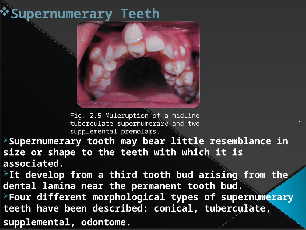
Supernumerary Teeth
.
Supernumerary tooth may bear little resemblance in size or shape to the teeth with which it is associated.It develop from a third tooth bud arising from the dental lamina near the permanent tooth bud.Four different morphological types of supernumerary teeth have been described: conical,
tuberculate, supplemental, odontome.
Fig. 2.5 Muleruption of a midline tuberculate supernumerary and twosupplemental premolars.
DEVELOPMENTAL DISTURBANCES IN STRUCTURE OF TEETH
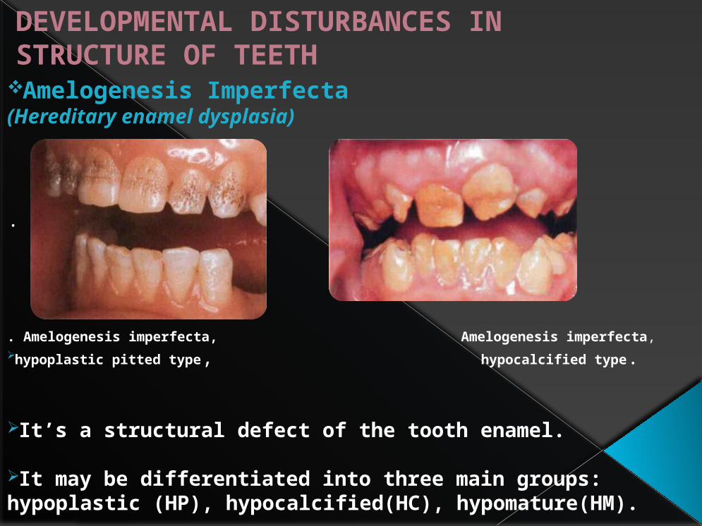
Amelogenesis Imperfecta(Hereditary enamel dysplasia)
.
. Amelogenesis imperfecta, Amelogenesis imperfecta,hypoplastic pitted type, hypocalcified type.
It’s a structural defect of the tooth enamel.
It may be differentiated into three main groups: hypoplastic (HP), hypocalcified(HC), hypomature(HM).

Environmental Enamel Hypoplasia
It is defined as an incomplete or defective formation of the organic enamel matrix of teeth.Enamel hypoplasia can cause due to nutritional deficiency, exanthematous diseases, congenital syphilis, hypocalcemia, birth injury, prematurity, local infection or trauma and ingestion of chemicals (chiefly fluoride).
Congenital syphilis: Hutchinson's teeth
Fluoride mottling.

Dentinogenesis Imperfecta
Affected teeth are gray to yellowish-brown and have broad crowns with constriction of the cervical area resulting in a ‘tulip’ shape.In Dentinogenesis imperfecta 1, teeth are blue-gray or amber brown and opalescent.In dentonogenesisi imperfecta 2, dentin is amber and smooth.
Shell tooth. In this severe form of dentinogenesis imperfecta
Dentinogenesis imperfecta showing the translucent appearanceof the teeth which are of normal morphology.
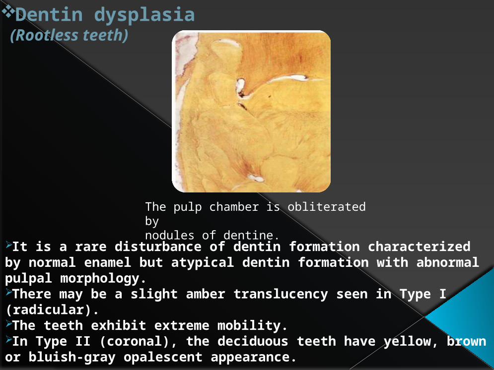
Dentin dysplasia(Rootless teeth)
It is a rare disturbance of dentin formation characterized by normal enamel but atypical dentin formation with abnormal pulpal morphology.There may be a slight amber translucency seen in Type I (radicular).The teeth exhibit extreme mobility.In Type II (coronal), the deciduous teeth have yellow, brown or bluish-gray opalescent appearance.
The pulp chamber is obliterated bynodules of dentine.

Regional Odontodysplasia(Odontodysplasia, ghost teeth)
Maxillary permanent incisors, lateral incisors, and cuspid are more commonly involved.The teeth affected by odontodysplasia exhibit either a delay or a total failure in eruption.Rdiographicaly teeth show a marked reduction in radiodensity so that the teeth assume a ghost appearance.
Both enamel and dentine aredeformed and there is calcification of the reduced enamel epithelium.
On one side of the midline the deciduous incisors have poor root formation withthin radicular dentine and enamel.
FISSURAL INCLUSION,DEVELOPMENTAL) CYSTS OF ORAL REGION
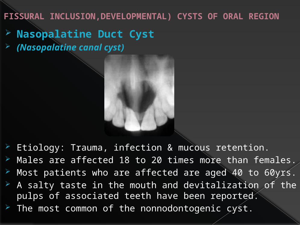
Nasopalatine Duct Cyst (Nasopalatine canal cyst)
Etiology: Trauma, infection & mucous retention. Males are affected 18 to 20 times more than females. Most patients who are affected are aged 40 to 60yrs. A salty taste in the mouth and devitalization of the pulps
of associated teeth have been reported. The most common of the nonnodontogenic cyst.

Globulomaxillary Cyst (fissural cyst)
Found within the bone between the maxillary lateral incisor and canine.Radiographicaly, it is a well defined radiolucency which frequently causes the roots of adjacent teeth to diverge.It may become infected and patient may complaint of local discomfort or pain in the area.
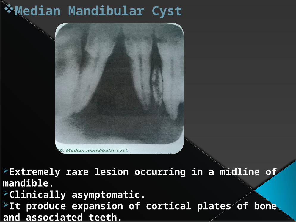
Median Mandibular Cyst
Extremely rare lesion occurring in a midline of mandible.Clinically asymptomatic.It produce expansion of cortical plates of bone and associated teeth.

Palatal and Alveolar Cysts of Newborns(Epstein’s pearls, Bohn’s nodules, gingival cyst of newborn)
Newborn cysts present at posterior hard palate and occasionally on anterior soft palate.Occasional cysts are located at some distance from midline.


![ORAL ~RADIOLOGY - dent.zaums.ac.irdent.zaums.ac.ir/uploads/1_296_chapter1.pdf · tal Disturbances of the Face and Jaws. ... 19 Inflammatory l esions of the Jaws, ]66 ... 29 Developmental](https://static.fdocuments.us/doc/165x107/5a97a0aa7f8b9ab6188ced90/oral-radiology-dentzaumsac-disturbances-of-the-face-and-jaws-19-inflammatory.jpg)