Development of the Polymerase Chain Reaction for …jcm.asm.org/content/31/3/659.full.pdf ·...
6
JOURNAL OF CLINICAL MICROBIOLOGY, Mar. 1993, p. 659-664 0095-1137/93/030659-06$02.00/0 Copyright © 1993, American Society for Microbiology Development of the Polymerase Chain Reaction for Diagnosis of Chancroid LINDA CHUI,1 WILLIAM ALBRITTON,l* BRUCE PASTER,2 IAN MACLEAN,3 AND RAYMOND MARUSYK' Provincial Laboratory of Public Health, University of Alberta, Edmonton, Alberta T6G 2J2,1 Canada; Forsyth Dental Center, Boston, Massachusetts 021152; and Department of Medical Microbiology, University of Manitoba, Winnipeg, Manitoba R3T 2N2,3 Canada Received 11 August 1992/Accepted 18 December 1992 The published nucleotide sequences of the 16S rRNA gene of Haemophilus ducreyi were used to develop primer sets and probes for the diagnosis of chancroid by polymerase chain reaction (PCR) DNA amplification. One set of broad specificity primers yielded a 303-bp PCR product from all bacteria tested. Two 16-base probes internal to this sequence were species specific for H. ducreyi when tested with 12 species of the families PasteureUaceae and Enterobacteriaceae. The two probes in combination with the broad specificity primers were 100% sensitive with 51 strains of H. ducreyi isolated from six continents over a 15-year period. The direct detection of H. ducreyi from 100 clinical specimens by PCR showed a sensitivity of 83 to 98% and a specificity of 51 to 67%, depending on the number of amplification cycles. The accuracy of the clinical diagnosis of genital ulcer disease as chancroid has been reported to be 75 to 80% at best (6, 10). Both false-positive and false-negative clinical variants of chancroid have been reported (2, 16, 20, 29, 32). The sensitivity of culture for the confirmation of clinical disease has been improved in recent years to 50 to 90% in experienced laboratories in areas where chancroid is en- demic (9, 31). Culture sensitivity in inexperienced laborato- ries during epidemics has been reported to be less than 50% (24). Efficient transport media for Haemophilus ducreyi that would allow culture diagnosis in distant laboratories with prolonged transportation times have not been reported. For these reasons, there has been considerable interest in non- culture methods for the diagnosis of chancroid. Direct examination of ulcer material by Gram's stain (3, 14) and electron microscopy of biopsy material (19) have both been suggested as diagnostic tests for chancroid. Re- cent studies have shown the sensitivity of the Gram stain to be less than 50% (5, 23), and the morphology of H. ducreyi is similar to those of many organisms found in the polymi- crobial flora of most genital ulcers (4). Improvements in reagents for direct examination by incorporating adsorbed polyclonal (7, 11) and monoclonal (11, 12, 15, 30) antibodies into immunofluorescence tests have been reported. These reagents have not been widely used in clinical studies to establish their performance characteristics under routine use conditions. With the introduction of DNA diagnostic methods (17) and the ability to amplify the signal with the polymerase chain reaction (PCR), newer tests should improve the sensitivity and specificity of diagnostic tests for chancroid. First-gener- ation DNA probes for H. ducreyi have been reported with sensitivities of 103 to 104 organisms and 100% specificity (25, 26). We report here our results with the development of primer sets and probes for the diagnosis of chancroid by DNA amplification using PCR. * Corresponding author. MATERIALS AND METHODS Bacterial strains. Fifty-one strains of H. ducreyi, including the type strain CIP542, isolated from six continents over a 15-year period were selected for assessing sensitivity. The following twelve species from the families Pasteurellaceae and Enterobacteriaceae were selected for assessing speci- ficity on the basis of rRNA sequence homology or because they are commonly isolated from genital ulcers: Haemo- philus paraphrophilus ATCC 29241, Haemophilus parainflu- enzae NCTC 7857, Haemophilus haemolyticus NCTC 10659, Haemophilus spp. (minor group 202), Pasteurella ureae NCTC 10219, Actinobacillus pleuropneumoniae ATCC 27088, Actinobacillus equuli NCTC 8529, Klebsiella pneumoniae ATCC 13883, Salmonella typhimurium LT2, Escherichia coli ATCC 25922, and Pseudomonas aeruginosa ATCC 27853. Nucleic acid extraction. Bacterial colonies were scraped from plates with cotton swabs and washed twice with 12 mM Tris-HCl, pH 7.6. Cells were standardized to an optical density of 0.12 at 540 nm in the same Tris buffer. One milliliter of the standardized suspension was pelleted and resuspended in 100 ,ul of Tris buffer. Crude DNA was obtained by subjecting the cell suspension to three cycles of boiling and freezing. Ten microliters of each sample was used for PCR analysis. Clinical specimens were taken in the Special Treatment Clinic in Nairobi, Kenya, from males with genital ulcers consistent with a clinical diagnosis of chancroid. The swabs were transported to the laboratory in Nairobi at ambient temperature in 1 ml of phosphate-buffered saline (PBS) containing chenodeoxycholate (Sigma) at 1 mg/ml and heated at 100°C for 15 min on arrival. Samples were stored at -70°C until transported to Edmonton, Canada. The method of extraction of nucleic acid from clinical specimens for PCR was adapted from Pollard et al. (27). A positive control containing 105 H. ducreyi organisms in chenodeoxycholate and a negative control of chenodeoxycholate were carried through each extraction procedure. Lysis buffer (200-,ul 0.1 M Tris-HCl [pH 8.0], 0.1 M NaCl, 5 mM EDTA, 1% sodium dodecyl sulfate [SDS], 50 ,ug of proteinase K per ml) was added to an equal volume of specimen and incubated at 37°C 659 Vol. 31, No. 3 on April 25, 2018 by guest http://jcm.asm.org/ Downloaded from
-
Upload
phungkhanh -
Category
Documents
-
view
217 -
download
1
Transcript of Development of the Polymerase Chain Reaction for …jcm.asm.org/content/31/3/659.full.pdf ·...
JOURNAL OF CLINICAL MICROBIOLOGY, Mar. 1993, p. 659-6640095-1137/93/030659-06$02.00/0Copyright © 1993, American Society for Microbiology
Development of the Polymerase Chain Reaction for
Diagnosis of ChancroidLINDA CHUI,1 WILLIAM ALBRITTON,l* BRUCE PASTER,2 IAN MACLEAN,3
AND RAYMOND MARUSYK'Provincial Laboratory of Public Health, University ofAlberta, Edmonton, Alberta T6G 2J2,1 Canada; Forsyth
Dental Center, Boston, Massachusetts 021152; and Department ofMedical Microbiology,
University ofManitoba, Winnipeg, Manitoba R3T 2N2,3 Canada
Received 11 August 1992/Accepted 18 December 1992
The published nucleotide sequences of the 16S rRNA gene of Haemophilus ducreyi were used to develop
primer sets and probes for the diagnosis of chancroid by polymerase chain reaction (PCR) DNA amplification.
One set ofbroad specificity primers yielded a 303-bp PCR product from all bacteria tested. Two 16-base probes
internal to this sequence were species specific for H. ducreyi when tested with 12 species of the familiesPasteureUaceae and Enterobacteriaceae. The two probes in combination with the broad specificity primers were100% sensitive with 51 strains of H. ducreyi isolated from six continents over a 15-year period. The directdetection of H. ducreyi from 100 clinical specimens by PCR showed a sensitivity of 83 to 98% and a specificityof 51 to 67%, depending on the number of amplification cycles.
The accuracy of the clinical diagnosis of genital ulcerdisease as chancroid has been reported to be 75 to 80% atbest (6, 10). Both false-positive and false-negative clinicalvariants of chancroid have been reported (2, 16, 20, 29, 32).The sensitivity of culture for the confirmation of clinical
disease has been improved in recent years to 50 to 90% inexperienced laboratories in areas where chancroid is en-
demic (9, 31). Culture sensitivity in inexperienced laborato-ries during epidemics has been reported to be less than 50%(24). Efficient transport media for Haemophilus ducreyi thatwould allow culture diagnosis in distant laboratories withprolonged transportation times have not been reported. Forthese reasons, there has been considerable interest in non-
culture methods for the diagnosis of chancroid.Direct examination of ulcer material by Gram's stain (3,
14) and electron microscopy of biopsy material (19) haveboth been suggested as diagnostic tests for chancroid. Re-cent studies have shown the sensitivity of the Gram stain tobe less than 50% (5, 23), and the morphology of H. ducreyiis similar to those of many organisms found in the polymi-crobial flora of most genital ulcers (4). Improvements inreagents for direct examination by incorporating adsorbedpolyclonal (7, 11) and monoclonal (11, 12, 15, 30) antibodiesinto immunofluorescence tests have been reported. These
reagents have not been widely used in clinical studies to
establish their performance characteristics under routine use
conditions.With the introduction ofDNA diagnostic methods (17) and
the ability to amplify the signal with the polymerase chainreaction (PCR), newer tests should improve the sensitivityand specificity of diagnostic tests for chancroid. First-gener-ation DNA probes for H. ducreyi have been reported withsensitivities of 103 to 104 organisms and 100% specificity (25,26). We report here our results with the development of
primer sets and probes for the diagnosis of chancroid byDNA amplification using PCR.
* Corresponding author.
MATERIALS AND METHODS
Bacterial strains. Fifty-one strains of H. ducreyi, includingthe type strain CIP542, isolated from six continents over a
15-year period were selected for assessing sensitivity. Thefollowing twelve species from the families Pasteurellaceaeand Enterobacteriaceae were selected for assessing speci-ficity on the basis of rRNA sequence homology or becausethey are commonly isolated from genital ulcers: Haemo-philusparaphrophilus ATCC 29241, Haemophilus parainflu-enzae NCTC 7857, Haemophilus haemolyticus NCTC10659, Haemophilus spp. (minor group 202), Pasteurellaureae NCTC 10219, Actinobacillus pleuropneumoniaeATCC 27088, Actinobacillus equuli NCTC 8529, Klebsiellapneumoniae ATCC 13883, Salmonella typhimurium LT2,Escherichia coli ATCC 25922, and Pseudomonas aeruginosa
ATCC 27853.Nucleic acid extraction. Bacterial colonies were scraped
from plates with cotton swabs and washed twice with 12 mMTris-HCl, pH 7.6. Cells were standardized to an opticaldensity of 0.12 at 540 nm in the same Tris buffer. Onemilliliter of the standardized suspension was pelleted andresuspended in 100 ,ul of Tris buffer. Crude DNA was
obtained by subjecting the cell suspension to three cycles ofboiling and freezing. Ten microliters of each sample was
used for PCR analysis.Clinical specimens were taken in the Special Treatment
Clinic in Nairobi, Kenya, from males with genital ulcersconsistent with a clinical diagnosis of chancroid. The swabswere transported to the laboratory in Nairobi at ambienttemperature in 1 ml of phosphate-buffered saline (PBS)containing chenodeoxycholate (Sigma) at 1 mg/ml andheated at 100°C for 15 min on arrival. Samples were stored at-70°C until transported to Edmonton, Canada. The methodof extraction of nucleic acid from clinical specimens for PCRwas adapted from Pollard et al. (27). A positive controlcontaining 105 H. ducreyi organisms in chenodeoxycholateand a negative control of chenodeoxycholate were carried
through each extraction procedure. Lysis buffer (200-,ul 0.1
M Tris-HCl [pH 8.0], 0.1 M NaCl, 5 mM EDTA, 1% sodium
dodecyl sulfate [SDS], 50 ,ug of proteinase K per ml) was
added to an equal volume of specimen and incubated at 37°C
659
Vol. 31, No. 3
on April 25, 2018 by guest
http://jcm.asm
.org/D
ownloaded from
660 CHUI ET AL.
Probe 1469-494
P0 mod8-22
799 bp
P0 modP3 modP1 broadP2 broadProbe 1Probe 2AProbe 2B
P3 mod787-806
Probe 2A/B1133-1147
P1 broad1046-1063
P2 broad1330-1347
16S rRNA
303 bp
5'- AGAG1TTGATC(AC)TGG -3'
5'- GGACTACCAGGGTATCTMT -3'5'- AGGTGCTGCATGGCTGTC -3'5'- CTAGCGATTCCGACTTCA -3'5'- CTGTGACTAACGTCAATCAATTTTGC -3'5'- ATGTAGTGATGGGMC -3'5'- ATGTAATGATGGGMC -3'
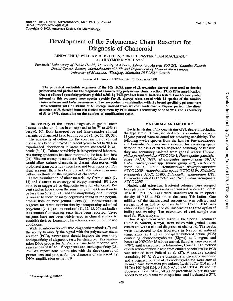
FIG. 1. Physical map and location of the oligonucleotide primers and probes within the 16S rRNA gene sequence. The sizes of the PCRamplification products are shown in the boxes. The nucleotide positions are those of Dewhirst et al. (8). P3 mod, P2 broad, and probe 1 arecomplementary to the published sequence.
for 2 h before extraction with phenol, phenol-chloroform,and chloroform. Nucleic acids were precipitated by additionof sodium acetate and ethanol and resolubilized in 100 ,u ofwater.An additional swab was taken and streaked onto a choc-
olated GC agar (Difco) plate supplemented with 5% (vol/vol)fetal calf serum and 3 mg of vancomycin per liter. The platewas incubated for 48 h at 33 to 35°C in a candle extinction jarfor isolation of H. ducreyi (9, 24).PCR. Two primer sets and two probes were selected from
the H. ducreyi 16S rRNA sequence. These oligonucleotideswere prepared by a PCR-MATE 391 DNA synthesizer(Applied Biosystems) according to the manufacturer's in-structions. Ten microliters of the crude DNA extract or 50 ,uof DNA extract from clinical material was added to a finalvolume of 100 RI of a PCR reaction mixture containing 30 pM(each) primer, 50 p.M (each) deoxynucleotide triphosphates(Pharmacia), 1 U of Taq polymerase, and 1 x reaction buffer(Bio/Can Scientific). The negative control containing noDNA and the positive control containing extracted DNAfrom H. ducreyi were included in all runs. Amplification ofbacterial DNA was carried out in an automated thermalcycler (Perkin-Elmer Cetus) in which samples were dena-tured at 94°C for 1 min and then primer annealed at 40°C for1 min and elongated at 72°C for 3 min. This cycle wasrepeated 25 times, and the amplified product was subjectedto electrophoresis for 90 min at 160 V in 1% (wt/vol) agarosegels containing 0.05 ,ug of ethidium bromide per ml andvisualized by UV illumination. DNA was transferred pas-sively onto nylon membranes (Hybond; Amersham), UVcross-linked, and prehybridized at the hybridization temper-ature in a solution containing 6x SSC (lx SSC is 0.15 MNaCl plus 0.015 M sodium citrate), 0.2% (wt/vol) SDS, 5xDenhardt solution, and 0.5 mg of sheared denatured salmonsperm DNA per ml. The probes were end labelled with[32P]ATP (Amersham) by using T4 polynucleotide kinase(Bethesda Research Laboratories) orwith digoxigenin (Boehr-inger Mannheim) by using terminal transferase according tothe manufacturer's instructions (Boehringer Mannheim).Hybridization was carried out at 650C with probe 1, 430Cwith probe 2A, and 400C with probe 2B overnight in a
shaking water bath. Washings for radioactive probes were
done according to the method of Maniatis et al. (18), andwashings for digoxigenin-labelled probes were done accord-
ing to manufacturer's instructions. The blot was exposed toKodak X-Omat AR film.
Sequencing of the PCR product. The amplified DNA prod-uct was excised and extracted with Geneclean (Bio 101). Thepurified DNA was sequenced directly with the dideoxyter-mination method, modified T7 DNA polymerase Sequenase(United States Biochemical), and the reagents provided inthe kit. The samples were subjected to electrophoresis in a6% (wt/vol) polyacrylamide-7 M urea sequencing gel, fixed,dried, and exposed to Kodak XAR-5 film.
Sensitivity of PCR primers. Cell pellets of H. ducreyi werewashed and standardized to an optical density of 0.12 at 540nm in 12 mM Tris-HCl, pH 7.6. Tenfold serial dilutions weremade from 10-1 to 10-8. The average number of CFU wasdetermined by plating 100 pul of each dilution in triplicate.Crude DNA was extracted from an equal volume of eachdilution, and 10 pul of the extract was used in the PCRreaction.
RESULTS
H. ducreyi primer sets and probes. Two primer sets wereevaluated for amplification of the 16S rRNA gene of H.ducreyi (Fig. 1). The choice of the 16S rRNA gene was basedon multiple copies of the gene within the genome, the abilityto use broad specificity primers for assessing amplification inculture-negative specimens, and the known sequence vari-ability within eubacteria. The P0 mod and P3 mod primershave been described previously (34) and are similar to thebroad specificity primers used by Weisburg et al. (33). TheP1 broad and P2 broad primers were selected for the highlevel of sequence homology between published eubacterial16S rRNA sequences. The probe 1 and probe 2 sequenceswere chosen from the known variable regions of publishedeubacterial 16S rRNA sequences and the published H.ducreyi sequence (8, 28). The probe 2A sequence was
originally chosen from the sequence of Dewhirst et al. (8),and probe 2B was from the sequence of Rossau et al. (28) forthe same region.
Sensitivity of H. ducreyi primers and probes. With the P0mod and P3 mod primers, a strong 799-bp PCR amplificationproduct was seen with only 48 of the 51 strains of H. ducreyitested. The combination of P0 mod and P3 mod primers withprobe 1 could detect only 105 to 106 organisms (data not
PROBES
PRIMERS
J. CLIN. MICROBIOL.
on April 25, 2018 by guest
http://jcm.asm
.org/D
ownloaded from

PCR FOR HAEMOPHILUS DUCREYI 661
7 8 9 10
a,
FIG. 2. Sensitivity of PCR detection of H. ducreyi. (a) Ethidiumbromide-stained agarose gel showing the PCR products generatedwith the P1 broad and P2 broad primers for serial dilutions of H.ducreyi V1157. Lanes 1 to 8 contain approximately 107, 106, 105, 104,103, 102, 101, and 100 organisms, respectively. Lane 9 is the negativecontrol consisting of PCR reagents only. Lane 10 contains DNA sizemarkers (pBR322 digested with HinFI) 154 to 517 bp in size. (b)Autoradiogram developed after Southern transfer of the PCR prod-ucts from panel a and hybridization with probe 2A.
shown). The P1 broad and P2 broad primers produced a
303-bp PCR product with all strains of H. ducreyi and allmembers of the Pasteurellaceae and Enterobactenaceaefamilies studied and with species commonly found in genitalulcers. The combination of P1 broad and P2 broad primerswith probe 2A could detect 102 to 103 organisms after one
round of 25 cycles of PCR (Fig. 2). When 25 ,ul of theamplification product from the first round of PCR was usedas the template for a second round of 25 cycles of PCR, the
G ATC
a .-
W._,
VW.
3-1
G A TC
- C
AAGGGTAGTG -o-AATGTA
sensitivity increased to 100 organisms (data not shown).However, beyond three rounds of 25 cycles, interferenceand a smearing of the amplification product in agarose gelswere noted. Special precautions, such as aerosol-free pipettetips, were required to prevent contamination when reampli-fication was carried out, and 25 ,ul from the negative controlof the previous round of amplification was always carriedthrough the reamplification cycles.
Specificity of H. ducreyi primers and probes. To avoid theproblem of false-negative results with clinical specimens(13), we deliberately chose primers with broad specificity.Probe 1 failed to hybridize to 3 of the 48 H. ducreyiamplification products with P0 mod and P3 mod primers(data not shown). Probe 2A hybridized strongly with 46 ofthe 51 H. ducreyi PCR amplification products with P1 broadand P2 broad primers. The failure of probe 2A to hybridizewith the remaining five strains ofH. ducreyi was investigatedby sequencing the PCR product from strains that hybridizedwith and failed to hybridize with this probe (Fig. 3). Bothstrand sequences confirmed an additional G at position 1144,as reported by Dewhirst et al. (8), and there was a G:Atransition at position 1137 in some strains (8, 28). A modifiedprobe 2B was synthesized (Fig. 1), and this probe recognizedall five strains that did not react with the probe 2A oligonu-cleotide (Fig. 4). Probe 2A or probe 2B oligonucleotideswere 100% specific for H. ducreyi when hybridized withamplification products from 10 species of eubacteria (Fig. 5).A similar PCR amplification product which failed to hybrid-ize with probes 2A and 2B was seen with H. paraphrophilusATCC 29241 and H. parainfluenzae NCTC 7857 (data notshown). Thus, the combination of P1 broad and P2 broadprimers with probe 2A or 2B was 100% sensitive and 100%specific for H. ducreyi with pure cultures of clinical isolates.
Detection of H. ducreyi in clinical specimens. Preliminarydata (not shown) indicated that dry swabs or swabs placed inPBS alone were less satisfactory for PCR than swabs heatedin chenodeoxycholate. Over a 6-month period, 100 speci-mens from patients with genital ulcer disease were processedby PCR and culture. Three specimens were contaminated
G ATC
b
..
im~
-AM
d
1.s
owp
" IM
0 ,
--P
TA
C
AT
AC
TAC
C
C
TTG
G A T C
FIG. 3. Autoradiogram showing the sequence of the PCR product corresponding to the probe 2A or 2B region. (a) Sequence of thesingle-stranded PCR product with the P1 broad primer. (b) Sequence of the single-stranded product with the P2 broad primer. Sequences on
the left were observed with isolates hybridizing to the probe 2A sequence. Sequences on the right were observed with isolates hybridizingto the probe 2B sequence.
1 2 34 5 6a
b
VOL. 31, 1993
L-7
on April 25, 2018 by guest
http://jcm.asm
.org/D
ownloaded from

J. CLIN. MICROBIOL.662 CHUI ET AL.
1 2 3 4 5 6 a
b
c
FIG. 4. Sequence specificity of the probe 2A and 2B oligonucle-otides. (a) Ethidium bromide-stained agarose gel showing the PCRproducts generated with the P1 broad and P2 broad primers for sixrepresentative strains of H. ducreyi. Lanes: 1, strain 557; 2, strain10945; 3, strain C147; 4, strain SA3114; 5, strain 9468; 6, strainV1157 (representative of the 46 strains hybridizing with probe 2A).(b) Autoradiogram developed after Southern transfer of the PCRproducts from panel a and hybridization with probe 2B. (c) Autora-diogram developed after Southern transfer of the PCR productsfrom panel a and hybridization with probe 2A.
upon culture, and the status for H. ducreyi could not bedetermined. Fifty-seven of the remaining specimens pro-duced an amplifiable product visible in agarose gels by usingthe P1 broad and P2 broad primers after the first 25 cycles ofPCR (Fig. 6a). The sensitivity for 30 culture-positive speci-mens was 83%, and the specificity for 27 culture-negativespecimens was 67%.An additional 14 culture-positive specimens failed to pro-
duce an amplifiable product visible in agarose gels after thefirst 25 cycles of PCR. Twelve of these specimens wereavailable for further study. These 12 specimens plus 5specimens that were culture positive and PCR positive butprobe negative; 13 specimens that were culture negative,PCR positive, and probe negative; and 25 specimens thatwere culture negative, PCR negative, and probe negativeafter the first amplification round of 25 cycles were carriedthrough additional rounds of 25 cycles of PCR. The com-bined sensitivity for 42 culture-positive specimens was 98%,and the combined specificity for 43 culture-negative speci-mens was 51% after three rounds of 25 cycles (Fig. 6b). Tenculture-negative, PCR-negative, and probe-negative speci-mens remained PCR negative after three rounds of 25 cyclesof PCR.
DISCUSSION
The laboratory diagnosis of chancroid has been difficultsince the original description of the etiological agent, H.ducreyi, by Auguste Ducrey over 100 years ago (1, 21). Theabsence of effective serological tests (22, 30), the difficultiesin culture diagnosis (9, 24, 31), and the increased incidence
FIG. 5. Sensitivity of the P1 broad and P2 broad primers andspecificity of probe 2A. (a) Ethidium bromide-stained agarose gelshowing the PCR products generated with the P1 broad and P2broad primers for 10 species of eubacteria. Lanes: 1, P. ureae; 2, H.haemolyticus; 3, A. pleuropneumoniae; 4, A. equuli; 5, H. ducreyiV1157; 6, Haemophilus sp. (minor group 202); 7,K pneumoniae; 8,S. typhimunum; 9, E. coli; 10, P. aeruginosa. Lane 11 is a negativecontrol containing PCR reagents only. Lane 12 contains DNA sizemarkers as in Fig. 2. (b) Autoradiogram developed after transfer ofthe PCR products and hybridization with probe 2A.
of disease in North America in recent years (31) haveheightened the need for nonculture diagnostic tests.DNA diagnostic tests are particularly attractive because
H. ducreyi appears to belong to a monospecific genus onlydistantly related to other members of the family Pasteurel-laceae (8). In addition, the current availability of amplifica-tion procedures will significantly improve the sensitivity ofDNA-based diagnostic tests (17).
Specific probes for the identification of H. ducreyi havebeen readily developed (26) because there is very littlehomology between strains of H. ducreyi and other eubacte-ria. This has been exploited by Rossau et al. (28) to developspecific rRNA-derived oligonucleotide probes, but the nu-cleotide sequences were not provided.The rRNA sequences have several characteristics that
make them especially suitable for the development of PCR-based diagnostic tests. First, rRNA genes exist in multiplecopies in most eubacteria and therefore increase the signalfor amplification. Second, the conserved regions can be usedfor broad specificity primers which should give positive
aCulture
+ 25 9Probe
- 5 18
b
Probe+
Culture+_
41 21
1 22
FIG. 6. Detection of H. ducreyi in 100 clinical specimens frommale patients with suspected chancroid. Only samples with a PCRamplification product with the broad primers P1 and P2 derived fromthe 16S rRNA gene sequence were probed with the H. ducreyi-specific probe 2A or 2B and included in the tables. Three specimenswere contaminated, and culture status could not be determined.Data after one (a) and three (b) rounds of 25 cycles of PCR are
shown.
a
b
Q..4 w *41~ *1
on April 25, 2018 by guest
http://jcm.asm
.org/D
ownloaded from

PCR FOR HAEMOPHILUS DUCREYI 663
signals with virtually all specimens, providing internal con-trols for the amplification reaction with clinical material.Finally, the variable regions can be used for the developmentof species-specific probes. The genetic homogeneity of H.ducreyi strains should allow the development of tests withhigh specificity without loss of sensitivity.We evaluated several primers and probes for the develop-
ment of the PCR for the diagnosis of chancroid. The P1broad and P2 broad primers amplified all bacterial speciestested and produced a 303-bp product from clinical speci-mens. PCR amplification with laboratory isolates was 100%sensitive and specific when used with a pair of 16-baseoligonucleotide probes (probes 2A and 2B). Preliminaryutilization of these primers and probes to detect H. ducreyiin ulcer material from a highly endemic area showed thesesequences to be 83 to 98% sensitive and 51 to 67% specific,depending on the number of PCR cycles used for amplifica-tion, compared with the culture isolation of H. ducreyi.Because the culture sensitivity in the Special TreatmentClinic in Nairobi has been reported to be only about 85% atbest, it is entirely likely that the lower specificity is due tofalse-negative cultures rather than false-positive PCR tests.Using the primers, probes, and methods described, wewould recommend probing only samples with a PCR ampli-fication product visible in agarose gels, and we would notamplify beyond three rounds of 25 cycles. Eighty-five of the95 specimens (89%) processed in the Special TreatmentClinic in Nairobi and transported to Edmonton were evalu-able when these criteria were used.Improvements in the methods by carefully evaluating
transport media and conditions, as well as the developmentof nonradioactive liquid-phase hybridization reactions andamplification methods other than PCR, should facilitate theease of diagnosis of chancroid and allow laboratory con-
firmation of the clinical diagnosis of chancroid in remoteareas.
Finally, it is possible that multiple agents may be presentin genital ulcers. The presence of H. ducreyi detected byPCR cannot exclude syphilis, herpes simplex virus, or otherorganisms associated with genital ulcers. Specific probes forthe 16S rRNA sequences of other eubacteria could beutilized with the primers described for amplification here,but agents other than eubacteria would require the develop-ment of additional amplification primers and probes.
ACKNOWLEDGMENTSThis work was supported in part by grant MA9770 from the
Medical Research Council of Canada to W.A. and grant DE08303from the National Institutes of Dental Research to B.P.We acknowledge the kind help of J. Kimani and 0. Anzala in
collecting the clinical specimens at the Special Treatment Clinic inNairobi.
REFERENCES1. Albritton, W. L. 1989. Biology of Haemophilus ducreyi. Micro-
biol. Rev. 53:377-389.2. Ballard, R. C., D. Abeck, H. C. Korting, Y. Dangor, and 0.
Braun-Falco. 1989. Morphologische Varianten des durch Hae-mophilus ducreyi bedingten Genitalulkus. Hautarzt 40:443-447.
3. Borchardt, K. A., and A. W. Hoke. 1970. Simplified laboratorytechnique for diagnosis of chancroid. Arch. Dermatol. 102:188-192.
4. Chapel, T., W. J. Brown, C. Jeffries, and J. A. Stewart. 1978.The microbiological flora of penile ulcerations. J. Infect. Dis.137:50-56.
5. Choudhary, B. P., S. Kumari, R. Bhatia, and D. S. Agarwal.
1982. Bacteriological study of chancroid. Indian J. Med. Res.76:379-385.
6. Dangor, Y., R. C. Ballard, F. D. Exposto, G. Fehler, S. D.Miller, and H. J. Koornhof. 1990. Accuracy of clinical diagnosisof genital ulcer disease. Sex. Transm. Dis. 17:184-189.
7. Denys, G. A., T. A. Chapel, and C. D. Jeffries. 1978. An indirectfluorescent antibody technique for Haemophilus ducreyi.Health Lab. Sci. 15:128-132.
8. Dewhirst, F. E., B. J. Paster, I. Olsen, and G. J. Fraser. 1992.Phylogeny of 54 representative strains of species in the familyPasteurellaceae as determined by comparison of 16S rRNAsequences. J. Bacteriol. 174:2002-2013.
9. Dylewski, J., H. Nsanze, G. Maitha, and A. Ronald. 1986.Laboratory diagnosis of Haemophilus ducreyi: sensitivity ofculture media. Diagn. Microbiol. Infect. Dis. 4:241-245.
10. Fast, M. V., L. J. D'Costa, H. Nsanze, P. Piot, J. Curran, P.Karasira, N. Mirza, I. W. Maclean, and A. R. Ronald. 1984. Theclinical diagnosis of genital ulcer disease in men in the tropics.Sex. Transm. Dis. 11:72-76.
11. Finn, G. Y., Q. N. Karim, and C. S. F. Easmon. 1990. Theproduction and characterization of rabbit antiserum and murinemonoclonal antibodies to Haemophilus ducreyi. J. Med. Micro-biol. 31:219-224.
12. Hansen, E. J., and T. A. Loftus. 1984. Monoclonal antibodiesreactive with all strains ofHaemophilus ducreyi. Infect. Immun.44:196-198.
13. Hayden, J. D., S. A. Ho, P. M. Hawkey, G. R. Taylor, and P.Quirke. 1991. The promises and pitfalls of PCR. Rev. Med.Microbiol. 2:129-137.
14. Heyman, A., P. B. Beeson, and W. H. Sheldon. 1945. Diagnosisof chancroid. JAMA 129:935-938.
15. Karim, Q. N., G. Y. Finn, C. S. F. Easmon, Y. Dangor, D. A. B.Dance, Y. F. Ngeow, and R. C. Ballard. 1989. Rapid detection ofHaemophilus ducreyi in clinical and experimental infectionsusing monoclonal antibody: a preliminary evaluation. Geni-tourin. Med. 65:361-365.
16. Kraus, S. J., B. S. Werman, J. W. Biddle, F. 0. Sottnek, andE. P. Ewing. 1982. Pseudogranuloma inguinale caused by Hae-mophilus ducreyi. Arch. Dermatol. 118:494-497.
17. Landegren, U., R. Kaiser, C. T. Caskey, and L. Hood. 1988.DNA diagnostics-molecular techniques and automation. Sci-ence 242:229-237.
18. Maniatis, T., E. F. Fritsch, and J. Sambrook. 1982. Molecularcloning: a laboratory manual. Cold Spring Harbor Laboratory,Cold Spring Harbor, N.Y.
19. Marsch, W. C., N. Haas, and G. Stuttgen. 1978. Ultrastructuraldetection of Haemophilus ducreyi in biopsies of chancroid.Arch. Dermatol. 263:153-157.
20. McCarley, M. E., P. D. Cruz, and R. D. Sontheimer. 1988.Chancroid: clinical variants and other findings from an epidemicin Dallas County, 1986-1987. J. Am. Acad. Dermatol. 19:330-337.
21. Morse, S. A. 1989. Chancroid and Haemophilus ducreyi. Clin.Microbiol. Rev. 2:137-157.
22. Museyi, K., E. van Dyck, T. Vervoort, D. Taylor, C. Hoge, andP. Piot. 1988. Use of an enzyme immunoassay to detect serum
IgG antibodies to Haemophilus ducreyi. J. Infect. Dis. 157:1039-1048.
23. Nsanze, H., M. V. Fast, L. J. D'Costa, P. Tukei, J. Curran, andA. R. Ronald. 1981. Genital ulcers in Kenya: a clinical andlaboratory study. Br. J. Vener. Dis. 59:378-381.
24. Nsanze, H., F. A. Plummer, and A. B. N. Maggwa. 1984.Comparison of media for the primary isolation of H. ducreyi.Sex. Transm. Dis. 11:6-9.
25. Parsons, L. M., M. Shayegani, A. L. Waring, and L. H. Bopp.1989. DNA probes for the identification of Haemophilus du-creyi. J. Clin. Microbiol. 27:1441-1445.
26. Parsons, L. M., M. Shayegani, A. L. Waring, and L. H. Bopp.1990. Construction of DNA probes for the identification ofHaemophilus ducreyi, p. 70-94. In A. J. L. Macario and E. C.de Macario (ed.), Gene probes for bacteria. Academic Press,Inc., Boston.
27. Pollard, D. R., S. D. Tyler, C.-W. Ng, and K. R. Rozee. 1989. A
VOL. 31, 1993
on April 25, 2018 by guest
http://jcm.asm
.org/D
ownloaded from
664 CHUI ET AL. J. CLIN. MICROBIOL.
PCR (polymerase chain reaction) protocol for the specific de-tection of Chlamydia spp. Mol. Cell. Probes 3:383-389.
28. Rossau, R., M. Duhamel, G. Jannes, J. L. Decourt, and H. V.Heuverswyn. 1991. The development of specific rRNA-derivedoligonucleotide probes for Haemophilus ducreyi, the causativeagent of chancroid. J. Gen. Microbiol. 137:277-285.
29. Salzman, R. S., S. J. Kraus, R. G. Miller, F. 0. Sottnek, andG. S. Kleris. 1984. Chancroidal ulcers that are not chancroid.Arch. Dermatol. 120:636-639.
30. Schalla, W. O., L. L. Sanders, G. P. Schmid, M. R. Tam, andS. A. Morse. 1986. Use of dot-immunobinding and immunoflu-orescence assays to investigate clinically suspected cases ofchancroid. J. Infect. Dis. 153:879-887.
31. Schmid, G. P., L. L. Sanders, J. H. Blount, and E. R. Alexander.1987. Chancroid in the United States: reestablishment of an olddisease. JAMA 258:3265-3268.
32. Sturm, A. W., G. J. Stolting, R. H. Cormane, and H. C. Zanen.1987. Clinical and microbiological evaluation of 46 episodes ofgenital ulceration. Genitourin. Med. 63:98-101.
33. Weisburg, W. G., S. M. Barns, D. A. Pelletier, and D. J. Lane.1991. 16S ribosomal DNA amplification for phylogenetic study.J. Bacteriol. 173:697-703.
34. Wilson, K. H., R. B. Blitchington, and R. C. Greene. 1990.Amplification of bacterial 16S ribosomal DNA with polymerasechain reaction. J. Clin. Microbiol. 28:1942-1946.
on April 25, 2018 by guest
http://jcm.asm
.org/D
ownloaded from