
Development of Teeth and their Supporting Tissues · 8/17/2013 · Essentials of Oral Histology...
61
Development of Teeth and their Supporting Tissues
Transcript of Development of Teeth and their Supporting Tissues · 8/17/2013 · Essentials of Oral Histology...

Development of Teeth andtheir Supporting Tissues
Some pictures may be disturbing or offensive to some.
They are presented with the intent to educate.
TOOTH & RELATED TISSUES: Developmental goal
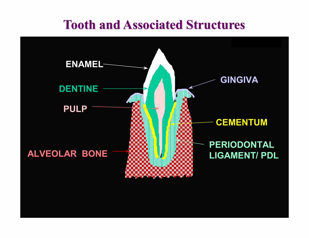
PULP
DENTINE
ENAMEL
CEMENTUM
PERIODONTAL LIGAMENT/ PDLALVEOLAR BONE
GINGIVA
WABeresford
Tooth and Associated Structures
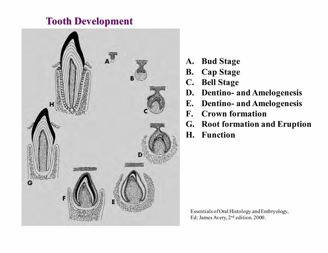
Tooth Development
A. Bud StageB. Cap StageC. Bell StageD. Dentino- and AmelogenesisE. Dentino- and AmelogenesisF. Crown formationG. Root formation and EruptionH. Function
Essentials of Oral Histology and Embryology,Ed: James Avery, 2nd edition. 2000.
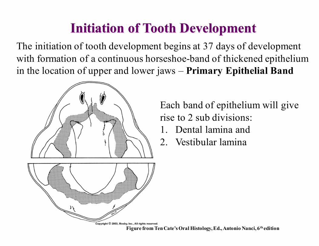
Initiation of Tooth DevelopmentThe initiation of tooth development begins at 37 days of developmentwith formation of a continuous horseshoe-band of thickened epitheliumin the location of upper and lower jaws – Primary Epithelial Band
Each band of epithelium will giverise to 2 sub divisions:1. Dental lamina and2. Vestibular lamina
Figure from Ten Cate’s Oral Histology, Ed., Antonio Nanci, 6th edition

Vestibular Lamina
Figure from Ten Cate’s Oral Histology, Ed., Antonio Nanci, 6th edition

Morphogenesis• The genesis of a morphe (of a shape; verb: to morph= to shape; change from
one image to another)• Molecular pathways
– Bone morphogenetic protein (BMP)• Inhibitory signals
– Fibroblast growth factor (FGF)• Stimulatory signals
– Sonic Hedgehog (Shh)• Initiation of tooth formation; cap stage development
– Wingless–related intergretion site (Wnt)• Stimulatory signals• Overexpression of Wnt inhibitor Dkk1 à NO DENTAL PLACODE
– Notch• Lateral inhibition or inductive signaling
– Ectodysplasin (Eda)• Formation of the placode• Inhibition à Oligodontia• Overactivation à more misshaped teeth

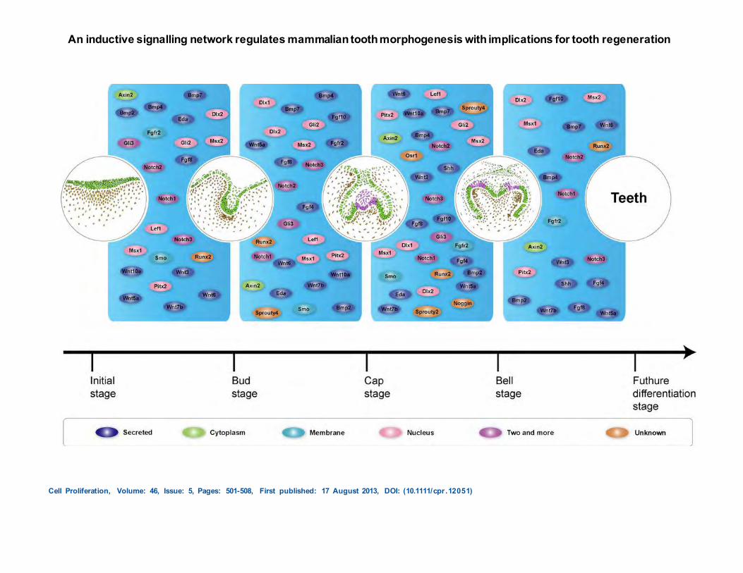
An inductive signalling network regulates mammalian tooth morphogenesis with implications for tooth regeneration
Cell Proliferation, Volume: 46, Issue: 5, Pages: 501-508, First published: 17 August 2013, DOI: (10.1111/cpr.12051)
BMPWnt
ShhNotchFGF

EDA
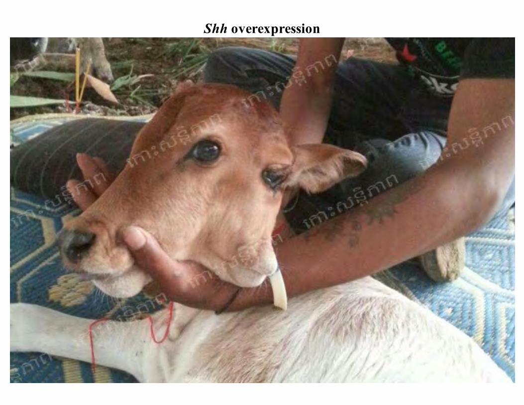
Shh overexpression


Some Genes Regulating Tooth Development
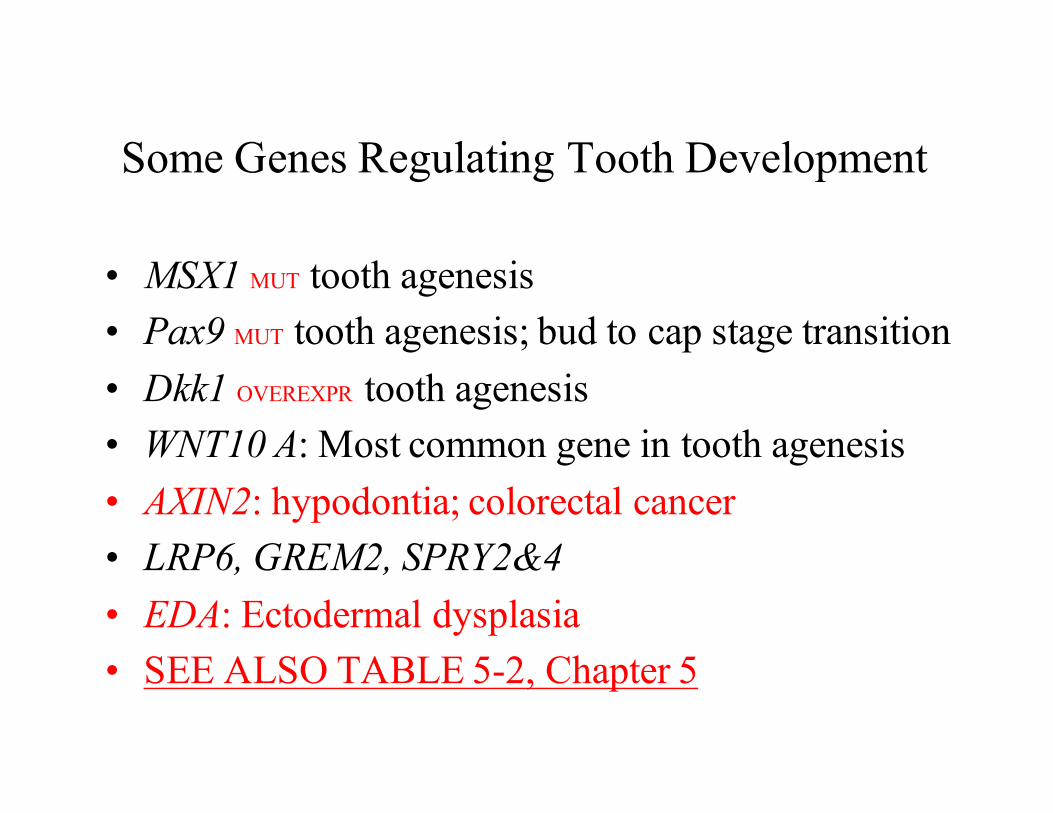
• MSX1 MUT tooth agenesis• Pax9 MUT tooth agenesis; bud to cap stage transition• Dkk1 OVEREXPR tooth agenesis• WNT10 A: Most common gene in tooth agenesis• AXIN2: hypodontia; colorectal cancer• LRP6, GREM2, SPRY2&4• EDA: Ectodermal dysplasia• SEE ALSO TABLE 5-2, Chapter 5

Stomatodeum
Developing Tongue
Dental lamina
Maxillary Process
Mandibular process
http://www.usc.edu/hsc/dental/ohisto/
Dental placodeRegulated by Notch, Eda, Wnt, Shh
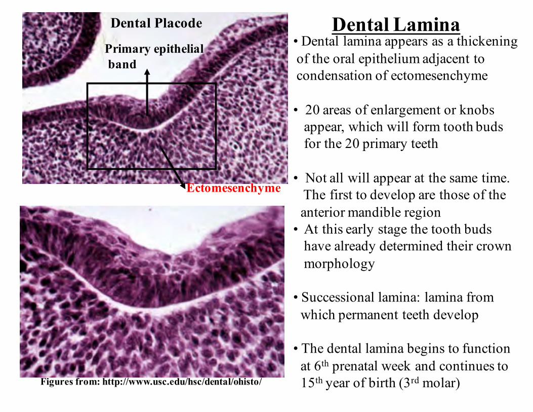
Dental Lamina• Dental lamina appears as a thickeningof the oral epithelium adjacent tocondensation of ectomesenchyme
• 20 areas of enlargement or knobsappear, which will form tooth budsfor the 20 primary teeth
• Not all will appear at the same time.The first to develop are those of theanterior mandible region
• At this early stage the tooth budshave already determined their crownmorphology
• Successional lamina: lamina fromwhich permanent teeth develop
• The dental lamina begins to functionat 6th prenatal week and continues to15th year of birth (3rd molar)
Primary epithelialband
Ectomesenchyme
Figures from: http://www.usc.edu/hsc/dental/ohisto/
Dental Placode
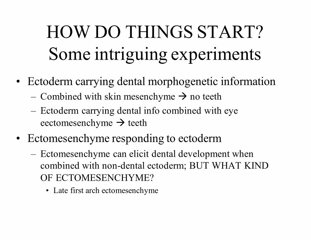
HOW DO THINGS START?Some intriguing experiments
• Ectoderm carrying dental morphogenetic information– Combined with skin mesenchyme à no teeth– Ectoderm carrying dental info combined with eye
eectomesenchyme à teeth• Ectomesenchyme responding to ectoderm
– Ectomesenchyme can elicit dental development when combined with non-dental ectoderm; BUT WHAT KIND OF ECTOMESENCHYME?
• Late first arch ectomesenchyme
An inductive signalling network regulates mammalian tooth morphogenesis with implications for tooth regeneration
Cell Proliferation, Volume: 46, Issue: 5, Pages: 501-508, First published: 17 August 2013, DOI: (10.1111/cpr.12051)
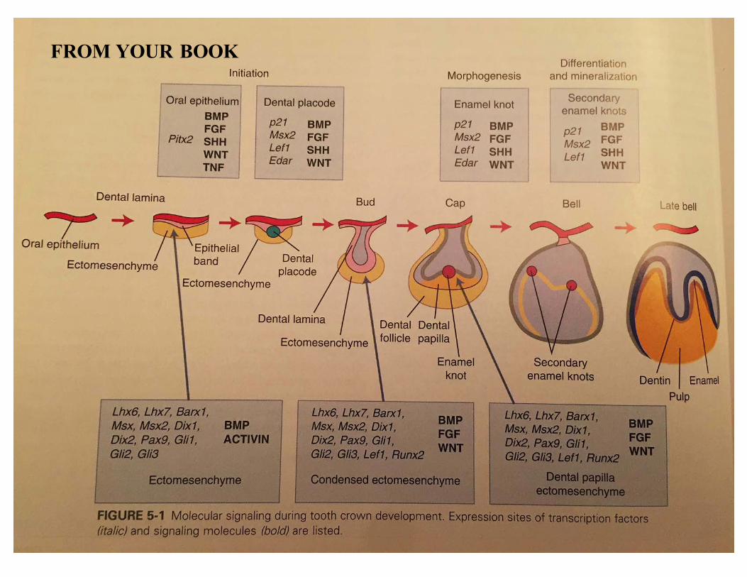
FROM YOUR BOOK

Tooth development is a continuous process, however can bedivided into 3 stages:
1. Bud Stage2. Cap Stage3. Bell Stage
No clear separation between stages
Sections may depict different stages, i.e. cap vs. bell
Histodifferentiation: End of cap stage and beginning of bell stage
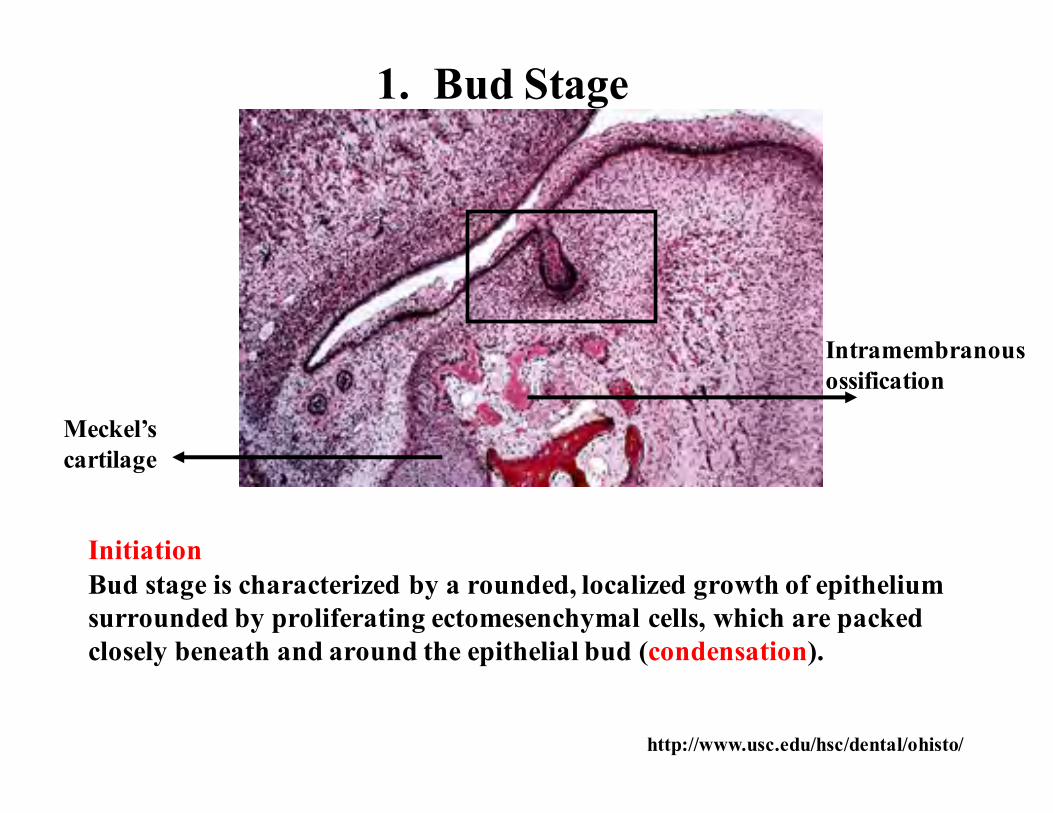
1. Bud Stage
InitiationBud stage is characterized by a rounded, localized growth of epithelium surrounded by proliferating ectomesenchymal cells, which are packed closely beneath and around the epithelial bud (condensation).
Meckel’scartilage
Intramembranousossification
http://www.usc.edu/hsc/dental/ohisto/

1. Bud Stage
In the bud stage, the enamel organ consists of peripherally locatedlow columnar cells and centrally located polygonal cells
http://www.usc.edu/hsc/dental/ohisto/
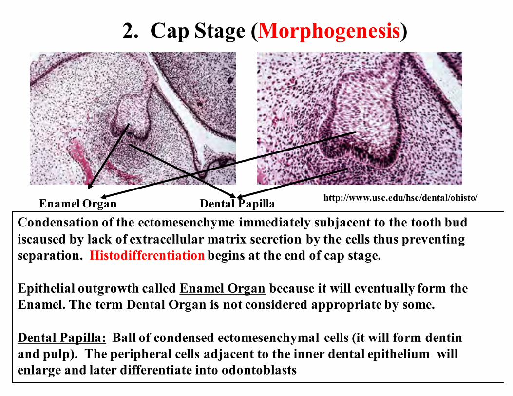
2. Cap Stage (Morphogenesis)
Condensation of the ectomesenchyme immediately subjacent to the tooth bud iscaused by lack of extracellular matrix secretion by the cells thus preventingseparation. Histodifferentiation begins at the end of cap stage.
Epithelial outgrowth called Enamel Organ because it will eventually form theEnamel. The term Dental Organ is not considered appropriate by some.
Dental Papilla: Ball of condensed ectomesenchymal cells (it will form dentinand pulp). The peripheral cells adjacent to the inner dental epithelium willenlarge and later differentiate into odontoblasts
Enamel Organ Dental Papilla http://www.usc.edu/hsc/dental/ohisto/

2. Cap Stage
Enamel organ
Dental papilla
Dental follicle or sacDental follicle or dental sac is the condensed ectomesenchymal tissuesurrounding the enamel organ and dental papilla. This gives rise tocementum and the periodontal ligament (support structures for tooth)
Enamel knot
http://www.usc.edu/hsc/dental/ohisto/
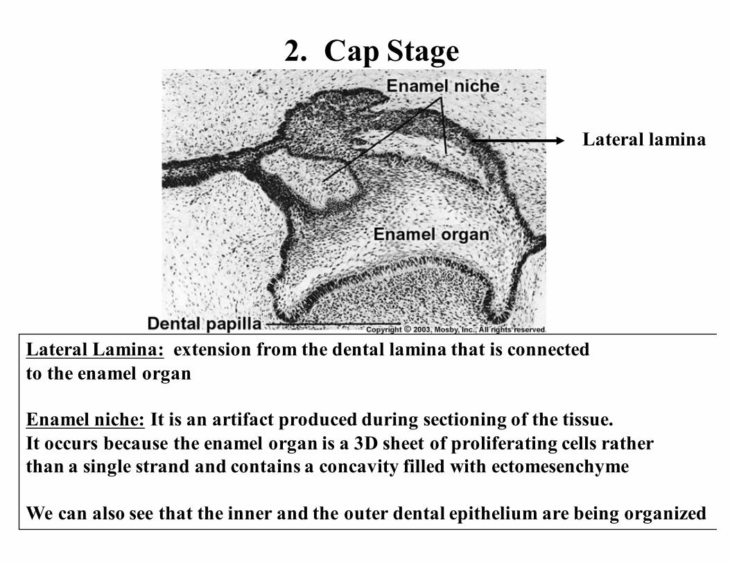
2. Cap Stage
Lateral Lamina: extension from the dental lamina that is connectedto the enamel organ
Enamel niche: It is an artifact produced during sectioning of the tissue.It occurs because the enamel organ is a 3D sheet of proliferating cells ratherthan a single strand and contains a concavity filled with ectomesenchyme
We can also see that the inner and the outer dental epithelium are being organized
Lateral lamina

Dental organ or tooth germ is a term used to constitute the structure that hasenamel organ, dental papilla and dental follicle
Enamel Knot: Densely packed accumulation of cells projecting from the inner enamel epithelium into dental papilla. Organizational center for cusp development. FGFs, BMPs, MSX2; shares similarities with ridges of developing limbs. Enamel Cord: Extension of the enamel knot.
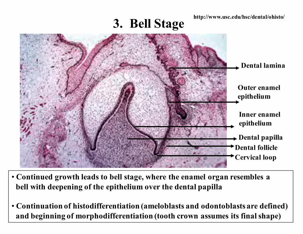
3. Bell Stage
• Continued growth leads to bell stage, where the enamel organ resembles abell with deepening of the epithelium over the dental papilla
• Continuation of histodifferentiation (ameloblasts and odontoblasts are defined)and beginning of morphodifferentiation (tooth crown assumes its final shape)
Dental lamina
Outer enamelepithelium
Inner enamelepithelium
Dental papillaDental follicleCervical loop
http://www.usc.edu/hsc/dental/ohisto/

3. Bell Stage (Early)
Inner enamel epithelium: Short columnar cells bordering the dental papilla.These will eventually become ameloblasts that will form the enamel of the tooth crown by differentiating into tall columnar cells. The cells of inner dentalepithelium exert an organizing influence on the underlying mesenchymal cellsin the dental papilla, which later differentiate into odontoblasts.
Outer enamel epithelium: Cuboidal cells that cover the enamel organ. Their function isto organize a network of capillaries that will bring nutrition to the ameloblasts. Inpreparation to formation of enamel, at the end of bell stage, the formerly smooth surfaceof the outer dental epithelium is laid in folds. Between the folds, adjacent mesenchymeof the dental sac forms papillae that contain capillary loops to provide nutritionalsupply for the intense metabolic activity of the avascular enamel organ
Stellate reticulum
Inner enamel epithelium
Stratum intermedium
Dental papilla
Outer enamel epithelium
http://www.usc.edu/hsc/dental/ohisto/

3. Bell Stage (Early)
Stellate reticulum
Inner dental epithelium
Stratum intermedium
Dental papilla
Stellate reticulum: Star-shaped cells with processes, present between the outerand the inner dental epithelium. These cells secrete glycosaminoglycans,which attract water, thereby swelling the cells and pushing them apart.However, they still maintain contact with each other, thus becoming star-shaped.They have a cushion-like consistency that may support and protect the delicateenamel organ. It is absent in the portion that outlines the root portions.
Stratum intermedium: Cell layer between the inner dental epithelium andstellate reticulum which have high alkaline phosphatase activity. They assistinner dental epithelium (ameloblasts) to form enamel.
Outer dental epithelium
http://www.usc.edu/hsc/dental/ohisto/
Stellate reticulum
Inner enamel epithelium
Stratum intermedium
Dental papilla
Outer enamel epithelium
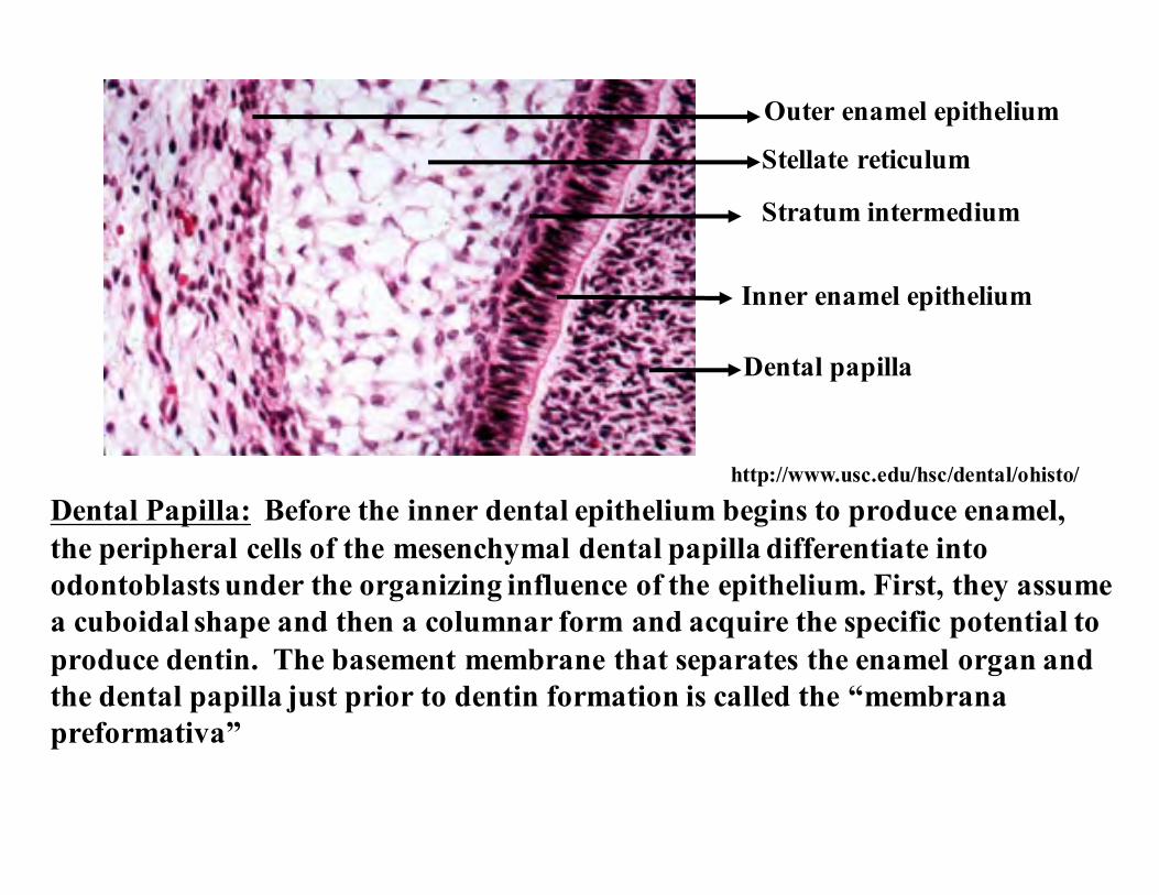
Dental Papilla: Before the inner dental epithelium begins to produce enamel,the peripheral cells of the mesenchymal dental papilla differentiate intoodontoblasts under the organizing influence of the epithelium. First, they assumea cuboidal shape and then a columnar form and acquire the specific potential toproduce dentin. The basement membrane that separates the enamel organ andthe dental papilla just prior to dentin formation is called the “membranapreformativa”
http://www.usc.edu/hsc/dental/ohisto/

3. Bell Stage
Higher power viewhttp://www.usc.edu/hsc/dental/ohisto/
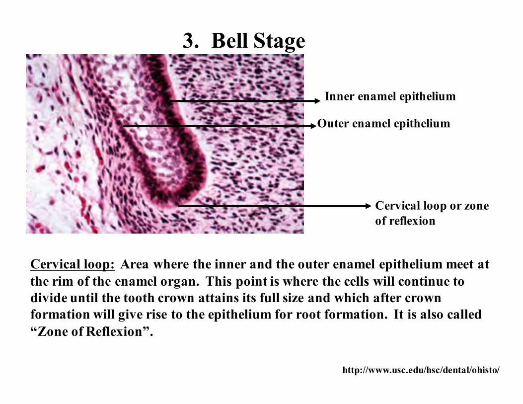
Inner enamel epithelium
Outer enamel epithelium
Cervical loop or zone of reflexion
Cervical loop: Area where the inner and the outer enamel epithelium meet atthe rim of the enamel organ. This point is where the cells will continue todivide until the tooth crown attains its full size and which after crownformation will give rise to the epithelium for root formation. It is also called“Zone of Reflexion”.
3. Bell Stage
http://www.usc.edu/hsc/dental/ohisto/
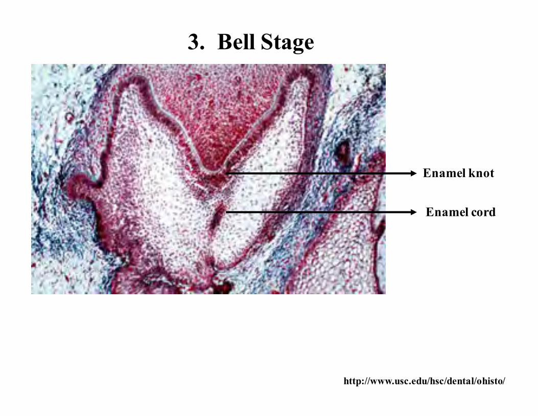
Enamel cord
Enamel knot
3. Bell Stage
http://www.usc.edu/hsc/dental/ohisto/

3. Bell Stage
Dental lamina (and the lateral lamina) will disintegrate and loose contactwith oral epithelium. Sometimes, these epithelial cells will persist whenthey are called “epithelial pearls” or “cell rests of Serres”
Clinical significance: Cysts will develop (eruption cysts) and preventeruption, or they may form odontomas (tumors) or may form supernumeraryteeth
http://www.usc.edu/hsc/dental/ohisto/

TOOTH TYPE DETERMINATION
• Homodonts and heterodonts
From a biology lecture; U of Miami
You and me and our cats and dogs
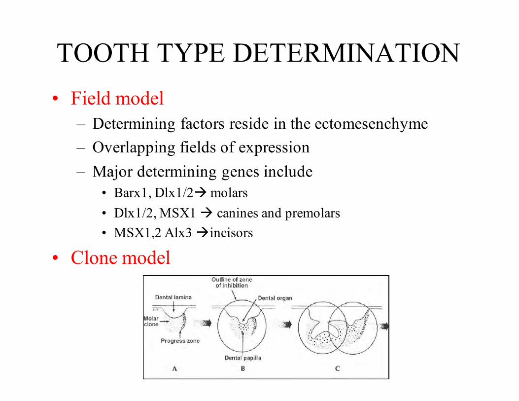
TOOTH TYPE DETERMINATION• Field model
– Determining factors reside in the ectomesenchyme– Overlapping fields of expression– Major determining genes include
• Barx1, Dlx1/2à molars• Dlx1/2, MSX1 à canines and premolars• MSX1,2 Alx3 àincisors
• Clone model
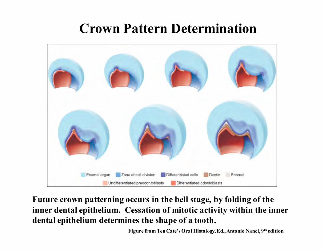
Future crown patterning occurs in the bell stage, by folding of theinner dental epithelium. Cessation of mitotic activity within the innerdental epithelium determines the shape of a tooth.
Crown Pattern Determination
Figure from Ten Cate’s Oral Histology, Ed., Antonio Nanci, 9th edition
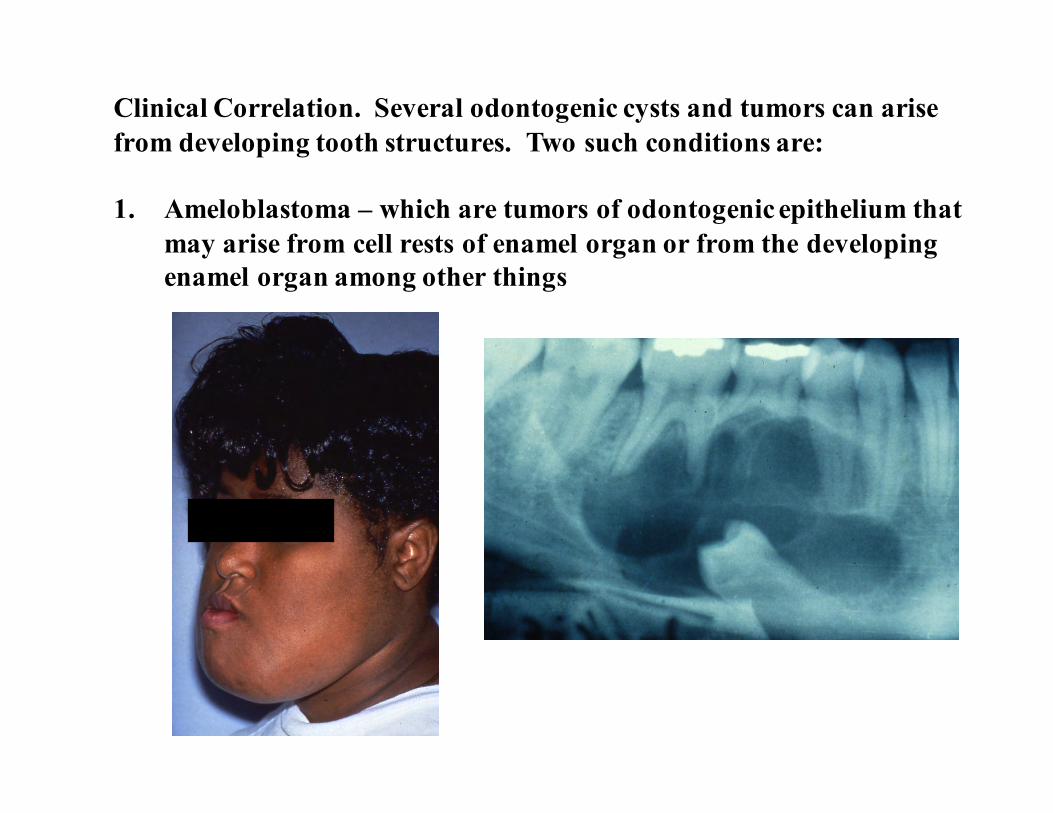
Clinical Correlation. Several odontogenic cysts and tumors can arisefrom developing tooth structures. Two such conditions are:
1. Ameloblastoma – which are tumors of odontogenic epithelium thatmay arise from cell rests of enamel organ or from the developingenamel organ among other things
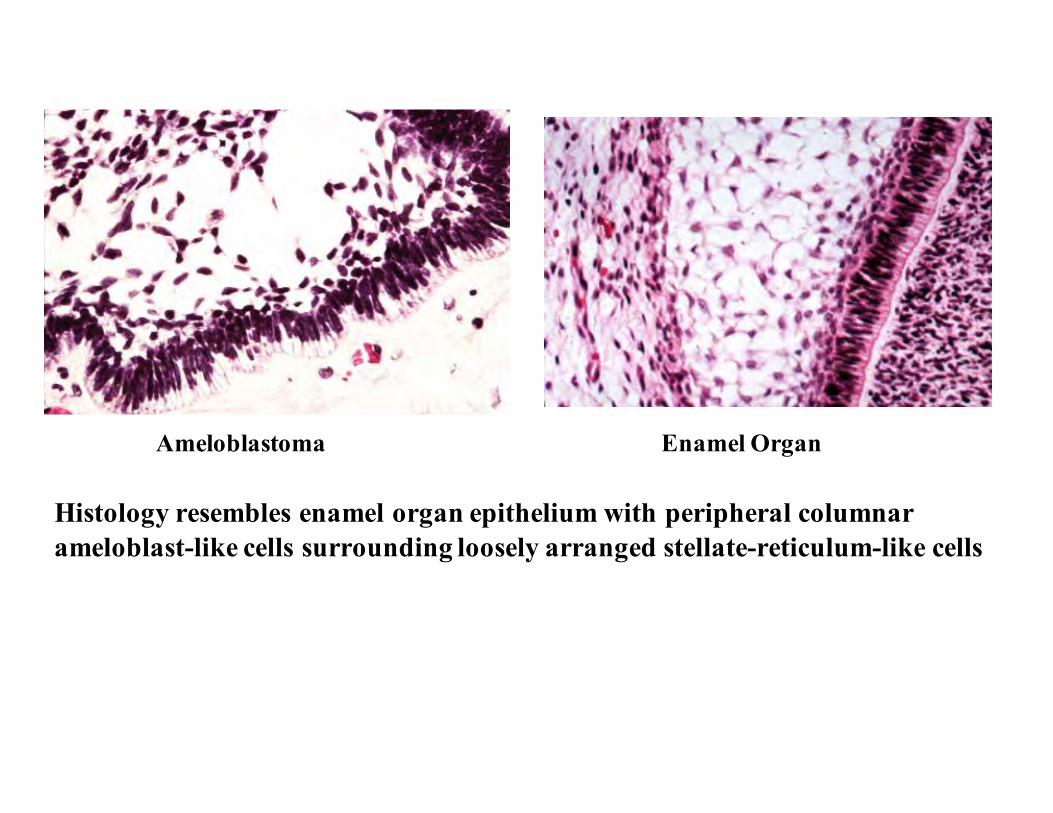
Histology resembles enamel organ epithelium with peripheral columnarameloblast-like cells surrounding loosely arranged stellate-reticulum-like cells
Ameloblastoma Enamel Organ

2. Odontogenic Myxoma: Tumor of the jaw that arise from odontogenicectomesenchyme. Histologically, looks similar to mesenchymal portionof a developing tooth (dental papilla).
Odontogenic myxoma Developing tooth
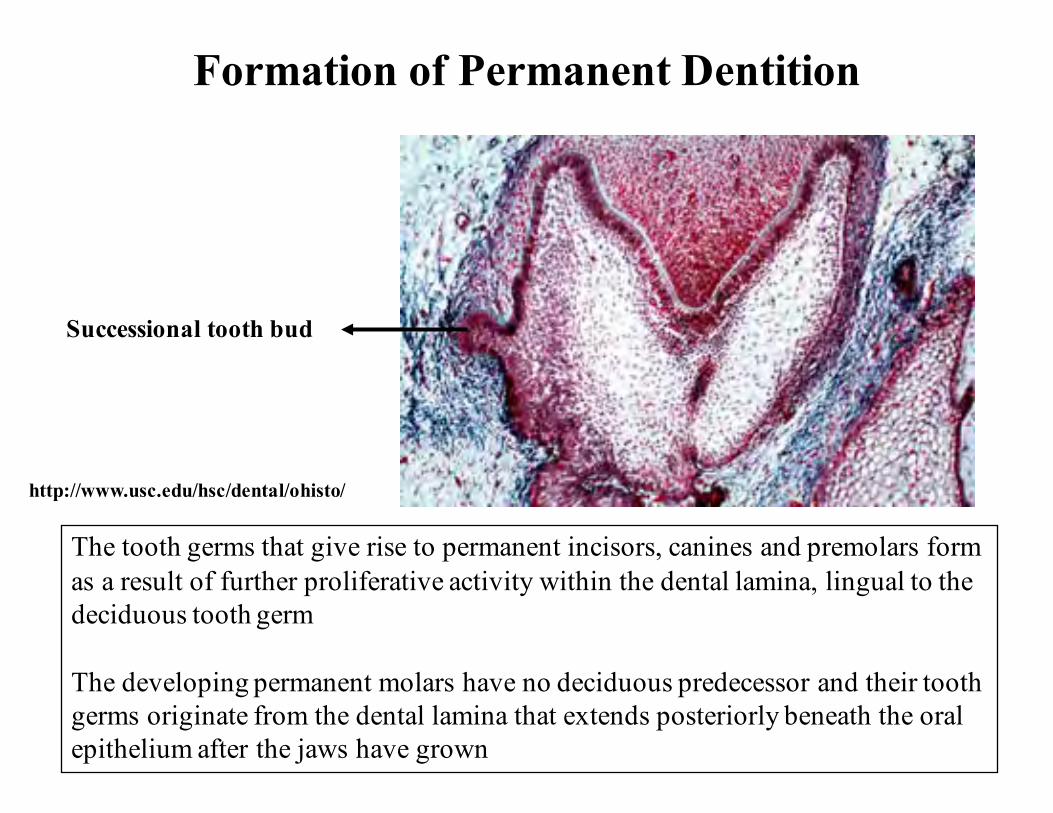
Formation of Permanent Dentition
Successional tooth bud
The tooth germs that give rise to permanent incisors, canines and premolars formas a result of further proliferative activity within the dental lamina, lingual to thedeciduous tooth germ
The developing permanent molars have no deciduous predecessor and their toothgerms originate from the dental lamina that extends posteriorly beneath the oralepithelium after the jaws have grown
http://www.usc.edu/hsc/dental/ohisto/

A timetable to rememberEntire primary dentition initiated between 6 and 8 weeks of embryonicdevelopment.
Successional permanent teeth initiated between 20th week in uteroand 10th month after birth
Permanent molars between 20th week in utero (first molar) and 5th year of life (third molar)
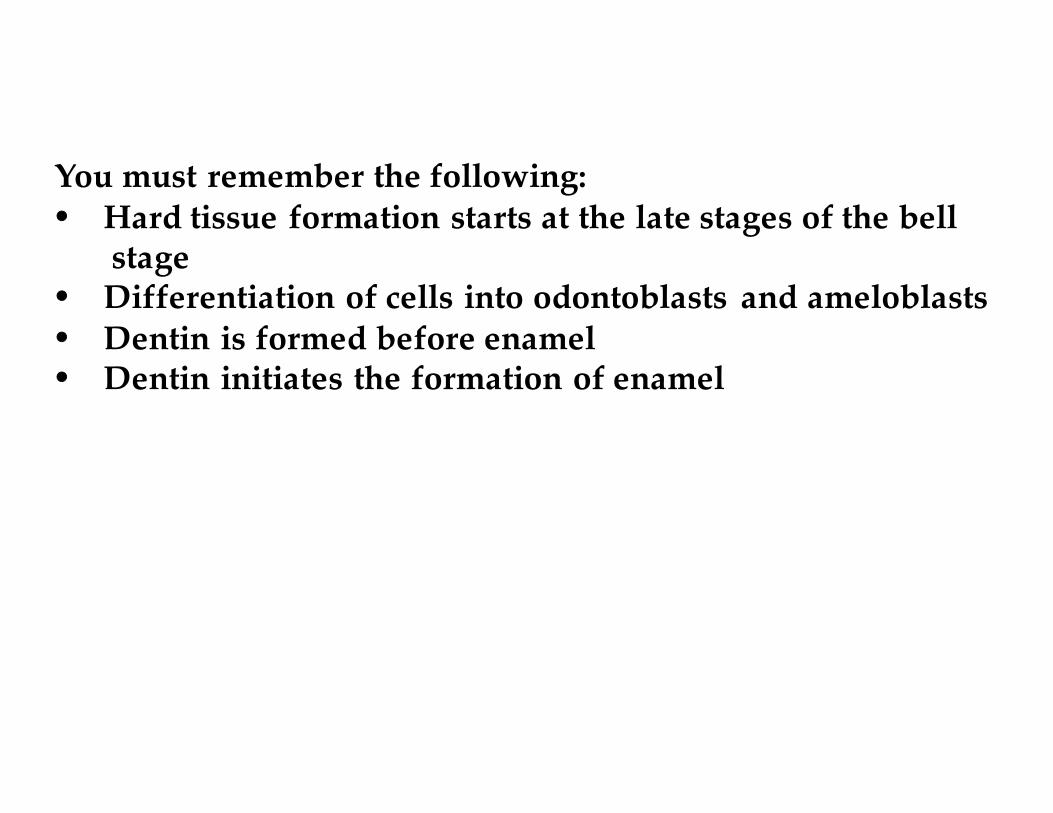
You must remember the following:• Hard tissue formation starts at the late stages of the bell
stage• Differentiation of cells into odontoblasts and ameloblasts• Dentin is formed before enamel• Dentin initiates the formation of enamel
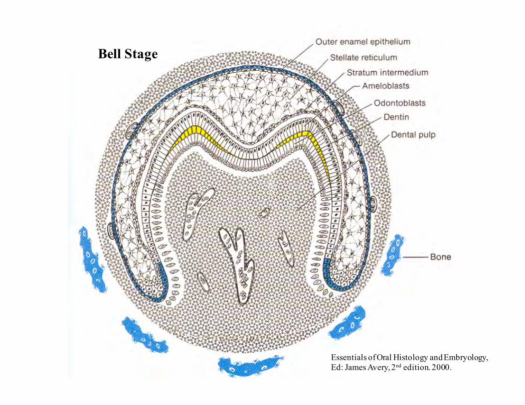
Bell Stage
Essentials of Oral Histology and Embryology,Ed: James Avery, 2nd edition. 2000.
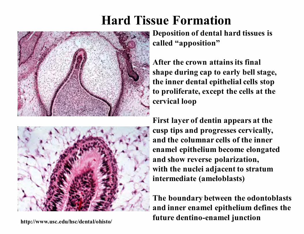
Hard Tissue FormationDeposition of dental hard tissues iscalled “apposition”
After the crown attains its finalshape during cap to early bell stage,the inner dental epithelial cells stopto proliferate, except the cells at thecervical loop
First layer of dentin appears at thecusp tips and progresses cervically,and the columnar cells of the innerenamel epithelium become elongatedand show reverse polarization,with the nuclei adjacent to stratumintermediate (ameloblasts)
The boundary between the odontoblastsand inner enamel epithelium defines thefuture dentino-enamel junction http://www.usc.edu/hsc/dental/ohisto/

For dentinogenesis and amelogenesis to take place normally,the differentiating odontoblasts and ameloblasts will receivesignals form each other – “reciprocal induction”
Stages of Apposition
1. Elongation of inner enamel epithelium2. Differentiation of odontoblasts3. Formation of dentin4. Formation of enamel

At the same time or soon after the first layer of dentin (mantle dentin) is formed,the inner enamel epithelial cells differentiate into ameloblasts and secrete enamelproteins. These proteins further will help in the terminal differentiation ofodontoblasts. The ameloblasts will then start laying down organic matrix ofenamel against the newly formed dentinal surface. The enamel matrix willmineralize immediately and form the first layer of enamel. The formation ofenamel is called amelogenesis.
Ameloblasts
First layer of enamel
Dentin
Odontoblasts
http://www.usc.edu/hsc/dental/ohisto/

AppositionAt the same time when the innerenamel epithelium is differentiating,the undifferentiated ectomesenchymalcells increase rapidly in size andultimately differentiate into odontoblasts
The increase in size of the papillarycells leads to elimination of the acellularzone between dental papilla and innerenamel epithelium
Differentiation of odontoblasts fromectomesenchymal cells are induced byinfluence from the inner enamel epithelium
Experiments have shown that if there isno inner enamel epithelium, there is nodentin formed
Odontoblasts Dentin
Enamel
Ameloblastshttp://www.usc.edu/hsc/dental/ohisto/
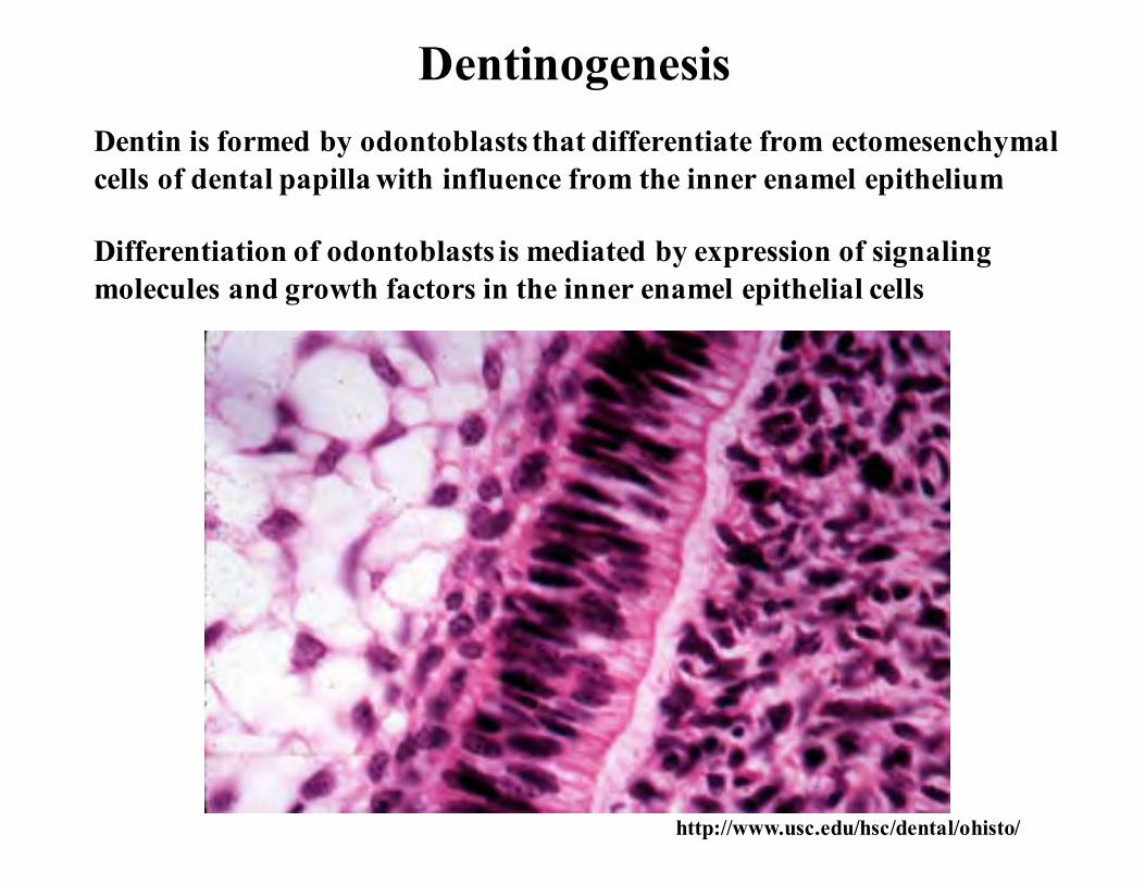
DentinogenesisDentin is formed by odontoblasts that differentiate from ectomesenchymalcells of dental papilla with influence from the inner enamel epithelium
Differentiation of odontoblasts is mediated by expression of signalingmolecules and growth factors in the inner enamel epithelial cells
http://www.usc.edu/hsc/dental/ohisto/

Time Line of Human Tooth Development(Table 5-2 in Text book)
Age Developmental Characteristics42 to 48 days Dental lamina formation
55 to 56 days Bud stage; deciduous incisors;canines and molars
14 weeks Bell stage for deciduous teeth; budstage for permanent teeth
18 weeks Dentin and functional ameloblastsin deciduous teeth
32 weeks Dentin and functional ameloblastsin permanent first molars

Incremental pattern of dentinand enamel formation frominitiation to completion
Growth areas of developing crown.Growth at cusp tip, intercuspal region,and cervical region
Essentials of Oral Histology and Embryology,Ed: James Avery, 2nd edition. 2000.
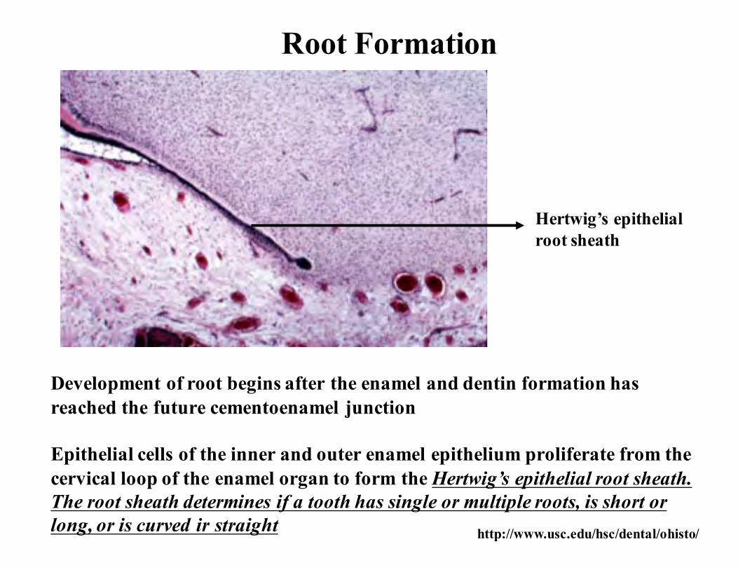
Root Formation
Development of root begins after the enamel and dentin formation hasreached the future cementoenamel junction
Epithelial cells of the inner and outer enamel epithelium proliferate from thecervical loop of the enamel organ to form the Hertwig’s epithelial root sheath.The root sheath determines if a tooth has single or multiple roots, is short orlong, or is curved ir straight
Hertwig’s epithelialroot sheath
http://www.usc.edu/hsc/dental/ohisto/

Hertwig’s epithelialroot sheath
Inner enamel epithelium
Outer enamel epithelium
Stratum intermedium
Eventually the root sheath will fragment to form several discrete clustersof epithelial cells known as epithelial cell rests of malassez. These will persist inadults within the periodontal ligament
http://www.usc.edu/hsc/dental/ohisto/
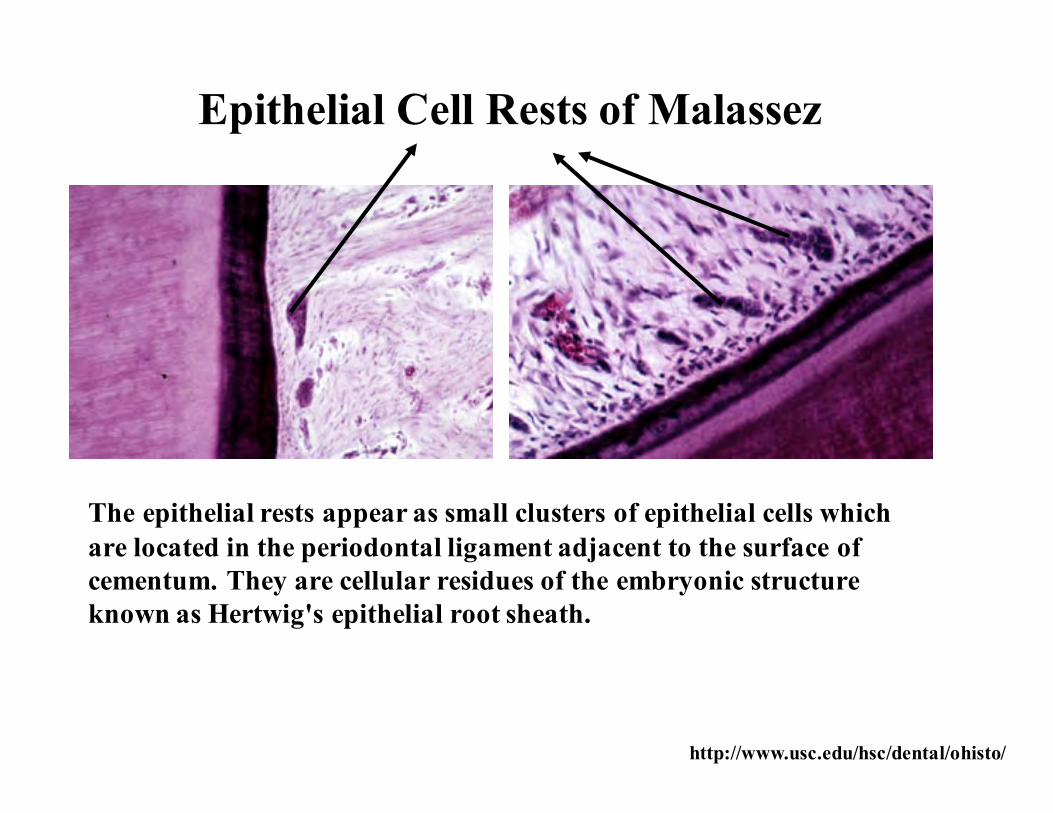
The epithelial rests appear as small clusters of epithelial cells which are located in the periodontal ligament adjacent to the surface of cementum. They are cellular residues of the embryonic structure known as Hertwig's epithelial root sheath.
Epithelial Cell Rests of Malassez
http://www.usc.edu/hsc/dental/ohisto/
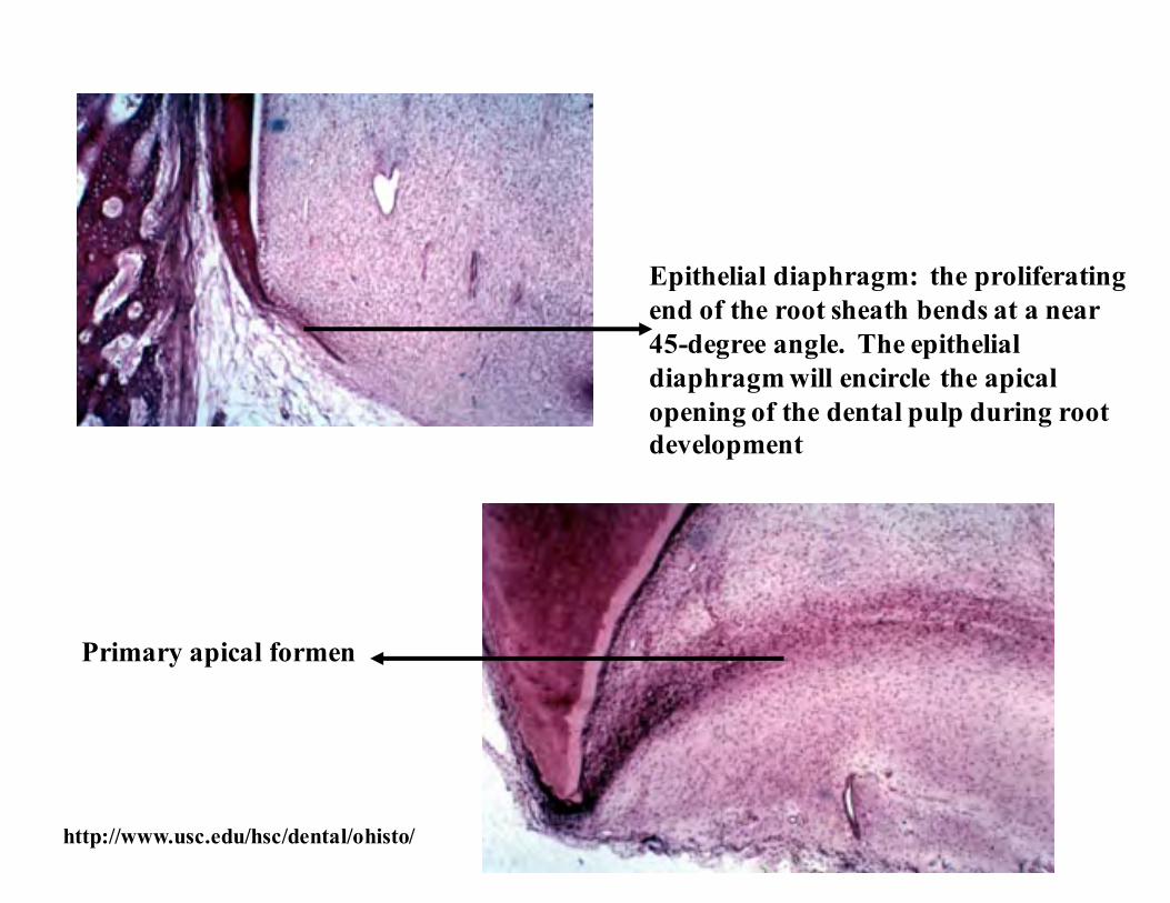
Primary apical formen
Epithelial diaphragm: the proliferatingend of the root sheath bends at a near45-degree angle. The epithelialdiaphragm will encircle the apicalopening of the dental pulp during rootdevelopment
http://www.usc.edu/hsc/dental/ohisto/

Secondary apical foramen form as a result of two or three tongues of epithelium growing inward toward each other resulting in multirooted teeth
Essentials of Oral Histology and Embryology,Ed: James Avery, 2nd edition. 2000
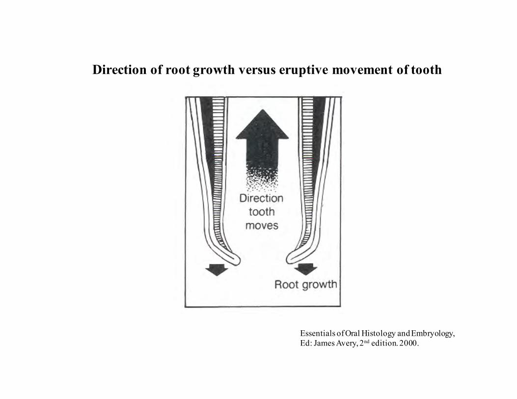
Direction of root growth versus eruptive movement of tooth
Essentials of Oral Histology and Embryology,Ed: James Avery, 2nd edition. 2000.
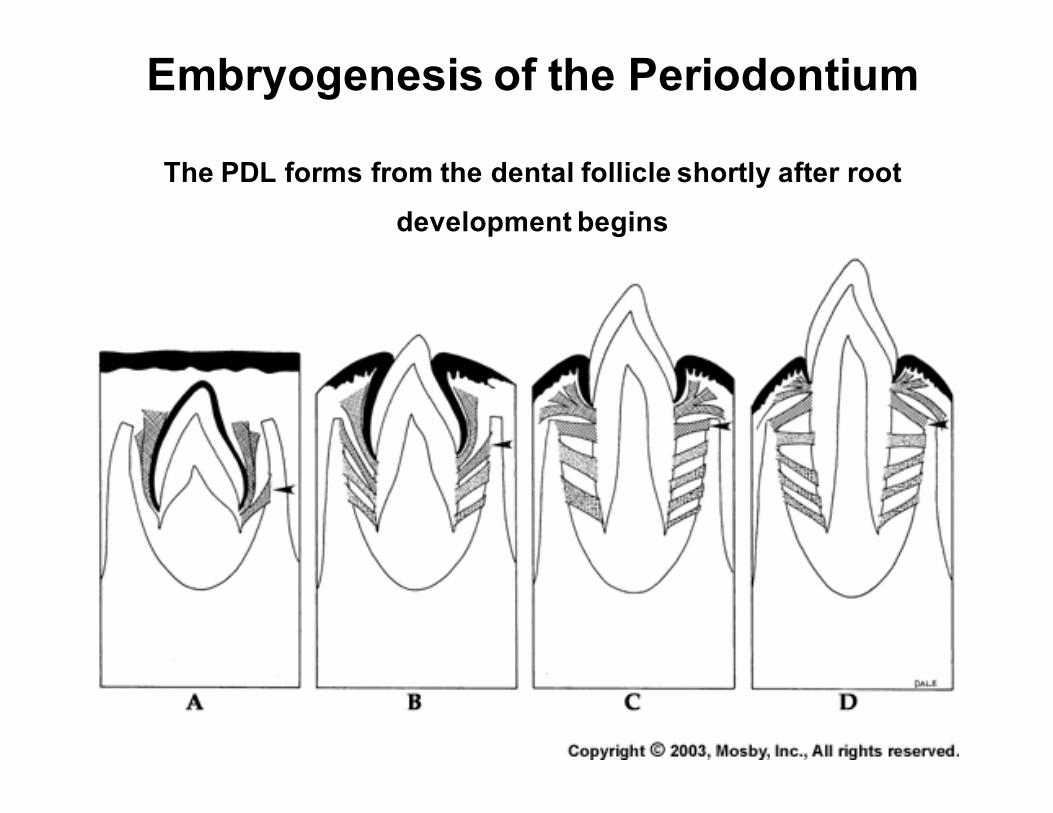
Embryogenesis of the Periodontium
The PDL forms from the dental follicle shortly after root
development begins
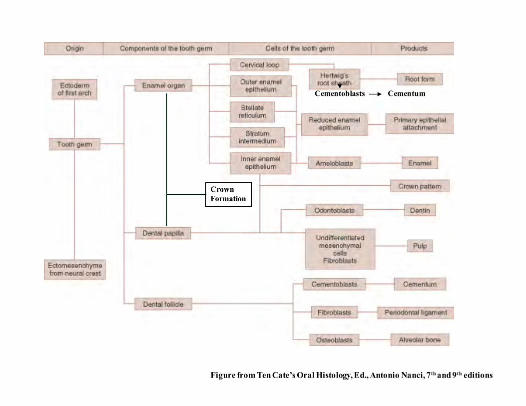
Figure from Ten Cate’s Oral Histology, Ed., Antonio Nanci, 7th and 9th editions
Cementoblasts Cementum
CrownFormation


![Foundations Lecture - Introduction to Human Development - Embryology · 2014-08-22 · Foundations Lecture - Introduction to Human Development from Embryology (14 Apr 2014) [show]](https://static.fdocuments.us/doc/165x107/5ea4aa2461a7f0668e469fa9/foundations-lecture-introduction-to-human-development-embryology-2014-08-22.jpg)


