Development of rational bioprocess design strategies for a ...
148
This document is downloaded from DR‑NTU (https://dr.ntu.edu.sg) Nanyang Technological University, Singapore. Development of rational bioprocess design strategies for a clinically relevant protein candidate Anindya Basu 2012 Anindya Basu. (2012). Development of rational bioprocess design strategies for a clinically relevant protein candidate. Doctoral thesis, Nanyang Technological University, Singapore. https://hdl.handle.net/10356/50661 https://doi.org/10.32657/10356/50661 Downloaded on 26 Jan 2022 00:34:52 SGT
Transcript of Development of rational bioprocess design strategies for a ...

This document is downloaded from DR‑NTU (httpsdrntuedusg)Nanyang Technological University Singapore
Development of rational bioprocess designstrategies for a clinically relevant proteincandidate
Anindya Basu
2012
Anindya Basu (2012) Development of rational bioprocess design strategies for a clinicallyrelevant protein candidate Doctoral thesis Nanyang Technological University Singapore
httpshdlhandlenet1035650661
httpsdoiorg10326571035650661
Downloaded on 26 Jan 2022 003452 SGT
DEVELOPMENT OF RATIONAL BIOPROCESS
DESIGN STRATEGIES FOR A CLINICALLY RELEVANT
PROTEIN CANDIDATE
ANINDYA BASU
SCHOOL OF CHEMICAL AND BIOMEDICAL ENGINEERING
2012
1
DEVELOPMENT OF RATIONAL BIOPROCESS
DESIGN STRATEGIES FOR A CLINICALLY RELEVANT
PROTEIN CANDIDATE
ANINDYA BASU
SCHOOL OF CHEMICAL AND BIOMEDICAL ENGINEERING
A thesis submitted to the Nanyang Technological University in partial fulfilment of the
requirement for the degree of Doctor of Philosophy
2
Acknowledgement
I would like to express my heartfelt gratitude to my supervisor Dr Susanna Leong Su Jan for
allowing me to be a part of her team I know no words to thank her for her expert guidance
and unrestricted support that constantly kept me motivated during the toughest times of my
project Without her valuable suggestions this thesis would not have been possible
My special thanks and appreciation goes to Prof William Chen for providing the plasmid
which formed the starting point of the current thesis
I am highly indebted to Dr Chen Yu the senior member of my lab Without his selfless
helping hand and patient considerations to all my queries this work could not have been
possible
I would also like to acknowledge the kind support from all my other lab-mates especially Dr
A K R Gobinath Mr Li Xiang Mr Derrick TK Sing and Dr Foo Jee Loon for their
unremitted help whenever I needed
I would like to thank the School of Chemical and Biomedical Engineering Nanyang
Technological University Singapore for providing me the scholarship without which this
work would not have been possible
Last but not the least I am grateful to each and every member of my family for their
unconditional support and encouragement to keep me upbeat and enthusiastic to do a good
work
3
Contents
LIST OF ABBREVIATIONS 8
ABSTRACT 9
CHAPTER 1 INTRODUCTION 11
1 INTRODUCTION 12
11 Aims and objectives 13
12 Thesis Organization 14
13 List of publications 15
CHAPTER 2 LITERATURE REVIEW 16
Abstract 17
21 Role of HBx in HCC 19
22 In vitro expression of HBx 20
221 Protein production in higher organisms 21
222 Cell culture based protein production 21
23 Protein folding mechanisms Forces involved 23
231 Protein folding models 23
2311 The thermodynamic hypothesis 23
2312 Molten Globule hypothesis 24
2313 Other similar models 24
2314 Energy Landscape theory 25
2315 Protein folding inside the cells role of chaperones 27
24 Protein refolding methods 28
241 Dilution refolding 29
242 Dialysis Methods 31
4
243 Chromatographic techniques 31
244 High hydrostatic pressure (HHP) technology 32
25 SVC measurement a rational approach to designing protein refolding recipes 34
251 Second virial coefficient theory and applications 34
26 Earlier attempts to produce recombinant HBx in microbial systems 36
CHAPTER 3 THE EFFECT OF A GST FUSION PARTNER ON HBX EXPRESSION AND
BIOPROCESSING IN AN E COLI MICROBIAL PLATFORM 43
Abstract 44
31 Introduction 45
32 Methods and materials 46
321 HBx plasmid transformation 46
322 DNA sequencing 46
323 GST-HBx protein expression 46
324 Cell disruption to recover GST-HBx 47
325 GST-HBx purification refolding and cleavage 47
3251 GST-HBx processing using Strategy I (pre-refolding purification of GST-HBx under denatured-
reduced conditions followed by dilution refolding) 47
3252 GST-HBx bioprocessing using Strategy II (protein solubilisation using sarkosyl under non-
denaturing non-reducing conditions and purification using Glutahione-Sepharose resins) 49
326 Analytical methods 50
33 Results and discussions 51
331 Characterization of the pGEX plasmid harbouring GST-HBx 51
332 Optimising culture conditions for higher soluble GST-HBx yield 51
3321 Effect of temperature on soluble GST-HBx expression 52
33211 Effect of carbon source on soluble expression of GST-HBx 53
333 Downstream processing of GST-HBx using Strategy I 54
3331 Solubilisation and purification of GST-HBx IBs 55
3332 Solubilisation studies of GST-HBx under non-denaturing conditions by dilution refolding
followed by Factor Xa cleavage 56
33321 GST-HBx solubility studies at pH 8-9 56
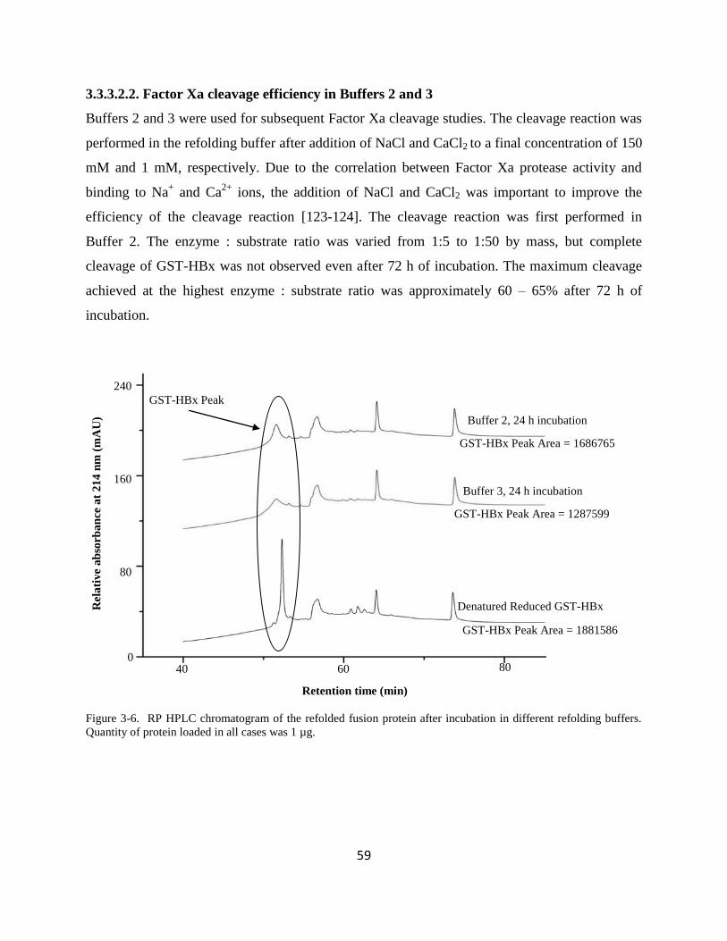
33322 Factor Xa cleavage efficiency in Buffers 2 and 3 59
33323 GST-HBx solubilisation and cleavage studies at pH 6 62
5
334 Downstream processing of GST-HBx using Strategy II 63
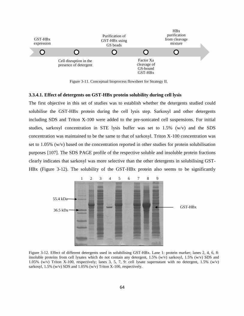
3341 Effect of detergents on GST-HBx protein solubility during cell lysis 64
3342 Binding and cleavage of GS-immobilised GST-HBx 65
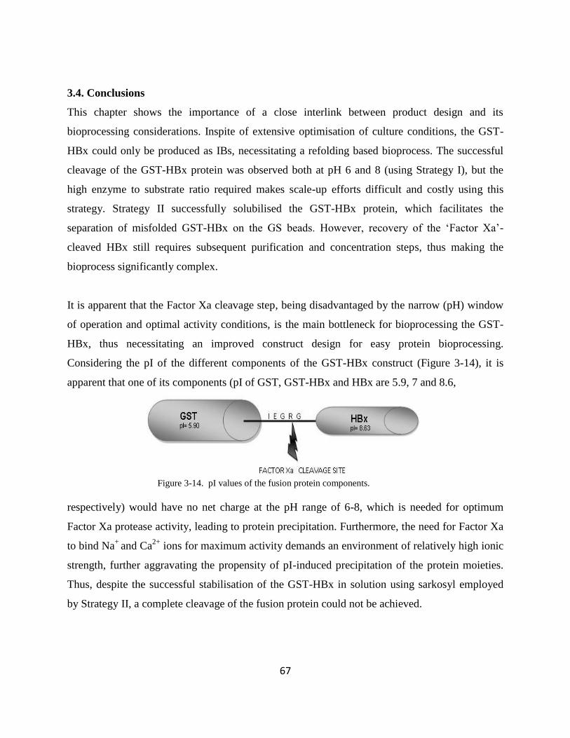
34 Conclusions 67
CHAPTER 4 STUDY OF PROTEIN INTERMOLECULAR INTERACTIONS TO
DETERMINE RATIONAL METHODS FOR HBX REFOLDING 69
Abstract 70
41 Introduction 71
42 Materials and methods 72
421 HBx plasmid transformation 72
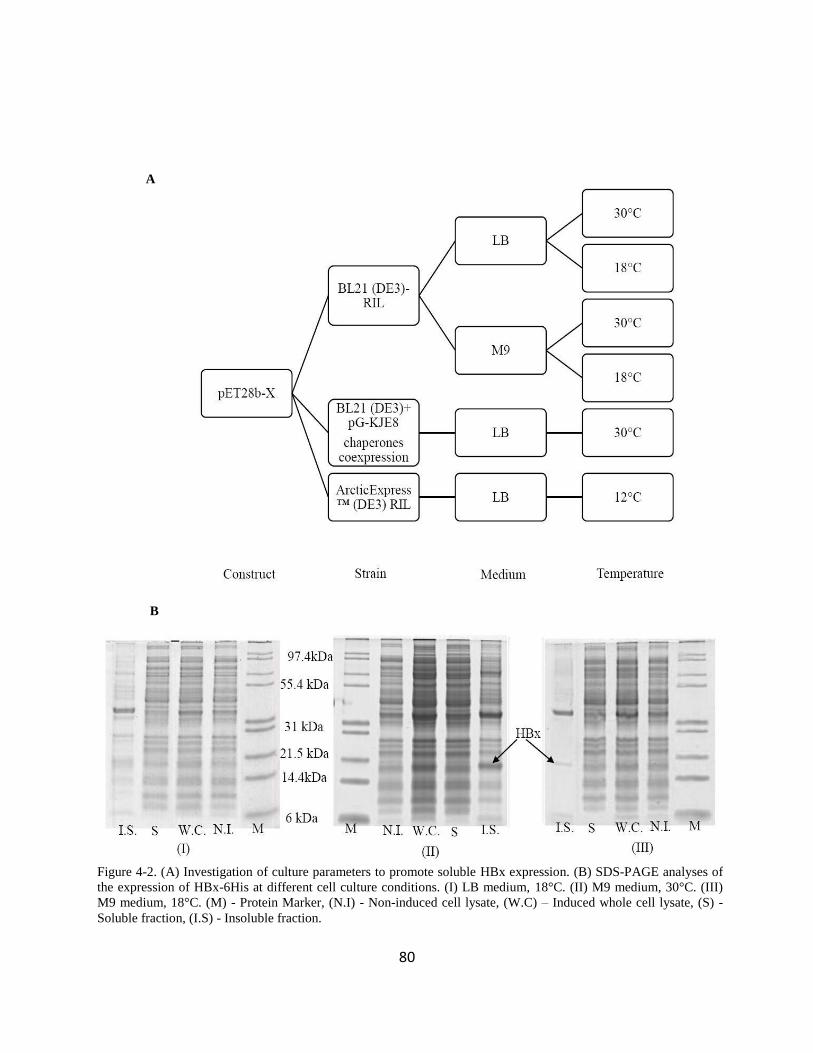
422 HBx cell culture optimisation studies 72
423 Recombinant HBx protein expression and IB recovery 73
424 HBx protein identification using Western Blot 73
425 HBx protein preparation for SVC measurements 73
426 Determination of SVC using SLS 74
427 Studying the effect of physicochemical environment on HBx solubility by DoE methodology 75
428 Dilution refolding of HBx 76
429 SEC-HPLC analysis 76
4291 Protein monomericity determination by SEC-FPLC 76
4210 p53 interaction test for HBx bioactivity determination 76
4211 RP-HPLC analysis 77
4212 Circular dichroism (CD) spectroscopy 77
4213 Other analytical protocols 78
43 Results and discussions 79
431 Optimising HBx gene expression conditions 79
432 HBx solubility studies by SVC measurements 82
433 HBx solubility and refolding studies 85
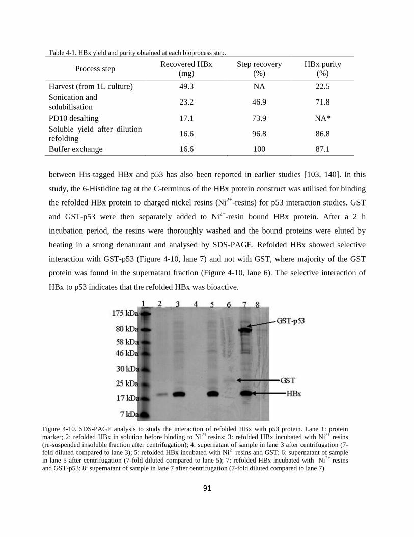
434 Refolded HBx bioactivity determination 90
435 Structural characterisation of soluble HBx post-refolding 92
4351 RP-HPLC analysis 92
4352 Secondary and tertiary HBx structure characterisation using CD spectroscopy 92
44 Conclusions 94
6
CHAPTER 5 DEVELOPMENT OF AN ELISA BASED ANALYTICAL PLATFORM FOR
DETERMINATION OF HBX REFOLDING YIELDS 96
Abstract 97
51 Introduction 98
52 Materials and methods 99
521 Construction of a calibration curve to quantify bound HBx 99
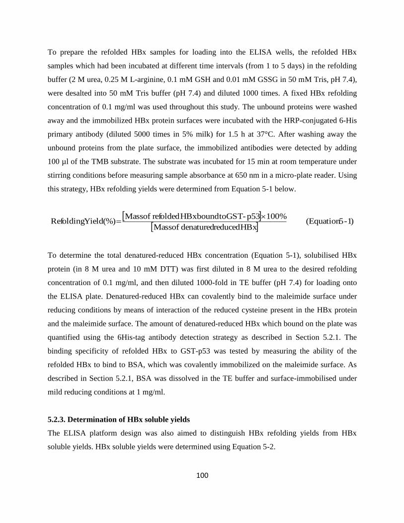
522 Determination of HBx refolding yield 99
523 Determination of HBx soluble yields 100
524 Analytical methods 101
525 Measurement System Analysis 101
5251 Precision 101
5252 Accuracy determination 102
53 Results and discussion 102
531 Immobilisation of proteins on the maleimide surface of the ELISA plates 102
532 Generation of calibration curve for the ELISA system using 6His-GST protein 104
533 Determination of HBx soluble and refolding yields 106
54 Conclusion 108
CHAPTER 6 HIGH PRODUCTIVITY CHROMATOGRAPHY REFOLDING PROCESS FOR
HBX GUIDED BY STATISTICAL DOE STUDIES 110
Abstract 111
61 Introduction 112
62 Methods and materials 112
621 HBx protein expression and solubilisation 112
622 IMAC refolding of the HBx protein 113
623 Dilution refolding of HBx IBs 113
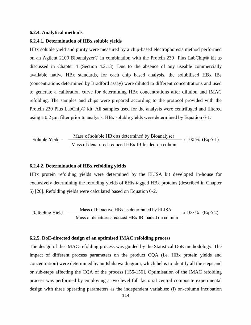
624 Analytical methods 114
6241 Determination of HBx soluble yields 114
6242 Determination of HBx refolding yields 114
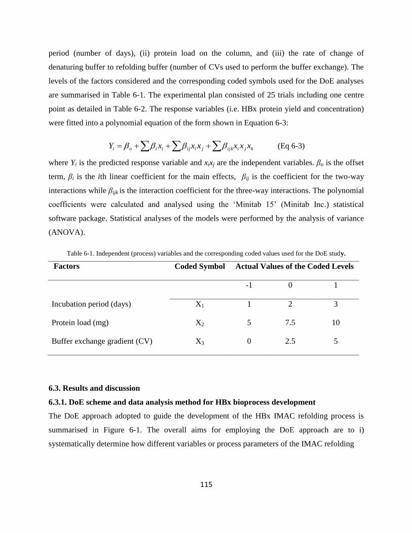
625 DoE-directed design of an optimised IMAC refolding process 114
63 Results and discussion 115
7
631 DoE scheme and data analysis method for HBx bioprocess development 115
632 Selection of IMAC refolding parameters for DoE studies 117
633 Development and optimisation of an IMAC refolding bioprocess for HBx production 119
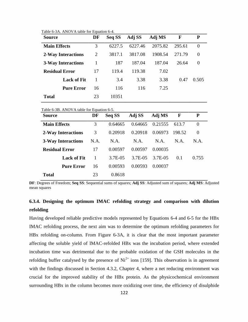
634 Designing the optimum IMAC refolding strategy and comparison with dilution refolding 122
64 Conclusions 127
CHAPTER 7 CONCLUSIONS AND FUTURE WORK 128
71 Conclusions 129
72 Future Work 131
721 Formulation and stability studies 131
7211 Formulation of HBx solution and stability studies 131
7212 HBx lyophilisation and stability studies 133
722 HBx structural studies 134
7221 HBx structural characterisation through crystallisation studies 135
7221 HBx structural characterisation through NMR spectroscopy 135
REFERENCES 137
8
List of Abbreviations
6His
6-Histidine
APS
Ammonium persulphate
BSA
Bovine serum albumin
CQA
Critical to Quality Attributes
CV
Column Volume
DoE
Design of Experiments
DTT
Dithiothreitol
ELISA
Enzyme Linked Immuno-Sorbent Assay
FPLC
Fast Protein Liquid Chromatography
GSH
Glutathione
GST
Glutathione S Transferase
HBV
Hepatitis B Virus
HBx
Hepatitis B Virus X
HCC
Hepatocellular carcinoma
His
Histidine
HPLC
High performance liquid chromatography
IBs
Inclusion Bodies
IMAC
Immobilised Metal Affinity Chromatography
IPTG
Isopropyl-1-thio-β-D-galactoside
MBP
Maltose Binding Protein
nRTIs
nucleos(t)idic reverse transcriptase inhibitors
OD
Optical density
PBS
Phosphate buffered saline
pI
Isoelectric pH
QbD
Quality by Design
RP HPLC Reverse phase high performance liquid chromatography
SDS
Sodium dodecyl sulphate
SDS PAGE
Sodium dodecyl sulphate poly-acryl amide gel
electrophoresis
SEC
Size Exclusion Chromatography
SEC HPLC Size Exclusion High Performance Liquid Chromatography
SLS
Static Light Scattering
SVC
Second Virial Coefficient
TEMED
Tetramethylethylenediamine
TFA
Tri-Fluoroacetic Acid
9
Abstract
The Hepatitis B Virus X (HBx) protein has been associated with the initiation and
development of hepatocellular carcinoma (HCC) a killer disease affecting millions of lives
worldwide Being a multifunctional viral regulator the HBx protein has been found to
modulate all major host cellular metabolic pathways causing scientists to hypothesize that it
can be a potential drug target for the disease However the HBx protein is expressed at very
low levels within the infected host cells Increase in HBx yield can be achieved by
recombinant production in bacteria host systems but this often results in the insoluble
expression of the protein The lack of pure bioactive HBx continues to hinder research
progress to study the protein‟s structure-function and hence the development of new anti-
HBx drug candidates Moreover the absence of native HBx also prevents quantitative
bioactivity determination of the protein making bioprocess design and scale-up studies
difficult to perform To overcome this roadblock my research project aims to develop a
scalable bioprocess for HBx production at amounts that are sufficient for subsequent
structural characterisation and drug designing studies
HBx expression and bioprocess development studies commenced with the use of a
glutathione S transferase (GST) tagged HBx construct The use of this construct however
was challenged by inefficient Factor Xa cleavage and poor economics Thereafter a new
6His-HBx protein construct was designed which only comprised a 6-histidine (His)tag to ease
recovery of the protein using immobilised metal affinity chromatography (IMAC) As the
6His-HBx protein was expressed as insoluble inclusion bodies (IBs) rational strategies
employing second virial coefficient (SVC) measurements in combination with a Statistical
design of experiments (DoE) platform were developed to facilitate rapid determination of
optimal physicochemical conditions necessary to retain HBx solubility and stability The
SVC studies clearly indicated the importance of a net reducing environment combined with
L-arginine (an aggregation inhibitor) for improved solubility of HBx a highly hydrophobic
protein with 9 cysteine residues The SVC results guided the rational design of a HBx
refolding buffer to maintain HBx in a stable soluble state leading to the development of a
dilution refolding based bioprocess for HBx Further improvement of the process was
impeded by the absence of an analytical platform to evaluate HBx refolding yields To
overcome this roadblock a novel ELISA platform for HBx was subsequently developed to
10
quantitatively determine HBx refolding yields The ELISA platform was designed based on
the well-characterised interaction between HBx and the tumour suppressor protein p53
GST-p53 molecules were immobilised on a glutathione (GSH) functionalised maleimide
surface to avail the p53 ligand for binding with bioactive 6His-HBx The amount of bioactive
HBx bound to the p53 molecules was determined by generating a 6His-GST calibration
curve where the 6His-GSTproteins were also immobilised on the maleimide plate surface
Armed with a protein stabilising buffer and a quantitative analytical method the stage was set
for the design and development of an intensified process for large scale production of HBx
An optimum IMAC refolding-based bioprocess was then developed for the HBx protein
using the Statistical DoE methodology which readily achieved the production of bioactive
HBx protein at 06 mgml and gt95 purity (at an overall refolding yield of 54) The DoE
methodology is advantaged by its capability to provide detailed understanding of how process
parameters like column incubation times protein load and refolding buffer exchange
gradients influenced the critical to quality attributes (CQA) of the target product This process
design approach also allows the attainment of data needed to generate the process design
space crucial for subsequent regulatory filing of the product
In conclusion using HBx as a model protein candidate this thesis presents rational strategies
that can be used for determining optimal refolding conditions for hydrophobic and cysteine-
rich protein candidates Furthermore the HBx ELISA platform presented here is the first of
its kind where both the soluble and refolding yields of the protein could be individually
determined The successful development of an efficient bioprocess platform for HBx
production reported in this thesis is expected to be readily adopted as a routine
platformflowsheet for the production of other novel protein candidates of biopharmaceutical
relevance
11
CHAPTER 1 Introduction
12
1 Introduction
HCC is a killer disease affecting over 350 million lives every year [3-4] Though highly
prevalent in Asia and sub-Saharan Africa [5-7] incidences of HCC are also on a steep rise in
developed countries like Japan and United States [6 8] In fact HCC is considered to be the
fifth most common malignancy in the world and the third most common cause of cancer-
related death worldwide [9] Through intense research in this field scientists have identified
that Hepatitis B Virus (HBV) infection as a major causative factor for the initiation and
development of the HCC within humans Currently the seven anti-viral medications approved
in the USA for treatment of this condition include (i) two type-1 interferons and (ii) five
nucleos(t)idic reverse transcriptase inhibitors (nRTIs) none of which has been found to
completely eradicate the infection [10] Furthermore long term usage of these medications
particularly the nRTIs may lead to development of virus resistance and cross-tolerance
thereby rendering these strategies ineffective Therefore new drug therapeutic strategies are
urgently needed to reduce patient suffering and mortality rates associated with HCC [11]
Over the years different studies have indicated that the HBx protein expressed by the HBV is
strongly associated with HCC development Various studies have suggested the involvement
of HBx in HBV transcription replication and interaction with host cellular machineries to
support its proliferation [12-14] leading to strong interests in developing HBx-targeted
therapeutic strategies for HCC treatment However the real bottleneck in identifying a
structure-based lead molecule against HBx is the lack of knowledge of the HBx structure
[12] thereby making its structural characterization a difficult task even with in silico
modelling and subsequent drug designing studies [15] Furthermore the low inherent
expression of the protein within the infected host cells makes structural characterization of
natively isolated proteins a highly challenging endeavour Biosynthesis of the HBx protein in
high-yielding bacteria platforms thus seems to be an obvious solution However previous
attempts to recombinantly produce the protein in different expression systems such as E coli
have consistently resulted in the production of insoluble and inactive HBx [16-19]
necessitating the development of a refolding based HBx bioprocess ldquo[20]rdquo
Apart from the protein insolubility problem development of a bacterial expression platform
for the HBx protein is not straightforward because of the following problems
i Due to the absence of a native protein standard it would be difficult to verify
structural characteristics of the recombinantly-derived protein
13
ii There are no analytical platforms reported to quantify HBx bioactivity thus hindering
bioprocess design and scale-up studies for the HBx protein
The unmet demand for HBx merits detailed research into the development of a bioprocess
capable of large scale production of the active protein which forms the basis of this thesis
The outcome of this study is aimed to pave the way for the development of better targeted
antibodies against HBx or the design of anti-HBx drug candidates Production of the HBx
protein in sufficient quantities will also allow easy structural characterization of the molecule
for better drug designing opportunities
Refolding the HBx protein is expected to be challenging because of its high hydrophobicity
and the number of cysteine residues (ie between eight to ten in different variants) [21] For
HBx variants containing odd number cysteines the tendency of the free cysteine moieties to
interact covalently and form non-native conformations such as dimers or other misfolded
conformations due to cysteine mispairing increases bioprocessing complexity Therefore the
successful design of the HBx bioprocess demands a highly systematic and rational approach
involving a thorough understanding of the protein behaviour under different physicochemical
environments leading to rational bioprocess design which will be researched in this thesis
The resulting bioprocess will not only meet rising demands for the HBx protein but also
other complex proteins that are rapidly being discovered in this post-genomic era [22-23]
11 Aims and objectives
The overall objective of this PhD research project is to design a robust bioprocess that can
be easily scaled up for the large scale production of the active HBx protein To achieve this
goal a set of studies with the following specific aims were conducted
Studying the effect of a GST fusion partner on HBx expression solubility behaviour
and bioprocessing considerations
Refolding of HBx and physiochemical optimisation of refolding conditions for high
bioactive protein yield guided by second virial coefficient measurements
Development of an ELISA bio-analytical platform for evaluating HBx soluble and
refolding yields
14
Development of a scalable and intensified chromatography refolding bioprocess
aligned with Quality by Design (QbD) principles for improved HBx productivity
The motivations of this research are stated below
The outcome of this research will form the basis for developing a generic bioprocess
platform for the rapid efficient cost effective and scalable production of other HBx
variants for structural and clinical studies of the protein
Studies from this project will also help in understanding how molecular complexity of
a protein such as the presence of odd number of cysteine residues can influence
protein refolding thereby laying the foundations for a general approach for refolding
processes of similar protein molecules
The adoption of rational and systematic approaches such as Statistical design of
experiments (DoE) methodologies and QbD concepts to every stage of the process
development studies will narrow existing knowledge gap related to designing scalable
bioprocesses for novel protein based drug candidates which will be of significant
value to the pharmaceutical industry
12 Thesis Organization
The following chapter (ie Chapter 2) reviews the role of HBx in HCC development and
discusses protein folding theories based on which current protein refolding strategies have
been designed Chapter 2 also reviews previous studies reported on the production of HBx
protein and discusses bioprocess limitations identified in those studies Chapter 3 reports the
studies of the effects of a GST tag on the solubility yield and bioprocessing ease of the HBx
protein Chapter 4 reports the design of a new HBx protein construct (ie 6His-HBx) and the
rational development of a dilution refolding process for this construct guided by static light
scattering experiments using a Statistical DoE platform Chapter 5 reports the development of
an ELISA based analytical platform for quantitative soluble and refolding yield determination
of 6His-HBx Chapter 6 presents the development of a chromatography based refolding
bioprocess for 6His-HBx following principles of QbD concepts Future work and conclusions
are presented in Chapter 7 A brief overview of the thesis flow rationale (Chapters 2-6) is
presented in Figure 1-1
15
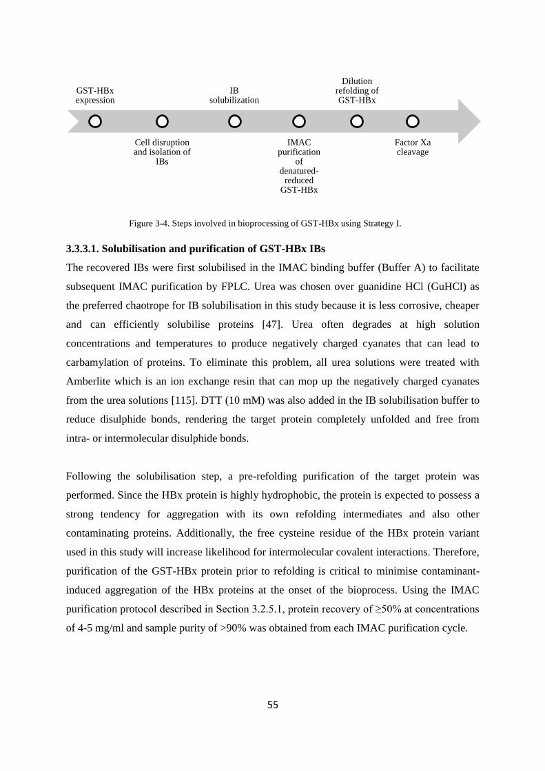
Figure 1-1 Thesis organisation
13 List of publications
1 A Basu WN Chen and SSJ Leong (2011) A rational design for hepatitis B virus
X protein refolding and bioprocess development guided by second virial coefficient
studies Applied Microbiology and Biotechnology 90(1) 181-191
2 A Basu L Xiang SSJ Leong (2011) Refolding of proteins from protein inclusion
bodies Rational design and recipes Applied Microbiology and Biotechnology 92(2)
241-251
3 A Basu SSJ Leong (2011) Development of an ELISA based analytical method for
HBx refolding yield quantification Analytical Biochemistry 418(1) 155-157
4 A Basu SSJ Leong (20112) High productivity chromatography refolding process
for Hepatitis B Virus X (HBx) protein guided by Statistical Design of Experiment
studies Journal of Chromatography A 1223(0) p 64-71
THESIS ORGANISATION
Chapter 1 Introduction
Chapter 2 Literature
review
Chapters 3-6 Results and Discussion
Chapter 3
Impact of GST tag on HBx bioprocessing
Chapter 4
Design and characterisation of
6His-HBx construct
Chapter 5
Development of an ELISA based bioanalytical
platform for quantifying HBx refolding yields
Chapter 6
HBx bioprocess intensification
studies
Chapter 7 Conclusions and
Future work
16
CHAPTER 2 Literature Review
17
Abstract
This chapter reviews the importance of the HBx protein in relation to HCC and presents some
basic protein refolding concepts that are essential to rationally design a bio-manufacturing
platform for the HBx protein From a pharmacological perspective the ability of the HBx
protein to modulate all major host cellular controlling systems (including cell cycle gene
expression and defence systems) indicates its potential as a therapeutic target against HCC
However since the protein is consistently produced as insoluble inclusion bodies in the
bacterial host systems bulk manufacturing of the protein would necessitate a refolding based
bioprocessing strategy To facilitate the development of a scalable HBx bioprocess a brief
overview of the different theories related to protein folding mechanisms are discussed in this
chapter
The protein folding phenomenon has been explained using different models to quantitatively
and qualitatively describe the events leading to the successful folding of a protein from its
denatured-reduced form From a protein refolding perspective the common point proposed in
all the theories is the presence of refolding intermediates which can interact with each other
leading to off pathway aggregation reactions and subsequent protein precipitation Different
technologies have emerged to facilitate protein refolding reactions for bulk manufacturing of
target proteins The most conventional method is dilution refolding although simple to
perform the huge dilution fold required to keep protein concentrations low incurs huge
capital investments and associated operating costs to scale-up the process Other refolding
technologies have subsequently been developed to overcome the limitations of dilution
refolding which include chromatography and high pressure refolding methods In parallel to
the development of new technologies significant efforts have also been directed towards
understanding the impact of different additives on protein refolding
Lack of a universal in situ monitoring platform however makes the protein refolding process
largely trial and error based Light scattering tools have shown to be effective in the quick
screening of the optimum refolding conditions for proteins and their potential in monitoring
and controlling protein refolding reactions are also discussed in this chapter Such tools can
be instrumental in developing rational protein refolding strategies for novel protein candidate
like HBx lacking commercially available native standards This will be particularly
important since earlier reports related to HBx have attempted to produce truncated or mutated
18
versions of the protein and also lack quantitative data which is needed for establishing
scalable bioprocesses The success and the failures associated with these earlier attempts to
produce HBx are also reviewed in this chapter
19
21 Role of HBx in HCC
Chronic HBV infection has been well associated with initiation and development of HCC
Within the small3 kb genome of HBV HBx has attracted particular attention amongst
scientists chiefly because of its capacity to affect myriads of metabolic pathways within the
infected cells However its mechanism of action is yet to be firmly established due to the
differential and sometimes contradictory roles played by the protein through its interaction
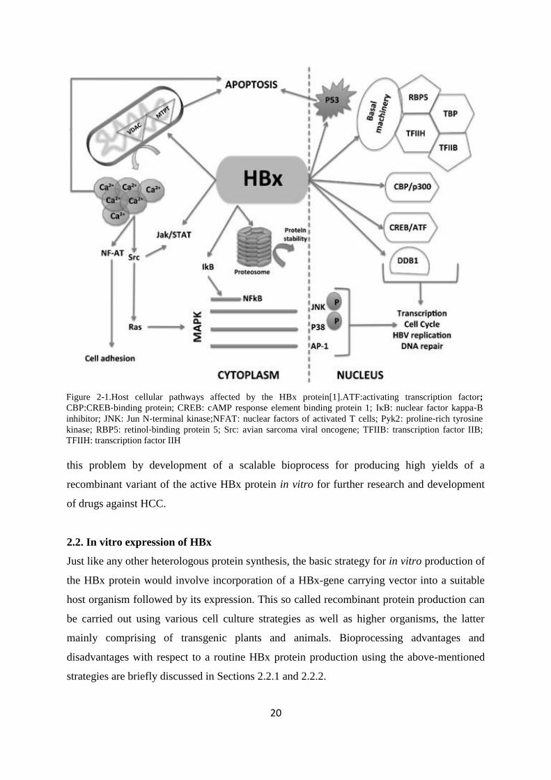
with other protein binding partners[24] Based on current understanding it is believed that
HBx can activate both viral and cellular transcription through direct binding to transcription
factors co-activators and components of the basal transcription machinery (summarised in
Figure2-1) It also has the capability to cause a mitochondria-mediated increase in cytosolic
calcium levels triggering transcription indirectly through stimulation of signalling pathways
like nuclear factor kappa-B (NFκB) Janus kinase (JAK) signal transducers and activators of
a transcription (STAT) and mitogen‐activated protein kinase (MAPK) In addition HBx
interacts with proteasome subunits to regulate protein degradation and stability Importantly
HBx can interact with various cellular proteins like damaged DNA binding protein 1 (DDB1)
and p53 thereby providing access to regulation of important cellular events like apoptosis
cell cycle transformation and viral replication [25-28] Based on these findings it seems that
the HBx protein not only facilitates HBV replication but can also lead to cellular
transformations within the infected hosts
Until now the main focus of HBx research seems to be directed towards unearthing its
interaction networks and understanding the cluster of genes or proteins regulated by it
Studies like these can help in understanding the probable pathways by which the HBx
establishes its control within the host cells and thus allows identification of potential drug
targets for the treatment of HCC To develop suitable drug candidates against HCC different
strategies can be adopted the major pathway in which the HBx is involved can be
manipulated so that the carcinoma is not induced An alternative and a much more direct
strategy would be to design suitable ligands that can specifically bind to HBx within the
cellular environment thereby inactivating it In either approach particularly for the latter
case a reliable and cost effective source of the active protein is required without which
research on HBx and HCC may be impeded Unfortunately the structural and biochemical
characteristics of the HBx protein are largely unknown due to the difficulties associated with
producing the protein in soluble form [1 25] My PhD research therefore aims to address
20
this problem by development of a scalable bioprocess for producing high yields of a
recombinant variant of the active HBx protein in vitro for further research and development
of drugs against HCC
22 In vitro expression of HBx
Just like any other heterologous protein synthesis the basic strategy for in vitro production of
the HBx protein would involve incorporation of a HBx-gene carrying vector into a suitable
host organism followed by its expression This so called recombinant protein production can
be carried out using various cell culture strategies as well as higher organisms the latter
mainly comprising of transgenic plants and animals Bioprocessing advantages and
disadvantages with respect to a routine HBx protein production using the above-mentioned
strategies are briefly discussed in Sections 221 and 222
Figure 2-1Host cellular pathways affected by the HBx protein[1]ATFactivating transcription factor
CBPCREB‐binding protein CREB cAMP response element binding protein 1 IκB nuclear factor kappa‐B
inhibitor JNK Jun N‐terminal kinaseNFAT nuclear factors of activated T cells Pyk2 proline‐rich tyrosine
kinase RBP5 retinol‐binding protein 5 Src avian sarcoma viral oncogene TFIIB transcription factor IIB
TFIIH transcription factor IIH
21
221 Protein production in higher organisms
Amongst higher organisms plants have gained wide popularity for large scale recombinant
protein production chiefly because of their safety and cost-effectiveness Protein production
in plants would require water minerals and sunlight However protein production in plants
suffers from several disadvantages like difficulty in genetic manipulation and frequent
contamination with other secondary metabolites due to less well-characterized plant
genomes Moreover problems associated with low expression yields and protein stability
within the harvested crops [29] imply that more investigation are required before such
systems can be used as a reliable protein production platform
Like plants transgenic animals (cows goats pigs etc) have been used for production of
recombinant proteins in milk egg white blood urine seminal plasma and silk worm
cocoons Though high titres of the target proteins have been achieved through the use of
transgenic animals the low production throughput (ie about 35 months for mice to 3 years
for cows) disadvantages their use for protein production Moreover production in transgenic
animals is associated with several ethical or regulatory challenges and has thus practically
stalled after the closure of the PPL therapeutics of Scotland which along with the Roslin
Institute cloned Dolly the sheep [30]
Thus considering the complications associated with large organisms it would be easier to
consider cell culture based protein production platforms for designing a scalable HBx
bioprocess
222 Cell culture based protein production
In this strategy the target genes are over-expressed within cellular hosts which can be either
eukaryotic or prokaryotic The cells are first transfected with the desired gene which is then
used to produce the protein (depending on the expression system) within the cytoplasm
periplasm or are secreted by the cell The most popular production platforms using the
eukaryotic systems include yeasts insect cells and mammalian cells The chief advantage of
using eukaryotic platforms is their capability to produce post-translationally modified
proteins Post-translational modifications are not important as far as the HBx protein
production is concerned but being a cysteine rich small protein expression in eukaryotic
hosts might provide significant benefits However as discussed later in section 26 attempts
22
to produce the HBx protein in yeast and insect cells led to some protein instability and
insolubility problems and hence were not considered in this research Protein production in
mammalian cultures on the other hand takes a significantly longer time for growth and
multiplication compared to bacterial expression systems thereby presenting considerable
challenges in maintaining sterility This constraint calls for high operating costs because of
the need to maintain a carefully controlled environment for cell growth in an expensive
medium [30]
Compared to mammalian expression systems production of proteins in bacterial hosts is
highly economical not only because of cheap growth media but also because of the higher
productivity Bacterial systems however are disadvantaged due to protein folding problems
chiefly when expressing cysteine-rich proteins because of the reducing environment
presented by the bacterial cytoplasm To overcome these problems different bacteria strains
have been engineered to facilitate heterologous protein production within bacterial hosts
which can be easily adopted for HBx production E coli (Gram negative) is widely used as a
host system for large scale protein production as it is a very well-studied system Other host
systems that are well-studied such as Staphylococcus aureus (Gram positive) are less
favoured because they express considerable amounts of proteases which impede heterologous
protein stability [30] For cost and productivity reasons E coli has been used for HBx protein
production in this study
In vitro production of the recombinant HBx protein in the Ecoli would involve the over-
expression of the protein using suitably engineered plasmids The recombinantly produced
protein can be extracted from the bacteria cells purified and subsequently if required
refolded to produce the desired product in a pure and biologically active form Producing the
protein of interest in the correctly folded and active state often poses a great challenge
particularly as seen in the case of the HBx protein (discussed in Section 26) where its over-
expression has been found to form IBs within the bacteria cells Insoluble protein expression
necessitates an in vitro refolding step which often gives sub-optimal yields A biomolecule-
centred process which is tailored around specific HBx molecular characteristics is thus
considered important to enhance downstream product yield Therefore the scope of my
research demands a proper understanding of the mechanism of protein folding A review of
23
some of the most accepted mechanisms of protein folding and protocols for the protein
refolding based on these mechanisms is presented in the following sections
23 Protein folding mechanisms Forces involved
The idea of folding proteins from an unfolded state emerged in the 1950s when a
ribonuclease protein was successfully refolded from denaturing conditions [31] This famous
experiment by Anfinsen indicated that the amino acid sequence of a polypeptide chain must
contain all the information required for its proper folding However the idea led to another
great realization which is popularly known as the Levinthal‟s paradox [32] It is an accepted
fact today that Levinthal rightly said that the protein folding process cannot be a completely
random trial-and-error process There must be shortcut pathways along which the polypeptide
chain passes through to acquire its native state within the stipulated time It is believed that
forces like electrostatic interactions hydrogen bonding hydrophobic interactions and
disulfide bridging play a key role in stabilizing the protein molecule in its native state Based
on the understanding of the roles of different forces in protein folding scientists have
hypothesized various models which can be used to understand how a given protein acquire
the native stable secondary tertiary and quaternary structures in a given environment [2 33]
231 Protein folding models
A crucial aim for all these models is to understand how a protein can find the bdquoright‟ pathway
and avoid others All these models are based on the evidence obtained through the metastable
or partially folded states of proteins from various experimental methods [2] Some of the
important models are discussed below
2311 The thermodynamic hypothesis
The thermodynamic approach for understanding the protein folding mechanism probably
came to the forefront right after Anfinsen‟s work According to this hypothesis folding of
polypeptides is driven by the need to attain a thermodynamically stable state where the
folded conformation under physiological conditions is more stable than the unfolded state
This theory does not take into consideration that the native state acquired may be the lowest
energy state of the protein molecule which is accessible within the physiologically
appropriate time scale Anfinsens hypothesis thus may be accepted as the basis for protein
folding processes through self assembly in general However the theory says very little about
24
the actual mechanism of protein folding [34] This issue was addressed to a certain extent in
the Molten Globule Hypothesis
2312 Molten Globule hypothesis
The Molten Globule hypothesis presents a very methodical approach to understand the
mechanism of protein folding The protein chain is thought to first collapse under the
influence of hydrophobic interactions between the hydrophobic amino acid residues The
elimination of the ordered water molecules from the hydrophobic sites can be considered to
be the driving force for this process A non-polar core is thus formed within the protein
molecule The stability of the polar peptide moieties within the core is achieved by the
formation of suitable H-bonds between the carbonyl and amide groups of the peptide
backbone in a particular pattern These interactions lead to the formation of regular secondary
structures or non-regular conformations which are compatible with the polarity of the
globule The resulting conformation of the protein is called the molten globule state [35-36]
The molten globule is considered to have a conformation close to the native structure the
difference being that it is inactive and may not be as compact as the native structure
According to Creighton various combinations of disulfide bonds [37] are formed broken and
reformed within this collapsed state until the correct linkage is formed leading to correct
folding Within the molten globule all the sulphide groups may not be easily accessible The
Molten Globule hypothesis was highly successful in explaining the protein folding process
However with time and based on new experimental outcomes scientists have proposed other
models to describe the protein folding mechanism as well
2313 Other similar models
Like the Molten Globule theory the protein folding models that subsequently followed have
their roots from the Anfinsen‟s protein folding concept itself where the thermodynamic
forces provide a major guidance towards attainment of the correctly folded state of the
protein Amongst these models the Nucleation Growth Model deserves particular attention It
was proposed that the residues adjacent in the sequence can itself form a nucleus for initiating
the folding process and ultimately leading to native structure formation The Framework
Model on the other hand suggested that at first the basic framework for native confirmation
is provided by local elements of secondary structure initiating the formation of native tertiary
25
structures through diffusion-collision mechanisms Some scientists on the other hand are
sceptical to accept the presence of only one pathway for the protein folding According to
them there are multiple pathways by which a protein can fold just like how the jigsaw puzzle
can be solved in a large variety of ways which is popularly known as the Jigsaw Model [34]
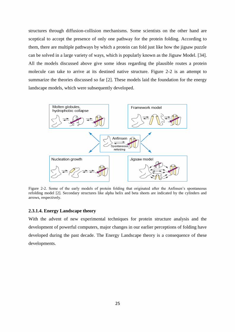
All the models discussed above give some ideas regarding the plausible routes a protein
molecule can take to arrive at its destined native structure Figure 2-2 is an attempt to
summarize the theories discussed so far [2] These models laid the foundation for the energy
landscape models which were subsequently developed
Figure 2-2 Some of the early models of protein folding that originated after the Anfinsen‟s spontaneous
refolding model [2] Secondary structures like alpha helix and beta sheets are indicated by the cylinders and
arrows respectively
2314 Energy Landscape theory
With the advent of new experimental techniques for protein structure analysis and the
development of powerful computers major changes in our earlier perceptions of folding have
developed during the past decade The Energy Landscape theory is a consequence of these
developments
26
In contrast to the classical view of protein folding which relies on discreet intermediate
refolding species the Energy Landscape theory describes folding using an ensemble of
partially folded structures According to this theory the refolding intermediates progressively
organise themselves to attain the final native folded state and the energy profile of the these
refolding intermediates can be described through an energy landscape diagram as shown in
Figure 2-3 [38] Denatured protein molecules having the highest energy remain at the top of
the landscape which ultimately guides them towards the final (low energy) conformation by
a bdquofunnel‟ On initiation of folding infinite pathways can take the denatured protein to its
native conformation a few of which might involve transient intermediates or local energy
minima However some of the routes can lead to formation of misfolded states leading to
significant kinetic traps It is therefore considered that lesser the number of intermediates the
smoother the surface of the funnel Thus the shape of the landscape directs all conformations
towards the native state (the bottom of the funnel) and avoids the need to sample all possible
conformations [2]
The energy landscape theory has several advantages over the previously proposed theories
First this model proposes that the process of folding will be difficult to change by mutation
as long as its stability is maintained based on the funnelling landscape Mild mutation will
thus probably lead to only slight shifts in the folding routes Second the funnelling
Figure 2-3 Schematic diagram of a folding energy landscape [2]
27
phenomenon limits the space the protein needs to search for the proper folding path Thus to
fold the system has to pass through ensembles of structures called bdquotransition state
ensembles‟ which consist of high energy intermediate structures The transition state
ensembles derive their stability from interactions present in the native structure The protein
would finally proceed to the bottom of the funnel through this bdquotransition state ensemble‟ to
attain the stable native state [33]
2315 Protein folding inside the cells role of chaperones
The discussion on the protein folding process presented so far has been restricted to in vitro
systems However understanding of the folding process is incomplete without the knowledge
of the challenges confronted by proteins inside the cellular environment where the folding
and biosynthesis of proteins are intimately coupled to each other The newly emerging
polypeptide chains particularly in the case of large protein molecules can start to fold before
synthesis is complete [39] Hence the lack of complete sequence information can lead to
incorrect tertiary structure formation leading to the exposure of hydrophobic surfaces
resulting in aggregation To make situations worse the over-crowded cellular environment
highly favours aggregation of partially folded protein molecules [40] All these factors
indicate that proteins are highly prone to aggregation during the protein folding operations
within the cell [41]
To address this aggregation problem living cells use an ingenious machinery called
chaperones [42] These proteins can sequester newly synthesised peptides until it has folded
to a state that that is much less prone to aggregation thereby smoothening the folding energy
landscape [43-44] Chaperones are also reported to play important roles in many cellular
events like signal transduction protein targeting and degradation Different types of
chaperones have been reported in the prokaryotic and eukaryotic cells for instance in the
case of E coli the GroEL-GroES system forms an important component of the chaperone
machinery Within the eukaryotic cells on the other hand the hsp70 and hsp60 chaperones
have been reported to co-operate in vivo forming a chaperone relay
The importance of the chaperonin systems is widely accepted today and has influenced many
scientists to use different molecular species that have the capability to behave like artificial
chaperones for protein refolding operations
28
24 Protein refolding methods
Refolding is generally referred to as the change in protein conformation from the unfolded to
folded state In this research the choice of refolding method will be highly dependent on the
HBx structure Hence understanding the protein folding mechanism will be very important
for proper design of in vitro refolding systems Aggregates popularly known as IBs are a
major hurdle for the biopharmaceutical industry in the efficient manufacture of commercially
important proteins However increasing demands of different protein-based
biopharmaceuticals has urged the industry to focus their attention towards development of
sustainable bioprocesses capable of producing active proteins from IBs Unfortunately
refolding of IBs is not a straightforward process The following review on protein refolding
methods describes the way in which contemporary knowledge of protein folding and
aggregation mechanisms have influenced scientists for devising protocols for in vitro protein
refolding
Undoubtedly the first step in refolding proteins from IBs would involve solubilisation of
protein aggregates in a suitable denaturant to unfold the polypeptide and render a
monomolecular dispersion of polypeptides prior to refolding The solubilisation efficiency
and structure of the proteins in the denatured state as well as in the subsequent refolding
process generally depends on the choice of the solubilising agents Due to their capability to
interfere with solute-solvent interactions high concentrations of common chaotropes like
urea or guanidine hydrochloride (GdnHCl) has been reported to reduce the hydrophillicity of
an aqueous medium In the presence of chaotropes the environment becomes sufficiently
hydrophobic so as to stabilize the hydrophobic amino acid residues within the protein
molecules and thus unfolds the protein Consequently urea or GdnHCl in high concentrations
have become an obvious choice as solubilising agents for IBs Detergents are also used
occasionally as denaturants However the main drawback of using only detergents for
solubilisation is that unlike urea or GdnHCl modulating the binding of detergents with
proteins for more efficient refolding is rather difficult [45] and detergent carry-over a
common phenomenon is highly undesirable in secondary processing steps
Solubilising-denaturing proteins containing disulphide bonds necessitates the use of reducing
agents like Dithiothreitol (DTT) or β Mercapto-ethanol to resist the formation of unwanted
29
disulphide bonds during solubilisation [46] Even in the unfolded state and in the presence of
high concentrations of denaturants non-native disulphide bonds are often easily formed
leading to the formation of non-native structures These unstable structures have a strong
tendency for aggregation or misfolding upon removal of denaturants during the subsequent
refolding steps The solubilised molecules can then be subjected to refolding by reducing the
concentration of denaturants The success of refolding lies in the success to minimise other
competing side-reactions which lead to misfolding and aggregation which occur in parallel to
the refolding process
The rates of refolding and other side-reactions generally depend on the procedure used to
reduce the denaturant concentration and the solvent conditions surrounding the protein as the
protein is subjected to different physicochemical environment A review of the most widely
used refolding protocols is discussed below
241 Dilution refolding
In dilution refolding native structure formation of the protein is induced by diluting the
denatured protein solution into a refolding buffer Dispersion of the protein in large volumes
will reduce protein concentration and hence the second or higher ordered aggregation
pathways A refolding reaction which is of first order reaction is described using Equation 2-
1
nNUkUkdt
dU21 Equation 2-1
where k1 is the net rate constant of refolding k2 is the net rate constant of aggregation U is
the concentration of unfolded protein t is refolding time N is aggregation number and n is
the reaction order of aggregation Thus at higher protein concentrations the protein
aggregation reaction would dominate over the refolding reactions while at lower protein
concentrations the refolding reaction would dominate as shown in Figure 2-4 [47-48]
30
Figure 2-4 Effect of concentration of denatured protein (cd) on the extent of in vitro refolding ( ) and
aggregation ( ) [48]
To minimize unwanted irreversible intramolecular interactions low denaturant
concentrations are often introduced in the refolding buffer to minimise the propensity of
aggregation during refolding For most proteins rapid dilution is favoured over a slow
dilution process to keep protein concentration low at the onset of refolding [49-52]
Pulsed dilution or fed-batch dilution refolding is also popular where the addition of
denatured protein is interspersed with the aim to maintain low concentrations of the
denatured protein species at any given point of time For example denatured protein is added
to the refolding mixture only when 80 of the refolding yield is reached Factors like the
increase of denaturant concentration with each cycle and the amount of protein added per
pulse need to be carefully considered with the use of such protocols [53-54]
Though it is simple to scale-up a dilution refolding process using a stirred-tank reactor the
main disadvantage of this strategy lies in its exorbitant scale-up costs Process scale up would
require the utilization of huge volumes of buffers and stirring tanks that can increase the
capital and operating costs of the overall process The need for subsequent treatment of such
huge volumes of buffers can further multiply the production costs to a significant extent [55]
31
242 Dialysis Methods
Instead of using an approach that drastically reduces the denaturant concentration which may
guide protein molecules into a wrong folding pathway many researchers like to use a much
more gradual approach for the removal of denaturants such as dialysis to obtain the native
proteins at good yields [54 56] However the dialysis refolding process is time consuming
Additionally the protein being subjected to a relatively slow change in denaturant
concentration can also provide ample opportunities for protein refolding intermediates to
interact with each other Refolding intermediates which still has a loosely packed
hydrophobic core also often get adsorbed onto the dialysis membrane resulting in protein
loss In a comparative study of the dilution and dialysis methods for the refolding of α-
fetoprotein a highly disulphide-bonded biopharmaceutical candidate the former process was
found to give higher refolding yields than the latter [57] However each protein having a
unique sequence may exhibit significant extents of amphiphilicity and may respond
differently to different refolding methods [55 58-60]
243 Chromatographic techniques
In recent years column chromatography has gained an increased interest among researchers
for protein refolding The ease of automation and scaling up using chromatography
techniques has increased the number of refolding studies based on chromatography tools in
the past decade The overall objective of on-column refolding is to adsorb solubilised
denatured proteins on a matrix to facilitate spatial isolation of the molecules The restricted
movement of the protein molecules within the immobilized matrix allows them to remain
dispersed avoiding inter-molecular interactions thus preventing aggregate formation In this
refolding method the chromatography column is first equilibrated with denaturant to allow
protein adsorption in the denatured form followed by a solvent exchange which favours
refolding of the protein molecules on-column After refolding the refolding buffer is then
displaced with an appropriate elution buffer to elute the refolded proteins Different
chromatographic matrices have been successfully used for on-column refolding including
size-exclusion chromatography ion-exchange chromatography affinity chromatography and
hydrophobic interaction chromatography [61-66] Due to the spatial isolation of the
immobilised protein molecules within the packed beads chromatography refolding has the
advantage of facilitating refolding at much higher protein concentrations compared with other
dilution refolding techniques without incurring extensive aggregation Simultaneous
32
purification and refolding can also be readily achieved on-column upon adequate process
optimisation [67]
Successful refolding using this strategy demands optimisation of several operational
parameters like proper selection of the chromatographic matrix column dimensions and
solvent flow rates Particularly the rate of change of the denaturing environment within the
column into a refolding one can significantly influence the topography of the energy
landscape of the refolding protein [60]
244 High hydrostatic pressure (HHP) technology
Another way to promote native refolding reactions is to create a refolding environment that
can destabilise off-pathway aggregation HHP technology was developed with this strategy in
mind where hydrostatic pressure is applied to bdquoensure‟ that only proteins refolded to the
native form and hence having the lowest specific volumes are stabilised [68-69] Application
of pressure can also help to solubilise the IBs directly and refold the target protein
spontaneously However modulation of hydrogen bonds and disulphide bridges needed for
solubilising the IBs and obtaining the correctly refolded product can only be achieved by an
optimised buffer system [70]
Based on the existing refolding techniques (summarised in Table 2-1) it is clear that the
design of existing refolding methods is focused mainly on controlling refolding
concentrations to obtain high refolding yields but optimisation of the physicochemical
environment still needs to be independently performed for each of these methods In recent
years high throughput-screening of refolding buffers for protein refolding based on protein
solubility has been reported [71] Some of these screening kits have been commercialized for
example Refold MasterTM
developed by Novexin Ltd (Cambridge UK) comprises a matrix
of different buffer compositions which allows screening of protein foldability in 96 well-plate
formats These screening kits however do not allow in situ monitoring of the refolding
reaction which can provide important insights into the influence of different refolding
additives on refolding yields The major hurdle for such monitoring is that different proteins
require different detection strategies and a generic screening platform is not practically
possible It would therefore be considerably more advantageous if new screening methods
could be developed to simultaneously provide quantitative information on aggregation
33
Table 2-1 A brief overview of existing refolding technologies
Refolding Strategy Rationale Advantages Disadvantages
Dialysis
1) Minimise denaturant carry over from
the solubilised IB solutions
2) Equilibrate the proteins within a buffer
capable of refolding the protein
Helpful when the denatured-
reduced protein concentration is
low
1) Protein loss to the dialysis membranes
2) Slow buffer exchange kinetics can induce
aggregation of the refolding intermediates
Dilution Reduced interaction amongst the refolding
intermediates by lowering protein
concentration
Better refolding yields compared
to dialysis in most cases
1) Success largely depends on the choice of the
refolding buffer composition
2) Large consumptions of expensive buffers can
negatively impact the overall process
economics
Chromatography
1) Spatial isolation of refolding
intermediates bound on chromatographic
matrices eliminates inter-molecular
interactions
2) Refolded proteins can be eluted from
the column at relatively high
concentrations
Useful for process intensification
1) May inhibit protein refolding due to loss of
flexibility of the bound polypetide chain
2) Intra-molecular protein interactions can lead
to unproductive refolding of the bound proteins
High Hydrostatic Pressure
Technology
1) High pressure helps to solubilise the
IBs
2) The pressure allows preferential
stabilisation of the natively folded protein
molecules
One-step conversion of IBs into
refolded products
Hydrogen and disulphide bonds within the IBs
cannot be broken by applying pressure
Successful solubilisation and refolding require
an optimised buffer system
34
kinetics under different refolding environments thus providing a better correlation between
physicochemical parameters and refolding yield for a given protein ldquo[72]rdquo The following
two sections present a brief review on how second virial coefficient (SVC) measurements can
be used not only as a valuable tool for measuring such data but also to develop rational
process designs and recipes for protein refolding
25 SVC measurement a rational approach to designing protein refolding recipes
Based on our discussion in Section 32 the random movement of protein molecules during
refolding is influenced by the inter-particle and hydrodynamic interactions amongst them
These interactions can be quantitatively assessed using a thermodynamic parameter known as
SVC Importance of SVC measurement in understanding protein behaviour came to the
forefront during the early 1990s when George and Wilson found that successful
crystallization takes place only when the SVC of the solutions lie within a specific value [73]
The ability to quantitatively predict environments conducive for protein crystallization
indicates that a convenient SVC measurement platform can also be instrumental for studying
protein-protein interaction within a refolding system by providing thermodynamic
information on protein phase behaviour Before discussing the applications of SVC in the
rational design of refolding recipes concepts of SVC is briefly reviewed to set the context of
the discussion ldquo[72]rdquo
251 Second virial coefficient theory and applications
SVC is a thermodynamic parameter which originates from the virial expansion of solution
osmotic pressure given in Equation 2-2
322
CB
RT Equation 2-2
where π is the solution osmotic pressure R is the gas constant T is the temperature ρ is the
solute density and B2 C etc are the osmotic virial coefficients B2 in the above equation is
known as the second virial coefficient and is a measure of two-body interactions within a
solution Throughout the rest of the thesis the term ldquoB2rdquo and ldquoSVCrdquo will be used
synonymously According to statistical mechanics SVC can be expressed as an integration of
the potential of mean force w2(r) between two molecules
0
2
222
2drre
M
NB k Trw
w
A Equation 2-3
35
In Equation 2-3 NA stands for the Avogadro‟s constant Mw is the molecular weight k is the
Boltzmann constant and r is the centre-to-centre distance between two molecules [74]
Therefore the SVC takes into account the same interactions (like electrostatic Van der
Waals excluded volume hydration forces and hydrophobic interactions) which are known to
regulate protein phase behaviour [75] Hence this parameter should correlate well with
protein solubility which is also evident from Equation 2-4 developed by Guo etal [76]
SM
S
SMRTB
ww
p
2
l n
2
12
Equation 2-4
where S is the protein solubility and Δμp is defined as the difference of the standard chemical
potentials of protein between two states (solution and crystal) Equation 2-4 was further
refined by Ruppert et al [77] to obtain a direct relationship between the solubility and
experimentally determined SVC
Based on these findings over the past decade experimentally determined SVC values have
been used by scientists to shed light on methods to manipulate protein thermodynamic
properties and phase behaviour [78-79] and even obtain partial structural information [80]
SVC values have thus been used as an unbiased estimator for protein-specific behavioral
patterns to guide determination of optimal crystallization conditions for proteins [73 81-82]
In view of protein refolding applications SVC values can accurately correlate the
relationship between refolding additives and protein interactions [83-86] and has been
exploited to predict (i) the propensity for aggregation during protein refolding [51] and (ii)
potential refolding yields of IB proteins [87] Therefore SVC measurements of refolding
mixtures carried out at statistically determined design points can rapidly provide quantitative
correlations between the choice and concentration of additives on the interactive behaviour of
proteins [75 88] Hence in the case of proteins which do not have standard bioactive
product(s) nor any analytical platforms for the quantitative bioactivity assay such as HBx
SVC can be a good analytical tool for determining protein stability andor solubility
Amongst diverse methods for SVC measurement both static light scattering (SLS) and static
interaction chromatography (SIC) have obtained wide popularity within the scientific
community SLS experiments are based on the measurement of the light intensities scattered
by the randomly moving protein molecules within a solution (discussed in Chapter 4) The
SIC methodology on the other hand involves measurement of the retention time between
36
protein molecules flowing over the same molecules covalently immobilised over a surface
(chromatographic bead or a chip) the retention time in turn is linked to B2 through the
potential of mean force [81] SLS measurements being a non-invasive technique is very
simple to conduct particularly with the modern commercially available equipment but is often
disadvantaged by the lack of throughput SIC in this regard can provide higher throughput but
the need to immobilise the protein on a surface might be problematic on many occasions In
this research project SLS was used to monitor the SVC values of the HBx protein solutions
(discussed in Chapter 4) ldquo[72]rdquo
26 Earlier attempts to produce recombinant HBx in microbial systems
A definitive proof for the existence of the HBx protein was established only in the late 1980s
from two independent studies [89-90] Since then several attempts have been directed
towards soluble production of the protein for its structural and biochemical characterisation
However the protein is consistently expressed in the insoluble form under all expression
conditions for all types of protein production platforms studied including insect cells [91-93]
Even attempts to produce the full length protein within yeast cells lead to some stability
problems [94] Since E coli is the most popular protein expression platform due to cost and
speed factors various strategies have been adopted by scientists to improve the soluble
expression of HBx in bacteria which include but are not limited to (i) co-expression of
chaperones to aid in protein folding [95-97] (ii) producing truncated version of the whole
length protein (iii) directing protein expression in the periplasmic space to provide an
oxidising environment for cysteine oxidation to form disulphide bonds or (iv) using
solubility enhancing fusion tags [98-102] Table 2-2 summarises some recent attempts to
express HBx in the soluble form where the most successful methodology involving the use
of maltose binding protein (MBP) fusion protein strategy by Liu D et al could only provide
sub-optimal yields of the HBx protein (10 mg HBx from 25 L of culture)
The exact cause for insoluble HBx production is yet to be fully established but amino acid
sequence analysis of the protein using the ProtParam program available from
wwwExPasyorg reveals some interesting results With approximately 40 of hydrophobic
amino acid residues the HBx sequence possesses a positive grand average of hydropathicity
(GRAVY) index indicating its high hydrophobicity Moreover the HBx protein sequence
often possesses an odd number of cysteine residues which further adds to its instability
37
during recombinant production as indicated by the instability index of 6039 classifying the
protein as unstable Based on these results it is not surprising that until now protein
scientists have had little success in expressing the HBx protein in the soluble form at
reasonable yields The absence of quantitative yields reported in earlier refolding studies of
HBx also further impedes the development of a sustainable bioprocess for large-scale
production of HBx For example Marczinovits and co-workers reported successful refolding
of a GST-fused truncated HBx protein using dilution refolding method [17] However a
complete analysis of the final yields of the bioactive protein was not reported in their study
Other groups reported the use of sequential dialysis in PIPES buffer systems to successfully
refold HBx and its mutated forms [103-104] but again the lack of quantitative yields impedes
implementation of bioprocess design for scale-up It is clear that the lack of quantitative HBx
refolding analyses coupled with ill-defined bioprocesses will continue to hinder the supply of
bioactive HBx in sufficient amounts for advanced structural and drug designing studies
Based on the discussion presented above on the earlier works it appears that successful
bioprocesses can be established to improve HBx solubility during expression by using
solubility-enhancing fusion tags Considering that the widely known MBP fusion tag only led
to suboptimal HBx yields other tags like NusA Trx or GST can be studied to obtain better
results Tags like NusA or Trx however are disadvantaged by the lack of any biospecific
ligand affinity leading to purification complexities [105] In comparison although the GST
tag may or may not solve the solubility problem [106-107] the bioactive target protein can be
readily purified using a glutathione affinity chromatography step Furthermore since a
truncated version of the HBx protein has been successfully produced with a GST-tag [17] we
deemed the use of a GST-tag for full length expression of HBx worth studying
38
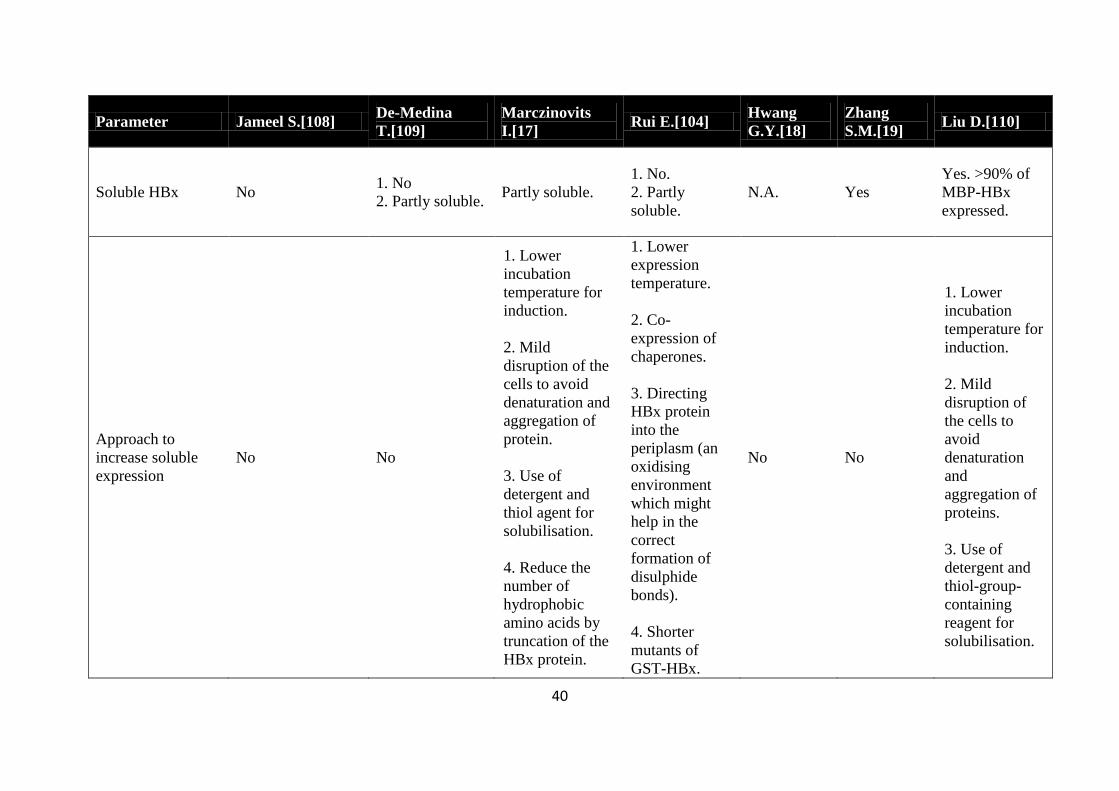
Table 2-2 A brief summary of the attempts to express HBx in the soluble form [NA implies not applicable not mentioned in the study]
Parameter Jameel S[108] De-Medina
T[109]
Marczinovits
I[17] Rui E[104]
Hwang
GY[18]
Zhang
SM[19] Liu D[110]
E coli strain BL21 (DE3) BL21 (DE3)
pLysS DH5α BL21 DH5α BL21 (DE3) JM109
Construct pET-8C vector 1 pET-8c vector
2pGEX1 vector
Truncated variant
of HBx cloned
into pGEX-3X
vector
psW202 vector pGEX-5X-1
vector
pGEX-4T-1
vector pMAL-c2x
Fusion tag No 1 No
2 GST GST
1 6-Histidine
2 GST GST GST MBP
Expression
medium NA 2xYT Luria broth LB LB LB LB
Expression
temperature 37degC 37degC 37degC 37degC 30degC 28degC 37degC
Point of induction NA OD 05 A600 06-07 OD 07 NA A550 06 OD600 06-07
Induction method 04 mM IPTG 1 mM IPTG 05 mM IPTG 1 mM IPTG 1 mM IPTG 01 mM
IPTG 003 mM IPTG
Post induction time 3 h 2 h 25 h 05 - 4 h 5 h 3 h 25 h
39
Parameter Jameel S[108] De-Medina
T[109]
Marczinovits
I[17] Rui E[104]
Hwang
GY[18]
Zhang
SM[19] Liu D[110]
Lysis buffer
50 mM Tris
(pH85) 10 mM
EDTA
50 mM HEPES
(pH80) 100
mM NaCl 05
mM EDTA 5
μgml PMSF 10
μgml aprotinin
20 μgml
benzamindine
and 2 μgml
pepstatin
10 mM Tris (pH
80) 150 mM
NaCl 1 mM
EDTA
incubation with
100 gml
lysozyme on ice
for 15 min 1
N-lauryl
sarcosine
Addition of 1
mgml
lysozyme
10 mM Tris
(pH80) 150
mM NaCl 1
mM EDTA)
addition of
100 μgml
lysozyme
incubated on
ice for 15
min 5 mM
DTT
sarkosyl
PBS pH
73 01
Triton X-
100
20 ml of 10 M
Tris-HCl pH
74 117 g of
NaCl 2 ml of
05 M EDTA
154 mg of
DTT
Homogenization
method
One freeze thaw
cycle sonication Sonication Sonication
Incubation at
room
temperature for
20 and 3 freeze
thaw cycles
Sonication Sonication Sonication
Buffer to dissolve
insoluble fraction
50 mM Tris (pH
85) 10 mM
EDTA 6 M urea
and 5 mM 2-
mercaptoethanol
7 M urea 8M urea (pH 65)
6 M
guanidinium-
HCl 100 mM
NaH2PO4 10
mM Tris-HCl
(pH 80)
NA NA NA
40
Parameter Jameel S[108] De-Medina
T[109]
Marczinovits
I[17] Rui E[104]
Hwang
GY[18]
Zhang
SM[19] Liu D[110]
Soluble HBx No 1 No
2 Partly soluble Partly soluble
1 No
2 Partly
soluble
NA Yes
Yes gt90 of
MBP-HBx
expressed
Approach to
increase soluble
expression
No No
1 Lower
incubation
temperature for
induction
2 Mild
disruption of the
cells to avoid
denaturation and
aggregation of
protein
3 Use of
detergent and
thiol agent for
solubilisation
4 Reduce the
number of
hydrophobic
amino acids by
truncation of the
HBx protein
1 Lower
expression
temperature
2 Co-
expression of
chaperones
3 Directing
HBx protein
into the
periplasm (an
oxidising
environment
which might
help in the
correct
formation of
disulphide
bonds)
4 Shorter
mutants of
GST-HBx
No No
1 Lower
incubation
temperature for
induction
2 Mild
disruption of
the cells to
avoid
denaturation
and
aggregation of
proteins
3 Use of
detergent and
thiol-group-
containing
reagent for
solubilisation
41
Parameter Jameel S[108] De-Medina
T[109]
Marczinovits
I[17] Rui E[104]
Hwang
GY[18]
Zhang
SM[19] Liu D[110]
Successful in
increasing soluble
expression
NA NA No
Truncated
proteins with
fusion tags
were partly
soluble but the
full length HBx
protein was
exclusively
found in the
insoluble
fraction
NA NA No
Hypothesis on why
the method failed
to increase soluble
HBx expression
NA NA
Due to structural
characteristics of
HBx (52 of the
amino acids is
hydrophobic and
4 disulphide
bonds)
1 Chaperones
used were not
involved in the
folding
pathway of the
HBx protein
2 HBx might
also form IBs
in periplasmatic
space
NA NA NA
In this research I aim to study both molecular and process-related factors which could affect
refoldability and solubility of HBx which have not been quantitatively studied in the earlier
works The sequence of the HBx model protein in this thesis contains 9 cysteine residues and
has close similarities with the protein from the HBV-A3 variant recently reported to be
prevalent in places like Cameroon Gambia and Gabon in Africa (Isolate
CameroonCMR9831994) [111] To the best of my knowledge it is the first time refolding
studies of this HBx variant are studied
43
CHAPTER 3 The Effect of a GST Fusion Partner on HBx
Expression and Bioprocessing in an E coli Microbial Platform
44
Abstract
Earlier efforts to produce the HBx protein by optimising culture conditions using various
protein expression systems have consistently resulted in the expression of HBx in the
insoluble form The hydrophobicity of HBx and the presence of odd cysteine residues are
likely causes for the protein‟s propensity to form aggregates in solution which pose a
roadblock to the development of a large scale HBx production platform for structure-
characterization studies This chapter investigates the impact of a solubility enhancing fusion
partner ie GST on the soluble expression and bioprocessing strategies for the HBx protein
Extensive fermentation optimisation strategies failed to improve the expression of the GST-
HBx in the soluble form in E coli Two downstream processing strategies for the GST-HBx
protein were subsequently designed and employed with the aim to recover purified HBx in
the soluble form Firstly GST-HBx was purified in the denatured-reduced state followed by
protein solubilisation and Factor Xa cleavage under non-denaturing conditions to recover
HBx Secondly sarkosyl was introduced into the cell lysate to facilitate selective
solubilisation of GST-HBx followed by purification and Factor Xa cleavage to recover HBx
The results from these studies revealed that designing an economically viable bioprocess for
HBx production would be extremely challenging using the GST-HBx construct In particular
solubility of the fusion protein or the HBx protein alone was difficult to achieve under
various physicochemical environment due to the pI range of the fusion protein components
Additionally the low efficiency of the Factor Xa cleavage reaction makes subsequent
recovery of bioactive HBx difficult and costly These results point to the need for a new HBx
construct design to facilitate more efficient and economical bioprocessing of this protein
which was subsequently investigated in Chapter 4
45
31 Introduction
The insolubility of recombinantly produced HBx in E coli is a major impeding factor for
large scale biomanufacturing of this protein The results from earlier HBx production studies
presented in Chapter 2 indicate that the way forward for developing a scalable HBx
bioprocess might be through the use of solubility enhancing fusion tags or optimised protein
refolding strategies for the HBx protein Both these strategies were studied in this thesis
wherein studies were first directed towards enhancing soluble protein production using a
GST-HBx construct which forms the basis of the studies reported in this chapter
Co-expression of HBx with fusion partners like GST or MBP has been reported in earlier
HBx production studies [17 104 110] Although the co-expression of HBx with MBP
resulted in the soluble expression of HBx the low cleavage efficiency coupled with high step
yield losses resulted in a sub-optimal HBx yields Despite its well-known solubility-
enhancing property the use of MBP is also disadvantaged by the increased metabolic burden
to produce such a large fusion partner (42 kDa) [112] thus resulting in lower peptide yields
due to the high fusion protein to peptide mass ratio Due to these problems the GST protein
was thought to be a more suitable fusion partner for HBx production since it has been
reported to improve recombinantly expressed protein solubility and facilitate its purification
as well [113] Previously researchers have tried to produce the GST-HBx protein using
mutated or truncated HBx variants but insoluble expression was consistently observed [17
104]
Studies reported in this chapter were first aimed at improving the soluble expression of GST-
HBx in E coli by varying culture conditions and media compositions Two approaches were
subsequently studied to purify and solubilise the IB-derived HBx protein Strategy I) pre-
refolding purification of GST-HBx under denatured-reduced conditions followed by dilution
refolding and Strategy II) protein solubilisation using suitable detergents under non-
denaturing non-reducing conditions followed by purification The purified fusion protein
stabilised under non-denaturing conditions was then subjected to Factor Xa cleavage for
removal of the GST-tag in both strategies
46
32 Methods and materials
321 HBx plasmid transformation
A pGEX-5X-1 plasmid containing the gene sequence of HBx-6His in frame with that of GST
was kindly provided by Prof William Chen Wei Ning (School of Chemical and Biomedical
Engineering NTU Singapore) The plasmid was dissolved in Tris-EDTA (TE) buffer and
transformed into E coli BL21DE3 (Stratagene) by the heat shock method The bacteria cells
were made competent by CaCl2-MgCl2 treatment using standard protocols [114] The
transformed cells were allowed to grow overnight on an ampicillin selective LB agar
medium A single colony from the plate was picked and used to inoculate a liquid LB
medium (5 ml) containing 01mgml of ampicillin and the culture was grown overnight The
cultures were then harvested for subsequent DNA sequencing and protein expression studies
322 DNA sequencing
Plasmids from 5 ml of the transformed E coli overnight culture were extracted using a
plasmid extraction kit (Qiagen) for DNA sequencing of the HBx insert to determine the
precise amino acid sequence of the protein Sequencing was performed by the National
Neuroscience Institute (NNI) Singapore using pGEX-5‟ and pGEX-3‟ primers (GE
Healthcare) The resulting DNA sequence data was translated into the corresponding amino
acid sequence using the online software bdquoTranslate‟ [httpwwwexpasyorgtoolstranslate]
323 GST-HBx protein expression
Overnight culture containing the transformed E coli (BL21 DE3) cells was used to inoculate
a fresh sterile 400 ml LB medium contained in a 2 L culture flask to obtain a 1 (vv)
bacterial suspension The bacteria cells were then allowed to grow in a shaking incubator at
37degC and 200 rpm shaking condition until the late log phase was attained (ie OD600 = 07)
Isopropyl-1-thio-β-D-galactoside (IPTG) (1st Base) was then added to a final concentration
of 025 mM to induce recombinant protein expression The cells were harvested at OD600 =
20 The 2 L culture was divided into 500 ml centrifuge tubes and centrifuged at 5200 g for
30 min The cell pellets were stored in a -20degC freezer (for a maximum duration of 1 week)
and thawed before cell lysis
For protein expression using the M9 medium the cell cultures (grown at 37degC in LB
medium) were centrifuged after reaching the late log phase (OD600 = 07) as specified earlier
47
The cell pellets were then re-suspended in fresh M9 minimal medium containing 1 (wv)
casamino acids and 1 (wv) glucose The M9 minimal medium consisted of 128 g
Na2HPO4 3 g KH2PO4 05 g NaCl 10 g NH4Cl 2 ml of 1 M MgSO4 01 ml of 1 M CaCl2
in 1 L sterile deionised water For low temperature protein expression (ie at 30degC and 18degC)
the cultures were grown at 30degC or 18degC for 30 min and induced with 01 mM IPTG after
reaching the late log phase (ie OD600 = 07)
324 Cell disruption to recover GST-HBx
The cell pellets were washed with phosphate buffered saline (PBS pH 72) and then re-
suspended in 200 ml of 50 mM Tris-HCl buffer (pH 82) The suspended cells were disrupted
by subjecting the cells to (i) French Press (Thermo Electron Corporation) at16000 psi
pressure in 3 consecutive passes or (ii) sonication (Branson sonifier 450 400 watts) for 3
min (pulses 5s on and 7s off) at 30 amplitude (maximum frequency = 20000 Hz) The
soluble and insoluble protein fractions were separated by centrifugation before further
analysis of the proteins
325 GST-HBx purification refolding and cleavage
Despite extensive optimisation of culture conditions the GST-HBx was inherently expressed
as IBs in E coli A refolding based downstream processing strategy was thus developed to
recover GST-HBx in a soluble form Since the Factor Xa protease activity is dependent on
the native structural conformation of the fusion protein studies to solubilise GST-HBx under
non-denaturing conditions were performed Two strategies were employed (ie Strategies I
and II) as described in Sections 3251 and 3252 to solubilise GST-HBx followed by
FactorXa cleavage
NB In the following sections of this chapter solubilisation of GST-HBx under non-
denaturing conditions is referred to as a refolding step
3251 GST-HBx processing using Strategy I (pre-refolding purification of GST-HBx
under denatured-reduced conditions followed by dilution refolding)
The GST-HBx protein expressed in LB medium (at 37degC Section 323) was used for further
downstream processing studies The IBs recovered from the disrupted cell suspensions were
centrifuged at 6500 g for 30 min Pre-refolding purification of the GST-HBx was performed
48
using IMAC with a 1 ml Ni2+
Sepharose HisTrap FF column (GE Healthcare) All
chromatography experiments were conducted using an AKTA Explorer Fast Protein Liquid
Chromatography (FPLC) System (GE Healthcare) HBx IBs were solubilised in the IMAC
binding buffer (Buffer A) comprising 8 M urea 05 M NaCl 20 mM Imidazole 10 mM
DTT in 20 mM sodium phosphate buffer (pH 74) and filtered through a 045 microm filter
After equilibrating the IMAC column with 10 column volumes (CVs) of Buffer A at 10
mlmin approximately 40 mg of the protein sample was loaded into the column The bound
protein was then further washed with 10 CVs of Buffer A at 10 mlmin to allow complete
removal of all unbound proteins GST-HBx was then eluted with 60 Buffer A and 40
elution buffer (Buffer B) (8 M urea 05 M NaCl 300 mM Imidazole 10 mM DTT in 20 mM
sodium phosphate buffer pH 74) in a single elution step over 10 CVs at 10 mlmin 15 ml
fractions of the eluate collected using the Frac-950 (AKTA GE Healthcare) fraction collector
were subjected to refolding studies Urea used in all the work reported in this thesis was pre-
treated with Amberlite (GE Healthcare) to capture cyanates which can carbamylate proteins
[115] Amberlite pre-treatment was achieved by stirring 9 M urea solution containing 1
(wv) Amberlite at room temperature for 1 h The urea solution was subsequently diluted to
the desired concentration
Purified GST-HBx was buffer exchanged into 8 M urea and 50 mM Tris (pH 7505) using a
PD 10 desalting column (GE Healthcare) to remove Imidazole and DTT which can interfere
with HBx refolding The buffer-exchanged protein solution was rapidly diluted into the
refolding buffer at different folds to obtain refolding protein concentrations ranging from 25
to 100 gml Four refolding buffer compositions were examined for protein solubilisation
optimization studies (i) Buffer 1 2 M urea 1 mM reduced glutathione (GSH) and 01 mM
oxidised glutathione (GSSG) 50 mM Tris pH 8005 (ii) Buffer 2 2 M urea 1 mM GSH
and 01 mM GSSG 03 M L-arginine 50 mM Tris pH 8005 (iii) Buffer 3 2 M urea 03
M L-arginine 50 mM Tris pH 8-9 (iv) Buffer 4 2 M urea 03 M L-arginine in 50 mM
acetate-citrate buffer pH 6 In all the refolding studies the proteins were incubated at 4degC for
1 to 3 h
After incubation the refolded GST-HBx samples were subjected to enzymatic cleavage by
Factor Xa (Part No P8010L New England Biolabs) The cleavage reaction was conducted at
room temperature with an optimised enzyme to substrate ratio of 125 (by mass) for 48 h
49
The cleavage buffer was prepared by adding NaCl and CaCl2 to the refolding buffer to a final
concentration of 150 mM and 1 mM respectively The cleavage reaction was arrested by
adding the supernatant of the cleavage sample into 2 x Laemmli buffer and storing at -20degC
HBx recovery from the cleavage reaction was determined by sodium dodecyl sulphate poly-
acrylamide gel electrophoresis (SDS PAGE) analysis The gels were stained either by the
Coomassie stain or the Silver staining kit (Catalogue No 161-0449 Biorad)
3252 GST-HBx bioprocessing using Strategy II (protein solubilisation using sarkosyl
under non-denaturing non-reducing conditions and purification using Glutahione-
Sepharose resins)
In Strategy II the GST-HBx IBs were solubilised with three types of detergents (ie sarkosyl
SDS and Triton X-100) during the cell lysis step and the proteins were then bound to
Glutahione-Sepharose 4B (GS) resins (GE Healthcare) followed by the Factor Xa cleavage
reaction
The cell pellets (1 g of cells obtained from LB culture as described in Section 323) were re-
suspended in 40 ml of STE buffer (150 mM NaCl 1 mM EDTA 10 mM Tris-HCl pH 80)
and divided into 15 ml aliquots in 50 ml Falcon tubes DTT and sarkosyl were then added to
the mixture to a final concentration of 5 mM and 02 (wv) respectively before the sample
was subjected to sonication for 5 min at 25 amplitude (pulses 5s on 7s off) The cell
lysates were transferred into 15 ml eppendorf tubes and centrifuged at 16000 g at 4degC for 25
min The supernatant which contained the clarified cell lysate was recovered and the
insoluble proteins were re-solubilised in 8 M urea for subsequent SDS PAGE analysis
Excess sarkosyl in the 1 ml clarified cell lysate was sequestered by adding Triton X-100 to a
final concentration of 2 The resultant mixture was added to 100 ndash 300 microl of 50 (vv)
slurry of GS beads suspended in PBS buffer The mixture was then incubated with gentle
agitation at room temperature for 2 h and the GS beads were then separated by centrifugation
at 500 g for 5 min After washing the bound proteins with Factor Xa cleavage buffer (50 mM
Tris-HCl 150 mM NaCl 1 mM CaCl2 pH 74) the mixture was treated by Factor Xa at an
enzyme substrate ratio of 15 and incubated up to 16 h After cleavage the GS beads were
removed from the protein mixture by centrifugation and the bound proteins were eluted with
3 bed column volumes of elution buffer (50 mM Tris-HCl 20 mM GSH pH 80) [113]
50
326 Analytical methods
Recombinant protein expression was qualitatively and quantitatively analysed by SDS
PAGE 12 Bis-Tris SDS PAGE resolving gel was prepared by mixing 188 ml of deionized
water 15 ml of 40 acrylamide 152 ml of 125 M Bis-Tris (pH67) 50 μl of 10 (wv)
SDS (1st BASE Singapore) 50 μl of 10 (wv) ammonium persulphate (APS) and 2 μl of
tetramethylethylenediamine (TEMED) (Biorad) This mixture was allowed to polymerize for
30 min For preparing the 5 stacking gel 11 ml of deionized water 025 ml of 40
acrylamide 061 ml of 125 M Bis-Tris (pH67) 20 μl of 10 (wv) SDS and 10 (wv)
APS were mixed and poured on top of the solidified resolving gel The gels were run at a
fixed voltage of 200 V for 50 min using MOPS running buffer containing 1 SDS The gels
were stained with Coomassie Blue Stain for 5-10 min and then incubated overnight in water
for de-staining The destained gels were scanned by the GS800 Calibrated Densitometric
scanner (BioRad) and analyzed by the GelPro (Version 30) densitometry software
Bradford assay was used together with the GelPro3 software to estimate the concentration of
the target protein in each sample 1 ml Bradford reagent (Biorad) was added to 20 μl protein
sample and incubated at room temperature for 15 min in the dark Sample absorbance was
then measured at optical density at 595 nm (OD595) using a spectrophotometer (Beckmann
Coulter DU 800) A standard curve of OD595 against bovine serum albumin (BSA)
concentrations was plotted The total amount of proteins in each sample was calculated by
linear regression of the standard curve [116]
The HBx fusion protein was also analysed by reverse phase high performance liquid
chromatography (RP HPLC) to determine protein hydrophobicity A C5 Jupiter RP column
(5 mm particle size 300 A˚ pore size 150 x 46 mm Phenomenex) was used on a Shimadzu
LC-20 AT HPLC system (Shimadzu Corporation) Tri-Fluoroacetic Acid (TFA) counter-ion
(005 vv) was added to all HPLC buffers Solvent delivery and formation of acetonitrile
gradients were achieved using a Shimadzu LC-20 AT solvent delivery module The RP
column was first subjected to an isocratic 20 vv acetonitrilendashwater gradient for 20 min
followed by a 20-80 vv acetonitrilendashwater gradient over the next 40 min The column was
then subjected to another isocratic 80 vv acetonitrile-water gradient for 20 min before a
final 20 min wash with 100 acetonitrile The solvent flow-rate was kept constant at 05
51
mlmin Absorbance was measured at 214 nm using a UV detector (Shimadzu SPD-20A)
Samples were passed through a 045 microm filter prior to injection into the column Data
acquisition was performed using the Shimadzu LC solution software [LC Solutions
Shimadzu Pte Ltd]
Aggregation kinetics during refolding was monitored by measuring sample turbidity via
absorbance measurement at OD450 using a spectrophotometer The protein sample was diluted
in the refolding buffer and incubated at 4degC for different time points up to 10 to 12 h
Refolding of the GST fusion protein was monitored using a Chloro-Di-Nitro Benzene
(CDNB) assay kit (Merck) Bioactivity determination of GST was performed using the
protocol specified by the supplier
The pI of the fusion protein and its fragments were calculated using online Proteomics
software from the website wwwExPASYcom
All chemicals used for the experiments unless specified were obtained from Sigma-Aldrich
33 Results and discussions
331 Characterization of the pGEX plasmid harbouring GST-HBx
DNA sequencing of the pGEX-5X-1 plasmid harbouring the GST-HBx sequence confirmed
that the HBx protein possessed 154 amino acid residues The GST fusion protein was located
at the N-terminus of the HBx protein while 6 histidine residues were present at the C
terminus About 40 of the HBx sequence comprised hydrophobic amino acids while the
presence of a total of 9 cysteine residues in the HBx protein sequence suggested that the HBx
protein studied is a variant from the HBV subtype A3
332 Optimising culture conditions for higher soluble GST-HBx yield
Optimisation studies to increase the soluble expression of GST-HBx were performed under
various culturing conditions The transformed BL21DE3 strains containing the pGEX-5X-1
plasmid harbouring GST-HBx were grown in LB medium and the effects of varying IPTG
concentrations and post-induction times were studied Protein solubility or insolubility was
determined by SDS PAGE analysis where it is clear that the HBx protein was expressed in
52
the insoluble form in all cases (Figure 3-1) Varying IPTG concentrations from 025 mM to
10 mM did not significantly affect GST-HBx expression yield However upon increasing
the post-induction time protein expression increased alongside the expression of contaminant
proteins for the insoluble protein fractions (Figure 3-1B)
Figure 3-1 SDS PAGE analysis showing the expression of GST-HBx in LB medium with varying (A) IPTG
concentrations and (B) post-induction times Harvested cells were centrifuged and the supernatant (soluble) and
pellet (insoluble) fractions were loaded on SDS gels Lane 1 protein marker lane 2 un-induced culture (A)
Lanes 4 6 8 10 insoluble protein fractions after induction with 025 05 075 and 10 mM IPTG respectively
for 1 h lanes 3 5 7 and 9 soluble protein fractions after induction with 025 05 075 and 10 mM IPTG
respectively for 1 h (B) Cultures induced by 025 mM IPTG Lanes 4 6 8 10 insoluble protein fractions after
1 2 3 and 4 h respectively lanes 3 5 7 and 9 soluble protein fractions after 1 2 3 and 4 h respectively
Based on these results recombinant protein expression was carried out in LB and induced
with 025 mM IPTG at OD600= 07 for all subsequent experiments The IPTG-induced cells
were harvested at OD600 = 20 (~15 to 2 h post-induction) The final yield obtained was
01plusmn003 g GST-HBx per litre of culture
3321 Effect of temperature on soluble GST-HBx expression
To investigate the effects of temperature on soluble expression of GST-HBx induction of
protein expression at lower temperature and IPTG concentration (ie 01 mM) was studied in
both LB and M9 minimal medium Reduction of culture temperature has been reported to
improve protein solubility [117] while the coupled use of lowered temperature and M9
medium has reportedly improved soluble protein expression by lowering overall cellular
metabolic rate [106 118] The SDS PAGE gels of whole cell lysates cultured at 30degC and
18degC are shown in Figure 3-2 It is clear that the GST-HBx was expressed as IBs in all the
GST-
HBx
40 kDa
10 9 2 8 7 6 5 4 3 1 1 2 3 4 5 6 7 8 9 10
B A
53
four conditions (ie A B C and D) The GST-HBx protein yield decreased when the
culturing temperature was lowered to 18degC compared to 30degC Even the reduced IPTG
concentration used for inducing the protein expression did not help in improving GST-HBx
soluble yields
Figure 3-2 SDS-PAGE analyses of GST-HBx expression at different cell culture conditions induced with 01
mM IPTG using (A) LB medium at 30degC (Lanes 1 2 3 4 and 5 non-induced whole cell lysate induced whole
cell lysate soluble cell lysate fraction insoluble cell lysate fraction and protein marker respectively) (B) LB
medium at 18degC (Lanes 1 2 3 4 and 5 protein marker non-induced whole cell lysate induced whole cell
lysate soluble cell lysate fraction and insoluble cell lysate fraction respectively) (C) M9 medium at 30degC
(Lanes 1 2 3 4 and 5 non-induced whole cell lysate induced whole cell lysate soluble cell lysate fraction
insoluble cell lysate and protein marker respectively) (D) M9 medium at 18degC (Lanes 1 2 3 4 and 5 protein
marker non-induced whole cell lysate induced whole cell lysate soluble cell lysate and insoluble cell lysate
respectively)
33211 Effect of carbon source on soluble expression of GST-HBx
The impact of carbon source on the expression of GST-HBx in M9 medium was tested at
30degC using two different nutrients glucose and glycerol Glucose is a common carbon source
[114] and has been effective in inducing soluble expression of GST-tagged proteins using M9
medium [106] Glycerol on the other hand is often used as an alternative carbon source since
it produces less acetate when metabolised as compared to glucose [119] Acetate
accumulation might affect the pH within the cells and hence affect soluble protein
production From Figure 3-3 it is clear that most of the GST-HBx protein was expressed in
GST-HBx-6His
GST-HBx-6His
554
365
310
215
144
554
365
310
215
144
554
365
310
215
144
554
365
310
215
144
NI WC S IS M M NI WC S IS
NI WC S IS M M NI WC S IS
A B
C D
1 2 3 4 5 1 2 3 4 5
GST-HBx-6His
GST-HBx-6His
554
365
310
215
144
554
365
310
215
144
554
365
310
215
144
554
365
310
215
144
NI WC S IS M M NI WC S IS
NI WC S IS M M NI WC S IS
A B
C DC D
1 2 3 4 5
1 2 3 4 5
54
the insoluble fraction for both carbon sources indicating that varying carbon sources had no
significant effect on the solubility of GST-HBx
Since the choice of medium (ie LB vs M9) did not improve soluble expression of GST-
HBx no further attempts were made to express the protein in any other culture medium and
LB was used throughout the work reported in this thesis
Figure 3-3 SDS-PAGE analysis of the expression of GST-HBx using glucose and glycerol as carbon source at
30degC for 5 h Lane 1 protein marker lanes 2 and 6 non-induced whole cell lysate lanes 3 and 7 whole cell
lysate lanes 4 and 8 soluble cell lysate lanes 5 and 9 insoluble cell lysate
333 Downstream processing of GST-HBx using Strategy I
Following the completion of culture optimization studies a conceptual downstream
bioprocess flowsheet was designed for bioprocessing of GST-HBx to recover the protein in a
soluble and bioactive form A bioprocess flowsheet that involves IB solubilisation followed
by a refolding step was designed (Figure 3-4) The GST tag could then be removed from the
fusion protein by cleavage using Factor Xa protease Sections 3331 to 33323 discuss the
results obtained from each processing step of this strategy
M NI WC S IS NI WC S IS
554
365
310
215
144
GST-HBx-6His
Glucose Glycerol
1 2 3 4 5 6 7 8 9
55
3331 Solubilisation and purification of GST-HBx IBs
The recovered IBs were first solubilised in the IMAC binding buffer (Buffer A) to facilitate
subsequent IMAC purification by FPLC Urea was chosen over guanidine HCl (GuHCl) as
the preferred chaotrope for IB solubilisation in this study because it is less corrosive cheaper
and can efficiently solubilise proteins [47] Urea often degrades at high solution
concentrations and temperatures to produce negatively charged cyanates that can lead to
carbamylation of proteins To eliminate this problem all urea solutions were treated with
Amberlite which is an ion exchange resin that can mop up the negatively charged cyanates
from the urea solutions [115] DTT (10 mM) was also added in the IB solubilisation buffer to
reduce disulphide bonds rendering the target protein completely unfolded and free from
intra- or intermolecular disulphide bonds
Following the solubilisation step a pre-refolding purification of the target protein was
performed Since the HBx protein is highly hydrophobic the protein is expected to possess a
strong tendency for aggregation with its own refolding intermediates and also other
contaminating proteins Additionally the free cysteine residue of the HBx protein variant
used in this study will increase likelihood for intermolecular covalent interactions Therefore
purification of the GST-HBx protein prior to refolding is critical to minimise contaminant-
induced aggregation of the HBx proteins at the onset of the bioprocess Using the IMAC
purification protocol described in Section 3251 protein recovery of ge50 at concentrations
of 4-5 mgml and sample purity of gt90 was obtained from each IMAC purification cycle
GST-HBx expression
Cell disruption and isolation of
IBs
IB solubilization
IMAC purification
of denatured-
reduced GST-HBx
Dilution refolding of GST-HBx
Factor Xa cleavage
Figure 3-4 Steps involved in bioprocessing of GST-HBx using Strategy I
56
3332 Solubilisation studies of GST-HBx under non-denaturing conditions by dilution
refolding followed by Factor Xa cleavage
In designing the GST-HBx refolding process it was important to consider the
physicochemical environment requirements of the cleavage reaction The supplier‟s
recommended cleavage conditions for Factor Xa are as follows
a) The substrate (ie GST-HBx protein) should be soluble in the cleavage buffer to allow
hydrolysis activity of Factor Xa on the substrate
b) The optimum pH range for Factor Xa activity is 6-8 (ie cleavage reaction needs to be
performed at this pH range)
Since the theoretical isoelectric pH (pI) of GST-HBx is 698 GST-HBx is likely to lose its
charged state and thus aggregate when solubilised at the pH 6-8 range and the refolding
intermediates would thus be susceptible to pI induced aggregation within the optimal pH
range (ie 6-8) To maintain solubility of GST-HBx it was therefore considered necessary to
conduct the cleavage reaction at pH values away from pH 71 but still within the pH range
that supports Factor Xa activity As such refolding had to be performed at pH 8 or 6 Besides
pH variation in the refolding buffer composition (ie redox and refolding additives) was also
performed to increase HBx solubility and Factor Xa cleavage efficiency The effects of
varying the refolding buffer composition on HBx solubility are presented in Figure 3-5 and
discussed in the following sections
33321 GST-HBx solubility studies at pH 8-9
Denatured-reduced HBx was diluted into 3 refolding buffer systems to a final GST-HBx
concentration of 005 to 01mgml Buffer 1 consisted of 2 M urea 50 mM Tris 1 mM GSH
and 01 mM GSSG at pH 8 The addition of GSSG and GSH was considered important to
facilitate oxido-shuffling of the disulphide bonds which is favoured at a pH range of 8-9
where the GSH molecules can easily form thiolate anions Generally a GSH and GSSG ratio
of 101 is widely used in renaturation buffers with the GSH concentration in the range of
01ndash1 mM [120-121] In most cases a slightly net reducing environment helps in oxido-
disulphide shuffling where mispaired disulphide bonds easily re-shuffle to form the correct
57
Figure 3-5 The effect of varying refolding buffer composition on HBx protein solubility
Buffer 1 Buffer composition = 2 M urea 1 mM GSH and 01 mM GSSG in 50 mM Tris buffer at pH 8 (Protein concentration = 005 mgml)
OBSERVATIONS
bullRapid protein precipitation
Buffer 2
Buffer composition = 2 M urea 1 mM GSH and 01 mM GSSG 03 M L-arginine in 50 mM Tris buffer at pH 8 (Protein concentration = 01 mgml)
OBSERVATIONS
bullImproved solubility (analysed by RP-HPLC)
bullPrecipitation with prolonged incubation (determined by GST activity assay and RP-HPLC)
bullReduced Factor Xa cleavage efficieny (60 - 65 determined by SDS PAGE analysis)
Buffer 3
Buffer composition = 2 M urea 03 M L-arginine in 50 mM Tris buffer at pH 8-9 (Protein concentration = 01 mgml)
OBSERVATIONS
bullReduced protein solubility compared to Buffer 2 - (analysed by RP-HPLC and GST activity assay)
bullIncreased Factor Xa cleavage efficiency - 100 (approx) cleavage observed
bullStrong propensity of HBx to precipitate (determined by SDS PAGE analysis)
Buffer 4 Buffer composition = 2 M urea 03 M L-arginine in 50 mM acetate-citrate buffer at pH 6 (Protein concentration = 0025 mgml)
OBSERVATIONS
bullImproved solubility observed with 11 GSHGSSG
bullBetter Factor Xa cleavage without any glutathione system (~100 cleavage as determined by SDS PAGE analysis)
58
native disulphide bond A non-denaturing urea concentration (ie 2 M) was used to maintain a
mild denaturing environment within the refolding buffer to minimise hydrophobicity-induced
aggregation Buffer 1 was unable to support GST-HBx solubility as indicated by the visible
turbidity of the protein solution when denatured protein was diluted into the refolding buffer
Since this rapid increase in turbidity suggested that the protein had aggregated Buffer 1 was not
considered further in subsequent experiments To suppress aggregation a widely used protein
aggregation inhibitor L-arginine was added into the refolding buffer [122] and this buffer
composition was named Buffer 2 Buffer 2 increased protein solubility where no visible
aggregates were observed after 10ndash12 h of incubation at 4oC following dilution of the denatured
protein into the buffer (although aggregates started appearing at 24 h of incubation) To prevent
compromising the bioactivity of Factor Xa protease (which has one disulphide bond) by the net
reducing environment imposed by excess GSH the glutathione redox couple was removed from
Buffer 2 which formed Buffer 3 (2 M urea 03 M L-arginine in 50 mM Tris buffer pH 8 ie
Buffer 2 without GSH and GSSG) Buffer 3 however was found to be poorer in protein
solubilisation ability compared to Buffer 2 (ie solution turbidity was observed within 10-12 h of
incubation) indicating the importance of disulphide shuffling in maintaining solubility and
stability of the GST-HBx protein
Apart from visual observations GST-HBx solubility in Buffers 2 and 3 was also determined
using RP HPLC The reduction in elution peak area for GST-HBx of the RP HPLC
chromatograms of the refolding protein samples (Figure 3-6) indicated reduced quantities of the
target protein in solution The fact that a higher elution peak area was observed for GST-HBx in
Buffer 2 compared to Buffer 3 suggests that GST-HBx has a higher solubility in Buffer 2 than
Buffer 3 The consistent retention time of the GST-HBx peak indicated that there was no change
in the hydrophobicity of the protein following incubation in the refolding buffer
The protein samples incubated in Buffers 2 and 3 were also subjected to GST activity assays
The assay results indicated reduction in GST activity with higher incubation times for both
Buffers 2 and 3 (Figure 3-7) which corresponds with decreased sample solubility with
increasing incubation time as observed by the turbidity studies
59
33322 Factor Xa cleavage efficiency in Buffers 2 and 3
Buffers 2 and 3 were used for subsequent Factor Xa cleavage studies The cleavage reaction was
performed in the refolding buffer after addition of NaCl and CaCl2 to a final concentration of 150
mM and 1 mM respectively Due to the correlation between Factor Xa protease activity and
binding to Na+ and Ca
2+ ions the addition of NaCl and CaCl2 was important to improve the
efficiency of the cleavage reaction [123-124] The cleavage reaction was first performed in
Buffer 2 The enzyme substrate ratio was varied from 15 to 150 by mass but complete
cleavage of GST-HBx was not observed even after 72 h of incubation The maximum cleavage
achieved at the highest enzyme substrate ratio was approximately 60 ndash 65 after 72 h of
incubation
Figure 3-6 RP HPLC chromatogram of the refolded fusion protein after incubation in different refolding buffers
Quantity of protein loaded in all cases was 1 microg
80
160
240
GST-HBx Peak
Denatured Reduced GST-HBx
Buffer 3 24 h incubation
Buffer 2 24 h incubation
GST-HBx Peak Area = 1287599
GST-HBx Peak Area = 1686765
GST-HBx Peak Area = 1881586
Retention time (min)
80 60 40 0
Rel
ati
ve
ab
sorb
an
ce a
t 21
4 n
m (
mA
U)
60
Figure 3-7 GST activity of the GST-HBx protein after incubation in Buffer 2 and 3 the respective incubation
periods are indicated in the x-axis The last set of bars corresponds to the amount and activity of a GST protein
standard
Cleavage of GST-HBx was next studied in Buffer 3 Since the post-refolding sample solubility
was lower in Buffer 3 compared to Buffer 2 (discussed in Section 33321) it was necessary to
identify the optimum time following incubation in the refolding buffer after which the cleavage
reaction should be started Since the cleavage reaction cannot occur in aggregating molecules
the optimum time point for the initiation of the reaction would be the point when the aggregation
induced turbidity of the solution is minimum The turbidity kinetics of GST-HBx when refolded
in Buffer 3 was subsequently studied and the results are presented in Figure 3-8 In order to
identify the impact of pH on the fusion protein solubility the pH of Buffer 3 was also varied
from 8-9
The results of the turbidity experiments indicated that irrespective of the refolding buffer pH
increase in turbidity of the protein solution could be expected after 2 to 3 h of protein dilution
into refolding buffer In fact the solutions started becoming visibly turbid at OD450 002-003
following which fluctuation in the solution turbidity was observed This may be due to the
precipitation of the formed aggregates Based on these results the time point of 2 h post-
refolding was chosen for the initiation of the cleavage reaction where complete cleavage of the
substrate was obtained at an enzyme substrate ratio of 125 (by mass) at room temperature after
48 h incubation (Figure 3-9) To maintain activity of the Factor Xa protease the final pH of the
refolding and cleavage buffer was kept at 8
0
05
1
15
2
25
3
35
4
Buffer 2 (1h) Buffer 2 (24h) Buffer 3 (1h) Buffer 3 (24h) GST (std)
Quantity of Protein(microg) GST Activity = ∆340(minml)
61
Since the molecular mass of the GST protein is approximately 15 times that of HBx the
expected (nominal) quantities of HBx recovered post-cleavage was calculated based on the
amount of GST present in the SDS-PAGE gel quantified by the Gel Pro software The quantity
of HBx recovered as quantified by the Gel Pro software was determined (Table 3-1) where
approximately close to 50 of HBx could be recovered post-cleavage in Buffer 3
Figure 3-8 Turbidity kinetics of the refolding sample diluted to 01 mgml in Buffer 3 at different pH values
0
0005
001
0015
002
0025
003
0035
0 1 2 3 4 5 6 7 8 9
OD
45
0
Incubation time (h)
9 85 8
GST-HBx
GST
HBx
1 2 3 4 5 6 7 8 9 10
17 kDa
25 kDa
46 kDa
Figure 3-9 SDS-PAGE analysis of the GST-HBx cleavage efficiency at a Factor Xa substrate ratio of 1 25 (by
mass) with varying cleavage incubation time at pH = 8 Lane 1 protein marker lanes 2 and 3 48 h lanes 5-6 24
h and 8-9 12 h lanes 4 7 and 10 corresponding controls without any treatment with Factor Xa
62
Table 3-1HBx protein recovery after cleavage
Considering that the refolding pH was within plusmn 05 pH units to the HBx pI (ie 86) it is
therefore highly possible that the loss of HBx could be attributed to pI-induced precipitation
[121] It was hypothesized that lowering the refolding buffer pH could help to reduce the
occurrence of HBx aggregation Therefore a refolding pH of 6 was subsequently studied (ie
Buffer 4) where the HBx protein would in theory be less prone to pI-induced precipitation
33323 GST-HBx solubilisation and cleavage studies at pH 6
Amongst the various buffer systems that could be used to maintain pH at 6 the acetate-citrate
buffer system was chosen for cost reasons Two buffer systems were selected for dilution
refolding (i) Buffer 4 (2 M urea and 03 M L-arginine in 50 mM acetate-citrate buffer) and (ii)
Buffer 4A (01 M urea and 03 M L-arginine in 50 mM acetate-citrate buffer) both at pH 6 The
final protein concentration within the refolding buffer was kept at 0025 mgml Turbidity was
observed after 12 h and 6 h of protein incubation in Buffers 4 and 4A respectively which
indicates the importance of keeping urea concentration modestly high to suppress hydrophobic-
induced aggregation during refolding
The Factor Xa cleavage efficiency of the fusion protein in the presence of Buffer 4 and Buffer
4A is shown in Figure 3-10 From the SDS PAGE results complete cleavage could be observed
upon incubation of GST-HBx with Factor Xa for 48 h in Buffer 4 containing 015 M NaCl and 1
mM CaCl2 Complete cleavage could not be observed in other buffer systems which contained
Amount of GST
detected (ng)
Nominal amount of
HBx (ng)
Amount of
HBx detected
(ng)
Recovery of
HBx
263 172 80 47
264 172 97 56
310 202 97 48
353 231 113 49
63
varying ratios of GSH-GSSG at a total molar concentration of 11 mM Based on these results
Buffer 4 was found to be the most optimum refolding and cleavage buffer system for HBx
bioprocessing However subsequent scale-up efforts to perform the cleavage reaction at higher
volumes and protein concentrations resulted in reduced yield coupled with protein precipitation
334 Downstream processing of GST-HBx using Strategy II
Considering that Strategy I failed to recover HBx in the soluble form a separate strategy based
on HBx solubilisation in the cell lysis step was studied The conceptual bioprocess design for
detergent-based solubilisation of GST-HBx (ie Strategy II) is illustrated in Figure 3-11 The aim
of this strategy is to recover HBx in the soluble and preferably bioactive form by solubilising the
GST-HBx IBs under non-denaturing conditions Sarkosyl which has been reported to selectively
solubilise GST-tagged proteins was used to stabilise GST-HBx under non-denaturing conditions
in this study [107] The solubilised fusion protein could then be purified in a single step by
binding the fusion protein to GS beads followed by Factor Xa cleavage
GST-HBx
17 kDa
25kDa
30kDa
46kDa
HBx
Factor Xa
fragment 1
Factor Xa
fragment 2
GST
1 2 3 4 5 6 7 8 9 10
A A
Figure 3-10 SDS PAGE analysis of the GST-HBx cleavage reaction performed in Buffers 4 and 4A stained using a
silver staining kit NaCl and CaCl2 were added to a final concentration of 150 mM and 1 mM respectively to all
cleavage reaction mixtures before Factor Xa addition Lane 1 protein marker lane 2 cleavage in Buffer 4 lanes 3-5
cleavage in Buffer 4 at various GSH and GSSG ratios (101 11 and 110 respectively) lane 6 cleavage in Buffer
4A lanes 7-9 cleavage in Buffer 4A at various concentrations of GSH and GSSG (101 11 and 110 respectively)
lane 10 no Factor Xa treatment
64
3341 Effect of detergents on GST-HBx protein solubility during cell lysis
The first objective in this set of studies was to establish whether the detergents studied could
solubilise the GST-HBx protein during the cell lysis step Sarkosyl and other detergents
including SDS and Triton X-100 were added to the pre-sonicated cell suspensions For initial
studies sarkosyl concentration in STE lysis buffer was set to 15 (wv) and the SDS
concentration was maintained to be the same to that of sarkosyl Triton X-100 concentration was
set to 105 (wv) based on the concentration reported in other studies for protein solubilisation
purposes [107] The SDS PAGE profile of the respective soluble and insoluble protein fractions
clearly indicates that sarkosyl was more selective than the other detergents in solubilising GST-
HBx (Figure 3-12) The solubility of the GST-HBx protein also seems to be significantly
GST-HBx expression
Cell disruption in the presence of detergent
Purification of
GST-HBx using
GS beads
Factor Xa cleavage of GS-bound GST-HBx
HBx purification
from cleavage mixture
Figure 3-12 Effect of different detergents used in solubilising GST-HBx Lane 1 protein marker lanes 2 4 6 8
insoluble proteins from cell lysates which do not contain any detergent 15 (wv) sarkosyl 15 (wv) SDS and
105 (wv) Triton X-100 respectively lanes 3 5 7 9 cell lysate supernatant with no detergent 15 (wv)
sarkosyl 15 (wv) SDS and 105 (wv) Triton X-100 respectively
1 2 3 4 5 6 7 8 9
GST-HBx 365 kDa
554 kDa
Figure 3-11 Conceptual bioprocess flowsheet for Strategy II
65
improved as indicated by a higher fraction of the protein recovered in the soluble fraction
compared to the insoluble fraction (ie Figure 3-12 lanes 4 and 5 respectively) The absence of
protein bands in lane 6 reflects the denaturing effect of SDS which can non-specifically
solubilise all proteins within the insoluble cell lysates fraction Triton-X 100 did not have any
effect on improving the GST-HBx solubility These results confirmed the capability of sarkosyl
to selectively solubilise the GST-HBx protein in the cell lysates Recovery of the HBx protein in
the correctly refolded or bioactive conformation is highly desirable to eliminate the need for
subsequent refolding step which can lower yields Further optimisation studies on sarkosyl
concentration showed that a final concentration of 02 (wv) yielded complete solubilisation of
the GST-HBx IBs A lower concentration of the detergent would not only benefit the overall
process economics but at concentrations lower than its critical micelle concentration (CMC) (ie
0428 (wv) [125]) sarkosyl would have less interference on the binding of GST-HBx on the
GS beads used in the subsequent protein purification step
3342 Binding and cleavage of GS-immobilised GST-HBx
Purification of GST-HBx from other contaminant proteins was performed using GS beads To
minimise the interference of sarkosyl on the binding of GST-HBx to the GS beads Triton X-100
a non-ionic detergent was employed to sequester sarkosyl carried over from the lysis step The
clarified cell lysates were incubated with GS beads at different concentrations of Triton X-100
and the bound GST-HBx was eluted at a high concentration of glutathione (20 mM) Figure 3-
13A shows that majority of the GST-HBx bound to the GS beads and could be recovered after
elution from the beads For binding of the GST tag to the glutathione moiety on the beads the
GST must be in a conformationally active form Therefore it can be concluded that the GST-tag
of the fusion protein was in the refolded conformation to allow binding to the glutathione moiety
on the beads No significant improvement in the binding capacity of GST-HBx could be
observed beyond 2 (wv) of Triton X-100 Subsequent optimization studies also showed that
increasing the volume of GS beads beyond 200 microl did not further increase the amount of GST-
HBx bound This result suggests that the misfolded GST moieties present in the sample failed to
bind to the GS beads The GS beads having bound GST-HBx were washed and re-suspended in
the cleavage buffer before subsequent addition of the Factor Xa enzyme to the desired enzyme
substrate ratio
66
Upon cleavage of the fusion protein the HBx protein is expected to remain soluble in the
cleavage buffer and hence can be easily separated from the GS beads by centrifugation The
bottleneck for the cleavage reaction is the high enzyme to substrate ratio required which is
prohibitively expensive The SDS PAGE gel characterising the cleavage reaction at 15 enzyme
substrate ratio is presented in Figure 3-13B It is clear that only partial cleavage of the fusion
protein occurred despite a prolonged cleavage incubation time of 16 h It is possible that the HBx
protein have either precipitated or remained strongly bound to the GS beads Recovery of HBx
from this partially cleaved protein mixture would require further separation operations and
probably refolding of the HBx protein which can lead to yield losses Improved Factor Xa
cleavage efficiency was not observed when purification was performed on Ni-NTA beads instead
of GS beads (data not shown)
1 2 3 4 5 6 7 8 9 1 2 3 4 5 6 7 8
GST-HBx 365 kDa
554kDa
365 kDa
554kDa
GST-HBx
FactorXa
fragment
GST
HBx
A B
Figure 3-13 (A) Analysis of GST-HBx binding to GS beads with 2 (wv) and 4 (wv) of Triton X-100 in binding
buffer containing 02 (wv) sarkosyl Lane 1 protein marker lane 2 cell lysate prior to addition of Triton X-100
lanes 3 and 6 GS beads with proteins bound in the presence of 4 (wv) and 2 (wv) Triton X-100 present in the
binding buffer respectively lanes 4 and 7 eluted protein fractions obtained from GS beads corresponding to lanes 3
and 6 respectively lanes 5 and 8 flowthrough obtained after binding with 4 (wv) and 2 (wv) Triton X-100
present in the binding buffer respectively (B) Determination of cleavage efficiency at enzyme to substrate ratio =
15 Lane 1 protein marker lanes 2 and 3 GS beads and the supernatant after 16 h incubation with no Factor Xa
added respectively lanes 4 6 8 GS beads after 4 8 and 16 h incubation respectively lanes 5 7 9 supernatant of
GS beads cleavage mixture after 4 8 and 16 h incubation respectively
67
34 Conclusions
This chapter shows the importance of a close interlink between product design and its
bioprocessing considerations Inspite of extensive optimisation of culture conditions the GST-
HBx could only be produced as IBs necessitating a refolding based bioprocess The successful
cleavage of the GST-HBx protein was observed both at pH 6 and 8 (using Strategy I) but the
high enzyme to substrate ratio required makes scale-up efforts difficult and costly using this
strategy Strategy II successfully solubilised the GST-HBx protein which facilitates the
separation of misfolded GST-HBx on the GS beads However recovery of the bdquoFactor Xa‟-
cleaved HBx still requires subsequent purification and concentration steps thus making the
bioprocess significantly complex
It is apparent that the Factor Xa cleavage step being disadvantaged by the narrow (pH) window
of operation and optimal activity conditions is the main bottleneck for bioprocessing the GST-
HBx thus necessitating an improved construct design for easy protein bioprocessing
Considering the pI of the different components of the GST-HBx construct (Figure 3-14) it is
apparent that one of its components (pI of GST GST-HBx and HBx are 59 7 and 86
respectively) would have no net charge at the pH range of 6-8 which is needed for optimum
Factor Xa protease activity leading to protein precipitation Furthermore the need for Factor Xa
to bind Na+
and Ca2+
ions for maximum activity demands an environment of relatively high ionic
strength further aggravating the propensity of pI-induced precipitation of the protein moieties
Thus despite the successful stabilisation of the GST-HBx in solution using sarkosyl employed
by Strategy II a complete cleavage of the fusion protein could not be achieved
Figure 3-14 pI values of the fusion protein components
68
The results presented in this chapter are to some extent in concert with the earlier findings of the
HBx production strategy with MBP-fusion partner [110] As discussed previously being a highly
solubility enhancing fusion partner MBP helped in the soluble expression of the HBx gene but
the inefficient fusion protein cleavage coupled with expensive post-cleavage purification steps
led only to sub-optimal HBx yields It is thus reasonable to say clear from this study that product
design for novel protein candidates demands careful consideration of its bioprocessing
requirements A highly complex downstream processing flowsheet such as that necessitated by
the GST-HBx construct which was kindly provided by a collaborator is undesirable because it
is expensive laborious and difficult to scale-up or validate These challenges merit further
studies to design a new HBx construct without a fusion partner which forms the aim of Chapter
4 The absence of a fusion partner will ease out the bioprocessing challenges confronted with
protein cleavage operations
69
CHAPTER 4 Study of Protein Intermolecular Interactions to
Determine Rational Methods for HBx Refolding
70
Abstract
The chief objective of this chapter is to develop a scalable bioprocess for the bulk production of
the HBx protein construct devoid of any solubility enhancing fusion tag The HBx protein was
expressed in the insoluble form in spite of extensive culture optimization studies involving
modifications of expression conditions as well as the E coli strains (ie through chaperone co-
expression systems) A refolding based bioprocess was thus imperative for designing a scalable
HBx bioprocess To guide refolding studies of HBx in vitro it was considered important to
establish a quantitative method to correlate HBx solubility with the protein‟s physicochemical
environment Specifically SVC measurements were employed to quantify the influence of
refolding-relevant physicochemical environment on HBx solubility behaviour in vitro
Denatured-reduced HBx was diluted into buffers having different compositions and SVC
measurements were performed on each sample The negative SVC values exhibited by HBx in a
highly denaturing environment such as 6 M urea (without adding the GSHGSSG redox couple)
indicated an attractive HBx protein-protein interaction results mainly due to intramolecular
andor intermolecular cysteine mispairing The increase in positive SVC values with increasing
GSHGSSG ratio indicated that a net reducing environment improves HBx solubility by
enhancing the oxido-shuffling of disulphides This observed redox-dependent variation in SVC is
not unexpected considering that the model HBx protein is stabilised by 4 disulphide bonds and a
free cysteine residue Thus using an SVC optimized refolding buffer the dilution refolded HBx
remained stable in solution over a prolonged period of time (at least 60 h) In line with the SVC
results size exclusion chromatography HPLC (SEC-HPLC) and reducing and non-reducing
SDS-PAGE analyses of the HBx refolding samples revealed that the protein was not subjected to
disulphide-induced inter-molecular aggregation Circular dichroism (CD) spectroscopy showed
that the refolded protein adopted a predominantly random coil secondary structure The ability of
the refolded HBx protein to bind with its substrate the p53 protein confirmed the biological
activity of the refolded protein The optimised refolding based bioprocess presented in this
chapter can be used for bulk manufacturing of the HBx protein for further structural and drug
designing studies This chapter also presents the applicability of SVC measurements for rapid
determination and optimisation of refolding conditions for novel protein candidates like HBx
71
41 Introduction
In Chapter 3 of this thesis problems associated with bioprocessing a GST-tagged HBx protein
were discussed where the use of a GST tag as a fusion protein failed to improve soluble
expression of HBx The ensuing complexities linked to poor enzymatic cleavage efficiency of
Factor Xa an expensive commercial enzyme coupled with additional post-cleavage protein
recovery steps that increased step yield losses indicated that bioprocessing the GST-HBx
construct is likely to be futile Efforts were thus directed towards designing a process for a new
HBx construct without any solubility-enhancing fusion partner The HBx protein used in the new
construct is from HBV subtype A3 HBx expression from this new construct resulted in
formation of HBx IBs despite efforts to optimise culture conditions to promote soluble HBx
expression From a bioprocessing perspective IB formation can be beneficial in many respects
apart from high product yields resistance towards proteolytic cleavage minimised product
toxicity towards the host cell [126] and relative ease of purification of the target protein from
other contaminants are strong advantages [127] for bulk manufacturing of the protein ldquo[72]rdquo
However designing a refolding process for a novel protein like HBx which lacks detailed
intermolecular interaction studies is particularly challenging because the physicochemical forces
influencing the protein refolding reactions are unknown Such information is critical in
determining the optimum conditions necessary for refolding a highly hydrophobic protein
consisting of an odd cysteine residue system where the refolding reaction would be complicated
by the hydrophobic and covalent interactions leading to off-pathway protein aggregation
reactions The primary objective of this chapter is thus to characterise HBx stability (in solution)
at the molecular level under different environmental conditions
As discussed in Chapter 2 the use of a SLS analytical platform allows SVC measurements of the
HBx protein solutions which provide vital thermodynamic information on protein solubility
[75] Since a positive SVC value of the HBx solution would mean that the protein molecules
repel each other because of their affinity towards the solvent molecules and a negative value
would mean the opposite SVC measurements can be used to evaluate the impact of diverse
environmental conditions on HBx through a statistical DoE methodology Statistical tools used
in the DoE methodology can yield valuable information on the impact of experimental
parameters which include (i) the solution environment consisting of various refolding-assisting
72
components and (ii) their corresponding interactions on the dependent variable (or the response
variable SVC) and hence are widely used for bioprocess optimization studies [128] Such
experimental strategies can thus be used to determine the type of interactions between the HBx
molecules within different environmental conditions Subsequently the results obtained from the
DoE studies can be verified by independent protein solubility or aggregation studies and used to
determine the optimum refolding conditions for the HBx protein thus laying the foundations for
a rationally designed dilution refolding based process for HBx production
42 Materials and methods
All chemicals used for the experiments performed were obtained from Sigma Aldrich unless
stated otherwise
421 HBx plasmid transformation
The sequence of HBx which originates from the HBV subtype A3 (Isolate Cameroon) is given as
follows (cysteine residue underlined)
MAARLYCQLDSSRDVLCLRPVGAESRGRPFSGPLGTLSSPSPSAVSSDHGAHLSLRGLPV
CAFSSAGPCALRFTSARCMATTVNAHQILPKVLHKRTLGLPAMSTTDLEAYFKDCLFKD
WEELGEEIRLKVFVLGGCRHKLVCAPSSCNFFTSA
The codon-optimised HBx gene was cloned between the NcoI and XhoI restriction sites of a
pET28b+ vector by Geneart The plasmid harbouring the HBx gene which is referred to as
pET28b-X throughout the thesis would yield HBx proteins with 6-Histidine (6-His) tags at the
C-terminus The pET28b-X plasmid was transformed into E coli BL21DE3RIL (Stratagene) by
heat shock The transformed cells were grown overnight on a kanamycin (50 microgml) selective LB
agar medium and inoculated into LB medium (50 ml) also containing 50 microgml kanamycin The
cells were then grown overnight (12 h) in a shaking incubator at 37oC and 200 rpm
422 HBx cell culture optimisation studies
To investigate the effects of temperature and co-expression of chaperones on the soluble
expression of HBx the pET28b-X plasmid was also incorporated into two different E coli
strains (i) E coli BL21 DE3 (Stratagene) harbouring the pG-KJE8 chaperones plasmid (Takara
73
Bio Inc) and (ii) ArcticExpresstrade (DE3) RIL (Stratagene) respectively The pG-KJE8
chaperone plasmid system employed can co-express a system of five different chaperones (ie
dnaK dnaJ grpE groES and groEL) The ArcticExpresstrade (DE3) RIL strain is specifically
engineered to induce soluble expression of recombinant proteins at low temperatures [129] The
effects of medium composition on soluble expression of HBx was also investigated where the
expression profile of HBx cultured in LB and M9 were compared at high (30degC) and low (18degC)
temperatures
423 Recombinant HBx protein expression and IB recovery
Overnight culture (obtained as described in Section 421) was inoculated into 400 ml LB
medium containing 50 microgml kanamycin to obtain a 1 bacterial suspension The bacterial cells
were grown at 37oC and 200 rpm until late log phase (OD600 = 07plusmn01) followed by 05 mM
IPTG induction to induce HBx expression The cells were harvested 2 h post-induction and the
culture medium containing the cells was centrifuged at 5200 g for 30 min The cell pellets
obtained were washed with PBS and then re-suspended in 80 ml of 50 mM Tris-HCl buffer (pH
8) 10 ml of this bacterial suspension was disrupted using a sonicator (Branson sonifier 450 400
watts) at 30 amplitude strength (Maximum frequency = 20000 Hz) for 3 min with 5 s pulse on
and 7 s pulse off The HBx IBs were recovered by centrifugation at 6500 g for 30 min at 4oC
424 HBx protein identification using Western Blot
Western blot of the insoluble protein fraction after cell lysis was performed using monoclonal
antibodies (Millipore) generated against the HBx antigen and anti-mouse IgG-AP (alkaline
phosphatase) Visualization of the bands was performed with Chemiluminescent Alkaline
Phosphatase (AP) substrate (Immobilon Western Millipore) detection system Luminescence
from the stained bands was captured by exposing the blots to a medical film (Konica Minolta)
and scanned by an image scanner (Image Scanner II Amersham Biosciences)
425 HBx protein preparation for SVC measurements
For SVC measurements the HBx IBs were IMAC purified using a 5 ml Ni2+
Sepharose HisTrap
FF column (GE Healthcare) using an AKTA Explorer FPLC System (GE Healthcare) The IBs
74
were solubilised under denaturing-reducing conditions in IMAC Binding Buffer (8 M urea 1 M
NaCl 10 mM DTT 20 mM Imidazole 50 mM Tris pH 74) and then bound to the
Ni2+
Sepharose FF column which was also equilibrated in the same buffer at 10 mlmin mobile
phase flow rate Column washing was performed with 2 CV of IMAC Binding Buffer and
bound proteins were eluted with 100 IMAC Elution Buffer (8 M urea 1 M NaCl 10 mM DTT
1 M Imidazole 50 mM Tris pH 74) at 50 mlmin flow rate NaCl Imidazole and DTT were
removed from the protein solutions using a PD10 desalting column (GE Healthcare) in 8 M urea
and 50 mM Tris pH = 74 The HBx solutions were subsequently diluted in refolding buffer
containing varying concentrations of urea arginine and reduced to oxidised glutathione
(GSHGSSG) ratios to the desired HBx concentrations (001 ndash 05 mgml) for SLS experiments
All protein solutions were centrifuged at 12000 g for 25 min and filtered through 02 microm filters
prior to SLS measurements All buffers used for SLS experiments were also filtered using 02
microm filters
426 Determination of SVC using SLS
The SVC value for each HBx protein sample was measured by performing SLS experiments
using a Zetasizer Nano (ZS) instrument (Malvern Instruments Ltd) The Zetasizer uses a non-
invasive back scatter (NIBS) technology for performing SLS measurements where a 633 nm
laser is focused on the HBx samples and the Rayleigh ratio measured using a detector at an angle
of 173deg All measurements were performed at room temperature SVC values of protein solutions
were determined using a Debye plot according to Equation 4-1 [130]
cBMR
Kc
w
221
)(
(4-1)
R(θ) is the excess Rayleigh ratio (cm-1
) of the HBx protein solution under investigation K (mol
cm2g) is the light-scattering optical constant c is the protein concentration (gml) K depends on
the solutions‟ scattering properties and is given by Equation 4-2
2
4
2 )]([4
A
s
N
dcdnnK (4-2)
75
NA is the Avogadro‟s number (mol-1
) λ is the wavelength of the incident light (cm) n is the
refractive index of the protein solution and ns is the refractive index of the solvent ns for each
solvent used was determined using a pocket refractometer (Atago) HPLC grade toluene was
used as the scattering standard for all light scattering experiments HBx concentrations were
obtained by measuring sample absorbance at 280 nm using extinction coefficient ε280 = 05
Lgcm [calculated using the ProtParam tool in the Expasy server (httpexpasyorgtools-
protparamhtml)]
427 Studying the effect of physicochemical environment on HBx solubility by DoE
methodology
A DoE-based study was designed to study and determine a combination of the experimental
variables (ie the concentrations of urea L-arginine and the redox potential) that can yield a
positive value of SVC for the HBx solution Two main reasons have been hypothesized for the
insoluble behaviour of the expressed recombinant HBx proteins Firstly the amino acid
composition of the HBx protein renders the protein strongly hydrophobic thereby leading to
hydrophobicity induced aggregation Secondly the significant number of cysteine residues (ie
eight to ten depending on the variant of HBx nine in the construct reported in this study) in the
HBx primary structure can lead to mispairing or incomplete oxidation of cysteines An optimum
physicochemical environment that can address these instability problems is critical in designing
the HBx refolding process The concentrations of urea L-arginine and GSHGSSG ratio were
varied in the refolding buffer Urea was chosen as a variable that can control protein
hydrophobicity in solution arginine is an anti-aggregating agent capable of preventing non-
specific protein aggregation reactions while GSHGSSG can control the redox environment to
induce oxido-shuffling of the cysteine residues
SVC values for the HBx protein solutions following IMAC purification were measured under
statistically designed experimental conditions Using the Minitab 15 software (Minitab Inc) a
two level full factorial statistical DoE approach (consisting of one centre point) was adopted for
this study The three parameters used in the DoE study were (i) urea concentration (ii) L-
arginine concentration and (iii) GSHGSSG molar ratio (Table 4-1) All SVC measurements
76
were performed thrice to determine the influence of each parameter and parameter interactions
on HBx solubility The turbidity of HBx samples was determined by absorbance measurement at
600 nm using a spectrophotometer (Beckman Coulter DU 800)
428 Dilution refolding of HBx
The recovered HBx IBs were first denatured-reduced and solubilised in 8 M urea and 10 mM
DTT (pH 74) and buffer-exchanged into 8 M urea using a PD10 column (GE Healthcare) The
DTT-free denatured HBx protein was rapidly diluted into the optimised refolding buffer (2 M
urea 01 mM GSH 001 mM GSSG and 025 M L-arginine in 50 mM Tris buffer pH 74) to a
final HBx concentration of 01 mgml under stirring conditions After stirring for 2 min the
solution was incubated in the refolding buffer at 4degC for at least 60 h
429 SEC-HPLC analysis
An analytical SEC column (BioSep-SEC-S3000 300 x 78mm) (Phenomenex) was used to
analyse the HBx samples at different time points SEC analysis was performed using a HPLC
system (LC-20 AT Shimadzu) at room temperature A constant mobile phase flow rate of 10
mlmin was used throughout the analysis The mobile phase composition was 2 M urea 025 M
L-arginine in 50 mM Tris buffer (pH 70) HPLC peak integration was performed using the LC
solution software (Shimadzu)
4291 Protein monomericity determination by SEC-FPLC
To determine the protein composition in chromatogram peaks obtained from SEC-HPLC
analyses refolded HBx samples were fractionated using a preparative SEC column (Superdex
200 10300 GL GE Healthcare) operated on an AKTA Explorer FPLC system (GE Healthcare)
The flow rate of the mobile phase (2 M urea 025 M L-arginine in 50 mM Tris buffer pH 7 ndash
74) was kept at 05 mlmin Protein fractions from the SEC column were concentrated using
Vivaspin concentrators (Sartorious) before SDS PAGE analysis
4210 p53 interaction test for HBx bioactivity determination
After 60 h of incubation in the optimised refolding buffer refolded HBx bioactivity was
determined by measuring the interaction of the refolded protein with the human tumour
77
suppressor protein p53 Ni-NTA resin (30 microl) (BioRad) was added to three 100 microl aliquots of
refolded HBx (buffer exchanged into 50 mM Tris-HCl) solution The first and second refolded
HBx aliquots contained 57 microg GST and 15 microg GST-p53 (Millipore) respectively which yielded
equimolar GST concentrations in both aliquots The third refolded HBx aliquot did not contain
any GST and served as a negative control After 2 h incubation under mild stirring conditions
the Ni-NTA resins were recovered by centrifugation and washed with 50 mM Tris-HCl buffer
Bound proteins were subsequently eluted by incubating the resins with equal volumes of 2x
Laemmli buffer and heating at 100degC for 10 min The eluate was subjected to SDS-PAGE
analysis
4211 RP-HPLC analysis
Conformational changes of the HBx protein following refolding was analysed by RP HPLC The
RP column was equilibrated in 10 vv acetonitrilendashwater gradient for 10 min and a 10-80 vv
acetonitrilendashwater gradient was employed over the next 30 min followed by column stripping
with 80 vv acetonitrile-water gradient for 10 min Absorbance was measured at 214 nm using
a UV detector (Shimadzu SPD-20A) All samples were passed through a 02 microm filter prior to
injection into the column
4212 Circular dichroism (CD) spectroscopy
Far- and near-UV CD spectra of the refolded and denatured HBx protein were measured using a
spectropolarimeter (Chirascan Applied Photophysics) at room temperature Quartz cuvettes with
1 and 10 mm path-lengths (Hellma) were used for far- and near-UV CD measurements
respectively All CD spectra were corrected by subtracting the buffer baseline and then averaged
over 5 scans for all measurements Denatured HBx was solubilised in 8 M urea and 1 mM DTT
(pH 74) 05 mgml and 1 mgml HBx protein were used for far- and near-UV spectra
respectively The far-UV CD spectrum of refolded HBx was deconvoluted using the CDNN
software package provided with the CD equipment The predicted data was verified using the
Hierarchical Neural Network (HNN) secondary structure prediction tool from the Expasy server
(httpexpasyorgtoolsproteome)
78
4213 Other analytical protocols
Aggregation kinetics for the HBx protein molecules when incubated within different refolding
buffers was tested by measuring the OD600 at different time points Protein refolding
concentrations were maintained at 01 mgml for all the samples
All protein samples were centrifuged and the supernatant filtered using a 045 microm filter to
remove insoluble proteins prior to SDS-PAGE and Bioanalyzer analyses
All gel electrophoreses were performed with 12 Bis-Tris gels and 50 mM MOPS running
buffer pH = 70 (containing 1 Sodium Dodecyl Sulphate) at 200 V constant voltage for 40
min For analysis of samples under denatured-reduced conditions equal volumes of the sample
and 2x Laemmli buffer were mixed and heated at 100degC before loading into the gels For
analysis of samples under denatured but non-reduced conditions the sample was mixed with
Nupage 4x LDS sample loading buffer (Invitrogen) which did not contain reducing agent
Protein band intensity was determined using a calibrated densitometer (GS-800 Densitometer
BioRad) while HBx purity was estimated using the Gel Pro3 software to correlate protein band
intensity with protein amount
HBx soluble yield and purity at different processing steps were determined using an Agilent
2100 Bioanalyzerreg
in combination with the Protein 80 Plus LabChip
kit The Agilent 2100
Bioanalyzerreg detection is based on laser-induced fluorescence of an intercalating dye which
interacts with protein-SDS complexes Denatured-reduced HBx (from 20 to 100 gml
measured using Bradford assay with BSA a standard) was used to generate a calibration curve
The samples and chips were prepared according to the protocol provided with the Protein 80 Plus
LabChip
kit All samples used for the analysis were centrifuged and filtered using 02 microm filter
prior to analysis
79
1 2 3 4 5
A B
43 Results and discussions
431 Optimising HBx gene expression conditions
Recombinant HBx was expressed predominantly in the insoluble form in the E coli
BL21(DE3)RIL strain (Figure 4-1A lane 5) The identity of the full length HBx protein was
confirmed by Western Blot (Figure 4-1B) Extensive optimisation studies were conducted to
understand the effects of varying culture conditions on HBx protein solubility during expression
(Figure 4-2A) The use of lower temperatures (ie 30degC and 18degC) for protein expression was
hypothesised to improve correct folding because the formation of properly folded proteins
having lower entropy than their denatured counterparts would be favoured at lower temperatures
[117] The effect of media composition on HBx expression was also investigated where earlier
studies have shown that the osmotic stress provided by the chemically defined minimal media
often promotes soluble expression of recombinant proteins [106 131] At 30degC HBx was still
predominantly expressed in the insoluble fraction while at 18degC the expression yield of HBx
was extremely poor (Figure 4-2B) These results showed that soluble HBx expression could not
be improved by modifying culture media and temperature which were in agreement with
previously reported protein expression studies albeit for different HBx constructs [17 104]
Attempts to improve protein solubility were continued by expressing the HBx protein at lower
Figure 4-1 (A) SDS PAGE analysis of the protein expression profile of E coli (BL21 DE3 RIL)
harbouring the plasmid containing HBx-6His Lane 1 protein marker lanes 2 and 3 un-induced and
induced whole cell proteins respectively lanes 4 and 5 soluble and insoluble protein fractions from
IPTG-induced cells respectively (B) Identification of the HBx protein within the insoluble protein
fraction using Western Blotting Lanes 1and 2 represent blot corresponding to 15 microg and 1 microg total
insoluble protein fraction load
80
A
B
Figure 4-2 (A) Investigation of culture parameters to promote soluble HBx expression (B) SDS-PAGE analyses of
the expression of HBx-6His at different cell culture conditions (I) LB medium 18degC (II) M9 medium 30degC (III)
M9 medium 18degC (M) - Protein Marker (NI) - Non-induced cell lysate (WC) ndash Induced whole cell lysate (S) -
Soluble fraction (IS) - Insoluble fraction
81
temperatures (12degC) using the (i) Arctic Express DE3 strain and (ii) chaperone co-expression
system where the HBx protein was expressed in an E coli strain harbouring the pG-KJE8
plasmid which produces the following five chaperones Dna-K Dna-J Grp-E Gro-EL and Gro-
ES As shown in Figure 4-3 HBx expression was significantly repressed at 12degC and no
improvement in expression yield could be observed even when IPTG induction was extended to
24 h suggesting that low-temperature stress was unfavourable for HBx expression The use of
the chaperone co-expressing strain also did not improve the soluble expression of the HBx
protein These results clearly demonstrate that the HBx protein was recalcitrantly insoluble in E
coli and hence a refolding-based bioprocess was the most reasonable way forward for large scale
HBx production which forms the basis for the mode of HBx production reported in this chapter
and subsequent chapters
Figure 4-3 SDS PAGE analysis of the expression of HBx-6His using ArcticExpresstrade strain Lanes on the left half
of the gel represent cells induced by 1 mM IPTG while those on the right represent cells induced by 01 mM IPTG
Lanes 1 and 6 non-induced cell lysates lanes 2 and 7 induced whole cell lysates lanes 3 and 8 soluble cell lysates
fraction lanes 4 and 9 insoluble cell lysates fraction lane 5 protein marker The molecular mass units of the
protein markers are in kDa
A HBx IB expression yield of 496 mg HBx per liter of cell culture was obtained under
optimised cell culture conditions (ie 37C 05 mM IPTG LB medium 2 h post induction time)
This expression yield was approximately 40-fold higher than the recently reported soluble HBx
expression yield in E coli [110] Besides an expression yield advantage insoluble expression of
1 2 3 4 5 6 7 8 9
82
HBx also yielded a relatively pure target protein (ie 70 purity) which can be directly used for
dilution refolding thus significantly simplifying bioprocessing of HBx compared to a fusion
protein derived HBx
432 HBx solubility studies by SVC measurements
SVC measurements (as obtained from the SLS experiments) were employed to determine the
effects of different physicochemical environment on HBx solubility using the statistical DoE
methodology Following IB solubilisation the denatured-reduced HBx protein was purified by
Ni2+
metal affinity chromatography purification to remove cellular contaminants (detailed in
Section 425) The purified denatured-reduced HBx was diluted into buffers containing varying
concentrations of urea L-arginine and GSHGSSG ratio for SVC measurements The SVC
results are presented in Table 4-1 The relative magnitude and statistical significance of each
varying parameter as well as parameter interaction effects obtained from the analysis of data are
shown using a Pareto chart in Figure 4-4 It is clear that GSHGSSG ratio as a single parameter
had the most significant impact on the SVC values of the HBx solutions while L-arginine had
the least impact It is also clear that parameter interactions played a significant role in varying the
HBx SVC values which confirmed a complex dependence of SVC on the range of refolding
additives studied
In accordance to the order of importance (Figure 4-4) the interaction effects of GSHGSSG ratio
versus urea and L-arginine were next studied Figures 4-5A and 4-5B show the intricacies of
interaction amongst parameters where the changes in mean SVC values with urea (Figure 4-5A)
and L-arginine (Figure 4-5B) concentrations at a fixed GSHGSSG ratio were plotted Each point
in Figure 4-5A was normalised against L-arginine concentration at any given combination of
GSHGSSG ratio and urea concentration (ie the plot was obtained from the mean of all the
SVCs recorded at data points that contain 0 and 05 M L-arginine in combination with the
concerned levels of urea and GSHGSSG ratio) Therefore Figure 4-5A represents the average
change in the SVC due to a change in the urea concentration at a given GSHGSSG ratio
irrespective of L-arginine concentration Similarly in the case of Figure 4-5B the data points
were normalised against urea concentration at any given combination of GSHGSSG ratio and L-
83
Table 4-1 DoE study to determine the effects of urea arginine and GSHGSSG redox on SVC of HBx samples in
50 mM Tris (pH 74)
Factor Settingsa SVC
(ml molg2) Urea (M) GSHGSSG ratio
b Arginine (M)
6 0 0 -004382
6 10 05 00704
6 10 0 -001353
2 10 0 0000633
2 0 0 -00226
4 5 025 00222
2 10 05 0030614
6 0 05 -0351
2 0 05 0003679
6 0 0 -00363
6 10 05 0080824
6 10 0 -001255
2 10 0 0000628
2 0 0 -002219
4 5 025 00228
2 10 05 0038047
6 0 05 -017964
2 0 05 0004479
6 0 0 -003996
6 10 05 00699
6 10 0 -001275
2 10 0 0000622
2 0 0 -002427
4 5 025 00238
2 10 05 003194
6 0 05 -03459
2 0 05 000397
84
arginine concentration From Figure 4-5A it is clear that in the presence of 101 GSHGSSG
ratio increasing urea concentration from 2 to 6 M had a relatively small effect on the SVC
values while in the absence of the redox couple increasing urea concentrations alone
increased the attractive interaction between the HBx proteins and hence will not promote HBx
solubility Figure 4-5B shows that in the absence of the glutathione redox couple the addition of
L-arginine from 0 to 05 M also decreased SVC values In the presence of 101 GSHGSSG ratio
however increasing L-arginine concentration increased HBx solubility as indicated by a linearly
increasing mean SVC value Urea at non-denaturing concentrations (ie 2 M) is widely known
to reduce protein-protein interaction by preferentially interacting with the hydrophobic amino
acid residues [132] However it appears that a mild reducing environment consisting of non-
denaturing concentrations of urea was insufficient to completely eliminate possibilities of protein
aggregation L-arginine on the other hand incurred a stronger influence on HBx solubility
(Figure 4-5B) as indicated by a steeper increase in SVC values with increasing concentrations
compared to urea L-arginine has been reported to reduce aggregation rates of the protein
transition states and improve the equilibrium solubility of the protein molecules in solution by
reducing the chemical potential of the refolding species [133-135] The positive synergistic
effect of L-arginine and the glutathione redox couple to yield a net increase in SVC values
2101
A Urea Concentration
B GSHGSSG ratio
C Arginine Concentration C
AC
A
ABC
AB
BC
B
0 1 2 3 4 5 6 7 8 9
Standardised effect
Ter
m
Figure 4-4 Pareto Chart for the relative effects on SVC due to varying experimental parameters Any term with a
standardized effect (a measure of how far an observation lies from its mean in units of standard deviation) ge 2101 is
considered to be significant at 95 confidence level
85
Figure 4-5 (A) Interaction plot showing the dependence of SVC on urea and GSHGSSG ratio (B) Interaction
plot showing the dependence of SVC on L-arginine and GSHGSSG ratio The centre-point ( ) represents the
SVC for HBx in a solution containing 4 M Urea 51 GSHGSSG ratio 025 M L-arginine
suggests that besides covalent aggregation other types of aggregates also contribute to HBx
protein destabilization and hence insolubility
Considering that both L-arginine and urea were ineffective in improving HBx solubility in the
absence of the glutathione redox couple it is likely that protein aggregates due to disulphide
mispairing were present which cannot be disrupted by L-arginine or urea Despite the extensive
use of L-arginine urea and redox couples in protein refolding the mechanism by which these
additives interact with each other to aid refolding remains largely unreported and not well-
understood The SVC results obtained in this study allow quantification of physicochemical
environment effects on HBx solubility thus opening the way for the rational design of a
refolding-based bioprocess for HBx production
433 HBx solubility and refolding studies
The SVC results obtained were used to guide the design of an optimised refolding buffer for in
vitro HBx refolding It is clear from Figures 4-5A and 4-5B that HBx solubility was enhanced by
GSHGSSG = 0
GSHGSSG = 101
Centre Point
A B
-02
-01
00
01
2 4 6 00 02 04 06
Urea (M) L-arginine (M)
SV
C (
ml
mo
lg
2)
86
a simultaneous increase in net reducing environment as well as L-arginine and urea
concentration Although the use of 101 GSHGSSG in the presence of 6 M urea gave the highest
positive SVC value (Figure 4-5A) the highly denaturing environment incurred by 6 M urea will
be unfavourable for HBx refolding 2 M urea is instead more suitable for refolding and has been
reported to be effective in solubilising hydrophobic-driven aggregates in earlier refolding studies
[115 120 136-137] and was hence employed in the HBx refolding buffer for this study The
location of the centre points in both Figures 4-5A and 4-5B suggest that decreasing the net
reducing environment from 101 to 51 while increasing urea or L-arginine concentrations to 4 M
and 025 M respectively retained positive SVC values This result indicates that a net reducing
environment was dominant in governing SVC values and remained unaffected by changing
concentrations of other additives within the concentration range tested Based on these SVC
results it was hypothesised that HBx solubility would increase in the following order Condition
1 (2 M urea) lt Condition 2 (2 M urea +101 GSHGSSG) lt Condition 3 (2 M urea + 101
GSHGSSG + 025 M L-arginine) lt Condition 4 (2 M urea + 101 GSHGSSG + 05 M L-
arginine) To test this hypothesis HBx solubility under Conditions 1 to 4 was determined by
absorbance measurement at 600 nm (A600) The turbidity kinetics data obtained confirmed the
SVC-inferred HBx solubility behaviour where relatively rapid precipitation of the HBx protein
was observed for Conditions 1 and 2 (ie visible aggregates were observed at A600gt 004)
compared to Conditions 3 and 4 where no turbidity was observed even after an incubation time
of 6 h (Figure 4-6) Condition 3 was subsequently chosen as the optimised refolding condition
which was subsequently employed for in vitro dilution HBx refolding throughout this thesis
HBx was thus refolded by diluting the denatured-reduced HBx IBs into the optimised refolding
buffer (2 M urea 01 mM GSH 001 mM GSSG and 025 M L-arginine pH 75) to attain a final
HBx refolding concentration of 01 mgml The progress of HBx refolding over time was also
monitored using SEC-HPLC (Figure 4-7) Figure 4-7A shows the SEC-HPLC chromatogram
profiles of HBx refolded over time Three protein peaks were consistently observed
87
From the calibration curve plotted using standard proteins of known molecular masses
monomeric refolded HBx was expected to be present in Peak 2 To confirm the protein
composition of each SEC-HPLC peak the 60 h refolded HBx sample was introduced into a
preparative SEC column which was operated on an FPLC system and protein fractions from
SEC-FPLC were collected for SDS-PAGE analysis A comparable chromatogram profile (Figure
4-7B) which contained 3 distinct peaks was obtained after SEC-FPLC fractionation SDS-PAGE
analysis of the SEC-FPLC eluate showed that all the HBx protein fractions were found in peak 2
(Figure 4-8A lane 3) and neither peak 1 nor peak 3 contained HBx (Figure 4-8A lanes 2 and 4
respectively) Peaks 1 and 3 might thus contain other contaminants present in the diluted HBx IB
solutions To study the occurrence of disulphide-induced inter-molecular protein aggregation the
reduced and non-reduced refolding samples were analysed at different time points by SDS-
PAGE analysis A similar SDS PAGE profile was observed for both the reduced and non-
reduced protein samples (Figure 4-8B) which indicates the absence of any disulphide-induced
protein aggregation after HBx was refolded under Condition 3 for 60 h
0
001
002
003
004
0 1 2 3 4 5 6 7
Ab
sorb
an
ce a
t 6
00
nm
(A
U)
Time (h)
Condition-1 Condition-2 Condition-3 Condition-4
Figure 4-6 Turbidity kinetics for HBx protein fractions under varying physicochemical environment Condition 1 =
2 M urea Condition 2 = 2 M urea + 101 GSHGSSG Condition 3 = 2 M urea + 101 GSHGSSG + 025 M L-
arginine Condition 4 = 2 M urea + 101 GSHGSSG + 05 M L-arginine All fractions contained 01 mgml HBx
and were buffered at pH 75 with 50 mM Tris
88
Figure 4-7 (A) SEC-HPLC chromatograms of refolded HBx at different time-points (B) SEC-FPLC chromatogram
of the 60 h refolded HBx protein sample
Elution Volume (ml)
A
B
Peak 1 Peak 2
Peak 3
0 5 10 15 20 25 30 35
60
40
30
20
10
00
-10
0 5 10 15
0
1500
2500
3500
4500
5500
6500
Elution Volume (ml)
Rel
ati
ve
Inte
nsi
ty (
mV
)
A 2
80
89
7 kDa
25 kDa
30 kDa
175 kDa
A B
Figure 4-9A illustrates the overall bioprocess flowsheet that was developed for the production of
recombinant HBx Figure 4-9B shows the protein profile from Bioanalyzer analysis for
determination of HBx soluble yield after refolding and buffer exchange steps Table 4-2 shows
the soluble HBx yields and respective protein purity obtained after each process step HBx yields
in this study were quantified by Bioanalyzer analysis Due to the absence of any native
commercial HBx standards for calibration purposes solubilised denatured-reduced HBx protein
were used as in-house standards for yield calculations during Bioanalyzer analysis
Figure 4-8 (A) SDS PAGE analysis of refolded HBx samples which were fractionated by SEC-FPLC (Figure 4-
7B) Lane 1 protein marker lane 2 sample corresponding to peak1 lane 3 sample corresponding to peak 2 lane
4 sample corresponding to peak 3 (B) SDS PAGE profile of the refolding samples analysed under reducing and
non-reducing conditions Equal volumes of the samples were loaded in each well Lanes 1 3 5 and 7 represent the
refolded samples incubated in the refolding buffer for 60 48 24 and 12 h respectively which were analysed under
non-reducing conditions Lanes 2 4 6 and 8 represent the refolded samples incubated in the refolding buffer for
60 48 24 and 12 h respectively which were analysed under reducing conditions Lane 9 protein marker
90
434 Refolded HBx bioactivity determination
Refolded HBx bioactivity was determined by testing the ability of the refolded HBx to interact
with the p53 tumour suppressor protein [14 138-139] It is believed that for survival within the
host cells the Hepatitis B virus needs to evade the host cellular apoptotic mechanism [12]
Native HBx has been widely shown to interact in vitro and in vivo with the tumour suppressor
p53 which leads to inactivation of this tumour suppressor protein [14 138-139] The interaction
A B
Figure 4-9 (A) Process flowsheet for HBx production via a dilution refolding based bioprocess (B) Bioanalyzer
analysis to determine HBx soluble yields L protein marker lanes 1-5 20 40 60 80 and 100 microgml of solubilised
denatured-reduced HBx protein respectively lane 6 refolded HBx sample after 60 h incubation in the refolding
buffer (centrifuged and filtered before analysis) lane 7 refolded HBx in 50 mM Tris and 1 mM EDTA buffer
(centrifuged and filtered before analysis)
91
Table 4-1 HBx yield and purity obtained at each bioprocess step
Process step Recovered HBx
(mg)
Step recovery
()
HBx purity
()
Harvest (from 1L culture) 493 NA 225
Sonication and
solubilisation 232 469 718
PD10 desalting 171 739 NA
Soluble yield after dilution
refolding 166 968 868
Buffer exchange 166 100 871
between His-tagged HBx and p53 has also been reported in earlier studies [103 140] In this
study the 6-Histidine tag at the C-terminus of the HBx protein construct was utilised for binding
the refolded HBx protein to charged nickel resins (Ni2+
-resins) for p53 interaction studies GST
and GST-p53 were then separately added to Ni2+
-resin bound HBx protein After a 2 h
incubation period the resins were thoroughly washed and the bound proteins were eluted by
heating in a strong denaturant and analysed by SDS-PAGE Refolded HBx showed selective
interaction with GST-p53 (Figure 4-10 lane 7) and not with GST where majority of the GST
protein was found in the supernatant fraction (Figure 4-10 lane 6) The selective interaction of
HBx to p53 indicates that the refolded HBx was bioactive
Figure 4-10 SDS-PAGE analysis to study the interaction of refolded HBx with p53 protein Lane 1 protein
marker 2 refolded HBx in solution before binding to Ni2+
resins 3 refolded HBx incubated with Ni2+
resins
(re-suspended insoluble fraction after centrifugation) 4 supernatant of sample in lane 3 after centrifugation (7-
fold diluted compared to lane 3) 5 refolded HBx incubated with Ni2+
resins and GST 6 supernatant of sample
in lane 5 after centrifugation (7-fold diluted compared to lane 5) 7 refolded HBx incubated with Ni2+
resins
and GST-p53 8 supernatant of sample in lane 7 after centrifugation (7-fold diluted compared to lane 7)
92
435 Structural characterisation of soluble HBx post-refolding
4351 RP-HPLC analysis
Comparison of conformational changes between the refolded and denatured-reduced HBx
protein were analysed using RP HPLC RP HPLC is a powerful method for analysing
conformations of a given protein including the analysis of disulphide-induced protein
conformational changes [141-144] Figure 4-11 shows that denatured-reduced and DTT-treated
refolded HBx obtained post-refolding eluted at the same retention time while refolded HBx
eluted at a slightly earlier retention time (Figure 4-11) The RP-HPLC chromatogram indicates
that both HBx proteins had quite a comparable hydrophobicity profile
4352 Secondary and tertiary HBx structure characterisation using CD spectroscopy
Refolded HBx was analysed by far- and near-UV CD spectroscopy to obtain the secondary and
tertiary structure fingerprints of the protein respectively The CD spectra in the near-UV region
Retention time (min)
Rel
ati
ve
inte
nsi
ty (
mV
)
Figure 4-11 RP-HPLC chromatogram for denatured-reduced refolded and DTT-reduced HBx after refolding
93
(250ndash300 nm) originates from aromatic amino acid residues and disulphide bonds thus
providing a unique fingerprint of the protein tertiary structure [145] Figure 4-12A represents the
tertiary structure fingerprint of the HBx protein measured by near-UV CD spectroscopy The
farndashUV CD spectrum of the refolded HBx protein (Figure 4-12B) shows good conformity with
the spectrum reported by a recent study [110] Deconvolution of the far-UV CD spectrum of
Wavelength (nm)
Mea
n r
esid
ue
ell
ipti
city
([θ
] d
eg c
m2 d
mo
l-1)
A
B
Wavelength (nm)
Mea
n r
esid
ue
elli
pti
city
([θ
] d
eg c
m2 d
mo
l-1)
Figure 4-12 (A) Near-UV and (B) far-UV CD spectra of denatured and refolded HBx protein
94
refolded HBx using the CDNN software predicted that approximately 42 of the refolded HBx
secondary structure is random coil while 23 of the remaining refolded protein is alpha helical
This spectral data analysis is in close conformity with the secondary structure of the HBx
predicted using the hierarchical neural network (HNN) tool (for structure prediction) which also
predicts the native HBx protein to possess a random coil structure (54) with traces of alpha
helix (25)
44 Conclusions
In this chapter the combination of employing SVC measurements and a DoE platform proved to
be crucial for a better understanding of the HBx inter-molecular interactions which laid the
foundations for the development of a rationally designed refolding process Although protein
concentration and physicochemical environment of the refolding buffer deserve equal
considerations while designing an optimal protein refolding strategy little progress has been
made in tool development to rapidly and rationally optimise the latter Although huge amounts of
information on the impact of different additives on the refolding reactions are available from
earlier studies from which different refolding buffer systems can be designed the lack of
quantitative data on aggregation kinetics to complement solubility data which is often used to
infer protein foldability in existing refolding screening methods increases the difficulty in design
and scale-up of refolding processes Accurate and systematic measurements of thermodynamic
parameters like SVC at statistically determined design points can breach this gap thus expediting
process development strategies for novel protein candidates like HBx as demonstrated by the
results of this chapter
An overall soluble HBx yield of 34 at a protein purity of 87 was obtained from the refolding-
based bioprocess developed in this chapter The bioactivity of the refolded HBx protein was
demonstrated by its selective interaction with the tumour suppressor protein p53 ldquo[88]rdquo Due to
the lack of native standards however only HBx soluble yield could be quantified in this chapter
while quantification of refolding or bioactive yield was not possible Since quantitative refolding
yield is a pre-requisite for subsequent design and bioprocess scale-up studies the following
chapter (ie Chapter 5) reports efforts to develop a quantitative analytical platform for
determining HBx refolding (or bioactive) yield The selective interaction of HBx with the p53
95
protein as described in Section 434 is exploited in developing the ELISA platform which will
be presented in detail in Chapter 5
96
CHAPTER 5 Development of an ELISA Based Analytical Platform
for Determination of HBx Refolding Yields
97
Abstract
This chapter reports the development of a novel ELISA platform to quantitate HBx protein
refolding yields which is critical for rational design and scale up of the HBx bioprocess HBx
refolding yields were measured by determining the amount of HBx bound to immobilized GST-
p53 on a GSH-functionalised maleimide surface This ELISA platform allows refolding yields to
be distinguished from soluble yields where the latter was determined by measuring total HBx
protein bound to a maleimide surface under reducing conditions The accuracy of the developed
ELISA platform was further validated using standard statistical techniques and data trend
analysis The ELISA platform is amenable to scale-up and will facilitate bioprocess
development for large scale production of HBx for structural and clinical studies thereby
expediting the development of HBx-based therapy for liver cancer
98
51 Introduction
In Chapter 4 a dilution refolding based HBx bioprocess was developed using an SVC optimized
refolding buffer However further process improvement and scale-up efforts are hindered due to
the lack of suitable bioanalytical platforms to quantitatively assess HBx refolding yields
Although in vitro activity of recombinantly produced HBx protein has been reported in some of
the previous studies [110 146] no quantitative method for HBx refolding yield determination
has been reported to date Confronted by this roadblock this chapter aims to develop an ELISA
platform for quantitative refolding yield determination of HBx (possessing a 6-His tag) The
design rationale of the ELISA analytical platform is based on the well-characterized HBx
cellular activities which are reported to be mediated through various protein-protein interactions
[147] Amongst the different cellular tumor suppressor factors modulated by HBx its interaction
with the p53 protein is one of the most well-established [14 103 138-140 147] The ELISA
platform was therefore designed to correlate HBx-p53 interaction with the HBx protein‟s
biological activity and hence refolding yield The basic strategy for the ELISA system would
include immobilisation of GST-p53 on a suitably functionalised micro-plate surface such that it
can bind with bioactive HBx (possessing a 6-His tag) Thereafter using the 6His-tag as a
common detection platform the amount of immobilised bioactive HBx can be detected from a
calibration curve generated using commercially available standard 6His-tagged proteins (such as
6His-GST) which can also be immobilised on a similar surface
Each step of the developed ELISA platform can be analysed through well established statistical
methods to identify the source of variation in the observed data such that it can be concluded
whether the measurement methodology or the sample variation leads to variability in the
observed signals In this study it would be reasonable to assume that the designed measurement
system (ie the HBx ELISA platform) is valid if the data obtained from any given sample
produces a consistent result (ie reproducible results are obtained using the strategy for any
given sample) indicating its precision Additionally the accuracy of the measurement system
can be gauged through the conformity of the ELISA derived HBx yield data to expected
refolding trends from the results of earlier studies
99
52 Materials and methods
521 Construction of a calibration curve to quantify bound HBx
A calibration curve to quantify the amount of bound HBx on the ELISA plate was constructed
using commercially available 6His-GST protein (Abcam) 50 microl of 6His-GST protein solutions at
varying known concentrations (0 to 20 microgml) were covalently bound to maleimide-
functionalized 96-well microplates (Sulfhydryl Bindtrade Corning) As per the supplier‟s
instructions the 6His-GST protein solutions were prepared in Tris-EDTA (TE) buffer (50 mM
Tris and 1 mM EDTA pH 65) containing 10-3
mM DTT before being added to the plates where
the samples were incubated for 1 h under moderate shaking conditions at room temperature The
wells containing the proteins were then thoroughly washed with TE buffer and incubated with a
5 non-fat milk solution for 1 h at 37degC for blocking The excess milk was washed off and the
wells were incubated with 50 microl of horse radish peroxidase (HRP)-conjugated 6His primary
antibody (Abcam diluted 5000 fold in 5 non-fat milk solution) for 15 hour at 37degC The
amounts of 6His-GST protein bound to the plate surfaces were then quantified by adding equal
volumes of TMB substrate developing solution (Millipore) and measuring sample absorbance at
650 nm in a microplate reader following 15 min incubation
522 Determination of HBx refolding yield
HBx refolding yield was determined based on the interaction of correctly refolded HBx with
p53 50 microl of 01 M GSH solution (in TE buffer) was incubated on the maleimide-functionalised
plate surface for 1 h at room temperature under moderate shaking conditions After washing
away the unbound GSH molecules the wells were blocked for at least 30 min by adding 100 microl
of 02 non-fat milk as per the plate supplier‟s protocol The milk was decanted and 50 microl of 10
microgml GST-p53 (Millipore) solution in 50 mM Tris buffer was added to the wells for
immobilisation to the GSH-functionalised surface After 2 h of incubation at room temperature
the unbound GST-p53 proteins were washed away The wells were then blocked for 1 h by
incubation with 5 milk at 37degC and after washing away the excess milk the bdquoGST-p53‟-
immobilized surfaces were incubated with IMAC purified 6His-HBx protein (refolded as
described in Section 428 Chapter 4) for 15 h at room temperature
100
To prepare the refolded HBx samples for loading into the ELISA wells the refolded HBx
samples which had been incubated at different time intervals (from 1 to 5 days) in the refolding
buffer (2 M urea 025 M L-arginine 01 mM GSH and 001 mM GSSG in 50 mM Tris pH 74)
were desalted into 50 mM Tris buffer (pH 74) and diluted 1000 times A fixed HBx refolding
concentration of 01 mgml was used throughout this study The unbound proteins were washed
away and the immobilized HBx protein surfaces were incubated with the HRP-conjugated 6-His
primary antibody (diluted 5000 times in 5 milk) for 15 h at 37degC After washing away the
unbound proteins from the plate surface the immobilized antibodies were detected by adding
100 microl of the TMB substrate The substrate was incubated for 15 min at room temperature under
stirring conditions before measuring sample absorbance at 650 nm in a micro-plate reader Using
this strategy HBx refolding yields were determined from Equation 5-1 below
)1-5Equation ( HBx reduced denatured of Mass
100p53-GST toboundHBx refolded of Mass () Yield Refolding
To determine the total denatured-reduced HBx concentration (Equation 5-1) solubilised HBx
protein (in 8 M urea and 10 mM DTT) was first diluted in 8 M urea to the desired refolding
concentration of 01 mgml and then diluted 1000-fold in TE buffer (pH 74) for loading onto
the ELISA plate Denatured-reduced HBx can covalently bind to the maleimide surface under
reducing conditions by means of interaction of the reduced cysteine present in the HBx protein
and the maleimide surface The amount of denatured-reduced HBx which bound on the plate was
quantified using the 6His-tag antibody detection strategy as described in Section 521 The
binding specificity of refolded HBx to GST-p53 was tested by measuring the ability of the
refolded HBx to bind to BSA which was covalently immobilized on the maleimide surface As
described in Section 521 BSA was dissolved in the TE buffer and surface-immobilised under
mild reducing conditions at 1 mgml
523 Determination of HBx soluble yields
The ELISA platform design was also aimed to distinguish HBx refolding yields from HBx
soluble yields HBx soluble yields were determined using Equation 5-2
101
2)-5(Equation HBx reduced denatured totalof Mass
100surface maleimide on the boundHBx of Mass () Yield Soluble
To determine the mass of HBx bound on the maleimide surface for determination of HBx soluble
yields the refolded HBx samples which were diluted 1000-fold in TE buffer was incubated in
10-3
mM DTT to enable covalent binding between the free cysteine residues of HBx and the
maleimide surface The amount of HBx bound was determined by using the 6-His tag antibody
detection strategy (as described in Sections 521 and 522)
524 Analytical methods
To determine whether the standard proteins (ie 6His-GST and GST-p53) have bound to the
ELISA plates surface 6His-GST and the GST-p53 proteins were added to the maleimide and the
GSH-functionalized maleimide surfaces respectively as per the methods described in Sections
521 and 522 Samples from the wells were then injected into a RP HPLC system for analysis
as per the method described in Section 4211 Chapter 4 The molecular weights of the standard
proteins (6His-GST and GST-p53) were further confirmed by SDS PAGE
Soluble yield data trend as obtained from the ELISA strategy was further validated
independently using a Bioanalyzer (protocol detailed in Section 4213 Chapter 4)
525 Measurement System Analysis
The two most important criteria for an ideal measurement system are (i) precision and (ii)
accuracy [148] This section explains the methods by which ldquoprecisionrdquo and ldquoaccuracyrdquo of the
developed ELISA platform was assessed
5251 Precision
The total variation within the observed response from any measurement system can be
considered as the sum of variation due to samples and the measurement system (Equation 5-3)
S2
Total = S2
sample+ S2
measurement (5-3)
where S2 represents the data variation under consideration Therefore from Equation 5-3 it can
be implied that a measurement system is ldquopreciserdquo if the S2
measurement forms only a miniscule
102
fraction (lt10) of the total observed variation (S2
Total) Only in such a situation can changes in
the response from the measurement system ldquoaccuratelyrdquo reflect the changes within the tested
samples (ie S2
Total asymp S2
sample)
For generation of the calibration curve signals (ie absorbance measurements at 650 nm)
obtained from the immobilized 6His-GST using the ELISA platform were directly used for
measurement system analysis using the Gage Repeatability and Reproducibility program in the
Minitab 15 software The test consisted of five samples (containing 6His-GST samples within
the concentration range of 125 ndash 15 microgml) with three repeats each For determining the
precision of the soluble and refolding yields data the yield values obtained using Equations 5-1
and 5-2 respectively were used for the analysis where a total of seven different samples were
considered for each case along with three corresponding repeats Data variation for all the test
cases was analysed using the Analysis of Variance (ANOVA) method
5252 Accuracy determination
The measurement system ldquoaccuracyrdquo can be qualitatively determined through the conformity of
the observed response from the ELISA strategy to expected trends For example the accuracy of
the calibration curve constructed using the 6His-GST protein can be assessed from the
correlation coefficient (R2) of the protein concentration versus absorbance at 650 nm (A650)
plot
53 Results and discussion
531 Immobilisation of proteins on the maleimide surface of the ELISA plates
Maleimide-functionalised surfaces were chosen for use in this study because of their interaction
specificity for sulphydryl groups within the pH range of 65 to 75 which allows binding of
cysteine-containing proteins like GST and HBx Maleimides (or maleic acid imides) (Figure 5-1
I) are obtained when maleic anhydride is reacted with ammonia or an amine derivative
Thereafter as shown in Figure 5-1 one of the double bonded carbon atoms in molecule I (the
maleimide) can undergo nucleophillic attacks by sulphydryl groups to form stable thioether
bonds (Figure 5-1 II) Based on this strategy maleimide-functionalised surfaces are widely used
to specifically immobilize proteins or peptides containing reduced cysteine residues [149] In this
103
study the use of a maleimide surface will facilitate covalent binding of the 6His-GST and HBx
molecules which both contain cysteine residues under mild reducing conditions Hence based on
this strategy the developed ELISA methodology can be schematically represented by Figure 5-2
(A and C) showing how the 6His-GST and HBx can be covalently bound to the maleimide
surface under mild reducing conditions
To determine HBx bioactivity and hence refolding yield GSH was first immobilized on the
maleimide-functionalized microplate via covalent binding of the free cysteine residue within
GSH and the maleimide surface (Figure 5-2B) Incubation of the GST-p53 protein on this
surface would allow binding of the GST protein to GSH and thus avail the p53 molecule to bind
with biologically active HBx proteins present in the refolding samples
To determine the amounts of 6His-GST and GST-p53 binding to the ELISA microplate surface
the proteins were incubated in the maleimide and the GSH-functionalised maleimide surfaces of
the microplates respectively as per the methods described in Sections 521 and 522 After
incubating the samples on the respective surfaces the supernatant of the sample recovered from
the ELISA plates (which would contain unbound proteins if any) was injected into a RP HPLC
column for analysis Peak integration of the RP HPLC chromatograms of the protein samples
before and after binding on the plates indicated that all the 6His-GST and GST-p53 proteins that
were loaded into the ELISA plates bound to the plate surfaces (Figure 5-3A and B) To further
confirm the identity of the eluted proteins the molecular weights of the corresponding protein
samples were verified using SDS PAGE analysis (Figure 5-3C)
Figure 5-1 Binding mechanism of sulphydryl groups on maleimide surfaces
104
532 Generation of calibration curve for the ELISA system using 6His-GST protein
A major hurdle for the development of a quantitative analytical platform for a protein like HBx is
the absence of native standards needed for generating the calibration curves to quantify the
amount of HBx bound on the ELISA plates This roadblock can be overcome by using the 6His
tag as a common platform to detect the amount of HBx protein bound on a given surface ie
using a commercially available 6His-tagged protein like the 6His-GST that can specifically bind
on the microplate surface under similar conditions The results in Figure 5-3 indicate negligible
Figure-5-2 Schematic of the developed ELISA platform to quantify HBx refolding and soluble yields The 6-His tags
were used as a common platform for detection of bound proteins (A) Strategy for the construction of calibration
curve 6His-GST protein at different known concentrations were covalently attached to the maleimide surface and
incubated with the HRP-conjugated 6His primary antibody Upon addition of the TMB substrate the intensity of the
colour development was measured by recording absorbance at 650 nm where increasing absorbance correlates with
increasing 6His primary antibody concentration and 6His proteins bound on the plate (B) Strategy to determine HBx
refolding yields GST-p53 proteins were first bound to a GSH-functionalized maleimide surface to which the
refolded HBx sample was added The amount of HBx protein bound to GST-p53 was determined by incubation with
HRP-conjugated 6His primary antibody (C) Soluble HBx yield was determined by covalently binding the HBx
proteins to the maleimide surface (under mild reducing conditions) followed by incubation with the HRP-conjugated
6His primary antibody for subsequent detection and quantitation of bound HBx NB The schematic diagram may
not reflect the true binding stoichiometry between the different proteins
105
Figure 5-3 RP-HPLC chromatograms of (A) pre-incubation and post-incubation of 6His-GST protein samples on the
maleimide-functionalized surface (B) pre-incubation and post-incubation GST-p53 samples on the GSH-
functionalized maleimide surface Both samples in (A) and (B) were diluted to same protein concentrations (C)
(Insert) SDS PAGE analysis of 6His-GST and GST-p53 pre-incubation samples used in RP-HPLC analysis in (A) and
(B)
protein loss due to non-binding Therefore the absorbance units (at 650 nm) corresponding to
known concentrations of 6His-GST can be used to correlate the amount of bound protein (nM)
which is employed to generate the calibration curve to quantify the amounts of immobilized HBx
on the ELISA plate Figure 5-4A shows that majority of the variation (gt99) in the observed
ELISA response for calibration curve is due to variation within samples indicating the precision
of the system (ie S2
Total asymp S2
Sample) The obtained calibration curve shown in Figure 5-4B also
possessed a high correlation coefficient (R2 = 0963) which indicates a strong correlation of the
observed signal with the amount of immobilized protein Based on these results it can be
concluded that the concentration of the immobilized 6His-tagged proteins can be measured
accurately and precisely using the calibration curve presented in Figure 5-4B
30 35 30 35
Post incubation on plate
Before incubation
A
-70000
-60000
-50000
-40000
-30000
-20000
-10000
0
Rel
ati
ve
inte
nsi
ty (
mA
u)
B
-5000
0
5000
10000
15000
Post incubation on plate
Before incubation
6His-GST (26 kDa)
GST-p53 (72 kDa)
C
Retention time (min) Retention time (min)
106
Figure 5-4 (A) The percentage contributions to the total variation by the measurement system and the samples for
the calibration curve generated (B) Calibration curve obtained by covalent immobilization of 6His-GST on the
maleimide-functionalized surface of the ELISA plate
533 Determination of HBx soluble and refolding yields
Upon establishment of a calibration curve to quantify the amount of HBx bound to GST-p53 the
ELISA platform was subsequently used to determine the soluble and refolding yields of the HBx
protein Denatured-reduced HBx samples were incubated over different incubation periods in the
refolding buffer and introduced to the ELISA platform for soluble and refolding yield
determination From Figures 5-5A and B it is clear that the measurement system contributes to a
very small proportion of the total variation (lt4) which indicates the relative accuracy of the
results obtained The trend of increasing soluble yield with refolding incubation time (Figure 5-
5C) agrees well with the HBx solubility behaviour with increasing incubation time reported in
Section 433 Chapter 4 and is further validated using an independent method of analysis as
shown in Figure 5-6 The decrease in refolding yield after 3 days is likely to be due to
unproductive interaction between misfolded proteins and the refolded protein andor oxidation of
the GSH [88] which is expected considering that the SVC results presented in Section 432
Chapter 4 indicated that the stability of the HBx protein largely depended on the successful
Measurement 043
Sample-to-Sample9957
Gage RampR
Part-to-Part
B A Y = 00022x
R2 = 0963
Measurement
043
Sample-to-
sample
9957
6His-GST (nM)
A 6
50
107
Figure 5-5 (A) Percentage contributions to the total variation by the measurement system and the samples obtained for
the HBx soluble yield determination step (B) percentage contributions to the total variation by the measurement
system and the samples obtained for the HBx refolding yield determination step (C) HBx soluble and refolding yields
determined using the ELISA platform (D) comparison of the specificity of HBx binding to GST-p53 and BSA All
data represented within the figure shows the mean plusmn standard deviation of the observed values
maintenance of a net reducing environment Figure 5-5D compares the amount of HBx protein
bound to GST-p53 with non-specific binding of the HBx protein to BSA Non-specific binding
of HBx to BSA (surface-immobilized at 1mgml) for each point was found to be significantly
smaller (plt001 data tested with Student‟s T-test) compared to HBx binding to GST-p53
Sample-to-sample
9839
Repeatability361
Part-to-part9639
Repeatability
Part-to-part
Measurement
361
Sample-to-sample
9639
Measurement 161
Sample-to-Sample9839
Repeatability
Part-to-part
Measurement
161
1 2 3 5 1 2 3 5
30
40
50
60
70
80
90
30
40
50
60
20
10
0
C D
A B S
olu
ble
yie
ld (
)
HB
x b
ind
ing
(
)
Refolding incubation time (Days) Refolding incubation time (Days)
108
(surface-immobilized at 10 microgml) Based on these results it is clear that the developed
measurement system produced precise data and the data trends are in good agreement with the
HBx solubility behavior (as a function of time) thus indicating its accuracy
54 Conclusion
This study reports the successful development of a novel ELISA analytical platform to determine
HBx refolding and soluble yields based on the specific interaction of HBx with the tumour
suppressor protein p53 [103 140] Since there are no high purity HBx standards that are
commercially available to date this ELISA platform will be instrumental in facilitating future
design optimisation and scale-up studies of HBx bioprocesses The maleimide attachment
chemistry [149] was used to create a GSH surface that was needed to immobilize the p53 protein
molecules so that it remains available for binding with the HBx The success of the strategy is
well-reflected not only by the reproducibility of the values at each data point (coefficient of
variation lt 6 for all the measured soluble and refolded HBx concentration measurements) but
also the data trends which agreed well with the previous findings on HBx solubility studies based
on SVC measurements as reported in Chapter 4 To the best of my knowledge this is the first
report on the development of an ELISA based analytical platform for evaluation of recombinant
HBx bioactivity where the distinction between the soluble and the refolding yields of the HBx
1 2 3 4 40
50
60
70
80
90
100
110
Refolding incubation time (days)
So
lub
le y
ield
(
)
Figure 5-6 Soluble HBx yield determined by Bioanalyzer analysis
109
protein could be established Such an analytical platform will be critical not only for evaluating
the quality of recombinantly produced HBx but also allows quantitative refolding yield
determination to facilitate HBx bioprocess design development and scale-up studies ldquo[20]rdquo as
reported in Chapter 6
110
CHAPTER 6 High Productivity Chromatography Refolding
Process for HBx Guided by Statistical DoE Studies
111
Abstract
This chapter reports how the rationally designed refolding buffer system and HBx bio-analytical
platform established in chapters 4 and 5 respectively can synergistically guide the development
of a bdquochromatography refolding‟-based bioprocess for HBx using immobilised metal affinity
chromatography (IMAC) In line with the principles of QbD a Statistical DoE methodology was
used to design an optimum IMAC refolding step which successfully delivered bioactive HBx at a
productivity of 022 mgmlh and refolding yield of 54 (at 10 mgml refolding concentration)
The use of a DoE-directed approach to design and optimise the IMAC refolding process
facilitated the rational development of an intensified IMAC-based bioprocessing platform which
enabled the production of bioactive HBx at ~ 350 mgL at gt95 purity in a highly streamlined
process flowsheet Compared with an intensified dilution refolding process the optimised IMAC
refolding process achieved HBx refolding yield and productivity which was higher by 250 and
400 respectively The DoE-based screening methodology adopted for the study also provided
important insights into the effects of different bioprocess parameters on the HBx productivity
and quality Such a bioprocess design approach can play a pivotal role in the development of
intensified processes for other novel proteins and hence expedite resolution of scale-up and
validation challenges faced by the biopharmaceutical industry today
112
61 Introduction
Having established the physicochemical environment essential for HBx refolding (in Chapter 4)
and an ELISA platform to quantify HBx refolding yield (in Chapter 5) the next objective of this
project was to establish a bioprocess that is robust enough to ensure efficient and scalable supply
of the HBx protein for subsequent structural characterisation and drug designing studies In this
chapter the C-terminus 6His-tag of the HBx protein was exploited to design a scalable
bioprocess based on IMAC refolding The aim of developing an IMAC refolding strategy was to
allow simultaneous purification and refolding of 6His-tagged proteins thus minimising the
number of unit operations involved to improve productivity and process costs [66 150] Instead
of adopting a trial and error approach for optimising the IMAC refolding process a Statistical
DoE methodology was used to extract maximum information about the impact of different
refolding process parameters on the quality of the HBx product IMAC on-column protein
refolding processes reported in the literature so far [151-152] lack this systematic DoE approach
for obtaining optimised processes which is crucial for identifying the interaction effects of the
different process parameters on the critical to quality attributes (CQA) of the product Besides
saving time and resources DoE approaches for designing and optimising the IMAC refolding
process align with the requirements by the drug regulatory authorities to implement Quality by
Design (QbD) principles in biopharmaceutical manufacturing [153-154] In the DoE-based
optimisation study of IMAC refolding important process parameters affecting the product CQA
were first determined after thorough consideration of each step of the probable process design
Selected parameters which might significantly affect the on-column refolding process were then
chosen as the independent variables within the DoE strategy
62 Methods and materials
All chemicals were obtained from Sigma-Aldrich (Singapore) unless stated otherwise
621 HBx protein expression and solubilisation
Expression and recovery of the HBx IBs were performed using the same methods as those
described in Section 423 Chapter 4 [88] The prepared HBx IBs were solubilised in a
denaturing-reducing buffer (8 M urea 1 M NaCl 10 mM DTT 20 mM Imidazole 50 mM Tris
pH 74) at 08 to 12 mgml protein concentration for 3 h The denaturing-reducing buffer used
113
for solubilising the HBx protein was also used for pre-equilibrating the IMAC columns and
would be referred to as IMAC binding buffer in the following sections
622 IMAC refolding of the HBx protein
Refolding studies for the HBx protein were performed on IMAC columns using the AKTA
Explorer FPLC system (GE Healthcare) All IMAC refolding experiments were carried out using
1 ml Ni2+
Sepharose HisTrap HP columns (GE Healthcare) at 10 mlmin flow-rate 5-10 mg of
solubilised denatured-reduced HBx IBs were loaded on the IMAC columns which were pre-
equilibrated with IMAC binding buffer After protein loading the column mobile phase was
changed from IMAC binding buffer to refolding buffer (2 M urea 025 M L-arginine 01 mM
GSH and 001 mM GSSG in 50 mM Tris pH 74) over 0-5 column volumes (CVs) followed by
equilibration with 15 CVs of refolding buffer The IMAC columns containing the bound proteins
were then incubated at 4degC for different time periods (1 to 3 days) and then eluted in a single
step with the refolding elution buffer (2 M urea 025 M L-arginine 01 mM GSH and 001 mM
GSSG 1 M Imidazole in 50 mM Tris pH 74) for refolding yield determination The column
was then washed with 10 CVs of IMAC binding buffer and a stripping step was performed using
the stripping buffer (8 M urea 1 M NaCl 10 mM DTT 1 M Imidazole 50 mM Tris pH 74) to
remove strongly bound proteins
623 Dilution refolding of HBx IBs
The prepared HBx IBs were first purified using a 1 ml Ni2+
Sepharose HisTrap HP column on an
AKTA Explorer FPLC system at a constant mobile phase flow rate of 10 mlmin 10 mg of the
solubilised denatured-reduced HBx IBs were loaded onto the column pre-equilibrated in the
IMAC binding buffer The bound proteins were then eluted with the stripping buffer Dilution
refolding was performed through a buffer exchange step using a Hi-Trap Desalting column (GE
Healthcare) equilibrated in the refolding buffer (2 M urea 025 M L-arginine 01 mM GSH and
001 mM GSSG in 50 mM Tris pH 74) The purified HBx IBs were loaded into the desalting
column and the eluate which was 133-fold diluted was collected and incubated overnight at
4degC followed by determination of the HBx refolding yield (Section 6242)
114
624 Analytical methods
6241 Determination of HBx soluble yields
HBx soluble yield and purity were measured by a chip-based electrophoresis method performed
on an Agilent 2100 Bioanalyzerreg in combination with the Protein 230 Plus LabChipreg kit as
discussed in Chapter 4 (Section 4213) Due to the absence of any useable commercially
available native HBx standards for each chip based analysis the solubilised HBx IBs
(concentrations determined by Bradford assay) were diluted to different concentrations and used
to generate a calibration curve for determining HBx concentrations after dilution and IMAC
refolding The samples and chips were prepared according to the protocol provided with the
Protein 230 Plus LabChipreg kit All samples used for the analysis were centrifuged and filtered
using a 02 μm filter prior to analysis HBx soluble yields were determined by Equation 6-1
6242 Determination of HBx refolding yields
HBx protein refolding yields were determined by the ELISA kit developed in-house for
exclusively determining the refolding yields of 6His-tagged HBx proteins (described in Chapter
5) [20] Refolding yields were calculated based on Equation 6-2
625 DoE-directed design of an optimised IMAC refolding process
The design of the IMAC refolding process was guided by the Statistical DoE methodology The
impact of different process parameters on the product CQA (ie HBx protein yields and
concentration) were determined by an Ishikawa diagram which helps to identify all the steps and
or sub-steps affecting the CQA of the process [155-156] Optimisation of the IMAC refolding
process was performed by employing a two level full factorial central composite experimental
design with three operating parameters as the independent variables (i) on-column incubation
115
period (number of days) (ii) protein load on the column and (iii) the rate of change of
denaturing buffer to refolding buffer (number of CVs used to perform the buffer exchange) The
levels of the factors considered and the corresponding coded symbols used for the DoE analyses
are summarised in Table 6-1 The experimental plan consisted of 25 trials including one centre
point as detailed in Table 6-2 The response variables (ie HBx protein yield and concentration)
were fitted into a polynomial equation of the form shown in Equation 6-3
kjiijkjiijiioi xxxxxxY
(Eq 6-3)
where Yi is the predicted response variable and xixj are the independent variables βo is the offset
term βi is the ith linear coefficient for the main effects βij is the coefficient for the two-way
interactions while βijk is the interaction coefficient for the three-way interactions The polynomial
coefficients were calculated and analysed using the bdquoMinitab 15‟ (Minitab Inc) statistical
software package Statistical analyses of the models were performed by the analysis of variance
(ANOVA)
Table 6-1 Independent (process) variables and the corresponding coded values used for the DoE study
Factors Coded Symbol Actual Values of the Coded Levels
-1 0 1
Incubation period (days) X1 1 2 3
Protein load (mg) X2 5 75 10
Buffer exchange gradient (CV) X3 0 25 5
63 Results and discussion
631 DoE scheme and data analysis method for HBx bioprocess development
The DoE approach adopted to guide the development of the HBx IMAC refolding process is
summarised in Figure 6-1 The overall aims for employing the DoE approach are to i)
systematically determine how different variables or process parameters of the IMAC refolding
116
process affect the process CQA (ie HBx refolding yield and concentration) and ii) obtain the
design space which can provide the optimum CQAs for the target product An Ishikawa diagram
was constructed to identify different process parameters that can directly impact the process
CQA for the DoE analysis [154-155] Based on the number of parameters selected a set of
experiments were then designed at statistically determined points to obtain maximum
information with minimum number of experiments The experimental design in general largely
depends on the number of parameters to be tested and the feasibility of the experimental set-up
required or the desired resolution [128] In this study a two-level full factorial central composite
Figure 6-1 DoE scheme employed for rational HBx bioprocess development
117
design was used along with one centre point to obtain full resolution data for the HBx refolding
process A standardised effects plot was then constructed to identify which of the parameters or
their combined interactions significantly affected the response variables The terms that
significantly influenced the response variables (or the CQAs) were then chosen to develop semi-
empirical models which would provide the predicted responses of the CQA parameters within
the tested range of the process variables Statistical quality of the models was assessed by
ANOVA studies which obtained a) the significance of the parameters used within the models
and b) the source of residual errors obtained from the models In general the residual error
possesses two important components lack of fit (error due to improper model fitting) and pure
error (experimental error) The exclusion of any critical process parameters from this study
would result in a significant lack of fit error (P lt 005) necessitating a reconsideration of the
process variables tested Graphical plots from the developed models were then used to determine
optimum process design which maintains the CQA parameters within the desired range The
model-predicted outcomes were subsequently validated with experimental results
632 Selection of IMAC refolding parameters for DoE studies
As mentioned in Section 61 the chief objective of this chapter is to develop a bioprocess that
would allow the production of HBx protein at concentrations and purity which will enable their
direct use in structural and drug designing studies To this end the two most important CQA
requirements that the process must achieve are (i) high HBx refolding yield and (ii) high HBx
protein concentration To accelerate data acquisition for the optimisation study of IMAC
refolding parameters HBx soluble yields were measured instead of HBx refolding yield for
preliminary screening of refolding conditions to indicate correct refolding This decision was
made based on the previous results (discussed in Section 533 Chapter 5) which showed that
HBx soluble yield positively correlated with HBx refolding yield Using the DoE approach the
process optimisation strategy was designed after thorough consideration of the different
parameters that can influence the IMAC column refolding process as shown in the Ishikawa
diagram (Figure 6-2)
Since all column refolding experiments were performed on pre-packed 1 ml Ni2+
Sepharose
columns (with fixed dimensions) at a consistent flow-rate parameters such as column
118
equilibration bed height and flow rate were fixed in this study Protein load was chosen as the
first process variable to be studied where protein binding behaviour on-column has been
reported to significantly affect refolding yield [151-152 157] It was hypothesised that a higher
protein load may reduce protein intermolecular distances leading to unwanted protein
aggregation during on-column refolding The highest protein concentration used in previously
reported IMAC refolding studies was approximately 5mg protein per ml of resin [152] Since the
aim of this study is to develop an intensified refolding platform to increase HBx productivity a
high protein load is desired The effects of protein concentration in the range of 5 ndash 10 mgml on
HBx refolding yield and concentration was therefore chosen to be studied
The success of any refolding process relies on the optimal refolding buffer design The SVC-
optimised dilution refolding buffer established in Chapter 4 was used for all the on-column
refolding experiments The choice of buffer was based on the consideration that upon
immobilisation of the denatured-reduced HBx protein molecules on the Ni2+
resin the majority
of the protein molecule would remain suspended in the mobile phase of the column Hence
successful protein refolding would depend on the flexibility of the HBx polypeptide to refold
Figure 6-2 Ishikawa diagram showing the various factors that can affect the performance of a column refolding
process
119
within the physicochemical environment provided [150 158] Furthermore it was also
hypothesised that refolding may be influenced by the rate of change of mobile phase from
denaturing to refolding which would largely depend on rate of mobile phase buffer exchange
Buffer exchange gradients were hence chosen as another process variable for the DoE study
With the aim to minimise refolding time for increased productivity buffer exchange gradients
from 0 - 5 CV were employed for the IMAC refolding studies As reported in Section 533
Chapter 5 dilution refolding achieved a maximum refolding yield of ~55 at a refolding
concentration of 01 mgml after 3 days of protein incubation at 4ordmC [20] which reflects slow
refolding kinetics for the HBx protein in the dilution refolding step Hence in this work the
incubation period (number of days) was chosen as the third process variable for the DoE study
The maximum incubation time under refolding conditions was kept at 3 days while the lower
level of the factor was kept at 1 day To maximise protein concentration during elution step
elutions rather than gradient elutions were employed for all the IMAC refolding experiments
The process variables (independent variables) thus used for the DoE along with the levels and
coded symbols are presented in Table 6-1
633 Development and optimisation of an IMAC refolding bioprocess for HBx production
By employing the DoE format refolding of HBx on the IMAC column was performed by
varying the three process variables chosen in Section 632 over a given range The impact of
different combinations of the process variables on HBx soluble yield and the final protein
concentration are summarised in Table 6-2 Preliminary ANOVA studies of the experimental
data did not show any significant contribution to data variability due to curvature or non-linearity
(Pgt 005) of the response variables within the limits of the tested independent variables Since
the centre point (X1 X2 X3 = 0 ie incubation period = 2 days protein load = 75 mg and buffer
exchange gradient = 25 CV respectively) in the DoE design is representative of the curvatures
within the experimental range this point was excluded during subsequent data analyses Figure
6-3 is a pareto chart which compares the importance of the different single and interaction
parameters studied on HBx soluble yield and protein concentration during IMAC as obtained
from Table 6-2 Figure 6-3 shows that the protein incubation period has a major impact on both
the soluble yield and concentration of the HBx protein The parameters having a significance
120
level higher than 95 (as indicated by the vertical line in Figure 6-3) were then chosen for
model development to predict HBx soluble yield and protein concentration as shown in
Equations 6-4 and 6-5 below
The quality of the developed models are indicated by the R2 R
2(pred) and R
2(adj) values where
R2 indicates the net variability within the data that is explained by the respective model
Therefore the closer the value is to 100 the better the model The predicted R2
[R2(pred)]
indicates how well the model will describe any future data while the Adjusted R2
[R2(adj)] is a
modified form of R2
that takes into consideration the number of terms used within the model
Hence the models developed through this DoE strategy explain the obtained experimental results
Figure 6-3 Pareto plot showing the influence of the parameters (in order of their importance) on (A) HBx soluble
yield and (B) concentration of the eluted HBx protein (under refolding conditions) A Incubation period (days) B
Protein loaded on to the column (mg) C Rate of buffer exchange (CVs) Bars crossing the 212 line are significant
at 95 confidence level
Concentration (mgml) = 03 ndash0092X1+0135X2+0019X3ndash0054X1X2ndash0066X1X3ndash0038X2X3 (Eq 6-5)
[R2 = 9931 R
2(pred) = 9862 R
2(adj) = 9906]
Soluble Yield () = 4004 ndash1396X1 +613X2+521X3ndash879X1X3ndash904X2X3 +279X1X2X3 (Eq 6-4)
[R2 = 9885 R
2(pred) = 9770 R
2(adj) = 9844]
Standardised effect Standardised effect
Ter
ms
Ter
ms
121
Table 6-2 DoE experimental design for HBx IMAC refolding and corresponding results
Coded Levels Soluble Yield
() Concentration
(mgml) X1 X2 X3
-1 +1 -1 55 05
+1 -1 +1 20 01
-1 -1 +1 70 03
+1 +1 -1 40 037
-1 -1 -1 20 007
+1 +1 +1 21 02
+1 -1 -1 20 015
+1 -1 -1 18 012
+1 +1 +1 25 02
-1 +1 -1 60 055
-1 +1 +1 65 065
+1 +1 +1 20 02
-1 +1 -1 60 054
-1 -1 -1 25 008
-1 +1 +1 61 061
+1 +1 -1 40 04
+1 -1 +1 24 012
-1 -1 +1 75 032
-1 -1 -1 20 009
-1 -1 +1 75 035
0 0 0 43 0315
+1 -1 +1 25 013
-1 +1 +1 62 062
+1 -1 -1 15 013
+1 +1 -1 45 035 Details of symbol explained in Table 6-1
well (shown in Table 6-2) and could therefore be used to predict the optimum IMAC refolding
strategy Furthermore ANOVA results for our developed models indicated that the error due to
lack of fit was insignificant (Pgt 005) (Table 6-3) which indicated that the variable process
parameters chosen for this investigation were sufficient to explain the observed data variability
observed in Table 6-2
122
Table 6-3A ANOVA table for Equation 6-4
Source DF Seq SS Adj SS Adj MS F P
Main Effects 3 62275 622746 207582 29561 0
2-Way Interactions 2 38171 381708 190854 27179 0
3-Way Interactions 1 187 18704 18704 2664 0
Residual Error 17 1194 11938 702
Lack of Fit 1 34 338 338 047 0505
Pure Error 16 116 116 725
Total 23 10351
Table 6-3B ANOVA table for Equation 6-5
Source DF Seq SS Adj SS Adj MS F P
Main Effects 3 064665 064665 021555 6137 0
2-Way Interactions 3 020918 020918 006973 19852 0
3-Way Interactions NA NA NA NA NA NA
Residual Error 17 000597 000597 000035
Lack of Fit 1 37E-05 37E-05 37E-05 01 0755
Pure Error 16 000593 000593 000037
Total 23 08618
DF Degrees of Freedom Seq SS Sequential sums of squares Adj SS Adjusted sum of squares Adj MS Adjusted
mean squares
634 Designing the optimum IMAC refolding strategy and comparison with dilution
refolding
Having developed reliable predictive models represented by Equations 6-4 and 6-5 for the HBx
IMAC refolding process the next aim was to determine the optimum refolding parameters for
HBx refolding on-column From Figure 6-3A it is clear that the most important parameter
affecting the soluble yield of IMAC-refolded HBx was the incubation period where extended
incubation time was detrimental due to the probable oxidation of the GSH molecules in the
refolding buffer catalysed by the presence of Ni2+
ions [159] This observation is in agreement
with the findings discussed in Section 432 Chapter 4 where a net reducing environment was
crucial for the improved stability of the HBx protein As the physicochemical environment
surrounding HBx in the column becomes more oxidizing over time the efficiency of disulphide
123
re-shuffling may be reduced The importance of incubation period on protein concentration is
also demonstrated in Figure 6-3B where it is the second most important parameter affecting the
concentration of IMAC-refolded HBx The results in Table 6-2 also show that incubation time
negatively impacts the two CQA parameters Therefore the optimum on-column incubation time
for the HBx protein was taken to be 1day
The investigation was next focused towards understanding how different protein loads and buffer
exchange gradients affect HBx soluble yield and concentration at a fixed on-column incubation
period of 1 day in the refolding buffer The results based on the prediction models (Equations 6-4
and 6-5) are shown in the form of contour plots in Figure 6-4 Although the best soluble yields
would be expected to be attained at lower protein loads (5 mg) and by using 5 CV buffer
exchange gradients (Figure 6-4A) the resulting protein concentration obtained would however
be relatively low (03 to 04 mgml) as shown in Figure 6-4B
It is also clear from Figure 6-4 that at a fixed protein load soluble yields can be improved by
increasing the length of buffer exchange gradients The refolding kinetics of different proteins
have been found to be significantly affected by the slope of the free energy landscape which in
turn is directly influenced by the refolding physicochemical environment [2 160-162] In this
5 6 7 8 9 10 5 6 7 8 9 10
Protein load (mg)
0
1
2
3
4
5
Gra
die
nt
(CV
)
0
1
2
3
4
5
Gra
die
nt
(CV
)
Protein load (mg)
A B
Figure 6-4 (A) Contour plot of soluble yield (S Yield) versus buffer exchange gradient and protein load at 1 day of
incubation (B) Contour plot of product concentration (Conc) versus buffer exchange gradient and protein load at 1
day of incubation
124
Figure 6-5 HBx refolding process flowsheets based on (1) IMAC refolding and (2) dilution refolding
study experimental results clearly demonstrate the importance of optimising the topography of
the free energy landscape in on-column refolding studies where the protein remains largely
flexible to refold as only the 6His tag portion of the protein remains bound to the charged Ni2+
resins Therefore the rate of change in the physicochemical environment will have a similarly
critical effect on governing the refolding behaviour such as efficiency and kinetics of the bound
HBx proteins as it would have on free HBx proteins in solution
Considering that a soluble yield of 60-70 at a concentration of gt06 mgml would provide an
acceptable product amount that enables their direct use for structural and other characterisation
studies it was concluded that the following parameters ie 10 mg protein load with 5 CV
refolding buffer exchange gradient and 1 day protein incubation would provide the optimal
parameter combination for the IMAC refolding process of the HBx protein Subsequently using
these conditions a HBx soluble yield of 63 and eluted HBx concentration of 064 mgml was
obtained which confirmed the validity of the predicted models (overall process scheme shown in
Figure 6-5 Scheme I) HBx soluble yield and concentration were analysed by the Bioanalyzer
(Figure 6-6) The bioactivity of the refolded HBx fraction was determined by ELISA which
125
gave a HBx refolding yield of 54 Although an iterative refolding strategy can be employed to
refold the recovered HBx proteins in the stripping step to further improve process yield [152]
the eluted protein concentration is likely to be lower than the expected range and hence was not
considered
Figure 6-6 (A) Electrophoresis results obtained from Bioanalyzer analysis of the optimised IMAC and dilution
refolding processes L Protein marker lanes 1-4 denatured reduced HBx IBs diluted to different concentrations
(consisting of 25 50 75 and 100 respectively of the solubilised HBx IB solution) lane 5 flowthrough fractions
lanes 6-7 eluted fractions after IMAC refolding 8 column stripping fractions 9 dilution-refolded HBx (B)
Calibration curve obtained from denatured-reduced HBx samples in lanes 1-4 Y = 041X R2 = 09797
L 1 2 3 4 5 6 7 8 9
240 kDa
150 kDa
95 kDa
63 kDa
46 kDa
28 kDa
15 kDa
100 200 300 400 500 600 700 800 900 1000 1100 1200 1300
100
200
300
400
500
Standard concentration (microgml)
Rel
ati
ve
con
cen
tra
tio
n (
microg
ml)
A
B
126
Therefore compared to dilution refolding (at 01 mgml) refolding the HBx protein using the
optimised IMAC refolding process achieved similar refolding yields in 1 day (at 10 mgml
protein concentration) Spatial isolation of the HBx molecules achieved through protein
immobilisation on an affinity chromatography based platform thus seems to be vital for
improving refolding productivity with the conventional dilution refolding method To verify this
hypothesis HBx was further refolded and purified using two process schemes ie a dilution
refolding based process and an IMAC-based process in parallel (Figure 6-5) starting with a
consistent initial protein amount of 10 mg The IMAC refolding process (Figure 6-5 Scheme I)
achieved a bioactive HBx amount and productivity that were 26 (260) and 44-fold higher than
the dilution refolding based process (Figure 6-5 Scheme II) respectively (Table 6-4) These
results are not unexpected considering the fact that the protein concentration range at which the
dilution refolding is performed (ie ge5 mgml) would almost certainly readily induce
intermolecular protein interaction thereby accelerating non-native off-pathway protein
aggregation reactions HBx protein production was thus found to be significantly improved
through the optimised IMAC refolding process The outcome of this study shows the
effectiveness of employing a DoE methodology to generate process parameter information to
successfully design develop and optimise HBx bioprocess in a time- and resource-saving
manner Such a methodology can be suitably adopted in future for designing the optimum
protein refolding process for any other novel proteins like HBx
Table 6-4 Comparison of the HBx product characteristics obtained from process schemes 1 and 2 at a fixed
denatured-reduced protein load of 10 mg
Process Scheme
1 2
HBx purity () gt95 gt95
HBx soluble yield () 63 plusmn 3 40 plusmn 4
HBx refolding yield () 54 plusmn 12 21 plusmn 3
HBx protein concentration(mgml) gt06 ~ 20
Overall bioactive HBx (mg) 54 plusmn 12 21 plusmn 03
Refolding process productivity (mgmlh) 021 plusmn 005 005 plusmn 001
127
64 Conclusions
Significant efforts are being directed to establish proper design spaces for the manufacturing of
biopharmaceuticals which forms a crucial part in establishing QbD approaches a mandate for
current pharmaceutical manufacturing practices to meet stringent regulatory requirements The
development and optimisation of a bioprocess platform based on a DoE design methodology
reported in this study is beneficial in establishing the optimised bioprocess right at the
development stage which saves resources whilst maximising information output As shown in
this work the DoE approach provides critical information on the important parameters affecting
the chromatography refolding behaviour for the target HBx protein while simultaneously
providing the design space needed for the subsequent regulatory approval procedures in the near
future The systematic methodology adopted for the current study is the first study of its kind for
refolding the HBx protein leading to the first intensified chromatography-based refolding
platform for HBx production to the best of my knowledge The models developed through this
study can be used to facilitate scale-up of the HBx IMAC refolding process which will then open
the way for an efficient and cost-effective supply of the HBx protein to the scientific community
striving to develop new drug candidates against HCC ldquo[163]rdquo
128
CHAPTER 7 Conclusions and Future Work
129
71 Conclusions
The HBx protein is an example of many candidate proteins that encounter bioprocessing
challenges typically faced in many academic and industrial laboratories The lack of easily
available native standards and a reliable bioanalytical platform (to assess the bioactivity of the
recombinantly produced protein) are particularly challenging for proteins like HBx that are
inherently insoluble and require a refolding based bioprocessing strategy This thesis presents a
methodical and rational approach to develop streamlined bioprocessing strategies for the HBx
protein It is clear from this study that product design and bioprocessing considerations are
closely interlinked where the process shapes the product and vice versa As discussed in Chapter
3 the presence of a GST fusion partner for a highly hydrophobic protein like HBx failed to
improve protein solubility even after extensive fermentation optimisation attempts The
physicochemical environment which was necessary for optimum Factor Xa cleavage activity like
the use of Na+
and Ca2+
ions within a pH range of 6-8 appeared to compromise HBx stability in
solution Faced by these bdquoproduct design‟-associated obstacles a new HBx construct was
designed to comprise only a C-terminus 6His tag to ease purification and detection Using this
construct a dilution refolding based HBx bioprocess was successfully developed in Chapter 4
The combination of SVC and DoE proved crucial for a better understanding of HBx solubility
and protein-protein interactions at the molecular level leading to the development of an
optimised refolding buffer for the HBx protein This outcome is significant considering that
although huge amounts of information on the impact of different types of additives on the
refolding reactions are available little progress has been made in tool development for rapid
optimisation of protein refolding buffers Although the HBx protein was found to be stable in the
soluble form during dilution refolding the absence of native standards or any bioactivity
analytical platform prevented accurate determination of bioactive protein yield which presented
another roadblock for subsequent scale-up efforts
This roadblock is addressed in Chapter 5 where a quantitative ELISA platform for determination
of HBx refolding (or bioactive) yield was successfully developed The well-studied selective
interaction of the HBx protein with the p53 molecule was exploited to determine the amount of
bioactive 6His-HBx bound to the ELISA wells which were immobilised with GST-p53 Protein
quantification was based on a calibration curve generated using 6His-GST immobilized on the
130
ELISA wells coupled with colorimetric detection of anti-6His antibodies This novel ELISA
platform allowed the determination of HBx refolding and soluble yields based on the 6His tag
detection strategy In the absence of commercially available high purity HBx standards the
developed ELISA platform will be instrumental in facilitating future design optimisation and
scale-up studies of HBx bioprocesses To the best of my knowledge this is the first study on the
development of an ELISA based analytical platform for evaluation of recombinant HBx
bioactivity where the distinction between the soluble and the refolding yields of the HBx protein
could be established
Armed with a rationally designed protein refolding buffer system and bio-analytical platform the
stage was set to steer ahead with process intensification studies for the HBx protein using an
IMAC based on-column refolding strategy which is reported in Chapter 6 The use of the
statistical DoE approach helped to rapidly establish how critical process parameters like buffer
exchange gradients protein load and on-column incubation period under refolding conditions
together affect the chromatography refolding behaviour of the HBx protein Based on the DoE
results the use of longer bufferndashexchange gradients (5 CV) in combination with smaller on-
column incubation period (1 day) at a higher protein load (10 mg) was able to yield HBx at
concentrations gt06 mgml at a purity of gt95 The final refolding yield (54plusmn12) obtained
from the optimised process was similar to that obtained using the dilution refolding based
process albeit at an incubation period of 3 days and a refolding concentration of 01 mgml thus
indicating improved process productivity using the IMAC based process The QbD aligned
bioprocess design strategy adopted in Chapter 6 also provides the foundations for generating the
design space needed for subsequent regulatory approval of other target products and hence can
be readily used for other novel protein candidates To the best of my knowledge the systematic
methodology adopted for HBx refolding and bioprocess design and development (summarised in
Figure 7-1) is the first study of its kind for the HBx protein leading to the first intensified
chromatography-based refolding platform for HBx production
The overall findings of this thesis will not only open the way for establishing an efficient and
cost-effective supply of the HBx protein to the scientific community but can also solve some of
131
the big challenges faced by the modern bio-pharmaceutical industry which is striving to deliver
novel products and meeting increasing demands from the drug regulatory authorities
Figure 7-1 Bioprocess design flowsheet for the HBx protein
72 Future Work
With a scalable bioprocess now established for the HBx protein the subsequent research goals
for the project are as follows (i) HBx stability studies for formulation development of the
protein and (ii) HBx protein structural characterisation
721 Formulation and stability studies
Research efforts would be directed towards formulation studies for the HBx protein to ensure
delivery of the product in its bioactive form to the end-user The protein can be delivered in a
solubilised form or in a lyophilised form depending on the end-user requirement and final
processing costs The following two sections discuss some of the tentative strategies that would
be taken for formulation development and stability testing for the HBx protein in solution and
lyophilised forms respectively
7211 Formulation of HBx solution and stability studies
Producing the HBx protein in the stable solution form would facilitate in vitro bioactivity studies
of the protein for improved understanding of the clinical functionsapplications of the product
However obtaining an optimum formulation in the liquid form would be challenging particularly
132
due to stability issues During storage protein molecules remain prone to chemical degradation
andor aggregation reactions which is dependent on the physicochemical environment
surrounding the protein including temperature pH and stabilising agents within the formulation
Thus detailed stability profiles for the designed protein formulations must be obtained to
determine the optimum storage conditions and corresponding shelf-lives of the protein solution
To determine the optimum formulation for the HBx protein in solution form the refolded HBx
protein (obtained through the optimised process presented in this thesis) would be buffer
exchanged into different protein stabilising buffers The use of protein refolding agents and other
protein stabilizers such as sugars and glycerol will be studied Chaotropes under non-denaturing
concentrations however are known to promote aggregation reactions because of its capability to
destabilise the protein by allowing the protein molecules search for other conformations and
hence will not be used As discussed in Chapter 4 SLS tools can guide the rational design of the
formulation buffer where buffers showing positive SVC values would be only chosen for further
protein stability studies
Following successful formulation development the next challenge would be to determine the
optimum storage temperature of the protein This is particularly important for determining the
experimental strategies to be adopted for subsequent stability studies Considering the tendency
of the HBx protein to aggregate particular attention would be devoted towards understanding its
aggregation kinetics As such dynamic light scattering (DLS) methods can be used for in situ
monitoring of the aggregation reaction process at different temperature Light scattering tools are
particularly advantageous in this regard because of its amenability to modelling studies for
methodology iteration Thermodynamic parameters like the association rate constants under
different environments can be obtained through DLS data [164] which in turn can be used to
calculate the free energy change for HBx aggregation reactions Under the same conditions SVC
values (obtained through SLS experiments) can be used to gauge the potential of mean force
(PMF) for such type of reactions All these information can be used as input for subsequent
Molecular Dynamics simulation studies These studies can provide detailed mechanistic insight
into the molecular interactions leading to HBx aggregation reactions Such knowledge will not
only open up ways to devise the best formulations for the protein but also help in obtaining
133
mathematical models for the HBx protein folding pathway which in turn can be used for further
improvement and better control of the HBx bioprocessing platform
The main aim of the HBx stability studies would be to determine aggregation rates for the HBx
protein when subjected to different stress conditions associated with temperature and protein
concentration for example Generally isothermal and non-isothermal approaches are taken for
determining protein aggregation reaction rates However considering that non-isothermal
methods allow faster determination of temperature dependence of the reaction rate constants
while also using lesser amounts of protein samples the latter method could be more readily
employed Furthermore non-isothermal methods are advantaged by their compatibility to
different mathematical and or statistical treatments for obtaining the best estimates of the
product shelf-life [165] However the rate of change of temperature is likely to influence the
aggregation rates during the non-isothermal treatment of the HBx solutions To overcome this
problem light scattering equipment provided with linear reciprocal logarithmic and exponential
heating algorithms will be used for monitoring the aggregation reactions to obtain the reaction
rates at different temperatures for final prediction purposes [165] Formulations possessing the
lowest aggregation rates would be used for subsequent stability studies as per the ICH Q1A-(R2)
guidelines to determine the final shelf-life of the product The bioactivity of the HBx protein
within these formulations would be determined using the ELISA methodology developed in
Chapter 5 for determining the most feasible HBx formulation [166]
7212 HBx lyophilisation and stability studies
Just like the behaviour of many proteins lyophilised HBx is expected to be more stable than
HBx in solution Apart from the chemical stability lyophilised protein molecules would be
particularly resistant to conformational changes associated with shear stress or surface
partitioning frequently encountered in protein solutions Therefore alongside the development of
a liquefied HBx formulation efforts would be directed towards developing a lyophilised HBx
product as well
The lyophilisation process involves the removal of water molecules from a frozen protein
solution by vacuum sublimation at temperatures below the triple point temperature of water
134
Although apparently simple to perform achieving an optimised formulation for a lyophilisation
process is not straightforward chiefly because freezing often induces denaturation of proteins
leading to irreversible aggregate formation The freezing rate of the protein solution thus needs
to be carefully optimised particularly because slow cooling might lead to crystallisation of
buffer constituents which can instigate crystallisation-induced protein aggregation reactions In
other situations precipitation of buffer constituents can compromise the pH of the protein
solution thus triggering a pH induced aggregation reaction Suitable stabilising agents would
thus be studied for designing the optimum formulation for the lyophilised product In general
sugars like terhalose and sucrose or other polymers and volatile salts are widely used as
stabilising agents for formulating lyophilised proteins The use of these compounds can be
advantageous in two respects first these additives prevent over-drying (induced by
lyophilisation) of the protein molecules by forming hydrogen bonds between the protein-adhered
water molecules and the peptide chain thereby preventing hydrophobic residues aggregating
with each other second these compounds form glasses under low temperatures thus preserving
the liquid properties even under freezing conditions and preventing undesirable crystallisation
and or chemical side reactions [143 167]
Stability of the reconstituted protein samples would then be studied with the optimised
lyophilisation process The process which provides maximum HBx activity following its
reconstitution would be chosen as the final lyophilisation process Following successful
optimisation of the HBx lyophilisation process the dried HBx product would be again subjected
to stability testing in accordance to the ICH Q1A-(R2) guidelines to obtain the shelf-life of the
final product [166]
722 HBx structural studies
Structural elucidation of the HBx protein is vital for subsequent drug designing efforts for HCC
treatment Having laid the foundations for the production of sufficient amounts of the HBx
protein at reasonably high concentrations efforts would be directed towards high resolution
structural elucidation through crystallisation and nuclear magnetic resonance (NMR)
methodologies The approach for the structural elucidation studies is briefly described in the
following two sections
135
7221 HBx structural characterisation through crystallisation studies
The primary requirement for any protein crystallisation studies are high purity and high protein
concentrations The high purity HBx obtained from the process described in Chapter 6 can be
directly used for crystallisation trials Following achievement of sufficient quantities of the
protein different buffer compositions would be screened using commercially available high
throughput crystallisation screening kits to determine optimum crystallising conditions for HBx
Alternatively aligned with the DoE strategy adopted in Chapter 4 the effect of different
additives can be assessed using SLS measurements to obtain semi-empirical models that can be
used to design SVC-optimised HBx crystallisation conditions
Besides HBx crystallisation studies efforts would also be directed towards studying
crystallisation of the HBx-p53 complex This study will have to be preceded by attempts to co-
express the HBx protein along with the p53 ligand to obtain the HBx-p53 complex in the bacteria
host system A suitable bioprocess will then need to be developed to obtain high concentrations
of the complex in purified form for subsequent crystallisation studies From a drug designing
perspective successful crystallization of this complex can provide vital information about the
binding sites of the HBx protein which in turn can be used to design tailor made anti-HBx lead
compounds Such studies would also allow better characterisation of the HBx-p53 interaction
which can be used for designing other bioanalytical platforms for the HBx protein as well
7221 HBx structural characterisation through NMR spectroscopy
Alongside HBx crystallisation attempts efforts will be directed towards structural
characterisation of the protein using NMR spectroscopy NMR spectroscopy is advantaged by its
capability to provide structural information in the protein solution form This method requires the
use of HBx molecules doped with N15
and C13
isotopes which in turn can be obtained by
culturing the E coli in chemically defined medium enriched with such isotopes Considering
substantial HBx bioprocessing information is provided within this thesis revalidating the HBx
bioprocess with the labelled isotopes is not expected to pose a great challenge
136
The outcomes from these future works will help in subsequent drug designing studies which will
open up new therapeutic strategies for treatment of HCC to hopefully eradicate this disease
137
References
1 Martin-Vilchez S et al The molecular and pathophysiological implications of hepatitis
B X antigen in chronic hepatitis B virus infection Reviews in Medical Virology 2011
21(5) p 315-329
2 Radford SE Protein folding progress made and promises ahead Trends in
Biochemical Sciences 2000 25(12) p 611-618
3 Lavanchy D Hepatitis B virus epidemiology disease burden treatment and current
and emerging prevention and control measures Journal of Viral Hepatitis 2004 11(2)
p 97-107
4 Parkin DM P Pisani and J Ferlay Estimates of the worldwide incidence of 25 major
cancers in 1990 International Journal of Cancer 1999 80(6) p 827-841
5 Bosch FX et al Epidemiology of hepatocellular carcinoma Clinics in Liver Disease
2005 9(2) p 191-211
6 Kew MC Epidemiology of hepatocellular carcinoma Toxicology 2002 p 181-182
7 Bosch FX J Ribes and J Borragraves Epidemiology of primary liver cancer Seminars in
Liver Disease 1999 19(3) p 271-285
8 Bergsland EK and AP Venook Hepatocellular carcinoma Current Opinion in
Oncology 2000 12(4) p 357-361
9 Metodieva SN Molecular pathogenesis of hepatocellular carcinoma Balkan Journal of
Medical Genetics 2007 10(2) p 15-22
10 Lok AS Navigating the Maze of Hepatitis B Treatments Gastroenterology 2007
132(4) p 1586-1594
11 Nassal M New insights into HBV replication New opportunities for improved
therapies Future Virology 2009 4(1) p 55-70
12 Benhenda S et al Chapter 4 Hepatitis B Virus X Protein Molecular Functions and Its
Role in Virus Life Cycle and Pathogenesis in Advances in Cancer Research 2009 p 75-
109
13 David H Nguyen LL and Jianming Hu Hepatitis B VirusndashCell Interactions and
Pathogenesis Journal of Cellular Physiology 2008 216 p 289-294
14 Tang H et al Molecular functions and biological roles of hepatitis B virus x protein
Cancer Science 2006 97(10) p 977-983
15 Mandal S M Moudgil and SK Mandal Rational drug design European Journal of
Pharmacology 2009 625(1-3) p 90-100
16 Gupta A et al Assignment of disulphide bonds in the X protein (HBx) of hepatitis B
virus Biochemical and Biophysical Research Communications 1995 212(3) p 919-
924
17 Marczinovits I et al An alternative purification protocol for producing hepatitis B
virus X antigen on a preparative scale in Escherichia coli Journal of Biotechnology
1997 56(2) p 81-88
18 Hwang GY et al Detection of the Hepatitis B Virus X Protein (HBx) Antigen and
Anti-HBx Antibodies in Cases of Human Hepatocellular Carcinoma Journal of Clinical
Microbiology 2003 41(12) p 5598-5603
138
19 Zhang SM et al HBx protein of hepatitis B virus (HBV) can form complex with
mitochondrial HSP60 and HSP70 Archives of Virology 2005 150(8) p 1579-1590
20 Basu A and SSJ Leong Development of an enzyme-linked immunosorbent assay
analytical platform for refolding yield determination of recombinant hepatitis B virus X
(HBx) protein Analytical Biochemistry 2011 418(1) p 155-157
21 AparnaGupta TKM NJayasuryan and Virander SChauhan Assignment of Disulphide
Bonds in the X Protein (HBx) of Hepatitis B Virus Biochemical and Biophysical
Research Communications 1995 212(3) p 919-924
22 Eisenberg D et al Protein function the post-genomic era Nature 2000 405(6788) p
823-826
23 Zhou C et al High potency and broad-spectrum antimicrobial peptides synthesized via
ring-opening polymerization of α-Aminoacid-N-carboxyanhydrides Biomacromolecules
2010 11(1) p 60-67
24 Ma J et al The role of hepatitis B virus X protein is related to its differential
intracellular localization Acta Biochimica et Biophysica Sinica 2011 43(8) p 583-
588
25 Murakami S Hepatitis B Virus X Protein Structure Function and Biology
Intervirology 1999 42 p 81ndash99
26 Hong Tang et al Molecular functions and biological roles of hepatitis B virus x protein
Cancer Research 2006 97(10) p 977-983
27 Bouchard JM and JR Schneider The Enigmatic X Gene of Hepatitis B Virus Journal
of Virology 2004 78 p 12725-12734
28 Murakami S Hepatitis B virus X protein a multifunctional viral regulator Journal of
Gastroenterology 2001 36(10) p 651-660
29 Fischer R et al Plant-based production of biopharmaceuticals Current Opinion in
Plant Biology 2004 7(2) p 152-158
30 Demain AL and P Vaishnav Production of recombinant proteins by microbes and
higher organisms Biotechnology Advances 2009 27(3) p 297-306
31 Sela M FH White and CB Anfinsen REDUCTIVE CLEAVAGE OF DISULFIDE
BRIDGES IN RIBONUCLEASE Science 1957 125(3250) p 691-692
32 Lehninger AL DL Nelson and MM Cox Lehninger principles of biochemistry
student cd-rom 2005 4th[1 CD-ROM]
33 Sayam SenGupta TS Folding Self-assembly and Conformational Switches of Proteins
Protein Folding-Misfolding Some Current Concepts in Protein Chemistry ed TS JP
Zbilut 2007 Nova Schience Publishers Inc 1-33
34 Pain R Protein folding for pleasure and for profit Trends in Biochemical Sciences
1987 12 p 309-312
35 Dolgikh DA et al [alpha]-lactalbumin compact state with fluctuating tertiary
structure FEBS Letters 1981 136(2) p 311-315
36 Mikio Ohgushi and A Wada Molten-globule statersquo a compact form of globular
proteins with mobile side-chains FEBS Letters 1983 164(1) p 21-24
37 Creighton TE Experimental studies of protein folding and unfolding Progress in
biophysics and molecular biology 1978 33(3) p 231-297
38 Dill KA and HS Chan From Levinthal to pathways to funnels Nat Struct Mol Biol
1997 4(1) p 10-19
139
39 Agashe VR and FU Hartl Roles of molecular chaperones in cytoplasmic protein
folding Seminars in Cell amp Developmental Biology 2000 11(1) p 15-25
40 Minton AP Implications of macromolecular crowding for protein assembly Current
Opinion in Structural Biology 2000 10(1) p 34-39
41 Bert van den Berg RJEa and C MDobson The EMBO Journal 1999 18(24) p
6927-6933
42 Ellis J Proteins as molecular chaperones Nature 1987 328(6129) p 378-379
43 Hue Sun Chan KAD A simple model of chaperonin-mediated protein folding
Proteins Structure Function and Genetics 1996 24(3) p 345-351
44 Todd MJ GH Lorimer and D Thirumalai Chaperonin-facilitated protein folding
optimization of rate and yield by an iterative annealing mechanism Proceedings of the
National Academy of Sciences of the United States of America 1996 93(9) p 4030-
4035
45 Kouhei Tsumoto et al Practical considerations in refolding proteins from inclusion
bodies Protein Expression and Purification 2003 28 p 1-8
46 Bhavesh NS et al NMR identification of local structural preferences in HIV-1
protease tethered heterodimer in 6 M guanidine hydrochloride FEBS Letters 2001
509(2) p 218-224
47 Jungbauer A and W Kaar Current status of technical protein refolding Journal of
Biotechnology 2007 128(3) p 587-596
48 Kiefhaber T et al Protein aggregation in vitro and in vivo A quantitative model of the
kinetic competition between folding and aggregation BioTechnology 1991 9(9) p
825-829
49 Vallejo LF and U Rinas Optimized Procedure for Renaturation of Recombinant
Human Bone Morphogenetic Protein-2 at High Protein Concentration Biotechnology
and Bioengineering 2004 85(6) p 601-609
50 Fischer B et al A novel sequential procedure to enhance the renaturation of
recombinant protein from Escherichia coli inclusion bodies Protein Engineering 1992
5(6) p 593-596
51 Ho JGS et al The likelihood of aggregation during protein renaturation can be
assessed using the second virial coefficient Protein Science 2003 12(4) p 708-716
52 Terashima M K Suzuki and S Katoh Effective refolding of fully reduced lysozyme
with a flow-type reactor Process Biochemistry 1996 31(4) p 341-345
53 De Bernardez Clark E E Schwarz and R Rudolph Inhibition of aggregation side
reactions during in vitro protein folding Methods in Enzymology 1999 309 p 217-
236
54 Maeda Y et al Effective renaturation of reduced lysozyme by gentle removal of urea
Protein Engineering 1995 8(2) p 201-205
55 Sahdev S SK Khattar and KS Saini Production of active eukaryotic proteins
through bacterial expression systems A review of the existing biotechnology strategies
Molecular and Cellular Biochemistry 2008 307(1-2) p 249-264
56 Varnerin JP et al Production of leptin in Escherichia coli A comparison of methods
Protein Expression and Purification 1998 14(3) p 335-342
57 Leong SSJ and APJ Middelberg Dilution Versus Dialysis A Quantitative Study of
the Oxidative Refolding of Recombinant Human Alpha-fetoprotein Food and Bioproducts
Processing 2006 84(1) p 9-17
140
58 West SM JB Chaudhuri and JA Howell Improved protein refolding using hollow-
fibre membrane dialysis Biotechnology and Bioengineering 1998 57(5) p 590-599
59 Yoshii H et al Refolding of denaturedreduced lysozyme at high concentration with
diafiltration Bioscience Biotechnology and Biochemistry 2000 64(6) p 1159-1165
60 Gu Z et al Chromatographic Methods for the Isolation of and Refolding of Proteins
from Escherichia coli Inclusion Bodies Protein Expression and Purification 2002
25(1) p 174-179
61 Batas B HR Jones and JB Chaudhuri Studies of the hydrodynamic volume changes
that occur during refolding of lysozyme using size-exclusion chromatography Journal of
Chromatography A 1997 766(1-2) p 109-119
62 Li M G Zhang and Z Su Dual gradient ion-exchange chromatography improved
refolding yield of lysozyme Journal of Chromatography A 2002 959(1-2) p 113-120
63 Li M and Z-G Su Refolding of superoxide dismutase by ion-exchange
chromatography Biotechnology Letters 2002 24(11) p 919-923
64 Anne BMason Q-YH Peter J Halbrooks Stephen J Everse Dmitry R Gumerov
Igor A Kaltashov Valerie C Smith Jeff Hewitt Ross T A MacGillivray Differential
effect of a His tag at the N- and C-termini Functional studies with recombinant human
serum transferrin Biochemistry 2002 41(30) p 9448-9454
65 Shi Y et al One-step on-column affinity refolding purification and functional analysis
of recombinant human VDAC1 Biochemical and Biophysical Research Communications
2003 303(2) p 475-482
66 Li M Z-G Su and J-C Janson In vitro protein refolding by chromatographic
procedures Protein Expression and Purification 2004 33(1) p 1-10
67 Geng X et al Refolding and purification of interferon-gamma in industry by
hydrophobic interaction chromatography Journal of Biotechnology 2004 113(1-3) p
137-149
68 Malavasi NV et al Protein refolding at high pressure Optimization using eGFP as a
model Process Biochemistry 2011 46(2) p 512-518
69 Seefeldt MB et al Application of high hydrostatic pressure to dissociate aggregates
and refold proteins Current Pharmaceutical Biotechnology 2009 10(4) p 447-455
70 Qoronfleh MW LK Hesterberg and MB Seefeldt Confronting high-throughput
protein refolding using high pressure and solution screens Protein Expression and
Purification 2007 55(2) p 209-224
71 Vincentelli R et al High-throughput automated refolding screening of inclusion
bodies Protein Science 2004 13(10) p 2782-2792
72 Basu A X Li and S Leong Refolding of proteins from inclusion bodies rational
design and recipes Applied Microbiology and Biotechnology 2011 92(2) p 241-251
73 George A and WW Wilson Predicting protein crystallization from a dilute solution
property Acta Crystallogr D 1994 50 p 361-365
74 Pan X and CE Glatz Solvent effects on the second virial coefficient of subtilisin and
solubility Crystal Growth and Design 2003 3(2) p 203-207
75 Valente JJ et al Colloidal behavior of proteins Effects of the second virial coefficient
on solubility crystallization and aggregation of proteins in aqueous solution Current
Pharmaceutical Biotechnology 2005 6(6) p 427-436
76 Guo B et al Correlation of second virial coefficients and solubilities useful in protein
crystal growth Journal of Crystal Growth 1999 196(2-4) p 424-433
141
77 Ruppert S SI Sandler and AM Lenhoff Correlation between the osmotic second
virial coefficient and the solubility of proteins Biotechnology Progress 2001 17(1) p
182-187
78 Curtis RA and L Lue A molecular approach to bioseparations Protein-protein and
protein-salt interactions Chemical Engineering Science 2006 61(3) p 907-923
79 Rosenbaum DF and CF Zukoski Protein interactions and crystallization Journal of
Crystal Growth 1996 169(4) p 752-758
80 Neal BL D Asthagiri and AM Lenhoff Molecular origins of osmotic second virial
coefficients of proteins Biophysical Journal 1998 75(5) p 2469-2477
81 Tessier PM and AM Lenhoff Measurements of protein self-association as a guide to
crystallization Current Opinion in Biotechnology 2003 14(5) p 512-516
82 Velev OD EW Kaler and AM Lenhoff Protein interactions in solution
characterized by light and neutron scattering Comparison of lysozyme and
chymotrypsinogen Biophysical Journal 1998 75(6) p 2682-2697
83 Kulkarni AM et al Effects of polyethylene glycol on protein interactions Journal of
Chemical Physics 2000 113(21) p 9863-9873
84 Kulkarni A and C Zukoski Depletion interactions and protein crystallization Journal
of Crystal Growth 2001 232(1-4) p 156-164
85 Curtis RA et al Proteinndashprotein interactions in concentrated electrolyte solutions
Biotechnology and Bioengineering 2002 79(4) p 367-380
86 Liu W et al Effect of alcohols on aqueous lysozyme-lysozyme interactions from static
light-scattering measurements Biophysical Chemistry 2004 107(3) p 289-298
87 Ho JGS and APJ Middelberg Estimating the potential refolding yield of recombinant
proteins expressed as inclusion bodies Biotechnology and Bioengineering 2004 87(5)
p 584-592
88 Basu A WN Chen and SSJ Leong A rational design for hepatitis B virus X protein
refolding and bioprocess development guided by second virial coefficient studies
Applied Microbiology and Biotechnology 2011 90(1) p 181-191
89 Pugh JC et al Expression of the X gene of hepatitis B virus Journal of Medical
Virology 1986 20(3) p 229-246
90 Kay A et al The HBV HBX gene expressed in E coli is recognised by sera from
hepatitis patients EMBO Journal 1985 4(5) p 1287-1292
91 Song CZ ZL Bai and QW Wang Aggregate formation of hepatitis B virus X protein
affects cell cycle and apoptosis World Journal of Gastroenterology 2003 9(7) p 1521-
1524
92 Henkler F et al Intracellular localization of the hepatitis B virus HBx protein Journal
of General Virology 2001 82(4) p 871-882
93 Urban S et al Isolation and molecular characterization of hepatitis B virus X-protein
from a baculovirus expression system Hepatology 1997 26(4) p 1045-1053
94 Melegari M PP Scaglioni and JR Wands Cloning and characterization of a novel
hepatitis B virus x binding protein that inhibits viral replication JOURNAL OF
VIROLOGY 1998 72(3) p 1737-1743
95 Nishihara K et al Chaperone coexpression plasmids Differential and synergistic roles
of DnaK-DnaJ-GrpE and GroEL-GroES in assisting folding of an allergen of Japanese
cedar pollen Cryj2 in Escherichia coli Applied and Environmental Microbiology 1998
64(5) p 1694-1699
142
96 Kirschner A J Altenbuchner and UT Bornscheuer Cloning expression and
characterization of a Baeyer-Villiger monooxygenase from Pseudomonas fluorescens
DSM 50106 in E coli Applied Microbiology and Biotechnology 2007 73(5) p 1065-
1072
97 de Marco A et al Chaperone-based procedure to increase yields of soluble
recombinant proteins produced in E coli BMC Biotechnology 2007 7
98 Bryksa BC et al A C-terminal glycine suppresses production of pleurocidin as a
fusion peptide in Escherichia coli Protein Expression and Purification 2006 45(1) p
88-98
99 Pazgier M and J Lubkowski Expression and purification of recombinant human α-
defensins in Escherichia coli Protein Expression and Purification 2006 49(1) p 1-8
100 Xu X et al Expression and purification of a recombinant antibacterial peptide
cecropin from Escherichia coli Protein Expression and Purification 2007 53(2) p 293-
301
101 Huang L et al Production of bioactive human beta-defensin 5 and 6 in Escherichia
coli by soluble fusion expression Protein Expression and Purification 2008 61(2) p
168-174
102 Huang L SSJ Leong and R Jiang Soluble fusion expression and characterization of
bioactive human beta-defensin 26 and 27 Applied Microbiology and Biotechnology
2009 84(2) p 301-308
103 de Moura PR et al The cysteine residues of the hepatitis B virus onco-protein HBx are
not required for its interaction with RNA or with human p53 Virus Research 2005
108(1-2) p 121-131
104 Rui E PR de Moura and J Kobarg Expression of deletion mutants of the hepatitis B
virus protein HBx in E coli and characterization of their RNA binding activities Virus
Research 2001 74(1-2) p 59-73
105 Esposito D and DK Chatterjee Enhancement of soluble protein expression through the
use of fusion tags Current Opinion in Biotechnology 2006 17(4) p 353-358
106 Khattar SK et al Enhanced soluble production of biologically active recombinant
human p38 mitogen-activated-protein kinase (MAPK) in Escherichia coli Protein and
Peptide Letters 2007 14(8) p 756-760
107 Frangioni JV and BG Neel Solubilization and purification of enzymatically active
glutathione S-transferase (pGEX) fusion proteins Analytical Biochemistry 1993 210(1)
p 179-187
108 Jameel S et al Hepatitis B virus X protein produced in Escherichia coli is biologically
functional Journal of Virology 1990 64(8) p 3963-3966
109 De-Medina T et al The X protein of hepatitis B virus has a ribodeoxy ATPase activity
Virology 1994 202(1) p 401-407
110 Liu D et al High-level expression and large-scale preparation of soluble HBx antigen
from Escherichia coli Biotechnology and Applied Biochemistry 2009 54(3) p 141-
147
111 Mbayed VA et al Molecular characterization of hepatitis B virus genotype A from
Argentina and Brazil Archives of Virology 2009 154(3) p 525-529
112 Hoffmann F and U Rinas Stress induced by recombinant protein production in
Escherichia coli Advances in biochemical engineeringbiotechnology 2004 89 p 73-
92
143
113 Harper S and DW Speicher Expression and purification of GST fusion proteins
Current Protocols in Protein Science 2008(SUPPL 52) p 661-6626
114 Sambrook J and DW Russell Molecular cloning a laboratory manual 3rd ed 2001
Cold Spring Harbor NY Cold Spring Harbor Laboratory Press
115 Leong SSJ and APJ Middelberg A simplified bioprocess for human alpha-
fetoprotein production from inclusion bodies Biotechnology and Bioengineering 2007
97(1) p 99-117
116 Bradford MM A rapid and sensitive method for the quantitation of microgram
quantities of protein utilizing the principle of protein dye binding Analytical
Biochemistry 1976 72(1-2) p 248-254
117 Privalov PL Thermodynamics of protein folding Journal of Chemical
Thermodynamics 1997 29(4) p 447-474
118 Francis DM and R Page Strategies to optimize protein expression in E coli Current
Protocols in Protein Science 2010(SUPPL 61) p 5241-52429
119 Flickinger MC and SW Drew Encyclopedia of bioprocess technology fermentation
biocatalysis and bioseperation 1999 New York John Wiley 5 v (2756 p)
120 Middelberg APJ Preparative protein refolding Trends in Biotechnology 2002
20(10) p 437-443
121 Fischer B I Sumner and P Goodenough Isolation renaturation and formation of
disulfide bonds of eukaryotic proteins expressed in Escherichia coli as inclusion bodies
Biotechnology and Bioengineering 1993 41(1) p 3-13
122 Tsumoto K et al Role of arginine in protein refolding solubilization and purification
Biotechnology Progress 2004 20(5) p 1301-1308
123 Jenny RJ KG Mann and RL Lundblad A critical review of the methods for cleavage
of fusion proteins with thrombin and factor Xa Protein Expression and Purification
2003 31(1) p 1-11
124 Underwood MC et al Thermodynamic linkage between the S1 site the Na+ site and
the Ca2+ site in the protease domain of human coagulation factor Xa Studies on
catalytic efficiency and inhibitor binding Journal of Biological Chemistry 2000
275(47) p 36876-36884
125 Aldrich S N-Lauroylsarcosine sodium salt 2010 [cited 2010 16 March 2010] N-
Lauroylsarcosine sodium salt properties] Available from
httpwwwsigmaaldrichcomcatalogProductDetaildolang=enampN4=L9150|SIGMAampN
5=SEARCH_CONCAT_PNO|BRAND_KEYampF=SPEC
126 Greenshields AL et al Strategies for recombinant expression of small highly
disulphide-bonded cationic antimicrobial peptides Protein and Peptide Letters 2008
15(9) p 985-994
127 Fahnert B H Lilie and P Neubauer Inclusion bodies formation and utilisation
Advances in biochemical engineeringbiotechnology 2004 89 p 93-142
128 Mandenius CF and A Brundin Bioprocess optimization using design-of-experiments
methodology Biotechnology Progress 2008 24(6) p 1191-1203
129 Ferrer M et al Chaperonins govern growth of Escherichia coli at low temperatures
[2] Nature Biotechnology 2003 21(11) p 1266-1267
130 Stockmayer WH Light scattering in multi-component systems The Journal of
Chemical Physics 1950 18(1) p 58-61
144
131 Blackwell JR and R Horgan A novel strategy for production of a highly expressed
recombinant protein in an active form FEBS Letters 1991 295(1-3) p 10-12
132 Tsumoto K et al Practical considerations in refolding proteins from inclusion bodies
Protein Expression and Purification 2003 28(1) p 1-8
133 Tischer A et al L-arginine hydrochloride increases the solubility of folded and
unfolded recombinant plasminogen activator rPA Protein Science 2010 19(9) p 1783-
1795
134 Reddy K RC et al L-Arginine increases the solubility of unfolded species of hen egg
white lysozyme Protein Science 2005 14(4) p 929-935
135 Hevehan DL and E De Bernardez Clark Oxidative renaturation of lysozyme at high
concentrations Biotechnology and Bioengineering 1997 54(3) p 221-230
136 Leong SSJ and APJ Middelberg The refolding of different α-fetoprotein variants
Protein Science 2006 15(9) p 2040-2050
137 Huang Z and SSJ Leong Molecular-assisted refolding Study of two different ionic
forms of recombinant human fibroblast growth factors Journal of Biotechnology 2009
142(2) p 157-163
138 Feitelson MA et al Hepatitis B x antigen and p53 are associated in vitro and in liver
tissues from patients with primary hepatocellular carcinoma Oncogene 1993 8(5) p
1109-1117
139 Truant R et al Direct interaction of the hepatitis B virus HBx protein with p53 leads to
inhibition by HBx of p53 response element-directed transactivation JOURNAL OF
VIROLOGY 1995 69(3) p 1851-1859
140 Lin Y et al The hepatitis B virus X protein is a co-activator of activated transcription
that modulates the transcription machinery and distal binding activators Journal of
Biological Chemistry 1998 273(42) p 27097-27103
141 Van Den Berg B et al The oxidative refolding of hen lysozyme and its catalysis by
protein disulfide isomerase EMBO Journal 1999 18(17) p 4794-4803
142 Daly NL RJ Clark and DJ Craik Disulfide folding pathways of cystine knot
proteins Tying the knot within the circular backbone of the cyclotides Journal of
Biological Chemistry 2003 278(8) p 6314-6322
143 Wang W Protein aggregation and its inhibition in biopharmaceutics International
Journal of Pharmaceutics 2005 289(1-2) p 1-30
144 Arolas JL et al Folding of small disulfide-rich proteins clarifying the puzzle Trends
in Biochemical Sciences 2006 31(5) p 292-301
145 Kelly SM TJ Jess and NC Price How to study proteins by circular dichroism
Biochimica et Biophysica Acta (BBA) - Proteins amp Proteomics 2005 1751(2) p 119-
139
146 Rui E et al Expression and spectroscopic analysis of a mutant hepatitis B virus onco-
protein HBx without cysteine residues Journal of Virological Methods 2005 126(1-2)
p 65-74
147 Kew MC Hepatitis B virus x protein in the pathogenesis of hepatitis B virus-induced
hepatocellular carcinoma Journal of Gastroenterology and Hepatology 2011
26(SUPPL 1) p 144-152
148 Vogel AI and J Mendham Vogels textbook of quantitative chemical analysis 6th ed
2008 India Pearson Education Limited 834 p
145
149 Hermanson GT Bioconjugate techniques 2nd ed 2008 Amsterdam Boston London
Burlington MA Elsevier Academic Press xxx 1202 p
150 Chen Y and SSJ Leong Adsorptive refolding of a highly disulfide-bonded inclusion
body protein using anion-exchange chromatography Journal of Chromatography A
2009 1216(24) p 4877-4886
151 Hutchinson MH and HA Chase Refolding strategies for ketosteroid isomerase
following insoluble expression in Escherichia coli Biotechnology and Bioengineering
2006 94(6) p 1089-1098
152 Hutchinson MH and HA Chase Adsorptive refolding of histidine-tagged glutathione
S-transferase using metal affinity chromatography Journal of Chromatography A 2006
1128(1-2) p 125-132
153 Rathore AS and H Winkle Quality by design for biopharmaceuticals Nat Biotech
2009 27(1) p 26-34
154 Harms J et al Defining Process Design Space for Biotech Products Case Study of
Pichia pastoris Fermentation Biotechnology Progress 2008 24(3) p 655-662
155 Jiang C et al Defining process design space for a hydrophobic interaction
chromatography (HIC) purification step Application of quality by design (QbD)
principles Biotechnology and Bioengineering 2010 107(6) p 985-997
156 Shukla AA et al Process characterization for metal-affinity chromatography of an Fc
fusion protein a design-of-experiments approach Biotechnology and Applied
Biochemistry 2001 34(2) p 71-80
157 Hutchinson MH et al Production of enzymatically active ketosteroid isomerase
following insoluble expression in Escherichia coli Biotechnology and Bioengineering
2006 95(4) p 724-733
158 Langenhof M et al Controlled oxidative protein refolding using an ion-exchange
column Journal of Chromatography A 2005 1069(2) p 195-201
159 Geng X and C Wang Protein folding liquid chromatography and its recent
developments Journal of Chromatography B 2007 849(1-2) p 69-80
160 Dobson CM Protein folding and misfolding Nature 2003 426(6968) p 884-890
161 Dobson CM Principles of protein folding misfolding and aggregation Seminars in
Cell amp Developmental Biology 2004 15(1) p 3-16
162 Oliveberg M et al The changing nature of the protein folding transition state
implications for the shape of the free-energy profile for folding Journal of Molecular
Biology 1998 277(4) p 933-943
163 Basu A and SSJ Leong High productivity chromatography refolding process for
Hepatitis B Virus X (HBx) protein guided by statistical design of experiment studies
Journal of Chromatography A (In Press)
164 Kuehner DE et al Interactions of lysozyme in concentrated electrolyte solutions from
dynamic light-scattering measurements Biophysical Journal 1997 73(6) p 3211-3224
165 Oliva A M Llabreacutes and JB Farintildea Data analysis in stability studies of
biopharmaceutical drugs with isothermal and non-isothermal assays TrAC - Trends in
Analytical Chemistry 2011 30(5) p 717-730
166 International Conference on Harmonization I Harmonized Tripartite Guideline for the
Stability Testing of New Drug Substances and Products Q1A-(R2) 2002 IFPMA
Geneva Switzerland
146
167 Frokjaer S and DE Otzen Protein drug stability A formulation challenge Nature
Reviews Drug Discovery 2005 4(4) p 298-306

DEVELOPMENT OF RATIONAL BIOPROCESS
DESIGN STRATEGIES FOR A CLINICALLY RELEVANT
PROTEIN CANDIDATE
ANINDYA BASU
SCHOOL OF CHEMICAL AND BIOMEDICAL ENGINEERING
2012
1
DEVELOPMENT OF RATIONAL BIOPROCESS
DESIGN STRATEGIES FOR A CLINICALLY RELEVANT
PROTEIN CANDIDATE
ANINDYA BASU
SCHOOL OF CHEMICAL AND BIOMEDICAL ENGINEERING
A thesis submitted to the Nanyang Technological University in partial fulfilment of the
requirement for the degree of Doctor of Philosophy
2
Acknowledgement
I would like to express my heartfelt gratitude to my supervisor Dr Susanna Leong Su Jan for
allowing me to be a part of her team I know no words to thank her for her expert guidance
and unrestricted support that constantly kept me motivated during the toughest times of my
project Without her valuable suggestions this thesis would not have been possible
My special thanks and appreciation goes to Prof William Chen for providing the plasmid
which formed the starting point of the current thesis
I am highly indebted to Dr Chen Yu the senior member of my lab Without his selfless
helping hand and patient considerations to all my queries this work could not have been
possible
I would also like to acknowledge the kind support from all my other lab-mates especially Dr
A K R Gobinath Mr Li Xiang Mr Derrick TK Sing and Dr Foo Jee Loon for their
unremitted help whenever I needed
I would like to thank the School of Chemical and Biomedical Engineering Nanyang
Technological University Singapore for providing me the scholarship without which this
work would not have been possible
Last but not the least I am grateful to each and every member of my family for their
unconditional support and encouragement to keep me upbeat and enthusiastic to do a good
work
3
Contents
LIST OF ABBREVIATIONS 8
ABSTRACT 9
CHAPTER 1 INTRODUCTION 11
1 INTRODUCTION 12
11 Aims and objectives 13
12 Thesis Organization 14
13 List of publications 15
CHAPTER 2 LITERATURE REVIEW 16
Abstract 17
21 Role of HBx in HCC 19
22 In vitro expression of HBx 20
221 Protein production in higher organisms 21
222 Cell culture based protein production 21
23 Protein folding mechanisms Forces involved 23
231 Protein folding models 23
2311 The thermodynamic hypothesis 23
2312 Molten Globule hypothesis 24
2313 Other similar models 24
2314 Energy Landscape theory 25
2315 Protein folding inside the cells role of chaperones 27
24 Protein refolding methods 28
241 Dilution refolding 29
242 Dialysis Methods 31
4
243 Chromatographic techniques 31
244 High hydrostatic pressure (HHP) technology 32
25 SVC measurement a rational approach to designing protein refolding recipes 34
251 Second virial coefficient theory and applications 34
26 Earlier attempts to produce recombinant HBx in microbial systems 36
CHAPTER 3 THE EFFECT OF A GST FUSION PARTNER ON HBX EXPRESSION AND
BIOPROCESSING IN AN E COLI MICROBIAL PLATFORM 43
Abstract 44
31 Introduction 45
32 Methods and materials 46
321 HBx plasmid transformation 46
322 DNA sequencing 46
323 GST-HBx protein expression 46
324 Cell disruption to recover GST-HBx 47
325 GST-HBx purification refolding and cleavage 47
3251 GST-HBx processing using Strategy I (pre-refolding purification of GST-HBx under denatured-
reduced conditions followed by dilution refolding) 47
3252 GST-HBx bioprocessing using Strategy II (protein solubilisation using sarkosyl under non-
denaturing non-reducing conditions and purification using Glutahione-Sepharose resins) 49
326 Analytical methods 50
33 Results and discussions 51
331 Characterization of the pGEX plasmid harbouring GST-HBx 51
332 Optimising culture conditions for higher soluble GST-HBx yield 51
3321 Effect of temperature on soluble GST-HBx expression 52
33211 Effect of carbon source on soluble expression of GST-HBx 53
333 Downstream processing of GST-HBx using Strategy I 54
3331 Solubilisation and purification of GST-HBx IBs 55
3332 Solubilisation studies of GST-HBx under non-denaturing conditions by dilution refolding
followed by Factor Xa cleavage 56
33321 GST-HBx solubility studies at pH 8-9 56
33322 Factor Xa cleavage efficiency in Buffers 2 and 3 59
33323 GST-HBx solubilisation and cleavage studies at pH 6 62
5
334 Downstream processing of GST-HBx using Strategy II 63
3341 Effect of detergents on GST-HBx protein solubility during cell lysis 64
3342 Binding and cleavage of GS-immobilised GST-HBx 65
34 Conclusions 67
CHAPTER 4 STUDY OF PROTEIN INTERMOLECULAR INTERACTIONS TO
DETERMINE RATIONAL METHODS FOR HBX REFOLDING 69
Abstract 70
41 Introduction 71
42 Materials and methods 72
421 HBx plasmid transformation 72
422 HBx cell culture optimisation studies 72
423 Recombinant HBx protein expression and IB recovery 73
424 HBx protein identification using Western Blot 73
425 HBx protein preparation for SVC measurements 73
426 Determination of SVC using SLS 74
427 Studying the effect of physicochemical environment on HBx solubility by DoE methodology 75
428 Dilution refolding of HBx 76
429 SEC-HPLC analysis 76
4291 Protein monomericity determination by SEC-FPLC 76
4210 p53 interaction test for HBx bioactivity determination 76
4211 RP-HPLC analysis 77
4212 Circular dichroism (CD) spectroscopy 77
4213 Other analytical protocols 78
43 Results and discussions 79
431 Optimising HBx gene expression conditions 79
432 HBx solubility studies by SVC measurements 82
433 HBx solubility and refolding studies 85
434 Refolded HBx bioactivity determination 90
435 Structural characterisation of soluble HBx post-refolding 92
4351 RP-HPLC analysis 92
4352 Secondary and tertiary HBx structure characterisation using CD spectroscopy 92
44 Conclusions 94
6
CHAPTER 5 DEVELOPMENT OF AN ELISA BASED ANALYTICAL PLATFORM FOR
DETERMINATION OF HBX REFOLDING YIELDS 96
Abstract 97
51 Introduction 98
52 Materials and methods 99
521 Construction of a calibration curve to quantify bound HBx 99
522 Determination of HBx refolding yield 99
523 Determination of HBx soluble yields 100
524 Analytical methods 101
525 Measurement System Analysis 101
5251 Precision 101
5252 Accuracy determination 102
53 Results and discussion 102
531 Immobilisation of proteins on the maleimide surface of the ELISA plates 102
532 Generation of calibration curve for the ELISA system using 6His-GST protein 104
533 Determination of HBx soluble and refolding yields 106
54 Conclusion 108
CHAPTER 6 HIGH PRODUCTIVITY CHROMATOGRAPHY REFOLDING PROCESS FOR
HBX GUIDED BY STATISTICAL DOE STUDIES 110
Abstract 111
61 Introduction 112
62 Methods and materials 112
621 HBx protein expression and solubilisation 112
622 IMAC refolding of the HBx protein 113
623 Dilution refolding of HBx IBs 113
624 Analytical methods 114
6241 Determination of HBx soluble yields 114
6242 Determination of HBx refolding yields 114
625 DoE-directed design of an optimised IMAC refolding process 114
63 Results and discussion 115
7
631 DoE scheme and data analysis method for HBx bioprocess development 115
632 Selection of IMAC refolding parameters for DoE studies 117
633 Development and optimisation of an IMAC refolding bioprocess for HBx production 119
634 Designing the optimum IMAC refolding strategy and comparison with dilution refolding 122
64 Conclusions 127
CHAPTER 7 CONCLUSIONS AND FUTURE WORK 128
71 Conclusions 129
72 Future Work 131
721 Formulation and stability studies 131
7211 Formulation of HBx solution and stability studies 131
7212 HBx lyophilisation and stability studies 133
722 HBx structural studies 134
7221 HBx structural characterisation through crystallisation studies 135
7221 HBx structural characterisation through NMR spectroscopy 135
REFERENCES 137
8
List of Abbreviations
6His
6-Histidine
APS
Ammonium persulphate
BSA
Bovine serum albumin
CQA
Critical to Quality Attributes
CV
Column Volume
DoE
Design of Experiments
DTT
Dithiothreitol
ELISA
Enzyme Linked Immuno-Sorbent Assay
FPLC
Fast Protein Liquid Chromatography
GSH
Glutathione
GST
Glutathione S Transferase
HBV
Hepatitis B Virus
HBx
Hepatitis B Virus X
HCC
Hepatocellular carcinoma
His
Histidine
HPLC
High performance liquid chromatography
IBs
Inclusion Bodies
IMAC
Immobilised Metal Affinity Chromatography
IPTG
Isopropyl-1-thio-β-D-galactoside
MBP
Maltose Binding Protein
nRTIs
nucleos(t)idic reverse transcriptase inhibitors
OD
Optical density
PBS
Phosphate buffered saline
pI
Isoelectric pH
QbD
Quality by Design
RP HPLC Reverse phase high performance liquid chromatography
SDS
Sodium dodecyl sulphate
SDS PAGE
Sodium dodecyl sulphate poly-acryl amide gel
electrophoresis
SEC
Size Exclusion Chromatography
SEC HPLC Size Exclusion High Performance Liquid Chromatography
SLS
Static Light Scattering
SVC
Second Virial Coefficient
TEMED
Tetramethylethylenediamine
TFA
Tri-Fluoroacetic Acid
9
Abstract
The Hepatitis B Virus X (HBx) protein has been associated with the initiation and
development of hepatocellular carcinoma (HCC) a killer disease affecting millions of lives
worldwide Being a multifunctional viral regulator the HBx protein has been found to
modulate all major host cellular metabolic pathways causing scientists to hypothesize that it
can be a potential drug target for the disease However the HBx protein is expressed at very
low levels within the infected host cells Increase in HBx yield can be achieved by
recombinant production in bacteria host systems but this often results in the insoluble
expression of the protein The lack of pure bioactive HBx continues to hinder research
progress to study the protein‟s structure-function and hence the development of new anti-
HBx drug candidates Moreover the absence of native HBx also prevents quantitative
bioactivity determination of the protein making bioprocess design and scale-up studies
difficult to perform To overcome this roadblock my research project aims to develop a
scalable bioprocess for HBx production at amounts that are sufficient for subsequent
structural characterisation and drug designing studies
HBx expression and bioprocess development studies commenced with the use of a
glutathione S transferase (GST) tagged HBx construct The use of this construct however
was challenged by inefficient Factor Xa cleavage and poor economics Thereafter a new
6His-HBx protein construct was designed which only comprised a 6-histidine (His)tag to ease
recovery of the protein using immobilised metal affinity chromatography (IMAC) As the
6His-HBx protein was expressed as insoluble inclusion bodies (IBs) rational strategies
employing second virial coefficient (SVC) measurements in combination with a Statistical
design of experiments (DoE) platform were developed to facilitate rapid determination of
optimal physicochemical conditions necessary to retain HBx solubility and stability The
SVC studies clearly indicated the importance of a net reducing environment combined with
L-arginine (an aggregation inhibitor) for improved solubility of HBx a highly hydrophobic
protein with 9 cysteine residues The SVC results guided the rational design of a HBx
refolding buffer to maintain HBx in a stable soluble state leading to the development of a
dilution refolding based bioprocess for HBx Further improvement of the process was
impeded by the absence of an analytical platform to evaluate HBx refolding yields To
overcome this roadblock a novel ELISA platform for HBx was subsequently developed to
10
quantitatively determine HBx refolding yields The ELISA platform was designed based on
the well-characterised interaction between HBx and the tumour suppressor protein p53
GST-p53 molecules were immobilised on a glutathione (GSH) functionalised maleimide
surface to avail the p53 ligand for binding with bioactive 6His-HBx The amount of bioactive
HBx bound to the p53 molecules was determined by generating a 6His-GST calibration
curve where the 6His-GSTproteins were also immobilised on the maleimide plate surface
Armed with a protein stabilising buffer and a quantitative analytical method the stage was set
for the design and development of an intensified process for large scale production of HBx
An optimum IMAC refolding-based bioprocess was then developed for the HBx protein
using the Statistical DoE methodology which readily achieved the production of bioactive
HBx protein at 06 mgml and gt95 purity (at an overall refolding yield of 54) The DoE
methodology is advantaged by its capability to provide detailed understanding of how process
parameters like column incubation times protein load and refolding buffer exchange
gradients influenced the critical to quality attributes (CQA) of the target product This process
design approach also allows the attainment of data needed to generate the process design
space crucial for subsequent regulatory filing of the product
In conclusion using HBx as a model protein candidate this thesis presents rational strategies
that can be used for determining optimal refolding conditions for hydrophobic and cysteine-
rich protein candidates Furthermore the HBx ELISA platform presented here is the first of
its kind where both the soluble and refolding yields of the protein could be individually
determined The successful development of an efficient bioprocess platform for HBx
production reported in this thesis is expected to be readily adopted as a routine
platformflowsheet for the production of other novel protein candidates of biopharmaceutical
relevance
11
CHAPTER 1 Introduction
12
1 Introduction
HCC is a killer disease affecting over 350 million lives every year [3-4] Though highly
prevalent in Asia and sub-Saharan Africa [5-7] incidences of HCC are also on a steep rise in
developed countries like Japan and United States [6 8] In fact HCC is considered to be the
fifth most common malignancy in the world and the third most common cause of cancer-
related death worldwide [9] Through intense research in this field scientists have identified
that Hepatitis B Virus (HBV) infection as a major causative factor for the initiation and
development of the HCC within humans Currently the seven anti-viral medications approved
in the USA for treatment of this condition include (i) two type-1 interferons and (ii) five
nucleos(t)idic reverse transcriptase inhibitors (nRTIs) none of which has been found to
completely eradicate the infection [10] Furthermore long term usage of these medications
particularly the nRTIs may lead to development of virus resistance and cross-tolerance
thereby rendering these strategies ineffective Therefore new drug therapeutic strategies are
urgently needed to reduce patient suffering and mortality rates associated with HCC [11]
Over the years different studies have indicated that the HBx protein expressed by the HBV is
strongly associated with HCC development Various studies have suggested the involvement
of HBx in HBV transcription replication and interaction with host cellular machineries to
support its proliferation [12-14] leading to strong interests in developing HBx-targeted
therapeutic strategies for HCC treatment However the real bottleneck in identifying a
structure-based lead molecule against HBx is the lack of knowledge of the HBx structure
[12] thereby making its structural characterization a difficult task even with in silico
modelling and subsequent drug designing studies [15] Furthermore the low inherent
expression of the protein within the infected host cells makes structural characterization of
natively isolated proteins a highly challenging endeavour Biosynthesis of the HBx protein in
high-yielding bacteria platforms thus seems to be an obvious solution However previous
attempts to recombinantly produce the protein in different expression systems such as E coli
have consistently resulted in the production of insoluble and inactive HBx [16-19]
necessitating the development of a refolding based HBx bioprocess ldquo[20]rdquo
Apart from the protein insolubility problem development of a bacterial expression platform
for the HBx protein is not straightforward because of the following problems
i Due to the absence of a native protein standard it would be difficult to verify
structural characteristics of the recombinantly-derived protein
13
ii There are no analytical platforms reported to quantify HBx bioactivity thus hindering
bioprocess design and scale-up studies for the HBx protein
The unmet demand for HBx merits detailed research into the development of a bioprocess
capable of large scale production of the active protein which forms the basis of this thesis
The outcome of this study is aimed to pave the way for the development of better targeted
antibodies against HBx or the design of anti-HBx drug candidates Production of the HBx
protein in sufficient quantities will also allow easy structural characterization of the molecule
for better drug designing opportunities
Refolding the HBx protein is expected to be challenging because of its high hydrophobicity
and the number of cysteine residues (ie between eight to ten in different variants) [21] For
HBx variants containing odd number cysteines the tendency of the free cysteine moieties to
interact covalently and form non-native conformations such as dimers or other misfolded
conformations due to cysteine mispairing increases bioprocessing complexity Therefore the
successful design of the HBx bioprocess demands a highly systematic and rational approach
involving a thorough understanding of the protein behaviour under different physicochemical
environments leading to rational bioprocess design which will be researched in this thesis
The resulting bioprocess will not only meet rising demands for the HBx protein but also
other complex proteins that are rapidly being discovered in this post-genomic era [22-23]
11 Aims and objectives
The overall objective of this PhD research project is to design a robust bioprocess that can
be easily scaled up for the large scale production of the active HBx protein To achieve this
goal a set of studies with the following specific aims were conducted
Studying the effect of a GST fusion partner on HBx expression solubility behaviour
and bioprocessing considerations
Refolding of HBx and physiochemical optimisation of refolding conditions for high
bioactive protein yield guided by second virial coefficient measurements
Development of an ELISA bio-analytical platform for evaluating HBx soluble and
refolding yields
14
Development of a scalable and intensified chromatography refolding bioprocess
aligned with Quality by Design (QbD) principles for improved HBx productivity
The motivations of this research are stated below
The outcome of this research will form the basis for developing a generic bioprocess
platform for the rapid efficient cost effective and scalable production of other HBx
variants for structural and clinical studies of the protein
Studies from this project will also help in understanding how molecular complexity of
a protein such as the presence of odd number of cysteine residues can influence
protein refolding thereby laying the foundations for a general approach for refolding
processes of similar protein molecules
The adoption of rational and systematic approaches such as Statistical design of
experiments (DoE) methodologies and QbD concepts to every stage of the process
development studies will narrow existing knowledge gap related to designing scalable
bioprocesses for novel protein based drug candidates which will be of significant
value to the pharmaceutical industry
12 Thesis Organization
The following chapter (ie Chapter 2) reviews the role of HBx in HCC development and
discusses protein folding theories based on which current protein refolding strategies have
been designed Chapter 2 also reviews previous studies reported on the production of HBx
protein and discusses bioprocess limitations identified in those studies Chapter 3 reports the
studies of the effects of a GST tag on the solubility yield and bioprocessing ease of the HBx
protein Chapter 4 reports the design of a new HBx protein construct (ie 6His-HBx) and the
rational development of a dilution refolding process for this construct guided by static light
scattering experiments using a Statistical DoE platform Chapter 5 reports the development of
an ELISA based analytical platform for quantitative soluble and refolding yield determination
of 6His-HBx Chapter 6 presents the development of a chromatography based refolding
bioprocess for 6His-HBx following principles of QbD concepts Future work and conclusions
are presented in Chapter 7 A brief overview of the thesis flow rationale (Chapters 2-6) is
presented in Figure 1-1
15
Figure 1-1 Thesis organisation
13 List of publications
1 A Basu WN Chen and SSJ Leong (2011) A rational design for hepatitis B virus
X protein refolding and bioprocess development guided by second virial coefficient
studies Applied Microbiology and Biotechnology 90(1) 181-191
2 A Basu L Xiang SSJ Leong (2011) Refolding of proteins from protein inclusion
bodies Rational design and recipes Applied Microbiology and Biotechnology 92(2)
241-251
3 A Basu SSJ Leong (2011) Development of an ELISA based analytical method for
HBx refolding yield quantification Analytical Biochemistry 418(1) 155-157
4 A Basu SSJ Leong (20112) High productivity chromatography refolding process
for Hepatitis B Virus X (HBx) protein guided by Statistical Design of Experiment
studies Journal of Chromatography A 1223(0) p 64-71
THESIS ORGANISATION
Chapter 1 Introduction
Chapter 2 Literature
review
Chapters 3-6 Results and Discussion
Chapter 3
Impact of GST tag on HBx bioprocessing
Chapter 4
Design and characterisation of
6His-HBx construct
Chapter 5
Development of an ELISA based bioanalytical
platform for quantifying HBx refolding yields
Chapter 6
HBx bioprocess intensification
studies
Chapter 7 Conclusions and
Future work
16
CHAPTER 2 Literature Review
17
Abstract
This chapter reviews the importance of the HBx protein in relation to HCC and presents some
basic protein refolding concepts that are essential to rationally design a bio-manufacturing
platform for the HBx protein From a pharmacological perspective the ability of the HBx
protein to modulate all major host cellular controlling systems (including cell cycle gene
expression and defence systems) indicates its potential as a therapeutic target against HCC
However since the protein is consistently produced as insoluble inclusion bodies in the
bacterial host systems bulk manufacturing of the protein would necessitate a refolding based
bioprocessing strategy To facilitate the development of a scalable HBx bioprocess a brief
overview of the different theories related to protein folding mechanisms are discussed in this
chapter
The protein folding phenomenon has been explained using different models to quantitatively
and qualitatively describe the events leading to the successful folding of a protein from its
denatured-reduced form From a protein refolding perspective the common point proposed in
all the theories is the presence of refolding intermediates which can interact with each other
leading to off pathway aggregation reactions and subsequent protein precipitation Different
technologies have emerged to facilitate protein refolding reactions for bulk manufacturing of
target proteins The most conventional method is dilution refolding although simple to
perform the huge dilution fold required to keep protein concentrations low incurs huge
capital investments and associated operating costs to scale-up the process Other refolding
technologies have subsequently been developed to overcome the limitations of dilution
refolding which include chromatography and high pressure refolding methods In parallel to
the development of new technologies significant efforts have also been directed towards
understanding the impact of different additives on protein refolding
Lack of a universal in situ monitoring platform however makes the protein refolding process
largely trial and error based Light scattering tools have shown to be effective in the quick
screening of the optimum refolding conditions for proteins and their potential in monitoring
and controlling protein refolding reactions are also discussed in this chapter Such tools can
be instrumental in developing rational protein refolding strategies for novel protein candidate
like HBx lacking commercially available native standards This will be particularly
important since earlier reports related to HBx have attempted to produce truncated or mutated
18
versions of the protein and also lack quantitative data which is needed for establishing
scalable bioprocesses The success and the failures associated with these earlier attempts to
produce HBx are also reviewed in this chapter
19
21 Role of HBx in HCC
Chronic HBV infection has been well associated with initiation and development of HCC
Within the small3 kb genome of HBV HBx has attracted particular attention amongst
scientists chiefly because of its capacity to affect myriads of metabolic pathways within the
infected cells However its mechanism of action is yet to be firmly established due to the
differential and sometimes contradictory roles played by the protein through its interaction
with other protein binding partners[24] Based on current understanding it is believed that
HBx can activate both viral and cellular transcription through direct binding to transcription
factors co-activators and components of the basal transcription machinery (summarised in
Figure2-1) It also has the capability to cause a mitochondria-mediated increase in cytosolic
calcium levels triggering transcription indirectly through stimulation of signalling pathways
like nuclear factor kappa-B (NFκB) Janus kinase (JAK) signal transducers and activators of
a transcription (STAT) and mitogen‐activated protein kinase (MAPK) In addition HBx
interacts with proteasome subunits to regulate protein degradation and stability Importantly
HBx can interact with various cellular proteins like damaged DNA binding protein 1 (DDB1)
and p53 thereby providing access to regulation of important cellular events like apoptosis
cell cycle transformation and viral replication [25-28] Based on these findings it seems that
the HBx protein not only facilitates HBV replication but can also lead to cellular
transformations within the infected hosts
Until now the main focus of HBx research seems to be directed towards unearthing its
interaction networks and understanding the cluster of genes or proteins regulated by it
Studies like these can help in understanding the probable pathways by which the HBx
establishes its control within the host cells and thus allows identification of potential drug
targets for the treatment of HCC To develop suitable drug candidates against HCC different
strategies can be adopted the major pathway in which the HBx is involved can be
manipulated so that the carcinoma is not induced An alternative and a much more direct
strategy would be to design suitable ligands that can specifically bind to HBx within the
cellular environment thereby inactivating it In either approach particularly for the latter
case a reliable and cost effective source of the active protein is required without which
research on HBx and HCC may be impeded Unfortunately the structural and biochemical
characteristics of the HBx protein are largely unknown due to the difficulties associated with
producing the protein in soluble form [1 25] My PhD research therefore aims to address
20
this problem by development of a scalable bioprocess for producing high yields of a
recombinant variant of the active HBx protein in vitro for further research and development
of drugs against HCC
22 In vitro expression of HBx
Just like any other heterologous protein synthesis the basic strategy for in vitro production of
the HBx protein would involve incorporation of a HBx-gene carrying vector into a suitable
host organism followed by its expression This so called recombinant protein production can
be carried out using various cell culture strategies as well as higher organisms the latter
mainly comprising of transgenic plants and animals Bioprocessing advantages and
disadvantages with respect to a routine HBx protein production using the above-mentioned
strategies are briefly discussed in Sections 221 and 222
Figure 2-1Host cellular pathways affected by the HBx protein[1]ATFactivating transcription factor
CBPCREB‐binding protein CREB cAMP response element binding protein 1 IκB nuclear factor kappa‐B
inhibitor JNK Jun N‐terminal kinaseNFAT nuclear factors of activated T cells Pyk2 proline‐rich tyrosine
kinase RBP5 retinol‐binding protein 5 Src avian sarcoma viral oncogene TFIIB transcription factor IIB
TFIIH transcription factor IIH
21
221 Protein production in higher organisms
Amongst higher organisms plants have gained wide popularity for large scale recombinant
protein production chiefly because of their safety and cost-effectiveness Protein production
in plants would require water minerals and sunlight However protein production in plants
suffers from several disadvantages like difficulty in genetic manipulation and frequent
contamination with other secondary metabolites due to less well-characterized plant
genomes Moreover problems associated with low expression yields and protein stability
within the harvested crops [29] imply that more investigation are required before such
systems can be used as a reliable protein production platform
Like plants transgenic animals (cows goats pigs etc) have been used for production of
recombinant proteins in milk egg white blood urine seminal plasma and silk worm
cocoons Though high titres of the target proteins have been achieved through the use of
transgenic animals the low production throughput (ie about 35 months for mice to 3 years
for cows) disadvantages their use for protein production Moreover production in transgenic
animals is associated with several ethical or regulatory challenges and has thus practically
stalled after the closure of the PPL therapeutics of Scotland which along with the Roslin
Institute cloned Dolly the sheep [30]
Thus considering the complications associated with large organisms it would be easier to
consider cell culture based protein production platforms for designing a scalable HBx
bioprocess
222 Cell culture based protein production
In this strategy the target genes are over-expressed within cellular hosts which can be either
eukaryotic or prokaryotic The cells are first transfected with the desired gene which is then
used to produce the protein (depending on the expression system) within the cytoplasm
periplasm or are secreted by the cell The most popular production platforms using the
eukaryotic systems include yeasts insect cells and mammalian cells The chief advantage of
using eukaryotic platforms is their capability to produce post-translationally modified
proteins Post-translational modifications are not important as far as the HBx protein
production is concerned but being a cysteine rich small protein expression in eukaryotic
hosts might provide significant benefits However as discussed later in section 26 attempts
22
to produce the HBx protein in yeast and insect cells led to some protein instability and
insolubility problems and hence were not considered in this research Protein production in
mammalian cultures on the other hand takes a significantly longer time for growth and
multiplication compared to bacterial expression systems thereby presenting considerable
challenges in maintaining sterility This constraint calls for high operating costs because of
the need to maintain a carefully controlled environment for cell growth in an expensive
medium [30]
Compared to mammalian expression systems production of proteins in bacterial hosts is
highly economical not only because of cheap growth media but also because of the higher
productivity Bacterial systems however are disadvantaged due to protein folding problems
chiefly when expressing cysteine-rich proteins because of the reducing environment
presented by the bacterial cytoplasm To overcome these problems different bacteria strains
have been engineered to facilitate heterologous protein production within bacterial hosts
which can be easily adopted for HBx production E coli (Gram negative) is widely used as a
host system for large scale protein production as it is a very well-studied system Other host
systems that are well-studied such as Staphylococcus aureus (Gram positive) are less
favoured because they express considerable amounts of proteases which impede heterologous
protein stability [30] For cost and productivity reasons E coli has been used for HBx protein
production in this study
In vitro production of the recombinant HBx protein in the Ecoli would involve the over-
expression of the protein using suitably engineered plasmids The recombinantly produced
protein can be extracted from the bacteria cells purified and subsequently if required
refolded to produce the desired product in a pure and biologically active form Producing the
protein of interest in the correctly folded and active state often poses a great challenge
particularly as seen in the case of the HBx protein (discussed in Section 26) where its over-
expression has been found to form IBs within the bacteria cells Insoluble protein expression
necessitates an in vitro refolding step which often gives sub-optimal yields A biomolecule-
centred process which is tailored around specific HBx molecular characteristics is thus
considered important to enhance downstream product yield Therefore the scope of my
research demands a proper understanding of the mechanism of protein folding A review of
23
some of the most accepted mechanisms of protein folding and protocols for the protein
refolding based on these mechanisms is presented in the following sections
23 Protein folding mechanisms Forces involved
The idea of folding proteins from an unfolded state emerged in the 1950s when a
ribonuclease protein was successfully refolded from denaturing conditions [31] This famous
experiment by Anfinsen indicated that the amino acid sequence of a polypeptide chain must
contain all the information required for its proper folding However the idea led to another
great realization which is popularly known as the Levinthal‟s paradox [32] It is an accepted
fact today that Levinthal rightly said that the protein folding process cannot be a completely
random trial-and-error process There must be shortcut pathways along which the polypeptide
chain passes through to acquire its native state within the stipulated time It is believed that
forces like electrostatic interactions hydrogen bonding hydrophobic interactions and
disulfide bridging play a key role in stabilizing the protein molecule in its native state Based
on the understanding of the roles of different forces in protein folding scientists have
hypothesized various models which can be used to understand how a given protein acquire
the native stable secondary tertiary and quaternary structures in a given environment [2 33]
231 Protein folding models
A crucial aim for all these models is to understand how a protein can find the bdquoright‟ pathway
and avoid others All these models are based on the evidence obtained through the metastable
or partially folded states of proteins from various experimental methods [2] Some of the
important models are discussed below
2311 The thermodynamic hypothesis
The thermodynamic approach for understanding the protein folding mechanism probably
came to the forefront right after Anfinsen‟s work According to this hypothesis folding of
polypeptides is driven by the need to attain a thermodynamically stable state where the
folded conformation under physiological conditions is more stable than the unfolded state
This theory does not take into consideration that the native state acquired may be the lowest
energy state of the protein molecule which is accessible within the physiologically
appropriate time scale Anfinsens hypothesis thus may be accepted as the basis for protein
folding processes through self assembly in general However the theory says very little about
24
the actual mechanism of protein folding [34] This issue was addressed to a certain extent in
the Molten Globule Hypothesis
2312 Molten Globule hypothesis
The Molten Globule hypothesis presents a very methodical approach to understand the
mechanism of protein folding The protein chain is thought to first collapse under the
influence of hydrophobic interactions between the hydrophobic amino acid residues The
elimination of the ordered water molecules from the hydrophobic sites can be considered to
be the driving force for this process A non-polar core is thus formed within the protein
molecule The stability of the polar peptide moieties within the core is achieved by the
formation of suitable H-bonds between the carbonyl and amide groups of the peptide
backbone in a particular pattern These interactions lead to the formation of regular secondary
structures or non-regular conformations which are compatible with the polarity of the
globule The resulting conformation of the protein is called the molten globule state [35-36]
The molten globule is considered to have a conformation close to the native structure the
difference being that it is inactive and may not be as compact as the native structure
According to Creighton various combinations of disulfide bonds [37] are formed broken and
reformed within this collapsed state until the correct linkage is formed leading to correct
folding Within the molten globule all the sulphide groups may not be easily accessible The
Molten Globule hypothesis was highly successful in explaining the protein folding process
However with time and based on new experimental outcomes scientists have proposed other
models to describe the protein folding mechanism as well
2313 Other similar models
Like the Molten Globule theory the protein folding models that subsequently followed have
their roots from the Anfinsen‟s protein folding concept itself where the thermodynamic
forces provide a major guidance towards attainment of the correctly folded state of the
protein Amongst these models the Nucleation Growth Model deserves particular attention It
was proposed that the residues adjacent in the sequence can itself form a nucleus for initiating
the folding process and ultimately leading to native structure formation The Framework
Model on the other hand suggested that at first the basic framework for native confirmation
is provided by local elements of secondary structure initiating the formation of native tertiary
25
structures through diffusion-collision mechanisms Some scientists on the other hand are
sceptical to accept the presence of only one pathway for the protein folding According to
them there are multiple pathways by which a protein can fold just like how the jigsaw puzzle
can be solved in a large variety of ways which is popularly known as the Jigsaw Model [34]
All the models discussed above give some ideas regarding the plausible routes a protein
molecule can take to arrive at its destined native structure Figure 2-2 is an attempt to
summarize the theories discussed so far [2] These models laid the foundation for the energy
landscape models which were subsequently developed
Figure 2-2 Some of the early models of protein folding that originated after the Anfinsen‟s spontaneous
refolding model [2] Secondary structures like alpha helix and beta sheets are indicated by the cylinders and
arrows respectively
2314 Energy Landscape theory
With the advent of new experimental techniques for protein structure analysis and the
development of powerful computers major changes in our earlier perceptions of folding have
developed during the past decade The Energy Landscape theory is a consequence of these
developments
26
In contrast to the classical view of protein folding which relies on discreet intermediate
refolding species the Energy Landscape theory describes folding using an ensemble of
partially folded structures According to this theory the refolding intermediates progressively
organise themselves to attain the final native folded state and the energy profile of the these
refolding intermediates can be described through an energy landscape diagram as shown in
Figure 2-3 [38] Denatured protein molecules having the highest energy remain at the top of
the landscape which ultimately guides them towards the final (low energy) conformation by
a bdquofunnel‟ On initiation of folding infinite pathways can take the denatured protein to its
native conformation a few of which might involve transient intermediates or local energy
minima However some of the routes can lead to formation of misfolded states leading to
significant kinetic traps It is therefore considered that lesser the number of intermediates the
smoother the surface of the funnel Thus the shape of the landscape directs all conformations
towards the native state (the bottom of the funnel) and avoids the need to sample all possible
conformations [2]
The energy landscape theory has several advantages over the previously proposed theories
First this model proposes that the process of folding will be difficult to change by mutation
as long as its stability is maintained based on the funnelling landscape Mild mutation will
thus probably lead to only slight shifts in the folding routes Second the funnelling
Figure 2-3 Schematic diagram of a folding energy landscape [2]
27
phenomenon limits the space the protein needs to search for the proper folding path Thus to
fold the system has to pass through ensembles of structures called bdquotransition state
ensembles‟ which consist of high energy intermediate structures The transition state
ensembles derive their stability from interactions present in the native structure The protein
would finally proceed to the bottom of the funnel through this bdquotransition state ensemble‟ to
attain the stable native state [33]
2315 Protein folding inside the cells role of chaperones
The discussion on the protein folding process presented so far has been restricted to in vitro
systems However understanding of the folding process is incomplete without the knowledge
of the challenges confronted by proteins inside the cellular environment where the folding
and biosynthesis of proteins are intimately coupled to each other The newly emerging
polypeptide chains particularly in the case of large protein molecules can start to fold before
synthesis is complete [39] Hence the lack of complete sequence information can lead to
incorrect tertiary structure formation leading to the exposure of hydrophobic surfaces
resulting in aggregation To make situations worse the over-crowded cellular environment
highly favours aggregation of partially folded protein molecules [40] All these factors
indicate that proteins are highly prone to aggregation during the protein folding operations
within the cell [41]
To address this aggregation problem living cells use an ingenious machinery called
chaperones [42] These proteins can sequester newly synthesised peptides until it has folded
to a state that that is much less prone to aggregation thereby smoothening the folding energy
landscape [43-44] Chaperones are also reported to play important roles in many cellular
events like signal transduction protein targeting and degradation Different types of
chaperones have been reported in the prokaryotic and eukaryotic cells for instance in the
case of E coli the GroEL-GroES system forms an important component of the chaperone
machinery Within the eukaryotic cells on the other hand the hsp70 and hsp60 chaperones
have been reported to co-operate in vivo forming a chaperone relay
The importance of the chaperonin systems is widely accepted today and has influenced many
scientists to use different molecular species that have the capability to behave like artificial
chaperones for protein refolding operations
28
24 Protein refolding methods
Refolding is generally referred to as the change in protein conformation from the unfolded to
folded state In this research the choice of refolding method will be highly dependent on the
HBx structure Hence understanding the protein folding mechanism will be very important
for proper design of in vitro refolding systems Aggregates popularly known as IBs are a
major hurdle for the biopharmaceutical industry in the efficient manufacture of commercially
important proteins However increasing demands of different protein-based
biopharmaceuticals has urged the industry to focus their attention towards development of
sustainable bioprocesses capable of producing active proteins from IBs Unfortunately
refolding of IBs is not a straightforward process The following review on protein refolding
methods describes the way in which contemporary knowledge of protein folding and
aggregation mechanisms have influenced scientists for devising protocols for in vitro protein
refolding
Undoubtedly the first step in refolding proteins from IBs would involve solubilisation of
protein aggregates in a suitable denaturant to unfold the polypeptide and render a
monomolecular dispersion of polypeptides prior to refolding The solubilisation efficiency
and structure of the proteins in the denatured state as well as in the subsequent refolding
process generally depends on the choice of the solubilising agents Due to their capability to
interfere with solute-solvent interactions high concentrations of common chaotropes like
urea or guanidine hydrochloride (GdnHCl) has been reported to reduce the hydrophillicity of
an aqueous medium In the presence of chaotropes the environment becomes sufficiently
hydrophobic so as to stabilize the hydrophobic amino acid residues within the protein
molecules and thus unfolds the protein Consequently urea or GdnHCl in high concentrations
have become an obvious choice as solubilising agents for IBs Detergents are also used
occasionally as denaturants However the main drawback of using only detergents for
solubilisation is that unlike urea or GdnHCl modulating the binding of detergents with
proteins for more efficient refolding is rather difficult [45] and detergent carry-over a
common phenomenon is highly undesirable in secondary processing steps
Solubilising-denaturing proteins containing disulphide bonds necessitates the use of reducing
agents like Dithiothreitol (DTT) or β Mercapto-ethanol to resist the formation of unwanted
29
disulphide bonds during solubilisation [46] Even in the unfolded state and in the presence of
high concentrations of denaturants non-native disulphide bonds are often easily formed
leading to the formation of non-native structures These unstable structures have a strong
tendency for aggregation or misfolding upon removal of denaturants during the subsequent
refolding steps The solubilised molecules can then be subjected to refolding by reducing the
concentration of denaturants The success of refolding lies in the success to minimise other
competing side-reactions which lead to misfolding and aggregation which occur in parallel to
the refolding process
The rates of refolding and other side-reactions generally depend on the procedure used to
reduce the denaturant concentration and the solvent conditions surrounding the protein as the
protein is subjected to different physicochemical environment A review of the most widely
used refolding protocols is discussed below
241 Dilution refolding
In dilution refolding native structure formation of the protein is induced by diluting the
denatured protein solution into a refolding buffer Dispersion of the protein in large volumes
will reduce protein concentration and hence the second or higher ordered aggregation
pathways A refolding reaction which is of first order reaction is described using Equation 2-
1
nNUkUkdt
dU21 Equation 2-1
where k1 is the net rate constant of refolding k2 is the net rate constant of aggregation U is
the concentration of unfolded protein t is refolding time N is aggregation number and n is
the reaction order of aggregation Thus at higher protein concentrations the protein
aggregation reaction would dominate over the refolding reactions while at lower protein
concentrations the refolding reaction would dominate as shown in Figure 2-4 [47-48]
30
Figure 2-4 Effect of concentration of denatured protein (cd) on the extent of in vitro refolding ( ) and
aggregation ( ) [48]
To minimize unwanted irreversible intramolecular interactions low denaturant
concentrations are often introduced in the refolding buffer to minimise the propensity of
aggregation during refolding For most proteins rapid dilution is favoured over a slow
dilution process to keep protein concentration low at the onset of refolding [49-52]
Pulsed dilution or fed-batch dilution refolding is also popular where the addition of
denatured protein is interspersed with the aim to maintain low concentrations of the
denatured protein species at any given point of time For example denatured protein is added
to the refolding mixture only when 80 of the refolding yield is reached Factors like the
increase of denaturant concentration with each cycle and the amount of protein added per
pulse need to be carefully considered with the use of such protocols [53-54]
Though it is simple to scale-up a dilution refolding process using a stirred-tank reactor the
main disadvantage of this strategy lies in its exorbitant scale-up costs Process scale up would
require the utilization of huge volumes of buffers and stirring tanks that can increase the
capital and operating costs of the overall process The need for subsequent treatment of such
huge volumes of buffers can further multiply the production costs to a significant extent [55]
31
242 Dialysis Methods
Instead of using an approach that drastically reduces the denaturant concentration which may
guide protein molecules into a wrong folding pathway many researchers like to use a much
more gradual approach for the removal of denaturants such as dialysis to obtain the native
proteins at good yields [54 56] However the dialysis refolding process is time consuming
Additionally the protein being subjected to a relatively slow change in denaturant
concentration can also provide ample opportunities for protein refolding intermediates to
interact with each other Refolding intermediates which still has a loosely packed
hydrophobic core also often get adsorbed onto the dialysis membrane resulting in protein
loss In a comparative study of the dilution and dialysis methods for the refolding of α-
fetoprotein a highly disulphide-bonded biopharmaceutical candidate the former process was
found to give higher refolding yields than the latter [57] However each protein having a
unique sequence may exhibit significant extents of amphiphilicity and may respond
differently to different refolding methods [55 58-60]
243 Chromatographic techniques
In recent years column chromatography has gained an increased interest among researchers
for protein refolding The ease of automation and scaling up using chromatography
techniques has increased the number of refolding studies based on chromatography tools in
the past decade The overall objective of on-column refolding is to adsorb solubilised
denatured proteins on a matrix to facilitate spatial isolation of the molecules The restricted
movement of the protein molecules within the immobilized matrix allows them to remain
dispersed avoiding inter-molecular interactions thus preventing aggregate formation In this
refolding method the chromatography column is first equilibrated with denaturant to allow
protein adsorption in the denatured form followed by a solvent exchange which favours
refolding of the protein molecules on-column After refolding the refolding buffer is then
displaced with an appropriate elution buffer to elute the refolded proteins Different
chromatographic matrices have been successfully used for on-column refolding including
size-exclusion chromatography ion-exchange chromatography affinity chromatography and
hydrophobic interaction chromatography [61-66] Due to the spatial isolation of the
immobilised protein molecules within the packed beads chromatography refolding has the
advantage of facilitating refolding at much higher protein concentrations compared with other
dilution refolding techniques without incurring extensive aggregation Simultaneous
32
purification and refolding can also be readily achieved on-column upon adequate process
optimisation [67]
Successful refolding using this strategy demands optimisation of several operational
parameters like proper selection of the chromatographic matrix column dimensions and
solvent flow rates Particularly the rate of change of the denaturing environment within the
column into a refolding one can significantly influence the topography of the energy
landscape of the refolding protein [60]
244 High hydrostatic pressure (HHP) technology
Another way to promote native refolding reactions is to create a refolding environment that
can destabilise off-pathway aggregation HHP technology was developed with this strategy in
mind where hydrostatic pressure is applied to bdquoensure‟ that only proteins refolded to the
native form and hence having the lowest specific volumes are stabilised [68-69] Application
of pressure can also help to solubilise the IBs directly and refold the target protein
spontaneously However modulation of hydrogen bonds and disulphide bridges needed for
solubilising the IBs and obtaining the correctly refolded product can only be achieved by an
optimised buffer system [70]
Based on the existing refolding techniques (summarised in Table 2-1) it is clear that the
design of existing refolding methods is focused mainly on controlling refolding
concentrations to obtain high refolding yields but optimisation of the physicochemical
environment still needs to be independently performed for each of these methods In recent
years high throughput-screening of refolding buffers for protein refolding based on protein
solubility has been reported [71] Some of these screening kits have been commercialized for
example Refold MasterTM
developed by Novexin Ltd (Cambridge UK) comprises a matrix
of different buffer compositions which allows screening of protein foldability in 96 well-plate
formats These screening kits however do not allow in situ monitoring of the refolding
reaction which can provide important insights into the influence of different refolding
additives on refolding yields The major hurdle for such monitoring is that different proteins
require different detection strategies and a generic screening platform is not practically
possible It would therefore be considerably more advantageous if new screening methods
could be developed to simultaneously provide quantitative information on aggregation
33
Table 2-1 A brief overview of existing refolding technologies
Refolding Strategy Rationale Advantages Disadvantages
Dialysis
1) Minimise denaturant carry over from
the solubilised IB solutions
2) Equilibrate the proteins within a buffer
capable of refolding the protein
Helpful when the denatured-
reduced protein concentration is
low
1) Protein loss to the dialysis membranes
2) Slow buffer exchange kinetics can induce
aggregation of the refolding intermediates
Dilution Reduced interaction amongst the refolding
intermediates by lowering protein
concentration
Better refolding yields compared
to dialysis in most cases
1) Success largely depends on the choice of the
refolding buffer composition
2) Large consumptions of expensive buffers can
negatively impact the overall process
economics
Chromatography
1) Spatial isolation of refolding
intermediates bound on chromatographic
matrices eliminates inter-molecular
interactions
2) Refolded proteins can be eluted from
the column at relatively high
concentrations
Useful for process intensification
1) May inhibit protein refolding due to loss of
flexibility of the bound polypetide chain
2) Intra-molecular protein interactions can lead
to unproductive refolding of the bound proteins
High Hydrostatic Pressure
Technology
1) High pressure helps to solubilise the
IBs
2) The pressure allows preferential
stabilisation of the natively folded protein
molecules
One-step conversion of IBs into
refolded products
Hydrogen and disulphide bonds within the IBs
cannot be broken by applying pressure
Successful solubilisation and refolding require
an optimised buffer system
34
kinetics under different refolding environments thus providing a better correlation between
physicochemical parameters and refolding yield for a given protein ldquo[72]rdquo The following
two sections present a brief review on how second virial coefficient (SVC) measurements can
be used not only as a valuable tool for measuring such data but also to develop rational
process designs and recipes for protein refolding
25 SVC measurement a rational approach to designing protein refolding recipes
Based on our discussion in Section 32 the random movement of protein molecules during
refolding is influenced by the inter-particle and hydrodynamic interactions amongst them
These interactions can be quantitatively assessed using a thermodynamic parameter known as
SVC Importance of SVC measurement in understanding protein behaviour came to the
forefront during the early 1990s when George and Wilson found that successful
crystallization takes place only when the SVC of the solutions lie within a specific value [73]
The ability to quantitatively predict environments conducive for protein crystallization
indicates that a convenient SVC measurement platform can also be instrumental for studying
protein-protein interaction within a refolding system by providing thermodynamic
information on protein phase behaviour Before discussing the applications of SVC in the
rational design of refolding recipes concepts of SVC is briefly reviewed to set the context of
the discussion ldquo[72]rdquo
251 Second virial coefficient theory and applications
SVC is a thermodynamic parameter which originates from the virial expansion of solution
osmotic pressure given in Equation 2-2
322
CB
RT Equation 2-2
where π is the solution osmotic pressure R is the gas constant T is the temperature ρ is the
solute density and B2 C etc are the osmotic virial coefficients B2 in the above equation is
known as the second virial coefficient and is a measure of two-body interactions within a
solution Throughout the rest of the thesis the term ldquoB2rdquo and ldquoSVCrdquo will be used
synonymously According to statistical mechanics SVC can be expressed as an integration of
the potential of mean force w2(r) between two molecules
0
2
222
2drre
M
NB k Trw
w
A Equation 2-3
35
In Equation 2-3 NA stands for the Avogadro‟s constant Mw is the molecular weight k is the
Boltzmann constant and r is the centre-to-centre distance between two molecules [74]
Therefore the SVC takes into account the same interactions (like electrostatic Van der
Waals excluded volume hydration forces and hydrophobic interactions) which are known to
regulate protein phase behaviour [75] Hence this parameter should correlate well with
protein solubility which is also evident from Equation 2-4 developed by Guo etal [76]
SM
S
SMRTB
ww
p
2
l n
2
12
Equation 2-4
where S is the protein solubility and Δμp is defined as the difference of the standard chemical
potentials of protein between two states (solution and crystal) Equation 2-4 was further
refined by Ruppert et al [77] to obtain a direct relationship between the solubility and
experimentally determined SVC
Based on these findings over the past decade experimentally determined SVC values have
been used by scientists to shed light on methods to manipulate protein thermodynamic
properties and phase behaviour [78-79] and even obtain partial structural information [80]
SVC values have thus been used as an unbiased estimator for protein-specific behavioral
patterns to guide determination of optimal crystallization conditions for proteins [73 81-82]
In view of protein refolding applications SVC values can accurately correlate the
relationship between refolding additives and protein interactions [83-86] and has been
exploited to predict (i) the propensity for aggregation during protein refolding [51] and (ii)
potential refolding yields of IB proteins [87] Therefore SVC measurements of refolding
mixtures carried out at statistically determined design points can rapidly provide quantitative
correlations between the choice and concentration of additives on the interactive behaviour of
proteins [75 88] Hence in the case of proteins which do not have standard bioactive
product(s) nor any analytical platforms for the quantitative bioactivity assay such as HBx
SVC can be a good analytical tool for determining protein stability andor solubility
Amongst diverse methods for SVC measurement both static light scattering (SLS) and static
interaction chromatography (SIC) have obtained wide popularity within the scientific
community SLS experiments are based on the measurement of the light intensities scattered
by the randomly moving protein molecules within a solution (discussed in Chapter 4) The
SIC methodology on the other hand involves measurement of the retention time between
36
protein molecules flowing over the same molecules covalently immobilised over a surface
(chromatographic bead or a chip) the retention time in turn is linked to B2 through the
potential of mean force [81] SLS measurements being a non-invasive technique is very
simple to conduct particularly with the modern commercially available equipment but is often
disadvantaged by the lack of throughput SIC in this regard can provide higher throughput but
the need to immobilise the protein on a surface might be problematic on many occasions In
this research project SLS was used to monitor the SVC values of the HBx protein solutions
(discussed in Chapter 4) ldquo[72]rdquo
26 Earlier attempts to produce recombinant HBx in microbial systems
A definitive proof for the existence of the HBx protein was established only in the late 1980s
from two independent studies [89-90] Since then several attempts have been directed
towards soluble production of the protein for its structural and biochemical characterisation
However the protein is consistently expressed in the insoluble form under all expression
conditions for all types of protein production platforms studied including insect cells [91-93]
Even attempts to produce the full length protein within yeast cells lead to some stability
problems [94] Since E coli is the most popular protein expression platform due to cost and
speed factors various strategies have been adopted by scientists to improve the soluble
expression of HBx in bacteria which include but are not limited to (i) co-expression of
chaperones to aid in protein folding [95-97] (ii) producing truncated version of the whole
length protein (iii) directing protein expression in the periplasmic space to provide an
oxidising environment for cysteine oxidation to form disulphide bonds or (iv) using
solubility enhancing fusion tags [98-102] Table 2-2 summarises some recent attempts to
express HBx in the soluble form where the most successful methodology involving the use
of maltose binding protein (MBP) fusion protein strategy by Liu D et al could only provide
sub-optimal yields of the HBx protein (10 mg HBx from 25 L of culture)
The exact cause for insoluble HBx production is yet to be fully established but amino acid
sequence analysis of the protein using the ProtParam program available from
wwwExPasyorg reveals some interesting results With approximately 40 of hydrophobic
amino acid residues the HBx sequence possesses a positive grand average of hydropathicity
(GRAVY) index indicating its high hydrophobicity Moreover the HBx protein sequence
often possesses an odd number of cysteine residues which further adds to its instability
37
during recombinant production as indicated by the instability index of 6039 classifying the
protein as unstable Based on these results it is not surprising that until now protein
scientists have had little success in expressing the HBx protein in the soluble form at
reasonable yields The absence of quantitative yields reported in earlier refolding studies of
HBx also further impedes the development of a sustainable bioprocess for large-scale
production of HBx For example Marczinovits and co-workers reported successful refolding
of a GST-fused truncated HBx protein using dilution refolding method [17] However a
complete analysis of the final yields of the bioactive protein was not reported in their study
Other groups reported the use of sequential dialysis in PIPES buffer systems to successfully
refold HBx and its mutated forms [103-104] but again the lack of quantitative yields impedes
implementation of bioprocess design for scale-up It is clear that the lack of quantitative HBx
refolding analyses coupled with ill-defined bioprocesses will continue to hinder the supply of
bioactive HBx in sufficient amounts for advanced structural and drug designing studies
Based on the discussion presented above on the earlier works it appears that successful
bioprocesses can be established to improve HBx solubility during expression by using
solubility-enhancing fusion tags Considering that the widely known MBP fusion tag only led
to suboptimal HBx yields other tags like NusA Trx or GST can be studied to obtain better
results Tags like NusA or Trx however are disadvantaged by the lack of any biospecific
ligand affinity leading to purification complexities [105] In comparison although the GST
tag may or may not solve the solubility problem [106-107] the bioactive target protein can be
readily purified using a glutathione affinity chromatography step Furthermore since a
truncated version of the HBx protein has been successfully produced with a GST-tag [17] we
deemed the use of a GST-tag for full length expression of HBx worth studying
38
Table 2-2 A brief summary of the attempts to express HBx in the soluble form [NA implies not applicable not mentioned in the study]
Parameter Jameel S[108] De-Medina
T[109]
Marczinovits
I[17] Rui E[104]
Hwang
GY[18]
Zhang
SM[19] Liu D[110]
E coli strain BL21 (DE3) BL21 (DE3)
pLysS DH5α BL21 DH5α BL21 (DE3) JM109
Construct pET-8C vector 1 pET-8c vector
2pGEX1 vector
Truncated variant
of HBx cloned
into pGEX-3X
vector
psW202 vector pGEX-5X-1
vector
pGEX-4T-1
vector pMAL-c2x
Fusion tag No 1 No
2 GST GST
1 6-Histidine
2 GST GST GST MBP
Expression
medium NA 2xYT Luria broth LB LB LB LB
Expression
temperature 37degC 37degC 37degC 37degC 30degC 28degC 37degC
Point of induction NA OD 05 A600 06-07 OD 07 NA A550 06 OD600 06-07
Induction method 04 mM IPTG 1 mM IPTG 05 mM IPTG 1 mM IPTG 1 mM IPTG 01 mM
IPTG 003 mM IPTG
Post induction time 3 h 2 h 25 h 05 - 4 h 5 h 3 h 25 h
39
Parameter Jameel S[108] De-Medina
T[109]
Marczinovits
I[17] Rui E[104]
Hwang
GY[18]
Zhang
SM[19] Liu D[110]
Lysis buffer
50 mM Tris
(pH85) 10 mM
EDTA
50 mM HEPES
(pH80) 100
mM NaCl 05
mM EDTA 5
μgml PMSF 10
μgml aprotinin
20 μgml
benzamindine
and 2 μgml
pepstatin
10 mM Tris (pH
80) 150 mM
NaCl 1 mM
EDTA
incubation with
100 gml
lysozyme on ice
for 15 min 1
N-lauryl
sarcosine
Addition of 1
mgml
lysozyme
10 mM Tris
(pH80) 150
mM NaCl 1
mM EDTA)
addition of
100 μgml
lysozyme
incubated on
ice for 15
min 5 mM
DTT
sarkosyl
PBS pH
73 01
Triton X-
100
20 ml of 10 M
Tris-HCl pH
74 117 g of
NaCl 2 ml of
05 M EDTA
154 mg of
DTT
Homogenization
method
One freeze thaw
cycle sonication Sonication Sonication
Incubation at
room
temperature for
20 and 3 freeze
thaw cycles
Sonication Sonication Sonication
Buffer to dissolve
insoluble fraction
50 mM Tris (pH
85) 10 mM
EDTA 6 M urea
and 5 mM 2-
mercaptoethanol
7 M urea 8M urea (pH 65)
6 M
guanidinium-
HCl 100 mM
NaH2PO4 10
mM Tris-HCl
(pH 80)
NA NA NA
40
Parameter Jameel S[108] De-Medina
T[109]
Marczinovits
I[17] Rui E[104]
Hwang
GY[18]
Zhang
SM[19] Liu D[110]
Soluble HBx No 1 No
2 Partly soluble Partly soluble
1 No
2 Partly
soluble
NA Yes
Yes gt90 of
MBP-HBx
expressed
Approach to
increase soluble
expression
No No
1 Lower
incubation
temperature for
induction
2 Mild
disruption of the
cells to avoid
denaturation and
aggregation of
protein
3 Use of
detergent and
thiol agent for
solubilisation
4 Reduce the
number of
hydrophobic
amino acids by
truncation of the
HBx protein
1 Lower
expression
temperature
2 Co-
expression of
chaperones
3 Directing
HBx protein
into the
periplasm (an
oxidising
environment
which might
help in the
correct
formation of
disulphide
bonds)
4 Shorter
mutants of
GST-HBx
No No
1 Lower
incubation
temperature for
induction
2 Mild
disruption of
the cells to
avoid
denaturation
and
aggregation of
proteins
3 Use of
detergent and
thiol-group-
containing
reagent for
solubilisation
41
Parameter Jameel S[108] De-Medina
T[109]
Marczinovits
I[17] Rui E[104]
Hwang
GY[18]
Zhang
SM[19] Liu D[110]
Successful in
increasing soluble
expression
NA NA No
Truncated
proteins with
fusion tags
were partly
soluble but the
full length HBx
protein was
exclusively
found in the
insoluble
fraction
NA NA No
Hypothesis on why
the method failed
to increase soluble
HBx expression
NA NA
Due to structural
characteristics of
HBx (52 of the
amino acids is
hydrophobic and
4 disulphide
bonds)
1 Chaperones
used were not
involved in the
folding
pathway of the
HBx protein
2 HBx might
also form IBs
in periplasmatic
space
NA NA NA
In this research I aim to study both molecular and process-related factors which could affect
refoldability and solubility of HBx which have not been quantitatively studied in the earlier
works The sequence of the HBx model protein in this thesis contains 9 cysteine residues and
has close similarities with the protein from the HBV-A3 variant recently reported to be
prevalent in places like Cameroon Gambia and Gabon in Africa (Isolate
CameroonCMR9831994) [111] To the best of my knowledge it is the first time refolding
studies of this HBx variant are studied
43
CHAPTER 3 The Effect of a GST Fusion Partner on HBx
Expression and Bioprocessing in an E coli Microbial Platform
44
Abstract
Earlier efforts to produce the HBx protein by optimising culture conditions using various
protein expression systems have consistently resulted in the expression of HBx in the
insoluble form The hydrophobicity of HBx and the presence of odd cysteine residues are
likely causes for the protein‟s propensity to form aggregates in solution which pose a
roadblock to the development of a large scale HBx production platform for structure-
characterization studies This chapter investigates the impact of a solubility enhancing fusion
partner ie GST on the soluble expression and bioprocessing strategies for the HBx protein
Extensive fermentation optimisation strategies failed to improve the expression of the GST-
HBx in the soluble form in E coli Two downstream processing strategies for the GST-HBx
protein were subsequently designed and employed with the aim to recover purified HBx in
the soluble form Firstly GST-HBx was purified in the denatured-reduced state followed by
protein solubilisation and Factor Xa cleavage under non-denaturing conditions to recover
HBx Secondly sarkosyl was introduced into the cell lysate to facilitate selective
solubilisation of GST-HBx followed by purification and Factor Xa cleavage to recover HBx
The results from these studies revealed that designing an economically viable bioprocess for
HBx production would be extremely challenging using the GST-HBx construct In particular
solubility of the fusion protein or the HBx protein alone was difficult to achieve under
various physicochemical environment due to the pI range of the fusion protein components
Additionally the low efficiency of the Factor Xa cleavage reaction makes subsequent
recovery of bioactive HBx difficult and costly These results point to the need for a new HBx
construct design to facilitate more efficient and economical bioprocessing of this protein
which was subsequently investigated in Chapter 4
45
31 Introduction
The insolubility of recombinantly produced HBx in E coli is a major impeding factor for
large scale biomanufacturing of this protein The results from earlier HBx production studies
presented in Chapter 2 indicate that the way forward for developing a scalable HBx
bioprocess might be through the use of solubility enhancing fusion tags or optimised protein
refolding strategies for the HBx protein Both these strategies were studied in this thesis
wherein studies were first directed towards enhancing soluble protein production using a
GST-HBx construct which forms the basis of the studies reported in this chapter
Co-expression of HBx with fusion partners like GST or MBP has been reported in earlier
HBx production studies [17 104 110] Although the co-expression of HBx with MBP
resulted in the soluble expression of HBx the low cleavage efficiency coupled with high step
yield losses resulted in a sub-optimal HBx yields Despite its well-known solubility-
enhancing property the use of MBP is also disadvantaged by the increased metabolic burden
to produce such a large fusion partner (42 kDa) [112] thus resulting in lower peptide yields
due to the high fusion protein to peptide mass ratio Due to these problems the GST protein
was thought to be a more suitable fusion partner for HBx production since it has been
reported to improve recombinantly expressed protein solubility and facilitate its purification
as well [113] Previously researchers have tried to produce the GST-HBx protein using
mutated or truncated HBx variants but insoluble expression was consistently observed [17
104]
Studies reported in this chapter were first aimed at improving the soluble expression of GST-
HBx in E coli by varying culture conditions and media compositions Two approaches were
subsequently studied to purify and solubilise the IB-derived HBx protein Strategy I) pre-
refolding purification of GST-HBx under denatured-reduced conditions followed by dilution
refolding and Strategy II) protein solubilisation using suitable detergents under non-
denaturing non-reducing conditions followed by purification The purified fusion protein
stabilised under non-denaturing conditions was then subjected to Factor Xa cleavage for
removal of the GST-tag in both strategies
46
32 Methods and materials
321 HBx plasmid transformation
A pGEX-5X-1 plasmid containing the gene sequence of HBx-6His in frame with that of GST
was kindly provided by Prof William Chen Wei Ning (School of Chemical and Biomedical
Engineering NTU Singapore) The plasmid was dissolved in Tris-EDTA (TE) buffer and
transformed into E coli BL21DE3 (Stratagene) by the heat shock method The bacteria cells
were made competent by CaCl2-MgCl2 treatment using standard protocols [114] The
transformed cells were allowed to grow overnight on an ampicillin selective LB agar
medium A single colony from the plate was picked and used to inoculate a liquid LB
medium (5 ml) containing 01mgml of ampicillin and the culture was grown overnight The
cultures were then harvested for subsequent DNA sequencing and protein expression studies
322 DNA sequencing
Plasmids from 5 ml of the transformed E coli overnight culture were extracted using a
plasmid extraction kit (Qiagen) for DNA sequencing of the HBx insert to determine the
precise amino acid sequence of the protein Sequencing was performed by the National
Neuroscience Institute (NNI) Singapore using pGEX-5‟ and pGEX-3‟ primers (GE
Healthcare) The resulting DNA sequence data was translated into the corresponding amino
acid sequence using the online software bdquoTranslate‟ [httpwwwexpasyorgtoolstranslate]
323 GST-HBx protein expression
Overnight culture containing the transformed E coli (BL21 DE3) cells was used to inoculate
a fresh sterile 400 ml LB medium contained in a 2 L culture flask to obtain a 1 (vv)
bacterial suspension The bacteria cells were then allowed to grow in a shaking incubator at
37degC and 200 rpm shaking condition until the late log phase was attained (ie OD600 = 07)
Isopropyl-1-thio-β-D-galactoside (IPTG) (1st Base) was then added to a final concentration
of 025 mM to induce recombinant protein expression The cells were harvested at OD600 =
20 The 2 L culture was divided into 500 ml centrifuge tubes and centrifuged at 5200 g for
30 min The cell pellets were stored in a -20degC freezer (for a maximum duration of 1 week)
and thawed before cell lysis
For protein expression using the M9 medium the cell cultures (grown at 37degC in LB
medium) were centrifuged after reaching the late log phase (OD600 = 07) as specified earlier
47
The cell pellets were then re-suspended in fresh M9 minimal medium containing 1 (wv)
casamino acids and 1 (wv) glucose The M9 minimal medium consisted of 128 g
Na2HPO4 3 g KH2PO4 05 g NaCl 10 g NH4Cl 2 ml of 1 M MgSO4 01 ml of 1 M CaCl2
in 1 L sterile deionised water For low temperature protein expression (ie at 30degC and 18degC)
the cultures were grown at 30degC or 18degC for 30 min and induced with 01 mM IPTG after
reaching the late log phase (ie OD600 = 07)
324 Cell disruption to recover GST-HBx
The cell pellets were washed with phosphate buffered saline (PBS pH 72) and then re-
suspended in 200 ml of 50 mM Tris-HCl buffer (pH 82) The suspended cells were disrupted
by subjecting the cells to (i) French Press (Thermo Electron Corporation) at16000 psi
pressure in 3 consecutive passes or (ii) sonication (Branson sonifier 450 400 watts) for 3
min (pulses 5s on and 7s off) at 30 amplitude (maximum frequency = 20000 Hz) The
soluble and insoluble protein fractions were separated by centrifugation before further
analysis of the proteins
325 GST-HBx purification refolding and cleavage
Despite extensive optimisation of culture conditions the GST-HBx was inherently expressed
as IBs in E coli A refolding based downstream processing strategy was thus developed to
recover GST-HBx in a soluble form Since the Factor Xa protease activity is dependent on
the native structural conformation of the fusion protein studies to solubilise GST-HBx under
non-denaturing conditions were performed Two strategies were employed (ie Strategies I
and II) as described in Sections 3251 and 3252 to solubilise GST-HBx followed by
FactorXa cleavage
NB In the following sections of this chapter solubilisation of GST-HBx under non-
denaturing conditions is referred to as a refolding step
3251 GST-HBx processing using Strategy I (pre-refolding purification of GST-HBx
under denatured-reduced conditions followed by dilution refolding)
The GST-HBx protein expressed in LB medium (at 37degC Section 323) was used for further
downstream processing studies The IBs recovered from the disrupted cell suspensions were
centrifuged at 6500 g for 30 min Pre-refolding purification of the GST-HBx was performed
48
using IMAC with a 1 ml Ni2+
Sepharose HisTrap FF column (GE Healthcare) All
chromatography experiments were conducted using an AKTA Explorer Fast Protein Liquid
Chromatography (FPLC) System (GE Healthcare) HBx IBs were solubilised in the IMAC
binding buffer (Buffer A) comprising 8 M urea 05 M NaCl 20 mM Imidazole 10 mM
DTT in 20 mM sodium phosphate buffer (pH 74) and filtered through a 045 microm filter
After equilibrating the IMAC column with 10 column volumes (CVs) of Buffer A at 10
mlmin approximately 40 mg of the protein sample was loaded into the column The bound
protein was then further washed with 10 CVs of Buffer A at 10 mlmin to allow complete
removal of all unbound proteins GST-HBx was then eluted with 60 Buffer A and 40
elution buffer (Buffer B) (8 M urea 05 M NaCl 300 mM Imidazole 10 mM DTT in 20 mM
sodium phosphate buffer pH 74) in a single elution step over 10 CVs at 10 mlmin 15 ml
fractions of the eluate collected using the Frac-950 (AKTA GE Healthcare) fraction collector
were subjected to refolding studies Urea used in all the work reported in this thesis was pre-
treated with Amberlite (GE Healthcare) to capture cyanates which can carbamylate proteins
[115] Amberlite pre-treatment was achieved by stirring 9 M urea solution containing 1
(wv) Amberlite at room temperature for 1 h The urea solution was subsequently diluted to
the desired concentration
Purified GST-HBx was buffer exchanged into 8 M urea and 50 mM Tris (pH 7505) using a
PD 10 desalting column (GE Healthcare) to remove Imidazole and DTT which can interfere
with HBx refolding The buffer-exchanged protein solution was rapidly diluted into the
refolding buffer at different folds to obtain refolding protein concentrations ranging from 25
to 100 gml Four refolding buffer compositions were examined for protein solubilisation
optimization studies (i) Buffer 1 2 M urea 1 mM reduced glutathione (GSH) and 01 mM
oxidised glutathione (GSSG) 50 mM Tris pH 8005 (ii) Buffer 2 2 M urea 1 mM GSH
and 01 mM GSSG 03 M L-arginine 50 mM Tris pH 8005 (iii) Buffer 3 2 M urea 03
M L-arginine 50 mM Tris pH 8-9 (iv) Buffer 4 2 M urea 03 M L-arginine in 50 mM
acetate-citrate buffer pH 6 In all the refolding studies the proteins were incubated at 4degC for
1 to 3 h
After incubation the refolded GST-HBx samples were subjected to enzymatic cleavage by
Factor Xa (Part No P8010L New England Biolabs) The cleavage reaction was conducted at
room temperature with an optimised enzyme to substrate ratio of 125 (by mass) for 48 h
49
The cleavage buffer was prepared by adding NaCl and CaCl2 to the refolding buffer to a final
concentration of 150 mM and 1 mM respectively The cleavage reaction was arrested by
adding the supernatant of the cleavage sample into 2 x Laemmli buffer and storing at -20degC
HBx recovery from the cleavage reaction was determined by sodium dodecyl sulphate poly-
acrylamide gel electrophoresis (SDS PAGE) analysis The gels were stained either by the
Coomassie stain or the Silver staining kit (Catalogue No 161-0449 Biorad)
3252 GST-HBx bioprocessing using Strategy II (protein solubilisation using sarkosyl
under non-denaturing non-reducing conditions and purification using Glutahione-
Sepharose resins)
In Strategy II the GST-HBx IBs were solubilised with three types of detergents (ie sarkosyl
SDS and Triton X-100) during the cell lysis step and the proteins were then bound to
Glutahione-Sepharose 4B (GS) resins (GE Healthcare) followed by the Factor Xa cleavage
reaction
The cell pellets (1 g of cells obtained from LB culture as described in Section 323) were re-
suspended in 40 ml of STE buffer (150 mM NaCl 1 mM EDTA 10 mM Tris-HCl pH 80)
and divided into 15 ml aliquots in 50 ml Falcon tubes DTT and sarkosyl were then added to
the mixture to a final concentration of 5 mM and 02 (wv) respectively before the sample
was subjected to sonication for 5 min at 25 amplitude (pulses 5s on 7s off) The cell
lysates were transferred into 15 ml eppendorf tubes and centrifuged at 16000 g at 4degC for 25
min The supernatant which contained the clarified cell lysate was recovered and the
insoluble proteins were re-solubilised in 8 M urea for subsequent SDS PAGE analysis
Excess sarkosyl in the 1 ml clarified cell lysate was sequestered by adding Triton X-100 to a
final concentration of 2 The resultant mixture was added to 100 ndash 300 microl of 50 (vv)
slurry of GS beads suspended in PBS buffer The mixture was then incubated with gentle
agitation at room temperature for 2 h and the GS beads were then separated by centrifugation
at 500 g for 5 min After washing the bound proteins with Factor Xa cleavage buffer (50 mM
Tris-HCl 150 mM NaCl 1 mM CaCl2 pH 74) the mixture was treated by Factor Xa at an
enzyme substrate ratio of 15 and incubated up to 16 h After cleavage the GS beads were
removed from the protein mixture by centrifugation and the bound proteins were eluted with
3 bed column volumes of elution buffer (50 mM Tris-HCl 20 mM GSH pH 80) [113]
50
326 Analytical methods
Recombinant protein expression was qualitatively and quantitatively analysed by SDS
PAGE 12 Bis-Tris SDS PAGE resolving gel was prepared by mixing 188 ml of deionized
water 15 ml of 40 acrylamide 152 ml of 125 M Bis-Tris (pH67) 50 μl of 10 (wv)
SDS (1st BASE Singapore) 50 μl of 10 (wv) ammonium persulphate (APS) and 2 μl of
tetramethylethylenediamine (TEMED) (Biorad) This mixture was allowed to polymerize for
30 min For preparing the 5 stacking gel 11 ml of deionized water 025 ml of 40
acrylamide 061 ml of 125 M Bis-Tris (pH67) 20 μl of 10 (wv) SDS and 10 (wv)
APS were mixed and poured on top of the solidified resolving gel The gels were run at a
fixed voltage of 200 V for 50 min using MOPS running buffer containing 1 SDS The gels
were stained with Coomassie Blue Stain for 5-10 min and then incubated overnight in water
for de-staining The destained gels were scanned by the GS800 Calibrated Densitometric
scanner (BioRad) and analyzed by the GelPro (Version 30) densitometry software
Bradford assay was used together with the GelPro3 software to estimate the concentration of
the target protein in each sample 1 ml Bradford reagent (Biorad) was added to 20 μl protein
sample and incubated at room temperature for 15 min in the dark Sample absorbance was
then measured at optical density at 595 nm (OD595) using a spectrophotometer (Beckmann
Coulter DU 800) A standard curve of OD595 against bovine serum albumin (BSA)
concentrations was plotted The total amount of proteins in each sample was calculated by
linear regression of the standard curve [116]
The HBx fusion protein was also analysed by reverse phase high performance liquid
chromatography (RP HPLC) to determine protein hydrophobicity A C5 Jupiter RP column
(5 mm particle size 300 A˚ pore size 150 x 46 mm Phenomenex) was used on a Shimadzu
LC-20 AT HPLC system (Shimadzu Corporation) Tri-Fluoroacetic Acid (TFA) counter-ion
(005 vv) was added to all HPLC buffers Solvent delivery and formation of acetonitrile
gradients were achieved using a Shimadzu LC-20 AT solvent delivery module The RP
column was first subjected to an isocratic 20 vv acetonitrilendashwater gradient for 20 min
followed by a 20-80 vv acetonitrilendashwater gradient over the next 40 min The column was
then subjected to another isocratic 80 vv acetonitrile-water gradient for 20 min before a
final 20 min wash with 100 acetonitrile The solvent flow-rate was kept constant at 05
51
mlmin Absorbance was measured at 214 nm using a UV detector (Shimadzu SPD-20A)
Samples were passed through a 045 microm filter prior to injection into the column Data
acquisition was performed using the Shimadzu LC solution software [LC Solutions
Shimadzu Pte Ltd]
Aggregation kinetics during refolding was monitored by measuring sample turbidity via
absorbance measurement at OD450 using a spectrophotometer The protein sample was diluted
in the refolding buffer and incubated at 4degC for different time points up to 10 to 12 h
Refolding of the GST fusion protein was monitored using a Chloro-Di-Nitro Benzene
(CDNB) assay kit (Merck) Bioactivity determination of GST was performed using the
protocol specified by the supplier
The pI of the fusion protein and its fragments were calculated using online Proteomics
software from the website wwwExPASYcom
All chemicals used for the experiments unless specified were obtained from Sigma-Aldrich
33 Results and discussions
331 Characterization of the pGEX plasmid harbouring GST-HBx
DNA sequencing of the pGEX-5X-1 plasmid harbouring the GST-HBx sequence confirmed
that the HBx protein possessed 154 amino acid residues The GST fusion protein was located
at the N-terminus of the HBx protein while 6 histidine residues were present at the C
terminus About 40 of the HBx sequence comprised hydrophobic amino acids while the
presence of a total of 9 cysteine residues in the HBx protein sequence suggested that the HBx
protein studied is a variant from the HBV subtype A3
332 Optimising culture conditions for higher soluble GST-HBx yield
Optimisation studies to increase the soluble expression of GST-HBx were performed under
various culturing conditions The transformed BL21DE3 strains containing the pGEX-5X-1
plasmid harbouring GST-HBx were grown in LB medium and the effects of varying IPTG
concentrations and post-induction times were studied Protein solubility or insolubility was
determined by SDS PAGE analysis where it is clear that the HBx protein was expressed in
52
the insoluble form in all cases (Figure 3-1) Varying IPTG concentrations from 025 mM to
10 mM did not significantly affect GST-HBx expression yield However upon increasing
the post-induction time protein expression increased alongside the expression of contaminant
proteins for the insoluble protein fractions (Figure 3-1B)
Figure 3-1 SDS PAGE analysis showing the expression of GST-HBx in LB medium with varying (A) IPTG
concentrations and (B) post-induction times Harvested cells were centrifuged and the supernatant (soluble) and
pellet (insoluble) fractions were loaded on SDS gels Lane 1 protein marker lane 2 un-induced culture (A)
Lanes 4 6 8 10 insoluble protein fractions after induction with 025 05 075 and 10 mM IPTG respectively
for 1 h lanes 3 5 7 and 9 soluble protein fractions after induction with 025 05 075 and 10 mM IPTG
respectively for 1 h (B) Cultures induced by 025 mM IPTG Lanes 4 6 8 10 insoluble protein fractions after
1 2 3 and 4 h respectively lanes 3 5 7 and 9 soluble protein fractions after 1 2 3 and 4 h respectively
Based on these results recombinant protein expression was carried out in LB and induced
with 025 mM IPTG at OD600= 07 for all subsequent experiments The IPTG-induced cells
were harvested at OD600 = 20 (~15 to 2 h post-induction) The final yield obtained was
01plusmn003 g GST-HBx per litre of culture
3321 Effect of temperature on soluble GST-HBx expression
To investigate the effects of temperature on soluble expression of GST-HBx induction of
protein expression at lower temperature and IPTG concentration (ie 01 mM) was studied in
both LB and M9 minimal medium Reduction of culture temperature has been reported to
improve protein solubility [117] while the coupled use of lowered temperature and M9
medium has reportedly improved soluble protein expression by lowering overall cellular
metabolic rate [106 118] The SDS PAGE gels of whole cell lysates cultured at 30degC and
18degC are shown in Figure 3-2 It is clear that the GST-HBx was expressed as IBs in all the
GST-
HBx
40 kDa
10 9 2 8 7 6 5 4 3 1 1 2 3 4 5 6 7 8 9 10
B A
53
four conditions (ie A B C and D) The GST-HBx protein yield decreased when the
culturing temperature was lowered to 18degC compared to 30degC Even the reduced IPTG
concentration used for inducing the protein expression did not help in improving GST-HBx
soluble yields
Figure 3-2 SDS-PAGE analyses of GST-HBx expression at different cell culture conditions induced with 01
mM IPTG using (A) LB medium at 30degC (Lanes 1 2 3 4 and 5 non-induced whole cell lysate induced whole
cell lysate soluble cell lysate fraction insoluble cell lysate fraction and protein marker respectively) (B) LB
medium at 18degC (Lanes 1 2 3 4 and 5 protein marker non-induced whole cell lysate induced whole cell
lysate soluble cell lysate fraction and insoluble cell lysate fraction respectively) (C) M9 medium at 30degC
(Lanes 1 2 3 4 and 5 non-induced whole cell lysate induced whole cell lysate soluble cell lysate fraction
insoluble cell lysate and protein marker respectively) (D) M9 medium at 18degC (Lanes 1 2 3 4 and 5 protein
marker non-induced whole cell lysate induced whole cell lysate soluble cell lysate and insoluble cell lysate
respectively)
33211 Effect of carbon source on soluble expression of GST-HBx
The impact of carbon source on the expression of GST-HBx in M9 medium was tested at
30degC using two different nutrients glucose and glycerol Glucose is a common carbon source
[114] and has been effective in inducing soluble expression of GST-tagged proteins using M9
medium [106] Glycerol on the other hand is often used as an alternative carbon source since
it produces less acetate when metabolised as compared to glucose [119] Acetate
accumulation might affect the pH within the cells and hence affect soluble protein
production From Figure 3-3 it is clear that most of the GST-HBx protein was expressed in
GST-HBx-6His
GST-HBx-6His
554
365
310
215
144
554
365
310
215
144
554
365
310
215
144
554
365
310
215
144
NI WC S IS M M NI WC S IS
NI WC S IS M M NI WC S IS
A B
C D
1 2 3 4 5 1 2 3 4 5
GST-HBx-6His
GST-HBx-6His
554
365
310
215
144
554
365
310
215
144
554
365
310
215
144
554
365
310
215
144
NI WC S IS M M NI WC S IS
NI WC S IS M M NI WC S IS
A B
C DC D
1 2 3 4 5
1 2 3 4 5
54
the insoluble fraction for both carbon sources indicating that varying carbon sources had no
significant effect on the solubility of GST-HBx
Since the choice of medium (ie LB vs M9) did not improve soluble expression of GST-
HBx no further attempts were made to express the protein in any other culture medium and
LB was used throughout the work reported in this thesis
Figure 3-3 SDS-PAGE analysis of the expression of GST-HBx using glucose and glycerol as carbon source at
30degC for 5 h Lane 1 protein marker lanes 2 and 6 non-induced whole cell lysate lanes 3 and 7 whole cell
lysate lanes 4 and 8 soluble cell lysate lanes 5 and 9 insoluble cell lysate
333 Downstream processing of GST-HBx using Strategy I
Following the completion of culture optimization studies a conceptual downstream
bioprocess flowsheet was designed for bioprocessing of GST-HBx to recover the protein in a
soluble and bioactive form A bioprocess flowsheet that involves IB solubilisation followed
by a refolding step was designed (Figure 3-4) The GST tag could then be removed from the
fusion protein by cleavage using Factor Xa protease Sections 3331 to 33323 discuss the
results obtained from each processing step of this strategy
M NI WC S IS NI WC S IS
554
365
310
215
144
GST-HBx-6His
Glucose Glycerol
1 2 3 4 5 6 7 8 9
55
3331 Solubilisation and purification of GST-HBx IBs
The recovered IBs were first solubilised in the IMAC binding buffer (Buffer A) to facilitate
subsequent IMAC purification by FPLC Urea was chosen over guanidine HCl (GuHCl) as
the preferred chaotrope for IB solubilisation in this study because it is less corrosive cheaper
and can efficiently solubilise proteins [47] Urea often degrades at high solution
concentrations and temperatures to produce negatively charged cyanates that can lead to
carbamylation of proteins To eliminate this problem all urea solutions were treated with
Amberlite which is an ion exchange resin that can mop up the negatively charged cyanates
from the urea solutions [115] DTT (10 mM) was also added in the IB solubilisation buffer to
reduce disulphide bonds rendering the target protein completely unfolded and free from
intra- or intermolecular disulphide bonds
Following the solubilisation step a pre-refolding purification of the target protein was
performed Since the HBx protein is highly hydrophobic the protein is expected to possess a
strong tendency for aggregation with its own refolding intermediates and also other
contaminating proteins Additionally the free cysteine residue of the HBx protein variant
used in this study will increase likelihood for intermolecular covalent interactions Therefore
purification of the GST-HBx protein prior to refolding is critical to minimise contaminant-
induced aggregation of the HBx proteins at the onset of the bioprocess Using the IMAC
purification protocol described in Section 3251 protein recovery of ge50 at concentrations
of 4-5 mgml and sample purity of gt90 was obtained from each IMAC purification cycle
GST-HBx expression
Cell disruption and isolation of
IBs
IB solubilization
IMAC purification
of denatured-
reduced GST-HBx
Dilution refolding of GST-HBx
Factor Xa cleavage
Figure 3-4 Steps involved in bioprocessing of GST-HBx using Strategy I
56
3332 Solubilisation studies of GST-HBx under non-denaturing conditions by dilution
refolding followed by Factor Xa cleavage
In designing the GST-HBx refolding process it was important to consider the
physicochemical environment requirements of the cleavage reaction The supplier‟s
recommended cleavage conditions for Factor Xa are as follows
a) The substrate (ie GST-HBx protein) should be soluble in the cleavage buffer to allow
hydrolysis activity of Factor Xa on the substrate
b) The optimum pH range for Factor Xa activity is 6-8 (ie cleavage reaction needs to be
performed at this pH range)
Since the theoretical isoelectric pH (pI) of GST-HBx is 698 GST-HBx is likely to lose its
charged state and thus aggregate when solubilised at the pH 6-8 range and the refolding
intermediates would thus be susceptible to pI induced aggregation within the optimal pH
range (ie 6-8) To maintain solubility of GST-HBx it was therefore considered necessary to
conduct the cleavage reaction at pH values away from pH 71 but still within the pH range
that supports Factor Xa activity As such refolding had to be performed at pH 8 or 6 Besides
pH variation in the refolding buffer composition (ie redox and refolding additives) was also
performed to increase HBx solubility and Factor Xa cleavage efficiency The effects of
varying the refolding buffer composition on HBx solubility are presented in Figure 3-5 and
discussed in the following sections
33321 GST-HBx solubility studies at pH 8-9
Denatured-reduced HBx was diluted into 3 refolding buffer systems to a final GST-HBx
concentration of 005 to 01mgml Buffer 1 consisted of 2 M urea 50 mM Tris 1 mM GSH
and 01 mM GSSG at pH 8 The addition of GSSG and GSH was considered important to
facilitate oxido-shuffling of the disulphide bonds which is favoured at a pH range of 8-9
where the GSH molecules can easily form thiolate anions Generally a GSH and GSSG ratio
of 101 is widely used in renaturation buffers with the GSH concentration in the range of
01ndash1 mM [120-121] In most cases a slightly net reducing environment helps in oxido-
disulphide shuffling where mispaired disulphide bonds easily re-shuffle to form the correct
57
Figure 3-5 The effect of varying refolding buffer composition on HBx protein solubility
Buffer 1 Buffer composition = 2 M urea 1 mM GSH and 01 mM GSSG in 50 mM Tris buffer at pH 8 (Protein concentration = 005 mgml)
OBSERVATIONS
bullRapid protein precipitation
Buffer 2
Buffer composition = 2 M urea 1 mM GSH and 01 mM GSSG 03 M L-arginine in 50 mM Tris buffer at pH 8 (Protein concentration = 01 mgml)
OBSERVATIONS
bullImproved solubility (analysed by RP-HPLC)
bullPrecipitation with prolonged incubation (determined by GST activity assay and RP-HPLC)
bullReduced Factor Xa cleavage efficieny (60 - 65 determined by SDS PAGE analysis)
Buffer 3
Buffer composition = 2 M urea 03 M L-arginine in 50 mM Tris buffer at pH 8-9 (Protein concentration = 01 mgml)
OBSERVATIONS
bullReduced protein solubility compared to Buffer 2 - (analysed by RP-HPLC and GST activity assay)
bullIncreased Factor Xa cleavage efficiency - 100 (approx) cleavage observed
bullStrong propensity of HBx to precipitate (determined by SDS PAGE analysis)
Buffer 4 Buffer composition = 2 M urea 03 M L-arginine in 50 mM acetate-citrate buffer at pH 6 (Protein concentration = 0025 mgml)
OBSERVATIONS
bullImproved solubility observed with 11 GSHGSSG
bullBetter Factor Xa cleavage without any glutathione system (~100 cleavage as determined by SDS PAGE analysis)
58
native disulphide bond A non-denaturing urea concentration (ie 2 M) was used to maintain a
mild denaturing environment within the refolding buffer to minimise hydrophobicity-induced
aggregation Buffer 1 was unable to support GST-HBx solubility as indicated by the visible
turbidity of the protein solution when denatured protein was diluted into the refolding buffer
Since this rapid increase in turbidity suggested that the protein had aggregated Buffer 1 was not
considered further in subsequent experiments To suppress aggregation a widely used protein
aggregation inhibitor L-arginine was added into the refolding buffer [122] and this buffer
composition was named Buffer 2 Buffer 2 increased protein solubility where no visible
aggregates were observed after 10ndash12 h of incubation at 4oC following dilution of the denatured
protein into the buffer (although aggregates started appearing at 24 h of incubation) To prevent
compromising the bioactivity of Factor Xa protease (which has one disulphide bond) by the net
reducing environment imposed by excess GSH the glutathione redox couple was removed from
Buffer 2 which formed Buffer 3 (2 M urea 03 M L-arginine in 50 mM Tris buffer pH 8 ie
Buffer 2 without GSH and GSSG) Buffer 3 however was found to be poorer in protein
solubilisation ability compared to Buffer 2 (ie solution turbidity was observed within 10-12 h of
incubation) indicating the importance of disulphide shuffling in maintaining solubility and
stability of the GST-HBx protein
Apart from visual observations GST-HBx solubility in Buffers 2 and 3 was also determined
using RP HPLC The reduction in elution peak area for GST-HBx of the RP HPLC
chromatograms of the refolding protein samples (Figure 3-6) indicated reduced quantities of the
target protein in solution The fact that a higher elution peak area was observed for GST-HBx in
Buffer 2 compared to Buffer 3 suggests that GST-HBx has a higher solubility in Buffer 2 than
Buffer 3 The consistent retention time of the GST-HBx peak indicated that there was no change
in the hydrophobicity of the protein following incubation in the refolding buffer
The protein samples incubated in Buffers 2 and 3 were also subjected to GST activity assays
The assay results indicated reduction in GST activity with higher incubation times for both
Buffers 2 and 3 (Figure 3-7) which corresponds with decreased sample solubility with
increasing incubation time as observed by the turbidity studies
59
33322 Factor Xa cleavage efficiency in Buffers 2 and 3
Buffers 2 and 3 were used for subsequent Factor Xa cleavage studies The cleavage reaction was
performed in the refolding buffer after addition of NaCl and CaCl2 to a final concentration of 150
mM and 1 mM respectively Due to the correlation between Factor Xa protease activity and
binding to Na+ and Ca
2+ ions the addition of NaCl and CaCl2 was important to improve the
efficiency of the cleavage reaction [123-124] The cleavage reaction was first performed in
Buffer 2 The enzyme substrate ratio was varied from 15 to 150 by mass but complete
cleavage of GST-HBx was not observed even after 72 h of incubation The maximum cleavage
achieved at the highest enzyme substrate ratio was approximately 60 ndash 65 after 72 h of
incubation
Figure 3-6 RP HPLC chromatogram of the refolded fusion protein after incubation in different refolding buffers
Quantity of protein loaded in all cases was 1 microg
80
160
240
GST-HBx Peak
Denatured Reduced GST-HBx
Buffer 3 24 h incubation
Buffer 2 24 h incubation
GST-HBx Peak Area = 1287599
GST-HBx Peak Area = 1686765
GST-HBx Peak Area = 1881586
Retention time (min)
80 60 40 0
Rel
ati
ve
ab
sorb
an
ce a
t 21
4 n
m (
mA
U)
60
Figure 3-7 GST activity of the GST-HBx protein after incubation in Buffer 2 and 3 the respective incubation
periods are indicated in the x-axis The last set of bars corresponds to the amount and activity of a GST protein
standard
Cleavage of GST-HBx was next studied in Buffer 3 Since the post-refolding sample solubility
was lower in Buffer 3 compared to Buffer 2 (discussed in Section 33321) it was necessary to
identify the optimum time following incubation in the refolding buffer after which the cleavage
reaction should be started Since the cleavage reaction cannot occur in aggregating molecules
the optimum time point for the initiation of the reaction would be the point when the aggregation
induced turbidity of the solution is minimum The turbidity kinetics of GST-HBx when refolded
in Buffer 3 was subsequently studied and the results are presented in Figure 3-8 In order to
identify the impact of pH on the fusion protein solubility the pH of Buffer 3 was also varied
from 8-9
The results of the turbidity experiments indicated that irrespective of the refolding buffer pH
increase in turbidity of the protein solution could be expected after 2 to 3 h of protein dilution
into refolding buffer In fact the solutions started becoming visibly turbid at OD450 002-003
following which fluctuation in the solution turbidity was observed This may be due to the
precipitation of the formed aggregates Based on these results the time point of 2 h post-
refolding was chosen for the initiation of the cleavage reaction where complete cleavage of the
substrate was obtained at an enzyme substrate ratio of 125 (by mass) at room temperature after
48 h incubation (Figure 3-9) To maintain activity of the Factor Xa protease the final pH of the
refolding and cleavage buffer was kept at 8
0
05
1
15
2
25
3
35
4
Buffer 2 (1h) Buffer 2 (24h) Buffer 3 (1h) Buffer 3 (24h) GST (std)
Quantity of Protein(microg) GST Activity = ∆340(minml)
61
Since the molecular mass of the GST protein is approximately 15 times that of HBx the
expected (nominal) quantities of HBx recovered post-cleavage was calculated based on the
amount of GST present in the SDS-PAGE gel quantified by the Gel Pro software The quantity
of HBx recovered as quantified by the Gel Pro software was determined (Table 3-1) where
approximately close to 50 of HBx could be recovered post-cleavage in Buffer 3
Figure 3-8 Turbidity kinetics of the refolding sample diluted to 01 mgml in Buffer 3 at different pH values
0
0005
001
0015
002
0025
003
0035
0 1 2 3 4 5 6 7 8 9
OD
45
0
Incubation time (h)
9 85 8
GST-HBx
GST
HBx
1 2 3 4 5 6 7 8 9 10
17 kDa
25 kDa
46 kDa
Figure 3-9 SDS-PAGE analysis of the GST-HBx cleavage efficiency at a Factor Xa substrate ratio of 1 25 (by
mass) with varying cleavage incubation time at pH = 8 Lane 1 protein marker lanes 2 and 3 48 h lanes 5-6 24
h and 8-9 12 h lanes 4 7 and 10 corresponding controls without any treatment with Factor Xa
62
Table 3-1HBx protein recovery after cleavage
Considering that the refolding pH was within plusmn 05 pH units to the HBx pI (ie 86) it is
therefore highly possible that the loss of HBx could be attributed to pI-induced precipitation
[121] It was hypothesized that lowering the refolding buffer pH could help to reduce the
occurrence of HBx aggregation Therefore a refolding pH of 6 was subsequently studied (ie
Buffer 4) where the HBx protein would in theory be less prone to pI-induced precipitation
33323 GST-HBx solubilisation and cleavage studies at pH 6
Amongst the various buffer systems that could be used to maintain pH at 6 the acetate-citrate
buffer system was chosen for cost reasons Two buffer systems were selected for dilution
refolding (i) Buffer 4 (2 M urea and 03 M L-arginine in 50 mM acetate-citrate buffer) and (ii)
Buffer 4A (01 M urea and 03 M L-arginine in 50 mM acetate-citrate buffer) both at pH 6 The
final protein concentration within the refolding buffer was kept at 0025 mgml Turbidity was
observed after 12 h and 6 h of protein incubation in Buffers 4 and 4A respectively which
indicates the importance of keeping urea concentration modestly high to suppress hydrophobic-
induced aggregation during refolding
The Factor Xa cleavage efficiency of the fusion protein in the presence of Buffer 4 and Buffer
4A is shown in Figure 3-10 From the SDS PAGE results complete cleavage could be observed
upon incubation of GST-HBx with Factor Xa for 48 h in Buffer 4 containing 015 M NaCl and 1
mM CaCl2 Complete cleavage could not be observed in other buffer systems which contained
Amount of GST
detected (ng)
Nominal amount of
HBx (ng)
Amount of
HBx detected
(ng)
Recovery of
HBx
263 172 80 47
264 172 97 56
310 202 97 48
353 231 113 49
63
varying ratios of GSH-GSSG at a total molar concentration of 11 mM Based on these results
Buffer 4 was found to be the most optimum refolding and cleavage buffer system for HBx
bioprocessing However subsequent scale-up efforts to perform the cleavage reaction at higher
volumes and protein concentrations resulted in reduced yield coupled with protein precipitation
334 Downstream processing of GST-HBx using Strategy II
Considering that Strategy I failed to recover HBx in the soluble form a separate strategy based
on HBx solubilisation in the cell lysis step was studied The conceptual bioprocess design for
detergent-based solubilisation of GST-HBx (ie Strategy II) is illustrated in Figure 3-11 The aim
of this strategy is to recover HBx in the soluble and preferably bioactive form by solubilising the
GST-HBx IBs under non-denaturing conditions Sarkosyl which has been reported to selectively
solubilise GST-tagged proteins was used to stabilise GST-HBx under non-denaturing conditions
in this study [107] The solubilised fusion protein could then be purified in a single step by
binding the fusion protein to GS beads followed by Factor Xa cleavage
GST-HBx
17 kDa
25kDa
30kDa
46kDa
HBx
Factor Xa
fragment 1
Factor Xa
fragment 2
GST
1 2 3 4 5 6 7 8 9 10
A A
Figure 3-10 SDS PAGE analysis of the GST-HBx cleavage reaction performed in Buffers 4 and 4A stained using a
silver staining kit NaCl and CaCl2 were added to a final concentration of 150 mM and 1 mM respectively to all
cleavage reaction mixtures before Factor Xa addition Lane 1 protein marker lane 2 cleavage in Buffer 4 lanes 3-5
cleavage in Buffer 4 at various GSH and GSSG ratios (101 11 and 110 respectively) lane 6 cleavage in Buffer
4A lanes 7-9 cleavage in Buffer 4A at various concentrations of GSH and GSSG (101 11 and 110 respectively)
lane 10 no Factor Xa treatment
64
3341 Effect of detergents on GST-HBx protein solubility during cell lysis
The first objective in this set of studies was to establish whether the detergents studied could
solubilise the GST-HBx protein during the cell lysis step Sarkosyl and other detergents
including SDS and Triton X-100 were added to the pre-sonicated cell suspensions For initial
studies sarkosyl concentration in STE lysis buffer was set to 15 (wv) and the SDS
concentration was maintained to be the same to that of sarkosyl Triton X-100 concentration was
set to 105 (wv) based on the concentration reported in other studies for protein solubilisation
purposes [107] The SDS PAGE profile of the respective soluble and insoluble protein fractions
clearly indicates that sarkosyl was more selective than the other detergents in solubilising GST-
HBx (Figure 3-12) The solubility of the GST-HBx protein also seems to be significantly
GST-HBx expression
Cell disruption in the presence of detergent
Purification of
GST-HBx using
GS beads
Factor Xa cleavage of GS-bound GST-HBx
HBx purification
from cleavage mixture
Figure 3-12 Effect of different detergents used in solubilising GST-HBx Lane 1 protein marker lanes 2 4 6 8
insoluble proteins from cell lysates which do not contain any detergent 15 (wv) sarkosyl 15 (wv) SDS and
105 (wv) Triton X-100 respectively lanes 3 5 7 9 cell lysate supernatant with no detergent 15 (wv)
sarkosyl 15 (wv) SDS and 105 (wv) Triton X-100 respectively
1 2 3 4 5 6 7 8 9
GST-HBx 365 kDa
554 kDa
Figure 3-11 Conceptual bioprocess flowsheet for Strategy II
65
improved as indicated by a higher fraction of the protein recovered in the soluble fraction
compared to the insoluble fraction (ie Figure 3-12 lanes 4 and 5 respectively) The absence of
protein bands in lane 6 reflects the denaturing effect of SDS which can non-specifically
solubilise all proteins within the insoluble cell lysates fraction Triton-X 100 did not have any
effect on improving the GST-HBx solubility These results confirmed the capability of sarkosyl
to selectively solubilise the GST-HBx protein in the cell lysates Recovery of the HBx protein in
the correctly refolded or bioactive conformation is highly desirable to eliminate the need for
subsequent refolding step which can lower yields Further optimisation studies on sarkosyl
concentration showed that a final concentration of 02 (wv) yielded complete solubilisation of
the GST-HBx IBs A lower concentration of the detergent would not only benefit the overall
process economics but at concentrations lower than its critical micelle concentration (CMC) (ie
0428 (wv) [125]) sarkosyl would have less interference on the binding of GST-HBx on the
GS beads used in the subsequent protein purification step
3342 Binding and cleavage of GS-immobilised GST-HBx
Purification of GST-HBx from other contaminant proteins was performed using GS beads To
minimise the interference of sarkosyl on the binding of GST-HBx to the GS beads Triton X-100
a non-ionic detergent was employed to sequester sarkosyl carried over from the lysis step The
clarified cell lysates were incubated with GS beads at different concentrations of Triton X-100
and the bound GST-HBx was eluted at a high concentration of glutathione (20 mM) Figure 3-
13A shows that majority of the GST-HBx bound to the GS beads and could be recovered after
elution from the beads For binding of the GST tag to the glutathione moiety on the beads the
GST must be in a conformationally active form Therefore it can be concluded that the GST-tag
of the fusion protein was in the refolded conformation to allow binding to the glutathione moiety
on the beads No significant improvement in the binding capacity of GST-HBx could be
observed beyond 2 (wv) of Triton X-100 Subsequent optimization studies also showed that
increasing the volume of GS beads beyond 200 microl did not further increase the amount of GST-
HBx bound This result suggests that the misfolded GST moieties present in the sample failed to
bind to the GS beads The GS beads having bound GST-HBx were washed and re-suspended in
the cleavage buffer before subsequent addition of the Factor Xa enzyme to the desired enzyme
substrate ratio
66
Upon cleavage of the fusion protein the HBx protein is expected to remain soluble in the
cleavage buffer and hence can be easily separated from the GS beads by centrifugation The
bottleneck for the cleavage reaction is the high enzyme to substrate ratio required which is
prohibitively expensive The SDS PAGE gel characterising the cleavage reaction at 15 enzyme
substrate ratio is presented in Figure 3-13B It is clear that only partial cleavage of the fusion
protein occurred despite a prolonged cleavage incubation time of 16 h It is possible that the HBx
protein have either precipitated or remained strongly bound to the GS beads Recovery of HBx
from this partially cleaved protein mixture would require further separation operations and
probably refolding of the HBx protein which can lead to yield losses Improved Factor Xa
cleavage efficiency was not observed when purification was performed on Ni-NTA beads instead
of GS beads (data not shown)
1 2 3 4 5 6 7 8 9 1 2 3 4 5 6 7 8
GST-HBx 365 kDa
554kDa
365 kDa
554kDa
GST-HBx
FactorXa
fragment
GST
HBx
A B
Figure 3-13 (A) Analysis of GST-HBx binding to GS beads with 2 (wv) and 4 (wv) of Triton X-100 in binding
buffer containing 02 (wv) sarkosyl Lane 1 protein marker lane 2 cell lysate prior to addition of Triton X-100
lanes 3 and 6 GS beads with proteins bound in the presence of 4 (wv) and 2 (wv) Triton X-100 present in the
binding buffer respectively lanes 4 and 7 eluted protein fractions obtained from GS beads corresponding to lanes 3
and 6 respectively lanes 5 and 8 flowthrough obtained after binding with 4 (wv) and 2 (wv) Triton X-100
present in the binding buffer respectively (B) Determination of cleavage efficiency at enzyme to substrate ratio =
15 Lane 1 protein marker lanes 2 and 3 GS beads and the supernatant after 16 h incubation with no Factor Xa
added respectively lanes 4 6 8 GS beads after 4 8 and 16 h incubation respectively lanes 5 7 9 supernatant of
GS beads cleavage mixture after 4 8 and 16 h incubation respectively
67
34 Conclusions
This chapter shows the importance of a close interlink between product design and its
bioprocessing considerations Inspite of extensive optimisation of culture conditions the GST-
HBx could only be produced as IBs necessitating a refolding based bioprocess The successful
cleavage of the GST-HBx protein was observed both at pH 6 and 8 (using Strategy I) but the
high enzyme to substrate ratio required makes scale-up efforts difficult and costly using this
strategy Strategy II successfully solubilised the GST-HBx protein which facilitates the
separation of misfolded GST-HBx on the GS beads However recovery of the bdquoFactor Xa‟-
cleaved HBx still requires subsequent purification and concentration steps thus making the
bioprocess significantly complex
It is apparent that the Factor Xa cleavage step being disadvantaged by the narrow (pH) window
of operation and optimal activity conditions is the main bottleneck for bioprocessing the GST-
HBx thus necessitating an improved construct design for easy protein bioprocessing
Considering the pI of the different components of the GST-HBx construct (Figure 3-14) it is
apparent that one of its components (pI of GST GST-HBx and HBx are 59 7 and 86
respectively) would have no net charge at the pH range of 6-8 which is needed for optimum
Factor Xa protease activity leading to protein precipitation Furthermore the need for Factor Xa
to bind Na+
and Ca2+
ions for maximum activity demands an environment of relatively high ionic
strength further aggravating the propensity of pI-induced precipitation of the protein moieties
Thus despite the successful stabilisation of the GST-HBx in solution using sarkosyl employed
by Strategy II a complete cleavage of the fusion protein could not be achieved
Figure 3-14 pI values of the fusion protein components
68
The results presented in this chapter are to some extent in concert with the earlier findings of the
HBx production strategy with MBP-fusion partner [110] As discussed previously being a highly
solubility enhancing fusion partner MBP helped in the soluble expression of the HBx gene but
the inefficient fusion protein cleavage coupled with expensive post-cleavage purification steps
led only to sub-optimal HBx yields It is thus reasonable to say clear from this study that product
design for novel protein candidates demands careful consideration of its bioprocessing
requirements A highly complex downstream processing flowsheet such as that necessitated by
the GST-HBx construct which was kindly provided by a collaborator is undesirable because it
is expensive laborious and difficult to scale-up or validate These challenges merit further
studies to design a new HBx construct without a fusion partner which forms the aim of Chapter
4 The absence of a fusion partner will ease out the bioprocessing challenges confronted with
protein cleavage operations
69
CHAPTER 4 Study of Protein Intermolecular Interactions to
Determine Rational Methods for HBx Refolding
70
Abstract
The chief objective of this chapter is to develop a scalable bioprocess for the bulk production of
the HBx protein construct devoid of any solubility enhancing fusion tag The HBx protein was
expressed in the insoluble form in spite of extensive culture optimization studies involving
modifications of expression conditions as well as the E coli strains (ie through chaperone co-
expression systems) A refolding based bioprocess was thus imperative for designing a scalable
HBx bioprocess To guide refolding studies of HBx in vitro it was considered important to
establish a quantitative method to correlate HBx solubility with the protein‟s physicochemical
environment Specifically SVC measurements were employed to quantify the influence of
refolding-relevant physicochemical environment on HBx solubility behaviour in vitro
Denatured-reduced HBx was diluted into buffers having different compositions and SVC
measurements were performed on each sample The negative SVC values exhibited by HBx in a
highly denaturing environment such as 6 M urea (without adding the GSHGSSG redox couple)
indicated an attractive HBx protein-protein interaction results mainly due to intramolecular
andor intermolecular cysteine mispairing The increase in positive SVC values with increasing
GSHGSSG ratio indicated that a net reducing environment improves HBx solubility by
enhancing the oxido-shuffling of disulphides This observed redox-dependent variation in SVC is
not unexpected considering that the model HBx protein is stabilised by 4 disulphide bonds and a
free cysteine residue Thus using an SVC optimized refolding buffer the dilution refolded HBx
remained stable in solution over a prolonged period of time (at least 60 h) In line with the SVC
results size exclusion chromatography HPLC (SEC-HPLC) and reducing and non-reducing
SDS-PAGE analyses of the HBx refolding samples revealed that the protein was not subjected to
disulphide-induced inter-molecular aggregation Circular dichroism (CD) spectroscopy showed
that the refolded protein adopted a predominantly random coil secondary structure The ability of
the refolded HBx protein to bind with its substrate the p53 protein confirmed the biological
activity of the refolded protein The optimised refolding based bioprocess presented in this
chapter can be used for bulk manufacturing of the HBx protein for further structural and drug
designing studies This chapter also presents the applicability of SVC measurements for rapid
determination and optimisation of refolding conditions for novel protein candidates like HBx
71
41 Introduction
In Chapter 3 of this thesis problems associated with bioprocessing a GST-tagged HBx protein
were discussed where the use of a GST tag as a fusion protein failed to improve soluble
expression of HBx The ensuing complexities linked to poor enzymatic cleavage efficiency of
Factor Xa an expensive commercial enzyme coupled with additional post-cleavage protein
recovery steps that increased step yield losses indicated that bioprocessing the GST-HBx
construct is likely to be futile Efforts were thus directed towards designing a process for a new
HBx construct without any solubility-enhancing fusion partner The HBx protein used in the new
construct is from HBV subtype A3 HBx expression from this new construct resulted in
formation of HBx IBs despite efforts to optimise culture conditions to promote soluble HBx
expression From a bioprocessing perspective IB formation can be beneficial in many respects
apart from high product yields resistance towards proteolytic cleavage minimised product
toxicity towards the host cell [126] and relative ease of purification of the target protein from
other contaminants are strong advantages [127] for bulk manufacturing of the protein ldquo[72]rdquo
However designing a refolding process for a novel protein like HBx which lacks detailed
intermolecular interaction studies is particularly challenging because the physicochemical forces
influencing the protein refolding reactions are unknown Such information is critical in
determining the optimum conditions necessary for refolding a highly hydrophobic protein
consisting of an odd cysteine residue system where the refolding reaction would be complicated
by the hydrophobic and covalent interactions leading to off-pathway protein aggregation
reactions The primary objective of this chapter is thus to characterise HBx stability (in solution)
at the molecular level under different environmental conditions
As discussed in Chapter 2 the use of a SLS analytical platform allows SVC measurements of the
HBx protein solutions which provide vital thermodynamic information on protein solubility
[75] Since a positive SVC value of the HBx solution would mean that the protein molecules
repel each other because of their affinity towards the solvent molecules and a negative value
would mean the opposite SVC measurements can be used to evaluate the impact of diverse
environmental conditions on HBx through a statistical DoE methodology Statistical tools used
in the DoE methodology can yield valuable information on the impact of experimental
parameters which include (i) the solution environment consisting of various refolding-assisting
72
components and (ii) their corresponding interactions on the dependent variable (or the response
variable SVC) and hence are widely used for bioprocess optimization studies [128] Such
experimental strategies can thus be used to determine the type of interactions between the HBx
molecules within different environmental conditions Subsequently the results obtained from the
DoE studies can be verified by independent protein solubility or aggregation studies and used to
determine the optimum refolding conditions for the HBx protein thus laying the foundations for
a rationally designed dilution refolding based process for HBx production
42 Materials and methods
All chemicals used for the experiments performed were obtained from Sigma Aldrich unless
stated otherwise
421 HBx plasmid transformation
The sequence of HBx which originates from the HBV subtype A3 (Isolate Cameroon) is given as
follows (cysteine residue underlined)
MAARLYCQLDSSRDVLCLRPVGAESRGRPFSGPLGTLSSPSPSAVSSDHGAHLSLRGLPV
CAFSSAGPCALRFTSARCMATTVNAHQILPKVLHKRTLGLPAMSTTDLEAYFKDCLFKD
WEELGEEIRLKVFVLGGCRHKLVCAPSSCNFFTSA
The codon-optimised HBx gene was cloned between the NcoI and XhoI restriction sites of a
pET28b+ vector by Geneart The plasmid harbouring the HBx gene which is referred to as
pET28b-X throughout the thesis would yield HBx proteins with 6-Histidine (6-His) tags at the
C-terminus The pET28b-X plasmid was transformed into E coli BL21DE3RIL (Stratagene) by
heat shock The transformed cells were grown overnight on a kanamycin (50 microgml) selective LB
agar medium and inoculated into LB medium (50 ml) also containing 50 microgml kanamycin The
cells were then grown overnight (12 h) in a shaking incubator at 37oC and 200 rpm
422 HBx cell culture optimisation studies
To investigate the effects of temperature and co-expression of chaperones on the soluble
expression of HBx the pET28b-X plasmid was also incorporated into two different E coli
strains (i) E coli BL21 DE3 (Stratagene) harbouring the pG-KJE8 chaperones plasmid (Takara
73
Bio Inc) and (ii) ArcticExpresstrade (DE3) RIL (Stratagene) respectively The pG-KJE8
chaperone plasmid system employed can co-express a system of five different chaperones (ie
dnaK dnaJ grpE groES and groEL) The ArcticExpresstrade (DE3) RIL strain is specifically
engineered to induce soluble expression of recombinant proteins at low temperatures [129] The
effects of medium composition on soluble expression of HBx was also investigated where the
expression profile of HBx cultured in LB and M9 were compared at high (30degC) and low (18degC)
temperatures
423 Recombinant HBx protein expression and IB recovery
Overnight culture (obtained as described in Section 421) was inoculated into 400 ml LB
medium containing 50 microgml kanamycin to obtain a 1 bacterial suspension The bacterial cells
were grown at 37oC and 200 rpm until late log phase (OD600 = 07plusmn01) followed by 05 mM
IPTG induction to induce HBx expression The cells were harvested 2 h post-induction and the
culture medium containing the cells was centrifuged at 5200 g for 30 min The cell pellets
obtained were washed with PBS and then re-suspended in 80 ml of 50 mM Tris-HCl buffer (pH
8) 10 ml of this bacterial suspension was disrupted using a sonicator (Branson sonifier 450 400
watts) at 30 amplitude strength (Maximum frequency = 20000 Hz) for 3 min with 5 s pulse on
and 7 s pulse off The HBx IBs were recovered by centrifugation at 6500 g for 30 min at 4oC
424 HBx protein identification using Western Blot
Western blot of the insoluble protein fraction after cell lysis was performed using monoclonal
antibodies (Millipore) generated against the HBx antigen and anti-mouse IgG-AP (alkaline
phosphatase) Visualization of the bands was performed with Chemiluminescent Alkaline
Phosphatase (AP) substrate (Immobilon Western Millipore) detection system Luminescence
from the stained bands was captured by exposing the blots to a medical film (Konica Minolta)
and scanned by an image scanner (Image Scanner II Amersham Biosciences)
425 HBx protein preparation for SVC measurements
For SVC measurements the HBx IBs were IMAC purified using a 5 ml Ni2+
Sepharose HisTrap
FF column (GE Healthcare) using an AKTA Explorer FPLC System (GE Healthcare) The IBs
74
were solubilised under denaturing-reducing conditions in IMAC Binding Buffer (8 M urea 1 M
NaCl 10 mM DTT 20 mM Imidazole 50 mM Tris pH 74) and then bound to the
Ni2+
Sepharose FF column which was also equilibrated in the same buffer at 10 mlmin mobile
phase flow rate Column washing was performed with 2 CV of IMAC Binding Buffer and
bound proteins were eluted with 100 IMAC Elution Buffer (8 M urea 1 M NaCl 10 mM DTT
1 M Imidazole 50 mM Tris pH 74) at 50 mlmin flow rate NaCl Imidazole and DTT were
removed from the protein solutions using a PD10 desalting column (GE Healthcare) in 8 M urea
and 50 mM Tris pH = 74 The HBx solutions were subsequently diluted in refolding buffer
containing varying concentrations of urea arginine and reduced to oxidised glutathione
(GSHGSSG) ratios to the desired HBx concentrations (001 ndash 05 mgml) for SLS experiments
All protein solutions were centrifuged at 12000 g for 25 min and filtered through 02 microm filters
prior to SLS measurements All buffers used for SLS experiments were also filtered using 02
microm filters
426 Determination of SVC using SLS
The SVC value for each HBx protein sample was measured by performing SLS experiments
using a Zetasizer Nano (ZS) instrument (Malvern Instruments Ltd) The Zetasizer uses a non-
invasive back scatter (NIBS) technology for performing SLS measurements where a 633 nm
laser is focused on the HBx samples and the Rayleigh ratio measured using a detector at an angle
of 173deg All measurements were performed at room temperature SVC values of protein solutions
were determined using a Debye plot according to Equation 4-1 [130]
cBMR
Kc
w
221
)(
(4-1)
R(θ) is the excess Rayleigh ratio (cm-1
) of the HBx protein solution under investigation K (mol
cm2g) is the light-scattering optical constant c is the protein concentration (gml) K depends on
the solutions‟ scattering properties and is given by Equation 4-2
2
4
2 )]([4
A
s
N
dcdnnK (4-2)
75
NA is the Avogadro‟s number (mol-1
) λ is the wavelength of the incident light (cm) n is the
refractive index of the protein solution and ns is the refractive index of the solvent ns for each
solvent used was determined using a pocket refractometer (Atago) HPLC grade toluene was
used as the scattering standard for all light scattering experiments HBx concentrations were
obtained by measuring sample absorbance at 280 nm using extinction coefficient ε280 = 05
Lgcm [calculated using the ProtParam tool in the Expasy server (httpexpasyorgtools-
protparamhtml)]
427 Studying the effect of physicochemical environment on HBx solubility by DoE
methodology
A DoE-based study was designed to study and determine a combination of the experimental
variables (ie the concentrations of urea L-arginine and the redox potential) that can yield a
positive value of SVC for the HBx solution Two main reasons have been hypothesized for the
insoluble behaviour of the expressed recombinant HBx proteins Firstly the amino acid
composition of the HBx protein renders the protein strongly hydrophobic thereby leading to
hydrophobicity induced aggregation Secondly the significant number of cysteine residues (ie
eight to ten depending on the variant of HBx nine in the construct reported in this study) in the
HBx primary structure can lead to mispairing or incomplete oxidation of cysteines An optimum
physicochemical environment that can address these instability problems is critical in designing
the HBx refolding process The concentrations of urea L-arginine and GSHGSSG ratio were
varied in the refolding buffer Urea was chosen as a variable that can control protein
hydrophobicity in solution arginine is an anti-aggregating agent capable of preventing non-
specific protein aggregation reactions while GSHGSSG can control the redox environment to
induce oxido-shuffling of the cysteine residues
SVC values for the HBx protein solutions following IMAC purification were measured under
statistically designed experimental conditions Using the Minitab 15 software (Minitab Inc) a
two level full factorial statistical DoE approach (consisting of one centre point) was adopted for
this study The three parameters used in the DoE study were (i) urea concentration (ii) L-
arginine concentration and (iii) GSHGSSG molar ratio (Table 4-1) All SVC measurements
76
were performed thrice to determine the influence of each parameter and parameter interactions
on HBx solubility The turbidity of HBx samples was determined by absorbance measurement at
600 nm using a spectrophotometer (Beckman Coulter DU 800)
428 Dilution refolding of HBx
The recovered HBx IBs were first denatured-reduced and solubilised in 8 M urea and 10 mM
DTT (pH 74) and buffer-exchanged into 8 M urea using a PD10 column (GE Healthcare) The
DTT-free denatured HBx protein was rapidly diluted into the optimised refolding buffer (2 M
urea 01 mM GSH 001 mM GSSG and 025 M L-arginine in 50 mM Tris buffer pH 74) to a
final HBx concentration of 01 mgml under stirring conditions After stirring for 2 min the
solution was incubated in the refolding buffer at 4degC for at least 60 h
429 SEC-HPLC analysis
An analytical SEC column (BioSep-SEC-S3000 300 x 78mm) (Phenomenex) was used to
analyse the HBx samples at different time points SEC analysis was performed using a HPLC
system (LC-20 AT Shimadzu) at room temperature A constant mobile phase flow rate of 10
mlmin was used throughout the analysis The mobile phase composition was 2 M urea 025 M
L-arginine in 50 mM Tris buffer (pH 70) HPLC peak integration was performed using the LC
solution software (Shimadzu)
4291 Protein monomericity determination by SEC-FPLC
To determine the protein composition in chromatogram peaks obtained from SEC-HPLC
analyses refolded HBx samples were fractionated using a preparative SEC column (Superdex
200 10300 GL GE Healthcare) operated on an AKTA Explorer FPLC system (GE Healthcare)
The flow rate of the mobile phase (2 M urea 025 M L-arginine in 50 mM Tris buffer pH 7 ndash
74) was kept at 05 mlmin Protein fractions from the SEC column were concentrated using
Vivaspin concentrators (Sartorious) before SDS PAGE analysis
4210 p53 interaction test for HBx bioactivity determination
After 60 h of incubation in the optimised refolding buffer refolded HBx bioactivity was
determined by measuring the interaction of the refolded protein with the human tumour
77
suppressor protein p53 Ni-NTA resin (30 microl) (BioRad) was added to three 100 microl aliquots of
refolded HBx (buffer exchanged into 50 mM Tris-HCl) solution The first and second refolded
HBx aliquots contained 57 microg GST and 15 microg GST-p53 (Millipore) respectively which yielded
equimolar GST concentrations in both aliquots The third refolded HBx aliquot did not contain
any GST and served as a negative control After 2 h incubation under mild stirring conditions
the Ni-NTA resins were recovered by centrifugation and washed with 50 mM Tris-HCl buffer
Bound proteins were subsequently eluted by incubating the resins with equal volumes of 2x
Laemmli buffer and heating at 100degC for 10 min The eluate was subjected to SDS-PAGE
analysis
4211 RP-HPLC analysis
Conformational changes of the HBx protein following refolding was analysed by RP HPLC The
RP column was equilibrated in 10 vv acetonitrilendashwater gradient for 10 min and a 10-80 vv
acetonitrilendashwater gradient was employed over the next 30 min followed by column stripping
with 80 vv acetonitrile-water gradient for 10 min Absorbance was measured at 214 nm using
a UV detector (Shimadzu SPD-20A) All samples were passed through a 02 microm filter prior to
injection into the column
4212 Circular dichroism (CD) spectroscopy
Far- and near-UV CD spectra of the refolded and denatured HBx protein were measured using a
spectropolarimeter (Chirascan Applied Photophysics) at room temperature Quartz cuvettes with
1 and 10 mm path-lengths (Hellma) were used for far- and near-UV CD measurements
respectively All CD spectra were corrected by subtracting the buffer baseline and then averaged
over 5 scans for all measurements Denatured HBx was solubilised in 8 M urea and 1 mM DTT
(pH 74) 05 mgml and 1 mgml HBx protein were used for far- and near-UV spectra
respectively The far-UV CD spectrum of refolded HBx was deconvoluted using the CDNN
software package provided with the CD equipment The predicted data was verified using the
Hierarchical Neural Network (HNN) secondary structure prediction tool from the Expasy server
(httpexpasyorgtoolsproteome)
78
4213 Other analytical protocols
Aggregation kinetics for the HBx protein molecules when incubated within different refolding
buffers was tested by measuring the OD600 at different time points Protein refolding
concentrations were maintained at 01 mgml for all the samples
All protein samples were centrifuged and the supernatant filtered using a 045 microm filter to
remove insoluble proteins prior to SDS-PAGE and Bioanalyzer analyses
All gel electrophoreses were performed with 12 Bis-Tris gels and 50 mM MOPS running
buffer pH = 70 (containing 1 Sodium Dodecyl Sulphate) at 200 V constant voltage for 40
min For analysis of samples under denatured-reduced conditions equal volumes of the sample
and 2x Laemmli buffer were mixed and heated at 100degC before loading into the gels For
analysis of samples under denatured but non-reduced conditions the sample was mixed with
Nupage 4x LDS sample loading buffer (Invitrogen) which did not contain reducing agent
Protein band intensity was determined using a calibrated densitometer (GS-800 Densitometer
BioRad) while HBx purity was estimated using the Gel Pro3 software to correlate protein band
intensity with protein amount
HBx soluble yield and purity at different processing steps were determined using an Agilent
2100 Bioanalyzerreg
in combination with the Protein 80 Plus LabChip
kit The Agilent 2100
Bioanalyzerreg detection is based on laser-induced fluorescence of an intercalating dye which
interacts with protein-SDS complexes Denatured-reduced HBx (from 20 to 100 gml
measured using Bradford assay with BSA a standard) was used to generate a calibration curve
The samples and chips were prepared according to the protocol provided with the Protein 80 Plus
LabChip
kit All samples used for the analysis were centrifuged and filtered using 02 microm filter
prior to analysis
79
1 2 3 4 5
A B
43 Results and discussions
431 Optimising HBx gene expression conditions
Recombinant HBx was expressed predominantly in the insoluble form in the E coli
BL21(DE3)RIL strain (Figure 4-1A lane 5) The identity of the full length HBx protein was
confirmed by Western Blot (Figure 4-1B) Extensive optimisation studies were conducted to
understand the effects of varying culture conditions on HBx protein solubility during expression
(Figure 4-2A) The use of lower temperatures (ie 30degC and 18degC) for protein expression was
hypothesised to improve correct folding because the formation of properly folded proteins
having lower entropy than their denatured counterparts would be favoured at lower temperatures
[117] The effect of media composition on HBx expression was also investigated where earlier
studies have shown that the osmotic stress provided by the chemically defined minimal media
often promotes soluble expression of recombinant proteins [106 131] At 30degC HBx was still
predominantly expressed in the insoluble fraction while at 18degC the expression yield of HBx
was extremely poor (Figure 4-2B) These results showed that soluble HBx expression could not
be improved by modifying culture media and temperature which were in agreement with
previously reported protein expression studies albeit for different HBx constructs [17 104]
Attempts to improve protein solubility were continued by expressing the HBx protein at lower
Figure 4-1 (A) SDS PAGE analysis of the protein expression profile of E coli (BL21 DE3 RIL)
harbouring the plasmid containing HBx-6His Lane 1 protein marker lanes 2 and 3 un-induced and
induced whole cell proteins respectively lanes 4 and 5 soluble and insoluble protein fractions from
IPTG-induced cells respectively (B) Identification of the HBx protein within the insoluble protein
fraction using Western Blotting Lanes 1and 2 represent blot corresponding to 15 microg and 1 microg total
insoluble protein fraction load
80
A
B
Figure 4-2 (A) Investigation of culture parameters to promote soluble HBx expression (B) SDS-PAGE analyses of
the expression of HBx-6His at different cell culture conditions (I) LB medium 18degC (II) M9 medium 30degC (III)
M9 medium 18degC (M) - Protein Marker (NI) - Non-induced cell lysate (WC) ndash Induced whole cell lysate (S) -
Soluble fraction (IS) - Insoluble fraction
81
temperatures (12degC) using the (i) Arctic Express DE3 strain and (ii) chaperone co-expression
system where the HBx protein was expressed in an E coli strain harbouring the pG-KJE8
plasmid which produces the following five chaperones Dna-K Dna-J Grp-E Gro-EL and Gro-
ES As shown in Figure 4-3 HBx expression was significantly repressed at 12degC and no
improvement in expression yield could be observed even when IPTG induction was extended to
24 h suggesting that low-temperature stress was unfavourable for HBx expression The use of
the chaperone co-expressing strain also did not improve the soluble expression of the HBx
protein These results clearly demonstrate that the HBx protein was recalcitrantly insoluble in E
coli and hence a refolding-based bioprocess was the most reasonable way forward for large scale
HBx production which forms the basis for the mode of HBx production reported in this chapter
and subsequent chapters
Figure 4-3 SDS PAGE analysis of the expression of HBx-6His using ArcticExpresstrade strain Lanes on the left half
of the gel represent cells induced by 1 mM IPTG while those on the right represent cells induced by 01 mM IPTG
Lanes 1 and 6 non-induced cell lysates lanes 2 and 7 induced whole cell lysates lanes 3 and 8 soluble cell lysates
fraction lanes 4 and 9 insoluble cell lysates fraction lane 5 protein marker The molecular mass units of the
protein markers are in kDa
A HBx IB expression yield of 496 mg HBx per liter of cell culture was obtained under
optimised cell culture conditions (ie 37C 05 mM IPTG LB medium 2 h post induction time)
This expression yield was approximately 40-fold higher than the recently reported soluble HBx
expression yield in E coli [110] Besides an expression yield advantage insoluble expression of
1 2 3 4 5 6 7 8 9
82
HBx also yielded a relatively pure target protein (ie 70 purity) which can be directly used for
dilution refolding thus significantly simplifying bioprocessing of HBx compared to a fusion
protein derived HBx
432 HBx solubility studies by SVC measurements
SVC measurements (as obtained from the SLS experiments) were employed to determine the
effects of different physicochemical environment on HBx solubility using the statistical DoE
methodology Following IB solubilisation the denatured-reduced HBx protein was purified by
Ni2+
metal affinity chromatography purification to remove cellular contaminants (detailed in
Section 425) The purified denatured-reduced HBx was diluted into buffers containing varying
concentrations of urea L-arginine and GSHGSSG ratio for SVC measurements The SVC
results are presented in Table 4-1 The relative magnitude and statistical significance of each
varying parameter as well as parameter interaction effects obtained from the analysis of data are
shown using a Pareto chart in Figure 4-4 It is clear that GSHGSSG ratio as a single parameter
had the most significant impact on the SVC values of the HBx solutions while L-arginine had
the least impact It is also clear that parameter interactions played a significant role in varying the
HBx SVC values which confirmed a complex dependence of SVC on the range of refolding
additives studied
In accordance to the order of importance (Figure 4-4) the interaction effects of GSHGSSG ratio
versus urea and L-arginine were next studied Figures 4-5A and 4-5B show the intricacies of
interaction amongst parameters where the changes in mean SVC values with urea (Figure 4-5A)
and L-arginine (Figure 4-5B) concentrations at a fixed GSHGSSG ratio were plotted Each point
in Figure 4-5A was normalised against L-arginine concentration at any given combination of
GSHGSSG ratio and urea concentration (ie the plot was obtained from the mean of all the
SVCs recorded at data points that contain 0 and 05 M L-arginine in combination with the
concerned levels of urea and GSHGSSG ratio) Therefore Figure 4-5A represents the average
change in the SVC due to a change in the urea concentration at a given GSHGSSG ratio
irrespective of L-arginine concentration Similarly in the case of Figure 4-5B the data points
were normalised against urea concentration at any given combination of GSHGSSG ratio and L-
83
Table 4-1 DoE study to determine the effects of urea arginine and GSHGSSG redox on SVC of HBx samples in
50 mM Tris (pH 74)
Factor Settingsa SVC
(ml molg2) Urea (M) GSHGSSG ratio
b Arginine (M)
6 0 0 -004382
6 10 05 00704
6 10 0 -001353
2 10 0 0000633
2 0 0 -00226
4 5 025 00222
2 10 05 0030614
6 0 05 -0351
2 0 05 0003679
6 0 0 -00363
6 10 05 0080824
6 10 0 -001255
2 10 0 0000628
2 0 0 -002219
4 5 025 00228
2 10 05 0038047
6 0 05 -017964
2 0 05 0004479
6 0 0 -003996
6 10 05 00699
6 10 0 -001275
2 10 0 0000622
2 0 0 -002427
4 5 025 00238
2 10 05 003194
6 0 05 -03459
2 0 05 000397
84
arginine concentration From Figure 4-5A it is clear that in the presence of 101 GSHGSSG
ratio increasing urea concentration from 2 to 6 M had a relatively small effect on the SVC
values while in the absence of the redox couple increasing urea concentrations alone
increased the attractive interaction between the HBx proteins and hence will not promote HBx
solubility Figure 4-5B shows that in the absence of the glutathione redox couple the addition of
L-arginine from 0 to 05 M also decreased SVC values In the presence of 101 GSHGSSG ratio
however increasing L-arginine concentration increased HBx solubility as indicated by a linearly
increasing mean SVC value Urea at non-denaturing concentrations (ie 2 M) is widely known
to reduce protein-protein interaction by preferentially interacting with the hydrophobic amino
acid residues [132] However it appears that a mild reducing environment consisting of non-
denaturing concentrations of urea was insufficient to completely eliminate possibilities of protein
aggregation L-arginine on the other hand incurred a stronger influence on HBx solubility
(Figure 4-5B) as indicated by a steeper increase in SVC values with increasing concentrations
compared to urea L-arginine has been reported to reduce aggregation rates of the protein
transition states and improve the equilibrium solubility of the protein molecules in solution by
reducing the chemical potential of the refolding species [133-135] The positive synergistic
effect of L-arginine and the glutathione redox couple to yield a net increase in SVC values
2101
A Urea Concentration
B GSHGSSG ratio
C Arginine Concentration C
AC
A
ABC
AB
BC
B
0 1 2 3 4 5 6 7 8 9
Standardised effect
Ter
m
Figure 4-4 Pareto Chart for the relative effects on SVC due to varying experimental parameters Any term with a
standardized effect (a measure of how far an observation lies from its mean in units of standard deviation) ge 2101 is
considered to be significant at 95 confidence level
85
Figure 4-5 (A) Interaction plot showing the dependence of SVC on urea and GSHGSSG ratio (B) Interaction
plot showing the dependence of SVC on L-arginine and GSHGSSG ratio The centre-point ( ) represents the
SVC for HBx in a solution containing 4 M Urea 51 GSHGSSG ratio 025 M L-arginine
suggests that besides covalent aggregation other types of aggregates also contribute to HBx
protein destabilization and hence insolubility
Considering that both L-arginine and urea were ineffective in improving HBx solubility in the
absence of the glutathione redox couple it is likely that protein aggregates due to disulphide
mispairing were present which cannot be disrupted by L-arginine or urea Despite the extensive
use of L-arginine urea and redox couples in protein refolding the mechanism by which these
additives interact with each other to aid refolding remains largely unreported and not well-
understood The SVC results obtained in this study allow quantification of physicochemical
environment effects on HBx solubility thus opening the way for the rational design of a
refolding-based bioprocess for HBx production
433 HBx solubility and refolding studies
The SVC results obtained were used to guide the design of an optimised refolding buffer for in
vitro HBx refolding It is clear from Figures 4-5A and 4-5B that HBx solubility was enhanced by
GSHGSSG = 0
GSHGSSG = 101
Centre Point
A B
-02
-01
00
01
2 4 6 00 02 04 06
Urea (M) L-arginine (M)
SV
C (
ml
mo
lg
2)
86
a simultaneous increase in net reducing environment as well as L-arginine and urea
concentration Although the use of 101 GSHGSSG in the presence of 6 M urea gave the highest
positive SVC value (Figure 4-5A) the highly denaturing environment incurred by 6 M urea will
be unfavourable for HBx refolding 2 M urea is instead more suitable for refolding and has been
reported to be effective in solubilising hydrophobic-driven aggregates in earlier refolding studies
[115 120 136-137] and was hence employed in the HBx refolding buffer for this study The
location of the centre points in both Figures 4-5A and 4-5B suggest that decreasing the net
reducing environment from 101 to 51 while increasing urea or L-arginine concentrations to 4 M
and 025 M respectively retained positive SVC values This result indicates that a net reducing
environment was dominant in governing SVC values and remained unaffected by changing
concentrations of other additives within the concentration range tested Based on these SVC
results it was hypothesised that HBx solubility would increase in the following order Condition
1 (2 M urea) lt Condition 2 (2 M urea +101 GSHGSSG) lt Condition 3 (2 M urea + 101
GSHGSSG + 025 M L-arginine) lt Condition 4 (2 M urea + 101 GSHGSSG + 05 M L-
arginine) To test this hypothesis HBx solubility under Conditions 1 to 4 was determined by
absorbance measurement at 600 nm (A600) The turbidity kinetics data obtained confirmed the
SVC-inferred HBx solubility behaviour where relatively rapid precipitation of the HBx protein
was observed for Conditions 1 and 2 (ie visible aggregates were observed at A600gt 004)
compared to Conditions 3 and 4 where no turbidity was observed even after an incubation time
of 6 h (Figure 4-6) Condition 3 was subsequently chosen as the optimised refolding condition
which was subsequently employed for in vitro dilution HBx refolding throughout this thesis
HBx was thus refolded by diluting the denatured-reduced HBx IBs into the optimised refolding
buffer (2 M urea 01 mM GSH 001 mM GSSG and 025 M L-arginine pH 75) to attain a final
HBx refolding concentration of 01 mgml The progress of HBx refolding over time was also
monitored using SEC-HPLC (Figure 4-7) Figure 4-7A shows the SEC-HPLC chromatogram
profiles of HBx refolded over time Three protein peaks were consistently observed
87
From the calibration curve plotted using standard proteins of known molecular masses
monomeric refolded HBx was expected to be present in Peak 2 To confirm the protein
composition of each SEC-HPLC peak the 60 h refolded HBx sample was introduced into a
preparative SEC column which was operated on an FPLC system and protein fractions from
SEC-FPLC were collected for SDS-PAGE analysis A comparable chromatogram profile (Figure
4-7B) which contained 3 distinct peaks was obtained after SEC-FPLC fractionation SDS-PAGE
analysis of the SEC-FPLC eluate showed that all the HBx protein fractions were found in peak 2
(Figure 4-8A lane 3) and neither peak 1 nor peak 3 contained HBx (Figure 4-8A lanes 2 and 4
respectively) Peaks 1 and 3 might thus contain other contaminants present in the diluted HBx IB
solutions To study the occurrence of disulphide-induced inter-molecular protein aggregation the
reduced and non-reduced refolding samples were analysed at different time points by SDS-
PAGE analysis A similar SDS PAGE profile was observed for both the reduced and non-
reduced protein samples (Figure 4-8B) which indicates the absence of any disulphide-induced
protein aggregation after HBx was refolded under Condition 3 for 60 h
0
001
002
003
004
0 1 2 3 4 5 6 7
Ab
sorb
an
ce a
t 6
00
nm
(A
U)
Time (h)
Condition-1 Condition-2 Condition-3 Condition-4
Figure 4-6 Turbidity kinetics for HBx protein fractions under varying physicochemical environment Condition 1 =
2 M urea Condition 2 = 2 M urea + 101 GSHGSSG Condition 3 = 2 M urea + 101 GSHGSSG + 025 M L-
arginine Condition 4 = 2 M urea + 101 GSHGSSG + 05 M L-arginine All fractions contained 01 mgml HBx
and were buffered at pH 75 with 50 mM Tris
88
Figure 4-7 (A) SEC-HPLC chromatograms of refolded HBx at different time-points (B) SEC-FPLC chromatogram
of the 60 h refolded HBx protein sample
Elution Volume (ml)
A
B
Peak 1 Peak 2
Peak 3
0 5 10 15 20 25 30 35
60
40
30
20
10
00
-10
0 5 10 15
0
1500
2500
3500
4500
5500
6500
Elution Volume (ml)
Rel
ati
ve
Inte
nsi
ty (
mV
)
A 2
80
89
7 kDa
25 kDa
30 kDa
175 kDa
A B
Figure 4-9A illustrates the overall bioprocess flowsheet that was developed for the production of
recombinant HBx Figure 4-9B shows the protein profile from Bioanalyzer analysis for
determination of HBx soluble yield after refolding and buffer exchange steps Table 4-2 shows
the soluble HBx yields and respective protein purity obtained after each process step HBx yields
in this study were quantified by Bioanalyzer analysis Due to the absence of any native
commercial HBx standards for calibration purposes solubilised denatured-reduced HBx protein
were used as in-house standards for yield calculations during Bioanalyzer analysis
Figure 4-8 (A) SDS PAGE analysis of refolded HBx samples which were fractionated by SEC-FPLC (Figure 4-
7B) Lane 1 protein marker lane 2 sample corresponding to peak1 lane 3 sample corresponding to peak 2 lane
4 sample corresponding to peak 3 (B) SDS PAGE profile of the refolding samples analysed under reducing and
non-reducing conditions Equal volumes of the samples were loaded in each well Lanes 1 3 5 and 7 represent the
refolded samples incubated in the refolding buffer for 60 48 24 and 12 h respectively which were analysed under
non-reducing conditions Lanes 2 4 6 and 8 represent the refolded samples incubated in the refolding buffer for
60 48 24 and 12 h respectively which were analysed under reducing conditions Lane 9 protein marker
90
434 Refolded HBx bioactivity determination
Refolded HBx bioactivity was determined by testing the ability of the refolded HBx to interact
with the p53 tumour suppressor protein [14 138-139] It is believed that for survival within the
host cells the Hepatitis B virus needs to evade the host cellular apoptotic mechanism [12]
Native HBx has been widely shown to interact in vitro and in vivo with the tumour suppressor
p53 which leads to inactivation of this tumour suppressor protein [14 138-139] The interaction
A B
Figure 4-9 (A) Process flowsheet for HBx production via a dilution refolding based bioprocess (B) Bioanalyzer
analysis to determine HBx soluble yields L protein marker lanes 1-5 20 40 60 80 and 100 microgml of solubilised
denatured-reduced HBx protein respectively lane 6 refolded HBx sample after 60 h incubation in the refolding
buffer (centrifuged and filtered before analysis) lane 7 refolded HBx in 50 mM Tris and 1 mM EDTA buffer
(centrifuged and filtered before analysis)
91
Table 4-1 HBx yield and purity obtained at each bioprocess step
Process step Recovered HBx
(mg)
Step recovery
()
HBx purity
()
Harvest (from 1L culture) 493 NA 225
Sonication and
solubilisation 232 469 718
PD10 desalting 171 739 NA
Soluble yield after dilution
refolding 166 968 868
Buffer exchange 166 100 871
between His-tagged HBx and p53 has also been reported in earlier studies [103 140] In this
study the 6-Histidine tag at the C-terminus of the HBx protein construct was utilised for binding
the refolded HBx protein to charged nickel resins (Ni2+
-resins) for p53 interaction studies GST
and GST-p53 were then separately added to Ni2+
-resin bound HBx protein After a 2 h
incubation period the resins were thoroughly washed and the bound proteins were eluted by
heating in a strong denaturant and analysed by SDS-PAGE Refolded HBx showed selective
interaction with GST-p53 (Figure 4-10 lane 7) and not with GST where majority of the GST
protein was found in the supernatant fraction (Figure 4-10 lane 6) The selective interaction of
HBx to p53 indicates that the refolded HBx was bioactive
Figure 4-10 SDS-PAGE analysis to study the interaction of refolded HBx with p53 protein Lane 1 protein
marker 2 refolded HBx in solution before binding to Ni2+
resins 3 refolded HBx incubated with Ni2+
resins
(re-suspended insoluble fraction after centrifugation) 4 supernatant of sample in lane 3 after centrifugation (7-
fold diluted compared to lane 3) 5 refolded HBx incubated with Ni2+
resins and GST 6 supernatant of sample
in lane 5 after centrifugation (7-fold diluted compared to lane 5) 7 refolded HBx incubated with Ni2+
resins
and GST-p53 8 supernatant of sample in lane 7 after centrifugation (7-fold diluted compared to lane 7)
92
435 Structural characterisation of soluble HBx post-refolding
4351 RP-HPLC analysis
Comparison of conformational changes between the refolded and denatured-reduced HBx
protein were analysed using RP HPLC RP HPLC is a powerful method for analysing
conformations of a given protein including the analysis of disulphide-induced protein
conformational changes [141-144] Figure 4-11 shows that denatured-reduced and DTT-treated
refolded HBx obtained post-refolding eluted at the same retention time while refolded HBx
eluted at a slightly earlier retention time (Figure 4-11) The RP-HPLC chromatogram indicates
that both HBx proteins had quite a comparable hydrophobicity profile
4352 Secondary and tertiary HBx structure characterisation using CD spectroscopy
Refolded HBx was analysed by far- and near-UV CD spectroscopy to obtain the secondary and
tertiary structure fingerprints of the protein respectively The CD spectra in the near-UV region
Retention time (min)
Rel
ati
ve
inte
nsi
ty (
mV
)
Figure 4-11 RP-HPLC chromatogram for denatured-reduced refolded and DTT-reduced HBx after refolding
93
(250ndash300 nm) originates from aromatic amino acid residues and disulphide bonds thus
providing a unique fingerprint of the protein tertiary structure [145] Figure 4-12A represents the
tertiary structure fingerprint of the HBx protein measured by near-UV CD spectroscopy The
farndashUV CD spectrum of the refolded HBx protein (Figure 4-12B) shows good conformity with
the spectrum reported by a recent study [110] Deconvolution of the far-UV CD spectrum of
Wavelength (nm)
Mea
n r
esid
ue
ell
ipti
city
([θ
] d
eg c
m2 d
mo
l-1)
A
B
Wavelength (nm)
Mea
n r
esid
ue
elli
pti
city
([θ
] d
eg c
m2 d
mo
l-1)
Figure 4-12 (A) Near-UV and (B) far-UV CD spectra of denatured and refolded HBx protein
94
refolded HBx using the CDNN software predicted that approximately 42 of the refolded HBx
secondary structure is random coil while 23 of the remaining refolded protein is alpha helical
This spectral data analysis is in close conformity with the secondary structure of the HBx
predicted using the hierarchical neural network (HNN) tool (for structure prediction) which also
predicts the native HBx protein to possess a random coil structure (54) with traces of alpha
helix (25)
44 Conclusions
In this chapter the combination of employing SVC measurements and a DoE platform proved to
be crucial for a better understanding of the HBx inter-molecular interactions which laid the
foundations for the development of a rationally designed refolding process Although protein
concentration and physicochemical environment of the refolding buffer deserve equal
considerations while designing an optimal protein refolding strategy little progress has been
made in tool development to rapidly and rationally optimise the latter Although huge amounts of
information on the impact of different additives on the refolding reactions are available from
earlier studies from which different refolding buffer systems can be designed the lack of
quantitative data on aggregation kinetics to complement solubility data which is often used to
infer protein foldability in existing refolding screening methods increases the difficulty in design
and scale-up of refolding processes Accurate and systematic measurements of thermodynamic
parameters like SVC at statistically determined design points can breach this gap thus expediting
process development strategies for novel protein candidates like HBx as demonstrated by the
results of this chapter
An overall soluble HBx yield of 34 at a protein purity of 87 was obtained from the refolding-
based bioprocess developed in this chapter The bioactivity of the refolded HBx protein was
demonstrated by its selective interaction with the tumour suppressor protein p53 ldquo[88]rdquo Due to
the lack of native standards however only HBx soluble yield could be quantified in this chapter
while quantification of refolding or bioactive yield was not possible Since quantitative refolding
yield is a pre-requisite for subsequent design and bioprocess scale-up studies the following
chapter (ie Chapter 5) reports efforts to develop a quantitative analytical platform for
determining HBx refolding (or bioactive) yield The selective interaction of HBx with the p53
95
protein as described in Section 434 is exploited in developing the ELISA platform which will
be presented in detail in Chapter 5
96
CHAPTER 5 Development of an ELISA Based Analytical Platform
for Determination of HBx Refolding Yields
97
Abstract
This chapter reports the development of a novel ELISA platform to quantitate HBx protein
refolding yields which is critical for rational design and scale up of the HBx bioprocess HBx
refolding yields were measured by determining the amount of HBx bound to immobilized GST-
p53 on a GSH-functionalised maleimide surface This ELISA platform allows refolding yields to
be distinguished from soluble yields where the latter was determined by measuring total HBx
protein bound to a maleimide surface under reducing conditions The accuracy of the developed
ELISA platform was further validated using standard statistical techniques and data trend
analysis The ELISA platform is amenable to scale-up and will facilitate bioprocess
development for large scale production of HBx for structural and clinical studies thereby
expediting the development of HBx-based therapy for liver cancer
98
51 Introduction
In Chapter 4 a dilution refolding based HBx bioprocess was developed using an SVC optimized
refolding buffer However further process improvement and scale-up efforts are hindered due to
the lack of suitable bioanalytical platforms to quantitatively assess HBx refolding yields
Although in vitro activity of recombinantly produced HBx protein has been reported in some of
the previous studies [110 146] no quantitative method for HBx refolding yield determination
has been reported to date Confronted by this roadblock this chapter aims to develop an ELISA
platform for quantitative refolding yield determination of HBx (possessing a 6-His tag) The
design rationale of the ELISA analytical platform is based on the well-characterized HBx
cellular activities which are reported to be mediated through various protein-protein interactions
[147] Amongst the different cellular tumor suppressor factors modulated by HBx its interaction
with the p53 protein is one of the most well-established [14 103 138-140 147] The ELISA
platform was therefore designed to correlate HBx-p53 interaction with the HBx protein‟s
biological activity and hence refolding yield The basic strategy for the ELISA system would
include immobilisation of GST-p53 on a suitably functionalised micro-plate surface such that it
can bind with bioactive HBx (possessing a 6-His tag) Thereafter using the 6His-tag as a
common detection platform the amount of immobilised bioactive HBx can be detected from a
calibration curve generated using commercially available standard 6His-tagged proteins (such as
6His-GST) which can also be immobilised on a similar surface
Each step of the developed ELISA platform can be analysed through well established statistical
methods to identify the source of variation in the observed data such that it can be concluded
whether the measurement methodology or the sample variation leads to variability in the
observed signals In this study it would be reasonable to assume that the designed measurement
system (ie the HBx ELISA platform) is valid if the data obtained from any given sample
produces a consistent result (ie reproducible results are obtained using the strategy for any
given sample) indicating its precision Additionally the accuracy of the measurement system
can be gauged through the conformity of the ELISA derived HBx yield data to expected
refolding trends from the results of earlier studies
99
52 Materials and methods
521 Construction of a calibration curve to quantify bound HBx
A calibration curve to quantify the amount of bound HBx on the ELISA plate was constructed
using commercially available 6His-GST protein (Abcam) 50 microl of 6His-GST protein solutions at
varying known concentrations (0 to 20 microgml) were covalently bound to maleimide-
functionalized 96-well microplates (Sulfhydryl Bindtrade Corning) As per the supplier‟s
instructions the 6His-GST protein solutions were prepared in Tris-EDTA (TE) buffer (50 mM
Tris and 1 mM EDTA pH 65) containing 10-3
mM DTT before being added to the plates where
the samples were incubated for 1 h under moderate shaking conditions at room temperature The
wells containing the proteins were then thoroughly washed with TE buffer and incubated with a
5 non-fat milk solution for 1 h at 37degC for blocking The excess milk was washed off and the
wells were incubated with 50 microl of horse radish peroxidase (HRP)-conjugated 6His primary
antibody (Abcam diluted 5000 fold in 5 non-fat milk solution) for 15 hour at 37degC The
amounts of 6His-GST protein bound to the plate surfaces were then quantified by adding equal
volumes of TMB substrate developing solution (Millipore) and measuring sample absorbance at
650 nm in a microplate reader following 15 min incubation
522 Determination of HBx refolding yield
HBx refolding yield was determined based on the interaction of correctly refolded HBx with
p53 50 microl of 01 M GSH solution (in TE buffer) was incubated on the maleimide-functionalised
plate surface for 1 h at room temperature under moderate shaking conditions After washing
away the unbound GSH molecules the wells were blocked for at least 30 min by adding 100 microl
of 02 non-fat milk as per the plate supplier‟s protocol The milk was decanted and 50 microl of 10
microgml GST-p53 (Millipore) solution in 50 mM Tris buffer was added to the wells for
immobilisation to the GSH-functionalised surface After 2 h of incubation at room temperature
the unbound GST-p53 proteins were washed away The wells were then blocked for 1 h by
incubation with 5 milk at 37degC and after washing away the excess milk the bdquoGST-p53‟-
immobilized surfaces were incubated with IMAC purified 6His-HBx protein (refolded as
described in Section 428 Chapter 4) for 15 h at room temperature
100
To prepare the refolded HBx samples for loading into the ELISA wells the refolded HBx
samples which had been incubated at different time intervals (from 1 to 5 days) in the refolding
buffer (2 M urea 025 M L-arginine 01 mM GSH and 001 mM GSSG in 50 mM Tris pH 74)
were desalted into 50 mM Tris buffer (pH 74) and diluted 1000 times A fixed HBx refolding
concentration of 01 mgml was used throughout this study The unbound proteins were washed
away and the immobilized HBx protein surfaces were incubated with the HRP-conjugated 6-His
primary antibody (diluted 5000 times in 5 milk) for 15 h at 37degC After washing away the
unbound proteins from the plate surface the immobilized antibodies were detected by adding
100 microl of the TMB substrate The substrate was incubated for 15 min at room temperature under
stirring conditions before measuring sample absorbance at 650 nm in a micro-plate reader Using
this strategy HBx refolding yields were determined from Equation 5-1 below
)1-5Equation ( HBx reduced denatured of Mass
100p53-GST toboundHBx refolded of Mass () Yield Refolding
To determine the total denatured-reduced HBx concentration (Equation 5-1) solubilised HBx
protein (in 8 M urea and 10 mM DTT) was first diluted in 8 M urea to the desired refolding
concentration of 01 mgml and then diluted 1000-fold in TE buffer (pH 74) for loading onto
the ELISA plate Denatured-reduced HBx can covalently bind to the maleimide surface under
reducing conditions by means of interaction of the reduced cysteine present in the HBx protein
and the maleimide surface The amount of denatured-reduced HBx which bound on the plate was
quantified using the 6His-tag antibody detection strategy as described in Section 521 The
binding specificity of refolded HBx to GST-p53 was tested by measuring the ability of the
refolded HBx to bind to BSA which was covalently immobilized on the maleimide surface As
described in Section 521 BSA was dissolved in the TE buffer and surface-immobilised under
mild reducing conditions at 1 mgml
523 Determination of HBx soluble yields
The ELISA platform design was also aimed to distinguish HBx refolding yields from HBx
soluble yields HBx soluble yields were determined using Equation 5-2
101
2)-5(Equation HBx reduced denatured totalof Mass
100surface maleimide on the boundHBx of Mass () Yield Soluble
To determine the mass of HBx bound on the maleimide surface for determination of HBx soluble
yields the refolded HBx samples which were diluted 1000-fold in TE buffer was incubated in
10-3
mM DTT to enable covalent binding between the free cysteine residues of HBx and the
maleimide surface The amount of HBx bound was determined by using the 6-His tag antibody
detection strategy (as described in Sections 521 and 522)
524 Analytical methods
To determine whether the standard proteins (ie 6His-GST and GST-p53) have bound to the
ELISA plates surface 6His-GST and the GST-p53 proteins were added to the maleimide and the
GSH-functionalized maleimide surfaces respectively as per the methods described in Sections
521 and 522 Samples from the wells were then injected into a RP HPLC system for analysis
as per the method described in Section 4211 Chapter 4 The molecular weights of the standard
proteins (6His-GST and GST-p53) were further confirmed by SDS PAGE
Soluble yield data trend as obtained from the ELISA strategy was further validated
independently using a Bioanalyzer (protocol detailed in Section 4213 Chapter 4)
525 Measurement System Analysis
The two most important criteria for an ideal measurement system are (i) precision and (ii)
accuracy [148] This section explains the methods by which ldquoprecisionrdquo and ldquoaccuracyrdquo of the
developed ELISA platform was assessed
5251 Precision
The total variation within the observed response from any measurement system can be
considered as the sum of variation due to samples and the measurement system (Equation 5-3)
S2
Total = S2
sample+ S2
measurement (5-3)
where S2 represents the data variation under consideration Therefore from Equation 5-3 it can
be implied that a measurement system is ldquopreciserdquo if the S2
measurement forms only a miniscule
102
fraction (lt10) of the total observed variation (S2
Total) Only in such a situation can changes in
the response from the measurement system ldquoaccuratelyrdquo reflect the changes within the tested
samples (ie S2
Total asymp S2
sample)
For generation of the calibration curve signals (ie absorbance measurements at 650 nm)
obtained from the immobilized 6His-GST using the ELISA platform were directly used for
measurement system analysis using the Gage Repeatability and Reproducibility program in the
Minitab 15 software The test consisted of five samples (containing 6His-GST samples within
the concentration range of 125 ndash 15 microgml) with three repeats each For determining the
precision of the soluble and refolding yields data the yield values obtained using Equations 5-1
and 5-2 respectively were used for the analysis where a total of seven different samples were
considered for each case along with three corresponding repeats Data variation for all the test
cases was analysed using the Analysis of Variance (ANOVA) method
5252 Accuracy determination
The measurement system ldquoaccuracyrdquo can be qualitatively determined through the conformity of
the observed response from the ELISA strategy to expected trends For example the accuracy of
the calibration curve constructed using the 6His-GST protein can be assessed from the
correlation coefficient (R2) of the protein concentration versus absorbance at 650 nm (A650)
plot
53 Results and discussion
531 Immobilisation of proteins on the maleimide surface of the ELISA plates
Maleimide-functionalised surfaces were chosen for use in this study because of their interaction
specificity for sulphydryl groups within the pH range of 65 to 75 which allows binding of
cysteine-containing proteins like GST and HBx Maleimides (or maleic acid imides) (Figure 5-1
I) are obtained when maleic anhydride is reacted with ammonia or an amine derivative
Thereafter as shown in Figure 5-1 one of the double bonded carbon atoms in molecule I (the
maleimide) can undergo nucleophillic attacks by sulphydryl groups to form stable thioether
bonds (Figure 5-1 II) Based on this strategy maleimide-functionalised surfaces are widely used
to specifically immobilize proteins or peptides containing reduced cysteine residues [149] In this
103
study the use of a maleimide surface will facilitate covalent binding of the 6His-GST and HBx
molecules which both contain cysteine residues under mild reducing conditions Hence based on
this strategy the developed ELISA methodology can be schematically represented by Figure 5-2
(A and C) showing how the 6His-GST and HBx can be covalently bound to the maleimide
surface under mild reducing conditions
To determine HBx bioactivity and hence refolding yield GSH was first immobilized on the
maleimide-functionalized microplate via covalent binding of the free cysteine residue within
GSH and the maleimide surface (Figure 5-2B) Incubation of the GST-p53 protein on this
surface would allow binding of the GST protein to GSH and thus avail the p53 molecule to bind
with biologically active HBx proteins present in the refolding samples
To determine the amounts of 6His-GST and GST-p53 binding to the ELISA microplate surface
the proteins were incubated in the maleimide and the GSH-functionalised maleimide surfaces of
the microplates respectively as per the methods described in Sections 521 and 522 After
incubating the samples on the respective surfaces the supernatant of the sample recovered from
the ELISA plates (which would contain unbound proteins if any) was injected into a RP HPLC
column for analysis Peak integration of the RP HPLC chromatograms of the protein samples
before and after binding on the plates indicated that all the 6His-GST and GST-p53 proteins that
were loaded into the ELISA plates bound to the plate surfaces (Figure 5-3A and B) To further
confirm the identity of the eluted proteins the molecular weights of the corresponding protein
samples were verified using SDS PAGE analysis (Figure 5-3C)
Figure 5-1 Binding mechanism of sulphydryl groups on maleimide surfaces
104
532 Generation of calibration curve for the ELISA system using 6His-GST protein
A major hurdle for the development of a quantitative analytical platform for a protein like HBx is
the absence of native standards needed for generating the calibration curves to quantify the
amount of HBx bound on the ELISA plates This roadblock can be overcome by using the 6His
tag as a common platform to detect the amount of HBx protein bound on a given surface ie
using a commercially available 6His-tagged protein like the 6His-GST that can specifically bind
on the microplate surface under similar conditions The results in Figure 5-3 indicate negligible
Figure-5-2 Schematic of the developed ELISA platform to quantify HBx refolding and soluble yields The 6-His tags
were used as a common platform for detection of bound proteins (A) Strategy for the construction of calibration
curve 6His-GST protein at different known concentrations were covalently attached to the maleimide surface and
incubated with the HRP-conjugated 6His primary antibody Upon addition of the TMB substrate the intensity of the
colour development was measured by recording absorbance at 650 nm where increasing absorbance correlates with
increasing 6His primary antibody concentration and 6His proteins bound on the plate (B) Strategy to determine HBx
refolding yields GST-p53 proteins were first bound to a GSH-functionalized maleimide surface to which the
refolded HBx sample was added The amount of HBx protein bound to GST-p53 was determined by incubation with
HRP-conjugated 6His primary antibody (C) Soluble HBx yield was determined by covalently binding the HBx
proteins to the maleimide surface (under mild reducing conditions) followed by incubation with the HRP-conjugated
6His primary antibody for subsequent detection and quantitation of bound HBx NB The schematic diagram may
not reflect the true binding stoichiometry between the different proteins
105
Figure 5-3 RP-HPLC chromatograms of (A) pre-incubation and post-incubation of 6His-GST protein samples on the
maleimide-functionalized surface (B) pre-incubation and post-incubation GST-p53 samples on the GSH-
functionalized maleimide surface Both samples in (A) and (B) were diluted to same protein concentrations (C)
(Insert) SDS PAGE analysis of 6His-GST and GST-p53 pre-incubation samples used in RP-HPLC analysis in (A) and
(B)
protein loss due to non-binding Therefore the absorbance units (at 650 nm) corresponding to
known concentrations of 6His-GST can be used to correlate the amount of bound protein (nM)
which is employed to generate the calibration curve to quantify the amounts of immobilized HBx
on the ELISA plate Figure 5-4A shows that majority of the variation (gt99) in the observed
ELISA response for calibration curve is due to variation within samples indicating the precision
of the system (ie S2
Total asymp S2
Sample) The obtained calibration curve shown in Figure 5-4B also
possessed a high correlation coefficient (R2 = 0963) which indicates a strong correlation of the
observed signal with the amount of immobilized protein Based on these results it can be
concluded that the concentration of the immobilized 6His-tagged proteins can be measured
accurately and precisely using the calibration curve presented in Figure 5-4B
30 35 30 35
Post incubation on plate
Before incubation
A
-70000
-60000
-50000
-40000
-30000
-20000
-10000
0
Rel
ati
ve
inte
nsi
ty (
mA
u)
B
-5000
0
5000
10000
15000
Post incubation on plate
Before incubation
6His-GST (26 kDa)
GST-p53 (72 kDa)
C
Retention time (min) Retention time (min)
106
Figure 5-4 (A) The percentage contributions to the total variation by the measurement system and the samples for
the calibration curve generated (B) Calibration curve obtained by covalent immobilization of 6His-GST on the
maleimide-functionalized surface of the ELISA plate
533 Determination of HBx soluble and refolding yields
Upon establishment of a calibration curve to quantify the amount of HBx bound to GST-p53 the
ELISA platform was subsequently used to determine the soluble and refolding yields of the HBx
protein Denatured-reduced HBx samples were incubated over different incubation periods in the
refolding buffer and introduced to the ELISA platform for soluble and refolding yield
determination From Figures 5-5A and B it is clear that the measurement system contributes to a
very small proportion of the total variation (lt4) which indicates the relative accuracy of the
results obtained The trend of increasing soluble yield with refolding incubation time (Figure 5-
5C) agrees well with the HBx solubility behaviour with increasing incubation time reported in
Section 433 Chapter 4 and is further validated using an independent method of analysis as
shown in Figure 5-6 The decrease in refolding yield after 3 days is likely to be due to
unproductive interaction between misfolded proteins and the refolded protein andor oxidation of
the GSH [88] which is expected considering that the SVC results presented in Section 432
Chapter 4 indicated that the stability of the HBx protein largely depended on the successful
Measurement 043
Sample-to-Sample9957
Gage RampR
Part-to-Part
B A Y = 00022x
R2 = 0963
Measurement
043
Sample-to-
sample
9957
6His-GST (nM)
A 6
50
107
Figure 5-5 (A) Percentage contributions to the total variation by the measurement system and the samples obtained for
the HBx soluble yield determination step (B) percentage contributions to the total variation by the measurement
system and the samples obtained for the HBx refolding yield determination step (C) HBx soluble and refolding yields
determined using the ELISA platform (D) comparison of the specificity of HBx binding to GST-p53 and BSA All
data represented within the figure shows the mean plusmn standard deviation of the observed values
maintenance of a net reducing environment Figure 5-5D compares the amount of HBx protein
bound to GST-p53 with non-specific binding of the HBx protein to BSA Non-specific binding
of HBx to BSA (surface-immobilized at 1mgml) for each point was found to be significantly
smaller (plt001 data tested with Student‟s T-test) compared to HBx binding to GST-p53
Sample-to-sample
9839
Repeatability361
Part-to-part9639
Repeatability
Part-to-part
Measurement
361
Sample-to-sample
9639
Measurement 161
Sample-to-Sample9839
Repeatability
Part-to-part
Measurement
161
1 2 3 5 1 2 3 5
30
40
50
60
70
80
90
30
40
50
60
20
10
0
C D
A B S
olu
ble
yie
ld (
)
HB
x b
ind
ing
(
)
Refolding incubation time (Days) Refolding incubation time (Days)
108
(surface-immobilized at 10 microgml) Based on these results it is clear that the developed
measurement system produced precise data and the data trends are in good agreement with the
HBx solubility behavior (as a function of time) thus indicating its accuracy
54 Conclusion
This study reports the successful development of a novel ELISA analytical platform to determine
HBx refolding and soluble yields based on the specific interaction of HBx with the tumour
suppressor protein p53 [103 140] Since there are no high purity HBx standards that are
commercially available to date this ELISA platform will be instrumental in facilitating future
design optimisation and scale-up studies of HBx bioprocesses The maleimide attachment
chemistry [149] was used to create a GSH surface that was needed to immobilize the p53 protein
molecules so that it remains available for binding with the HBx The success of the strategy is
well-reflected not only by the reproducibility of the values at each data point (coefficient of
variation lt 6 for all the measured soluble and refolded HBx concentration measurements) but
also the data trends which agreed well with the previous findings on HBx solubility studies based
on SVC measurements as reported in Chapter 4 To the best of my knowledge this is the first
report on the development of an ELISA based analytical platform for evaluation of recombinant
HBx bioactivity where the distinction between the soluble and the refolding yields of the HBx
1 2 3 4 40
50
60
70
80
90
100
110
Refolding incubation time (days)
So
lub
le y
ield
(
)
Figure 5-6 Soluble HBx yield determined by Bioanalyzer analysis
109
protein could be established Such an analytical platform will be critical not only for evaluating
the quality of recombinantly produced HBx but also allows quantitative refolding yield
determination to facilitate HBx bioprocess design development and scale-up studies ldquo[20]rdquo as
reported in Chapter 6
110
CHAPTER 6 High Productivity Chromatography Refolding
Process for HBx Guided by Statistical DoE Studies
111
Abstract
This chapter reports how the rationally designed refolding buffer system and HBx bio-analytical
platform established in chapters 4 and 5 respectively can synergistically guide the development
of a bdquochromatography refolding‟-based bioprocess for HBx using immobilised metal affinity
chromatography (IMAC) In line with the principles of QbD a Statistical DoE methodology was
used to design an optimum IMAC refolding step which successfully delivered bioactive HBx at a
productivity of 022 mgmlh and refolding yield of 54 (at 10 mgml refolding concentration)
The use of a DoE-directed approach to design and optimise the IMAC refolding process
facilitated the rational development of an intensified IMAC-based bioprocessing platform which
enabled the production of bioactive HBx at ~ 350 mgL at gt95 purity in a highly streamlined
process flowsheet Compared with an intensified dilution refolding process the optimised IMAC
refolding process achieved HBx refolding yield and productivity which was higher by 250 and
400 respectively The DoE-based screening methodology adopted for the study also provided
important insights into the effects of different bioprocess parameters on the HBx productivity
and quality Such a bioprocess design approach can play a pivotal role in the development of
intensified processes for other novel proteins and hence expedite resolution of scale-up and
validation challenges faced by the biopharmaceutical industry today
112
61 Introduction
Having established the physicochemical environment essential for HBx refolding (in Chapter 4)
and an ELISA platform to quantify HBx refolding yield (in Chapter 5) the next objective of this
project was to establish a bioprocess that is robust enough to ensure efficient and scalable supply
of the HBx protein for subsequent structural characterisation and drug designing studies In this
chapter the C-terminus 6His-tag of the HBx protein was exploited to design a scalable
bioprocess based on IMAC refolding The aim of developing an IMAC refolding strategy was to
allow simultaneous purification and refolding of 6His-tagged proteins thus minimising the
number of unit operations involved to improve productivity and process costs [66 150] Instead
of adopting a trial and error approach for optimising the IMAC refolding process a Statistical
DoE methodology was used to extract maximum information about the impact of different
refolding process parameters on the quality of the HBx product IMAC on-column protein
refolding processes reported in the literature so far [151-152] lack this systematic DoE approach
for obtaining optimised processes which is crucial for identifying the interaction effects of the
different process parameters on the critical to quality attributes (CQA) of the product Besides
saving time and resources DoE approaches for designing and optimising the IMAC refolding
process align with the requirements by the drug regulatory authorities to implement Quality by
Design (QbD) principles in biopharmaceutical manufacturing [153-154] In the DoE-based
optimisation study of IMAC refolding important process parameters affecting the product CQA
were first determined after thorough consideration of each step of the probable process design
Selected parameters which might significantly affect the on-column refolding process were then
chosen as the independent variables within the DoE strategy
62 Methods and materials
All chemicals were obtained from Sigma-Aldrich (Singapore) unless stated otherwise
621 HBx protein expression and solubilisation
Expression and recovery of the HBx IBs were performed using the same methods as those
described in Section 423 Chapter 4 [88] The prepared HBx IBs were solubilised in a
denaturing-reducing buffer (8 M urea 1 M NaCl 10 mM DTT 20 mM Imidazole 50 mM Tris
pH 74) at 08 to 12 mgml protein concentration for 3 h The denaturing-reducing buffer used
113
for solubilising the HBx protein was also used for pre-equilibrating the IMAC columns and
would be referred to as IMAC binding buffer in the following sections
622 IMAC refolding of the HBx protein
Refolding studies for the HBx protein were performed on IMAC columns using the AKTA
Explorer FPLC system (GE Healthcare) All IMAC refolding experiments were carried out using
1 ml Ni2+
Sepharose HisTrap HP columns (GE Healthcare) at 10 mlmin flow-rate 5-10 mg of
solubilised denatured-reduced HBx IBs were loaded on the IMAC columns which were pre-
equilibrated with IMAC binding buffer After protein loading the column mobile phase was
changed from IMAC binding buffer to refolding buffer (2 M urea 025 M L-arginine 01 mM
GSH and 001 mM GSSG in 50 mM Tris pH 74) over 0-5 column volumes (CVs) followed by
equilibration with 15 CVs of refolding buffer The IMAC columns containing the bound proteins
were then incubated at 4degC for different time periods (1 to 3 days) and then eluted in a single
step with the refolding elution buffer (2 M urea 025 M L-arginine 01 mM GSH and 001 mM
GSSG 1 M Imidazole in 50 mM Tris pH 74) for refolding yield determination The column
was then washed with 10 CVs of IMAC binding buffer and a stripping step was performed using
the stripping buffer (8 M urea 1 M NaCl 10 mM DTT 1 M Imidazole 50 mM Tris pH 74) to
remove strongly bound proteins
623 Dilution refolding of HBx IBs
The prepared HBx IBs were first purified using a 1 ml Ni2+
Sepharose HisTrap HP column on an
AKTA Explorer FPLC system at a constant mobile phase flow rate of 10 mlmin 10 mg of the
solubilised denatured-reduced HBx IBs were loaded onto the column pre-equilibrated in the
IMAC binding buffer The bound proteins were then eluted with the stripping buffer Dilution
refolding was performed through a buffer exchange step using a Hi-Trap Desalting column (GE
Healthcare) equilibrated in the refolding buffer (2 M urea 025 M L-arginine 01 mM GSH and
001 mM GSSG in 50 mM Tris pH 74) The purified HBx IBs were loaded into the desalting
column and the eluate which was 133-fold diluted was collected and incubated overnight at
4degC followed by determination of the HBx refolding yield (Section 6242)
114
624 Analytical methods
6241 Determination of HBx soluble yields
HBx soluble yield and purity were measured by a chip-based electrophoresis method performed
on an Agilent 2100 Bioanalyzerreg in combination with the Protein 230 Plus LabChipreg kit as
discussed in Chapter 4 (Section 4213) Due to the absence of any useable commercially
available native HBx standards for each chip based analysis the solubilised HBx IBs
(concentrations determined by Bradford assay) were diluted to different concentrations and used
to generate a calibration curve for determining HBx concentrations after dilution and IMAC
refolding The samples and chips were prepared according to the protocol provided with the
Protein 230 Plus LabChipreg kit All samples used for the analysis were centrifuged and filtered
using a 02 μm filter prior to analysis HBx soluble yields were determined by Equation 6-1
6242 Determination of HBx refolding yields
HBx protein refolding yields were determined by the ELISA kit developed in-house for
exclusively determining the refolding yields of 6His-tagged HBx proteins (described in Chapter
5) [20] Refolding yields were calculated based on Equation 6-2
625 DoE-directed design of an optimised IMAC refolding process
The design of the IMAC refolding process was guided by the Statistical DoE methodology The
impact of different process parameters on the product CQA (ie HBx protein yields and
concentration) were determined by an Ishikawa diagram which helps to identify all the steps and
or sub-steps affecting the CQA of the process [155-156] Optimisation of the IMAC refolding
process was performed by employing a two level full factorial central composite experimental
design with three operating parameters as the independent variables (i) on-column incubation
115
period (number of days) (ii) protein load on the column and (iii) the rate of change of
denaturing buffer to refolding buffer (number of CVs used to perform the buffer exchange) The
levels of the factors considered and the corresponding coded symbols used for the DoE analyses
are summarised in Table 6-1 The experimental plan consisted of 25 trials including one centre
point as detailed in Table 6-2 The response variables (ie HBx protein yield and concentration)
were fitted into a polynomial equation of the form shown in Equation 6-3
kjiijkjiijiioi xxxxxxY
(Eq 6-3)
where Yi is the predicted response variable and xixj are the independent variables βo is the offset
term βi is the ith linear coefficient for the main effects βij is the coefficient for the two-way
interactions while βijk is the interaction coefficient for the three-way interactions The polynomial
coefficients were calculated and analysed using the bdquoMinitab 15‟ (Minitab Inc) statistical
software package Statistical analyses of the models were performed by the analysis of variance
(ANOVA)
Table 6-1 Independent (process) variables and the corresponding coded values used for the DoE study
Factors Coded Symbol Actual Values of the Coded Levels
-1 0 1
Incubation period (days) X1 1 2 3
Protein load (mg) X2 5 75 10
Buffer exchange gradient (CV) X3 0 25 5
63 Results and discussion
631 DoE scheme and data analysis method for HBx bioprocess development
The DoE approach adopted to guide the development of the HBx IMAC refolding process is
summarised in Figure 6-1 The overall aims for employing the DoE approach are to i)
systematically determine how different variables or process parameters of the IMAC refolding
116
process affect the process CQA (ie HBx refolding yield and concentration) and ii) obtain the
design space which can provide the optimum CQAs for the target product An Ishikawa diagram
was constructed to identify different process parameters that can directly impact the process
CQA for the DoE analysis [154-155] Based on the number of parameters selected a set of
experiments were then designed at statistically determined points to obtain maximum
information with minimum number of experiments The experimental design in general largely
depends on the number of parameters to be tested and the feasibility of the experimental set-up
required or the desired resolution [128] In this study a two-level full factorial central composite
Figure 6-1 DoE scheme employed for rational HBx bioprocess development
117
design was used along with one centre point to obtain full resolution data for the HBx refolding
process A standardised effects plot was then constructed to identify which of the parameters or
their combined interactions significantly affected the response variables The terms that
significantly influenced the response variables (or the CQAs) were then chosen to develop semi-
empirical models which would provide the predicted responses of the CQA parameters within
the tested range of the process variables Statistical quality of the models was assessed by
ANOVA studies which obtained a) the significance of the parameters used within the models
and b) the source of residual errors obtained from the models In general the residual error
possesses two important components lack of fit (error due to improper model fitting) and pure
error (experimental error) The exclusion of any critical process parameters from this study
would result in a significant lack of fit error (P lt 005) necessitating a reconsideration of the
process variables tested Graphical plots from the developed models were then used to determine
optimum process design which maintains the CQA parameters within the desired range The
model-predicted outcomes were subsequently validated with experimental results
632 Selection of IMAC refolding parameters for DoE studies
As mentioned in Section 61 the chief objective of this chapter is to develop a bioprocess that
would allow the production of HBx protein at concentrations and purity which will enable their
direct use in structural and drug designing studies To this end the two most important CQA
requirements that the process must achieve are (i) high HBx refolding yield and (ii) high HBx
protein concentration To accelerate data acquisition for the optimisation study of IMAC
refolding parameters HBx soluble yields were measured instead of HBx refolding yield for
preliminary screening of refolding conditions to indicate correct refolding This decision was
made based on the previous results (discussed in Section 533 Chapter 5) which showed that
HBx soluble yield positively correlated with HBx refolding yield Using the DoE approach the
process optimisation strategy was designed after thorough consideration of the different
parameters that can influence the IMAC column refolding process as shown in the Ishikawa
diagram (Figure 6-2)
Since all column refolding experiments were performed on pre-packed 1 ml Ni2+
Sepharose
columns (with fixed dimensions) at a consistent flow-rate parameters such as column
118
equilibration bed height and flow rate were fixed in this study Protein load was chosen as the
first process variable to be studied where protein binding behaviour on-column has been
reported to significantly affect refolding yield [151-152 157] It was hypothesised that a higher
protein load may reduce protein intermolecular distances leading to unwanted protein
aggregation during on-column refolding The highest protein concentration used in previously
reported IMAC refolding studies was approximately 5mg protein per ml of resin [152] Since the
aim of this study is to develop an intensified refolding platform to increase HBx productivity a
high protein load is desired The effects of protein concentration in the range of 5 ndash 10 mgml on
HBx refolding yield and concentration was therefore chosen to be studied
The success of any refolding process relies on the optimal refolding buffer design The SVC-
optimised dilution refolding buffer established in Chapter 4 was used for all the on-column
refolding experiments The choice of buffer was based on the consideration that upon
immobilisation of the denatured-reduced HBx protein molecules on the Ni2+
resin the majority
of the protein molecule would remain suspended in the mobile phase of the column Hence
successful protein refolding would depend on the flexibility of the HBx polypeptide to refold
Figure 6-2 Ishikawa diagram showing the various factors that can affect the performance of a column refolding
process
119
within the physicochemical environment provided [150 158] Furthermore it was also
hypothesised that refolding may be influenced by the rate of change of mobile phase from
denaturing to refolding which would largely depend on rate of mobile phase buffer exchange
Buffer exchange gradients were hence chosen as another process variable for the DoE study
With the aim to minimise refolding time for increased productivity buffer exchange gradients
from 0 - 5 CV were employed for the IMAC refolding studies As reported in Section 533
Chapter 5 dilution refolding achieved a maximum refolding yield of ~55 at a refolding
concentration of 01 mgml after 3 days of protein incubation at 4ordmC [20] which reflects slow
refolding kinetics for the HBx protein in the dilution refolding step Hence in this work the
incubation period (number of days) was chosen as the third process variable for the DoE study
The maximum incubation time under refolding conditions was kept at 3 days while the lower
level of the factor was kept at 1 day To maximise protein concentration during elution step
elutions rather than gradient elutions were employed for all the IMAC refolding experiments
The process variables (independent variables) thus used for the DoE along with the levels and
coded symbols are presented in Table 6-1
633 Development and optimisation of an IMAC refolding bioprocess for HBx production
By employing the DoE format refolding of HBx on the IMAC column was performed by
varying the three process variables chosen in Section 632 over a given range The impact of
different combinations of the process variables on HBx soluble yield and the final protein
concentration are summarised in Table 6-2 Preliminary ANOVA studies of the experimental
data did not show any significant contribution to data variability due to curvature or non-linearity
(Pgt 005) of the response variables within the limits of the tested independent variables Since
the centre point (X1 X2 X3 = 0 ie incubation period = 2 days protein load = 75 mg and buffer
exchange gradient = 25 CV respectively) in the DoE design is representative of the curvatures
within the experimental range this point was excluded during subsequent data analyses Figure
6-3 is a pareto chart which compares the importance of the different single and interaction
parameters studied on HBx soluble yield and protein concentration during IMAC as obtained
from Table 6-2 Figure 6-3 shows that the protein incubation period has a major impact on both
the soluble yield and concentration of the HBx protein The parameters having a significance
120
level higher than 95 (as indicated by the vertical line in Figure 6-3) were then chosen for
model development to predict HBx soluble yield and protein concentration as shown in
Equations 6-4 and 6-5 below
The quality of the developed models are indicated by the R2 R
2(pred) and R
2(adj) values where
R2 indicates the net variability within the data that is explained by the respective model
Therefore the closer the value is to 100 the better the model The predicted R2
[R2(pred)]
indicates how well the model will describe any future data while the Adjusted R2
[R2(adj)] is a
modified form of R2
that takes into consideration the number of terms used within the model
Hence the models developed through this DoE strategy explain the obtained experimental results
Figure 6-3 Pareto plot showing the influence of the parameters (in order of their importance) on (A) HBx soluble
yield and (B) concentration of the eluted HBx protein (under refolding conditions) A Incubation period (days) B
Protein loaded on to the column (mg) C Rate of buffer exchange (CVs) Bars crossing the 212 line are significant
at 95 confidence level
Concentration (mgml) = 03 ndash0092X1+0135X2+0019X3ndash0054X1X2ndash0066X1X3ndash0038X2X3 (Eq 6-5)
[R2 = 9931 R
2(pred) = 9862 R
2(adj) = 9906]
Soluble Yield () = 4004 ndash1396X1 +613X2+521X3ndash879X1X3ndash904X2X3 +279X1X2X3 (Eq 6-4)
[R2 = 9885 R
2(pred) = 9770 R
2(adj) = 9844]
Standardised effect Standardised effect
Ter
ms
Ter
ms
121
Table 6-2 DoE experimental design for HBx IMAC refolding and corresponding results
Coded Levels Soluble Yield
() Concentration
(mgml) X1 X2 X3
-1 +1 -1 55 05
+1 -1 +1 20 01
-1 -1 +1 70 03
+1 +1 -1 40 037
-1 -1 -1 20 007
+1 +1 +1 21 02
+1 -1 -1 20 015
+1 -1 -1 18 012
+1 +1 +1 25 02
-1 +1 -1 60 055
-1 +1 +1 65 065
+1 +1 +1 20 02
-1 +1 -1 60 054
-1 -1 -1 25 008
-1 +1 +1 61 061
+1 +1 -1 40 04
+1 -1 +1 24 012
-1 -1 +1 75 032
-1 -1 -1 20 009
-1 -1 +1 75 035
0 0 0 43 0315
+1 -1 +1 25 013
-1 +1 +1 62 062
+1 -1 -1 15 013
+1 +1 -1 45 035 Details of symbol explained in Table 6-1
well (shown in Table 6-2) and could therefore be used to predict the optimum IMAC refolding
strategy Furthermore ANOVA results for our developed models indicated that the error due to
lack of fit was insignificant (Pgt 005) (Table 6-3) which indicated that the variable process
parameters chosen for this investigation were sufficient to explain the observed data variability
observed in Table 6-2
122
Table 6-3A ANOVA table for Equation 6-4
Source DF Seq SS Adj SS Adj MS F P
Main Effects 3 62275 622746 207582 29561 0
2-Way Interactions 2 38171 381708 190854 27179 0
3-Way Interactions 1 187 18704 18704 2664 0
Residual Error 17 1194 11938 702
Lack of Fit 1 34 338 338 047 0505
Pure Error 16 116 116 725
Total 23 10351
Table 6-3B ANOVA table for Equation 6-5
Source DF Seq SS Adj SS Adj MS F P
Main Effects 3 064665 064665 021555 6137 0
2-Way Interactions 3 020918 020918 006973 19852 0
3-Way Interactions NA NA NA NA NA NA
Residual Error 17 000597 000597 000035
Lack of Fit 1 37E-05 37E-05 37E-05 01 0755
Pure Error 16 000593 000593 000037
Total 23 08618
DF Degrees of Freedom Seq SS Sequential sums of squares Adj SS Adjusted sum of squares Adj MS Adjusted
mean squares
634 Designing the optimum IMAC refolding strategy and comparison with dilution
refolding
Having developed reliable predictive models represented by Equations 6-4 and 6-5 for the HBx
IMAC refolding process the next aim was to determine the optimum refolding parameters for
HBx refolding on-column From Figure 6-3A it is clear that the most important parameter
affecting the soluble yield of IMAC-refolded HBx was the incubation period where extended
incubation time was detrimental due to the probable oxidation of the GSH molecules in the
refolding buffer catalysed by the presence of Ni2+
ions [159] This observation is in agreement
with the findings discussed in Section 432 Chapter 4 where a net reducing environment was
crucial for the improved stability of the HBx protein As the physicochemical environment
surrounding HBx in the column becomes more oxidizing over time the efficiency of disulphide
123
re-shuffling may be reduced The importance of incubation period on protein concentration is
also demonstrated in Figure 6-3B where it is the second most important parameter affecting the
concentration of IMAC-refolded HBx The results in Table 6-2 also show that incubation time
negatively impacts the two CQA parameters Therefore the optimum on-column incubation time
for the HBx protein was taken to be 1day
The investigation was next focused towards understanding how different protein loads and buffer
exchange gradients affect HBx soluble yield and concentration at a fixed on-column incubation
period of 1 day in the refolding buffer The results based on the prediction models (Equations 6-4
and 6-5) are shown in the form of contour plots in Figure 6-4 Although the best soluble yields
would be expected to be attained at lower protein loads (5 mg) and by using 5 CV buffer
exchange gradients (Figure 6-4A) the resulting protein concentration obtained would however
be relatively low (03 to 04 mgml) as shown in Figure 6-4B
It is also clear from Figure 6-4 that at a fixed protein load soluble yields can be improved by
increasing the length of buffer exchange gradients The refolding kinetics of different proteins
have been found to be significantly affected by the slope of the free energy landscape which in
turn is directly influenced by the refolding physicochemical environment [2 160-162] In this
5 6 7 8 9 10 5 6 7 8 9 10
Protein load (mg)
0
1
2
3
4
5
Gra
die
nt
(CV
)
0
1
2
3
4
5
Gra
die
nt
(CV
)
Protein load (mg)
A B
Figure 6-4 (A) Contour plot of soluble yield (S Yield) versus buffer exchange gradient and protein load at 1 day of
incubation (B) Contour plot of product concentration (Conc) versus buffer exchange gradient and protein load at 1
day of incubation
124
Figure 6-5 HBx refolding process flowsheets based on (1) IMAC refolding and (2) dilution refolding
study experimental results clearly demonstrate the importance of optimising the topography of
the free energy landscape in on-column refolding studies where the protein remains largely
flexible to refold as only the 6His tag portion of the protein remains bound to the charged Ni2+
resins Therefore the rate of change in the physicochemical environment will have a similarly
critical effect on governing the refolding behaviour such as efficiency and kinetics of the bound
HBx proteins as it would have on free HBx proteins in solution
Considering that a soluble yield of 60-70 at a concentration of gt06 mgml would provide an
acceptable product amount that enables their direct use for structural and other characterisation
studies it was concluded that the following parameters ie 10 mg protein load with 5 CV
refolding buffer exchange gradient and 1 day protein incubation would provide the optimal
parameter combination for the IMAC refolding process of the HBx protein Subsequently using
these conditions a HBx soluble yield of 63 and eluted HBx concentration of 064 mgml was
obtained which confirmed the validity of the predicted models (overall process scheme shown in
Figure 6-5 Scheme I) HBx soluble yield and concentration were analysed by the Bioanalyzer
(Figure 6-6) The bioactivity of the refolded HBx fraction was determined by ELISA which
125
gave a HBx refolding yield of 54 Although an iterative refolding strategy can be employed to
refold the recovered HBx proteins in the stripping step to further improve process yield [152]
the eluted protein concentration is likely to be lower than the expected range and hence was not
considered
Figure 6-6 (A) Electrophoresis results obtained from Bioanalyzer analysis of the optimised IMAC and dilution
refolding processes L Protein marker lanes 1-4 denatured reduced HBx IBs diluted to different concentrations
(consisting of 25 50 75 and 100 respectively of the solubilised HBx IB solution) lane 5 flowthrough fractions
lanes 6-7 eluted fractions after IMAC refolding 8 column stripping fractions 9 dilution-refolded HBx (B)
Calibration curve obtained from denatured-reduced HBx samples in lanes 1-4 Y = 041X R2 = 09797
L 1 2 3 4 5 6 7 8 9
240 kDa
150 kDa
95 kDa
63 kDa
46 kDa
28 kDa
15 kDa
100 200 300 400 500 600 700 800 900 1000 1100 1200 1300
100
200
300
400
500
Standard concentration (microgml)
Rel
ati
ve
con
cen
tra
tio
n (
microg
ml)
A
B
126
Therefore compared to dilution refolding (at 01 mgml) refolding the HBx protein using the
optimised IMAC refolding process achieved similar refolding yields in 1 day (at 10 mgml
protein concentration) Spatial isolation of the HBx molecules achieved through protein
immobilisation on an affinity chromatography based platform thus seems to be vital for
improving refolding productivity with the conventional dilution refolding method To verify this
hypothesis HBx was further refolded and purified using two process schemes ie a dilution
refolding based process and an IMAC-based process in parallel (Figure 6-5) starting with a
consistent initial protein amount of 10 mg The IMAC refolding process (Figure 6-5 Scheme I)
achieved a bioactive HBx amount and productivity that were 26 (260) and 44-fold higher than
the dilution refolding based process (Figure 6-5 Scheme II) respectively (Table 6-4) These
results are not unexpected considering the fact that the protein concentration range at which the
dilution refolding is performed (ie ge5 mgml) would almost certainly readily induce
intermolecular protein interaction thereby accelerating non-native off-pathway protein
aggregation reactions HBx protein production was thus found to be significantly improved
through the optimised IMAC refolding process The outcome of this study shows the
effectiveness of employing a DoE methodology to generate process parameter information to
successfully design develop and optimise HBx bioprocess in a time- and resource-saving
manner Such a methodology can be suitably adopted in future for designing the optimum
protein refolding process for any other novel proteins like HBx
Table 6-4 Comparison of the HBx product characteristics obtained from process schemes 1 and 2 at a fixed
denatured-reduced protein load of 10 mg
Process Scheme
1 2
HBx purity () gt95 gt95
HBx soluble yield () 63 plusmn 3 40 plusmn 4
HBx refolding yield () 54 plusmn 12 21 plusmn 3
HBx protein concentration(mgml) gt06 ~ 20
Overall bioactive HBx (mg) 54 plusmn 12 21 plusmn 03
Refolding process productivity (mgmlh) 021 plusmn 005 005 plusmn 001
127
64 Conclusions
Significant efforts are being directed to establish proper design spaces for the manufacturing of
biopharmaceuticals which forms a crucial part in establishing QbD approaches a mandate for
current pharmaceutical manufacturing practices to meet stringent regulatory requirements The
development and optimisation of a bioprocess platform based on a DoE design methodology
reported in this study is beneficial in establishing the optimised bioprocess right at the
development stage which saves resources whilst maximising information output As shown in
this work the DoE approach provides critical information on the important parameters affecting
the chromatography refolding behaviour for the target HBx protein while simultaneously
providing the design space needed for the subsequent regulatory approval procedures in the near
future The systematic methodology adopted for the current study is the first study of its kind for
refolding the HBx protein leading to the first intensified chromatography-based refolding
platform for HBx production to the best of my knowledge The models developed through this
study can be used to facilitate scale-up of the HBx IMAC refolding process which will then open
the way for an efficient and cost-effective supply of the HBx protein to the scientific community
striving to develop new drug candidates against HCC ldquo[163]rdquo
128
CHAPTER 7 Conclusions and Future Work
129
71 Conclusions
The HBx protein is an example of many candidate proteins that encounter bioprocessing
challenges typically faced in many academic and industrial laboratories The lack of easily
available native standards and a reliable bioanalytical platform (to assess the bioactivity of the
recombinantly produced protein) are particularly challenging for proteins like HBx that are
inherently insoluble and require a refolding based bioprocessing strategy This thesis presents a
methodical and rational approach to develop streamlined bioprocessing strategies for the HBx
protein It is clear from this study that product design and bioprocessing considerations are
closely interlinked where the process shapes the product and vice versa As discussed in Chapter
3 the presence of a GST fusion partner for a highly hydrophobic protein like HBx failed to
improve protein solubility even after extensive fermentation optimisation attempts The
physicochemical environment which was necessary for optimum Factor Xa cleavage activity like
the use of Na+
and Ca2+
ions within a pH range of 6-8 appeared to compromise HBx stability in
solution Faced by these bdquoproduct design‟-associated obstacles a new HBx construct was
designed to comprise only a C-terminus 6His tag to ease purification and detection Using this
construct a dilution refolding based HBx bioprocess was successfully developed in Chapter 4
The combination of SVC and DoE proved crucial for a better understanding of HBx solubility
and protein-protein interactions at the molecular level leading to the development of an
optimised refolding buffer for the HBx protein This outcome is significant considering that
although huge amounts of information on the impact of different types of additives on the
refolding reactions are available little progress has been made in tool development for rapid
optimisation of protein refolding buffers Although the HBx protein was found to be stable in the
soluble form during dilution refolding the absence of native standards or any bioactivity
analytical platform prevented accurate determination of bioactive protein yield which presented
another roadblock for subsequent scale-up efforts
This roadblock is addressed in Chapter 5 where a quantitative ELISA platform for determination
of HBx refolding (or bioactive) yield was successfully developed The well-studied selective
interaction of the HBx protein with the p53 molecule was exploited to determine the amount of
bioactive 6His-HBx bound to the ELISA wells which were immobilised with GST-p53 Protein
quantification was based on a calibration curve generated using 6His-GST immobilized on the
130
ELISA wells coupled with colorimetric detection of anti-6His antibodies This novel ELISA
platform allowed the determination of HBx refolding and soluble yields based on the 6His tag
detection strategy In the absence of commercially available high purity HBx standards the
developed ELISA platform will be instrumental in facilitating future design optimisation and
scale-up studies of HBx bioprocesses To the best of my knowledge this is the first study on the
development of an ELISA based analytical platform for evaluation of recombinant HBx
bioactivity where the distinction between the soluble and the refolding yields of the HBx protein
could be established
Armed with a rationally designed protein refolding buffer system and bio-analytical platform the
stage was set to steer ahead with process intensification studies for the HBx protein using an
IMAC based on-column refolding strategy which is reported in Chapter 6 The use of the
statistical DoE approach helped to rapidly establish how critical process parameters like buffer
exchange gradients protein load and on-column incubation period under refolding conditions
together affect the chromatography refolding behaviour of the HBx protein Based on the DoE
results the use of longer bufferndashexchange gradients (5 CV) in combination with smaller on-
column incubation period (1 day) at a higher protein load (10 mg) was able to yield HBx at
concentrations gt06 mgml at a purity of gt95 The final refolding yield (54plusmn12) obtained
from the optimised process was similar to that obtained using the dilution refolding based
process albeit at an incubation period of 3 days and a refolding concentration of 01 mgml thus
indicating improved process productivity using the IMAC based process The QbD aligned
bioprocess design strategy adopted in Chapter 6 also provides the foundations for generating the
design space needed for subsequent regulatory approval of other target products and hence can
be readily used for other novel protein candidates To the best of my knowledge the systematic
methodology adopted for HBx refolding and bioprocess design and development (summarised in
Figure 7-1) is the first study of its kind for the HBx protein leading to the first intensified
chromatography-based refolding platform for HBx production
The overall findings of this thesis will not only open the way for establishing an efficient and
cost-effective supply of the HBx protein to the scientific community but can also solve some of
131
the big challenges faced by the modern bio-pharmaceutical industry which is striving to deliver
novel products and meeting increasing demands from the drug regulatory authorities
Figure 7-1 Bioprocess design flowsheet for the HBx protein
72 Future Work
With a scalable bioprocess now established for the HBx protein the subsequent research goals
for the project are as follows (i) HBx stability studies for formulation development of the
protein and (ii) HBx protein structural characterisation
721 Formulation and stability studies
Research efforts would be directed towards formulation studies for the HBx protein to ensure
delivery of the product in its bioactive form to the end-user The protein can be delivered in a
solubilised form or in a lyophilised form depending on the end-user requirement and final
processing costs The following two sections discuss some of the tentative strategies that would
be taken for formulation development and stability testing for the HBx protein in solution and
lyophilised forms respectively
7211 Formulation of HBx solution and stability studies
Producing the HBx protein in the stable solution form would facilitate in vitro bioactivity studies
of the protein for improved understanding of the clinical functionsapplications of the product
However obtaining an optimum formulation in the liquid form would be challenging particularly
132
due to stability issues During storage protein molecules remain prone to chemical degradation
andor aggregation reactions which is dependent on the physicochemical environment
surrounding the protein including temperature pH and stabilising agents within the formulation
Thus detailed stability profiles for the designed protein formulations must be obtained to
determine the optimum storage conditions and corresponding shelf-lives of the protein solution
To determine the optimum formulation for the HBx protein in solution form the refolded HBx
protein (obtained through the optimised process presented in this thesis) would be buffer
exchanged into different protein stabilising buffers The use of protein refolding agents and other
protein stabilizers such as sugars and glycerol will be studied Chaotropes under non-denaturing
concentrations however are known to promote aggregation reactions because of its capability to
destabilise the protein by allowing the protein molecules search for other conformations and
hence will not be used As discussed in Chapter 4 SLS tools can guide the rational design of the
formulation buffer where buffers showing positive SVC values would be only chosen for further
protein stability studies
Following successful formulation development the next challenge would be to determine the
optimum storage temperature of the protein This is particularly important for determining the
experimental strategies to be adopted for subsequent stability studies Considering the tendency
of the HBx protein to aggregate particular attention would be devoted towards understanding its
aggregation kinetics As such dynamic light scattering (DLS) methods can be used for in situ
monitoring of the aggregation reaction process at different temperature Light scattering tools are
particularly advantageous in this regard because of its amenability to modelling studies for
methodology iteration Thermodynamic parameters like the association rate constants under
different environments can be obtained through DLS data [164] which in turn can be used to
calculate the free energy change for HBx aggregation reactions Under the same conditions SVC
values (obtained through SLS experiments) can be used to gauge the potential of mean force
(PMF) for such type of reactions All these information can be used as input for subsequent
Molecular Dynamics simulation studies These studies can provide detailed mechanistic insight
into the molecular interactions leading to HBx aggregation reactions Such knowledge will not
only open up ways to devise the best formulations for the protein but also help in obtaining
133
mathematical models for the HBx protein folding pathway which in turn can be used for further
improvement and better control of the HBx bioprocessing platform
The main aim of the HBx stability studies would be to determine aggregation rates for the HBx
protein when subjected to different stress conditions associated with temperature and protein
concentration for example Generally isothermal and non-isothermal approaches are taken for
determining protein aggregation reaction rates However considering that non-isothermal
methods allow faster determination of temperature dependence of the reaction rate constants
while also using lesser amounts of protein samples the latter method could be more readily
employed Furthermore non-isothermal methods are advantaged by their compatibility to
different mathematical and or statistical treatments for obtaining the best estimates of the
product shelf-life [165] However the rate of change of temperature is likely to influence the
aggregation rates during the non-isothermal treatment of the HBx solutions To overcome this
problem light scattering equipment provided with linear reciprocal logarithmic and exponential
heating algorithms will be used for monitoring the aggregation reactions to obtain the reaction
rates at different temperatures for final prediction purposes [165] Formulations possessing the
lowest aggregation rates would be used for subsequent stability studies as per the ICH Q1A-(R2)
guidelines to determine the final shelf-life of the product The bioactivity of the HBx protein
within these formulations would be determined using the ELISA methodology developed in
Chapter 5 for determining the most feasible HBx formulation [166]
7212 HBx lyophilisation and stability studies
Just like the behaviour of many proteins lyophilised HBx is expected to be more stable than
HBx in solution Apart from the chemical stability lyophilised protein molecules would be
particularly resistant to conformational changes associated with shear stress or surface
partitioning frequently encountered in protein solutions Therefore alongside the development of
a liquefied HBx formulation efforts would be directed towards developing a lyophilised HBx
product as well
The lyophilisation process involves the removal of water molecules from a frozen protein
solution by vacuum sublimation at temperatures below the triple point temperature of water
134
Although apparently simple to perform achieving an optimised formulation for a lyophilisation
process is not straightforward chiefly because freezing often induces denaturation of proteins
leading to irreversible aggregate formation The freezing rate of the protein solution thus needs
to be carefully optimised particularly because slow cooling might lead to crystallisation of
buffer constituents which can instigate crystallisation-induced protein aggregation reactions In
other situations precipitation of buffer constituents can compromise the pH of the protein
solution thus triggering a pH induced aggregation reaction Suitable stabilising agents would
thus be studied for designing the optimum formulation for the lyophilised product In general
sugars like terhalose and sucrose or other polymers and volatile salts are widely used as
stabilising agents for formulating lyophilised proteins The use of these compounds can be
advantageous in two respects first these additives prevent over-drying (induced by
lyophilisation) of the protein molecules by forming hydrogen bonds between the protein-adhered
water molecules and the peptide chain thereby preventing hydrophobic residues aggregating
with each other second these compounds form glasses under low temperatures thus preserving
the liquid properties even under freezing conditions and preventing undesirable crystallisation
and or chemical side reactions [143 167]
Stability of the reconstituted protein samples would then be studied with the optimised
lyophilisation process The process which provides maximum HBx activity following its
reconstitution would be chosen as the final lyophilisation process Following successful
optimisation of the HBx lyophilisation process the dried HBx product would be again subjected
to stability testing in accordance to the ICH Q1A-(R2) guidelines to obtain the shelf-life of the
final product [166]
722 HBx structural studies
Structural elucidation of the HBx protein is vital for subsequent drug designing efforts for HCC
treatment Having laid the foundations for the production of sufficient amounts of the HBx
protein at reasonably high concentrations efforts would be directed towards high resolution
structural elucidation through crystallisation and nuclear magnetic resonance (NMR)
methodologies The approach for the structural elucidation studies is briefly described in the
following two sections
135
7221 HBx structural characterisation through crystallisation studies
The primary requirement for any protein crystallisation studies are high purity and high protein
concentrations The high purity HBx obtained from the process described in Chapter 6 can be
directly used for crystallisation trials Following achievement of sufficient quantities of the
protein different buffer compositions would be screened using commercially available high
throughput crystallisation screening kits to determine optimum crystallising conditions for HBx
Alternatively aligned with the DoE strategy adopted in Chapter 4 the effect of different
additives can be assessed using SLS measurements to obtain semi-empirical models that can be
used to design SVC-optimised HBx crystallisation conditions
Besides HBx crystallisation studies efforts would also be directed towards studying
crystallisation of the HBx-p53 complex This study will have to be preceded by attempts to co-
express the HBx protein along with the p53 ligand to obtain the HBx-p53 complex in the bacteria
host system A suitable bioprocess will then need to be developed to obtain high concentrations
of the complex in purified form for subsequent crystallisation studies From a drug designing
perspective successful crystallization of this complex can provide vital information about the
binding sites of the HBx protein which in turn can be used to design tailor made anti-HBx lead
compounds Such studies would also allow better characterisation of the HBx-p53 interaction
which can be used for designing other bioanalytical platforms for the HBx protein as well
7221 HBx structural characterisation through NMR spectroscopy
Alongside HBx crystallisation attempts efforts will be directed towards structural
characterisation of the protein using NMR spectroscopy NMR spectroscopy is advantaged by its
capability to provide structural information in the protein solution form This method requires the
use of HBx molecules doped with N15
and C13
isotopes which in turn can be obtained by
culturing the E coli in chemically defined medium enriched with such isotopes Considering
substantial HBx bioprocessing information is provided within this thesis revalidating the HBx
bioprocess with the labelled isotopes is not expected to pose a great challenge
136
The outcomes from these future works will help in subsequent drug designing studies which will
open up new therapeutic strategies for treatment of HCC to hopefully eradicate this disease
137
References
1 Martin-Vilchez S et al The molecular and pathophysiological implications of hepatitis
B X antigen in chronic hepatitis B virus infection Reviews in Medical Virology 2011
21(5) p 315-329
2 Radford SE Protein folding progress made and promises ahead Trends in
Biochemical Sciences 2000 25(12) p 611-618
3 Lavanchy D Hepatitis B virus epidemiology disease burden treatment and current
and emerging prevention and control measures Journal of Viral Hepatitis 2004 11(2)
p 97-107
4 Parkin DM P Pisani and J Ferlay Estimates of the worldwide incidence of 25 major
cancers in 1990 International Journal of Cancer 1999 80(6) p 827-841
5 Bosch FX et al Epidemiology of hepatocellular carcinoma Clinics in Liver Disease
2005 9(2) p 191-211
6 Kew MC Epidemiology of hepatocellular carcinoma Toxicology 2002 p 181-182
7 Bosch FX J Ribes and J Borragraves Epidemiology of primary liver cancer Seminars in
Liver Disease 1999 19(3) p 271-285
8 Bergsland EK and AP Venook Hepatocellular carcinoma Current Opinion in
Oncology 2000 12(4) p 357-361
9 Metodieva SN Molecular pathogenesis of hepatocellular carcinoma Balkan Journal of
Medical Genetics 2007 10(2) p 15-22
10 Lok AS Navigating the Maze of Hepatitis B Treatments Gastroenterology 2007
132(4) p 1586-1594
11 Nassal M New insights into HBV replication New opportunities for improved
therapies Future Virology 2009 4(1) p 55-70
12 Benhenda S et al Chapter 4 Hepatitis B Virus X Protein Molecular Functions and Its
Role in Virus Life Cycle and Pathogenesis in Advances in Cancer Research 2009 p 75-
109
13 David H Nguyen LL and Jianming Hu Hepatitis B VirusndashCell Interactions and
Pathogenesis Journal of Cellular Physiology 2008 216 p 289-294
14 Tang H et al Molecular functions and biological roles of hepatitis B virus x protein
Cancer Science 2006 97(10) p 977-983
15 Mandal S M Moudgil and SK Mandal Rational drug design European Journal of
Pharmacology 2009 625(1-3) p 90-100
16 Gupta A et al Assignment of disulphide bonds in the X protein (HBx) of hepatitis B
virus Biochemical and Biophysical Research Communications 1995 212(3) p 919-
924
17 Marczinovits I et al An alternative purification protocol for producing hepatitis B
virus X antigen on a preparative scale in Escherichia coli Journal of Biotechnology
1997 56(2) p 81-88
18 Hwang GY et al Detection of the Hepatitis B Virus X Protein (HBx) Antigen and
Anti-HBx Antibodies in Cases of Human Hepatocellular Carcinoma Journal of Clinical
Microbiology 2003 41(12) p 5598-5603
138
19 Zhang SM et al HBx protein of hepatitis B virus (HBV) can form complex with
mitochondrial HSP60 and HSP70 Archives of Virology 2005 150(8) p 1579-1590
20 Basu A and SSJ Leong Development of an enzyme-linked immunosorbent assay
analytical platform for refolding yield determination of recombinant hepatitis B virus X
(HBx) protein Analytical Biochemistry 2011 418(1) p 155-157
21 AparnaGupta TKM NJayasuryan and Virander SChauhan Assignment of Disulphide
Bonds in the X Protein (HBx) of Hepatitis B Virus Biochemical and Biophysical
Research Communications 1995 212(3) p 919-924
22 Eisenberg D et al Protein function the post-genomic era Nature 2000 405(6788) p
823-826
23 Zhou C et al High potency and broad-spectrum antimicrobial peptides synthesized via
ring-opening polymerization of α-Aminoacid-N-carboxyanhydrides Biomacromolecules
2010 11(1) p 60-67
24 Ma J et al The role of hepatitis B virus X protein is related to its differential
intracellular localization Acta Biochimica et Biophysica Sinica 2011 43(8) p 583-
588
25 Murakami S Hepatitis B Virus X Protein Structure Function and Biology
Intervirology 1999 42 p 81ndash99
26 Hong Tang et al Molecular functions and biological roles of hepatitis B virus x protein
Cancer Research 2006 97(10) p 977-983
27 Bouchard JM and JR Schneider The Enigmatic X Gene of Hepatitis B Virus Journal
of Virology 2004 78 p 12725-12734
28 Murakami S Hepatitis B virus X protein a multifunctional viral regulator Journal of
Gastroenterology 2001 36(10) p 651-660
29 Fischer R et al Plant-based production of biopharmaceuticals Current Opinion in
Plant Biology 2004 7(2) p 152-158
30 Demain AL and P Vaishnav Production of recombinant proteins by microbes and
higher organisms Biotechnology Advances 2009 27(3) p 297-306
31 Sela M FH White and CB Anfinsen REDUCTIVE CLEAVAGE OF DISULFIDE
BRIDGES IN RIBONUCLEASE Science 1957 125(3250) p 691-692
32 Lehninger AL DL Nelson and MM Cox Lehninger principles of biochemistry
student cd-rom 2005 4th[1 CD-ROM]
33 Sayam SenGupta TS Folding Self-assembly and Conformational Switches of Proteins
Protein Folding-Misfolding Some Current Concepts in Protein Chemistry ed TS JP
Zbilut 2007 Nova Schience Publishers Inc 1-33
34 Pain R Protein folding for pleasure and for profit Trends in Biochemical Sciences
1987 12 p 309-312
35 Dolgikh DA et al [alpha]-lactalbumin compact state with fluctuating tertiary
structure FEBS Letters 1981 136(2) p 311-315
36 Mikio Ohgushi and A Wada Molten-globule statersquo a compact form of globular
proteins with mobile side-chains FEBS Letters 1983 164(1) p 21-24
37 Creighton TE Experimental studies of protein folding and unfolding Progress in
biophysics and molecular biology 1978 33(3) p 231-297
38 Dill KA and HS Chan From Levinthal to pathways to funnels Nat Struct Mol Biol
1997 4(1) p 10-19
139
39 Agashe VR and FU Hartl Roles of molecular chaperones in cytoplasmic protein
folding Seminars in Cell amp Developmental Biology 2000 11(1) p 15-25
40 Minton AP Implications of macromolecular crowding for protein assembly Current
Opinion in Structural Biology 2000 10(1) p 34-39
41 Bert van den Berg RJEa and C MDobson The EMBO Journal 1999 18(24) p
6927-6933
42 Ellis J Proteins as molecular chaperones Nature 1987 328(6129) p 378-379
43 Hue Sun Chan KAD A simple model of chaperonin-mediated protein folding
Proteins Structure Function and Genetics 1996 24(3) p 345-351
44 Todd MJ GH Lorimer and D Thirumalai Chaperonin-facilitated protein folding
optimization of rate and yield by an iterative annealing mechanism Proceedings of the
National Academy of Sciences of the United States of America 1996 93(9) p 4030-
4035
45 Kouhei Tsumoto et al Practical considerations in refolding proteins from inclusion
bodies Protein Expression and Purification 2003 28 p 1-8
46 Bhavesh NS et al NMR identification of local structural preferences in HIV-1
protease tethered heterodimer in 6 M guanidine hydrochloride FEBS Letters 2001
509(2) p 218-224
47 Jungbauer A and W Kaar Current status of technical protein refolding Journal of
Biotechnology 2007 128(3) p 587-596
48 Kiefhaber T et al Protein aggregation in vitro and in vivo A quantitative model of the
kinetic competition between folding and aggregation BioTechnology 1991 9(9) p
825-829
49 Vallejo LF and U Rinas Optimized Procedure for Renaturation of Recombinant
Human Bone Morphogenetic Protein-2 at High Protein Concentration Biotechnology
and Bioengineering 2004 85(6) p 601-609
50 Fischer B et al A novel sequential procedure to enhance the renaturation of
recombinant protein from Escherichia coli inclusion bodies Protein Engineering 1992
5(6) p 593-596
51 Ho JGS et al The likelihood of aggregation during protein renaturation can be
assessed using the second virial coefficient Protein Science 2003 12(4) p 708-716
52 Terashima M K Suzuki and S Katoh Effective refolding of fully reduced lysozyme
with a flow-type reactor Process Biochemistry 1996 31(4) p 341-345
53 De Bernardez Clark E E Schwarz and R Rudolph Inhibition of aggregation side
reactions during in vitro protein folding Methods in Enzymology 1999 309 p 217-
236
54 Maeda Y et al Effective renaturation of reduced lysozyme by gentle removal of urea
Protein Engineering 1995 8(2) p 201-205
55 Sahdev S SK Khattar and KS Saini Production of active eukaryotic proteins
through bacterial expression systems A review of the existing biotechnology strategies
Molecular and Cellular Biochemistry 2008 307(1-2) p 249-264
56 Varnerin JP et al Production of leptin in Escherichia coli A comparison of methods
Protein Expression and Purification 1998 14(3) p 335-342
57 Leong SSJ and APJ Middelberg Dilution Versus Dialysis A Quantitative Study of
the Oxidative Refolding of Recombinant Human Alpha-fetoprotein Food and Bioproducts
Processing 2006 84(1) p 9-17
140
58 West SM JB Chaudhuri and JA Howell Improved protein refolding using hollow-
fibre membrane dialysis Biotechnology and Bioengineering 1998 57(5) p 590-599
59 Yoshii H et al Refolding of denaturedreduced lysozyme at high concentration with
diafiltration Bioscience Biotechnology and Biochemistry 2000 64(6) p 1159-1165
60 Gu Z et al Chromatographic Methods for the Isolation of and Refolding of Proteins
from Escherichia coli Inclusion Bodies Protein Expression and Purification 2002
25(1) p 174-179
61 Batas B HR Jones and JB Chaudhuri Studies of the hydrodynamic volume changes
that occur during refolding of lysozyme using size-exclusion chromatography Journal of
Chromatography A 1997 766(1-2) p 109-119
62 Li M G Zhang and Z Su Dual gradient ion-exchange chromatography improved
refolding yield of lysozyme Journal of Chromatography A 2002 959(1-2) p 113-120
63 Li M and Z-G Su Refolding of superoxide dismutase by ion-exchange
chromatography Biotechnology Letters 2002 24(11) p 919-923
64 Anne BMason Q-YH Peter J Halbrooks Stephen J Everse Dmitry R Gumerov
Igor A Kaltashov Valerie C Smith Jeff Hewitt Ross T A MacGillivray Differential
effect of a His tag at the N- and C-termini Functional studies with recombinant human
serum transferrin Biochemistry 2002 41(30) p 9448-9454
65 Shi Y et al One-step on-column affinity refolding purification and functional analysis
of recombinant human VDAC1 Biochemical and Biophysical Research Communications
2003 303(2) p 475-482
66 Li M Z-G Su and J-C Janson In vitro protein refolding by chromatographic
procedures Protein Expression and Purification 2004 33(1) p 1-10
67 Geng X et al Refolding and purification of interferon-gamma in industry by
hydrophobic interaction chromatography Journal of Biotechnology 2004 113(1-3) p
137-149
68 Malavasi NV et al Protein refolding at high pressure Optimization using eGFP as a
model Process Biochemistry 2011 46(2) p 512-518
69 Seefeldt MB et al Application of high hydrostatic pressure to dissociate aggregates
and refold proteins Current Pharmaceutical Biotechnology 2009 10(4) p 447-455
70 Qoronfleh MW LK Hesterberg and MB Seefeldt Confronting high-throughput
protein refolding using high pressure and solution screens Protein Expression and
Purification 2007 55(2) p 209-224
71 Vincentelli R et al High-throughput automated refolding screening of inclusion
bodies Protein Science 2004 13(10) p 2782-2792
72 Basu A X Li and S Leong Refolding of proteins from inclusion bodies rational
design and recipes Applied Microbiology and Biotechnology 2011 92(2) p 241-251
73 George A and WW Wilson Predicting protein crystallization from a dilute solution
property Acta Crystallogr D 1994 50 p 361-365
74 Pan X and CE Glatz Solvent effects on the second virial coefficient of subtilisin and
solubility Crystal Growth and Design 2003 3(2) p 203-207
75 Valente JJ et al Colloidal behavior of proteins Effects of the second virial coefficient
on solubility crystallization and aggregation of proteins in aqueous solution Current
Pharmaceutical Biotechnology 2005 6(6) p 427-436
76 Guo B et al Correlation of second virial coefficients and solubilities useful in protein
crystal growth Journal of Crystal Growth 1999 196(2-4) p 424-433
141
77 Ruppert S SI Sandler and AM Lenhoff Correlation between the osmotic second
virial coefficient and the solubility of proteins Biotechnology Progress 2001 17(1) p
182-187
78 Curtis RA and L Lue A molecular approach to bioseparations Protein-protein and
protein-salt interactions Chemical Engineering Science 2006 61(3) p 907-923
79 Rosenbaum DF and CF Zukoski Protein interactions and crystallization Journal of
Crystal Growth 1996 169(4) p 752-758
80 Neal BL D Asthagiri and AM Lenhoff Molecular origins of osmotic second virial
coefficients of proteins Biophysical Journal 1998 75(5) p 2469-2477
81 Tessier PM and AM Lenhoff Measurements of protein self-association as a guide to
crystallization Current Opinion in Biotechnology 2003 14(5) p 512-516
82 Velev OD EW Kaler and AM Lenhoff Protein interactions in solution
characterized by light and neutron scattering Comparison of lysozyme and
chymotrypsinogen Biophysical Journal 1998 75(6) p 2682-2697
83 Kulkarni AM et al Effects of polyethylene glycol on protein interactions Journal of
Chemical Physics 2000 113(21) p 9863-9873
84 Kulkarni A and C Zukoski Depletion interactions and protein crystallization Journal
of Crystal Growth 2001 232(1-4) p 156-164
85 Curtis RA et al Proteinndashprotein interactions in concentrated electrolyte solutions
Biotechnology and Bioengineering 2002 79(4) p 367-380
86 Liu W et al Effect of alcohols on aqueous lysozyme-lysozyme interactions from static
light-scattering measurements Biophysical Chemistry 2004 107(3) p 289-298
87 Ho JGS and APJ Middelberg Estimating the potential refolding yield of recombinant
proteins expressed as inclusion bodies Biotechnology and Bioengineering 2004 87(5)
p 584-592
88 Basu A WN Chen and SSJ Leong A rational design for hepatitis B virus X protein
refolding and bioprocess development guided by second virial coefficient studies
Applied Microbiology and Biotechnology 2011 90(1) p 181-191
89 Pugh JC et al Expression of the X gene of hepatitis B virus Journal of Medical
Virology 1986 20(3) p 229-246
90 Kay A et al The HBV HBX gene expressed in E coli is recognised by sera from
hepatitis patients EMBO Journal 1985 4(5) p 1287-1292
91 Song CZ ZL Bai and QW Wang Aggregate formation of hepatitis B virus X protein
affects cell cycle and apoptosis World Journal of Gastroenterology 2003 9(7) p 1521-
1524
92 Henkler F et al Intracellular localization of the hepatitis B virus HBx protein Journal
of General Virology 2001 82(4) p 871-882
93 Urban S et al Isolation and molecular characterization of hepatitis B virus X-protein
from a baculovirus expression system Hepatology 1997 26(4) p 1045-1053
94 Melegari M PP Scaglioni and JR Wands Cloning and characterization of a novel
hepatitis B virus x binding protein that inhibits viral replication JOURNAL OF
VIROLOGY 1998 72(3) p 1737-1743
95 Nishihara K et al Chaperone coexpression plasmids Differential and synergistic roles
of DnaK-DnaJ-GrpE and GroEL-GroES in assisting folding of an allergen of Japanese
cedar pollen Cryj2 in Escherichia coli Applied and Environmental Microbiology 1998
64(5) p 1694-1699
142
96 Kirschner A J Altenbuchner and UT Bornscheuer Cloning expression and
characterization of a Baeyer-Villiger monooxygenase from Pseudomonas fluorescens
DSM 50106 in E coli Applied Microbiology and Biotechnology 2007 73(5) p 1065-
1072
97 de Marco A et al Chaperone-based procedure to increase yields of soluble
recombinant proteins produced in E coli BMC Biotechnology 2007 7
98 Bryksa BC et al A C-terminal glycine suppresses production of pleurocidin as a
fusion peptide in Escherichia coli Protein Expression and Purification 2006 45(1) p
88-98
99 Pazgier M and J Lubkowski Expression and purification of recombinant human α-
defensins in Escherichia coli Protein Expression and Purification 2006 49(1) p 1-8
100 Xu X et al Expression and purification of a recombinant antibacterial peptide
cecropin from Escherichia coli Protein Expression and Purification 2007 53(2) p 293-
301
101 Huang L et al Production of bioactive human beta-defensin 5 and 6 in Escherichia
coli by soluble fusion expression Protein Expression and Purification 2008 61(2) p
168-174
102 Huang L SSJ Leong and R Jiang Soluble fusion expression and characterization of
bioactive human beta-defensin 26 and 27 Applied Microbiology and Biotechnology
2009 84(2) p 301-308
103 de Moura PR et al The cysteine residues of the hepatitis B virus onco-protein HBx are
not required for its interaction with RNA or with human p53 Virus Research 2005
108(1-2) p 121-131
104 Rui E PR de Moura and J Kobarg Expression of deletion mutants of the hepatitis B
virus protein HBx in E coli and characterization of their RNA binding activities Virus
Research 2001 74(1-2) p 59-73
105 Esposito D and DK Chatterjee Enhancement of soluble protein expression through the
use of fusion tags Current Opinion in Biotechnology 2006 17(4) p 353-358
106 Khattar SK et al Enhanced soluble production of biologically active recombinant
human p38 mitogen-activated-protein kinase (MAPK) in Escherichia coli Protein and
Peptide Letters 2007 14(8) p 756-760
107 Frangioni JV and BG Neel Solubilization and purification of enzymatically active
glutathione S-transferase (pGEX) fusion proteins Analytical Biochemistry 1993 210(1)
p 179-187
108 Jameel S et al Hepatitis B virus X protein produced in Escherichia coli is biologically
functional Journal of Virology 1990 64(8) p 3963-3966
109 De-Medina T et al The X protein of hepatitis B virus has a ribodeoxy ATPase activity
Virology 1994 202(1) p 401-407
110 Liu D et al High-level expression and large-scale preparation of soluble HBx antigen
from Escherichia coli Biotechnology and Applied Biochemistry 2009 54(3) p 141-
147
111 Mbayed VA et al Molecular characterization of hepatitis B virus genotype A from
Argentina and Brazil Archives of Virology 2009 154(3) p 525-529
112 Hoffmann F and U Rinas Stress induced by recombinant protein production in
Escherichia coli Advances in biochemical engineeringbiotechnology 2004 89 p 73-
92
143
113 Harper S and DW Speicher Expression and purification of GST fusion proteins
Current Protocols in Protein Science 2008(SUPPL 52) p 661-6626
114 Sambrook J and DW Russell Molecular cloning a laboratory manual 3rd ed 2001
Cold Spring Harbor NY Cold Spring Harbor Laboratory Press
115 Leong SSJ and APJ Middelberg A simplified bioprocess for human alpha-
fetoprotein production from inclusion bodies Biotechnology and Bioengineering 2007
97(1) p 99-117
116 Bradford MM A rapid and sensitive method for the quantitation of microgram
quantities of protein utilizing the principle of protein dye binding Analytical
Biochemistry 1976 72(1-2) p 248-254
117 Privalov PL Thermodynamics of protein folding Journal of Chemical
Thermodynamics 1997 29(4) p 447-474
118 Francis DM and R Page Strategies to optimize protein expression in E coli Current
Protocols in Protein Science 2010(SUPPL 61) p 5241-52429
119 Flickinger MC and SW Drew Encyclopedia of bioprocess technology fermentation
biocatalysis and bioseperation 1999 New York John Wiley 5 v (2756 p)
120 Middelberg APJ Preparative protein refolding Trends in Biotechnology 2002
20(10) p 437-443
121 Fischer B I Sumner and P Goodenough Isolation renaturation and formation of
disulfide bonds of eukaryotic proteins expressed in Escherichia coli as inclusion bodies
Biotechnology and Bioengineering 1993 41(1) p 3-13
122 Tsumoto K et al Role of arginine in protein refolding solubilization and purification
Biotechnology Progress 2004 20(5) p 1301-1308
123 Jenny RJ KG Mann and RL Lundblad A critical review of the methods for cleavage
of fusion proteins with thrombin and factor Xa Protein Expression and Purification
2003 31(1) p 1-11
124 Underwood MC et al Thermodynamic linkage between the S1 site the Na+ site and
the Ca2+ site in the protease domain of human coagulation factor Xa Studies on
catalytic efficiency and inhibitor binding Journal of Biological Chemistry 2000
275(47) p 36876-36884
125 Aldrich S N-Lauroylsarcosine sodium salt 2010 [cited 2010 16 March 2010] N-
Lauroylsarcosine sodium salt properties] Available from
httpwwwsigmaaldrichcomcatalogProductDetaildolang=enampN4=L9150|SIGMAampN
5=SEARCH_CONCAT_PNO|BRAND_KEYampF=SPEC
126 Greenshields AL et al Strategies for recombinant expression of small highly
disulphide-bonded cationic antimicrobial peptides Protein and Peptide Letters 2008
15(9) p 985-994
127 Fahnert B H Lilie and P Neubauer Inclusion bodies formation and utilisation
Advances in biochemical engineeringbiotechnology 2004 89 p 93-142
128 Mandenius CF and A Brundin Bioprocess optimization using design-of-experiments
methodology Biotechnology Progress 2008 24(6) p 1191-1203
129 Ferrer M et al Chaperonins govern growth of Escherichia coli at low temperatures
[2] Nature Biotechnology 2003 21(11) p 1266-1267
130 Stockmayer WH Light scattering in multi-component systems The Journal of
Chemical Physics 1950 18(1) p 58-61
144
131 Blackwell JR and R Horgan A novel strategy for production of a highly expressed
recombinant protein in an active form FEBS Letters 1991 295(1-3) p 10-12
132 Tsumoto K et al Practical considerations in refolding proteins from inclusion bodies
Protein Expression and Purification 2003 28(1) p 1-8
133 Tischer A et al L-arginine hydrochloride increases the solubility of folded and
unfolded recombinant plasminogen activator rPA Protein Science 2010 19(9) p 1783-
1795
134 Reddy K RC et al L-Arginine increases the solubility of unfolded species of hen egg
white lysozyme Protein Science 2005 14(4) p 929-935
135 Hevehan DL and E De Bernardez Clark Oxidative renaturation of lysozyme at high
concentrations Biotechnology and Bioengineering 1997 54(3) p 221-230
136 Leong SSJ and APJ Middelberg The refolding of different α-fetoprotein variants
Protein Science 2006 15(9) p 2040-2050
137 Huang Z and SSJ Leong Molecular-assisted refolding Study of two different ionic
forms of recombinant human fibroblast growth factors Journal of Biotechnology 2009
142(2) p 157-163
138 Feitelson MA et al Hepatitis B x antigen and p53 are associated in vitro and in liver
tissues from patients with primary hepatocellular carcinoma Oncogene 1993 8(5) p
1109-1117
139 Truant R et al Direct interaction of the hepatitis B virus HBx protein with p53 leads to
inhibition by HBx of p53 response element-directed transactivation JOURNAL OF
VIROLOGY 1995 69(3) p 1851-1859
140 Lin Y et al The hepatitis B virus X protein is a co-activator of activated transcription
that modulates the transcription machinery and distal binding activators Journal of
Biological Chemistry 1998 273(42) p 27097-27103
141 Van Den Berg B et al The oxidative refolding of hen lysozyme and its catalysis by
protein disulfide isomerase EMBO Journal 1999 18(17) p 4794-4803
142 Daly NL RJ Clark and DJ Craik Disulfide folding pathways of cystine knot
proteins Tying the knot within the circular backbone of the cyclotides Journal of
Biological Chemistry 2003 278(8) p 6314-6322
143 Wang W Protein aggregation and its inhibition in biopharmaceutics International
Journal of Pharmaceutics 2005 289(1-2) p 1-30
144 Arolas JL et al Folding of small disulfide-rich proteins clarifying the puzzle Trends
in Biochemical Sciences 2006 31(5) p 292-301
145 Kelly SM TJ Jess and NC Price How to study proteins by circular dichroism
Biochimica et Biophysica Acta (BBA) - Proteins amp Proteomics 2005 1751(2) p 119-
139
146 Rui E et al Expression and spectroscopic analysis of a mutant hepatitis B virus onco-
protein HBx without cysteine residues Journal of Virological Methods 2005 126(1-2)
p 65-74
147 Kew MC Hepatitis B virus x protein in the pathogenesis of hepatitis B virus-induced
hepatocellular carcinoma Journal of Gastroenterology and Hepatology 2011
26(SUPPL 1) p 144-152
148 Vogel AI and J Mendham Vogels textbook of quantitative chemical analysis 6th ed
2008 India Pearson Education Limited 834 p
145
149 Hermanson GT Bioconjugate techniques 2nd ed 2008 Amsterdam Boston London
Burlington MA Elsevier Academic Press xxx 1202 p
150 Chen Y and SSJ Leong Adsorptive refolding of a highly disulfide-bonded inclusion
body protein using anion-exchange chromatography Journal of Chromatography A
2009 1216(24) p 4877-4886
151 Hutchinson MH and HA Chase Refolding strategies for ketosteroid isomerase
following insoluble expression in Escherichia coli Biotechnology and Bioengineering
2006 94(6) p 1089-1098
152 Hutchinson MH and HA Chase Adsorptive refolding of histidine-tagged glutathione
S-transferase using metal affinity chromatography Journal of Chromatography A 2006
1128(1-2) p 125-132
153 Rathore AS and H Winkle Quality by design for biopharmaceuticals Nat Biotech
2009 27(1) p 26-34
154 Harms J et al Defining Process Design Space for Biotech Products Case Study of
Pichia pastoris Fermentation Biotechnology Progress 2008 24(3) p 655-662
155 Jiang C et al Defining process design space for a hydrophobic interaction
chromatography (HIC) purification step Application of quality by design (QbD)
principles Biotechnology and Bioengineering 2010 107(6) p 985-997
156 Shukla AA et al Process characterization for metal-affinity chromatography of an Fc
fusion protein a design-of-experiments approach Biotechnology and Applied
Biochemistry 2001 34(2) p 71-80
157 Hutchinson MH et al Production of enzymatically active ketosteroid isomerase
following insoluble expression in Escherichia coli Biotechnology and Bioengineering
2006 95(4) p 724-733
158 Langenhof M et al Controlled oxidative protein refolding using an ion-exchange
column Journal of Chromatography A 2005 1069(2) p 195-201
159 Geng X and C Wang Protein folding liquid chromatography and its recent
developments Journal of Chromatography B 2007 849(1-2) p 69-80
160 Dobson CM Protein folding and misfolding Nature 2003 426(6968) p 884-890
161 Dobson CM Principles of protein folding misfolding and aggregation Seminars in
Cell amp Developmental Biology 2004 15(1) p 3-16
162 Oliveberg M et al The changing nature of the protein folding transition state
implications for the shape of the free-energy profile for folding Journal of Molecular
Biology 1998 277(4) p 933-943
163 Basu A and SSJ Leong High productivity chromatography refolding process for
Hepatitis B Virus X (HBx) protein guided by statistical design of experiment studies
Journal of Chromatography A (In Press)
164 Kuehner DE et al Interactions of lysozyme in concentrated electrolyte solutions from
dynamic light-scattering measurements Biophysical Journal 1997 73(6) p 3211-3224
165 Oliva A M Llabreacutes and JB Farintildea Data analysis in stability studies of
biopharmaceutical drugs with isothermal and non-isothermal assays TrAC - Trends in
Analytical Chemistry 2011 30(5) p 717-730
166 International Conference on Harmonization I Harmonized Tripartite Guideline for the
Stability Testing of New Drug Substances and Products Q1A-(R2) 2002 IFPMA
Geneva Switzerland
146
167 Frokjaer S and DE Otzen Protein drug stability A formulation challenge Nature
Reviews Drug Discovery 2005 4(4) p 298-306

1
DEVELOPMENT OF RATIONAL BIOPROCESS
DESIGN STRATEGIES FOR A CLINICALLY RELEVANT
PROTEIN CANDIDATE
ANINDYA BASU
SCHOOL OF CHEMICAL AND BIOMEDICAL ENGINEERING
A thesis submitted to the Nanyang Technological University in partial fulfilment of the
requirement for the degree of Doctor of Philosophy
2
Acknowledgement
I would like to express my heartfelt gratitude to my supervisor Dr Susanna Leong Su Jan for
allowing me to be a part of her team I know no words to thank her for her expert guidance
and unrestricted support that constantly kept me motivated during the toughest times of my
project Without her valuable suggestions this thesis would not have been possible
My special thanks and appreciation goes to Prof William Chen for providing the plasmid
which formed the starting point of the current thesis
I am highly indebted to Dr Chen Yu the senior member of my lab Without his selfless
helping hand and patient considerations to all my queries this work could not have been
possible
I would also like to acknowledge the kind support from all my other lab-mates especially Dr
A K R Gobinath Mr Li Xiang Mr Derrick TK Sing and Dr Foo Jee Loon for their
unremitted help whenever I needed
I would like to thank the School of Chemical and Biomedical Engineering Nanyang
Technological University Singapore for providing me the scholarship without which this
work would not have been possible
Last but not the least I am grateful to each and every member of my family for their
unconditional support and encouragement to keep me upbeat and enthusiastic to do a good
work
3
Contents
LIST OF ABBREVIATIONS 8
ABSTRACT 9
CHAPTER 1 INTRODUCTION 11
1 INTRODUCTION 12
11 Aims and objectives 13
12 Thesis Organization 14
13 List of publications 15
CHAPTER 2 LITERATURE REVIEW 16
Abstract 17
21 Role of HBx in HCC 19
22 In vitro expression of HBx 20
221 Protein production in higher organisms 21
222 Cell culture based protein production 21
23 Protein folding mechanisms Forces involved 23
231 Protein folding models 23
2311 The thermodynamic hypothesis 23
2312 Molten Globule hypothesis 24
2313 Other similar models 24
2314 Energy Landscape theory 25
2315 Protein folding inside the cells role of chaperones 27
24 Protein refolding methods 28
241 Dilution refolding 29
242 Dialysis Methods 31
4
243 Chromatographic techniques 31
244 High hydrostatic pressure (HHP) technology 32
25 SVC measurement a rational approach to designing protein refolding recipes 34
251 Second virial coefficient theory and applications 34
26 Earlier attempts to produce recombinant HBx in microbial systems 36
CHAPTER 3 THE EFFECT OF A GST FUSION PARTNER ON HBX EXPRESSION AND
BIOPROCESSING IN AN E COLI MICROBIAL PLATFORM 43
Abstract 44
31 Introduction 45
32 Methods and materials 46
321 HBx plasmid transformation 46
322 DNA sequencing 46
323 GST-HBx protein expression 46
324 Cell disruption to recover GST-HBx 47
325 GST-HBx purification refolding and cleavage 47
3251 GST-HBx processing using Strategy I (pre-refolding purification of GST-HBx under denatured-
reduced conditions followed by dilution refolding) 47
3252 GST-HBx bioprocessing using Strategy II (protein solubilisation using sarkosyl under non-
denaturing non-reducing conditions and purification using Glutahione-Sepharose resins) 49
326 Analytical methods 50
33 Results and discussions 51
331 Characterization of the pGEX plasmid harbouring GST-HBx 51
332 Optimising culture conditions for higher soluble GST-HBx yield 51
3321 Effect of temperature on soluble GST-HBx expression 52
33211 Effect of carbon source on soluble expression of GST-HBx 53
333 Downstream processing of GST-HBx using Strategy I 54
3331 Solubilisation and purification of GST-HBx IBs 55
3332 Solubilisation studies of GST-HBx under non-denaturing conditions by dilution refolding
followed by Factor Xa cleavage 56
33321 GST-HBx solubility studies at pH 8-9 56
33322 Factor Xa cleavage efficiency in Buffers 2 and 3 59
33323 GST-HBx solubilisation and cleavage studies at pH 6 62
5
334 Downstream processing of GST-HBx using Strategy II 63
3341 Effect of detergents on GST-HBx protein solubility during cell lysis 64
3342 Binding and cleavage of GS-immobilised GST-HBx 65
34 Conclusions 67
CHAPTER 4 STUDY OF PROTEIN INTERMOLECULAR INTERACTIONS TO
DETERMINE RATIONAL METHODS FOR HBX REFOLDING 69
Abstract 70
41 Introduction 71
42 Materials and methods 72
421 HBx plasmid transformation 72
422 HBx cell culture optimisation studies 72
423 Recombinant HBx protein expression and IB recovery 73
424 HBx protein identification using Western Blot 73
425 HBx protein preparation for SVC measurements 73
426 Determination of SVC using SLS 74
427 Studying the effect of physicochemical environment on HBx solubility by DoE methodology 75
428 Dilution refolding of HBx 76
429 SEC-HPLC analysis 76
4291 Protein monomericity determination by SEC-FPLC 76
4210 p53 interaction test for HBx bioactivity determination 76
4211 RP-HPLC analysis 77
4212 Circular dichroism (CD) spectroscopy 77
4213 Other analytical protocols 78
43 Results and discussions 79
431 Optimising HBx gene expression conditions 79
432 HBx solubility studies by SVC measurements 82
433 HBx solubility and refolding studies 85
434 Refolded HBx bioactivity determination 90
435 Structural characterisation of soluble HBx post-refolding 92
4351 RP-HPLC analysis 92
4352 Secondary and tertiary HBx structure characterisation using CD spectroscopy 92
44 Conclusions 94
6
CHAPTER 5 DEVELOPMENT OF AN ELISA BASED ANALYTICAL PLATFORM FOR
DETERMINATION OF HBX REFOLDING YIELDS 96
Abstract 97
51 Introduction 98
52 Materials and methods 99
521 Construction of a calibration curve to quantify bound HBx 99
522 Determination of HBx refolding yield 99
523 Determination of HBx soluble yields 100
524 Analytical methods 101
525 Measurement System Analysis 101
5251 Precision 101
5252 Accuracy determination 102
53 Results and discussion 102
531 Immobilisation of proteins on the maleimide surface of the ELISA plates 102
532 Generation of calibration curve for the ELISA system using 6His-GST protein 104
533 Determination of HBx soluble and refolding yields 106
54 Conclusion 108
CHAPTER 6 HIGH PRODUCTIVITY CHROMATOGRAPHY REFOLDING PROCESS FOR
HBX GUIDED BY STATISTICAL DOE STUDIES 110
Abstract 111
61 Introduction 112
62 Methods and materials 112
621 HBx protein expression and solubilisation 112
622 IMAC refolding of the HBx protein 113
623 Dilution refolding of HBx IBs 113
624 Analytical methods 114
6241 Determination of HBx soluble yields 114
6242 Determination of HBx refolding yields 114
625 DoE-directed design of an optimised IMAC refolding process 114
63 Results and discussion 115
7
631 DoE scheme and data analysis method for HBx bioprocess development 115
632 Selection of IMAC refolding parameters for DoE studies 117
633 Development and optimisation of an IMAC refolding bioprocess for HBx production 119
634 Designing the optimum IMAC refolding strategy and comparison with dilution refolding 122
64 Conclusions 127
CHAPTER 7 CONCLUSIONS AND FUTURE WORK 128
71 Conclusions 129
72 Future Work 131
721 Formulation and stability studies 131
7211 Formulation of HBx solution and stability studies 131
7212 HBx lyophilisation and stability studies 133
722 HBx structural studies 134
7221 HBx structural characterisation through crystallisation studies 135
7221 HBx structural characterisation through NMR spectroscopy 135
REFERENCES 137
8
List of Abbreviations
6His
6-Histidine
APS
Ammonium persulphate
BSA
Bovine serum albumin
CQA
Critical to Quality Attributes
CV
Column Volume
DoE
Design of Experiments
DTT
Dithiothreitol
ELISA
Enzyme Linked Immuno-Sorbent Assay
FPLC
Fast Protein Liquid Chromatography
GSH
Glutathione
GST
Glutathione S Transferase
HBV
Hepatitis B Virus
HBx
Hepatitis B Virus X
HCC
Hepatocellular carcinoma
His
Histidine
HPLC
High performance liquid chromatography
IBs
Inclusion Bodies
IMAC
Immobilised Metal Affinity Chromatography
IPTG
Isopropyl-1-thio-β-D-galactoside
MBP
Maltose Binding Protein
nRTIs
nucleos(t)idic reverse transcriptase inhibitors
OD
Optical density
PBS
Phosphate buffered saline
pI
Isoelectric pH
QbD
Quality by Design
RP HPLC Reverse phase high performance liquid chromatography
SDS
Sodium dodecyl sulphate
SDS PAGE
Sodium dodecyl sulphate poly-acryl amide gel
electrophoresis
SEC
Size Exclusion Chromatography
SEC HPLC Size Exclusion High Performance Liquid Chromatography
SLS
Static Light Scattering
SVC
Second Virial Coefficient
TEMED
Tetramethylethylenediamine
TFA
Tri-Fluoroacetic Acid
9
Abstract
The Hepatitis B Virus X (HBx) protein has been associated with the initiation and
development of hepatocellular carcinoma (HCC) a killer disease affecting millions of lives
worldwide Being a multifunctional viral regulator the HBx protein has been found to
modulate all major host cellular metabolic pathways causing scientists to hypothesize that it
can be a potential drug target for the disease However the HBx protein is expressed at very
low levels within the infected host cells Increase in HBx yield can be achieved by
recombinant production in bacteria host systems but this often results in the insoluble
expression of the protein The lack of pure bioactive HBx continues to hinder research
progress to study the protein‟s structure-function and hence the development of new anti-
HBx drug candidates Moreover the absence of native HBx also prevents quantitative
bioactivity determination of the protein making bioprocess design and scale-up studies
difficult to perform To overcome this roadblock my research project aims to develop a
scalable bioprocess for HBx production at amounts that are sufficient for subsequent
structural characterisation and drug designing studies
HBx expression and bioprocess development studies commenced with the use of a
glutathione S transferase (GST) tagged HBx construct The use of this construct however
was challenged by inefficient Factor Xa cleavage and poor economics Thereafter a new
6His-HBx protein construct was designed which only comprised a 6-histidine (His)tag to ease
recovery of the protein using immobilised metal affinity chromatography (IMAC) As the
6His-HBx protein was expressed as insoluble inclusion bodies (IBs) rational strategies
employing second virial coefficient (SVC) measurements in combination with a Statistical
design of experiments (DoE) platform were developed to facilitate rapid determination of
optimal physicochemical conditions necessary to retain HBx solubility and stability The
SVC studies clearly indicated the importance of a net reducing environment combined with
L-arginine (an aggregation inhibitor) for improved solubility of HBx a highly hydrophobic
protein with 9 cysteine residues The SVC results guided the rational design of a HBx
refolding buffer to maintain HBx in a stable soluble state leading to the development of a
dilution refolding based bioprocess for HBx Further improvement of the process was
impeded by the absence of an analytical platform to evaluate HBx refolding yields To
overcome this roadblock a novel ELISA platform for HBx was subsequently developed to
10
quantitatively determine HBx refolding yields The ELISA platform was designed based on
the well-characterised interaction between HBx and the tumour suppressor protein p53
GST-p53 molecules were immobilised on a glutathione (GSH) functionalised maleimide
surface to avail the p53 ligand for binding with bioactive 6His-HBx The amount of bioactive
HBx bound to the p53 molecules was determined by generating a 6His-GST calibration
curve where the 6His-GSTproteins were also immobilised on the maleimide plate surface
Armed with a protein stabilising buffer and a quantitative analytical method the stage was set
for the design and development of an intensified process for large scale production of HBx
An optimum IMAC refolding-based bioprocess was then developed for the HBx protein
using the Statistical DoE methodology which readily achieved the production of bioactive
HBx protein at 06 mgml and gt95 purity (at an overall refolding yield of 54) The DoE
methodology is advantaged by its capability to provide detailed understanding of how process
parameters like column incubation times protein load and refolding buffer exchange
gradients influenced the critical to quality attributes (CQA) of the target product This process
design approach also allows the attainment of data needed to generate the process design
space crucial for subsequent regulatory filing of the product
In conclusion using HBx as a model protein candidate this thesis presents rational strategies
that can be used for determining optimal refolding conditions for hydrophobic and cysteine-
rich protein candidates Furthermore the HBx ELISA platform presented here is the first of
its kind where both the soluble and refolding yields of the protein could be individually
determined The successful development of an efficient bioprocess platform for HBx
production reported in this thesis is expected to be readily adopted as a routine
platformflowsheet for the production of other novel protein candidates of biopharmaceutical
relevance
11
CHAPTER 1 Introduction
12
1 Introduction
HCC is a killer disease affecting over 350 million lives every year [3-4] Though highly
prevalent in Asia and sub-Saharan Africa [5-7] incidences of HCC are also on a steep rise in
developed countries like Japan and United States [6 8] In fact HCC is considered to be the
fifth most common malignancy in the world and the third most common cause of cancer-
related death worldwide [9] Through intense research in this field scientists have identified
that Hepatitis B Virus (HBV) infection as a major causative factor for the initiation and
development of the HCC within humans Currently the seven anti-viral medications approved
in the USA for treatment of this condition include (i) two type-1 interferons and (ii) five
nucleos(t)idic reverse transcriptase inhibitors (nRTIs) none of which has been found to
completely eradicate the infection [10] Furthermore long term usage of these medications
particularly the nRTIs may lead to development of virus resistance and cross-tolerance
thereby rendering these strategies ineffective Therefore new drug therapeutic strategies are
urgently needed to reduce patient suffering and mortality rates associated with HCC [11]
Over the years different studies have indicated that the HBx protein expressed by the HBV is
strongly associated with HCC development Various studies have suggested the involvement
of HBx in HBV transcription replication and interaction with host cellular machineries to
support its proliferation [12-14] leading to strong interests in developing HBx-targeted
therapeutic strategies for HCC treatment However the real bottleneck in identifying a
structure-based lead molecule against HBx is the lack of knowledge of the HBx structure
[12] thereby making its structural characterization a difficult task even with in silico
modelling and subsequent drug designing studies [15] Furthermore the low inherent
expression of the protein within the infected host cells makes structural characterization of
natively isolated proteins a highly challenging endeavour Biosynthesis of the HBx protein in
high-yielding bacteria platforms thus seems to be an obvious solution However previous
attempts to recombinantly produce the protein in different expression systems such as E coli
have consistently resulted in the production of insoluble and inactive HBx [16-19]
necessitating the development of a refolding based HBx bioprocess ldquo[20]rdquo
Apart from the protein insolubility problem development of a bacterial expression platform
for the HBx protein is not straightforward because of the following problems
i Due to the absence of a native protein standard it would be difficult to verify
structural characteristics of the recombinantly-derived protein
13
ii There are no analytical platforms reported to quantify HBx bioactivity thus hindering
bioprocess design and scale-up studies for the HBx protein
The unmet demand for HBx merits detailed research into the development of a bioprocess
capable of large scale production of the active protein which forms the basis of this thesis
The outcome of this study is aimed to pave the way for the development of better targeted
antibodies against HBx or the design of anti-HBx drug candidates Production of the HBx
protein in sufficient quantities will also allow easy structural characterization of the molecule
for better drug designing opportunities
Refolding the HBx protein is expected to be challenging because of its high hydrophobicity
and the number of cysteine residues (ie between eight to ten in different variants) [21] For
HBx variants containing odd number cysteines the tendency of the free cysteine moieties to
interact covalently and form non-native conformations such as dimers or other misfolded
conformations due to cysteine mispairing increases bioprocessing complexity Therefore the
successful design of the HBx bioprocess demands a highly systematic and rational approach
involving a thorough understanding of the protein behaviour under different physicochemical
environments leading to rational bioprocess design which will be researched in this thesis
The resulting bioprocess will not only meet rising demands for the HBx protein but also
other complex proteins that are rapidly being discovered in this post-genomic era [22-23]
11 Aims and objectives
The overall objective of this PhD research project is to design a robust bioprocess that can
be easily scaled up for the large scale production of the active HBx protein To achieve this
goal a set of studies with the following specific aims were conducted
Studying the effect of a GST fusion partner on HBx expression solubility behaviour
and bioprocessing considerations
Refolding of HBx and physiochemical optimisation of refolding conditions for high
bioactive protein yield guided by second virial coefficient measurements
Development of an ELISA bio-analytical platform for evaluating HBx soluble and
refolding yields
14
Development of a scalable and intensified chromatography refolding bioprocess
aligned with Quality by Design (QbD) principles for improved HBx productivity
The motivations of this research are stated below
The outcome of this research will form the basis for developing a generic bioprocess
platform for the rapid efficient cost effective and scalable production of other HBx
variants for structural and clinical studies of the protein
Studies from this project will also help in understanding how molecular complexity of
a protein such as the presence of odd number of cysteine residues can influence
protein refolding thereby laying the foundations for a general approach for refolding
processes of similar protein molecules
The adoption of rational and systematic approaches such as Statistical design of
experiments (DoE) methodologies and QbD concepts to every stage of the process
development studies will narrow existing knowledge gap related to designing scalable
bioprocesses for novel protein based drug candidates which will be of significant
value to the pharmaceutical industry
12 Thesis Organization
The following chapter (ie Chapter 2) reviews the role of HBx in HCC development and
discusses protein folding theories based on which current protein refolding strategies have
been designed Chapter 2 also reviews previous studies reported on the production of HBx
protein and discusses bioprocess limitations identified in those studies Chapter 3 reports the
studies of the effects of a GST tag on the solubility yield and bioprocessing ease of the HBx
protein Chapter 4 reports the design of a new HBx protein construct (ie 6His-HBx) and the
rational development of a dilution refolding process for this construct guided by static light
scattering experiments using a Statistical DoE platform Chapter 5 reports the development of
an ELISA based analytical platform for quantitative soluble and refolding yield determination
of 6His-HBx Chapter 6 presents the development of a chromatography based refolding
bioprocess for 6His-HBx following principles of QbD concepts Future work and conclusions
are presented in Chapter 7 A brief overview of the thesis flow rationale (Chapters 2-6) is
presented in Figure 1-1
15
Figure 1-1 Thesis organisation
13 List of publications
1 A Basu WN Chen and SSJ Leong (2011) A rational design for hepatitis B virus
X protein refolding and bioprocess development guided by second virial coefficient
studies Applied Microbiology and Biotechnology 90(1) 181-191
2 A Basu L Xiang SSJ Leong (2011) Refolding of proteins from protein inclusion
bodies Rational design and recipes Applied Microbiology and Biotechnology 92(2)
241-251
3 A Basu SSJ Leong (2011) Development of an ELISA based analytical method for
HBx refolding yield quantification Analytical Biochemistry 418(1) 155-157
4 A Basu SSJ Leong (20112) High productivity chromatography refolding process
for Hepatitis B Virus X (HBx) protein guided by Statistical Design of Experiment
studies Journal of Chromatography A 1223(0) p 64-71
THESIS ORGANISATION
Chapter 1 Introduction
Chapter 2 Literature
review
Chapters 3-6 Results and Discussion
Chapter 3
Impact of GST tag on HBx bioprocessing
Chapter 4
Design and characterisation of
6His-HBx construct
Chapter 5
Development of an ELISA based bioanalytical
platform for quantifying HBx refolding yields
Chapter 6
HBx bioprocess intensification
studies
Chapter 7 Conclusions and
Future work
16
CHAPTER 2 Literature Review
17
Abstract
This chapter reviews the importance of the HBx protein in relation to HCC and presents some
basic protein refolding concepts that are essential to rationally design a bio-manufacturing
platform for the HBx protein From a pharmacological perspective the ability of the HBx
protein to modulate all major host cellular controlling systems (including cell cycle gene
expression and defence systems) indicates its potential as a therapeutic target against HCC
However since the protein is consistently produced as insoluble inclusion bodies in the
bacterial host systems bulk manufacturing of the protein would necessitate a refolding based
bioprocessing strategy To facilitate the development of a scalable HBx bioprocess a brief
overview of the different theories related to protein folding mechanisms are discussed in this
chapter
The protein folding phenomenon has been explained using different models to quantitatively
and qualitatively describe the events leading to the successful folding of a protein from its
denatured-reduced form From a protein refolding perspective the common point proposed in
all the theories is the presence of refolding intermediates which can interact with each other
leading to off pathway aggregation reactions and subsequent protein precipitation Different
technologies have emerged to facilitate protein refolding reactions for bulk manufacturing of
target proteins The most conventional method is dilution refolding although simple to
perform the huge dilution fold required to keep protein concentrations low incurs huge
capital investments and associated operating costs to scale-up the process Other refolding
technologies have subsequently been developed to overcome the limitations of dilution
refolding which include chromatography and high pressure refolding methods In parallel to
the development of new technologies significant efforts have also been directed towards
understanding the impact of different additives on protein refolding
Lack of a universal in situ monitoring platform however makes the protein refolding process
largely trial and error based Light scattering tools have shown to be effective in the quick
screening of the optimum refolding conditions for proteins and their potential in monitoring
and controlling protein refolding reactions are also discussed in this chapter Such tools can
be instrumental in developing rational protein refolding strategies for novel protein candidate
like HBx lacking commercially available native standards This will be particularly
important since earlier reports related to HBx have attempted to produce truncated or mutated
18
versions of the protein and also lack quantitative data which is needed for establishing
scalable bioprocesses The success and the failures associated with these earlier attempts to
produce HBx are also reviewed in this chapter
19
21 Role of HBx in HCC
Chronic HBV infection has been well associated with initiation and development of HCC
Within the small3 kb genome of HBV HBx has attracted particular attention amongst
scientists chiefly because of its capacity to affect myriads of metabolic pathways within the
infected cells However its mechanism of action is yet to be firmly established due to the
differential and sometimes contradictory roles played by the protein through its interaction
with other protein binding partners[24] Based on current understanding it is believed that
HBx can activate both viral and cellular transcription through direct binding to transcription
factors co-activators and components of the basal transcription machinery (summarised in
Figure2-1) It also has the capability to cause a mitochondria-mediated increase in cytosolic
calcium levels triggering transcription indirectly through stimulation of signalling pathways
like nuclear factor kappa-B (NFκB) Janus kinase (JAK) signal transducers and activators of
a transcription (STAT) and mitogen‐activated protein kinase (MAPK) In addition HBx
interacts with proteasome subunits to regulate protein degradation and stability Importantly
HBx can interact with various cellular proteins like damaged DNA binding protein 1 (DDB1)
and p53 thereby providing access to regulation of important cellular events like apoptosis
cell cycle transformation and viral replication [25-28] Based on these findings it seems that
the HBx protein not only facilitates HBV replication but can also lead to cellular
transformations within the infected hosts
Until now the main focus of HBx research seems to be directed towards unearthing its
interaction networks and understanding the cluster of genes or proteins regulated by it
Studies like these can help in understanding the probable pathways by which the HBx
establishes its control within the host cells and thus allows identification of potential drug
targets for the treatment of HCC To develop suitable drug candidates against HCC different
strategies can be adopted the major pathway in which the HBx is involved can be
manipulated so that the carcinoma is not induced An alternative and a much more direct
strategy would be to design suitable ligands that can specifically bind to HBx within the
cellular environment thereby inactivating it In either approach particularly for the latter
case a reliable and cost effective source of the active protein is required without which
research on HBx and HCC may be impeded Unfortunately the structural and biochemical
characteristics of the HBx protein are largely unknown due to the difficulties associated with
producing the protein in soluble form [1 25] My PhD research therefore aims to address
20
this problem by development of a scalable bioprocess for producing high yields of a
recombinant variant of the active HBx protein in vitro for further research and development
of drugs against HCC
22 In vitro expression of HBx
Just like any other heterologous protein synthesis the basic strategy for in vitro production of
the HBx protein would involve incorporation of a HBx-gene carrying vector into a suitable
host organism followed by its expression This so called recombinant protein production can
be carried out using various cell culture strategies as well as higher organisms the latter
mainly comprising of transgenic plants and animals Bioprocessing advantages and
disadvantages with respect to a routine HBx protein production using the above-mentioned
strategies are briefly discussed in Sections 221 and 222
Figure 2-1Host cellular pathways affected by the HBx protein[1]ATFactivating transcription factor
CBPCREB‐binding protein CREB cAMP response element binding protein 1 IκB nuclear factor kappa‐B
inhibitor JNK Jun N‐terminal kinaseNFAT nuclear factors of activated T cells Pyk2 proline‐rich tyrosine
kinase RBP5 retinol‐binding protein 5 Src avian sarcoma viral oncogene TFIIB transcription factor IIB
TFIIH transcription factor IIH
21
221 Protein production in higher organisms
Amongst higher organisms plants have gained wide popularity for large scale recombinant
protein production chiefly because of their safety and cost-effectiveness Protein production
in plants would require water minerals and sunlight However protein production in plants
suffers from several disadvantages like difficulty in genetic manipulation and frequent
contamination with other secondary metabolites due to less well-characterized plant
genomes Moreover problems associated with low expression yields and protein stability
within the harvested crops [29] imply that more investigation are required before such
systems can be used as a reliable protein production platform
Like plants transgenic animals (cows goats pigs etc) have been used for production of
recombinant proteins in milk egg white blood urine seminal plasma and silk worm
cocoons Though high titres of the target proteins have been achieved through the use of
transgenic animals the low production throughput (ie about 35 months for mice to 3 years
for cows) disadvantages their use for protein production Moreover production in transgenic
animals is associated with several ethical or regulatory challenges and has thus practically
stalled after the closure of the PPL therapeutics of Scotland which along with the Roslin
Institute cloned Dolly the sheep [30]
Thus considering the complications associated with large organisms it would be easier to
consider cell culture based protein production platforms for designing a scalable HBx
bioprocess
222 Cell culture based protein production
In this strategy the target genes are over-expressed within cellular hosts which can be either
eukaryotic or prokaryotic The cells are first transfected with the desired gene which is then
used to produce the protein (depending on the expression system) within the cytoplasm
periplasm or are secreted by the cell The most popular production platforms using the
eukaryotic systems include yeasts insect cells and mammalian cells The chief advantage of
using eukaryotic platforms is their capability to produce post-translationally modified
proteins Post-translational modifications are not important as far as the HBx protein
production is concerned but being a cysteine rich small protein expression in eukaryotic
hosts might provide significant benefits However as discussed later in section 26 attempts
22
to produce the HBx protein in yeast and insect cells led to some protein instability and
insolubility problems and hence were not considered in this research Protein production in
mammalian cultures on the other hand takes a significantly longer time for growth and
multiplication compared to bacterial expression systems thereby presenting considerable
challenges in maintaining sterility This constraint calls for high operating costs because of
the need to maintain a carefully controlled environment for cell growth in an expensive
medium [30]
Compared to mammalian expression systems production of proteins in bacterial hosts is
highly economical not only because of cheap growth media but also because of the higher
productivity Bacterial systems however are disadvantaged due to protein folding problems
chiefly when expressing cysteine-rich proteins because of the reducing environment
presented by the bacterial cytoplasm To overcome these problems different bacteria strains
have been engineered to facilitate heterologous protein production within bacterial hosts
which can be easily adopted for HBx production E coli (Gram negative) is widely used as a
host system for large scale protein production as it is a very well-studied system Other host
systems that are well-studied such as Staphylococcus aureus (Gram positive) are less
favoured because they express considerable amounts of proteases which impede heterologous
protein stability [30] For cost and productivity reasons E coli has been used for HBx protein
production in this study
In vitro production of the recombinant HBx protein in the Ecoli would involve the over-
expression of the protein using suitably engineered plasmids The recombinantly produced
protein can be extracted from the bacteria cells purified and subsequently if required
refolded to produce the desired product in a pure and biologically active form Producing the
protein of interest in the correctly folded and active state often poses a great challenge
particularly as seen in the case of the HBx protein (discussed in Section 26) where its over-
expression has been found to form IBs within the bacteria cells Insoluble protein expression
necessitates an in vitro refolding step which often gives sub-optimal yields A biomolecule-
centred process which is tailored around specific HBx molecular characteristics is thus
considered important to enhance downstream product yield Therefore the scope of my
research demands a proper understanding of the mechanism of protein folding A review of
23
some of the most accepted mechanisms of protein folding and protocols for the protein
refolding based on these mechanisms is presented in the following sections
23 Protein folding mechanisms Forces involved
The idea of folding proteins from an unfolded state emerged in the 1950s when a
ribonuclease protein was successfully refolded from denaturing conditions [31] This famous
experiment by Anfinsen indicated that the amino acid sequence of a polypeptide chain must
contain all the information required for its proper folding However the idea led to another
great realization which is popularly known as the Levinthal‟s paradox [32] It is an accepted
fact today that Levinthal rightly said that the protein folding process cannot be a completely
random trial-and-error process There must be shortcut pathways along which the polypeptide
chain passes through to acquire its native state within the stipulated time It is believed that
forces like electrostatic interactions hydrogen bonding hydrophobic interactions and
disulfide bridging play a key role in stabilizing the protein molecule in its native state Based
on the understanding of the roles of different forces in protein folding scientists have
hypothesized various models which can be used to understand how a given protein acquire
the native stable secondary tertiary and quaternary structures in a given environment [2 33]
231 Protein folding models
A crucial aim for all these models is to understand how a protein can find the bdquoright‟ pathway
and avoid others All these models are based on the evidence obtained through the metastable
or partially folded states of proteins from various experimental methods [2] Some of the
important models are discussed below
2311 The thermodynamic hypothesis
The thermodynamic approach for understanding the protein folding mechanism probably
came to the forefront right after Anfinsen‟s work According to this hypothesis folding of
polypeptides is driven by the need to attain a thermodynamically stable state where the
folded conformation under physiological conditions is more stable than the unfolded state
This theory does not take into consideration that the native state acquired may be the lowest
energy state of the protein molecule which is accessible within the physiologically
appropriate time scale Anfinsens hypothesis thus may be accepted as the basis for protein
folding processes through self assembly in general However the theory says very little about
24
the actual mechanism of protein folding [34] This issue was addressed to a certain extent in
the Molten Globule Hypothesis
2312 Molten Globule hypothesis
The Molten Globule hypothesis presents a very methodical approach to understand the
mechanism of protein folding The protein chain is thought to first collapse under the
influence of hydrophobic interactions between the hydrophobic amino acid residues The
elimination of the ordered water molecules from the hydrophobic sites can be considered to
be the driving force for this process A non-polar core is thus formed within the protein
molecule The stability of the polar peptide moieties within the core is achieved by the
formation of suitable H-bonds between the carbonyl and amide groups of the peptide
backbone in a particular pattern These interactions lead to the formation of regular secondary
structures or non-regular conformations which are compatible with the polarity of the
globule The resulting conformation of the protein is called the molten globule state [35-36]
The molten globule is considered to have a conformation close to the native structure the
difference being that it is inactive and may not be as compact as the native structure
According to Creighton various combinations of disulfide bonds [37] are formed broken and
reformed within this collapsed state until the correct linkage is formed leading to correct
folding Within the molten globule all the sulphide groups may not be easily accessible The
Molten Globule hypothesis was highly successful in explaining the protein folding process
However with time and based on new experimental outcomes scientists have proposed other
models to describe the protein folding mechanism as well
2313 Other similar models
Like the Molten Globule theory the protein folding models that subsequently followed have
their roots from the Anfinsen‟s protein folding concept itself where the thermodynamic
forces provide a major guidance towards attainment of the correctly folded state of the
protein Amongst these models the Nucleation Growth Model deserves particular attention It
was proposed that the residues adjacent in the sequence can itself form a nucleus for initiating
the folding process and ultimately leading to native structure formation The Framework
Model on the other hand suggested that at first the basic framework for native confirmation
is provided by local elements of secondary structure initiating the formation of native tertiary
25
structures through diffusion-collision mechanisms Some scientists on the other hand are
sceptical to accept the presence of only one pathway for the protein folding According to
them there are multiple pathways by which a protein can fold just like how the jigsaw puzzle
can be solved in a large variety of ways which is popularly known as the Jigsaw Model [34]
All the models discussed above give some ideas regarding the plausible routes a protein
molecule can take to arrive at its destined native structure Figure 2-2 is an attempt to
summarize the theories discussed so far [2] These models laid the foundation for the energy
landscape models which were subsequently developed
Figure 2-2 Some of the early models of protein folding that originated after the Anfinsen‟s spontaneous
refolding model [2] Secondary structures like alpha helix and beta sheets are indicated by the cylinders and
arrows respectively
2314 Energy Landscape theory
With the advent of new experimental techniques for protein structure analysis and the
development of powerful computers major changes in our earlier perceptions of folding have
developed during the past decade The Energy Landscape theory is a consequence of these
developments
26
In contrast to the classical view of protein folding which relies on discreet intermediate
refolding species the Energy Landscape theory describes folding using an ensemble of
partially folded structures According to this theory the refolding intermediates progressively
organise themselves to attain the final native folded state and the energy profile of the these
refolding intermediates can be described through an energy landscape diagram as shown in
Figure 2-3 [38] Denatured protein molecules having the highest energy remain at the top of
the landscape which ultimately guides them towards the final (low energy) conformation by
a bdquofunnel‟ On initiation of folding infinite pathways can take the denatured protein to its
native conformation a few of which might involve transient intermediates or local energy
minima However some of the routes can lead to formation of misfolded states leading to
significant kinetic traps It is therefore considered that lesser the number of intermediates the
smoother the surface of the funnel Thus the shape of the landscape directs all conformations
towards the native state (the bottom of the funnel) and avoids the need to sample all possible
conformations [2]
The energy landscape theory has several advantages over the previously proposed theories
First this model proposes that the process of folding will be difficult to change by mutation
as long as its stability is maintained based on the funnelling landscape Mild mutation will
thus probably lead to only slight shifts in the folding routes Second the funnelling
Figure 2-3 Schematic diagram of a folding energy landscape [2]
27
phenomenon limits the space the protein needs to search for the proper folding path Thus to
fold the system has to pass through ensembles of structures called bdquotransition state
ensembles‟ which consist of high energy intermediate structures The transition state
ensembles derive their stability from interactions present in the native structure The protein
would finally proceed to the bottom of the funnel through this bdquotransition state ensemble‟ to
attain the stable native state [33]
2315 Protein folding inside the cells role of chaperones
The discussion on the protein folding process presented so far has been restricted to in vitro
systems However understanding of the folding process is incomplete without the knowledge
of the challenges confronted by proteins inside the cellular environment where the folding
and biosynthesis of proteins are intimately coupled to each other The newly emerging
polypeptide chains particularly in the case of large protein molecules can start to fold before
synthesis is complete [39] Hence the lack of complete sequence information can lead to
incorrect tertiary structure formation leading to the exposure of hydrophobic surfaces
resulting in aggregation To make situations worse the over-crowded cellular environment
highly favours aggregation of partially folded protein molecules [40] All these factors
indicate that proteins are highly prone to aggregation during the protein folding operations
within the cell [41]
To address this aggregation problem living cells use an ingenious machinery called
chaperones [42] These proteins can sequester newly synthesised peptides until it has folded
to a state that that is much less prone to aggregation thereby smoothening the folding energy
landscape [43-44] Chaperones are also reported to play important roles in many cellular
events like signal transduction protein targeting and degradation Different types of
chaperones have been reported in the prokaryotic and eukaryotic cells for instance in the
case of E coli the GroEL-GroES system forms an important component of the chaperone
machinery Within the eukaryotic cells on the other hand the hsp70 and hsp60 chaperones
have been reported to co-operate in vivo forming a chaperone relay
The importance of the chaperonin systems is widely accepted today and has influenced many
scientists to use different molecular species that have the capability to behave like artificial
chaperones for protein refolding operations
28
24 Protein refolding methods
Refolding is generally referred to as the change in protein conformation from the unfolded to
folded state In this research the choice of refolding method will be highly dependent on the
HBx structure Hence understanding the protein folding mechanism will be very important
for proper design of in vitro refolding systems Aggregates popularly known as IBs are a
major hurdle for the biopharmaceutical industry in the efficient manufacture of commercially
important proteins However increasing demands of different protein-based
biopharmaceuticals has urged the industry to focus their attention towards development of
sustainable bioprocesses capable of producing active proteins from IBs Unfortunately
refolding of IBs is not a straightforward process The following review on protein refolding
methods describes the way in which contemporary knowledge of protein folding and
aggregation mechanisms have influenced scientists for devising protocols for in vitro protein
refolding
Undoubtedly the first step in refolding proteins from IBs would involve solubilisation of
protein aggregates in a suitable denaturant to unfold the polypeptide and render a
monomolecular dispersion of polypeptides prior to refolding The solubilisation efficiency
and structure of the proteins in the denatured state as well as in the subsequent refolding
process generally depends on the choice of the solubilising agents Due to their capability to
interfere with solute-solvent interactions high concentrations of common chaotropes like
urea or guanidine hydrochloride (GdnHCl) has been reported to reduce the hydrophillicity of
an aqueous medium In the presence of chaotropes the environment becomes sufficiently
hydrophobic so as to stabilize the hydrophobic amino acid residues within the protein
molecules and thus unfolds the protein Consequently urea or GdnHCl in high concentrations
have become an obvious choice as solubilising agents for IBs Detergents are also used
occasionally as denaturants However the main drawback of using only detergents for
solubilisation is that unlike urea or GdnHCl modulating the binding of detergents with
proteins for more efficient refolding is rather difficult [45] and detergent carry-over a
common phenomenon is highly undesirable in secondary processing steps
Solubilising-denaturing proteins containing disulphide bonds necessitates the use of reducing
agents like Dithiothreitol (DTT) or β Mercapto-ethanol to resist the formation of unwanted
29
disulphide bonds during solubilisation [46] Even in the unfolded state and in the presence of
high concentrations of denaturants non-native disulphide bonds are often easily formed
leading to the formation of non-native structures These unstable structures have a strong
tendency for aggregation or misfolding upon removal of denaturants during the subsequent
refolding steps The solubilised molecules can then be subjected to refolding by reducing the
concentration of denaturants The success of refolding lies in the success to minimise other
competing side-reactions which lead to misfolding and aggregation which occur in parallel to
the refolding process
The rates of refolding and other side-reactions generally depend on the procedure used to
reduce the denaturant concentration and the solvent conditions surrounding the protein as the
protein is subjected to different physicochemical environment A review of the most widely
used refolding protocols is discussed below
241 Dilution refolding
In dilution refolding native structure formation of the protein is induced by diluting the
denatured protein solution into a refolding buffer Dispersion of the protein in large volumes
will reduce protein concentration and hence the second or higher ordered aggregation
pathways A refolding reaction which is of first order reaction is described using Equation 2-
1
nNUkUkdt
dU21 Equation 2-1
where k1 is the net rate constant of refolding k2 is the net rate constant of aggregation U is
the concentration of unfolded protein t is refolding time N is aggregation number and n is
the reaction order of aggregation Thus at higher protein concentrations the protein
aggregation reaction would dominate over the refolding reactions while at lower protein
concentrations the refolding reaction would dominate as shown in Figure 2-4 [47-48]
30
Figure 2-4 Effect of concentration of denatured protein (cd) on the extent of in vitro refolding ( ) and
aggregation ( ) [48]
To minimize unwanted irreversible intramolecular interactions low denaturant
concentrations are often introduced in the refolding buffer to minimise the propensity of
aggregation during refolding For most proteins rapid dilution is favoured over a slow
dilution process to keep protein concentration low at the onset of refolding [49-52]
Pulsed dilution or fed-batch dilution refolding is also popular where the addition of
denatured protein is interspersed with the aim to maintain low concentrations of the
denatured protein species at any given point of time For example denatured protein is added
to the refolding mixture only when 80 of the refolding yield is reached Factors like the
increase of denaturant concentration with each cycle and the amount of protein added per
pulse need to be carefully considered with the use of such protocols [53-54]
Though it is simple to scale-up a dilution refolding process using a stirred-tank reactor the
main disadvantage of this strategy lies in its exorbitant scale-up costs Process scale up would
require the utilization of huge volumes of buffers and stirring tanks that can increase the
capital and operating costs of the overall process The need for subsequent treatment of such
huge volumes of buffers can further multiply the production costs to a significant extent [55]
31
242 Dialysis Methods
Instead of using an approach that drastically reduces the denaturant concentration which may
guide protein molecules into a wrong folding pathway many researchers like to use a much
more gradual approach for the removal of denaturants such as dialysis to obtain the native
proteins at good yields [54 56] However the dialysis refolding process is time consuming
Additionally the protein being subjected to a relatively slow change in denaturant
concentration can also provide ample opportunities for protein refolding intermediates to
interact with each other Refolding intermediates which still has a loosely packed
hydrophobic core also often get adsorbed onto the dialysis membrane resulting in protein
loss In a comparative study of the dilution and dialysis methods for the refolding of α-
fetoprotein a highly disulphide-bonded biopharmaceutical candidate the former process was
found to give higher refolding yields than the latter [57] However each protein having a
unique sequence may exhibit significant extents of amphiphilicity and may respond
differently to different refolding methods [55 58-60]
243 Chromatographic techniques
In recent years column chromatography has gained an increased interest among researchers
for protein refolding The ease of automation and scaling up using chromatography
techniques has increased the number of refolding studies based on chromatography tools in
the past decade The overall objective of on-column refolding is to adsorb solubilised
denatured proteins on a matrix to facilitate spatial isolation of the molecules The restricted
movement of the protein molecules within the immobilized matrix allows them to remain
dispersed avoiding inter-molecular interactions thus preventing aggregate formation In this
refolding method the chromatography column is first equilibrated with denaturant to allow
protein adsorption in the denatured form followed by a solvent exchange which favours
refolding of the protein molecules on-column After refolding the refolding buffer is then
displaced with an appropriate elution buffer to elute the refolded proteins Different
chromatographic matrices have been successfully used for on-column refolding including
size-exclusion chromatography ion-exchange chromatography affinity chromatography and
hydrophobic interaction chromatography [61-66] Due to the spatial isolation of the
immobilised protein molecules within the packed beads chromatography refolding has the
advantage of facilitating refolding at much higher protein concentrations compared with other
dilution refolding techniques without incurring extensive aggregation Simultaneous
32
purification and refolding can also be readily achieved on-column upon adequate process
optimisation [67]
Successful refolding using this strategy demands optimisation of several operational
parameters like proper selection of the chromatographic matrix column dimensions and
solvent flow rates Particularly the rate of change of the denaturing environment within the
column into a refolding one can significantly influence the topography of the energy
landscape of the refolding protein [60]
244 High hydrostatic pressure (HHP) technology
Another way to promote native refolding reactions is to create a refolding environment that
can destabilise off-pathway aggregation HHP technology was developed with this strategy in
mind where hydrostatic pressure is applied to bdquoensure‟ that only proteins refolded to the
native form and hence having the lowest specific volumes are stabilised [68-69] Application
of pressure can also help to solubilise the IBs directly and refold the target protein
spontaneously However modulation of hydrogen bonds and disulphide bridges needed for
solubilising the IBs and obtaining the correctly refolded product can only be achieved by an
optimised buffer system [70]
Based on the existing refolding techniques (summarised in Table 2-1) it is clear that the
design of existing refolding methods is focused mainly on controlling refolding
concentrations to obtain high refolding yields but optimisation of the physicochemical
environment still needs to be independently performed for each of these methods In recent
years high throughput-screening of refolding buffers for protein refolding based on protein
solubility has been reported [71] Some of these screening kits have been commercialized for
example Refold MasterTM
developed by Novexin Ltd (Cambridge UK) comprises a matrix
of different buffer compositions which allows screening of protein foldability in 96 well-plate
formats These screening kits however do not allow in situ monitoring of the refolding
reaction which can provide important insights into the influence of different refolding
additives on refolding yields The major hurdle for such monitoring is that different proteins
require different detection strategies and a generic screening platform is not practically
possible It would therefore be considerably more advantageous if new screening methods
could be developed to simultaneously provide quantitative information on aggregation
33
Table 2-1 A brief overview of existing refolding technologies
Refolding Strategy Rationale Advantages Disadvantages
Dialysis
1) Minimise denaturant carry over from
the solubilised IB solutions
2) Equilibrate the proteins within a buffer
capable of refolding the protein
Helpful when the denatured-
reduced protein concentration is
low
1) Protein loss to the dialysis membranes
2) Slow buffer exchange kinetics can induce
aggregation of the refolding intermediates
Dilution Reduced interaction amongst the refolding
intermediates by lowering protein
concentration
Better refolding yields compared
to dialysis in most cases
1) Success largely depends on the choice of the
refolding buffer composition
2) Large consumptions of expensive buffers can
negatively impact the overall process
economics
Chromatography
1) Spatial isolation of refolding
intermediates bound on chromatographic
matrices eliminates inter-molecular
interactions
2) Refolded proteins can be eluted from
the column at relatively high
concentrations
Useful for process intensification
1) May inhibit protein refolding due to loss of
flexibility of the bound polypetide chain
2) Intra-molecular protein interactions can lead
to unproductive refolding of the bound proteins
High Hydrostatic Pressure
Technology
1) High pressure helps to solubilise the
IBs
2) The pressure allows preferential
stabilisation of the natively folded protein
molecules
One-step conversion of IBs into
refolded products
Hydrogen and disulphide bonds within the IBs
cannot be broken by applying pressure
Successful solubilisation and refolding require
an optimised buffer system
34
kinetics under different refolding environments thus providing a better correlation between
physicochemical parameters and refolding yield for a given protein ldquo[72]rdquo The following
two sections present a brief review on how second virial coefficient (SVC) measurements can
be used not only as a valuable tool for measuring such data but also to develop rational
process designs and recipes for protein refolding
25 SVC measurement a rational approach to designing protein refolding recipes
Based on our discussion in Section 32 the random movement of protein molecules during
refolding is influenced by the inter-particle and hydrodynamic interactions amongst them
These interactions can be quantitatively assessed using a thermodynamic parameter known as
SVC Importance of SVC measurement in understanding protein behaviour came to the
forefront during the early 1990s when George and Wilson found that successful
crystallization takes place only when the SVC of the solutions lie within a specific value [73]
The ability to quantitatively predict environments conducive for protein crystallization
indicates that a convenient SVC measurement platform can also be instrumental for studying
protein-protein interaction within a refolding system by providing thermodynamic
information on protein phase behaviour Before discussing the applications of SVC in the
rational design of refolding recipes concepts of SVC is briefly reviewed to set the context of
the discussion ldquo[72]rdquo
251 Second virial coefficient theory and applications
SVC is a thermodynamic parameter which originates from the virial expansion of solution
osmotic pressure given in Equation 2-2
322
CB
RT Equation 2-2
where π is the solution osmotic pressure R is the gas constant T is the temperature ρ is the
solute density and B2 C etc are the osmotic virial coefficients B2 in the above equation is
known as the second virial coefficient and is a measure of two-body interactions within a
solution Throughout the rest of the thesis the term ldquoB2rdquo and ldquoSVCrdquo will be used
synonymously According to statistical mechanics SVC can be expressed as an integration of
the potential of mean force w2(r) between two molecules
0
2
222
2drre
M
NB k Trw
w
A Equation 2-3
35
In Equation 2-3 NA stands for the Avogadro‟s constant Mw is the molecular weight k is the
Boltzmann constant and r is the centre-to-centre distance between two molecules [74]
Therefore the SVC takes into account the same interactions (like electrostatic Van der
Waals excluded volume hydration forces and hydrophobic interactions) which are known to
regulate protein phase behaviour [75] Hence this parameter should correlate well with
protein solubility which is also evident from Equation 2-4 developed by Guo etal [76]
SM
S
SMRTB
ww
p
2
l n
2
12
Equation 2-4
where S is the protein solubility and Δμp is defined as the difference of the standard chemical
potentials of protein between two states (solution and crystal) Equation 2-4 was further
refined by Ruppert et al [77] to obtain a direct relationship between the solubility and
experimentally determined SVC
Based on these findings over the past decade experimentally determined SVC values have
been used by scientists to shed light on methods to manipulate protein thermodynamic
properties and phase behaviour [78-79] and even obtain partial structural information [80]
SVC values have thus been used as an unbiased estimator for protein-specific behavioral
patterns to guide determination of optimal crystallization conditions for proteins [73 81-82]
In view of protein refolding applications SVC values can accurately correlate the
relationship between refolding additives and protein interactions [83-86] and has been
exploited to predict (i) the propensity for aggregation during protein refolding [51] and (ii)
potential refolding yields of IB proteins [87] Therefore SVC measurements of refolding
mixtures carried out at statistically determined design points can rapidly provide quantitative
correlations between the choice and concentration of additives on the interactive behaviour of
proteins [75 88] Hence in the case of proteins which do not have standard bioactive
product(s) nor any analytical platforms for the quantitative bioactivity assay such as HBx
SVC can be a good analytical tool for determining protein stability andor solubility
Amongst diverse methods for SVC measurement both static light scattering (SLS) and static
interaction chromatography (SIC) have obtained wide popularity within the scientific
community SLS experiments are based on the measurement of the light intensities scattered
by the randomly moving protein molecules within a solution (discussed in Chapter 4) The
SIC methodology on the other hand involves measurement of the retention time between
36
protein molecules flowing over the same molecules covalently immobilised over a surface
(chromatographic bead or a chip) the retention time in turn is linked to B2 through the
potential of mean force [81] SLS measurements being a non-invasive technique is very
simple to conduct particularly with the modern commercially available equipment but is often
disadvantaged by the lack of throughput SIC in this regard can provide higher throughput but
the need to immobilise the protein on a surface might be problematic on many occasions In
this research project SLS was used to monitor the SVC values of the HBx protein solutions
(discussed in Chapter 4) ldquo[72]rdquo
26 Earlier attempts to produce recombinant HBx in microbial systems
A definitive proof for the existence of the HBx protein was established only in the late 1980s
from two independent studies [89-90] Since then several attempts have been directed
towards soluble production of the protein for its structural and biochemical characterisation
However the protein is consistently expressed in the insoluble form under all expression
conditions for all types of protein production platforms studied including insect cells [91-93]
Even attempts to produce the full length protein within yeast cells lead to some stability
problems [94] Since E coli is the most popular protein expression platform due to cost and
speed factors various strategies have been adopted by scientists to improve the soluble
expression of HBx in bacteria which include but are not limited to (i) co-expression of
chaperones to aid in protein folding [95-97] (ii) producing truncated version of the whole
length protein (iii) directing protein expression in the periplasmic space to provide an
oxidising environment for cysteine oxidation to form disulphide bonds or (iv) using
solubility enhancing fusion tags [98-102] Table 2-2 summarises some recent attempts to
express HBx in the soluble form where the most successful methodology involving the use
of maltose binding protein (MBP) fusion protein strategy by Liu D et al could only provide
sub-optimal yields of the HBx protein (10 mg HBx from 25 L of culture)
The exact cause for insoluble HBx production is yet to be fully established but amino acid
sequence analysis of the protein using the ProtParam program available from
wwwExPasyorg reveals some interesting results With approximately 40 of hydrophobic
amino acid residues the HBx sequence possesses a positive grand average of hydropathicity
(GRAVY) index indicating its high hydrophobicity Moreover the HBx protein sequence
often possesses an odd number of cysteine residues which further adds to its instability
37
during recombinant production as indicated by the instability index of 6039 classifying the
protein as unstable Based on these results it is not surprising that until now protein
scientists have had little success in expressing the HBx protein in the soluble form at
reasonable yields The absence of quantitative yields reported in earlier refolding studies of
HBx also further impedes the development of a sustainable bioprocess for large-scale
production of HBx For example Marczinovits and co-workers reported successful refolding
of a GST-fused truncated HBx protein using dilution refolding method [17] However a
complete analysis of the final yields of the bioactive protein was not reported in their study
Other groups reported the use of sequential dialysis in PIPES buffer systems to successfully
refold HBx and its mutated forms [103-104] but again the lack of quantitative yields impedes
implementation of bioprocess design for scale-up It is clear that the lack of quantitative HBx
refolding analyses coupled with ill-defined bioprocesses will continue to hinder the supply of
bioactive HBx in sufficient amounts for advanced structural and drug designing studies
Based on the discussion presented above on the earlier works it appears that successful
bioprocesses can be established to improve HBx solubility during expression by using
solubility-enhancing fusion tags Considering that the widely known MBP fusion tag only led
to suboptimal HBx yields other tags like NusA Trx or GST can be studied to obtain better
results Tags like NusA or Trx however are disadvantaged by the lack of any biospecific
ligand affinity leading to purification complexities [105] In comparison although the GST
tag may or may not solve the solubility problem [106-107] the bioactive target protein can be
readily purified using a glutathione affinity chromatography step Furthermore since a
truncated version of the HBx protein has been successfully produced with a GST-tag [17] we
deemed the use of a GST-tag for full length expression of HBx worth studying
38
Table 2-2 A brief summary of the attempts to express HBx in the soluble form [NA implies not applicable not mentioned in the study]
Parameter Jameel S[108] De-Medina
T[109]
Marczinovits
I[17] Rui E[104]
Hwang
GY[18]
Zhang
SM[19] Liu D[110]
E coli strain BL21 (DE3) BL21 (DE3)
pLysS DH5α BL21 DH5α BL21 (DE3) JM109
Construct pET-8C vector 1 pET-8c vector
2pGEX1 vector
Truncated variant
of HBx cloned
into pGEX-3X
vector
psW202 vector pGEX-5X-1
vector
pGEX-4T-1
vector pMAL-c2x
Fusion tag No 1 No
2 GST GST
1 6-Histidine
2 GST GST GST MBP
Expression
medium NA 2xYT Luria broth LB LB LB LB
Expression
temperature 37degC 37degC 37degC 37degC 30degC 28degC 37degC
Point of induction NA OD 05 A600 06-07 OD 07 NA A550 06 OD600 06-07
Induction method 04 mM IPTG 1 mM IPTG 05 mM IPTG 1 mM IPTG 1 mM IPTG 01 mM
IPTG 003 mM IPTG
Post induction time 3 h 2 h 25 h 05 - 4 h 5 h 3 h 25 h
39
Parameter Jameel S[108] De-Medina
T[109]
Marczinovits
I[17] Rui E[104]
Hwang
GY[18]
Zhang
SM[19] Liu D[110]
Lysis buffer
50 mM Tris
(pH85) 10 mM
EDTA
50 mM HEPES
(pH80) 100
mM NaCl 05
mM EDTA 5
μgml PMSF 10
μgml aprotinin
20 μgml
benzamindine
and 2 μgml
pepstatin
10 mM Tris (pH
80) 150 mM
NaCl 1 mM
EDTA
incubation with
100 gml
lysozyme on ice
for 15 min 1
N-lauryl
sarcosine
Addition of 1
mgml
lysozyme
10 mM Tris
(pH80) 150
mM NaCl 1
mM EDTA)
addition of
100 μgml
lysozyme
incubated on
ice for 15
min 5 mM
DTT
sarkosyl
PBS pH
73 01
Triton X-
100
20 ml of 10 M
Tris-HCl pH
74 117 g of
NaCl 2 ml of
05 M EDTA
154 mg of
DTT
Homogenization
method
One freeze thaw
cycle sonication Sonication Sonication
Incubation at
room
temperature for
20 and 3 freeze
thaw cycles
Sonication Sonication Sonication
Buffer to dissolve
insoluble fraction
50 mM Tris (pH
85) 10 mM
EDTA 6 M urea
and 5 mM 2-
mercaptoethanol
7 M urea 8M urea (pH 65)
6 M
guanidinium-
HCl 100 mM
NaH2PO4 10
mM Tris-HCl
(pH 80)
NA NA NA
40
Parameter Jameel S[108] De-Medina
T[109]
Marczinovits
I[17] Rui E[104]
Hwang
GY[18]
Zhang
SM[19] Liu D[110]
Soluble HBx No 1 No
2 Partly soluble Partly soluble
1 No
2 Partly
soluble
NA Yes
Yes gt90 of
MBP-HBx
expressed
Approach to
increase soluble
expression
No No
1 Lower
incubation
temperature for
induction
2 Mild
disruption of the
cells to avoid
denaturation and
aggregation of
protein
3 Use of
detergent and
thiol agent for
solubilisation
4 Reduce the
number of
hydrophobic
amino acids by
truncation of the
HBx protein
1 Lower
expression
temperature
2 Co-
expression of
chaperones
3 Directing
HBx protein
into the
periplasm (an
oxidising
environment
which might
help in the
correct
formation of
disulphide
bonds)
4 Shorter
mutants of
GST-HBx
No No
1 Lower
incubation
temperature for
induction
2 Mild
disruption of
the cells to
avoid
denaturation
and
aggregation of
proteins
3 Use of
detergent and
thiol-group-
containing
reagent for
solubilisation
41
Parameter Jameel S[108] De-Medina
T[109]
Marczinovits
I[17] Rui E[104]
Hwang
GY[18]
Zhang
SM[19] Liu D[110]
Successful in
increasing soluble
expression
NA NA No
Truncated
proteins with
fusion tags
were partly
soluble but the
full length HBx
protein was
exclusively
found in the
insoluble
fraction
NA NA No
Hypothesis on why
the method failed
to increase soluble
HBx expression
NA NA
Due to structural
characteristics of
HBx (52 of the
amino acids is
hydrophobic and
4 disulphide
bonds)
1 Chaperones
used were not
involved in the
folding
pathway of the
HBx protein
2 HBx might
also form IBs
in periplasmatic
space
NA NA NA
In this research I aim to study both molecular and process-related factors which could affect
refoldability and solubility of HBx which have not been quantitatively studied in the earlier
works The sequence of the HBx model protein in this thesis contains 9 cysteine residues and
has close similarities with the protein from the HBV-A3 variant recently reported to be
prevalent in places like Cameroon Gambia and Gabon in Africa (Isolate
CameroonCMR9831994) [111] To the best of my knowledge it is the first time refolding
studies of this HBx variant are studied
43
CHAPTER 3 The Effect of a GST Fusion Partner on HBx
Expression and Bioprocessing in an E coli Microbial Platform
44
Abstract
Earlier efforts to produce the HBx protein by optimising culture conditions using various
protein expression systems have consistently resulted in the expression of HBx in the
insoluble form The hydrophobicity of HBx and the presence of odd cysteine residues are
likely causes for the protein‟s propensity to form aggregates in solution which pose a
roadblock to the development of a large scale HBx production platform for structure-
characterization studies This chapter investigates the impact of a solubility enhancing fusion
partner ie GST on the soluble expression and bioprocessing strategies for the HBx protein
Extensive fermentation optimisation strategies failed to improve the expression of the GST-
HBx in the soluble form in E coli Two downstream processing strategies for the GST-HBx
protein were subsequently designed and employed with the aim to recover purified HBx in
the soluble form Firstly GST-HBx was purified in the denatured-reduced state followed by
protein solubilisation and Factor Xa cleavage under non-denaturing conditions to recover
HBx Secondly sarkosyl was introduced into the cell lysate to facilitate selective
solubilisation of GST-HBx followed by purification and Factor Xa cleavage to recover HBx
The results from these studies revealed that designing an economically viable bioprocess for
HBx production would be extremely challenging using the GST-HBx construct In particular
solubility of the fusion protein or the HBx protein alone was difficult to achieve under
various physicochemical environment due to the pI range of the fusion protein components
Additionally the low efficiency of the Factor Xa cleavage reaction makes subsequent
recovery of bioactive HBx difficult and costly These results point to the need for a new HBx
construct design to facilitate more efficient and economical bioprocessing of this protein
which was subsequently investigated in Chapter 4
45
31 Introduction
The insolubility of recombinantly produced HBx in E coli is a major impeding factor for
large scale biomanufacturing of this protein The results from earlier HBx production studies
presented in Chapter 2 indicate that the way forward for developing a scalable HBx
bioprocess might be through the use of solubility enhancing fusion tags or optimised protein
refolding strategies for the HBx protein Both these strategies were studied in this thesis
wherein studies were first directed towards enhancing soluble protein production using a
GST-HBx construct which forms the basis of the studies reported in this chapter
Co-expression of HBx with fusion partners like GST or MBP has been reported in earlier
HBx production studies [17 104 110] Although the co-expression of HBx with MBP
resulted in the soluble expression of HBx the low cleavage efficiency coupled with high step
yield losses resulted in a sub-optimal HBx yields Despite its well-known solubility-
enhancing property the use of MBP is also disadvantaged by the increased metabolic burden
to produce such a large fusion partner (42 kDa) [112] thus resulting in lower peptide yields
due to the high fusion protein to peptide mass ratio Due to these problems the GST protein
was thought to be a more suitable fusion partner for HBx production since it has been
reported to improve recombinantly expressed protein solubility and facilitate its purification
as well [113] Previously researchers have tried to produce the GST-HBx protein using
mutated or truncated HBx variants but insoluble expression was consistently observed [17
104]
Studies reported in this chapter were first aimed at improving the soluble expression of GST-
HBx in E coli by varying culture conditions and media compositions Two approaches were
subsequently studied to purify and solubilise the IB-derived HBx protein Strategy I) pre-
refolding purification of GST-HBx under denatured-reduced conditions followed by dilution
refolding and Strategy II) protein solubilisation using suitable detergents under non-
denaturing non-reducing conditions followed by purification The purified fusion protein
stabilised under non-denaturing conditions was then subjected to Factor Xa cleavage for
removal of the GST-tag in both strategies
46
32 Methods and materials
321 HBx plasmid transformation
A pGEX-5X-1 plasmid containing the gene sequence of HBx-6His in frame with that of GST
was kindly provided by Prof William Chen Wei Ning (School of Chemical and Biomedical
Engineering NTU Singapore) The plasmid was dissolved in Tris-EDTA (TE) buffer and
transformed into E coli BL21DE3 (Stratagene) by the heat shock method The bacteria cells
were made competent by CaCl2-MgCl2 treatment using standard protocols [114] The
transformed cells were allowed to grow overnight on an ampicillin selective LB agar
medium A single colony from the plate was picked and used to inoculate a liquid LB
medium (5 ml) containing 01mgml of ampicillin and the culture was grown overnight The
cultures were then harvested for subsequent DNA sequencing and protein expression studies
322 DNA sequencing
Plasmids from 5 ml of the transformed E coli overnight culture were extracted using a
plasmid extraction kit (Qiagen) for DNA sequencing of the HBx insert to determine the
precise amino acid sequence of the protein Sequencing was performed by the National
Neuroscience Institute (NNI) Singapore using pGEX-5‟ and pGEX-3‟ primers (GE
Healthcare) The resulting DNA sequence data was translated into the corresponding amino
acid sequence using the online software bdquoTranslate‟ [httpwwwexpasyorgtoolstranslate]
323 GST-HBx protein expression
Overnight culture containing the transformed E coli (BL21 DE3) cells was used to inoculate
a fresh sterile 400 ml LB medium contained in a 2 L culture flask to obtain a 1 (vv)
bacterial suspension The bacteria cells were then allowed to grow in a shaking incubator at
37degC and 200 rpm shaking condition until the late log phase was attained (ie OD600 = 07)
Isopropyl-1-thio-β-D-galactoside (IPTG) (1st Base) was then added to a final concentration
of 025 mM to induce recombinant protein expression The cells were harvested at OD600 =
20 The 2 L culture was divided into 500 ml centrifuge tubes and centrifuged at 5200 g for
30 min The cell pellets were stored in a -20degC freezer (for a maximum duration of 1 week)
and thawed before cell lysis
For protein expression using the M9 medium the cell cultures (grown at 37degC in LB
medium) were centrifuged after reaching the late log phase (OD600 = 07) as specified earlier
47
The cell pellets were then re-suspended in fresh M9 minimal medium containing 1 (wv)
casamino acids and 1 (wv) glucose The M9 minimal medium consisted of 128 g
Na2HPO4 3 g KH2PO4 05 g NaCl 10 g NH4Cl 2 ml of 1 M MgSO4 01 ml of 1 M CaCl2
in 1 L sterile deionised water For low temperature protein expression (ie at 30degC and 18degC)
the cultures were grown at 30degC or 18degC for 30 min and induced with 01 mM IPTG after
reaching the late log phase (ie OD600 = 07)
324 Cell disruption to recover GST-HBx
The cell pellets were washed with phosphate buffered saline (PBS pH 72) and then re-
suspended in 200 ml of 50 mM Tris-HCl buffer (pH 82) The suspended cells were disrupted
by subjecting the cells to (i) French Press (Thermo Electron Corporation) at16000 psi
pressure in 3 consecutive passes or (ii) sonication (Branson sonifier 450 400 watts) for 3
min (pulses 5s on and 7s off) at 30 amplitude (maximum frequency = 20000 Hz) The
soluble and insoluble protein fractions were separated by centrifugation before further
analysis of the proteins
325 GST-HBx purification refolding and cleavage
Despite extensive optimisation of culture conditions the GST-HBx was inherently expressed
as IBs in E coli A refolding based downstream processing strategy was thus developed to
recover GST-HBx in a soluble form Since the Factor Xa protease activity is dependent on
the native structural conformation of the fusion protein studies to solubilise GST-HBx under
non-denaturing conditions were performed Two strategies were employed (ie Strategies I
and II) as described in Sections 3251 and 3252 to solubilise GST-HBx followed by
FactorXa cleavage
NB In the following sections of this chapter solubilisation of GST-HBx under non-
denaturing conditions is referred to as a refolding step
3251 GST-HBx processing using Strategy I (pre-refolding purification of GST-HBx
under denatured-reduced conditions followed by dilution refolding)
The GST-HBx protein expressed in LB medium (at 37degC Section 323) was used for further
downstream processing studies The IBs recovered from the disrupted cell suspensions were
centrifuged at 6500 g for 30 min Pre-refolding purification of the GST-HBx was performed
48
using IMAC with a 1 ml Ni2+
Sepharose HisTrap FF column (GE Healthcare) All
chromatography experiments were conducted using an AKTA Explorer Fast Protein Liquid
Chromatography (FPLC) System (GE Healthcare) HBx IBs were solubilised in the IMAC
binding buffer (Buffer A) comprising 8 M urea 05 M NaCl 20 mM Imidazole 10 mM
DTT in 20 mM sodium phosphate buffer (pH 74) and filtered through a 045 microm filter
After equilibrating the IMAC column with 10 column volumes (CVs) of Buffer A at 10
mlmin approximately 40 mg of the protein sample was loaded into the column The bound
protein was then further washed with 10 CVs of Buffer A at 10 mlmin to allow complete
removal of all unbound proteins GST-HBx was then eluted with 60 Buffer A and 40
elution buffer (Buffer B) (8 M urea 05 M NaCl 300 mM Imidazole 10 mM DTT in 20 mM
sodium phosphate buffer pH 74) in a single elution step over 10 CVs at 10 mlmin 15 ml
fractions of the eluate collected using the Frac-950 (AKTA GE Healthcare) fraction collector
were subjected to refolding studies Urea used in all the work reported in this thesis was pre-
treated with Amberlite (GE Healthcare) to capture cyanates which can carbamylate proteins
[115] Amberlite pre-treatment was achieved by stirring 9 M urea solution containing 1
(wv) Amberlite at room temperature for 1 h The urea solution was subsequently diluted to
the desired concentration
Purified GST-HBx was buffer exchanged into 8 M urea and 50 mM Tris (pH 7505) using a
PD 10 desalting column (GE Healthcare) to remove Imidazole and DTT which can interfere
with HBx refolding The buffer-exchanged protein solution was rapidly diluted into the
refolding buffer at different folds to obtain refolding protein concentrations ranging from 25
to 100 gml Four refolding buffer compositions were examined for protein solubilisation
optimization studies (i) Buffer 1 2 M urea 1 mM reduced glutathione (GSH) and 01 mM
oxidised glutathione (GSSG) 50 mM Tris pH 8005 (ii) Buffer 2 2 M urea 1 mM GSH
and 01 mM GSSG 03 M L-arginine 50 mM Tris pH 8005 (iii) Buffer 3 2 M urea 03
M L-arginine 50 mM Tris pH 8-9 (iv) Buffer 4 2 M urea 03 M L-arginine in 50 mM
acetate-citrate buffer pH 6 In all the refolding studies the proteins were incubated at 4degC for
1 to 3 h
After incubation the refolded GST-HBx samples were subjected to enzymatic cleavage by
Factor Xa (Part No P8010L New England Biolabs) The cleavage reaction was conducted at
room temperature with an optimised enzyme to substrate ratio of 125 (by mass) for 48 h
49
The cleavage buffer was prepared by adding NaCl and CaCl2 to the refolding buffer to a final
concentration of 150 mM and 1 mM respectively The cleavage reaction was arrested by
adding the supernatant of the cleavage sample into 2 x Laemmli buffer and storing at -20degC
HBx recovery from the cleavage reaction was determined by sodium dodecyl sulphate poly-
acrylamide gel electrophoresis (SDS PAGE) analysis The gels were stained either by the
Coomassie stain or the Silver staining kit (Catalogue No 161-0449 Biorad)
3252 GST-HBx bioprocessing using Strategy II (protein solubilisation using sarkosyl
under non-denaturing non-reducing conditions and purification using Glutahione-
Sepharose resins)
In Strategy II the GST-HBx IBs were solubilised with three types of detergents (ie sarkosyl
SDS and Triton X-100) during the cell lysis step and the proteins were then bound to
Glutahione-Sepharose 4B (GS) resins (GE Healthcare) followed by the Factor Xa cleavage
reaction
The cell pellets (1 g of cells obtained from LB culture as described in Section 323) were re-
suspended in 40 ml of STE buffer (150 mM NaCl 1 mM EDTA 10 mM Tris-HCl pH 80)
and divided into 15 ml aliquots in 50 ml Falcon tubes DTT and sarkosyl were then added to
the mixture to a final concentration of 5 mM and 02 (wv) respectively before the sample
was subjected to sonication for 5 min at 25 amplitude (pulses 5s on 7s off) The cell
lysates were transferred into 15 ml eppendorf tubes and centrifuged at 16000 g at 4degC for 25
min The supernatant which contained the clarified cell lysate was recovered and the
insoluble proteins were re-solubilised in 8 M urea for subsequent SDS PAGE analysis
Excess sarkosyl in the 1 ml clarified cell lysate was sequestered by adding Triton X-100 to a
final concentration of 2 The resultant mixture was added to 100 ndash 300 microl of 50 (vv)
slurry of GS beads suspended in PBS buffer The mixture was then incubated with gentle
agitation at room temperature for 2 h and the GS beads were then separated by centrifugation
at 500 g for 5 min After washing the bound proteins with Factor Xa cleavage buffer (50 mM
Tris-HCl 150 mM NaCl 1 mM CaCl2 pH 74) the mixture was treated by Factor Xa at an
enzyme substrate ratio of 15 and incubated up to 16 h After cleavage the GS beads were
removed from the protein mixture by centrifugation and the bound proteins were eluted with
3 bed column volumes of elution buffer (50 mM Tris-HCl 20 mM GSH pH 80) [113]
50
326 Analytical methods
Recombinant protein expression was qualitatively and quantitatively analysed by SDS
PAGE 12 Bis-Tris SDS PAGE resolving gel was prepared by mixing 188 ml of deionized
water 15 ml of 40 acrylamide 152 ml of 125 M Bis-Tris (pH67) 50 μl of 10 (wv)
SDS (1st BASE Singapore) 50 μl of 10 (wv) ammonium persulphate (APS) and 2 μl of
tetramethylethylenediamine (TEMED) (Biorad) This mixture was allowed to polymerize for
30 min For preparing the 5 stacking gel 11 ml of deionized water 025 ml of 40
acrylamide 061 ml of 125 M Bis-Tris (pH67) 20 μl of 10 (wv) SDS and 10 (wv)
APS were mixed and poured on top of the solidified resolving gel The gels were run at a
fixed voltage of 200 V for 50 min using MOPS running buffer containing 1 SDS The gels
were stained with Coomassie Blue Stain for 5-10 min and then incubated overnight in water
for de-staining The destained gels were scanned by the GS800 Calibrated Densitometric
scanner (BioRad) and analyzed by the GelPro (Version 30) densitometry software
Bradford assay was used together with the GelPro3 software to estimate the concentration of
the target protein in each sample 1 ml Bradford reagent (Biorad) was added to 20 μl protein
sample and incubated at room temperature for 15 min in the dark Sample absorbance was
then measured at optical density at 595 nm (OD595) using a spectrophotometer (Beckmann
Coulter DU 800) A standard curve of OD595 against bovine serum albumin (BSA)
concentrations was plotted The total amount of proteins in each sample was calculated by
linear regression of the standard curve [116]
The HBx fusion protein was also analysed by reverse phase high performance liquid
chromatography (RP HPLC) to determine protein hydrophobicity A C5 Jupiter RP column
(5 mm particle size 300 A˚ pore size 150 x 46 mm Phenomenex) was used on a Shimadzu
LC-20 AT HPLC system (Shimadzu Corporation) Tri-Fluoroacetic Acid (TFA) counter-ion
(005 vv) was added to all HPLC buffers Solvent delivery and formation of acetonitrile
gradients were achieved using a Shimadzu LC-20 AT solvent delivery module The RP
column was first subjected to an isocratic 20 vv acetonitrilendashwater gradient for 20 min
followed by a 20-80 vv acetonitrilendashwater gradient over the next 40 min The column was
then subjected to another isocratic 80 vv acetonitrile-water gradient for 20 min before a
final 20 min wash with 100 acetonitrile The solvent flow-rate was kept constant at 05
51
mlmin Absorbance was measured at 214 nm using a UV detector (Shimadzu SPD-20A)
Samples were passed through a 045 microm filter prior to injection into the column Data
acquisition was performed using the Shimadzu LC solution software [LC Solutions
Shimadzu Pte Ltd]
Aggregation kinetics during refolding was monitored by measuring sample turbidity via
absorbance measurement at OD450 using a spectrophotometer The protein sample was diluted
in the refolding buffer and incubated at 4degC for different time points up to 10 to 12 h
Refolding of the GST fusion protein was monitored using a Chloro-Di-Nitro Benzene
(CDNB) assay kit (Merck) Bioactivity determination of GST was performed using the
protocol specified by the supplier
The pI of the fusion protein and its fragments were calculated using online Proteomics
software from the website wwwExPASYcom
All chemicals used for the experiments unless specified were obtained from Sigma-Aldrich
33 Results and discussions
331 Characterization of the pGEX plasmid harbouring GST-HBx
DNA sequencing of the pGEX-5X-1 plasmid harbouring the GST-HBx sequence confirmed
that the HBx protein possessed 154 amino acid residues The GST fusion protein was located
at the N-terminus of the HBx protein while 6 histidine residues were present at the C
terminus About 40 of the HBx sequence comprised hydrophobic amino acids while the
presence of a total of 9 cysteine residues in the HBx protein sequence suggested that the HBx
protein studied is a variant from the HBV subtype A3
332 Optimising culture conditions for higher soluble GST-HBx yield
Optimisation studies to increase the soluble expression of GST-HBx were performed under
various culturing conditions The transformed BL21DE3 strains containing the pGEX-5X-1
plasmid harbouring GST-HBx were grown in LB medium and the effects of varying IPTG
concentrations and post-induction times were studied Protein solubility or insolubility was
determined by SDS PAGE analysis where it is clear that the HBx protein was expressed in
52
the insoluble form in all cases (Figure 3-1) Varying IPTG concentrations from 025 mM to
10 mM did not significantly affect GST-HBx expression yield However upon increasing
the post-induction time protein expression increased alongside the expression of contaminant
proteins for the insoluble protein fractions (Figure 3-1B)
Figure 3-1 SDS PAGE analysis showing the expression of GST-HBx in LB medium with varying (A) IPTG
concentrations and (B) post-induction times Harvested cells were centrifuged and the supernatant (soluble) and
pellet (insoluble) fractions were loaded on SDS gels Lane 1 protein marker lane 2 un-induced culture (A)
Lanes 4 6 8 10 insoluble protein fractions after induction with 025 05 075 and 10 mM IPTG respectively
for 1 h lanes 3 5 7 and 9 soluble protein fractions after induction with 025 05 075 and 10 mM IPTG
respectively for 1 h (B) Cultures induced by 025 mM IPTG Lanes 4 6 8 10 insoluble protein fractions after
1 2 3 and 4 h respectively lanes 3 5 7 and 9 soluble protein fractions after 1 2 3 and 4 h respectively
Based on these results recombinant protein expression was carried out in LB and induced
with 025 mM IPTG at OD600= 07 for all subsequent experiments The IPTG-induced cells
were harvested at OD600 = 20 (~15 to 2 h post-induction) The final yield obtained was
01plusmn003 g GST-HBx per litre of culture
3321 Effect of temperature on soluble GST-HBx expression
To investigate the effects of temperature on soluble expression of GST-HBx induction of
protein expression at lower temperature and IPTG concentration (ie 01 mM) was studied in
both LB and M9 minimal medium Reduction of culture temperature has been reported to
improve protein solubility [117] while the coupled use of lowered temperature and M9
medium has reportedly improved soluble protein expression by lowering overall cellular
metabolic rate [106 118] The SDS PAGE gels of whole cell lysates cultured at 30degC and
18degC are shown in Figure 3-2 It is clear that the GST-HBx was expressed as IBs in all the
GST-
HBx
40 kDa
10 9 2 8 7 6 5 4 3 1 1 2 3 4 5 6 7 8 9 10
B A
53
four conditions (ie A B C and D) The GST-HBx protein yield decreased when the
culturing temperature was lowered to 18degC compared to 30degC Even the reduced IPTG
concentration used for inducing the protein expression did not help in improving GST-HBx
soluble yields
Figure 3-2 SDS-PAGE analyses of GST-HBx expression at different cell culture conditions induced with 01
mM IPTG using (A) LB medium at 30degC (Lanes 1 2 3 4 and 5 non-induced whole cell lysate induced whole
cell lysate soluble cell lysate fraction insoluble cell lysate fraction and protein marker respectively) (B) LB
medium at 18degC (Lanes 1 2 3 4 and 5 protein marker non-induced whole cell lysate induced whole cell
lysate soluble cell lysate fraction and insoluble cell lysate fraction respectively) (C) M9 medium at 30degC
(Lanes 1 2 3 4 and 5 non-induced whole cell lysate induced whole cell lysate soluble cell lysate fraction
insoluble cell lysate and protein marker respectively) (D) M9 medium at 18degC (Lanes 1 2 3 4 and 5 protein
marker non-induced whole cell lysate induced whole cell lysate soluble cell lysate and insoluble cell lysate
respectively)
33211 Effect of carbon source on soluble expression of GST-HBx
The impact of carbon source on the expression of GST-HBx in M9 medium was tested at
30degC using two different nutrients glucose and glycerol Glucose is a common carbon source
[114] and has been effective in inducing soluble expression of GST-tagged proteins using M9
medium [106] Glycerol on the other hand is often used as an alternative carbon source since
it produces less acetate when metabolised as compared to glucose [119] Acetate
accumulation might affect the pH within the cells and hence affect soluble protein
production From Figure 3-3 it is clear that most of the GST-HBx protein was expressed in
GST-HBx-6His
GST-HBx-6His
554
365
310
215
144
554
365
310
215
144
554
365
310
215
144
554
365
310
215
144
NI WC S IS M M NI WC S IS
NI WC S IS M M NI WC S IS
A B
C D
1 2 3 4 5 1 2 3 4 5
GST-HBx-6His
GST-HBx-6His
554
365
310
215
144
554
365
310
215
144
554
365
310
215
144
554
365
310
215
144
NI WC S IS M M NI WC S IS
NI WC S IS M M NI WC S IS
A B
C DC D
1 2 3 4 5
1 2 3 4 5
54
the insoluble fraction for both carbon sources indicating that varying carbon sources had no
significant effect on the solubility of GST-HBx
Since the choice of medium (ie LB vs M9) did not improve soluble expression of GST-
HBx no further attempts were made to express the protein in any other culture medium and
LB was used throughout the work reported in this thesis
Figure 3-3 SDS-PAGE analysis of the expression of GST-HBx using glucose and glycerol as carbon source at
30degC for 5 h Lane 1 protein marker lanes 2 and 6 non-induced whole cell lysate lanes 3 and 7 whole cell
lysate lanes 4 and 8 soluble cell lysate lanes 5 and 9 insoluble cell lysate
333 Downstream processing of GST-HBx using Strategy I
Following the completion of culture optimization studies a conceptual downstream
bioprocess flowsheet was designed for bioprocessing of GST-HBx to recover the protein in a
soluble and bioactive form A bioprocess flowsheet that involves IB solubilisation followed
by a refolding step was designed (Figure 3-4) The GST tag could then be removed from the
fusion protein by cleavage using Factor Xa protease Sections 3331 to 33323 discuss the
results obtained from each processing step of this strategy
M NI WC S IS NI WC S IS
554
365
310
215
144
GST-HBx-6His
Glucose Glycerol
1 2 3 4 5 6 7 8 9
55
3331 Solubilisation and purification of GST-HBx IBs
The recovered IBs were first solubilised in the IMAC binding buffer (Buffer A) to facilitate
subsequent IMAC purification by FPLC Urea was chosen over guanidine HCl (GuHCl) as
the preferred chaotrope for IB solubilisation in this study because it is less corrosive cheaper
and can efficiently solubilise proteins [47] Urea often degrades at high solution
concentrations and temperatures to produce negatively charged cyanates that can lead to
carbamylation of proteins To eliminate this problem all urea solutions were treated with
Amberlite which is an ion exchange resin that can mop up the negatively charged cyanates
from the urea solutions [115] DTT (10 mM) was also added in the IB solubilisation buffer to
reduce disulphide bonds rendering the target protein completely unfolded and free from
intra- or intermolecular disulphide bonds
Following the solubilisation step a pre-refolding purification of the target protein was
performed Since the HBx protein is highly hydrophobic the protein is expected to possess a
strong tendency for aggregation with its own refolding intermediates and also other
contaminating proteins Additionally the free cysteine residue of the HBx protein variant
used in this study will increase likelihood for intermolecular covalent interactions Therefore
purification of the GST-HBx protein prior to refolding is critical to minimise contaminant-
induced aggregation of the HBx proteins at the onset of the bioprocess Using the IMAC
purification protocol described in Section 3251 protein recovery of ge50 at concentrations
of 4-5 mgml and sample purity of gt90 was obtained from each IMAC purification cycle
GST-HBx expression
Cell disruption and isolation of
IBs
IB solubilization
IMAC purification
of denatured-
reduced GST-HBx
Dilution refolding of GST-HBx
Factor Xa cleavage
Figure 3-4 Steps involved in bioprocessing of GST-HBx using Strategy I
56
3332 Solubilisation studies of GST-HBx under non-denaturing conditions by dilution
refolding followed by Factor Xa cleavage
In designing the GST-HBx refolding process it was important to consider the
physicochemical environment requirements of the cleavage reaction The supplier‟s
recommended cleavage conditions for Factor Xa are as follows
a) The substrate (ie GST-HBx protein) should be soluble in the cleavage buffer to allow
hydrolysis activity of Factor Xa on the substrate
b) The optimum pH range for Factor Xa activity is 6-8 (ie cleavage reaction needs to be
performed at this pH range)
Since the theoretical isoelectric pH (pI) of GST-HBx is 698 GST-HBx is likely to lose its
charged state and thus aggregate when solubilised at the pH 6-8 range and the refolding
intermediates would thus be susceptible to pI induced aggregation within the optimal pH
range (ie 6-8) To maintain solubility of GST-HBx it was therefore considered necessary to
conduct the cleavage reaction at pH values away from pH 71 but still within the pH range
that supports Factor Xa activity As such refolding had to be performed at pH 8 or 6 Besides
pH variation in the refolding buffer composition (ie redox and refolding additives) was also
performed to increase HBx solubility and Factor Xa cleavage efficiency The effects of
varying the refolding buffer composition on HBx solubility are presented in Figure 3-5 and
discussed in the following sections
33321 GST-HBx solubility studies at pH 8-9
Denatured-reduced HBx was diluted into 3 refolding buffer systems to a final GST-HBx
concentration of 005 to 01mgml Buffer 1 consisted of 2 M urea 50 mM Tris 1 mM GSH
and 01 mM GSSG at pH 8 The addition of GSSG and GSH was considered important to
facilitate oxido-shuffling of the disulphide bonds which is favoured at a pH range of 8-9
where the GSH molecules can easily form thiolate anions Generally a GSH and GSSG ratio
of 101 is widely used in renaturation buffers with the GSH concentration in the range of
01ndash1 mM [120-121] In most cases a slightly net reducing environment helps in oxido-
disulphide shuffling where mispaired disulphide bonds easily re-shuffle to form the correct
57
Figure 3-5 The effect of varying refolding buffer composition on HBx protein solubility
Buffer 1 Buffer composition = 2 M urea 1 mM GSH and 01 mM GSSG in 50 mM Tris buffer at pH 8 (Protein concentration = 005 mgml)
OBSERVATIONS
bullRapid protein precipitation
Buffer 2
Buffer composition = 2 M urea 1 mM GSH and 01 mM GSSG 03 M L-arginine in 50 mM Tris buffer at pH 8 (Protein concentration = 01 mgml)
OBSERVATIONS
bullImproved solubility (analysed by RP-HPLC)
bullPrecipitation with prolonged incubation (determined by GST activity assay and RP-HPLC)
bullReduced Factor Xa cleavage efficieny (60 - 65 determined by SDS PAGE analysis)
Buffer 3
Buffer composition = 2 M urea 03 M L-arginine in 50 mM Tris buffer at pH 8-9 (Protein concentration = 01 mgml)
OBSERVATIONS
bullReduced protein solubility compared to Buffer 2 - (analysed by RP-HPLC and GST activity assay)
bullIncreased Factor Xa cleavage efficiency - 100 (approx) cleavage observed
bullStrong propensity of HBx to precipitate (determined by SDS PAGE analysis)
Buffer 4 Buffer composition = 2 M urea 03 M L-arginine in 50 mM acetate-citrate buffer at pH 6 (Protein concentration = 0025 mgml)
OBSERVATIONS
bullImproved solubility observed with 11 GSHGSSG
bullBetter Factor Xa cleavage without any glutathione system (~100 cleavage as determined by SDS PAGE analysis)
58
native disulphide bond A non-denaturing urea concentration (ie 2 M) was used to maintain a
mild denaturing environment within the refolding buffer to minimise hydrophobicity-induced
aggregation Buffer 1 was unable to support GST-HBx solubility as indicated by the visible
turbidity of the protein solution when denatured protein was diluted into the refolding buffer
Since this rapid increase in turbidity suggested that the protein had aggregated Buffer 1 was not
considered further in subsequent experiments To suppress aggregation a widely used protein
aggregation inhibitor L-arginine was added into the refolding buffer [122] and this buffer
composition was named Buffer 2 Buffer 2 increased protein solubility where no visible
aggregates were observed after 10ndash12 h of incubation at 4oC following dilution of the denatured
protein into the buffer (although aggregates started appearing at 24 h of incubation) To prevent
compromising the bioactivity of Factor Xa protease (which has one disulphide bond) by the net
reducing environment imposed by excess GSH the glutathione redox couple was removed from
Buffer 2 which formed Buffer 3 (2 M urea 03 M L-arginine in 50 mM Tris buffer pH 8 ie
Buffer 2 without GSH and GSSG) Buffer 3 however was found to be poorer in protein
solubilisation ability compared to Buffer 2 (ie solution turbidity was observed within 10-12 h of
incubation) indicating the importance of disulphide shuffling in maintaining solubility and
stability of the GST-HBx protein
Apart from visual observations GST-HBx solubility in Buffers 2 and 3 was also determined
using RP HPLC The reduction in elution peak area for GST-HBx of the RP HPLC
chromatograms of the refolding protein samples (Figure 3-6) indicated reduced quantities of the
target protein in solution The fact that a higher elution peak area was observed for GST-HBx in
Buffer 2 compared to Buffer 3 suggests that GST-HBx has a higher solubility in Buffer 2 than
Buffer 3 The consistent retention time of the GST-HBx peak indicated that there was no change
in the hydrophobicity of the protein following incubation in the refolding buffer
The protein samples incubated in Buffers 2 and 3 were also subjected to GST activity assays
The assay results indicated reduction in GST activity with higher incubation times for both
Buffers 2 and 3 (Figure 3-7) which corresponds with decreased sample solubility with
increasing incubation time as observed by the turbidity studies
59
33322 Factor Xa cleavage efficiency in Buffers 2 and 3
Buffers 2 and 3 were used for subsequent Factor Xa cleavage studies The cleavage reaction was
performed in the refolding buffer after addition of NaCl and CaCl2 to a final concentration of 150
mM and 1 mM respectively Due to the correlation between Factor Xa protease activity and
binding to Na+ and Ca
2+ ions the addition of NaCl and CaCl2 was important to improve the
efficiency of the cleavage reaction [123-124] The cleavage reaction was first performed in
Buffer 2 The enzyme substrate ratio was varied from 15 to 150 by mass but complete
cleavage of GST-HBx was not observed even after 72 h of incubation The maximum cleavage
achieved at the highest enzyme substrate ratio was approximately 60 ndash 65 after 72 h of
incubation
Figure 3-6 RP HPLC chromatogram of the refolded fusion protein after incubation in different refolding buffers
Quantity of protein loaded in all cases was 1 microg
80
160
240
GST-HBx Peak
Denatured Reduced GST-HBx
Buffer 3 24 h incubation
Buffer 2 24 h incubation
GST-HBx Peak Area = 1287599
GST-HBx Peak Area = 1686765
GST-HBx Peak Area = 1881586
Retention time (min)
80 60 40 0
Rel
ati
ve
ab
sorb
an
ce a
t 21
4 n
m (
mA
U)
60
Figure 3-7 GST activity of the GST-HBx protein after incubation in Buffer 2 and 3 the respective incubation
periods are indicated in the x-axis The last set of bars corresponds to the amount and activity of a GST protein
standard
Cleavage of GST-HBx was next studied in Buffer 3 Since the post-refolding sample solubility
was lower in Buffer 3 compared to Buffer 2 (discussed in Section 33321) it was necessary to
identify the optimum time following incubation in the refolding buffer after which the cleavage
reaction should be started Since the cleavage reaction cannot occur in aggregating molecules
the optimum time point for the initiation of the reaction would be the point when the aggregation
induced turbidity of the solution is minimum The turbidity kinetics of GST-HBx when refolded
in Buffer 3 was subsequently studied and the results are presented in Figure 3-8 In order to
identify the impact of pH on the fusion protein solubility the pH of Buffer 3 was also varied
from 8-9
The results of the turbidity experiments indicated that irrespective of the refolding buffer pH
increase in turbidity of the protein solution could be expected after 2 to 3 h of protein dilution
into refolding buffer In fact the solutions started becoming visibly turbid at OD450 002-003
following which fluctuation in the solution turbidity was observed This may be due to the
precipitation of the formed aggregates Based on these results the time point of 2 h post-
refolding was chosen for the initiation of the cleavage reaction where complete cleavage of the
substrate was obtained at an enzyme substrate ratio of 125 (by mass) at room temperature after
48 h incubation (Figure 3-9) To maintain activity of the Factor Xa protease the final pH of the
refolding and cleavage buffer was kept at 8
0
05
1
15
2
25
3
35
4
Buffer 2 (1h) Buffer 2 (24h) Buffer 3 (1h) Buffer 3 (24h) GST (std)
Quantity of Protein(microg) GST Activity = ∆340(minml)
61
Since the molecular mass of the GST protein is approximately 15 times that of HBx the
expected (nominal) quantities of HBx recovered post-cleavage was calculated based on the
amount of GST present in the SDS-PAGE gel quantified by the Gel Pro software The quantity
of HBx recovered as quantified by the Gel Pro software was determined (Table 3-1) where
approximately close to 50 of HBx could be recovered post-cleavage in Buffer 3
Figure 3-8 Turbidity kinetics of the refolding sample diluted to 01 mgml in Buffer 3 at different pH values
0
0005
001
0015
002
0025
003
0035
0 1 2 3 4 5 6 7 8 9
OD
45
0
Incubation time (h)
9 85 8
GST-HBx
GST
HBx
1 2 3 4 5 6 7 8 9 10
17 kDa
25 kDa
46 kDa
Figure 3-9 SDS-PAGE analysis of the GST-HBx cleavage efficiency at a Factor Xa substrate ratio of 1 25 (by
mass) with varying cleavage incubation time at pH = 8 Lane 1 protein marker lanes 2 and 3 48 h lanes 5-6 24
h and 8-9 12 h lanes 4 7 and 10 corresponding controls without any treatment with Factor Xa
62
Table 3-1HBx protein recovery after cleavage
Considering that the refolding pH was within plusmn 05 pH units to the HBx pI (ie 86) it is
therefore highly possible that the loss of HBx could be attributed to pI-induced precipitation
[121] It was hypothesized that lowering the refolding buffer pH could help to reduce the
occurrence of HBx aggregation Therefore a refolding pH of 6 was subsequently studied (ie
Buffer 4) where the HBx protein would in theory be less prone to pI-induced precipitation
33323 GST-HBx solubilisation and cleavage studies at pH 6
Amongst the various buffer systems that could be used to maintain pH at 6 the acetate-citrate
buffer system was chosen for cost reasons Two buffer systems were selected for dilution
refolding (i) Buffer 4 (2 M urea and 03 M L-arginine in 50 mM acetate-citrate buffer) and (ii)
Buffer 4A (01 M urea and 03 M L-arginine in 50 mM acetate-citrate buffer) both at pH 6 The
final protein concentration within the refolding buffer was kept at 0025 mgml Turbidity was
observed after 12 h and 6 h of protein incubation in Buffers 4 and 4A respectively which
indicates the importance of keeping urea concentration modestly high to suppress hydrophobic-
induced aggregation during refolding
The Factor Xa cleavage efficiency of the fusion protein in the presence of Buffer 4 and Buffer
4A is shown in Figure 3-10 From the SDS PAGE results complete cleavage could be observed
upon incubation of GST-HBx with Factor Xa for 48 h in Buffer 4 containing 015 M NaCl and 1
mM CaCl2 Complete cleavage could not be observed in other buffer systems which contained
Amount of GST
detected (ng)
Nominal amount of
HBx (ng)
Amount of
HBx detected
(ng)
Recovery of
HBx
263 172 80 47
264 172 97 56
310 202 97 48
353 231 113 49
63
varying ratios of GSH-GSSG at a total molar concentration of 11 mM Based on these results
Buffer 4 was found to be the most optimum refolding and cleavage buffer system for HBx
bioprocessing However subsequent scale-up efforts to perform the cleavage reaction at higher
volumes and protein concentrations resulted in reduced yield coupled with protein precipitation
334 Downstream processing of GST-HBx using Strategy II
Considering that Strategy I failed to recover HBx in the soluble form a separate strategy based
on HBx solubilisation in the cell lysis step was studied The conceptual bioprocess design for
detergent-based solubilisation of GST-HBx (ie Strategy II) is illustrated in Figure 3-11 The aim
of this strategy is to recover HBx in the soluble and preferably bioactive form by solubilising the
GST-HBx IBs under non-denaturing conditions Sarkosyl which has been reported to selectively
solubilise GST-tagged proteins was used to stabilise GST-HBx under non-denaturing conditions
in this study [107] The solubilised fusion protein could then be purified in a single step by
binding the fusion protein to GS beads followed by Factor Xa cleavage
GST-HBx
17 kDa
25kDa
30kDa
46kDa
HBx
Factor Xa
fragment 1
Factor Xa
fragment 2
GST
1 2 3 4 5 6 7 8 9 10
A A
Figure 3-10 SDS PAGE analysis of the GST-HBx cleavage reaction performed in Buffers 4 and 4A stained using a
silver staining kit NaCl and CaCl2 were added to a final concentration of 150 mM and 1 mM respectively to all
cleavage reaction mixtures before Factor Xa addition Lane 1 protein marker lane 2 cleavage in Buffer 4 lanes 3-5
cleavage in Buffer 4 at various GSH and GSSG ratios (101 11 and 110 respectively) lane 6 cleavage in Buffer
4A lanes 7-9 cleavage in Buffer 4A at various concentrations of GSH and GSSG (101 11 and 110 respectively)
lane 10 no Factor Xa treatment
64
3341 Effect of detergents on GST-HBx protein solubility during cell lysis
The first objective in this set of studies was to establish whether the detergents studied could
solubilise the GST-HBx protein during the cell lysis step Sarkosyl and other detergents
including SDS and Triton X-100 were added to the pre-sonicated cell suspensions For initial
studies sarkosyl concentration in STE lysis buffer was set to 15 (wv) and the SDS
concentration was maintained to be the same to that of sarkosyl Triton X-100 concentration was
set to 105 (wv) based on the concentration reported in other studies for protein solubilisation
purposes [107] The SDS PAGE profile of the respective soluble and insoluble protein fractions
clearly indicates that sarkosyl was more selective than the other detergents in solubilising GST-
HBx (Figure 3-12) The solubility of the GST-HBx protein also seems to be significantly
GST-HBx expression
Cell disruption in the presence of detergent
Purification of
GST-HBx using
GS beads
Factor Xa cleavage of GS-bound GST-HBx
HBx purification
from cleavage mixture
Figure 3-12 Effect of different detergents used in solubilising GST-HBx Lane 1 protein marker lanes 2 4 6 8
insoluble proteins from cell lysates which do not contain any detergent 15 (wv) sarkosyl 15 (wv) SDS and
105 (wv) Triton X-100 respectively lanes 3 5 7 9 cell lysate supernatant with no detergent 15 (wv)
sarkosyl 15 (wv) SDS and 105 (wv) Triton X-100 respectively
1 2 3 4 5 6 7 8 9
GST-HBx 365 kDa
554 kDa
Figure 3-11 Conceptual bioprocess flowsheet for Strategy II
65
improved as indicated by a higher fraction of the protein recovered in the soluble fraction
compared to the insoluble fraction (ie Figure 3-12 lanes 4 and 5 respectively) The absence of
protein bands in lane 6 reflects the denaturing effect of SDS which can non-specifically
solubilise all proteins within the insoluble cell lysates fraction Triton-X 100 did not have any
effect on improving the GST-HBx solubility These results confirmed the capability of sarkosyl
to selectively solubilise the GST-HBx protein in the cell lysates Recovery of the HBx protein in
the correctly refolded or bioactive conformation is highly desirable to eliminate the need for
subsequent refolding step which can lower yields Further optimisation studies on sarkosyl
concentration showed that a final concentration of 02 (wv) yielded complete solubilisation of
the GST-HBx IBs A lower concentration of the detergent would not only benefit the overall
process economics but at concentrations lower than its critical micelle concentration (CMC) (ie
0428 (wv) [125]) sarkosyl would have less interference on the binding of GST-HBx on the
GS beads used in the subsequent protein purification step
3342 Binding and cleavage of GS-immobilised GST-HBx
Purification of GST-HBx from other contaminant proteins was performed using GS beads To
minimise the interference of sarkosyl on the binding of GST-HBx to the GS beads Triton X-100
a non-ionic detergent was employed to sequester sarkosyl carried over from the lysis step The
clarified cell lysates were incubated with GS beads at different concentrations of Triton X-100
and the bound GST-HBx was eluted at a high concentration of glutathione (20 mM) Figure 3-
13A shows that majority of the GST-HBx bound to the GS beads and could be recovered after
elution from the beads For binding of the GST tag to the glutathione moiety on the beads the
GST must be in a conformationally active form Therefore it can be concluded that the GST-tag
of the fusion protein was in the refolded conformation to allow binding to the glutathione moiety
on the beads No significant improvement in the binding capacity of GST-HBx could be
observed beyond 2 (wv) of Triton X-100 Subsequent optimization studies also showed that
increasing the volume of GS beads beyond 200 microl did not further increase the amount of GST-
HBx bound This result suggests that the misfolded GST moieties present in the sample failed to
bind to the GS beads The GS beads having bound GST-HBx were washed and re-suspended in
the cleavage buffer before subsequent addition of the Factor Xa enzyme to the desired enzyme
substrate ratio
66
Upon cleavage of the fusion protein the HBx protein is expected to remain soluble in the
cleavage buffer and hence can be easily separated from the GS beads by centrifugation The
bottleneck for the cleavage reaction is the high enzyme to substrate ratio required which is
prohibitively expensive The SDS PAGE gel characterising the cleavage reaction at 15 enzyme
substrate ratio is presented in Figure 3-13B It is clear that only partial cleavage of the fusion
protein occurred despite a prolonged cleavage incubation time of 16 h It is possible that the HBx
protein have either precipitated or remained strongly bound to the GS beads Recovery of HBx
from this partially cleaved protein mixture would require further separation operations and
probably refolding of the HBx protein which can lead to yield losses Improved Factor Xa
cleavage efficiency was not observed when purification was performed on Ni-NTA beads instead
of GS beads (data not shown)
1 2 3 4 5 6 7 8 9 1 2 3 4 5 6 7 8
GST-HBx 365 kDa
554kDa
365 kDa
554kDa
GST-HBx
FactorXa
fragment
GST
HBx
A B
Figure 3-13 (A) Analysis of GST-HBx binding to GS beads with 2 (wv) and 4 (wv) of Triton X-100 in binding
buffer containing 02 (wv) sarkosyl Lane 1 protein marker lane 2 cell lysate prior to addition of Triton X-100
lanes 3 and 6 GS beads with proteins bound in the presence of 4 (wv) and 2 (wv) Triton X-100 present in the
binding buffer respectively lanes 4 and 7 eluted protein fractions obtained from GS beads corresponding to lanes 3
and 6 respectively lanes 5 and 8 flowthrough obtained after binding with 4 (wv) and 2 (wv) Triton X-100
present in the binding buffer respectively (B) Determination of cleavage efficiency at enzyme to substrate ratio =
15 Lane 1 protein marker lanes 2 and 3 GS beads and the supernatant after 16 h incubation with no Factor Xa
added respectively lanes 4 6 8 GS beads after 4 8 and 16 h incubation respectively lanes 5 7 9 supernatant of
GS beads cleavage mixture after 4 8 and 16 h incubation respectively
67
34 Conclusions
This chapter shows the importance of a close interlink between product design and its
bioprocessing considerations Inspite of extensive optimisation of culture conditions the GST-
HBx could only be produced as IBs necessitating a refolding based bioprocess The successful
cleavage of the GST-HBx protein was observed both at pH 6 and 8 (using Strategy I) but the
high enzyme to substrate ratio required makes scale-up efforts difficult and costly using this
strategy Strategy II successfully solubilised the GST-HBx protein which facilitates the
separation of misfolded GST-HBx on the GS beads However recovery of the bdquoFactor Xa‟-
cleaved HBx still requires subsequent purification and concentration steps thus making the
bioprocess significantly complex
It is apparent that the Factor Xa cleavage step being disadvantaged by the narrow (pH) window
of operation and optimal activity conditions is the main bottleneck for bioprocessing the GST-
HBx thus necessitating an improved construct design for easy protein bioprocessing
Considering the pI of the different components of the GST-HBx construct (Figure 3-14) it is
apparent that one of its components (pI of GST GST-HBx and HBx are 59 7 and 86
respectively) would have no net charge at the pH range of 6-8 which is needed for optimum
Factor Xa protease activity leading to protein precipitation Furthermore the need for Factor Xa
to bind Na+
and Ca2+
ions for maximum activity demands an environment of relatively high ionic
strength further aggravating the propensity of pI-induced precipitation of the protein moieties
Thus despite the successful stabilisation of the GST-HBx in solution using sarkosyl employed
by Strategy II a complete cleavage of the fusion protein could not be achieved
Figure 3-14 pI values of the fusion protein components
68
The results presented in this chapter are to some extent in concert with the earlier findings of the
HBx production strategy with MBP-fusion partner [110] As discussed previously being a highly
solubility enhancing fusion partner MBP helped in the soluble expression of the HBx gene but
the inefficient fusion protein cleavage coupled with expensive post-cleavage purification steps
led only to sub-optimal HBx yields It is thus reasonable to say clear from this study that product
design for novel protein candidates demands careful consideration of its bioprocessing
requirements A highly complex downstream processing flowsheet such as that necessitated by
the GST-HBx construct which was kindly provided by a collaborator is undesirable because it
is expensive laborious and difficult to scale-up or validate These challenges merit further
studies to design a new HBx construct without a fusion partner which forms the aim of Chapter
4 The absence of a fusion partner will ease out the bioprocessing challenges confronted with
protein cleavage operations
69
CHAPTER 4 Study of Protein Intermolecular Interactions to
Determine Rational Methods for HBx Refolding
70
Abstract
The chief objective of this chapter is to develop a scalable bioprocess for the bulk production of
the HBx protein construct devoid of any solubility enhancing fusion tag The HBx protein was
expressed in the insoluble form in spite of extensive culture optimization studies involving
modifications of expression conditions as well as the E coli strains (ie through chaperone co-
expression systems) A refolding based bioprocess was thus imperative for designing a scalable
HBx bioprocess To guide refolding studies of HBx in vitro it was considered important to
establish a quantitative method to correlate HBx solubility with the protein‟s physicochemical
environment Specifically SVC measurements were employed to quantify the influence of
refolding-relevant physicochemical environment on HBx solubility behaviour in vitro
Denatured-reduced HBx was diluted into buffers having different compositions and SVC
measurements were performed on each sample The negative SVC values exhibited by HBx in a
highly denaturing environment such as 6 M urea (without adding the GSHGSSG redox couple)
indicated an attractive HBx protein-protein interaction results mainly due to intramolecular
andor intermolecular cysteine mispairing The increase in positive SVC values with increasing
GSHGSSG ratio indicated that a net reducing environment improves HBx solubility by
enhancing the oxido-shuffling of disulphides This observed redox-dependent variation in SVC is
not unexpected considering that the model HBx protein is stabilised by 4 disulphide bonds and a
free cysteine residue Thus using an SVC optimized refolding buffer the dilution refolded HBx
remained stable in solution over a prolonged period of time (at least 60 h) In line with the SVC
results size exclusion chromatography HPLC (SEC-HPLC) and reducing and non-reducing
SDS-PAGE analyses of the HBx refolding samples revealed that the protein was not subjected to
disulphide-induced inter-molecular aggregation Circular dichroism (CD) spectroscopy showed
that the refolded protein adopted a predominantly random coil secondary structure The ability of
the refolded HBx protein to bind with its substrate the p53 protein confirmed the biological
activity of the refolded protein The optimised refolding based bioprocess presented in this
chapter can be used for bulk manufacturing of the HBx protein for further structural and drug
designing studies This chapter also presents the applicability of SVC measurements for rapid
determination and optimisation of refolding conditions for novel protein candidates like HBx
71
41 Introduction
In Chapter 3 of this thesis problems associated with bioprocessing a GST-tagged HBx protein
were discussed where the use of a GST tag as a fusion protein failed to improve soluble
expression of HBx The ensuing complexities linked to poor enzymatic cleavage efficiency of
Factor Xa an expensive commercial enzyme coupled with additional post-cleavage protein
recovery steps that increased step yield losses indicated that bioprocessing the GST-HBx
construct is likely to be futile Efforts were thus directed towards designing a process for a new
HBx construct without any solubility-enhancing fusion partner The HBx protein used in the new
construct is from HBV subtype A3 HBx expression from this new construct resulted in
formation of HBx IBs despite efforts to optimise culture conditions to promote soluble HBx
expression From a bioprocessing perspective IB formation can be beneficial in many respects
apart from high product yields resistance towards proteolytic cleavage minimised product
toxicity towards the host cell [126] and relative ease of purification of the target protein from
other contaminants are strong advantages [127] for bulk manufacturing of the protein ldquo[72]rdquo
However designing a refolding process for a novel protein like HBx which lacks detailed
intermolecular interaction studies is particularly challenging because the physicochemical forces
influencing the protein refolding reactions are unknown Such information is critical in
determining the optimum conditions necessary for refolding a highly hydrophobic protein
consisting of an odd cysteine residue system where the refolding reaction would be complicated
by the hydrophobic and covalent interactions leading to off-pathway protein aggregation
reactions The primary objective of this chapter is thus to characterise HBx stability (in solution)
at the molecular level under different environmental conditions
As discussed in Chapter 2 the use of a SLS analytical platform allows SVC measurements of the
HBx protein solutions which provide vital thermodynamic information on protein solubility
[75] Since a positive SVC value of the HBx solution would mean that the protein molecules
repel each other because of their affinity towards the solvent molecules and a negative value
would mean the opposite SVC measurements can be used to evaluate the impact of diverse
environmental conditions on HBx through a statistical DoE methodology Statistical tools used
in the DoE methodology can yield valuable information on the impact of experimental
parameters which include (i) the solution environment consisting of various refolding-assisting
72
components and (ii) their corresponding interactions on the dependent variable (or the response
variable SVC) and hence are widely used for bioprocess optimization studies [128] Such
experimental strategies can thus be used to determine the type of interactions between the HBx
molecules within different environmental conditions Subsequently the results obtained from the
DoE studies can be verified by independent protein solubility or aggregation studies and used to
determine the optimum refolding conditions for the HBx protein thus laying the foundations for
a rationally designed dilution refolding based process for HBx production
42 Materials and methods
All chemicals used for the experiments performed were obtained from Sigma Aldrich unless
stated otherwise
421 HBx plasmid transformation
The sequence of HBx which originates from the HBV subtype A3 (Isolate Cameroon) is given as
follows (cysteine residue underlined)
MAARLYCQLDSSRDVLCLRPVGAESRGRPFSGPLGTLSSPSPSAVSSDHGAHLSLRGLPV
CAFSSAGPCALRFTSARCMATTVNAHQILPKVLHKRTLGLPAMSTTDLEAYFKDCLFKD
WEELGEEIRLKVFVLGGCRHKLVCAPSSCNFFTSA
The codon-optimised HBx gene was cloned between the NcoI and XhoI restriction sites of a
pET28b+ vector by Geneart The plasmid harbouring the HBx gene which is referred to as
pET28b-X throughout the thesis would yield HBx proteins with 6-Histidine (6-His) tags at the
C-terminus The pET28b-X plasmid was transformed into E coli BL21DE3RIL (Stratagene) by
heat shock The transformed cells were grown overnight on a kanamycin (50 microgml) selective LB
agar medium and inoculated into LB medium (50 ml) also containing 50 microgml kanamycin The
cells were then grown overnight (12 h) in a shaking incubator at 37oC and 200 rpm
422 HBx cell culture optimisation studies
To investigate the effects of temperature and co-expression of chaperones on the soluble
expression of HBx the pET28b-X plasmid was also incorporated into two different E coli
strains (i) E coli BL21 DE3 (Stratagene) harbouring the pG-KJE8 chaperones plasmid (Takara
73
Bio Inc) and (ii) ArcticExpresstrade (DE3) RIL (Stratagene) respectively The pG-KJE8
chaperone plasmid system employed can co-express a system of five different chaperones (ie
dnaK dnaJ grpE groES and groEL) The ArcticExpresstrade (DE3) RIL strain is specifically
engineered to induce soluble expression of recombinant proteins at low temperatures [129] The
effects of medium composition on soluble expression of HBx was also investigated where the
expression profile of HBx cultured in LB and M9 were compared at high (30degC) and low (18degC)
temperatures
423 Recombinant HBx protein expression and IB recovery
Overnight culture (obtained as described in Section 421) was inoculated into 400 ml LB
medium containing 50 microgml kanamycin to obtain a 1 bacterial suspension The bacterial cells
were grown at 37oC and 200 rpm until late log phase (OD600 = 07plusmn01) followed by 05 mM
IPTG induction to induce HBx expression The cells were harvested 2 h post-induction and the
culture medium containing the cells was centrifuged at 5200 g for 30 min The cell pellets
obtained were washed with PBS and then re-suspended in 80 ml of 50 mM Tris-HCl buffer (pH
8) 10 ml of this bacterial suspension was disrupted using a sonicator (Branson sonifier 450 400
watts) at 30 amplitude strength (Maximum frequency = 20000 Hz) for 3 min with 5 s pulse on
and 7 s pulse off The HBx IBs were recovered by centrifugation at 6500 g for 30 min at 4oC
424 HBx protein identification using Western Blot
Western blot of the insoluble protein fraction after cell lysis was performed using monoclonal
antibodies (Millipore) generated against the HBx antigen and anti-mouse IgG-AP (alkaline
phosphatase) Visualization of the bands was performed with Chemiluminescent Alkaline
Phosphatase (AP) substrate (Immobilon Western Millipore) detection system Luminescence
from the stained bands was captured by exposing the blots to a medical film (Konica Minolta)
and scanned by an image scanner (Image Scanner II Amersham Biosciences)
425 HBx protein preparation for SVC measurements
For SVC measurements the HBx IBs were IMAC purified using a 5 ml Ni2+
Sepharose HisTrap
FF column (GE Healthcare) using an AKTA Explorer FPLC System (GE Healthcare) The IBs
74
were solubilised under denaturing-reducing conditions in IMAC Binding Buffer (8 M urea 1 M
NaCl 10 mM DTT 20 mM Imidazole 50 mM Tris pH 74) and then bound to the
Ni2+
Sepharose FF column which was also equilibrated in the same buffer at 10 mlmin mobile
phase flow rate Column washing was performed with 2 CV of IMAC Binding Buffer and
bound proteins were eluted with 100 IMAC Elution Buffer (8 M urea 1 M NaCl 10 mM DTT
1 M Imidazole 50 mM Tris pH 74) at 50 mlmin flow rate NaCl Imidazole and DTT were
removed from the protein solutions using a PD10 desalting column (GE Healthcare) in 8 M urea
and 50 mM Tris pH = 74 The HBx solutions were subsequently diluted in refolding buffer
containing varying concentrations of urea arginine and reduced to oxidised glutathione
(GSHGSSG) ratios to the desired HBx concentrations (001 ndash 05 mgml) for SLS experiments
All protein solutions were centrifuged at 12000 g for 25 min and filtered through 02 microm filters
prior to SLS measurements All buffers used for SLS experiments were also filtered using 02
microm filters
426 Determination of SVC using SLS
The SVC value for each HBx protein sample was measured by performing SLS experiments
using a Zetasizer Nano (ZS) instrument (Malvern Instruments Ltd) The Zetasizer uses a non-
invasive back scatter (NIBS) technology for performing SLS measurements where a 633 nm
laser is focused on the HBx samples and the Rayleigh ratio measured using a detector at an angle
of 173deg All measurements were performed at room temperature SVC values of protein solutions
were determined using a Debye plot according to Equation 4-1 [130]
cBMR
Kc
w
221
)(
(4-1)
R(θ) is the excess Rayleigh ratio (cm-1
) of the HBx protein solution under investigation K (mol
cm2g) is the light-scattering optical constant c is the protein concentration (gml) K depends on
the solutions‟ scattering properties and is given by Equation 4-2
2
4
2 )]([4
A
s
N
dcdnnK (4-2)
75
NA is the Avogadro‟s number (mol-1
) λ is the wavelength of the incident light (cm) n is the
refractive index of the protein solution and ns is the refractive index of the solvent ns for each
solvent used was determined using a pocket refractometer (Atago) HPLC grade toluene was
used as the scattering standard for all light scattering experiments HBx concentrations were
obtained by measuring sample absorbance at 280 nm using extinction coefficient ε280 = 05
Lgcm [calculated using the ProtParam tool in the Expasy server (httpexpasyorgtools-
protparamhtml)]
427 Studying the effect of physicochemical environment on HBx solubility by DoE
methodology
A DoE-based study was designed to study and determine a combination of the experimental
variables (ie the concentrations of urea L-arginine and the redox potential) that can yield a
positive value of SVC for the HBx solution Two main reasons have been hypothesized for the
insoluble behaviour of the expressed recombinant HBx proteins Firstly the amino acid
composition of the HBx protein renders the protein strongly hydrophobic thereby leading to
hydrophobicity induced aggregation Secondly the significant number of cysteine residues (ie
eight to ten depending on the variant of HBx nine in the construct reported in this study) in the
HBx primary structure can lead to mispairing or incomplete oxidation of cysteines An optimum
physicochemical environment that can address these instability problems is critical in designing
the HBx refolding process The concentrations of urea L-arginine and GSHGSSG ratio were
varied in the refolding buffer Urea was chosen as a variable that can control protein
hydrophobicity in solution arginine is an anti-aggregating agent capable of preventing non-
specific protein aggregation reactions while GSHGSSG can control the redox environment to
induce oxido-shuffling of the cysteine residues
SVC values for the HBx protein solutions following IMAC purification were measured under
statistically designed experimental conditions Using the Minitab 15 software (Minitab Inc) a
two level full factorial statistical DoE approach (consisting of one centre point) was adopted for
this study The three parameters used in the DoE study were (i) urea concentration (ii) L-
arginine concentration and (iii) GSHGSSG molar ratio (Table 4-1) All SVC measurements
76
were performed thrice to determine the influence of each parameter and parameter interactions
on HBx solubility The turbidity of HBx samples was determined by absorbance measurement at
600 nm using a spectrophotometer (Beckman Coulter DU 800)
428 Dilution refolding of HBx
The recovered HBx IBs were first denatured-reduced and solubilised in 8 M urea and 10 mM
DTT (pH 74) and buffer-exchanged into 8 M urea using a PD10 column (GE Healthcare) The
DTT-free denatured HBx protein was rapidly diluted into the optimised refolding buffer (2 M
urea 01 mM GSH 001 mM GSSG and 025 M L-arginine in 50 mM Tris buffer pH 74) to a
final HBx concentration of 01 mgml under stirring conditions After stirring for 2 min the
solution was incubated in the refolding buffer at 4degC for at least 60 h
429 SEC-HPLC analysis
An analytical SEC column (BioSep-SEC-S3000 300 x 78mm) (Phenomenex) was used to
analyse the HBx samples at different time points SEC analysis was performed using a HPLC
system (LC-20 AT Shimadzu) at room temperature A constant mobile phase flow rate of 10
mlmin was used throughout the analysis The mobile phase composition was 2 M urea 025 M
L-arginine in 50 mM Tris buffer (pH 70) HPLC peak integration was performed using the LC
solution software (Shimadzu)
4291 Protein monomericity determination by SEC-FPLC
To determine the protein composition in chromatogram peaks obtained from SEC-HPLC
analyses refolded HBx samples were fractionated using a preparative SEC column (Superdex
200 10300 GL GE Healthcare) operated on an AKTA Explorer FPLC system (GE Healthcare)
The flow rate of the mobile phase (2 M urea 025 M L-arginine in 50 mM Tris buffer pH 7 ndash
74) was kept at 05 mlmin Protein fractions from the SEC column were concentrated using
Vivaspin concentrators (Sartorious) before SDS PAGE analysis
4210 p53 interaction test for HBx bioactivity determination
After 60 h of incubation in the optimised refolding buffer refolded HBx bioactivity was
determined by measuring the interaction of the refolded protein with the human tumour
77
suppressor protein p53 Ni-NTA resin (30 microl) (BioRad) was added to three 100 microl aliquots of
refolded HBx (buffer exchanged into 50 mM Tris-HCl) solution The first and second refolded
HBx aliquots contained 57 microg GST and 15 microg GST-p53 (Millipore) respectively which yielded
equimolar GST concentrations in both aliquots The third refolded HBx aliquot did not contain
any GST and served as a negative control After 2 h incubation under mild stirring conditions
the Ni-NTA resins were recovered by centrifugation and washed with 50 mM Tris-HCl buffer
Bound proteins were subsequently eluted by incubating the resins with equal volumes of 2x
Laemmli buffer and heating at 100degC for 10 min The eluate was subjected to SDS-PAGE
analysis
4211 RP-HPLC analysis
Conformational changes of the HBx protein following refolding was analysed by RP HPLC The
RP column was equilibrated in 10 vv acetonitrilendashwater gradient for 10 min and a 10-80 vv
acetonitrilendashwater gradient was employed over the next 30 min followed by column stripping
with 80 vv acetonitrile-water gradient for 10 min Absorbance was measured at 214 nm using
a UV detector (Shimadzu SPD-20A) All samples were passed through a 02 microm filter prior to
injection into the column
4212 Circular dichroism (CD) spectroscopy
Far- and near-UV CD spectra of the refolded and denatured HBx protein were measured using a
spectropolarimeter (Chirascan Applied Photophysics) at room temperature Quartz cuvettes with
1 and 10 mm path-lengths (Hellma) were used for far- and near-UV CD measurements
respectively All CD spectra were corrected by subtracting the buffer baseline and then averaged
over 5 scans for all measurements Denatured HBx was solubilised in 8 M urea and 1 mM DTT
(pH 74) 05 mgml and 1 mgml HBx protein were used for far- and near-UV spectra
respectively The far-UV CD spectrum of refolded HBx was deconvoluted using the CDNN
software package provided with the CD equipment The predicted data was verified using the
Hierarchical Neural Network (HNN) secondary structure prediction tool from the Expasy server
(httpexpasyorgtoolsproteome)
78
4213 Other analytical protocols
Aggregation kinetics for the HBx protein molecules when incubated within different refolding
buffers was tested by measuring the OD600 at different time points Protein refolding
concentrations were maintained at 01 mgml for all the samples
All protein samples were centrifuged and the supernatant filtered using a 045 microm filter to
remove insoluble proteins prior to SDS-PAGE and Bioanalyzer analyses
All gel electrophoreses were performed with 12 Bis-Tris gels and 50 mM MOPS running
buffer pH = 70 (containing 1 Sodium Dodecyl Sulphate) at 200 V constant voltage for 40
min For analysis of samples under denatured-reduced conditions equal volumes of the sample
and 2x Laemmli buffer were mixed and heated at 100degC before loading into the gels For
analysis of samples under denatured but non-reduced conditions the sample was mixed with
Nupage 4x LDS sample loading buffer (Invitrogen) which did not contain reducing agent
Protein band intensity was determined using a calibrated densitometer (GS-800 Densitometer
BioRad) while HBx purity was estimated using the Gel Pro3 software to correlate protein band
intensity with protein amount
HBx soluble yield and purity at different processing steps were determined using an Agilent
2100 Bioanalyzerreg
in combination with the Protein 80 Plus LabChip
kit The Agilent 2100
Bioanalyzerreg detection is based on laser-induced fluorescence of an intercalating dye which
interacts with protein-SDS complexes Denatured-reduced HBx (from 20 to 100 gml
measured using Bradford assay with BSA a standard) was used to generate a calibration curve
The samples and chips were prepared according to the protocol provided with the Protein 80 Plus
LabChip
kit All samples used for the analysis were centrifuged and filtered using 02 microm filter
prior to analysis
79
1 2 3 4 5
A B
43 Results and discussions
431 Optimising HBx gene expression conditions
Recombinant HBx was expressed predominantly in the insoluble form in the E coli
BL21(DE3)RIL strain (Figure 4-1A lane 5) The identity of the full length HBx protein was
confirmed by Western Blot (Figure 4-1B) Extensive optimisation studies were conducted to
understand the effects of varying culture conditions on HBx protein solubility during expression
(Figure 4-2A) The use of lower temperatures (ie 30degC and 18degC) for protein expression was
hypothesised to improve correct folding because the formation of properly folded proteins
having lower entropy than their denatured counterparts would be favoured at lower temperatures
[117] The effect of media composition on HBx expression was also investigated where earlier
studies have shown that the osmotic stress provided by the chemically defined minimal media
often promotes soluble expression of recombinant proteins [106 131] At 30degC HBx was still
predominantly expressed in the insoluble fraction while at 18degC the expression yield of HBx
was extremely poor (Figure 4-2B) These results showed that soluble HBx expression could not
be improved by modifying culture media and temperature which were in agreement with
previously reported protein expression studies albeit for different HBx constructs [17 104]
Attempts to improve protein solubility were continued by expressing the HBx protein at lower
Figure 4-1 (A) SDS PAGE analysis of the protein expression profile of E coli (BL21 DE3 RIL)
harbouring the plasmid containing HBx-6His Lane 1 protein marker lanes 2 and 3 un-induced and
induced whole cell proteins respectively lanes 4 and 5 soluble and insoluble protein fractions from
IPTG-induced cells respectively (B) Identification of the HBx protein within the insoluble protein
fraction using Western Blotting Lanes 1and 2 represent blot corresponding to 15 microg and 1 microg total
insoluble protein fraction load
80
A
B
Figure 4-2 (A) Investigation of culture parameters to promote soluble HBx expression (B) SDS-PAGE analyses of
the expression of HBx-6His at different cell culture conditions (I) LB medium 18degC (II) M9 medium 30degC (III)
M9 medium 18degC (M) - Protein Marker (NI) - Non-induced cell lysate (WC) ndash Induced whole cell lysate (S) -
Soluble fraction (IS) - Insoluble fraction
81
temperatures (12degC) using the (i) Arctic Express DE3 strain and (ii) chaperone co-expression
system where the HBx protein was expressed in an E coli strain harbouring the pG-KJE8
plasmid which produces the following five chaperones Dna-K Dna-J Grp-E Gro-EL and Gro-
ES As shown in Figure 4-3 HBx expression was significantly repressed at 12degC and no
improvement in expression yield could be observed even when IPTG induction was extended to
24 h suggesting that low-temperature stress was unfavourable for HBx expression The use of
the chaperone co-expressing strain also did not improve the soluble expression of the HBx
protein These results clearly demonstrate that the HBx protein was recalcitrantly insoluble in E
coli and hence a refolding-based bioprocess was the most reasonable way forward for large scale
HBx production which forms the basis for the mode of HBx production reported in this chapter
and subsequent chapters
Figure 4-3 SDS PAGE analysis of the expression of HBx-6His using ArcticExpresstrade strain Lanes on the left half
of the gel represent cells induced by 1 mM IPTG while those on the right represent cells induced by 01 mM IPTG
Lanes 1 and 6 non-induced cell lysates lanes 2 and 7 induced whole cell lysates lanes 3 and 8 soluble cell lysates
fraction lanes 4 and 9 insoluble cell lysates fraction lane 5 protein marker The molecular mass units of the
protein markers are in kDa
A HBx IB expression yield of 496 mg HBx per liter of cell culture was obtained under
optimised cell culture conditions (ie 37C 05 mM IPTG LB medium 2 h post induction time)
This expression yield was approximately 40-fold higher than the recently reported soluble HBx
expression yield in E coli [110] Besides an expression yield advantage insoluble expression of
1 2 3 4 5 6 7 8 9
82
HBx also yielded a relatively pure target protein (ie 70 purity) which can be directly used for
dilution refolding thus significantly simplifying bioprocessing of HBx compared to a fusion
protein derived HBx
432 HBx solubility studies by SVC measurements
SVC measurements (as obtained from the SLS experiments) were employed to determine the
effects of different physicochemical environment on HBx solubility using the statistical DoE
methodology Following IB solubilisation the denatured-reduced HBx protein was purified by
Ni2+
metal affinity chromatography purification to remove cellular contaminants (detailed in
Section 425) The purified denatured-reduced HBx was diluted into buffers containing varying
concentrations of urea L-arginine and GSHGSSG ratio for SVC measurements The SVC
results are presented in Table 4-1 The relative magnitude and statistical significance of each
varying parameter as well as parameter interaction effects obtained from the analysis of data are
shown using a Pareto chart in Figure 4-4 It is clear that GSHGSSG ratio as a single parameter
had the most significant impact on the SVC values of the HBx solutions while L-arginine had
the least impact It is also clear that parameter interactions played a significant role in varying the
HBx SVC values which confirmed a complex dependence of SVC on the range of refolding
additives studied
In accordance to the order of importance (Figure 4-4) the interaction effects of GSHGSSG ratio
versus urea and L-arginine were next studied Figures 4-5A and 4-5B show the intricacies of
interaction amongst parameters where the changes in mean SVC values with urea (Figure 4-5A)
and L-arginine (Figure 4-5B) concentrations at a fixed GSHGSSG ratio were plotted Each point
in Figure 4-5A was normalised against L-arginine concentration at any given combination of
GSHGSSG ratio and urea concentration (ie the plot was obtained from the mean of all the
SVCs recorded at data points that contain 0 and 05 M L-arginine in combination with the
concerned levels of urea and GSHGSSG ratio) Therefore Figure 4-5A represents the average
change in the SVC due to a change in the urea concentration at a given GSHGSSG ratio
irrespective of L-arginine concentration Similarly in the case of Figure 4-5B the data points
were normalised against urea concentration at any given combination of GSHGSSG ratio and L-
83
Table 4-1 DoE study to determine the effects of urea arginine and GSHGSSG redox on SVC of HBx samples in
50 mM Tris (pH 74)
Factor Settingsa SVC
(ml molg2) Urea (M) GSHGSSG ratio
b Arginine (M)
6 0 0 -004382
6 10 05 00704
6 10 0 -001353
2 10 0 0000633
2 0 0 -00226
4 5 025 00222
2 10 05 0030614
6 0 05 -0351
2 0 05 0003679
6 0 0 -00363
6 10 05 0080824
6 10 0 -001255
2 10 0 0000628
2 0 0 -002219
4 5 025 00228
2 10 05 0038047
6 0 05 -017964
2 0 05 0004479
6 0 0 -003996
6 10 05 00699
6 10 0 -001275
2 10 0 0000622
2 0 0 -002427
4 5 025 00238
2 10 05 003194
6 0 05 -03459
2 0 05 000397
84
arginine concentration From Figure 4-5A it is clear that in the presence of 101 GSHGSSG
ratio increasing urea concentration from 2 to 6 M had a relatively small effect on the SVC
values while in the absence of the redox couple increasing urea concentrations alone
increased the attractive interaction between the HBx proteins and hence will not promote HBx
solubility Figure 4-5B shows that in the absence of the glutathione redox couple the addition of
L-arginine from 0 to 05 M also decreased SVC values In the presence of 101 GSHGSSG ratio
however increasing L-arginine concentration increased HBx solubility as indicated by a linearly
increasing mean SVC value Urea at non-denaturing concentrations (ie 2 M) is widely known
to reduce protein-protein interaction by preferentially interacting with the hydrophobic amino
acid residues [132] However it appears that a mild reducing environment consisting of non-
denaturing concentrations of urea was insufficient to completely eliminate possibilities of protein
aggregation L-arginine on the other hand incurred a stronger influence on HBx solubility
(Figure 4-5B) as indicated by a steeper increase in SVC values with increasing concentrations
compared to urea L-arginine has been reported to reduce aggregation rates of the protein
transition states and improve the equilibrium solubility of the protein molecules in solution by
reducing the chemical potential of the refolding species [133-135] The positive synergistic
effect of L-arginine and the glutathione redox couple to yield a net increase in SVC values
2101
A Urea Concentration
B GSHGSSG ratio
C Arginine Concentration C
AC
A
ABC
AB
BC
B
0 1 2 3 4 5 6 7 8 9
Standardised effect
Ter
m
Figure 4-4 Pareto Chart for the relative effects on SVC due to varying experimental parameters Any term with a
standardized effect (a measure of how far an observation lies from its mean in units of standard deviation) ge 2101 is
considered to be significant at 95 confidence level
85
Figure 4-5 (A) Interaction plot showing the dependence of SVC on urea and GSHGSSG ratio (B) Interaction
plot showing the dependence of SVC on L-arginine and GSHGSSG ratio The centre-point ( ) represents the
SVC for HBx in a solution containing 4 M Urea 51 GSHGSSG ratio 025 M L-arginine
suggests that besides covalent aggregation other types of aggregates also contribute to HBx
protein destabilization and hence insolubility
Considering that both L-arginine and urea were ineffective in improving HBx solubility in the
absence of the glutathione redox couple it is likely that protein aggregates due to disulphide
mispairing were present which cannot be disrupted by L-arginine or urea Despite the extensive
use of L-arginine urea and redox couples in protein refolding the mechanism by which these
additives interact with each other to aid refolding remains largely unreported and not well-
understood The SVC results obtained in this study allow quantification of physicochemical
environment effects on HBx solubility thus opening the way for the rational design of a
refolding-based bioprocess for HBx production
433 HBx solubility and refolding studies
The SVC results obtained were used to guide the design of an optimised refolding buffer for in
vitro HBx refolding It is clear from Figures 4-5A and 4-5B that HBx solubility was enhanced by
GSHGSSG = 0
GSHGSSG = 101
Centre Point
A B
-02
-01
00
01
2 4 6 00 02 04 06
Urea (M) L-arginine (M)
SV
C (
ml
mo
lg
2)
86
a simultaneous increase in net reducing environment as well as L-arginine and urea
concentration Although the use of 101 GSHGSSG in the presence of 6 M urea gave the highest
positive SVC value (Figure 4-5A) the highly denaturing environment incurred by 6 M urea will
be unfavourable for HBx refolding 2 M urea is instead more suitable for refolding and has been
reported to be effective in solubilising hydrophobic-driven aggregates in earlier refolding studies
[115 120 136-137] and was hence employed in the HBx refolding buffer for this study The
location of the centre points in both Figures 4-5A and 4-5B suggest that decreasing the net
reducing environment from 101 to 51 while increasing urea or L-arginine concentrations to 4 M
and 025 M respectively retained positive SVC values This result indicates that a net reducing
environment was dominant in governing SVC values and remained unaffected by changing
concentrations of other additives within the concentration range tested Based on these SVC
results it was hypothesised that HBx solubility would increase in the following order Condition
1 (2 M urea) lt Condition 2 (2 M urea +101 GSHGSSG) lt Condition 3 (2 M urea + 101
GSHGSSG + 025 M L-arginine) lt Condition 4 (2 M urea + 101 GSHGSSG + 05 M L-
arginine) To test this hypothesis HBx solubility under Conditions 1 to 4 was determined by
absorbance measurement at 600 nm (A600) The turbidity kinetics data obtained confirmed the
SVC-inferred HBx solubility behaviour where relatively rapid precipitation of the HBx protein
was observed for Conditions 1 and 2 (ie visible aggregates were observed at A600gt 004)
compared to Conditions 3 and 4 where no turbidity was observed even after an incubation time
of 6 h (Figure 4-6) Condition 3 was subsequently chosen as the optimised refolding condition
which was subsequently employed for in vitro dilution HBx refolding throughout this thesis
HBx was thus refolded by diluting the denatured-reduced HBx IBs into the optimised refolding
buffer (2 M urea 01 mM GSH 001 mM GSSG and 025 M L-arginine pH 75) to attain a final
HBx refolding concentration of 01 mgml The progress of HBx refolding over time was also
monitored using SEC-HPLC (Figure 4-7) Figure 4-7A shows the SEC-HPLC chromatogram
profiles of HBx refolded over time Three protein peaks were consistently observed
87
From the calibration curve plotted using standard proteins of known molecular masses
monomeric refolded HBx was expected to be present in Peak 2 To confirm the protein
composition of each SEC-HPLC peak the 60 h refolded HBx sample was introduced into a
preparative SEC column which was operated on an FPLC system and protein fractions from
SEC-FPLC were collected for SDS-PAGE analysis A comparable chromatogram profile (Figure
4-7B) which contained 3 distinct peaks was obtained after SEC-FPLC fractionation SDS-PAGE
analysis of the SEC-FPLC eluate showed that all the HBx protein fractions were found in peak 2
(Figure 4-8A lane 3) and neither peak 1 nor peak 3 contained HBx (Figure 4-8A lanes 2 and 4
respectively) Peaks 1 and 3 might thus contain other contaminants present in the diluted HBx IB
solutions To study the occurrence of disulphide-induced inter-molecular protein aggregation the
reduced and non-reduced refolding samples were analysed at different time points by SDS-
PAGE analysis A similar SDS PAGE profile was observed for both the reduced and non-
reduced protein samples (Figure 4-8B) which indicates the absence of any disulphide-induced
protein aggregation after HBx was refolded under Condition 3 for 60 h
0
001
002
003
004
0 1 2 3 4 5 6 7
Ab
sorb
an
ce a
t 6
00
nm
(A
U)
Time (h)
Condition-1 Condition-2 Condition-3 Condition-4
Figure 4-6 Turbidity kinetics for HBx protein fractions under varying physicochemical environment Condition 1 =
2 M urea Condition 2 = 2 M urea + 101 GSHGSSG Condition 3 = 2 M urea + 101 GSHGSSG + 025 M L-
arginine Condition 4 = 2 M urea + 101 GSHGSSG + 05 M L-arginine All fractions contained 01 mgml HBx
and were buffered at pH 75 with 50 mM Tris
88
Figure 4-7 (A) SEC-HPLC chromatograms of refolded HBx at different time-points (B) SEC-FPLC chromatogram
of the 60 h refolded HBx protein sample
Elution Volume (ml)
A
B
Peak 1 Peak 2
Peak 3
0 5 10 15 20 25 30 35
60
40
30
20
10
00
-10
0 5 10 15
0
1500
2500
3500
4500
5500
6500
Elution Volume (ml)
Rel
ati
ve
Inte
nsi
ty (
mV
)
A 2
80
89
7 kDa
25 kDa
30 kDa
175 kDa
A B
Figure 4-9A illustrates the overall bioprocess flowsheet that was developed for the production of
recombinant HBx Figure 4-9B shows the protein profile from Bioanalyzer analysis for
determination of HBx soluble yield after refolding and buffer exchange steps Table 4-2 shows
the soluble HBx yields and respective protein purity obtained after each process step HBx yields
in this study were quantified by Bioanalyzer analysis Due to the absence of any native
commercial HBx standards for calibration purposes solubilised denatured-reduced HBx protein
were used as in-house standards for yield calculations during Bioanalyzer analysis
Figure 4-8 (A) SDS PAGE analysis of refolded HBx samples which were fractionated by SEC-FPLC (Figure 4-
7B) Lane 1 protein marker lane 2 sample corresponding to peak1 lane 3 sample corresponding to peak 2 lane
4 sample corresponding to peak 3 (B) SDS PAGE profile of the refolding samples analysed under reducing and
non-reducing conditions Equal volumes of the samples were loaded in each well Lanes 1 3 5 and 7 represent the
refolded samples incubated in the refolding buffer for 60 48 24 and 12 h respectively which were analysed under
non-reducing conditions Lanes 2 4 6 and 8 represent the refolded samples incubated in the refolding buffer for
60 48 24 and 12 h respectively which were analysed under reducing conditions Lane 9 protein marker
90
434 Refolded HBx bioactivity determination
Refolded HBx bioactivity was determined by testing the ability of the refolded HBx to interact
with the p53 tumour suppressor protein [14 138-139] It is believed that for survival within the
host cells the Hepatitis B virus needs to evade the host cellular apoptotic mechanism [12]
Native HBx has been widely shown to interact in vitro and in vivo with the tumour suppressor
p53 which leads to inactivation of this tumour suppressor protein [14 138-139] The interaction
A B
Figure 4-9 (A) Process flowsheet for HBx production via a dilution refolding based bioprocess (B) Bioanalyzer
analysis to determine HBx soluble yields L protein marker lanes 1-5 20 40 60 80 and 100 microgml of solubilised
denatured-reduced HBx protein respectively lane 6 refolded HBx sample after 60 h incubation in the refolding
buffer (centrifuged and filtered before analysis) lane 7 refolded HBx in 50 mM Tris and 1 mM EDTA buffer
(centrifuged and filtered before analysis)
91
Table 4-1 HBx yield and purity obtained at each bioprocess step
Process step Recovered HBx
(mg)
Step recovery
()
HBx purity
()
Harvest (from 1L culture) 493 NA 225
Sonication and
solubilisation 232 469 718
PD10 desalting 171 739 NA
Soluble yield after dilution
refolding 166 968 868
Buffer exchange 166 100 871
between His-tagged HBx and p53 has also been reported in earlier studies [103 140] In this
study the 6-Histidine tag at the C-terminus of the HBx protein construct was utilised for binding
the refolded HBx protein to charged nickel resins (Ni2+
-resins) for p53 interaction studies GST
and GST-p53 were then separately added to Ni2+
-resin bound HBx protein After a 2 h
incubation period the resins were thoroughly washed and the bound proteins were eluted by
heating in a strong denaturant and analysed by SDS-PAGE Refolded HBx showed selective
interaction with GST-p53 (Figure 4-10 lane 7) and not with GST where majority of the GST
protein was found in the supernatant fraction (Figure 4-10 lane 6) The selective interaction of
HBx to p53 indicates that the refolded HBx was bioactive
Figure 4-10 SDS-PAGE analysis to study the interaction of refolded HBx with p53 protein Lane 1 protein
marker 2 refolded HBx in solution before binding to Ni2+
resins 3 refolded HBx incubated with Ni2+
resins
(re-suspended insoluble fraction after centrifugation) 4 supernatant of sample in lane 3 after centrifugation (7-
fold diluted compared to lane 3) 5 refolded HBx incubated with Ni2+
resins and GST 6 supernatant of sample
in lane 5 after centrifugation (7-fold diluted compared to lane 5) 7 refolded HBx incubated with Ni2+
resins
and GST-p53 8 supernatant of sample in lane 7 after centrifugation (7-fold diluted compared to lane 7)
92
435 Structural characterisation of soluble HBx post-refolding
4351 RP-HPLC analysis
Comparison of conformational changes between the refolded and denatured-reduced HBx
protein were analysed using RP HPLC RP HPLC is a powerful method for analysing
conformations of a given protein including the analysis of disulphide-induced protein
conformational changes [141-144] Figure 4-11 shows that denatured-reduced and DTT-treated
refolded HBx obtained post-refolding eluted at the same retention time while refolded HBx
eluted at a slightly earlier retention time (Figure 4-11) The RP-HPLC chromatogram indicates
that both HBx proteins had quite a comparable hydrophobicity profile
4352 Secondary and tertiary HBx structure characterisation using CD spectroscopy
Refolded HBx was analysed by far- and near-UV CD spectroscopy to obtain the secondary and
tertiary structure fingerprints of the protein respectively The CD spectra in the near-UV region
Retention time (min)
Rel
ati
ve
inte
nsi
ty (
mV
)
Figure 4-11 RP-HPLC chromatogram for denatured-reduced refolded and DTT-reduced HBx after refolding
93
(250ndash300 nm) originates from aromatic amino acid residues and disulphide bonds thus
providing a unique fingerprint of the protein tertiary structure [145] Figure 4-12A represents the
tertiary structure fingerprint of the HBx protein measured by near-UV CD spectroscopy The
farndashUV CD spectrum of the refolded HBx protein (Figure 4-12B) shows good conformity with
the spectrum reported by a recent study [110] Deconvolution of the far-UV CD spectrum of
Wavelength (nm)
Mea
n r
esid
ue
ell
ipti
city
([θ
] d
eg c
m2 d
mo
l-1)
A
B
Wavelength (nm)
Mea
n r
esid
ue
elli
pti
city
([θ
] d
eg c
m2 d
mo
l-1)
Figure 4-12 (A) Near-UV and (B) far-UV CD spectra of denatured and refolded HBx protein
94
refolded HBx using the CDNN software predicted that approximately 42 of the refolded HBx
secondary structure is random coil while 23 of the remaining refolded protein is alpha helical
This spectral data analysis is in close conformity with the secondary structure of the HBx
predicted using the hierarchical neural network (HNN) tool (for structure prediction) which also
predicts the native HBx protein to possess a random coil structure (54) with traces of alpha
helix (25)
44 Conclusions
In this chapter the combination of employing SVC measurements and a DoE platform proved to
be crucial for a better understanding of the HBx inter-molecular interactions which laid the
foundations for the development of a rationally designed refolding process Although protein
concentration and physicochemical environment of the refolding buffer deserve equal
considerations while designing an optimal protein refolding strategy little progress has been
made in tool development to rapidly and rationally optimise the latter Although huge amounts of
information on the impact of different additives on the refolding reactions are available from
earlier studies from which different refolding buffer systems can be designed the lack of
quantitative data on aggregation kinetics to complement solubility data which is often used to
infer protein foldability in existing refolding screening methods increases the difficulty in design
and scale-up of refolding processes Accurate and systematic measurements of thermodynamic
parameters like SVC at statistically determined design points can breach this gap thus expediting
process development strategies for novel protein candidates like HBx as demonstrated by the
results of this chapter
An overall soluble HBx yield of 34 at a protein purity of 87 was obtained from the refolding-
based bioprocess developed in this chapter The bioactivity of the refolded HBx protein was
demonstrated by its selective interaction with the tumour suppressor protein p53 ldquo[88]rdquo Due to
the lack of native standards however only HBx soluble yield could be quantified in this chapter
while quantification of refolding or bioactive yield was not possible Since quantitative refolding
yield is a pre-requisite for subsequent design and bioprocess scale-up studies the following
chapter (ie Chapter 5) reports efforts to develop a quantitative analytical platform for
determining HBx refolding (or bioactive) yield The selective interaction of HBx with the p53
95
protein as described in Section 434 is exploited in developing the ELISA platform which will
be presented in detail in Chapter 5
96
CHAPTER 5 Development of an ELISA Based Analytical Platform
for Determination of HBx Refolding Yields
97
Abstract
This chapter reports the development of a novel ELISA platform to quantitate HBx protein
refolding yields which is critical for rational design and scale up of the HBx bioprocess HBx
refolding yields were measured by determining the amount of HBx bound to immobilized GST-
p53 on a GSH-functionalised maleimide surface This ELISA platform allows refolding yields to
be distinguished from soluble yields where the latter was determined by measuring total HBx
protein bound to a maleimide surface under reducing conditions The accuracy of the developed
ELISA platform was further validated using standard statistical techniques and data trend
analysis The ELISA platform is amenable to scale-up and will facilitate bioprocess
development for large scale production of HBx for structural and clinical studies thereby
expediting the development of HBx-based therapy for liver cancer
98
51 Introduction
In Chapter 4 a dilution refolding based HBx bioprocess was developed using an SVC optimized
refolding buffer However further process improvement and scale-up efforts are hindered due to
the lack of suitable bioanalytical platforms to quantitatively assess HBx refolding yields
Although in vitro activity of recombinantly produced HBx protein has been reported in some of
the previous studies [110 146] no quantitative method for HBx refolding yield determination
has been reported to date Confronted by this roadblock this chapter aims to develop an ELISA
platform for quantitative refolding yield determination of HBx (possessing a 6-His tag) The
design rationale of the ELISA analytical platform is based on the well-characterized HBx
cellular activities which are reported to be mediated through various protein-protein interactions
[147] Amongst the different cellular tumor suppressor factors modulated by HBx its interaction
with the p53 protein is one of the most well-established [14 103 138-140 147] The ELISA
platform was therefore designed to correlate HBx-p53 interaction with the HBx protein‟s
biological activity and hence refolding yield The basic strategy for the ELISA system would
include immobilisation of GST-p53 on a suitably functionalised micro-plate surface such that it
can bind with bioactive HBx (possessing a 6-His tag) Thereafter using the 6His-tag as a
common detection platform the amount of immobilised bioactive HBx can be detected from a
calibration curve generated using commercially available standard 6His-tagged proteins (such as
6His-GST) which can also be immobilised on a similar surface
Each step of the developed ELISA platform can be analysed through well established statistical
methods to identify the source of variation in the observed data such that it can be concluded
whether the measurement methodology or the sample variation leads to variability in the
observed signals In this study it would be reasonable to assume that the designed measurement
system (ie the HBx ELISA platform) is valid if the data obtained from any given sample
produces a consistent result (ie reproducible results are obtained using the strategy for any
given sample) indicating its precision Additionally the accuracy of the measurement system
can be gauged through the conformity of the ELISA derived HBx yield data to expected
refolding trends from the results of earlier studies
99
52 Materials and methods
521 Construction of a calibration curve to quantify bound HBx
A calibration curve to quantify the amount of bound HBx on the ELISA plate was constructed
using commercially available 6His-GST protein (Abcam) 50 microl of 6His-GST protein solutions at
varying known concentrations (0 to 20 microgml) were covalently bound to maleimide-
functionalized 96-well microplates (Sulfhydryl Bindtrade Corning) As per the supplier‟s
instructions the 6His-GST protein solutions were prepared in Tris-EDTA (TE) buffer (50 mM
Tris and 1 mM EDTA pH 65) containing 10-3
mM DTT before being added to the plates where
the samples were incubated for 1 h under moderate shaking conditions at room temperature The
wells containing the proteins were then thoroughly washed with TE buffer and incubated with a
5 non-fat milk solution for 1 h at 37degC for blocking The excess milk was washed off and the
wells were incubated with 50 microl of horse radish peroxidase (HRP)-conjugated 6His primary
antibody (Abcam diluted 5000 fold in 5 non-fat milk solution) for 15 hour at 37degC The
amounts of 6His-GST protein bound to the plate surfaces were then quantified by adding equal
volumes of TMB substrate developing solution (Millipore) and measuring sample absorbance at
650 nm in a microplate reader following 15 min incubation
522 Determination of HBx refolding yield
HBx refolding yield was determined based on the interaction of correctly refolded HBx with
p53 50 microl of 01 M GSH solution (in TE buffer) was incubated on the maleimide-functionalised
plate surface for 1 h at room temperature under moderate shaking conditions After washing
away the unbound GSH molecules the wells were blocked for at least 30 min by adding 100 microl
of 02 non-fat milk as per the plate supplier‟s protocol The milk was decanted and 50 microl of 10
microgml GST-p53 (Millipore) solution in 50 mM Tris buffer was added to the wells for
immobilisation to the GSH-functionalised surface After 2 h of incubation at room temperature
the unbound GST-p53 proteins were washed away The wells were then blocked for 1 h by
incubation with 5 milk at 37degC and after washing away the excess milk the bdquoGST-p53‟-
immobilized surfaces were incubated with IMAC purified 6His-HBx protein (refolded as
described in Section 428 Chapter 4) for 15 h at room temperature
100
To prepare the refolded HBx samples for loading into the ELISA wells the refolded HBx
samples which had been incubated at different time intervals (from 1 to 5 days) in the refolding
buffer (2 M urea 025 M L-arginine 01 mM GSH and 001 mM GSSG in 50 mM Tris pH 74)
were desalted into 50 mM Tris buffer (pH 74) and diluted 1000 times A fixed HBx refolding
concentration of 01 mgml was used throughout this study The unbound proteins were washed
away and the immobilized HBx protein surfaces were incubated with the HRP-conjugated 6-His
primary antibody (diluted 5000 times in 5 milk) for 15 h at 37degC After washing away the
unbound proteins from the plate surface the immobilized antibodies were detected by adding
100 microl of the TMB substrate The substrate was incubated for 15 min at room temperature under
stirring conditions before measuring sample absorbance at 650 nm in a micro-plate reader Using
this strategy HBx refolding yields were determined from Equation 5-1 below
)1-5Equation ( HBx reduced denatured of Mass
100p53-GST toboundHBx refolded of Mass () Yield Refolding
To determine the total denatured-reduced HBx concentration (Equation 5-1) solubilised HBx
protein (in 8 M urea and 10 mM DTT) was first diluted in 8 M urea to the desired refolding
concentration of 01 mgml and then diluted 1000-fold in TE buffer (pH 74) for loading onto
the ELISA plate Denatured-reduced HBx can covalently bind to the maleimide surface under
reducing conditions by means of interaction of the reduced cysteine present in the HBx protein
and the maleimide surface The amount of denatured-reduced HBx which bound on the plate was
quantified using the 6His-tag antibody detection strategy as described in Section 521 The
binding specificity of refolded HBx to GST-p53 was tested by measuring the ability of the
refolded HBx to bind to BSA which was covalently immobilized on the maleimide surface As
described in Section 521 BSA was dissolved in the TE buffer and surface-immobilised under
mild reducing conditions at 1 mgml
523 Determination of HBx soluble yields
The ELISA platform design was also aimed to distinguish HBx refolding yields from HBx
soluble yields HBx soluble yields were determined using Equation 5-2
101
2)-5(Equation HBx reduced denatured totalof Mass
100surface maleimide on the boundHBx of Mass () Yield Soluble
To determine the mass of HBx bound on the maleimide surface for determination of HBx soluble
yields the refolded HBx samples which were diluted 1000-fold in TE buffer was incubated in
10-3
mM DTT to enable covalent binding between the free cysteine residues of HBx and the
maleimide surface The amount of HBx bound was determined by using the 6-His tag antibody
detection strategy (as described in Sections 521 and 522)
524 Analytical methods
To determine whether the standard proteins (ie 6His-GST and GST-p53) have bound to the
ELISA plates surface 6His-GST and the GST-p53 proteins were added to the maleimide and the
GSH-functionalized maleimide surfaces respectively as per the methods described in Sections
521 and 522 Samples from the wells were then injected into a RP HPLC system for analysis
as per the method described in Section 4211 Chapter 4 The molecular weights of the standard
proteins (6His-GST and GST-p53) were further confirmed by SDS PAGE
Soluble yield data trend as obtained from the ELISA strategy was further validated
independently using a Bioanalyzer (protocol detailed in Section 4213 Chapter 4)
525 Measurement System Analysis
The two most important criteria for an ideal measurement system are (i) precision and (ii)
accuracy [148] This section explains the methods by which ldquoprecisionrdquo and ldquoaccuracyrdquo of the
developed ELISA platform was assessed
5251 Precision
The total variation within the observed response from any measurement system can be
considered as the sum of variation due to samples and the measurement system (Equation 5-3)
S2
Total = S2
sample+ S2
measurement (5-3)
where S2 represents the data variation under consideration Therefore from Equation 5-3 it can
be implied that a measurement system is ldquopreciserdquo if the S2
measurement forms only a miniscule
102
fraction (lt10) of the total observed variation (S2
Total) Only in such a situation can changes in
the response from the measurement system ldquoaccuratelyrdquo reflect the changes within the tested
samples (ie S2
Total asymp S2
sample)
For generation of the calibration curve signals (ie absorbance measurements at 650 nm)
obtained from the immobilized 6His-GST using the ELISA platform were directly used for
measurement system analysis using the Gage Repeatability and Reproducibility program in the
Minitab 15 software The test consisted of five samples (containing 6His-GST samples within
the concentration range of 125 ndash 15 microgml) with three repeats each For determining the
precision of the soluble and refolding yields data the yield values obtained using Equations 5-1
and 5-2 respectively were used for the analysis where a total of seven different samples were
considered for each case along with three corresponding repeats Data variation for all the test
cases was analysed using the Analysis of Variance (ANOVA) method
5252 Accuracy determination
The measurement system ldquoaccuracyrdquo can be qualitatively determined through the conformity of
the observed response from the ELISA strategy to expected trends For example the accuracy of
the calibration curve constructed using the 6His-GST protein can be assessed from the
correlation coefficient (R2) of the protein concentration versus absorbance at 650 nm (A650)
plot
53 Results and discussion
531 Immobilisation of proteins on the maleimide surface of the ELISA plates
Maleimide-functionalised surfaces were chosen for use in this study because of their interaction
specificity for sulphydryl groups within the pH range of 65 to 75 which allows binding of
cysteine-containing proteins like GST and HBx Maleimides (or maleic acid imides) (Figure 5-1
I) are obtained when maleic anhydride is reacted with ammonia or an amine derivative
Thereafter as shown in Figure 5-1 one of the double bonded carbon atoms in molecule I (the
maleimide) can undergo nucleophillic attacks by sulphydryl groups to form stable thioether
bonds (Figure 5-1 II) Based on this strategy maleimide-functionalised surfaces are widely used
to specifically immobilize proteins or peptides containing reduced cysteine residues [149] In this
103
study the use of a maleimide surface will facilitate covalent binding of the 6His-GST and HBx
molecules which both contain cysteine residues under mild reducing conditions Hence based on
this strategy the developed ELISA methodology can be schematically represented by Figure 5-2
(A and C) showing how the 6His-GST and HBx can be covalently bound to the maleimide
surface under mild reducing conditions
To determine HBx bioactivity and hence refolding yield GSH was first immobilized on the
maleimide-functionalized microplate via covalent binding of the free cysteine residue within
GSH and the maleimide surface (Figure 5-2B) Incubation of the GST-p53 protein on this
surface would allow binding of the GST protein to GSH and thus avail the p53 molecule to bind
with biologically active HBx proteins present in the refolding samples
To determine the amounts of 6His-GST and GST-p53 binding to the ELISA microplate surface
the proteins were incubated in the maleimide and the GSH-functionalised maleimide surfaces of
the microplates respectively as per the methods described in Sections 521 and 522 After
incubating the samples on the respective surfaces the supernatant of the sample recovered from
the ELISA plates (which would contain unbound proteins if any) was injected into a RP HPLC
column for analysis Peak integration of the RP HPLC chromatograms of the protein samples
before and after binding on the plates indicated that all the 6His-GST and GST-p53 proteins that
were loaded into the ELISA plates bound to the plate surfaces (Figure 5-3A and B) To further
confirm the identity of the eluted proteins the molecular weights of the corresponding protein
samples were verified using SDS PAGE analysis (Figure 5-3C)
Figure 5-1 Binding mechanism of sulphydryl groups on maleimide surfaces
104
532 Generation of calibration curve for the ELISA system using 6His-GST protein
A major hurdle for the development of a quantitative analytical platform for a protein like HBx is
the absence of native standards needed for generating the calibration curves to quantify the
amount of HBx bound on the ELISA plates This roadblock can be overcome by using the 6His
tag as a common platform to detect the amount of HBx protein bound on a given surface ie
using a commercially available 6His-tagged protein like the 6His-GST that can specifically bind
on the microplate surface under similar conditions The results in Figure 5-3 indicate negligible
Figure-5-2 Schematic of the developed ELISA platform to quantify HBx refolding and soluble yields The 6-His tags
were used as a common platform for detection of bound proteins (A) Strategy for the construction of calibration
curve 6His-GST protein at different known concentrations were covalently attached to the maleimide surface and
incubated with the HRP-conjugated 6His primary antibody Upon addition of the TMB substrate the intensity of the
colour development was measured by recording absorbance at 650 nm where increasing absorbance correlates with
increasing 6His primary antibody concentration and 6His proteins bound on the plate (B) Strategy to determine HBx
refolding yields GST-p53 proteins were first bound to a GSH-functionalized maleimide surface to which the
refolded HBx sample was added The amount of HBx protein bound to GST-p53 was determined by incubation with
HRP-conjugated 6His primary antibody (C) Soluble HBx yield was determined by covalently binding the HBx
proteins to the maleimide surface (under mild reducing conditions) followed by incubation with the HRP-conjugated
6His primary antibody for subsequent detection and quantitation of bound HBx NB The schematic diagram may
not reflect the true binding stoichiometry between the different proteins
105
Figure 5-3 RP-HPLC chromatograms of (A) pre-incubation and post-incubation of 6His-GST protein samples on the
maleimide-functionalized surface (B) pre-incubation and post-incubation GST-p53 samples on the GSH-
functionalized maleimide surface Both samples in (A) and (B) were diluted to same protein concentrations (C)
(Insert) SDS PAGE analysis of 6His-GST and GST-p53 pre-incubation samples used in RP-HPLC analysis in (A) and
(B)
protein loss due to non-binding Therefore the absorbance units (at 650 nm) corresponding to
known concentrations of 6His-GST can be used to correlate the amount of bound protein (nM)
which is employed to generate the calibration curve to quantify the amounts of immobilized HBx
on the ELISA plate Figure 5-4A shows that majority of the variation (gt99) in the observed
ELISA response for calibration curve is due to variation within samples indicating the precision
of the system (ie S2
Total asymp S2
Sample) The obtained calibration curve shown in Figure 5-4B also
possessed a high correlation coefficient (R2 = 0963) which indicates a strong correlation of the
observed signal with the amount of immobilized protein Based on these results it can be
concluded that the concentration of the immobilized 6His-tagged proteins can be measured
accurately and precisely using the calibration curve presented in Figure 5-4B
30 35 30 35
Post incubation on plate
Before incubation
A
-70000
-60000
-50000
-40000
-30000
-20000
-10000
0
Rel
ati
ve
inte
nsi
ty (
mA
u)
B
-5000
0
5000
10000
15000
Post incubation on plate
Before incubation
6His-GST (26 kDa)
GST-p53 (72 kDa)
C
Retention time (min) Retention time (min)
106
Figure 5-4 (A) The percentage contributions to the total variation by the measurement system and the samples for
the calibration curve generated (B) Calibration curve obtained by covalent immobilization of 6His-GST on the
maleimide-functionalized surface of the ELISA plate
533 Determination of HBx soluble and refolding yields
Upon establishment of a calibration curve to quantify the amount of HBx bound to GST-p53 the
ELISA platform was subsequently used to determine the soluble and refolding yields of the HBx
protein Denatured-reduced HBx samples were incubated over different incubation periods in the
refolding buffer and introduced to the ELISA platform for soluble and refolding yield
determination From Figures 5-5A and B it is clear that the measurement system contributes to a
very small proportion of the total variation (lt4) which indicates the relative accuracy of the
results obtained The trend of increasing soluble yield with refolding incubation time (Figure 5-
5C) agrees well with the HBx solubility behaviour with increasing incubation time reported in
Section 433 Chapter 4 and is further validated using an independent method of analysis as
shown in Figure 5-6 The decrease in refolding yield after 3 days is likely to be due to
unproductive interaction between misfolded proteins and the refolded protein andor oxidation of
the GSH [88] which is expected considering that the SVC results presented in Section 432
Chapter 4 indicated that the stability of the HBx protein largely depended on the successful
Measurement 043
Sample-to-Sample9957
Gage RampR
Part-to-Part
B A Y = 00022x
R2 = 0963
Measurement
043
Sample-to-
sample
9957
6His-GST (nM)
A 6
50
107
Figure 5-5 (A) Percentage contributions to the total variation by the measurement system and the samples obtained for
the HBx soluble yield determination step (B) percentage contributions to the total variation by the measurement
system and the samples obtained for the HBx refolding yield determination step (C) HBx soluble and refolding yields
determined using the ELISA platform (D) comparison of the specificity of HBx binding to GST-p53 and BSA All
data represented within the figure shows the mean plusmn standard deviation of the observed values
maintenance of a net reducing environment Figure 5-5D compares the amount of HBx protein
bound to GST-p53 with non-specific binding of the HBx protein to BSA Non-specific binding
of HBx to BSA (surface-immobilized at 1mgml) for each point was found to be significantly
smaller (plt001 data tested with Student‟s T-test) compared to HBx binding to GST-p53
Sample-to-sample
9839
Repeatability361
Part-to-part9639
Repeatability
Part-to-part
Measurement
361
Sample-to-sample
9639
Measurement 161
Sample-to-Sample9839
Repeatability
Part-to-part
Measurement
161
1 2 3 5 1 2 3 5
30
40
50
60
70
80
90
30
40
50
60
20
10
0
C D
A B S
olu
ble
yie
ld (
)
HB
x b
ind
ing
(
)
Refolding incubation time (Days) Refolding incubation time (Days)
108
(surface-immobilized at 10 microgml) Based on these results it is clear that the developed
measurement system produced precise data and the data trends are in good agreement with the
HBx solubility behavior (as a function of time) thus indicating its accuracy
54 Conclusion
This study reports the successful development of a novel ELISA analytical platform to determine
HBx refolding and soluble yields based on the specific interaction of HBx with the tumour
suppressor protein p53 [103 140] Since there are no high purity HBx standards that are
commercially available to date this ELISA platform will be instrumental in facilitating future
design optimisation and scale-up studies of HBx bioprocesses The maleimide attachment
chemistry [149] was used to create a GSH surface that was needed to immobilize the p53 protein
molecules so that it remains available for binding with the HBx The success of the strategy is
well-reflected not only by the reproducibility of the values at each data point (coefficient of
variation lt 6 for all the measured soluble and refolded HBx concentration measurements) but
also the data trends which agreed well with the previous findings on HBx solubility studies based
on SVC measurements as reported in Chapter 4 To the best of my knowledge this is the first
report on the development of an ELISA based analytical platform for evaluation of recombinant
HBx bioactivity where the distinction between the soluble and the refolding yields of the HBx
1 2 3 4 40
50
60
70
80
90
100
110
Refolding incubation time (days)
So
lub
le y
ield
(
)
Figure 5-6 Soluble HBx yield determined by Bioanalyzer analysis
109
protein could be established Such an analytical platform will be critical not only for evaluating
the quality of recombinantly produced HBx but also allows quantitative refolding yield
determination to facilitate HBx bioprocess design development and scale-up studies ldquo[20]rdquo as
reported in Chapter 6
110
CHAPTER 6 High Productivity Chromatography Refolding
Process for HBx Guided by Statistical DoE Studies
111
Abstract
This chapter reports how the rationally designed refolding buffer system and HBx bio-analytical
platform established in chapters 4 and 5 respectively can synergistically guide the development
of a bdquochromatography refolding‟-based bioprocess for HBx using immobilised metal affinity
chromatography (IMAC) In line with the principles of QbD a Statistical DoE methodology was
used to design an optimum IMAC refolding step which successfully delivered bioactive HBx at a
productivity of 022 mgmlh and refolding yield of 54 (at 10 mgml refolding concentration)
The use of a DoE-directed approach to design and optimise the IMAC refolding process
facilitated the rational development of an intensified IMAC-based bioprocessing platform which
enabled the production of bioactive HBx at ~ 350 mgL at gt95 purity in a highly streamlined
process flowsheet Compared with an intensified dilution refolding process the optimised IMAC
refolding process achieved HBx refolding yield and productivity which was higher by 250 and
400 respectively The DoE-based screening methodology adopted for the study also provided
important insights into the effects of different bioprocess parameters on the HBx productivity
and quality Such a bioprocess design approach can play a pivotal role in the development of
intensified processes for other novel proteins and hence expedite resolution of scale-up and
validation challenges faced by the biopharmaceutical industry today
112
61 Introduction
Having established the physicochemical environment essential for HBx refolding (in Chapter 4)
and an ELISA platform to quantify HBx refolding yield (in Chapter 5) the next objective of this
project was to establish a bioprocess that is robust enough to ensure efficient and scalable supply
of the HBx protein for subsequent structural characterisation and drug designing studies In this
chapter the C-terminus 6His-tag of the HBx protein was exploited to design a scalable
bioprocess based on IMAC refolding The aim of developing an IMAC refolding strategy was to
allow simultaneous purification and refolding of 6His-tagged proteins thus minimising the
number of unit operations involved to improve productivity and process costs [66 150] Instead
of adopting a trial and error approach for optimising the IMAC refolding process a Statistical
DoE methodology was used to extract maximum information about the impact of different
refolding process parameters on the quality of the HBx product IMAC on-column protein
refolding processes reported in the literature so far [151-152] lack this systematic DoE approach
for obtaining optimised processes which is crucial for identifying the interaction effects of the
different process parameters on the critical to quality attributes (CQA) of the product Besides
saving time and resources DoE approaches for designing and optimising the IMAC refolding
process align with the requirements by the drug regulatory authorities to implement Quality by
Design (QbD) principles in biopharmaceutical manufacturing [153-154] In the DoE-based
optimisation study of IMAC refolding important process parameters affecting the product CQA
were first determined after thorough consideration of each step of the probable process design
Selected parameters which might significantly affect the on-column refolding process were then
chosen as the independent variables within the DoE strategy
62 Methods and materials
All chemicals were obtained from Sigma-Aldrich (Singapore) unless stated otherwise
621 HBx protein expression and solubilisation
Expression and recovery of the HBx IBs were performed using the same methods as those
described in Section 423 Chapter 4 [88] The prepared HBx IBs were solubilised in a
denaturing-reducing buffer (8 M urea 1 M NaCl 10 mM DTT 20 mM Imidazole 50 mM Tris
pH 74) at 08 to 12 mgml protein concentration for 3 h The denaturing-reducing buffer used
113
for solubilising the HBx protein was also used for pre-equilibrating the IMAC columns and
would be referred to as IMAC binding buffer in the following sections
622 IMAC refolding of the HBx protein
Refolding studies for the HBx protein were performed on IMAC columns using the AKTA
Explorer FPLC system (GE Healthcare) All IMAC refolding experiments were carried out using
1 ml Ni2+
Sepharose HisTrap HP columns (GE Healthcare) at 10 mlmin flow-rate 5-10 mg of
solubilised denatured-reduced HBx IBs were loaded on the IMAC columns which were pre-
equilibrated with IMAC binding buffer After protein loading the column mobile phase was
changed from IMAC binding buffer to refolding buffer (2 M urea 025 M L-arginine 01 mM
GSH and 001 mM GSSG in 50 mM Tris pH 74) over 0-5 column volumes (CVs) followed by
equilibration with 15 CVs of refolding buffer The IMAC columns containing the bound proteins
were then incubated at 4degC for different time periods (1 to 3 days) and then eluted in a single
step with the refolding elution buffer (2 M urea 025 M L-arginine 01 mM GSH and 001 mM
GSSG 1 M Imidazole in 50 mM Tris pH 74) for refolding yield determination The column
was then washed with 10 CVs of IMAC binding buffer and a stripping step was performed using
the stripping buffer (8 M urea 1 M NaCl 10 mM DTT 1 M Imidazole 50 mM Tris pH 74) to
remove strongly bound proteins
623 Dilution refolding of HBx IBs
The prepared HBx IBs were first purified using a 1 ml Ni2+
Sepharose HisTrap HP column on an
AKTA Explorer FPLC system at a constant mobile phase flow rate of 10 mlmin 10 mg of the
solubilised denatured-reduced HBx IBs were loaded onto the column pre-equilibrated in the
IMAC binding buffer The bound proteins were then eluted with the stripping buffer Dilution
refolding was performed through a buffer exchange step using a Hi-Trap Desalting column (GE
Healthcare) equilibrated in the refolding buffer (2 M urea 025 M L-arginine 01 mM GSH and
001 mM GSSG in 50 mM Tris pH 74) The purified HBx IBs were loaded into the desalting
column and the eluate which was 133-fold diluted was collected and incubated overnight at
4degC followed by determination of the HBx refolding yield (Section 6242)
114
624 Analytical methods
6241 Determination of HBx soluble yields
HBx soluble yield and purity were measured by a chip-based electrophoresis method performed
on an Agilent 2100 Bioanalyzerreg in combination with the Protein 230 Plus LabChipreg kit as
discussed in Chapter 4 (Section 4213) Due to the absence of any useable commercially
available native HBx standards for each chip based analysis the solubilised HBx IBs
(concentrations determined by Bradford assay) were diluted to different concentrations and used
to generate a calibration curve for determining HBx concentrations after dilution and IMAC
refolding The samples and chips were prepared according to the protocol provided with the
Protein 230 Plus LabChipreg kit All samples used for the analysis were centrifuged and filtered
using a 02 μm filter prior to analysis HBx soluble yields were determined by Equation 6-1
6242 Determination of HBx refolding yields
HBx protein refolding yields were determined by the ELISA kit developed in-house for
exclusively determining the refolding yields of 6His-tagged HBx proteins (described in Chapter
5) [20] Refolding yields were calculated based on Equation 6-2
625 DoE-directed design of an optimised IMAC refolding process
The design of the IMAC refolding process was guided by the Statistical DoE methodology The
impact of different process parameters on the product CQA (ie HBx protein yields and
concentration) were determined by an Ishikawa diagram which helps to identify all the steps and
or sub-steps affecting the CQA of the process [155-156] Optimisation of the IMAC refolding
process was performed by employing a two level full factorial central composite experimental
design with three operating parameters as the independent variables (i) on-column incubation
115
period (number of days) (ii) protein load on the column and (iii) the rate of change of
denaturing buffer to refolding buffer (number of CVs used to perform the buffer exchange) The
levels of the factors considered and the corresponding coded symbols used for the DoE analyses
are summarised in Table 6-1 The experimental plan consisted of 25 trials including one centre
point as detailed in Table 6-2 The response variables (ie HBx protein yield and concentration)
were fitted into a polynomial equation of the form shown in Equation 6-3
kjiijkjiijiioi xxxxxxY
(Eq 6-3)
where Yi is the predicted response variable and xixj are the independent variables βo is the offset
term βi is the ith linear coefficient for the main effects βij is the coefficient for the two-way
interactions while βijk is the interaction coefficient for the three-way interactions The polynomial
coefficients were calculated and analysed using the bdquoMinitab 15‟ (Minitab Inc) statistical
software package Statistical analyses of the models were performed by the analysis of variance
(ANOVA)
Table 6-1 Independent (process) variables and the corresponding coded values used for the DoE study
Factors Coded Symbol Actual Values of the Coded Levels
-1 0 1
Incubation period (days) X1 1 2 3
Protein load (mg) X2 5 75 10
Buffer exchange gradient (CV) X3 0 25 5
63 Results and discussion
631 DoE scheme and data analysis method for HBx bioprocess development
The DoE approach adopted to guide the development of the HBx IMAC refolding process is
summarised in Figure 6-1 The overall aims for employing the DoE approach are to i)
systematically determine how different variables or process parameters of the IMAC refolding
116
process affect the process CQA (ie HBx refolding yield and concentration) and ii) obtain the
design space which can provide the optimum CQAs for the target product An Ishikawa diagram
was constructed to identify different process parameters that can directly impact the process
CQA for the DoE analysis [154-155] Based on the number of parameters selected a set of
experiments were then designed at statistically determined points to obtain maximum
information with minimum number of experiments The experimental design in general largely
depends on the number of parameters to be tested and the feasibility of the experimental set-up
required or the desired resolution [128] In this study a two-level full factorial central composite
Figure 6-1 DoE scheme employed for rational HBx bioprocess development
117
design was used along with one centre point to obtain full resolution data for the HBx refolding
process A standardised effects plot was then constructed to identify which of the parameters or
their combined interactions significantly affected the response variables The terms that
significantly influenced the response variables (or the CQAs) were then chosen to develop semi-
empirical models which would provide the predicted responses of the CQA parameters within
the tested range of the process variables Statistical quality of the models was assessed by
ANOVA studies which obtained a) the significance of the parameters used within the models
and b) the source of residual errors obtained from the models In general the residual error
possesses two important components lack of fit (error due to improper model fitting) and pure
error (experimental error) The exclusion of any critical process parameters from this study
would result in a significant lack of fit error (P lt 005) necessitating a reconsideration of the
process variables tested Graphical plots from the developed models were then used to determine
optimum process design which maintains the CQA parameters within the desired range The
model-predicted outcomes were subsequently validated with experimental results
632 Selection of IMAC refolding parameters for DoE studies
As mentioned in Section 61 the chief objective of this chapter is to develop a bioprocess that
would allow the production of HBx protein at concentrations and purity which will enable their
direct use in structural and drug designing studies To this end the two most important CQA
requirements that the process must achieve are (i) high HBx refolding yield and (ii) high HBx
protein concentration To accelerate data acquisition for the optimisation study of IMAC
refolding parameters HBx soluble yields were measured instead of HBx refolding yield for
preliminary screening of refolding conditions to indicate correct refolding This decision was
made based on the previous results (discussed in Section 533 Chapter 5) which showed that
HBx soluble yield positively correlated with HBx refolding yield Using the DoE approach the
process optimisation strategy was designed after thorough consideration of the different
parameters that can influence the IMAC column refolding process as shown in the Ishikawa
diagram (Figure 6-2)
Since all column refolding experiments were performed on pre-packed 1 ml Ni2+
Sepharose
columns (with fixed dimensions) at a consistent flow-rate parameters such as column
118
equilibration bed height and flow rate were fixed in this study Protein load was chosen as the
first process variable to be studied where protein binding behaviour on-column has been
reported to significantly affect refolding yield [151-152 157] It was hypothesised that a higher
protein load may reduce protein intermolecular distances leading to unwanted protein
aggregation during on-column refolding The highest protein concentration used in previously
reported IMAC refolding studies was approximately 5mg protein per ml of resin [152] Since the
aim of this study is to develop an intensified refolding platform to increase HBx productivity a
high protein load is desired The effects of protein concentration in the range of 5 ndash 10 mgml on
HBx refolding yield and concentration was therefore chosen to be studied
The success of any refolding process relies on the optimal refolding buffer design The SVC-
optimised dilution refolding buffer established in Chapter 4 was used for all the on-column
refolding experiments The choice of buffer was based on the consideration that upon
immobilisation of the denatured-reduced HBx protein molecules on the Ni2+
resin the majority
of the protein molecule would remain suspended in the mobile phase of the column Hence
successful protein refolding would depend on the flexibility of the HBx polypeptide to refold
Figure 6-2 Ishikawa diagram showing the various factors that can affect the performance of a column refolding
process
119
within the physicochemical environment provided [150 158] Furthermore it was also
hypothesised that refolding may be influenced by the rate of change of mobile phase from
denaturing to refolding which would largely depend on rate of mobile phase buffer exchange
Buffer exchange gradients were hence chosen as another process variable for the DoE study
With the aim to minimise refolding time for increased productivity buffer exchange gradients
from 0 - 5 CV were employed for the IMAC refolding studies As reported in Section 533
Chapter 5 dilution refolding achieved a maximum refolding yield of ~55 at a refolding
concentration of 01 mgml after 3 days of protein incubation at 4ordmC [20] which reflects slow
refolding kinetics for the HBx protein in the dilution refolding step Hence in this work the
incubation period (number of days) was chosen as the third process variable for the DoE study
The maximum incubation time under refolding conditions was kept at 3 days while the lower
level of the factor was kept at 1 day To maximise protein concentration during elution step
elutions rather than gradient elutions were employed for all the IMAC refolding experiments
The process variables (independent variables) thus used for the DoE along with the levels and
coded symbols are presented in Table 6-1
633 Development and optimisation of an IMAC refolding bioprocess for HBx production
By employing the DoE format refolding of HBx on the IMAC column was performed by
varying the three process variables chosen in Section 632 over a given range The impact of
different combinations of the process variables on HBx soluble yield and the final protein
concentration are summarised in Table 6-2 Preliminary ANOVA studies of the experimental
data did not show any significant contribution to data variability due to curvature or non-linearity
(Pgt 005) of the response variables within the limits of the tested independent variables Since
the centre point (X1 X2 X3 = 0 ie incubation period = 2 days protein load = 75 mg and buffer
exchange gradient = 25 CV respectively) in the DoE design is representative of the curvatures
within the experimental range this point was excluded during subsequent data analyses Figure
6-3 is a pareto chart which compares the importance of the different single and interaction
parameters studied on HBx soluble yield and protein concentration during IMAC as obtained
from Table 6-2 Figure 6-3 shows that the protein incubation period has a major impact on both
the soluble yield and concentration of the HBx protein The parameters having a significance
120
level higher than 95 (as indicated by the vertical line in Figure 6-3) were then chosen for
model development to predict HBx soluble yield and protein concentration as shown in
Equations 6-4 and 6-5 below
The quality of the developed models are indicated by the R2 R
2(pred) and R
2(adj) values where
R2 indicates the net variability within the data that is explained by the respective model
Therefore the closer the value is to 100 the better the model The predicted R2
[R2(pred)]
indicates how well the model will describe any future data while the Adjusted R2
[R2(adj)] is a
modified form of R2
that takes into consideration the number of terms used within the model
Hence the models developed through this DoE strategy explain the obtained experimental results
Figure 6-3 Pareto plot showing the influence of the parameters (in order of their importance) on (A) HBx soluble
yield and (B) concentration of the eluted HBx protein (under refolding conditions) A Incubation period (days) B
Protein loaded on to the column (mg) C Rate of buffer exchange (CVs) Bars crossing the 212 line are significant
at 95 confidence level
Concentration (mgml) = 03 ndash0092X1+0135X2+0019X3ndash0054X1X2ndash0066X1X3ndash0038X2X3 (Eq 6-5)
[R2 = 9931 R
2(pred) = 9862 R
2(adj) = 9906]
Soluble Yield () = 4004 ndash1396X1 +613X2+521X3ndash879X1X3ndash904X2X3 +279X1X2X3 (Eq 6-4)
[R2 = 9885 R
2(pred) = 9770 R
2(adj) = 9844]
Standardised effect Standardised effect
Ter
ms
Ter
ms
121
Table 6-2 DoE experimental design for HBx IMAC refolding and corresponding results
Coded Levels Soluble Yield
() Concentration
(mgml) X1 X2 X3
-1 +1 -1 55 05
+1 -1 +1 20 01
-1 -1 +1 70 03
+1 +1 -1 40 037
-1 -1 -1 20 007
+1 +1 +1 21 02
+1 -1 -1 20 015
+1 -1 -1 18 012
+1 +1 +1 25 02
-1 +1 -1 60 055
-1 +1 +1 65 065
+1 +1 +1 20 02
-1 +1 -1 60 054
-1 -1 -1 25 008
-1 +1 +1 61 061
+1 +1 -1 40 04
+1 -1 +1 24 012
-1 -1 +1 75 032
-1 -1 -1 20 009
-1 -1 +1 75 035
0 0 0 43 0315
+1 -1 +1 25 013
-1 +1 +1 62 062
+1 -1 -1 15 013
+1 +1 -1 45 035 Details of symbol explained in Table 6-1
well (shown in Table 6-2) and could therefore be used to predict the optimum IMAC refolding
strategy Furthermore ANOVA results for our developed models indicated that the error due to
lack of fit was insignificant (Pgt 005) (Table 6-3) which indicated that the variable process
parameters chosen for this investigation were sufficient to explain the observed data variability
observed in Table 6-2
122
Table 6-3A ANOVA table for Equation 6-4
Source DF Seq SS Adj SS Adj MS F P
Main Effects 3 62275 622746 207582 29561 0
2-Way Interactions 2 38171 381708 190854 27179 0
3-Way Interactions 1 187 18704 18704 2664 0
Residual Error 17 1194 11938 702
Lack of Fit 1 34 338 338 047 0505
Pure Error 16 116 116 725
Total 23 10351
Table 6-3B ANOVA table for Equation 6-5
Source DF Seq SS Adj SS Adj MS F P
Main Effects 3 064665 064665 021555 6137 0
2-Way Interactions 3 020918 020918 006973 19852 0
3-Way Interactions NA NA NA NA NA NA
Residual Error 17 000597 000597 000035
Lack of Fit 1 37E-05 37E-05 37E-05 01 0755
Pure Error 16 000593 000593 000037
Total 23 08618
DF Degrees of Freedom Seq SS Sequential sums of squares Adj SS Adjusted sum of squares Adj MS Adjusted
mean squares
634 Designing the optimum IMAC refolding strategy and comparison with dilution
refolding
Having developed reliable predictive models represented by Equations 6-4 and 6-5 for the HBx
IMAC refolding process the next aim was to determine the optimum refolding parameters for
HBx refolding on-column From Figure 6-3A it is clear that the most important parameter
affecting the soluble yield of IMAC-refolded HBx was the incubation period where extended
incubation time was detrimental due to the probable oxidation of the GSH molecules in the
refolding buffer catalysed by the presence of Ni2+
ions [159] This observation is in agreement
with the findings discussed in Section 432 Chapter 4 where a net reducing environment was
crucial for the improved stability of the HBx protein As the physicochemical environment
surrounding HBx in the column becomes more oxidizing over time the efficiency of disulphide
123
re-shuffling may be reduced The importance of incubation period on protein concentration is
also demonstrated in Figure 6-3B where it is the second most important parameter affecting the
concentration of IMAC-refolded HBx The results in Table 6-2 also show that incubation time
negatively impacts the two CQA parameters Therefore the optimum on-column incubation time
for the HBx protein was taken to be 1day
The investigation was next focused towards understanding how different protein loads and buffer
exchange gradients affect HBx soluble yield and concentration at a fixed on-column incubation
period of 1 day in the refolding buffer The results based on the prediction models (Equations 6-4
and 6-5) are shown in the form of contour plots in Figure 6-4 Although the best soluble yields
would be expected to be attained at lower protein loads (5 mg) and by using 5 CV buffer
exchange gradients (Figure 6-4A) the resulting protein concentration obtained would however
be relatively low (03 to 04 mgml) as shown in Figure 6-4B
It is also clear from Figure 6-4 that at a fixed protein load soluble yields can be improved by
increasing the length of buffer exchange gradients The refolding kinetics of different proteins
have been found to be significantly affected by the slope of the free energy landscape which in
turn is directly influenced by the refolding physicochemical environment [2 160-162] In this
5 6 7 8 9 10 5 6 7 8 9 10
Protein load (mg)
0
1
2
3
4
5
Gra
die
nt
(CV
)
0
1
2
3
4
5
Gra
die
nt
(CV
)
Protein load (mg)
A B
Figure 6-4 (A) Contour plot of soluble yield (S Yield) versus buffer exchange gradient and protein load at 1 day of
incubation (B) Contour plot of product concentration (Conc) versus buffer exchange gradient and protein load at 1
day of incubation
124
Figure 6-5 HBx refolding process flowsheets based on (1) IMAC refolding and (2) dilution refolding
study experimental results clearly demonstrate the importance of optimising the topography of
the free energy landscape in on-column refolding studies where the protein remains largely
flexible to refold as only the 6His tag portion of the protein remains bound to the charged Ni2+
resins Therefore the rate of change in the physicochemical environment will have a similarly
critical effect on governing the refolding behaviour such as efficiency and kinetics of the bound
HBx proteins as it would have on free HBx proteins in solution
Considering that a soluble yield of 60-70 at a concentration of gt06 mgml would provide an
acceptable product amount that enables their direct use for structural and other characterisation
studies it was concluded that the following parameters ie 10 mg protein load with 5 CV
refolding buffer exchange gradient and 1 day protein incubation would provide the optimal
parameter combination for the IMAC refolding process of the HBx protein Subsequently using
these conditions a HBx soluble yield of 63 and eluted HBx concentration of 064 mgml was
obtained which confirmed the validity of the predicted models (overall process scheme shown in
Figure 6-5 Scheme I) HBx soluble yield and concentration were analysed by the Bioanalyzer
(Figure 6-6) The bioactivity of the refolded HBx fraction was determined by ELISA which
125
gave a HBx refolding yield of 54 Although an iterative refolding strategy can be employed to
refold the recovered HBx proteins in the stripping step to further improve process yield [152]
the eluted protein concentration is likely to be lower than the expected range and hence was not
considered
Figure 6-6 (A) Electrophoresis results obtained from Bioanalyzer analysis of the optimised IMAC and dilution
refolding processes L Protein marker lanes 1-4 denatured reduced HBx IBs diluted to different concentrations
(consisting of 25 50 75 and 100 respectively of the solubilised HBx IB solution) lane 5 flowthrough fractions
lanes 6-7 eluted fractions after IMAC refolding 8 column stripping fractions 9 dilution-refolded HBx (B)
Calibration curve obtained from denatured-reduced HBx samples in lanes 1-4 Y = 041X R2 = 09797
L 1 2 3 4 5 6 7 8 9
240 kDa
150 kDa
95 kDa
63 kDa
46 kDa
28 kDa
15 kDa
100 200 300 400 500 600 700 800 900 1000 1100 1200 1300
100
200
300
400
500
Standard concentration (microgml)
Rel
ati
ve
con
cen
tra
tio
n (
microg
ml)
A
B
126
Therefore compared to dilution refolding (at 01 mgml) refolding the HBx protein using the
optimised IMAC refolding process achieved similar refolding yields in 1 day (at 10 mgml
protein concentration) Spatial isolation of the HBx molecules achieved through protein
immobilisation on an affinity chromatography based platform thus seems to be vital for
improving refolding productivity with the conventional dilution refolding method To verify this
hypothesis HBx was further refolded and purified using two process schemes ie a dilution
refolding based process and an IMAC-based process in parallel (Figure 6-5) starting with a
consistent initial protein amount of 10 mg The IMAC refolding process (Figure 6-5 Scheme I)
achieved a bioactive HBx amount and productivity that were 26 (260) and 44-fold higher than
the dilution refolding based process (Figure 6-5 Scheme II) respectively (Table 6-4) These
results are not unexpected considering the fact that the protein concentration range at which the
dilution refolding is performed (ie ge5 mgml) would almost certainly readily induce
intermolecular protein interaction thereby accelerating non-native off-pathway protein
aggregation reactions HBx protein production was thus found to be significantly improved
through the optimised IMAC refolding process The outcome of this study shows the
effectiveness of employing a DoE methodology to generate process parameter information to
successfully design develop and optimise HBx bioprocess in a time- and resource-saving
manner Such a methodology can be suitably adopted in future for designing the optimum
protein refolding process for any other novel proteins like HBx
Table 6-4 Comparison of the HBx product characteristics obtained from process schemes 1 and 2 at a fixed
denatured-reduced protein load of 10 mg
Process Scheme
1 2
HBx purity () gt95 gt95
HBx soluble yield () 63 plusmn 3 40 plusmn 4
HBx refolding yield () 54 plusmn 12 21 plusmn 3
HBx protein concentration(mgml) gt06 ~ 20
Overall bioactive HBx (mg) 54 plusmn 12 21 plusmn 03
Refolding process productivity (mgmlh) 021 plusmn 005 005 plusmn 001
127
64 Conclusions
Significant efforts are being directed to establish proper design spaces for the manufacturing of
biopharmaceuticals which forms a crucial part in establishing QbD approaches a mandate for
current pharmaceutical manufacturing practices to meet stringent regulatory requirements The
development and optimisation of a bioprocess platform based on a DoE design methodology
reported in this study is beneficial in establishing the optimised bioprocess right at the
development stage which saves resources whilst maximising information output As shown in
this work the DoE approach provides critical information on the important parameters affecting
the chromatography refolding behaviour for the target HBx protein while simultaneously
providing the design space needed for the subsequent regulatory approval procedures in the near
future The systematic methodology adopted for the current study is the first study of its kind for
refolding the HBx protein leading to the first intensified chromatography-based refolding
platform for HBx production to the best of my knowledge The models developed through this
study can be used to facilitate scale-up of the HBx IMAC refolding process which will then open
the way for an efficient and cost-effective supply of the HBx protein to the scientific community
striving to develop new drug candidates against HCC ldquo[163]rdquo
128
CHAPTER 7 Conclusions and Future Work
129
71 Conclusions
The HBx protein is an example of many candidate proteins that encounter bioprocessing
challenges typically faced in many academic and industrial laboratories The lack of easily
available native standards and a reliable bioanalytical platform (to assess the bioactivity of the
recombinantly produced protein) are particularly challenging for proteins like HBx that are
inherently insoluble and require a refolding based bioprocessing strategy This thesis presents a
methodical and rational approach to develop streamlined bioprocessing strategies for the HBx
protein It is clear from this study that product design and bioprocessing considerations are
closely interlinked where the process shapes the product and vice versa As discussed in Chapter
3 the presence of a GST fusion partner for a highly hydrophobic protein like HBx failed to
improve protein solubility even after extensive fermentation optimisation attempts The
physicochemical environment which was necessary for optimum Factor Xa cleavage activity like
the use of Na+
and Ca2+
ions within a pH range of 6-8 appeared to compromise HBx stability in
solution Faced by these bdquoproduct design‟-associated obstacles a new HBx construct was
designed to comprise only a C-terminus 6His tag to ease purification and detection Using this
construct a dilution refolding based HBx bioprocess was successfully developed in Chapter 4
The combination of SVC and DoE proved crucial for a better understanding of HBx solubility
and protein-protein interactions at the molecular level leading to the development of an
optimised refolding buffer for the HBx protein This outcome is significant considering that
although huge amounts of information on the impact of different types of additives on the
refolding reactions are available little progress has been made in tool development for rapid
optimisation of protein refolding buffers Although the HBx protein was found to be stable in the
soluble form during dilution refolding the absence of native standards or any bioactivity
analytical platform prevented accurate determination of bioactive protein yield which presented
another roadblock for subsequent scale-up efforts
This roadblock is addressed in Chapter 5 where a quantitative ELISA platform for determination
of HBx refolding (or bioactive) yield was successfully developed The well-studied selective
interaction of the HBx protein with the p53 molecule was exploited to determine the amount of
bioactive 6His-HBx bound to the ELISA wells which were immobilised with GST-p53 Protein
quantification was based on a calibration curve generated using 6His-GST immobilized on the
130
ELISA wells coupled with colorimetric detection of anti-6His antibodies This novel ELISA
platform allowed the determination of HBx refolding and soluble yields based on the 6His tag
detection strategy In the absence of commercially available high purity HBx standards the
developed ELISA platform will be instrumental in facilitating future design optimisation and
scale-up studies of HBx bioprocesses To the best of my knowledge this is the first study on the
development of an ELISA based analytical platform for evaluation of recombinant HBx
bioactivity where the distinction between the soluble and the refolding yields of the HBx protein
could be established
Armed with a rationally designed protein refolding buffer system and bio-analytical platform the
stage was set to steer ahead with process intensification studies for the HBx protein using an
IMAC based on-column refolding strategy which is reported in Chapter 6 The use of the
statistical DoE approach helped to rapidly establish how critical process parameters like buffer
exchange gradients protein load and on-column incubation period under refolding conditions
together affect the chromatography refolding behaviour of the HBx protein Based on the DoE
results the use of longer bufferndashexchange gradients (5 CV) in combination with smaller on-
column incubation period (1 day) at a higher protein load (10 mg) was able to yield HBx at
concentrations gt06 mgml at a purity of gt95 The final refolding yield (54plusmn12) obtained
from the optimised process was similar to that obtained using the dilution refolding based
process albeit at an incubation period of 3 days and a refolding concentration of 01 mgml thus
indicating improved process productivity using the IMAC based process The QbD aligned
bioprocess design strategy adopted in Chapter 6 also provides the foundations for generating the
design space needed for subsequent regulatory approval of other target products and hence can
be readily used for other novel protein candidates To the best of my knowledge the systematic
methodology adopted for HBx refolding and bioprocess design and development (summarised in
Figure 7-1) is the first study of its kind for the HBx protein leading to the first intensified
chromatography-based refolding platform for HBx production
The overall findings of this thesis will not only open the way for establishing an efficient and
cost-effective supply of the HBx protein to the scientific community but can also solve some of
131
the big challenges faced by the modern bio-pharmaceutical industry which is striving to deliver
novel products and meeting increasing demands from the drug regulatory authorities
Figure 7-1 Bioprocess design flowsheet for the HBx protein
72 Future Work
With a scalable bioprocess now established for the HBx protein the subsequent research goals
for the project are as follows (i) HBx stability studies for formulation development of the
protein and (ii) HBx protein structural characterisation
721 Formulation and stability studies
Research efforts would be directed towards formulation studies for the HBx protein to ensure
delivery of the product in its bioactive form to the end-user The protein can be delivered in a
solubilised form or in a lyophilised form depending on the end-user requirement and final
processing costs The following two sections discuss some of the tentative strategies that would
be taken for formulation development and stability testing for the HBx protein in solution and
lyophilised forms respectively
7211 Formulation of HBx solution and stability studies
Producing the HBx protein in the stable solution form would facilitate in vitro bioactivity studies
of the protein for improved understanding of the clinical functionsapplications of the product
However obtaining an optimum formulation in the liquid form would be challenging particularly
132
due to stability issues During storage protein molecules remain prone to chemical degradation
andor aggregation reactions which is dependent on the physicochemical environment
surrounding the protein including temperature pH and stabilising agents within the formulation
Thus detailed stability profiles for the designed protein formulations must be obtained to
determine the optimum storage conditions and corresponding shelf-lives of the protein solution
To determine the optimum formulation for the HBx protein in solution form the refolded HBx
protein (obtained through the optimised process presented in this thesis) would be buffer
exchanged into different protein stabilising buffers The use of protein refolding agents and other
protein stabilizers such as sugars and glycerol will be studied Chaotropes under non-denaturing
concentrations however are known to promote aggregation reactions because of its capability to
destabilise the protein by allowing the protein molecules search for other conformations and
hence will not be used As discussed in Chapter 4 SLS tools can guide the rational design of the
formulation buffer where buffers showing positive SVC values would be only chosen for further
protein stability studies
Following successful formulation development the next challenge would be to determine the
optimum storage temperature of the protein This is particularly important for determining the
experimental strategies to be adopted for subsequent stability studies Considering the tendency
of the HBx protein to aggregate particular attention would be devoted towards understanding its
aggregation kinetics As such dynamic light scattering (DLS) methods can be used for in situ
monitoring of the aggregation reaction process at different temperature Light scattering tools are
particularly advantageous in this regard because of its amenability to modelling studies for
methodology iteration Thermodynamic parameters like the association rate constants under
different environments can be obtained through DLS data [164] which in turn can be used to
calculate the free energy change for HBx aggregation reactions Under the same conditions SVC
values (obtained through SLS experiments) can be used to gauge the potential of mean force
(PMF) for such type of reactions All these information can be used as input for subsequent
Molecular Dynamics simulation studies These studies can provide detailed mechanistic insight
into the molecular interactions leading to HBx aggregation reactions Such knowledge will not
only open up ways to devise the best formulations for the protein but also help in obtaining
133
mathematical models for the HBx protein folding pathway which in turn can be used for further
improvement and better control of the HBx bioprocessing platform
The main aim of the HBx stability studies would be to determine aggregation rates for the HBx
protein when subjected to different stress conditions associated with temperature and protein
concentration for example Generally isothermal and non-isothermal approaches are taken for
determining protein aggregation reaction rates However considering that non-isothermal
methods allow faster determination of temperature dependence of the reaction rate constants
while also using lesser amounts of protein samples the latter method could be more readily
employed Furthermore non-isothermal methods are advantaged by their compatibility to
different mathematical and or statistical treatments for obtaining the best estimates of the
product shelf-life [165] However the rate of change of temperature is likely to influence the
aggregation rates during the non-isothermal treatment of the HBx solutions To overcome this
problem light scattering equipment provided with linear reciprocal logarithmic and exponential
heating algorithms will be used for monitoring the aggregation reactions to obtain the reaction
rates at different temperatures for final prediction purposes [165] Formulations possessing the
lowest aggregation rates would be used for subsequent stability studies as per the ICH Q1A-(R2)
guidelines to determine the final shelf-life of the product The bioactivity of the HBx protein
within these formulations would be determined using the ELISA methodology developed in
Chapter 5 for determining the most feasible HBx formulation [166]
7212 HBx lyophilisation and stability studies
Just like the behaviour of many proteins lyophilised HBx is expected to be more stable than
HBx in solution Apart from the chemical stability lyophilised protein molecules would be
particularly resistant to conformational changes associated with shear stress or surface
partitioning frequently encountered in protein solutions Therefore alongside the development of
a liquefied HBx formulation efforts would be directed towards developing a lyophilised HBx
product as well
The lyophilisation process involves the removal of water molecules from a frozen protein
solution by vacuum sublimation at temperatures below the triple point temperature of water
134
Although apparently simple to perform achieving an optimised formulation for a lyophilisation
process is not straightforward chiefly because freezing often induces denaturation of proteins
leading to irreversible aggregate formation The freezing rate of the protein solution thus needs
to be carefully optimised particularly because slow cooling might lead to crystallisation of
buffer constituents which can instigate crystallisation-induced protein aggregation reactions In
other situations precipitation of buffer constituents can compromise the pH of the protein
solution thus triggering a pH induced aggregation reaction Suitable stabilising agents would
thus be studied for designing the optimum formulation for the lyophilised product In general
sugars like terhalose and sucrose or other polymers and volatile salts are widely used as
stabilising agents for formulating lyophilised proteins The use of these compounds can be
advantageous in two respects first these additives prevent over-drying (induced by
lyophilisation) of the protein molecules by forming hydrogen bonds between the protein-adhered
water molecules and the peptide chain thereby preventing hydrophobic residues aggregating
with each other second these compounds form glasses under low temperatures thus preserving
the liquid properties even under freezing conditions and preventing undesirable crystallisation
and or chemical side reactions [143 167]
Stability of the reconstituted protein samples would then be studied with the optimised
lyophilisation process The process which provides maximum HBx activity following its
reconstitution would be chosen as the final lyophilisation process Following successful
optimisation of the HBx lyophilisation process the dried HBx product would be again subjected
to stability testing in accordance to the ICH Q1A-(R2) guidelines to obtain the shelf-life of the
final product [166]
722 HBx structural studies
Structural elucidation of the HBx protein is vital for subsequent drug designing efforts for HCC
treatment Having laid the foundations for the production of sufficient amounts of the HBx
protein at reasonably high concentrations efforts would be directed towards high resolution
structural elucidation through crystallisation and nuclear magnetic resonance (NMR)
methodologies The approach for the structural elucidation studies is briefly described in the
following two sections
135
7221 HBx structural characterisation through crystallisation studies
The primary requirement for any protein crystallisation studies are high purity and high protein
concentrations The high purity HBx obtained from the process described in Chapter 6 can be
directly used for crystallisation trials Following achievement of sufficient quantities of the
protein different buffer compositions would be screened using commercially available high
throughput crystallisation screening kits to determine optimum crystallising conditions for HBx
Alternatively aligned with the DoE strategy adopted in Chapter 4 the effect of different
additives can be assessed using SLS measurements to obtain semi-empirical models that can be
used to design SVC-optimised HBx crystallisation conditions
Besides HBx crystallisation studies efforts would also be directed towards studying
crystallisation of the HBx-p53 complex This study will have to be preceded by attempts to co-
express the HBx protein along with the p53 ligand to obtain the HBx-p53 complex in the bacteria
host system A suitable bioprocess will then need to be developed to obtain high concentrations
of the complex in purified form for subsequent crystallisation studies From a drug designing
perspective successful crystallization of this complex can provide vital information about the
binding sites of the HBx protein which in turn can be used to design tailor made anti-HBx lead
compounds Such studies would also allow better characterisation of the HBx-p53 interaction
which can be used for designing other bioanalytical platforms for the HBx protein as well
7221 HBx structural characterisation through NMR spectroscopy
Alongside HBx crystallisation attempts efforts will be directed towards structural
characterisation of the protein using NMR spectroscopy NMR spectroscopy is advantaged by its
capability to provide structural information in the protein solution form This method requires the
use of HBx molecules doped with N15
and C13
isotopes which in turn can be obtained by
culturing the E coli in chemically defined medium enriched with such isotopes Considering
substantial HBx bioprocessing information is provided within this thesis revalidating the HBx
bioprocess with the labelled isotopes is not expected to pose a great challenge
136
The outcomes from these future works will help in subsequent drug designing studies which will
open up new therapeutic strategies for treatment of HCC to hopefully eradicate this disease
137
References
1 Martin-Vilchez S et al The molecular and pathophysiological implications of hepatitis
B X antigen in chronic hepatitis B virus infection Reviews in Medical Virology 2011
21(5) p 315-329
2 Radford SE Protein folding progress made and promises ahead Trends in
Biochemical Sciences 2000 25(12) p 611-618
3 Lavanchy D Hepatitis B virus epidemiology disease burden treatment and current
and emerging prevention and control measures Journal of Viral Hepatitis 2004 11(2)
p 97-107
4 Parkin DM P Pisani and J Ferlay Estimates of the worldwide incidence of 25 major
cancers in 1990 International Journal of Cancer 1999 80(6) p 827-841
5 Bosch FX et al Epidemiology of hepatocellular carcinoma Clinics in Liver Disease
2005 9(2) p 191-211
6 Kew MC Epidemiology of hepatocellular carcinoma Toxicology 2002 p 181-182
7 Bosch FX J Ribes and J Borragraves Epidemiology of primary liver cancer Seminars in
Liver Disease 1999 19(3) p 271-285
8 Bergsland EK and AP Venook Hepatocellular carcinoma Current Opinion in
Oncology 2000 12(4) p 357-361
9 Metodieva SN Molecular pathogenesis of hepatocellular carcinoma Balkan Journal of
Medical Genetics 2007 10(2) p 15-22
10 Lok AS Navigating the Maze of Hepatitis B Treatments Gastroenterology 2007
132(4) p 1586-1594
11 Nassal M New insights into HBV replication New opportunities for improved
therapies Future Virology 2009 4(1) p 55-70
12 Benhenda S et al Chapter 4 Hepatitis B Virus X Protein Molecular Functions and Its
Role in Virus Life Cycle and Pathogenesis in Advances in Cancer Research 2009 p 75-
109
13 David H Nguyen LL and Jianming Hu Hepatitis B VirusndashCell Interactions and
Pathogenesis Journal of Cellular Physiology 2008 216 p 289-294
14 Tang H et al Molecular functions and biological roles of hepatitis B virus x protein
Cancer Science 2006 97(10) p 977-983
15 Mandal S M Moudgil and SK Mandal Rational drug design European Journal of
Pharmacology 2009 625(1-3) p 90-100
16 Gupta A et al Assignment of disulphide bonds in the X protein (HBx) of hepatitis B
virus Biochemical and Biophysical Research Communications 1995 212(3) p 919-
924
17 Marczinovits I et al An alternative purification protocol for producing hepatitis B
virus X antigen on a preparative scale in Escherichia coli Journal of Biotechnology
1997 56(2) p 81-88
18 Hwang GY et al Detection of the Hepatitis B Virus X Protein (HBx) Antigen and
Anti-HBx Antibodies in Cases of Human Hepatocellular Carcinoma Journal of Clinical
Microbiology 2003 41(12) p 5598-5603
138
19 Zhang SM et al HBx protein of hepatitis B virus (HBV) can form complex with
mitochondrial HSP60 and HSP70 Archives of Virology 2005 150(8) p 1579-1590
20 Basu A and SSJ Leong Development of an enzyme-linked immunosorbent assay
analytical platform for refolding yield determination of recombinant hepatitis B virus X
(HBx) protein Analytical Biochemistry 2011 418(1) p 155-157
21 AparnaGupta TKM NJayasuryan and Virander SChauhan Assignment of Disulphide
Bonds in the X Protein (HBx) of Hepatitis B Virus Biochemical and Biophysical
Research Communications 1995 212(3) p 919-924
22 Eisenberg D et al Protein function the post-genomic era Nature 2000 405(6788) p
823-826
23 Zhou C et al High potency and broad-spectrum antimicrobial peptides synthesized via
ring-opening polymerization of α-Aminoacid-N-carboxyanhydrides Biomacromolecules
2010 11(1) p 60-67
24 Ma J et al The role of hepatitis B virus X protein is related to its differential
intracellular localization Acta Biochimica et Biophysica Sinica 2011 43(8) p 583-
588
25 Murakami S Hepatitis B Virus X Protein Structure Function and Biology
Intervirology 1999 42 p 81ndash99
26 Hong Tang et al Molecular functions and biological roles of hepatitis B virus x protein
Cancer Research 2006 97(10) p 977-983
27 Bouchard JM and JR Schneider The Enigmatic X Gene of Hepatitis B Virus Journal
of Virology 2004 78 p 12725-12734
28 Murakami S Hepatitis B virus X protein a multifunctional viral regulator Journal of
Gastroenterology 2001 36(10) p 651-660
29 Fischer R et al Plant-based production of biopharmaceuticals Current Opinion in
Plant Biology 2004 7(2) p 152-158
30 Demain AL and P Vaishnav Production of recombinant proteins by microbes and
higher organisms Biotechnology Advances 2009 27(3) p 297-306
31 Sela M FH White and CB Anfinsen REDUCTIVE CLEAVAGE OF DISULFIDE
BRIDGES IN RIBONUCLEASE Science 1957 125(3250) p 691-692
32 Lehninger AL DL Nelson and MM Cox Lehninger principles of biochemistry
student cd-rom 2005 4th[1 CD-ROM]
33 Sayam SenGupta TS Folding Self-assembly and Conformational Switches of Proteins
Protein Folding-Misfolding Some Current Concepts in Protein Chemistry ed TS JP
Zbilut 2007 Nova Schience Publishers Inc 1-33
34 Pain R Protein folding for pleasure and for profit Trends in Biochemical Sciences
1987 12 p 309-312
35 Dolgikh DA et al [alpha]-lactalbumin compact state with fluctuating tertiary
structure FEBS Letters 1981 136(2) p 311-315
36 Mikio Ohgushi and A Wada Molten-globule statersquo a compact form of globular
proteins with mobile side-chains FEBS Letters 1983 164(1) p 21-24
37 Creighton TE Experimental studies of protein folding and unfolding Progress in
biophysics and molecular biology 1978 33(3) p 231-297
38 Dill KA and HS Chan From Levinthal to pathways to funnels Nat Struct Mol Biol
1997 4(1) p 10-19
139
39 Agashe VR and FU Hartl Roles of molecular chaperones in cytoplasmic protein
folding Seminars in Cell amp Developmental Biology 2000 11(1) p 15-25
40 Minton AP Implications of macromolecular crowding for protein assembly Current
Opinion in Structural Biology 2000 10(1) p 34-39
41 Bert van den Berg RJEa and C MDobson The EMBO Journal 1999 18(24) p
6927-6933
42 Ellis J Proteins as molecular chaperones Nature 1987 328(6129) p 378-379
43 Hue Sun Chan KAD A simple model of chaperonin-mediated protein folding
Proteins Structure Function and Genetics 1996 24(3) p 345-351
44 Todd MJ GH Lorimer and D Thirumalai Chaperonin-facilitated protein folding
optimization of rate and yield by an iterative annealing mechanism Proceedings of the
National Academy of Sciences of the United States of America 1996 93(9) p 4030-
4035
45 Kouhei Tsumoto et al Practical considerations in refolding proteins from inclusion
bodies Protein Expression and Purification 2003 28 p 1-8
46 Bhavesh NS et al NMR identification of local structural preferences in HIV-1
protease tethered heterodimer in 6 M guanidine hydrochloride FEBS Letters 2001
509(2) p 218-224
47 Jungbauer A and W Kaar Current status of technical protein refolding Journal of
Biotechnology 2007 128(3) p 587-596
48 Kiefhaber T et al Protein aggregation in vitro and in vivo A quantitative model of the
kinetic competition between folding and aggregation BioTechnology 1991 9(9) p
825-829
49 Vallejo LF and U Rinas Optimized Procedure for Renaturation of Recombinant
Human Bone Morphogenetic Protein-2 at High Protein Concentration Biotechnology
and Bioengineering 2004 85(6) p 601-609
50 Fischer B et al A novel sequential procedure to enhance the renaturation of
recombinant protein from Escherichia coli inclusion bodies Protein Engineering 1992
5(6) p 593-596
51 Ho JGS et al The likelihood of aggregation during protein renaturation can be
assessed using the second virial coefficient Protein Science 2003 12(4) p 708-716
52 Terashima M K Suzuki and S Katoh Effective refolding of fully reduced lysozyme
with a flow-type reactor Process Biochemistry 1996 31(4) p 341-345
53 De Bernardez Clark E E Schwarz and R Rudolph Inhibition of aggregation side
reactions during in vitro protein folding Methods in Enzymology 1999 309 p 217-
236
54 Maeda Y et al Effective renaturation of reduced lysozyme by gentle removal of urea
Protein Engineering 1995 8(2) p 201-205
55 Sahdev S SK Khattar and KS Saini Production of active eukaryotic proteins
through bacterial expression systems A review of the existing biotechnology strategies
Molecular and Cellular Biochemistry 2008 307(1-2) p 249-264
56 Varnerin JP et al Production of leptin in Escherichia coli A comparison of methods
Protein Expression and Purification 1998 14(3) p 335-342
57 Leong SSJ and APJ Middelberg Dilution Versus Dialysis A Quantitative Study of
the Oxidative Refolding of Recombinant Human Alpha-fetoprotein Food and Bioproducts
Processing 2006 84(1) p 9-17
140
58 West SM JB Chaudhuri and JA Howell Improved protein refolding using hollow-
fibre membrane dialysis Biotechnology and Bioengineering 1998 57(5) p 590-599
59 Yoshii H et al Refolding of denaturedreduced lysozyme at high concentration with
diafiltration Bioscience Biotechnology and Biochemistry 2000 64(6) p 1159-1165
60 Gu Z et al Chromatographic Methods for the Isolation of and Refolding of Proteins
from Escherichia coli Inclusion Bodies Protein Expression and Purification 2002
25(1) p 174-179
61 Batas B HR Jones and JB Chaudhuri Studies of the hydrodynamic volume changes
that occur during refolding of lysozyme using size-exclusion chromatography Journal of
Chromatography A 1997 766(1-2) p 109-119
62 Li M G Zhang and Z Su Dual gradient ion-exchange chromatography improved
refolding yield of lysozyme Journal of Chromatography A 2002 959(1-2) p 113-120
63 Li M and Z-G Su Refolding of superoxide dismutase by ion-exchange
chromatography Biotechnology Letters 2002 24(11) p 919-923
64 Anne BMason Q-YH Peter J Halbrooks Stephen J Everse Dmitry R Gumerov
Igor A Kaltashov Valerie C Smith Jeff Hewitt Ross T A MacGillivray Differential
effect of a His tag at the N- and C-termini Functional studies with recombinant human
serum transferrin Biochemistry 2002 41(30) p 9448-9454
65 Shi Y et al One-step on-column affinity refolding purification and functional analysis
of recombinant human VDAC1 Biochemical and Biophysical Research Communications
2003 303(2) p 475-482
66 Li M Z-G Su and J-C Janson In vitro protein refolding by chromatographic
procedures Protein Expression and Purification 2004 33(1) p 1-10
67 Geng X et al Refolding and purification of interferon-gamma in industry by
hydrophobic interaction chromatography Journal of Biotechnology 2004 113(1-3) p
137-149
68 Malavasi NV et al Protein refolding at high pressure Optimization using eGFP as a
model Process Biochemistry 2011 46(2) p 512-518
69 Seefeldt MB et al Application of high hydrostatic pressure to dissociate aggregates
and refold proteins Current Pharmaceutical Biotechnology 2009 10(4) p 447-455
70 Qoronfleh MW LK Hesterberg and MB Seefeldt Confronting high-throughput
protein refolding using high pressure and solution screens Protein Expression and
Purification 2007 55(2) p 209-224
71 Vincentelli R et al High-throughput automated refolding screening of inclusion
bodies Protein Science 2004 13(10) p 2782-2792
72 Basu A X Li and S Leong Refolding of proteins from inclusion bodies rational
design and recipes Applied Microbiology and Biotechnology 2011 92(2) p 241-251
73 George A and WW Wilson Predicting protein crystallization from a dilute solution
property Acta Crystallogr D 1994 50 p 361-365
74 Pan X and CE Glatz Solvent effects on the second virial coefficient of subtilisin and
solubility Crystal Growth and Design 2003 3(2) p 203-207
75 Valente JJ et al Colloidal behavior of proteins Effects of the second virial coefficient
on solubility crystallization and aggregation of proteins in aqueous solution Current
Pharmaceutical Biotechnology 2005 6(6) p 427-436
76 Guo B et al Correlation of second virial coefficients and solubilities useful in protein
crystal growth Journal of Crystal Growth 1999 196(2-4) p 424-433
141
77 Ruppert S SI Sandler and AM Lenhoff Correlation between the osmotic second
virial coefficient and the solubility of proteins Biotechnology Progress 2001 17(1) p
182-187
78 Curtis RA and L Lue A molecular approach to bioseparations Protein-protein and
protein-salt interactions Chemical Engineering Science 2006 61(3) p 907-923
79 Rosenbaum DF and CF Zukoski Protein interactions and crystallization Journal of
Crystal Growth 1996 169(4) p 752-758
80 Neal BL D Asthagiri and AM Lenhoff Molecular origins of osmotic second virial
coefficients of proteins Biophysical Journal 1998 75(5) p 2469-2477
81 Tessier PM and AM Lenhoff Measurements of protein self-association as a guide to
crystallization Current Opinion in Biotechnology 2003 14(5) p 512-516
82 Velev OD EW Kaler and AM Lenhoff Protein interactions in solution
characterized by light and neutron scattering Comparison of lysozyme and
chymotrypsinogen Biophysical Journal 1998 75(6) p 2682-2697
83 Kulkarni AM et al Effects of polyethylene glycol on protein interactions Journal of
Chemical Physics 2000 113(21) p 9863-9873
84 Kulkarni A and C Zukoski Depletion interactions and protein crystallization Journal
of Crystal Growth 2001 232(1-4) p 156-164
85 Curtis RA et al Proteinndashprotein interactions in concentrated electrolyte solutions
Biotechnology and Bioengineering 2002 79(4) p 367-380
86 Liu W et al Effect of alcohols on aqueous lysozyme-lysozyme interactions from static
light-scattering measurements Biophysical Chemistry 2004 107(3) p 289-298
87 Ho JGS and APJ Middelberg Estimating the potential refolding yield of recombinant
proteins expressed as inclusion bodies Biotechnology and Bioengineering 2004 87(5)
p 584-592
88 Basu A WN Chen and SSJ Leong A rational design for hepatitis B virus X protein
refolding and bioprocess development guided by second virial coefficient studies
Applied Microbiology and Biotechnology 2011 90(1) p 181-191
89 Pugh JC et al Expression of the X gene of hepatitis B virus Journal of Medical
Virology 1986 20(3) p 229-246
90 Kay A et al The HBV HBX gene expressed in E coli is recognised by sera from
hepatitis patients EMBO Journal 1985 4(5) p 1287-1292
91 Song CZ ZL Bai and QW Wang Aggregate formation of hepatitis B virus X protein
affects cell cycle and apoptosis World Journal of Gastroenterology 2003 9(7) p 1521-
1524
92 Henkler F et al Intracellular localization of the hepatitis B virus HBx protein Journal
of General Virology 2001 82(4) p 871-882
93 Urban S et al Isolation and molecular characterization of hepatitis B virus X-protein
from a baculovirus expression system Hepatology 1997 26(4) p 1045-1053
94 Melegari M PP Scaglioni and JR Wands Cloning and characterization of a novel
hepatitis B virus x binding protein that inhibits viral replication JOURNAL OF
VIROLOGY 1998 72(3) p 1737-1743
95 Nishihara K et al Chaperone coexpression plasmids Differential and synergistic roles
of DnaK-DnaJ-GrpE and GroEL-GroES in assisting folding of an allergen of Japanese
cedar pollen Cryj2 in Escherichia coli Applied and Environmental Microbiology 1998
64(5) p 1694-1699
142
96 Kirschner A J Altenbuchner and UT Bornscheuer Cloning expression and
characterization of a Baeyer-Villiger monooxygenase from Pseudomonas fluorescens
DSM 50106 in E coli Applied Microbiology and Biotechnology 2007 73(5) p 1065-
1072
97 de Marco A et al Chaperone-based procedure to increase yields of soluble
recombinant proteins produced in E coli BMC Biotechnology 2007 7
98 Bryksa BC et al A C-terminal glycine suppresses production of pleurocidin as a
fusion peptide in Escherichia coli Protein Expression and Purification 2006 45(1) p
88-98
99 Pazgier M and J Lubkowski Expression and purification of recombinant human α-
defensins in Escherichia coli Protein Expression and Purification 2006 49(1) p 1-8
100 Xu X et al Expression and purification of a recombinant antibacterial peptide
cecropin from Escherichia coli Protein Expression and Purification 2007 53(2) p 293-
301
101 Huang L et al Production of bioactive human beta-defensin 5 and 6 in Escherichia
coli by soluble fusion expression Protein Expression and Purification 2008 61(2) p
168-174
102 Huang L SSJ Leong and R Jiang Soluble fusion expression and characterization of
bioactive human beta-defensin 26 and 27 Applied Microbiology and Biotechnology
2009 84(2) p 301-308
103 de Moura PR et al The cysteine residues of the hepatitis B virus onco-protein HBx are
not required for its interaction with RNA or with human p53 Virus Research 2005
108(1-2) p 121-131
104 Rui E PR de Moura and J Kobarg Expression of deletion mutants of the hepatitis B
virus protein HBx in E coli and characterization of their RNA binding activities Virus
Research 2001 74(1-2) p 59-73
105 Esposito D and DK Chatterjee Enhancement of soluble protein expression through the
use of fusion tags Current Opinion in Biotechnology 2006 17(4) p 353-358
106 Khattar SK et al Enhanced soluble production of biologically active recombinant
human p38 mitogen-activated-protein kinase (MAPK) in Escherichia coli Protein and
Peptide Letters 2007 14(8) p 756-760
107 Frangioni JV and BG Neel Solubilization and purification of enzymatically active
glutathione S-transferase (pGEX) fusion proteins Analytical Biochemistry 1993 210(1)
p 179-187
108 Jameel S et al Hepatitis B virus X protein produced in Escherichia coli is biologically
functional Journal of Virology 1990 64(8) p 3963-3966
109 De-Medina T et al The X protein of hepatitis B virus has a ribodeoxy ATPase activity
Virology 1994 202(1) p 401-407
110 Liu D et al High-level expression and large-scale preparation of soluble HBx antigen
from Escherichia coli Biotechnology and Applied Biochemistry 2009 54(3) p 141-
147
111 Mbayed VA et al Molecular characterization of hepatitis B virus genotype A from
Argentina and Brazil Archives of Virology 2009 154(3) p 525-529
112 Hoffmann F and U Rinas Stress induced by recombinant protein production in
Escherichia coli Advances in biochemical engineeringbiotechnology 2004 89 p 73-
92
143
113 Harper S and DW Speicher Expression and purification of GST fusion proteins
Current Protocols in Protein Science 2008(SUPPL 52) p 661-6626
114 Sambrook J and DW Russell Molecular cloning a laboratory manual 3rd ed 2001
Cold Spring Harbor NY Cold Spring Harbor Laboratory Press
115 Leong SSJ and APJ Middelberg A simplified bioprocess for human alpha-
fetoprotein production from inclusion bodies Biotechnology and Bioengineering 2007
97(1) p 99-117
116 Bradford MM A rapid and sensitive method for the quantitation of microgram
quantities of protein utilizing the principle of protein dye binding Analytical
Biochemistry 1976 72(1-2) p 248-254
117 Privalov PL Thermodynamics of protein folding Journal of Chemical
Thermodynamics 1997 29(4) p 447-474
118 Francis DM and R Page Strategies to optimize protein expression in E coli Current
Protocols in Protein Science 2010(SUPPL 61) p 5241-52429
119 Flickinger MC and SW Drew Encyclopedia of bioprocess technology fermentation
biocatalysis and bioseperation 1999 New York John Wiley 5 v (2756 p)
120 Middelberg APJ Preparative protein refolding Trends in Biotechnology 2002
20(10) p 437-443
121 Fischer B I Sumner and P Goodenough Isolation renaturation and formation of
disulfide bonds of eukaryotic proteins expressed in Escherichia coli as inclusion bodies
Biotechnology and Bioengineering 1993 41(1) p 3-13
122 Tsumoto K et al Role of arginine in protein refolding solubilization and purification
Biotechnology Progress 2004 20(5) p 1301-1308
123 Jenny RJ KG Mann and RL Lundblad A critical review of the methods for cleavage
of fusion proteins with thrombin and factor Xa Protein Expression and Purification
2003 31(1) p 1-11
124 Underwood MC et al Thermodynamic linkage between the S1 site the Na+ site and
the Ca2+ site in the protease domain of human coagulation factor Xa Studies on
catalytic efficiency and inhibitor binding Journal of Biological Chemistry 2000
275(47) p 36876-36884
125 Aldrich S N-Lauroylsarcosine sodium salt 2010 [cited 2010 16 March 2010] N-
Lauroylsarcosine sodium salt properties] Available from
httpwwwsigmaaldrichcomcatalogProductDetaildolang=enampN4=L9150|SIGMAampN
5=SEARCH_CONCAT_PNO|BRAND_KEYampF=SPEC
126 Greenshields AL et al Strategies for recombinant expression of small highly
disulphide-bonded cationic antimicrobial peptides Protein and Peptide Letters 2008
15(9) p 985-994
127 Fahnert B H Lilie and P Neubauer Inclusion bodies formation and utilisation
Advances in biochemical engineeringbiotechnology 2004 89 p 93-142
128 Mandenius CF and A Brundin Bioprocess optimization using design-of-experiments
methodology Biotechnology Progress 2008 24(6) p 1191-1203
129 Ferrer M et al Chaperonins govern growth of Escherichia coli at low temperatures
[2] Nature Biotechnology 2003 21(11) p 1266-1267
130 Stockmayer WH Light scattering in multi-component systems The Journal of
Chemical Physics 1950 18(1) p 58-61
144
131 Blackwell JR and R Horgan A novel strategy for production of a highly expressed
recombinant protein in an active form FEBS Letters 1991 295(1-3) p 10-12
132 Tsumoto K et al Practical considerations in refolding proteins from inclusion bodies
Protein Expression and Purification 2003 28(1) p 1-8
133 Tischer A et al L-arginine hydrochloride increases the solubility of folded and
unfolded recombinant plasminogen activator rPA Protein Science 2010 19(9) p 1783-
1795
134 Reddy K RC et al L-Arginine increases the solubility of unfolded species of hen egg
white lysozyme Protein Science 2005 14(4) p 929-935
135 Hevehan DL and E De Bernardez Clark Oxidative renaturation of lysozyme at high
concentrations Biotechnology and Bioengineering 1997 54(3) p 221-230
136 Leong SSJ and APJ Middelberg The refolding of different α-fetoprotein variants
Protein Science 2006 15(9) p 2040-2050
137 Huang Z and SSJ Leong Molecular-assisted refolding Study of two different ionic
forms of recombinant human fibroblast growth factors Journal of Biotechnology 2009
142(2) p 157-163
138 Feitelson MA et al Hepatitis B x antigen and p53 are associated in vitro and in liver
tissues from patients with primary hepatocellular carcinoma Oncogene 1993 8(5) p
1109-1117
139 Truant R et al Direct interaction of the hepatitis B virus HBx protein with p53 leads to
inhibition by HBx of p53 response element-directed transactivation JOURNAL OF
VIROLOGY 1995 69(3) p 1851-1859
140 Lin Y et al The hepatitis B virus X protein is a co-activator of activated transcription
that modulates the transcription machinery and distal binding activators Journal of
Biological Chemistry 1998 273(42) p 27097-27103
141 Van Den Berg B et al The oxidative refolding of hen lysozyme and its catalysis by
protein disulfide isomerase EMBO Journal 1999 18(17) p 4794-4803
142 Daly NL RJ Clark and DJ Craik Disulfide folding pathways of cystine knot
proteins Tying the knot within the circular backbone of the cyclotides Journal of
Biological Chemistry 2003 278(8) p 6314-6322
143 Wang W Protein aggregation and its inhibition in biopharmaceutics International
Journal of Pharmaceutics 2005 289(1-2) p 1-30
144 Arolas JL et al Folding of small disulfide-rich proteins clarifying the puzzle Trends
in Biochemical Sciences 2006 31(5) p 292-301
145 Kelly SM TJ Jess and NC Price How to study proteins by circular dichroism
Biochimica et Biophysica Acta (BBA) - Proteins amp Proteomics 2005 1751(2) p 119-
139
146 Rui E et al Expression and spectroscopic analysis of a mutant hepatitis B virus onco-
protein HBx without cysteine residues Journal of Virological Methods 2005 126(1-2)
p 65-74
147 Kew MC Hepatitis B virus x protein in the pathogenesis of hepatitis B virus-induced
hepatocellular carcinoma Journal of Gastroenterology and Hepatology 2011
26(SUPPL 1) p 144-152
148 Vogel AI and J Mendham Vogels textbook of quantitative chemical analysis 6th ed
2008 India Pearson Education Limited 834 p
145
149 Hermanson GT Bioconjugate techniques 2nd ed 2008 Amsterdam Boston London
Burlington MA Elsevier Academic Press xxx 1202 p
150 Chen Y and SSJ Leong Adsorptive refolding of a highly disulfide-bonded inclusion
body protein using anion-exchange chromatography Journal of Chromatography A
2009 1216(24) p 4877-4886
151 Hutchinson MH and HA Chase Refolding strategies for ketosteroid isomerase
following insoluble expression in Escherichia coli Biotechnology and Bioengineering
2006 94(6) p 1089-1098
152 Hutchinson MH and HA Chase Adsorptive refolding of histidine-tagged glutathione
S-transferase using metal affinity chromatography Journal of Chromatography A 2006
1128(1-2) p 125-132
153 Rathore AS and H Winkle Quality by design for biopharmaceuticals Nat Biotech
2009 27(1) p 26-34
154 Harms J et al Defining Process Design Space for Biotech Products Case Study of
Pichia pastoris Fermentation Biotechnology Progress 2008 24(3) p 655-662
155 Jiang C et al Defining process design space for a hydrophobic interaction
chromatography (HIC) purification step Application of quality by design (QbD)
principles Biotechnology and Bioengineering 2010 107(6) p 985-997
156 Shukla AA et al Process characterization for metal-affinity chromatography of an Fc
fusion protein a design-of-experiments approach Biotechnology and Applied
Biochemistry 2001 34(2) p 71-80
157 Hutchinson MH et al Production of enzymatically active ketosteroid isomerase
following insoluble expression in Escherichia coli Biotechnology and Bioengineering
2006 95(4) p 724-733
158 Langenhof M et al Controlled oxidative protein refolding using an ion-exchange
column Journal of Chromatography A 2005 1069(2) p 195-201
159 Geng X and C Wang Protein folding liquid chromatography and its recent
developments Journal of Chromatography B 2007 849(1-2) p 69-80
160 Dobson CM Protein folding and misfolding Nature 2003 426(6968) p 884-890
161 Dobson CM Principles of protein folding misfolding and aggregation Seminars in
Cell amp Developmental Biology 2004 15(1) p 3-16
162 Oliveberg M et al The changing nature of the protein folding transition state
implications for the shape of the free-energy profile for folding Journal of Molecular
Biology 1998 277(4) p 933-943
163 Basu A and SSJ Leong High productivity chromatography refolding process for
Hepatitis B Virus X (HBx) protein guided by statistical design of experiment studies
Journal of Chromatography A (In Press)
164 Kuehner DE et al Interactions of lysozyme in concentrated electrolyte solutions from
dynamic light-scattering measurements Biophysical Journal 1997 73(6) p 3211-3224
165 Oliva A M Llabreacutes and JB Farintildea Data analysis in stability studies of
biopharmaceutical drugs with isothermal and non-isothermal assays TrAC - Trends in
Analytical Chemistry 2011 30(5) p 717-730
166 International Conference on Harmonization I Harmonized Tripartite Guideline for the
Stability Testing of New Drug Substances and Products Q1A-(R2) 2002 IFPMA
Geneva Switzerland
146
167 Frokjaer S and DE Otzen Protein drug stability A formulation challenge Nature
Reviews Drug Discovery 2005 4(4) p 298-306

2
Acknowledgement
I would like to express my heartfelt gratitude to my supervisor Dr Susanna Leong Su Jan for
allowing me to be a part of her team I know no words to thank her for her expert guidance
and unrestricted support that constantly kept me motivated during the toughest times of my
project Without her valuable suggestions this thesis would not have been possible
My special thanks and appreciation goes to Prof William Chen for providing the plasmid
which formed the starting point of the current thesis
I am highly indebted to Dr Chen Yu the senior member of my lab Without his selfless
helping hand and patient considerations to all my queries this work could not have been
possible
I would also like to acknowledge the kind support from all my other lab-mates especially Dr
A K R Gobinath Mr Li Xiang Mr Derrick TK Sing and Dr Foo Jee Loon for their
unremitted help whenever I needed
I would like to thank the School of Chemical and Biomedical Engineering Nanyang
Technological University Singapore for providing me the scholarship without which this
work would not have been possible
Last but not the least I am grateful to each and every member of my family for their
unconditional support and encouragement to keep me upbeat and enthusiastic to do a good
work
3
Contents
LIST OF ABBREVIATIONS 8
ABSTRACT 9
CHAPTER 1 INTRODUCTION 11
1 INTRODUCTION 12
11 Aims and objectives 13
12 Thesis Organization 14
13 List of publications 15
CHAPTER 2 LITERATURE REVIEW 16
Abstract 17
21 Role of HBx in HCC 19
22 In vitro expression of HBx 20
221 Protein production in higher organisms 21
222 Cell culture based protein production 21
23 Protein folding mechanisms Forces involved 23
231 Protein folding models 23
2311 The thermodynamic hypothesis 23
2312 Molten Globule hypothesis 24
2313 Other similar models 24
2314 Energy Landscape theory 25
2315 Protein folding inside the cells role of chaperones 27
24 Protein refolding methods 28
241 Dilution refolding 29
242 Dialysis Methods 31
4
243 Chromatographic techniques 31
244 High hydrostatic pressure (HHP) technology 32
25 SVC measurement a rational approach to designing protein refolding recipes 34
251 Second virial coefficient theory and applications 34
26 Earlier attempts to produce recombinant HBx in microbial systems 36
CHAPTER 3 THE EFFECT OF A GST FUSION PARTNER ON HBX EXPRESSION AND
BIOPROCESSING IN AN E COLI MICROBIAL PLATFORM 43
Abstract 44
31 Introduction 45
32 Methods and materials 46
321 HBx plasmid transformation 46
322 DNA sequencing 46
323 GST-HBx protein expression 46
324 Cell disruption to recover GST-HBx 47
325 GST-HBx purification refolding and cleavage 47
3251 GST-HBx processing using Strategy I (pre-refolding purification of GST-HBx under denatured-
reduced conditions followed by dilution refolding) 47
3252 GST-HBx bioprocessing using Strategy II (protein solubilisation using sarkosyl under non-
denaturing non-reducing conditions and purification using Glutahione-Sepharose resins) 49
326 Analytical methods 50
33 Results and discussions 51
331 Characterization of the pGEX plasmid harbouring GST-HBx 51
332 Optimising culture conditions for higher soluble GST-HBx yield 51
3321 Effect of temperature on soluble GST-HBx expression 52
33211 Effect of carbon source on soluble expression of GST-HBx 53
333 Downstream processing of GST-HBx using Strategy I 54
3331 Solubilisation and purification of GST-HBx IBs 55
3332 Solubilisation studies of GST-HBx under non-denaturing conditions by dilution refolding
followed by Factor Xa cleavage 56
33321 GST-HBx solubility studies at pH 8-9 56
33322 Factor Xa cleavage efficiency in Buffers 2 and 3 59
33323 GST-HBx solubilisation and cleavage studies at pH 6 62
5
334 Downstream processing of GST-HBx using Strategy II 63
3341 Effect of detergents on GST-HBx protein solubility during cell lysis 64
3342 Binding and cleavage of GS-immobilised GST-HBx 65
34 Conclusions 67
CHAPTER 4 STUDY OF PROTEIN INTERMOLECULAR INTERACTIONS TO
DETERMINE RATIONAL METHODS FOR HBX REFOLDING 69
Abstract 70
41 Introduction 71
42 Materials and methods 72
421 HBx plasmid transformation 72
422 HBx cell culture optimisation studies 72
423 Recombinant HBx protein expression and IB recovery 73
424 HBx protein identification using Western Blot 73
425 HBx protein preparation for SVC measurements 73
426 Determination of SVC using SLS 74
427 Studying the effect of physicochemical environment on HBx solubility by DoE methodology 75
428 Dilution refolding of HBx 76
429 SEC-HPLC analysis 76
4291 Protein monomericity determination by SEC-FPLC 76
4210 p53 interaction test for HBx bioactivity determination 76
4211 RP-HPLC analysis 77
4212 Circular dichroism (CD) spectroscopy 77
4213 Other analytical protocols 78
43 Results and discussions 79
431 Optimising HBx gene expression conditions 79
432 HBx solubility studies by SVC measurements 82
433 HBx solubility and refolding studies 85
434 Refolded HBx bioactivity determination 90
435 Structural characterisation of soluble HBx post-refolding 92
4351 RP-HPLC analysis 92
4352 Secondary and tertiary HBx structure characterisation using CD spectroscopy 92
44 Conclusions 94
6
CHAPTER 5 DEVELOPMENT OF AN ELISA BASED ANALYTICAL PLATFORM FOR
DETERMINATION OF HBX REFOLDING YIELDS 96
Abstract 97
51 Introduction 98
52 Materials and methods 99
521 Construction of a calibration curve to quantify bound HBx 99
522 Determination of HBx refolding yield 99
523 Determination of HBx soluble yields 100
524 Analytical methods 101
525 Measurement System Analysis 101
5251 Precision 101
5252 Accuracy determination 102
53 Results and discussion 102
531 Immobilisation of proteins on the maleimide surface of the ELISA plates 102
532 Generation of calibration curve for the ELISA system using 6His-GST protein 104
533 Determination of HBx soluble and refolding yields 106
54 Conclusion 108
CHAPTER 6 HIGH PRODUCTIVITY CHROMATOGRAPHY REFOLDING PROCESS FOR
HBX GUIDED BY STATISTICAL DOE STUDIES 110
Abstract 111
61 Introduction 112
62 Methods and materials 112
621 HBx protein expression and solubilisation 112
622 IMAC refolding of the HBx protein 113
623 Dilution refolding of HBx IBs 113
624 Analytical methods 114
6241 Determination of HBx soluble yields 114
6242 Determination of HBx refolding yields 114
625 DoE-directed design of an optimised IMAC refolding process 114
63 Results and discussion 115
7
631 DoE scheme and data analysis method for HBx bioprocess development 115
632 Selection of IMAC refolding parameters for DoE studies 117
633 Development and optimisation of an IMAC refolding bioprocess for HBx production 119
634 Designing the optimum IMAC refolding strategy and comparison with dilution refolding 122
64 Conclusions 127
CHAPTER 7 CONCLUSIONS AND FUTURE WORK 128
71 Conclusions 129
72 Future Work 131
721 Formulation and stability studies 131
7211 Formulation of HBx solution and stability studies 131
7212 HBx lyophilisation and stability studies 133
722 HBx structural studies 134
7221 HBx structural characterisation through crystallisation studies 135
7221 HBx structural characterisation through NMR spectroscopy 135
REFERENCES 137
8
List of Abbreviations
6His
6-Histidine
APS
Ammonium persulphate
BSA
Bovine serum albumin
CQA
Critical to Quality Attributes
CV
Column Volume
DoE
Design of Experiments
DTT
Dithiothreitol
ELISA
Enzyme Linked Immuno-Sorbent Assay
FPLC
Fast Protein Liquid Chromatography
GSH
Glutathione
GST
Glutathione S Transferase
HBV
Hepatitis B Virus
HBx
Hepatitis B Virus X
HCC
Hepatocellular carcinoma
His
Histidine
HPLC
High performance liquid chromatography
IBs
Inclusion Bodies
IMAC
Immobilised Metal Affinity Chromatography
IPTG
Isopropyl-1-thio-β-D-galactoside
MBP
Maltose Binding Protein
nRTIs
nucleos(t)idic reverse transcriptase inhibitors
OD
Optical density
PBS
Phosphate buffered saline
pI
Isoelectric pH
QbD
Quality by Design
RP HPLC Reverse phase high performance liquid chromatography
SDS
Sodium dodecyl sulphate
SDS PAGE
Sodium dodecyl sulphate poly-acryl amide gel
electrophoresis
SEC
Size Exclusion Chromatography
SEC HPLC Size Exclusion High Performance Liquid Chromatography
SLS
Static Light Scattering
SVC
Second Virial Coefficient
TEMED
Tetramethylethylenediamine
TFA
Tri-Fluoroacetic Acid
9
Abstract
The Hepatitis B Virus X (HBx) protein has been associated with the initiation and
development of hepatocellular carcinoma (HCC) a killer disease affecting millions of lives
worldwide Being a multifunctional viral regulator the HBx protein has been found to
modulate all major host cellular metabolic pathways causing scientists to hypothesize that it
can be a potential drug target for the disease However the HBx protein is expressed at very
low levels within the infected host cells Increase in HBx yield can be achieved by
recombinant production in bacteria host systems but this often results in the insoluble
expression of the protein The lack of pure bioactive HBx continues to hinder research
progress to study the protein‟s structure-function and hence the development of new anti-
HBx drug candidates Moreover the absence of native HBx also prevents quantitative
bioactivity determination of the protein making bioprocess design and scale-up studies
difficult to perform To overcome this roadblock my research project aims to develop a
scalable bioprocess for HBx production at amounts that are sufficient for subsequent
structural characterisation and drug designing studies
HBx expression and bioprocess development studies commenced with the use of a
glutathione S transferase (GST) tagged HBx construct The use of this construct however
was challenged by inefficient Factor Xa cleavage and poor economics Thereafter a new
6His-HBx protein construct was designed which only comprised a 6-histidine (His)tag to ease
recovery of the protein using immobilised metal affinity chromatography (IMAC) As the
6His-HBx protein was expressed as insoluble inclusion bodies (IBs) rational strategies
employing second virial coefficient (SVC) measurements in combination with a Statistical
design of experiments (DoE) platform were developed to facilitate rapid determination of
optimal physicochemical conditions necessary to retain HBx solubility and stability The
SVC studies clearly indicated the importance of a net reducing environment combined with
L-arginine (an aggregation inhibitor) for improved solubility of HBx a highly hydrophobic
protein with 9 cysteine residues The SVC results guided the rational design of a HBx
refolding buffer to maintain HBx in a stable soluble state leading to the development of a
dilution refolding based bioprocess for HBx Further improvement of the process was
impeded by the absence of an analytical platform to evaluate HBx refolding yields To
overcome this roadblock a novel ELISA platform for HBx was subsequently developed to
10
quantitatively determine HBx refolding yields The ELISA platform was designed based on
the well-characterised interaction between HBx and the tumour suppressor protein p53
GST-p53 molecules were immobilised on a glutathione (GSH) functionalised maleimide
surface to avail the p53 ligand for binding with bioactive 6His-HBx The amount of bioactive
HBx bound to the p53 molecules was determined by generating a 6His-GST calibration
curve where the 6His-GSTproteins were also immobilised on the maleimide plate surface
Armed with a protein stabilising buffer and a quantitative analytical method the stage was set
for the design and development of an intensified process for large scale production of HBx
An optimum IMAC refolding-based bioprocess was then developed for the HBx protein
using the Statistical DoE methodology which readily achieved the production of bioactive
HBx protein at 06 mgml and gt95 purity (at an overall refolding yield of 54) The DoE
methodology is advantaged by its capability to provide detailed understanding of how process
parameters like column incubation times protein load and refolding buffer exchange
gradients influenced the critical to quality attributes (CQA) of the target product This process
design approach also allows the attainment of data needed to generate the process design
space crucial for subsequent regulatory filing of the product
In conclusion using HBx as a model protein candidate this thesis presents rational strategies
that can be used for determining optimal refolding conditions for hydrophobic and cysteine-
rich protein candidates Furthermore the HBx ELISA platform presented here is the first of
its kind where both the soluble and refolding yields of the protein could be individually
determined The successful development of an efficient bioprocess platform for HBx
production reported in this thesis is expected to be readily adopted as a routine
platformflowsheet for the production of other novel protein candidates of biopharmaceutical
relevance
11
CHAPTER 1 Introduction
12
1 Introduction
HCC is a killer disease affecting over 350 million lives every year [3-4] Though highly
prevalent in Asia and sub-Saharan Africa [5-7] incidences of HCC are also on a steep rise in
developed countries like Japan and United States [6 8] In fact HCC is considered to be the
fifth most common malignancy in the world and the third most common cause of cancer-
related death worldwide [9] Through intense research in this field scientists have identified
that Hepatitis B Virus (HBV) infection as a major causative factor for the initiation and
development of the HCC within humans Currently the seven anti-viral medications approved
in the USA for treatment of this condition include (i) two type-1 interferons and (ii) five
nucleos(t)idic reverse transcriptase inhibitors (nRTIs) none of which has been found to
completely eradicate the infection [10] Furthermore long term usage of these medications
particularly the nRTIs may lead to development of virus resistance and cross-tolerance
thereby rendering these strategies ineffective Therefore new drug therapeutic strategies are
urgently needed to reduce patient suffering and mortality rates associated with HCC [11]
Over the years different studies have indicated that the HBx protein expressed by the HBV is
strongly associated with HCC development Various studies have suggested the involvement
of HBx in HBV transcription replication and interaction with host cellular machineries to
support its proliferation [12-14] leading to strong interests in developing HBx-targeted
therapeutic strategies for HCC treatment However the real bottleneck in identifying a
structure-based lead molecule against HBx is the lack of knowledge of the HBx structure
[12] thereby making its structural characterization a difficult task even with in silico
modelling and subsequent drug designing studies [15] Furthermore the low inherent
expression of the protein within the infected host cells makes structural characterization of
natively isolated proteins a highly challenging endeavour Biosynthesis of the HBx protein in
high-yielding bacteria platforms thus seems to be an obvious solution However previous
attempts to recombinantly produce the protein in different expression systems such as E coli
have consistently resulted in the production of insoluble and inactive HBx [16-19]
necessitating the development of a refolding based HBx bioprocess ldquo[20]rdquo
Apart from the protein insolubility problem development of a bacterial expression platform
for the HBx protein is not straightforward because of the following problems
i Due to the absence of a native protein standard it would be difficult to verify
structural characteristics of the recombinantly-derived protein
13
ii There are no analytical platforms reported to quantify HBx bioactivity thus hindering
bioprocess design and scale-up studies for the HBx protein
The unmet demand for HBx merits detailed research into the development of a bioprocess
capable of large scale production of the active protein which forms the basis of this thesis
The outcome of this study is aimed to pave the way for the development of better targeted
antibodies against HBx or the design of anti-HBx drug candidates Production of the HBx
protein in sufficient quantities will also allow easy structural characterization of the molecule
for better drug designing opportunities
Refolding the HBx protein is expected to be challenging because of its high hydrophobicity
and the number of cysteine residues (ie between eight to ten in different variants) [21] For
HBx variants containing odd number cysteines the tendency of the free cysteine moieties to
interact covalently and form non-native conformations such as dimers or other misfolded
conformations due to cysteine mispairing increases bioprocessing complexity Therefore the
successful design of the HBx bioprocess demands a highly systematic and rational approach
involving a thorough understanding of the protein behaviour under different physicochemical
environments leading to rational bioprocess design which will be researched in this thesis
The resulting bioprocess will not only meet rising demands for the HBx protein but also
other complex proteins that are rapidly being discovered in this post-genomic era [22-23]
11 Aims and objectives
The overall objective of this PhD research project is to design a robust bioprocess that can
be easily scaled up for the large scale production of the active HBx protein To achieve this
goal a set of studies with the following specific aims were conducted
Studying the effect of a GST fusion partner on HBx expression solubility behaviour
and bioprocessing considerations
Refolding of HBx and physiochemical optimisation of refolding conditions for high
bioactive protein yield guided by second virial coefficient measurements
Development of an ELISA bio-analytical platform for evaluating HBx soluble and
refolding yields
14
Development of a scalable and intensified chromatography refolding bioprocess
aligned with Quality by Design (QbD) principles for improved HBx productivity
The motivations of this research are stated below
The outcome of this research will form the basis for developing a generic bioprocess
platform for the rapid efficient cost effective and scalable production of other HBx
variants for structural and clinical studies of the protein
Studies from this project will also help in understanding how molecular complexity of
a protein such as the presence of odd number of cysteine residues can influence
protein refolding thereby laying the foundations for a general approach for refolding
processes of similar protein molecules
The adoption of rational and systematic approaches such as Statistical design of
experiments (DoE) methodologies and QbD concepts to every stage of the process
development studies will narrow existing knowledge gap related to designing scalable
bioprocesses for novel protein based drug candidates which will be of significant
value to the pharmaceutical industry
12 Thesis Organization
The following chapter (ie Chapter 2) reviews the role of HBx in HCC development and
discusses protein folding theories based on which current protein refolding strategies have
been designed Chapter 2 also reviews previous studies reported on the production of HBx
protein and discusses bioprocess limitations identified in those studies Chapter 3 reports the
studies of the effects of a GST tag on the solubility yield and bioprocessing ease of the HBx
protein Chapter 4 reports the design of a new HBx protein construct (ie 6His-HBx) and the
rational development of a dilution refolding process for this construct guided by static light
scattering experiments using a Statistical DoE platform Chapter 5 reports the development of
an ELISA based analytical platform for quantitative soluble and refolding yield determination
of 6His-HBx Chapter 6 presents the development of a chromatography based refolding
bioprocess for 6His-HBx following principles of QbD concepts Future work and conclusions
are presented in Chapter 7 A brief overview of the thesis flow rationale (Chapters 2-6) is
presented in Figure 1-1
15
Figure 1-1 Thesis organisation
13 List of publications
1 A Basu WN Chen and SSJ Leong (2011) A rational design for hepatitis B virus
X protein refolding and bioprocess development guided by second virial coefficient
studies Applied Microbiology and Biotechnology 90(1) 181-191
2 A Basu L Xiang SSJ Leong (2011) Refolding of proteins from protein inclusion
bodies Rational design and recipes Applied Microbiology and Biotechnology 92(2)
241-251
3 A Basu SSJ Leong (2011) Development of an ELISA based analytical method for
HBx refolding yield quantification Analytical Biochemistry 418(1) 155-157
4 A Basu SSJ Leong (20112) High productivity chromatography refolding process
for Hepatitis B Virus X (HBx) protein guided by Statistical Design of Experiment
studies Journal of Chromatography A 1223(0) p 64-71
THESIS ORGANISATION
Chapter 1 Introduction
Chapter 2 Literature
review
Chapters 3-6 Results and Discussion
Chapter 3
Impact of GST tag on HBx bioprocessing
Chapter 4
Design and characterisation of
6His-HBx construct
Chapter 5
Development of an ELISA based bioanalytical
platform for quantifying HBx refolding yields
Chapter 6
HBx bioprocess intensification
studies
Chapter 7 Conclusions and
Future work
16
CHAPTER 2 Literature Review
17
Abstract
This chapter reviews the importance of the HBx protein in relation to HCC and presents some
basic protein refolding concepts that are essential to rationally design a bio-manufacturing
platform for the HBx protein From a pharmacological perspective the ability of the HBx
protein to modulate all major host cellular controlling systems (including cell cycle gene
expression and defence systems) indicates its potential as a therapeutic target against HCC
However since the protein is consistently produced as insoluble inclusion bodies in the
bacterial host systems bulk manufacturing of the protein would necessitate a refolding based
bioprocessing strategy To facilitate the development of a scalable HBx bioprocess a brief
overview of the different theories related to protein folding mechanisms are discussed in this
chapter
The protein folding phenomenon has been explained using different models to quantitatively
and qualitatively describe the events leading to the successful folding of a protein from its
denatured-reduced form From a protein refolding perspective the common point proposed in
all the theories is the presence of refolding intermediates which can interact with each other
leading to off pathway aggregation reactions and subsequent protein precipitation Different
technologies have emerged to facilitate protein refolding reactions for bulk manufacturing of
target proteins The most conventional method is dilution refolding although simple to
perform the huge dilution fold required to keep protein concentrations low incurs huge
capital investments and associated operating costs to scale-up the process Other refolding
technologies have subsequently been developed to overcome the limitations of dilution
refolding which include chromatography and high pressure refolding methods In parallel to
the development of new technologies significant efforts have also been directed towards
understanding the impact of different additives on protein refolding
Lack of a universal in situ monitoring platform however makes the protein refolding process
largely trial and error based Light scattering tools have shown to be effective in the quick
screening of the optimum refolding conditions for proteins and their potential in monitoring
and controlling protein refolding reactions are also discussed in this chapter Such tools can
be instrumental in developing rational protein refolding strategies for novel protein candidate
like HBx lacking commercially available native standards This will be particularly
important since earlier reports related to HBx have attempted to produce truncated or mutated
18
versions of the protein and also lack quantitative data which is needed for establishing
scalable bioprocesses The success and the failures associated with these earlier attempts to
produce HBx are also reviewed in this chapter
19
21 Role of HBx in HCC
Chronic HBV infection has been well associated with initiation and development of HCC
Within the small3 kb genome of HBV HBx has attracted particular attention amongst
scientists chiefly because of its capacity to affect myriads of metabolic pathways within the
infected cells However its mechanism of action is yet to be firmly established due to the
differential and sometimes contradictory roles played by the protein through its interaction
with other protein binding partners[24] Based on current understanding it is believed that
HBx can activate both viral and cellular transcription through direct binding to transcription
factors co-activators and components of the basal transcription machinery (summarised in
Figure2-1) It also has the capability to cause a mitochondria-mediated increase in cytosolic
calcium levels triggering transcription indirectly through stimulation of signalling pathways
like nuclear factor kappa-B (NFκB) Janus kinase (JAK) signal transducers and activators of
a transcription (STAT) and mitogen‐activated protein kinase (MAPK) In addition HBx
interacts with proteasome subunits to regulate protein degradation and stability Importantly
HBx can interact with various cellular proteins like damaged DNA binding protein 1 (DDB1)
and p53 thereby providing access to regulation of important cellular events like apoptosis
cell cycle transformation and viral replication [25-28] Based on these findings it seems that
the HBx protein not only facilitates HBV replication but can also lead to cellular
transformations within the infected hosts
Until now the main focus of HBx research seems to be directed towards unearthing its
interaction networks and understanding the cluster of genes or proteins regulated by it
Studies like these can help in understanding the probable pathways by which the HBx
establishes its control within the host cells and thus allows identification of potential drug
targets for the treatment of HCC To develop suitable drug candidates against HCC different
strategies can be adopted the major pathway in which the HBx is involved can be
manipulated so that the carcinoma is not induced An alternative and a much more direct
strategy would be to design suitable ligands that can specifically bind to HBx within the
cellular environment thereby inactivating it In either approach particularly for the latter
case a reliable and cost effective source of the active protein is required without which
research on HBx and HCC may be impeded Unfortunately the structural and biochemical
characteristics of the HBx protein are largely unknown due to the difficulties associated with
producing the protein in soluble form [1 25] My PhD research therefore aims to address
20
this problem by development of a scalable bioprocess for producing high yields of a
recombinant variant of the active HBx protein in vitro for further research and development
of drugs against HCC
22 In vitro expression of HBx
Just like any other heterologous protein synthesis the basic strategy for in vitro production of
the HBx protein would involve incorporation of a HBx-gene carrying vector into a suitable
host organism followed by its expression This so called recombinant protein production can
be carried out using various cell culture strategies as well as higher organisms the latter
mainly comprising of transgenic plants and animals Bioprocessing advantages and
disadvantages with respect to a routine HBx protein production using the above-mentioned
strategies are briefly discussed in Sections 221 and 222
Figure 2-1Host cellular pathways affected by the HBx protein[1]ATFactivating transcription factor
CBPCREB‐binding protein CREB cAMP response element binding protein 1 IκB nuclear factor kappa‐B
inhibitor JNK Jun N‐terminal kinaseNFAT nuclear factors of activated T cells Pyk2 proline‐rich tyrosine
kinase RBP5 retinol‐binding protein 5 Src avian sarcoma viral oncogene TFIIB transcription factor IIB
TFIIH transcription factor IIH
21
221 Protein production in higher organisms
Amongst higher organisms plants have gained wide popularity for large scale recombinant
protein production chiefly because of their safety and cost-effectiveness Protein production
in plants would require water minerals and sunlight However protein production in plants
suffers from several disadvantages like difficulty in genetic manipulation and frequent
contamination with other secondary metabolites due to less well-characterized plant
genomes Moreover problems associated with low expression yields and protein stability
within the harvested crops [29] imply that more investigation are required before such
systems can be used as a reliable protein production platform
Like plants transgenic animals (cows goats pigs etc) have been used for production of
recombinant proteins in milk egg white blood urine seminal plasma and silk worm
cocoons Though high titres of the target proteins have been achieved through the use of
transgenic animals the low production throughput (ie about 35 months for mice to 3 years
for cows) disadvantages their use for protein production Moreover production in transgenic
animals is associated with several ethical or regulatory challenges and has thus practically
stalled after the closure of the PPL therapeutics of Scotland which along with the Roslin
Institute cloned Dolly the sheep [30]
Thus considering the complications associated with large organisms it would be easier to
consider cell culture based protein production platforms for designing a scalable HBx
bioprocess
222 Cell culture based protein production
In this strategy the target genes are over-expressed within cellular hosts which can be either
eukaryotic or prokaryotic The cells are first transfected with the desired gene which is then
used to produce the protein (depending on the expression system) within the cytoplasm
periplasm or are secreted by the cell The most popular production platforms using the
eukaryotic systems include yeasts insect cells and mammalian cells The chief advantage of
using eukaryotic platforms is their capability to produce post-translationally modified
proteins Post-translational modifications are not important as far as the HBx protein
production is concerned but being a cysteine rich small protein expression in eukaryotic
hosts might provide significant benefits However as discussed later in section 26 attempts
22
to produce the HBx protein in yeast and insect cells led to some protein instability and
insolubility problems and hence were not considered in this research Protein production in
mammalian cultures on the other hand takes a significantly longer time for growth and
multiplication compared to bacterial expression systems thereby presenting considerable
challenges in maintaining sterility This constraint calls for high operating costs because of
the need to maintain a carefully controlled environment for cell growth in an expensive
medium [30]
Compared to mammalian expression systems production of proteins in bacterial hosts is
highly economical not only because of cheap growth media but also because of the higher
productivity Bacterial systems however are disadvantaged due to protein folding problems
chiefly when expressing cysteine-rich proteins because of the reducing environment
presented by the bacterial cytoplasm To overcome these problems different bacteria strains
have been engineered to facilitate heterologous protein production within bacterial hosts
which can be easily adopted for HBx production E coli (Gram negative) is widely used as a
host system for large scale protein production as it is a very well-studied system Other host
systems that are well-studied such as Staphylococcus aureus (Gram positive) are less
favoured because they express considerable amounts of proteases which impede heterologous
protein stability [30] For cost and productivity reasons E coli has been used for HBx protein
production in this study
In vitro production of the recombinant HBx protein in the Ecoli would involve the over-
expression of the protein using suitably engineered plasmids The recombinantly produced
protein can be extracted from the bacteria cells purified and subsequently if required
refolded to produce the desired product in a pure and biologically active form Producing the
protein of interest in the correctly folded and active state often poses a great challenge
particularly as seen in the case of the HBx protein (discussed in Section 26) where its over-
expression has been found to form IBs within the bacteria cells Insoluble protein expression
necessitates an in vitro refolding step which often gives sub-optimal yields A biomolecule-
centred process which is tailored around specific HBx molecular characteristics is thus
considered important to enhance downstream product yield Therefore the scope of my
research demands a proper understanding of the mechanism of protein folding A review of
23
some of the most accepted mechanisms of protein folding and protocols for the protein
refolding based on these mechanisms is presented in the following sections
23 Protein folding mechanisms Forces involved
The idea of folding proteins from an unfolded state emerged in the 1950s when a
ribonuclease protein was successfully refolded from denaturing conditions [31] This famous
experiment by Anfinsen indicated that the amino acid sequence of a polypeptide chain must
contain all the information required for its proper folding However the idea led to another
great realization which is popularly known as the Levinthal‟s paradox [32] It is an accepted
fact today that Levinthal rightly said that the protein folding process cannot be a completely
random trial-and-error process There must be shortcut pathways along which the polypeptide
chain passes through to acquire its native state within the stipulated time It is believed that
forces like electrostatic interactions hydrogen bonding hydrophobic interactions and
disulfide bridging play a key role in stabilizing the protein molecule in its native state Based
on the understanding of the roles of different forces in protein folding scientists have
hypothesized various models which can be used to understand how a given protein acquire
the native stable secondary tertiary and quaternary structures in a given environment [2 33]
231 Protein folding models
A crucial aim for all these models is to understand how a protein can find the bdquoright‟ pathway
and avoid others All these models are based on the evidence obtained through the metastable
or partially folded states of proteins from various experimental methods [2] Some of the
important models are discussed below
2311 The thermodynamic hypothesis
The thermodynamic approach for understanding the protein folding mechanism probably
came to the forefront right after Anfinsen‟s work According to this hypothesis folding of
polypeptides is driven by the need to attain a thermodynamically stable state where the
folded conformation under physiological conditions is more stable than the unfolded state
This theory does not take into consideration that the native state acquired may be the lowest
energy state of the protein molecule which is accessible within the physiologically
appropriate time scale Anfinsens hypothesis thus may be accepted as the basis for protein
folding processes through self assembly in general However the theory says very little about
24
the actual mechanism of protein folding [34] This issue was addressed to a certain extent in
the Molten Globule Hypothesis
2312 Molten Globule hypothesis
The Molten Globule hypothesis presents a very methodical approach to understand the
mechanism of protein folding The protein chain is thought to first collapse under the
influence of hydrophobic interactions between the hydrophobic amino acid residues The
elimination of the ordered water molecules from the hydrophobic sites can be considered to
be the driving force for this process A non-polar core is thus formed within the protein
molecule The stability of the polar peptide moieties within the core is achieved by the
formation of suitable H-bonds between the carbonyl and amide groups of the peptide
backbone in a particular pattern These interactions lead to the formation of regular secondary
structures or non-regular conformations which are compatible with the polarity of the
globule The resulting conformation of the protein is called the molten globule state [35-36]
The molten globule is considered to have a conformation close to the native structure the
difference being that it is inactive and may not be as compact as the native structure
According to Creighton various combinations of disulfide bonds [37] are formed broken and
reformed within this collapsed state until the correct linkage is formed leading to correct
folding Within the molten globule all the sulphide groups may not be easily accessible The
Molten Globule hypothesis was highly successful in explaining the protein folding process
However with time and based on new experimental outcomes scientists have proposed other
models to describe the protein folding mechanism as well
2313 Other similar models
Like the Molten Globule theory the protein folding models that subsequently followed have
their roots from the Anfinsen‟s protein folding concept itself where the thermodynamic
forces provide a major guidance towards attainment of the correctly folded state of the
protein Amongst these models the Nucleation Growth Model deserves particular attention It
was proposed that the residues adjacent in the sequence can itself form a nucleus for initiating
the folding process and ultimately leading to native structure formation The Framework
Model on the other hand suggested that at first the basic framework for native confirmation
is provided by local elements of secondary structure initiating the formation of native tertiary
25
structures through diffusion-collision mechanisms Some scientists on the other hand are
sceptical to accept the presence of only one pathway for the protein folding According to
them there are multiple pathways by which a protein can fold just like how the jigsaw puzzle
can be solved in a large variety of ways which is popularly known as the Jigsaw Model [34]
All the models discussed above give some ideas regarding the plausible routes a protein
molecule can take to arrive at its destined native structure Figure 2-2 is an attempt to
summarize the theories discussed so far [2] These models laid the foundation for the energy
landscape models which were subsequently developed
Figure 2-2 Some of the early models of protein folding that originated after the Anfinsen‟s spontaneous
refolding model [2] Secondary structures like alpha helix and beta sheets are indicated by the cylinders and
arrows respectively
2314 Energy Landscape theory
With the advent of new experimental techniques for protein structure analysis and the
development of powerful computers major changes in our earlier perceptions of folding have
developed during the past decade The Energy Landscape theory is a consequence of these
developments
26
In contrast to the classical view of protein folding which relies on discreet intermediate
refolding species the Energy Landscape theory describes folding using an ensemble of
partially folded structures According to this theory the refolding intermediates progressively
organise themselves to attain the final native folded state and the energy profile of the these
refolding intermediates can be described through an energy landscape diagram as shown in
Figure 2-3 [38] Denatured protein molecules having the highest energy remain at the top of
the landscape which ultimately guides them towards the final (low energy) conformation by
a bdquofunnel‟ On initiation of folding infinite pathways can take the denatured protein to its
native conformation a few of which might involve transient intermediates or local energy
minima However some of the routes can lead to formation of misfolded states leading to
significant kinetic traps It is therefore considered that lesser the number of intermediates the
smoother the surface of the funnel Thus the shape of the landscape directs all conformations
towards the native state (the bottom of the funnel) and avoids the need to sample all possible
conformations [2]
The energy landscape theory has several advantages over the previously proposed theories
First this model proposes that the process of folding will be difficult to change by mutation
as long as its stability is maintained based on the funnelling landscape Mild mutation will
thus probably lead to only slight shifts in the folding routes Second the funnelling
Figure 2-3 Schematic diagram of a folding energy landscape [2]
27
phenomenon limits the space the protein needs to search for the proper folding path Thus to
fold the system has to pass through ensembles of structures called bdquotransition state
ensembles‟ which consist of high energy intermediate structures The transition state
ensembles derive their stability from interactions present in the native structure The protein
would finally proceed to the bottom of the funnel through this bdquotransition state ensemble‟ to
attain the stable native state [33]
2315 Protein folding inside the cells role of chaperones
The discussion on the protein folding process presented so far has been restricted to in vitro
systems However understanding of the folding process is incomplete without the knowledge
of the challenges confronted by proteins inside the cellular environment where the folding
and biosynthesis of proteins are intimately coupled to each other The newly emerging
polypeptide chains particularly in the case of large protein molecules can start to fold before
synthesis is complete [39] Hence the lack of complete sequence information can lead to
incorrect tertiary structure formation leading to the exposure of hydrophobic surfaces
resulting in aggregation To make situations worse the over-crowded cellular environment
highly favours aggregation of partially folded protein molecules [40] All these factors
indicate that proteins are highly prone to aggregation during the protein folding operations
within the cell [41]
To address this aggregation problem living cells use an ingenious machinery called
chaperones [42] These proteins can sequester newly synthesised peptides until it has folded
to a state that that is much less prone to aggregation thereby smoothening the folding energy
landscape [43-44] Chaperones are also reported to play important roles in many cellular
events like signal transduction protein targeting and degradation Different types of
chaperones have been reported in the prokaryotic and eukaryotic cells for instance in the
case of E coli the GroEL-GroES system forms an important component of the chaperone
machinery Within the eukaryotic cells on the other hand the hsp70 and hsp60 chaperones
have been reported to co-operate in vivo forming a chaperone relay
The importance of the chaperonin systems is widely accepted today and has influenced many
scientists to use different molecular species that have the capability to behave like artificial
chaperones for protein refolding operations
28
24 Protein refolding methods
Refolding is generally referred to as the change in protein conformation from the unfolded to
folded state In this research the choice of refolding method will be highly dependent on the
HBx structure Hence understanding the protein folding mechanism will be very important
for proper design of in vitro refolding systems Aggregates popularly known as IBs are a
major hurdle for the biopharmaceutical industry in the efficient manufacture of commercially
important proteins However increasing demands of different protein-based
biopharmaceuticals has urged the industry to focus their attention towards development of
sustainable bioprocesses capable of producing active proteins from IBs Unfortunately
refolding of IBs is not a straightforward process The following review on protein refolding
methods describes the way in which contemporary knowledge of protein folding and
aggregation mechanisms have influenced scientists for devising protocols for in vitro protein
refolding
Undoubtedly the first step in refolding proteins from IBs would involve solubilisation of
protein aggregates in a suitable denaturant to unfold the polypeptide and render a
monomolecular dispersion of polypeptides prior to refolding The solubilisation efficiency
and structure of the proteins in the denatured state as well as in the subsequent refolding
process generally depends on the choice of the solubilising agents Due to their capability to
interfere with solute-solvent interactions high concentrations of common chaotropes like
urea or guanidine hydrochloride (GdnHCl) has been reported to reduce the hydrophillicity of
an aqueous medium In the presence of chaotropes the environment becomes sufficiently
hydrophobic so as to stabilize the hydrophobic amino acid residues within the protein
molecules and thus unfolds the protein Consequently urea or GdnHCl in high concentrations
have become an obvious choice as solubilising agents for IBs Detergents are also used
occasionally as denaturants However the main drawback of using only detergents for
solubilisation is that unlike urea or GdnHCl modulating the binding of detergents with
proteins for more efficient refolding is rather difficult [45] and detergent carry-over a
common phenomenon is highly undesirable in secondary processing steps
Solubilising-denaturing proteins containing disulphide bonds necessitates the use of reducing
agents like Dithiothreitol (DTT) or β Mercapto-ethanol to resist the formation of unwanted
29
disulphide bonds during solubilisation [46] Even in the unfolded state and in the presence of
high concentrations of denaturants non-native disulphide bonds are often easily formed
leading to the formation of non-native structures These unstable structures have a strong
tendency for aggregation or misfolding upon removal of denaturants during the subsequent
refolding steps The solubilised molecules can then be subjected to refolding by reducing the
concentration of denaturants The success of refolding lies in the success to minimise other
competing side-reactions which lead to misfolding and aggregation which occur in parallel to
the refolding process
The rates of refolding and other side-reactions generally depend on the procedure used to
reduce the denaturant concentration and the solvent conditions surrounding the protein as the
protein is subjected to different physicochemical environment A review of the most widely
used refolding protocols is discussed below
241 Dilution refolding
In dilution refolding native structure formation of the protein is induced by diluting the
denatured protein solution into a refolding buffer Dispersion of the protein in large volumes
will reduce protein concentration and hence the second or higher ordered aggregation
pathways A refolding reaction which is of first order reaction is described using Equation 2-
1
nNUkUkdt
dU21 Equation 2-1
where k1 is the net rate constant of refolding k2 is the net rate constant of aggregation U is
the concentration of unfolded protein t is refolding time N is aggregation number and n is
the reaction order of aggregation Thus at higher protein concentrations the protein
aggregation reaction would dominate over the refolding reactions while at lower protein
concentrations the refolding reaction would dominate as shown in Figure 2-4 [47-48]
30
Figure 2-4 Effect of concentration of denatured protein (cd) on the extent of in vitro refolding ( ) and
aggregation ( ) [48]
To minimize unwanted irreversible intramolecular interactions low denaturant
concentrations are often introduced in the refolding buffer to minimise the propensity of
aggregation during refolding For most proteins rapid dilution is favoured over a slow
dilution process to keep protein concentration low at the onset of refolding [49-52]
Pulsed dilution or fed-batch dilution refolding is also popular where the addition of
denatured protein is interspersed with the aim to maintain low concentrations of the
denatured protein species at any given point of time For example denatured protein is added
to the refolding mixture only when 80 of the refolding yield is reached Factors like the
increase of denaturant concentration with each cycle and the amount of protein added per
pulse need to be carefully considered with the use of such protocols [53-54]
Though it is simple to scale-up a dilution refolding process using a stirred-tank reactor the
main disadvantage of this strategy lies in its exorbitant scale-up costs Process scale up would
require the utilization of huge volumes of buffers and stirring tanks that can increase the
capital and operating costs of the overall process The need for subsequent treatment of such
huge volumes of buffers can further multiply the production costs to a significant extent [55]
31
242 Dialysis Methods
Instead of using an approach that drastically reduces the denaturant concentration which may
guide protein molecules into a wrong folding pathway many researchers like to use a much
more gradual approach for the removal of denaturants such as dialysis to obtain the native
proteins at good yields [54 56] However the dialysis refolding process is time consuming
Additionally the protein being subjected to a relatively slow change in denaturant
concentration can also provide ample opportunities for protein refolding intermediates to
interact with each other Refolding intermediates which still has a loosely packed
hydrophobic core also often get adsorbed onto the dialysis membrane resulting in protein
loss In a comparative study of the dilution and dialysis methods for the refolding of α-
fetoprotein a highly disulphide-bonded biopharmaceutical candidate the former process was
found to give higher refolding yields than the latter [57] However each protein having a
unique sequence may exhibit significant extents of amphiphilicity and may respond
differently to different refolding methods [55 58-60]
243 Chromatographic techniques
In recent years column chromatography has gained an increased interest among researchers
for protein refolding The ease of automation and scaling up using chromatography
techniques has increased the number of refolding studies based on chromatography tools in
the past decade The overall objective of on-column refolding is to adsorb solubilised
denatured proteins on a matrix to facilitate spatial isolation of the molecules The restricted
movement of the protein molecules within the immobilized matrix allows them to remain
dispersed avoiding inter-molecular interactions thus preventing aggregate formation In this
refolding method the chromatography column is first equilibrated with denaturant to allow
protein adsorption in the denatured form followed by a solvent exchange which favours
refolding of the protein molecules on-column After refolding the refolding buffer is then
displaced with an appropriate elution buffer to elute the refolded proteins Different
chromatographic matrices have been successfully used for on-column refolding including
size-exclusion chromatography ion-exchange chromatography affinity chromatography and
hydrophobic interaction chromatography [61-66] Due to the spatial isolation of the
immobilised protein molecules within the packed beads chromatography refolding has the
advantage of facilitating refolding at much higher protein concentrations compared with other
dilution refolding techniques without incurring extensive aggregation Simultaneous
32
purification and refolding can also be readily achieved on-column upon adequate process
optimisation [67]
Successful refolding using this strategy demands optimisation of several operational
parameters like proper selection of the chromatographic matrix column dimensions and
solvent flow rates Particularly the rate of change of the denaturing environment within the
column into a refolding one can significantly influence the topography of the energy
landscape of the refolding protein [60]
244 High hydrostatic pressure (HHP) technology
Another way to promote native refolding reactions is to create a refolding environment that
can destabilise off-pathway aggregation HHP technology was developed with this strategy in
mind where hydrostatic pressure is applied to bdquoensure‟ that only proteins refolded to the
native form and hence having the lowest specific volumes are stabilised [68-69] Application
of pressure can also help to solubilise the IBs directly and refold the target protein
spontaneously However modulation of hydrogen bonds and disulphide bridges needed for
solubilising the IBs and obtaining the correctly refolded product can only be achieved by an
optimised buffer system [70]
Based on the existing refolding techniques (summarised in Table 2-1) it is clear that the
design of existing refolding methods is focused mainly on controlling refolding
concentrations to obtain high refolding yields but optimisation of the physicochemical
environment still needs to be independently performed for each of these methods In recent
years high throughput-screening of refolding buffers for protein refolding based on protein
solubility has been reported [71] Some of these screening kits have been commercialized for
example Refold MasterTM
developed by Novexin Ltd (Cambridge UK) comprises a matrix
of different buffer compositions which allows screening of protein foldability in 96 well-plate
formats These screening kits however do not allow in situ monitoring of the refolding
reaction which can provide important insights into the influence of different refolding
additives on refolding yields The major hurdle for such monitoring is that different proteins
require different detection strategies and a generic screening platform is not practically
possible It would therefore be considerably more advantageous if new screening methods
could be developed to simultaneously provide quantitative information on aggregation
33
Table 2-1 A brief overview of existing refolding technologies
Refolding Strategy Rationale Advantages Disadvantages
Dialysis
1) Minimise denaturant carry over from
the solubilised IB solutions
2) Equilibrate the proteins within a buffer
capable of refolding the protein
Helpful when the denatured-
reduced protein concentration is
low
1) Protein loss to the dialysis membranes
2) Slow buffer exchange kinetics can induce
aggregation of the refolding intermediates
Dilution Reduced interaction amongst the refolding
intermediates by lowering protein
concentration
Better refolding yields compared
to dialysis in most cases
1) Success largely depends on the choice of the
refolding buffer composition
2) Large consumptions of expensive buffers can
negatively impact the overall process
economics
Chromatography
1) Spatial isolation of refolding
intermediates bound on chromatographic
matrices eliminates inter-molecular
interactions
2) Refolded proteins can be eluted from
the column at relatively high
concentrations
Useful for process intensification
1) May inhibit protein refolding due to loss of
flexibility of the bound polypetide chain
2) Intra-molecular protein interactions can lead
to unproductive refolding of the bound proteins
High Hydrostatic Pressure
Technology
1) High pressure helps to solubilise the
IBs
2) The pressure allows preferential
stabilisation of the natively folded protein
molecules
One-step conversion of IBs into
refolded products
Hydrogen and disulphide bonds within the IBs
cannot be broken by applying pressure
Successful solubilisation and refolding require
an optimised buffer system
34
kinetics under different refolding environments thus providing a better correlation between
physicochemical parameters and refolding yield for a given protein ldquo[72]rdquo The following
two sections present a brief review on how second virial coefficient (SVC) measurements can
be used not only as a valuable tool for measuring such data but also to develop rational
process designs and recipes for protein refolding
25 SVC measurement a rational approach to designing protein refolding recipes
Based on our discussion in Section 32 the random movement of protein molecules during
refolding is influenced by the inter-particle and hydrodynamic interactions amongst them
These interactions can be quantitatively assessed using a thermodynamic parameter known as
SVC Importance of SVC measurement in understanding protein behaviour came to the
forefront during the early 1990s when George and Wilson found that successful
crystallization takes place only when the SVC of the solutions lie within a specific value [73]
The ability to quantitatively predict environments conducive for protein crystallization
indicates that a convenient SVC measurement platform can also be instrumental for studying
protein-protein interaction within a refolding system by providing thermodynamic
information on protein phase behaviour Before discussing the applications of SVC in the
rational design of refolding recipes concepts of SVC is briefly reviewed to set the context of
the discussion ldquo[72]rdquo
251 Second virial coefficient theory and applications
SVC is a thermodynamic parameter which originates from the virial expansion of solution
osmotic pressure given in Equation 2-2
322
CB
RT Equation 2-2
where π is the solution osmotic pressure R is the gas constant T is the temperature ρ is the
solute density and B2 C etc are the osmotic virial coefficients B2 in the above equation is
known as the second virial coefficient and is a measure of two-body interactions within a
solution Throughout the rest of the thesis the term ldquoB2rdquo and ldquoSVCrdquo will be used
synonymously According to statistical mechanics SVC can be expressed as an integration of
the potential of mean force w2(r) between two molecules
0
2
222
2drre
M
NB k Trw
w
A Equation 2-3
35
In Equation 2-3 NA stands for the Avogadro‟s constant Mw is the molecular weight k is the
Boltzmann constant and r is the centre-to-centre distance between two molecules [74]
Therefore the SVC takes into account the same interactions (like electrostatic Van der
Waals excluded volume hydration forces and hydrophobic interactions) which are known to
regulate protein phase behaviour [75] Hence this parameter should correlate well with
protein solubility which is also evident from Equation 2-4 developed by Guo etal [76]
SM
S
SMRTB
ww
p
2
l n
2
12
Equation 2-4
where S is the protein solubility and Δμp is defined as the difference of the standard chemical
potentials of protein between two states (solution and crystal) Equation 2-4 was further
refined by Ruppert et al [77] to obtain a direct relationship between the solubility and
experimentally determined SVC
Based on these findings over the past decade experimentally determined SVC values have
been used by scientists to shed light on methods to manipulate protein thermodynamic
properties and phase behaviour [78-79] and even obtain partial structural information [80]
SVC values have thus been used as an unbiased estimator for protein-specific behavioral
patterns to guide determination of optimal crystallization conditions for proteins [73 81-82]
In view of protein refolding applications SVC values can accurately correlate the
relationship between refolding additives and protein interactions [83-86] and has been
exploited to predict (i) the propensity for aggregation during protein refolding [51] and (ii)
potential refolding yields of IB proteins [87] Therefore SVC measurements of refolding
mixtures carried out at statistically determined design points can rapidly provide quantitative
correlations between the choice and concentration of additives on the interactive behaviour of
proteins [75 88] Hence in the case of proteins which do not have standard bioactive
product(s) nor any analytical platforms for the quantitative bioactivity assay such as HBx
SVC can be a good analytical tool for determining protein stability andor solubility
Amongst diverse methods for SVC measurement both static light scattering (SLS) and static
interaction chromatography (SIC) have obtained wide popularity within the scientific
community SLS experiments are based on the measurement of the light intensities scattered
by the randomly moving protein molecules within a solution (discussed in Chapter 4) The
SIC methodology on the other hand involves measurement of the retention time between
36
protein molecules flowing over the same molecules covalently immobilised over a surface
(chromatographic bead or a chip) the retention time in turn is linked to B2 through the
potential of mean force [81] SLS measurements being a non-invasive technique is very
simple to conduct particularly with the modern commercially available equipment but is often
disadvantaged by the lack of throughput SIC in this regard can provide higher throughput but
the need to immobilise the protein on a surface might be problematic on many occasions In
this research project SLS was used to monitor the SVC values of the HBx protein solutions
(discussed in Chapter 4) ldquo[72]rdquo
26 Earlier attempts to produce recombinant HBx in microbial systems
A definitive proof for the existence of the HBx protein was established only in the late 1980s
from two independent studies [89-90] Since then several attempts have been directed
towards soluble production of the protein for its structural and biochemical characterisation
However the protein is consistently expressed in the insoluble form under all expression
conditions for all types of protein production platforms studied including insect cells [91-93]
Even attempts to produce the full length protein within yeast cells lead to some stability
problems [94] Since E coli is the most popular protein expression platform due to cost and
speed factors various strategies have been adopted by scientists to improve the soluble
expression of HBx in bacteria which include but are not limited to (i) co-expression of
chaperones to aid in protein folding [95-97] (ii) producing truncated version of the whole
length protein (iii) directing protein expression in the periplasmic space to provide an
oxidising environment for cysteine oxidation to form disulphide bonds or (iv) using
solubility enhancing fusion tags [98-102] Table 2-2 summarises some recent attempts to
express HBx in the soluble form where the most successful methodology involving the use
of maltose binding protein (MBP) fusion protein strategy by Liu D et al could only provide
sub-optimal yields of the HBx protein (10 mg HBx from 25 L of culture)
The exact cause for insoluble HBx production is yet to be fully established but amino acid
sequence analysis of the protein using the ProtParam program available from
wwwExPasyorg reveals some interesting results With approximately 40 of hydrophobic
amino acid residues the HBx sequence possesses a positive grand average of hydropathicity
(GRAVY) index indicating its high hydrophobicity Moreover the HBx protein sequence
often possesses an odd number of cysteine residues which further adds to its instability
37
during recombinant production as indicated by the instability index of 6039 classifying the
protein as unstable Based on these results it is not surprising that until now protein
scientists have had little success in expressing the HBx protein in the soluble form at
reasonable yields The absence of quantitative yields reported in earlier refolding studies of
HBx also further impedes the development of a sustainable bioprocess for large-scale
production of HBx For example Marczinovits and co-workers reported successful refolding
of a GST-fused truncated HBx protein using dilution refolding method [17] However a
complete analysis of the final yields of the bioactive protein was not reported in their study
Other groups reported the use of sequential dialysis in PIPES buffer systems to successfully
refold HBx and its mutated forms [103-104] but again the lack of quantitative yields impedes
implementation of bioprocess design for scale-up It is clear that the lack of quantitative HBx
refolding analyses coupled with ill-defined bioprocesses will continue to hinder the supply of
bioactive HBx in sufficient amounts for advanced structural and drug designing studies
Based on the discussion presented above on the earlier works it appears that successful
bioprocesses can be established to improve HBx solubility during expression by using
solubility-enhancing fusion tags Considering that the widely known MBP fusion tag only led
to suboptimal HBx yields other tags like NusA Trx or GST can be studied to obtain better
results Tags like NusA or Trx however are disadvantaged by the lack of any biospecific
ligand affinity leading to purification complexities [105] In comparison although the GST
tag may or may not solve the solubility problem [106-107] the bioactive target protein can be
readily purified using a glutathione affinity chromatography step Furthermore since a
truncated version of the HBx protein has been successfully produced with a GST-tag [17] we
deemed the use of a GST-tag for full length expression of HBx worth studying
38
Table 2-2 A brief summary of the attempts to express HBx in the soluble form [NA implies not applicable not mentioned in the study]
Parameter Jameel S[108] De-Medina
T[109]
Marczinovits
I[17] Rui E[104]
Hwang
GY[18]
Zhang
SM[19] Liu D[110]
E coli strain BL21 (DE3) BL21 (DE3)
pLysS DH5α BL21 DH5α BL21 (DE3) JM109
Construct pET-8C vector 1 pET-8c vector
2pGEX1 vector
Truncated variant
of HBx cloned
into pGEX-3X
vector
psW202 vector pGEX-5X-1
vector
pGEX-4T-1
vector pMAL-c2x
Fusion tag No 1 No
2 GST GST
1 6-Histidine
2 GST GST GST MBP
Expression
medium NA 2xYT Luria broth LB LB LB LB
Expression
temperature 37degC 37degC 37degC 37degC 30degC 28degC 37degC
Point of induction NA OD 05 A600 06-07 OD 07 NA A550 06 OD600 06-07
Induction method 04 mM IPTG 1 mM IPTG 05 mM IPTG 1 mM IPTG 1 mM IPTG 01 mM
IPTG 003 mM IPTG
Post induction time 3 h 2 h 25 h 05 - 4 h 5 h 3 h 25 h
39
Parameter Jameel S[108] De-Medina
T[109]
Marczinovits
I[17] Rui E[104]
Hwang
GY[18]
Zhang
SM[19] Liu D[110]
Lysis buffer
50 mM Tris
(pH85) 10 mM
EDTA
50 mM HEPES
(pH80) 100
mM NaCl 05
mM EDTA 5
μgml PMSF 10
μgml aprotinin
20 μgml
benzamindine
and 2 μgml
pepstatin
10 mM Tris (pH
80) 150 mM
NaCl 1 mM
EDTA
incubation with
100 gml
lysozyme on ice
for 15 min 1
N-lauryl
sarcosine
Addition of 1
mgml
lysozyme
10 mM Tris
(pH80) 150
mM NaCl 1
mM EDTA)
addition of
100 μgml
lysozyme
incubated on
ice for 15
min 5 mM
DTT
sarkosyl
PBS pH
73 01
Triton X-
100
20 ml of 10 M
Tris-HCl pH
74 117 g of
NaCl 2 ml of
05 M EDTA
154 mg of
DTT
Homogenization
method
One freeze thaw
cycle sonication Sonication Sonication
Incubation at
room
temperature for
20 and 3 freeze
thaw cycles
Sonication Sonication Sonication
Buffer to dissolve
insoluble fraction
50 mM Tris (pH
85) 10 mM
EDTA 6 M urea
and 5 mM 2-
mercaptoethanol
7 M urea 8M urea (pH 65)
6 M
guanidinium-
HCl 100 mM
NaH2PO4 10
mM Tris-HCl
(pH 80)
NA NA NA
40
Parameter Jameel S[108] De-Medina
T[109]
Marczinovits
I[17] Rui E[104]
Hwang
GY[18]
Zhang
SM[19] Liu D[110]
Soluble HBx No 1 No
2 Partly soluble Partly soluble
1 No
2 Partly
soluble
NA Yes
Yes gt90 of
MBP-HBx
expressed
Approach to
increase soluble
expression
No No
1 Lower
incubation
temperature for
induction
2 Mild
disruption of the
cells to avoid
denaturation and
aggregation of
protein
3 Use of
detergent and
thiol agent for
solubilisation
4 Reduce the
number of
hydrophobic
amino acids by
truncation of the
HBx protein
1 Lower
expression
temperature
2 Co-
expression of
chaperones
3 Directing
HBx protein
into the
periplasm (an
oxidising
environment
which might
help in the
correct
formation of
disulphide
bonds)
4 Shorter
mutants of
GST-HBx
No No
1 Lower
incubation
temperature for
induction
2 Mild
disruption of
the cells to
avoid
denaturation
and
aggregation of
proteins
3 Use of
detergent and
thiol-group-
containing
reagent for
solubilisation
41
Parameter Jameel S[108] De-Medina
T[109]
Marczinovits
I[17] Rui E[104]
Hwang
GY[18]
Zhang
SM[19] Liu D[110]
Successful in
increasing soluble
expression
NA NA No
Truncated
proteins with
fusion tags
were partly
soluble but the
full length HBx
protein was
exclusively
found in the
insoluble
fraction
NA NA No
Hypothesis on why
the method failed
to increase soluble
HBx expression
NA NA
Due to structural
characteristics of
HBx (52 of the
amino acids is
hydrophobic and
4 disulphide
bonds)
1 Chaperones
used were not
involved in the
folding
pathway of the
HBx protein
2 HBx might
also form IBs
in periplasmatic
space
NA NA NA
In this research I aim to study both molecular and process-related factors which could affect
refoldability and solubility of HBx which have not been quantitatively studied in the earlier
works The sequence of the HBx model protein in this thesis contains 9 cysteine residues and
has close similarities with the protein from the HBV-A3 variant recently reported to be
prevalent in places like Cameroon Gambia and Gabon in Africa (Isolate
CameroonCMR9831994) [111] To the best of my knowledge it is the first time refolding
studies of this HBx variant are studied
43
CHAPTER 3 The Effect of a GST Fusion Partner on HBx
Expression and Bioprocessing in an E coli Microbial Platform
44
Abstract
Earlier efforts to produce the HBx protein by optimising culture conditions using various
protein expression systems have consistently resulted in the expression of HBx in the
insoluble form The hydrophobicity of HBx and the presence of odd cysteine residues are
likely causes for the protein‟s propensity to form aggregates in solution which pose a
roadblock to the development of a large scale HBx production platform for structure-
characterization studies This chapter investigates the impact of a solubility enhancing fusion
partner ie GST on the soluble expression and bioprocessing strategies for the HBx protein
Extensive fermentation optimisation strategies failed to improve the expression of the GST-
HBx in the soluble form in E coli Two downstream processing strategies for the GST-HBx
protein were subsequently designed and employed with the aim to recover purified HBx in
the soluble form Firstly GST-HBx was purified in the denatured-reduced state followed by
protein solubilisation and Factor Xa cleavage under non-denaturing conditions to recover
HBx Secondly sarkosyl was introduced into the cell lysate to facilitate selective
solubilisation of GST-HBx followed by purification and Factor Xa cleavage to recover HBx
The results from these studies revealed that designing an economically viable bioprocess for
HBx production would be extremely challenging using the GST-HBx construct In particular
solubility of the fusion protein or the HBx protein alone was difficult to achieve under
various physicochemical environment due to the pI range of the fusion protein components
Additionally the low efficiency of the Factor Xa cleavage reaction makes subsequent
recovery of bioactive HBx difficult and costly These results point to the need for a new HBx
construct design to facilitate more efficient and economical bioprocessing of this protein
which was subsequently investigated in Chapter 4
45
31 Introduction
The insolubility of recombinantly produced HBx in E coli is a major impeding factor for
large scale biomanufacturing of this protein The results from earlier HBx production studies
presented in Chapter 2 indicate that the way forward for developing a scalable HBx
bioprocess might be through the use of solubility enhancing fusion tags or optimised protein
refolding strategies for the HBx protein Both these strategies were studied in this thesis
wherein studies were first directed towards enhancing soluble protein production using a
GST-HBx construct which forms the basis of the studies reported in this chapter
Co-expression of HBx with fusion partners like GST or MBP has been reported in earlier
HBx production studies [17 104 110] Although the co-expression of HBx with MBP
resulted in the soluble expression of HBx the low cleavage efficiency coupled with high step
yield losses resulted in a sub-optimal HBx yields Despite its well-known solubility-
enhancing property the use of MBP is also disadvantaged by the increased metabolic burden
to produce such a large fusion partner (42 kDa) [112] thus resulting in lower peptide yields
due to the high fusion protein to peptide mass ratio Due to these problems the GST protein
was thought to be a more suitable fusion partner for HBx production since it has been
reported to improve recombinantly expressed protein solubility and facilitate its purification
as well [113] Previously researchers have tried to produce the GST-HBx protein using
mutated or truncated HBx variants but insoluble expression was consistently observed [17
104]
Studies reported in this chapter were first aimed at improving the soluble expression of GST-
HBx in E coli by varying culture conditions and media compositions Two approaches were
subsequently studied to purify and solubilise the IB-derived HBx protein Strategy I) pre-
refolding purification of GST-HBx under denatured-reduced conditions followed by dilution
refolding and Strategy II) protein solubilisation using suitable detergents under non-
denaturing non-reducing conditions followed by purification The purified fusion protein
stabilised under non-denaturing conditions was then subjected to Factor Xa cleavage for
removal of the GST-tag in both strategies
46
32 Methods and materials
321 HBx plasmid transformation
A pGEX-5X-1 plasmid containing the gene sequence of HBx-6His in frame with that of GST
was kindly provided by Prof William Chen Wei Ning (School of Chemical and Biomedical
Engineering NTU Singapore) The plasmid was dissolved in Tris-EDTA (TE) buffer and
transformed into E coli BL21DE3 (Stratagene) by the heat shock method The bacteria cells
were made competent by CaCl2-MgCl2 treatment using standard protocols [114] The
transformed cells were allowed to grow overnight on an ampicillin selective LB agar
medium A single colony from the plate was picked and used to inoculate a liquid LB
medium (5 ml) containing 01mgml of ampicillin and the culture was grown overnight The
cultures were then harvested for subsequent DNA sequencing and protein expression studies
322 DNA sequencing
Plasmids from 5 ml of the transformed E coli overnight culture were extracted using a
plasmid extraction kit (Qiagen) for DNA sequencing of the HBx insert to determine the
precise amino acid sequence of the protein Sequencing was performed by the National
Neuroscience Institute (NNI) Singapore using pGEX-5‟ and pGEX-3‟ primers (GE
Healthcare) The resulting DNA sequence data was translated into the corresponding amino
acid sequence using the online software bdquoTranslate‟ [httpwwwexpasyorgtoolstranslate]
323 GST-HBx protein expression
Overnight culture containing the transformed E coli (BL21 DE3) cells was used to inoculate
a fresh sterile 400 ml LB medium contained in a 2 L culture flask to obtain a 1 (vv)
bacterial suspension The bacteria cells were then allowed to grow in a shaking incubator at
37degC and 200 rpm shaking condition until the late log phase was attained (ie OD600 = 07)
Isopropyl-1-thio-β-D-galactoside (IPTG) (1st Base) was then added to a final concentration
of 025 mM to induce recombinant protein expression The cells were harvested at OD600 =
20 The 2 L culture was divided into 500 ml centrifuge tubes and centrifuged at 5200 g for
30 min The cell pellets were stored in a -20degC freezer (for a maximum duration of 1 week)
and thawed before cell lysis
For protein expression using the M9 medium the cell cultures (grown at 37degC in LB
medium) were centrifuged after reaching the late log phase (OD600 = 07) as specified earlier
47
The cell pellets were then re-suspended in fresh M9 minimal medium containing 1 (wv)
casamino acids and 1 (wv) glucose The M9 minimal medium consisted of 128 g
Na2HPO4 3 g KH2PO4 05 g NaCl 10 g NH4Cl 2 ml of 1 M MgSO4 01 ml of 1 M CaCl2
in 1 L sterile deionised water For low temperature protein expression (ie at 30degC and 18degC)
the cultures were grown at 30degC or 18degC for 30 min and induced with 01 mM IPTG after
reaching the late log phase (ie OD600 = 07)
324 Cell disruption to recover GST-HBx
The cell pellets were washed with phosphate buffered saline (PBS pH 72) and then re-
suspended in 200 ml of 50 mM Tris-HCl buffer (pH 82) The suspended cells were disrupted
by subjecting the cells to (i) French Press (Thermo Electron Corporation) at16000 psi
pressure in 3 consecutive passes or (ii) sonication (Branson sonifier 450 400 watts) for 3
min (pulses 5s on and 7s off) at 30 amplitude (maximum frequency = 20000 Hz) The
soluble and insoluble protein fractions were separated by centrifugation before further
analysis of the proteins
325 GST-HBx purification refolding and cleavage
Despite extensive optimisation of culture conditions the GST-HBx was inherently expressed
as IBs in E coli A refolding based downstream processing strategy was thus developed to
recover GST-HBx in a soluble form Since the Factor Xa protease activity is dependent on
the native structural conformation of the fusion protein studies to solubilise GST-HBx under
non-denaturing conditions were performed Two strategies were employed (ie Strategies I
and II) as described in Sections 3251 and 3252 to solubilise GST-HBx followed by
FactorXa cleavage
NB In the following sections of this chapter solubilisation of GST-HBx under non-
denaturing conditions is referred to as a refolding step
3251 GST-HBx processing using Strategy I (pre-refolding purification of GST-HBx
under denatured-reduced conditions followed by dilution refolding)
The GST-HBx protein expressed in LB medium (at 37degC Section 323) was used for further
downstream processing studies The IBs recovered from the disrupted cell suspensions were
centrifuged at 6500 g for 30 min Pre-refolding purification of the GST-HBx was performed
48
using IMAC with a 1 ml Ni2+
Sepharose HisTrap FF column (GE Healthcare) All
chromatography experiments were conducted using an AKTA Explorer Fast Protein Liquid
Chromatography (FPLC) System (GE Healthcare) HBx IBs were solubilised in the IMAC
binding buffer (Buffer A) comprising 8 M urea 05 M NaCl 20 mM Imidazole 10 mM
DTT in 20 mM sodium phosphate buffer (pH 74) and filtered through a 045 microm filter
After equilibrating the IMAC column with 10 column volumes (CVs) of Buffer A at 10
mlmin approximately 40 mg of the protein sample was loaded into the column The bound
protein was then further washed with 10 CVs of Buffer A at 10 mlmin to allow complete
removal of all unbound proteins GST-HBx was then eluted with 60 Buffer A and 40
elution buffer (Buffer B) (8 M urea 05 M NaCl 300 mM Imidazole 10 mM DTT in 20 mM
sodium phosphate buffer pH 74) in a single elution step over 10 CVs at 10 mlmin 15 ml
fractions of the eluate collected using the Frac-950 (AKTA GE Healthcare) fraction collector
were subjected to refolding studies Urea used in all the work reported in this thesis was pre-
treated with Amberlite (GE Healthcare) to capture cyanates which can carbamylate proteins
[115] Amberlite pre-treatment was achieved by stirring 9 M urea solution containing 1
(wv) Amberlite at room temperature for 1 h The urea solution was subsequently diluted to
the desired concentration
Purified GST-HBx was buffer exchanged into 8 M urea and 50 mM Tris (pH 7505) using a
PD 10 desalting column (GE Healthcare) to remove Imidazole and DTT which can interfere
with HBx refolding The buffer-exchanged protein solution was rapidly diluted into the
refolding buffer at different folds to obtain refolding protein concentrations ranging from 25
to 100 gml Four refolding buffer compositions were examined for protein solubilisation
optimization studies (i) Buffer 1 2 M urea 1 mM reduced glutathione (GSH) and 01 mM
oxidised glutathione (GSSG) 50 mM Tris pH 8005 (ii) Buffer 2 2 M urea 1 mM GSH
and 01 mM GSSG 03 M L-arginine 50 mM Tris pH 8005 (iii) Buffer 3 2 M urea 03
M L-arginine 50 mM Tris pH 8-9 (iv) Buffer 4 2 M urea 03 M L-arginine in 50 mM
acetate-citrate buffer pH 6 In all the refolding studies the proteins were incubated at 4degC for
1 to 3 h
After incubation the refolded GST-HBx samples were subjected to enzymatic cleavage by
Factor Xa (Part No P8010L New England Biolabs) The cleavage reaction was conducted at
room temperature with an optimised enzyme to substrate ratio of 125 (by mass) for 48 h
49
The cleavage buffer was prepared by adding NaCl and CaCl2 to the refolding buffer to a final
concentration of 150 mM and 1 mM respectively The cleavage reaction was arrested by
adding the supernatant of the cleavage sample into 2 x Laemmli buffer and storing at -20degC
HBx recovery from the cleavage reaction was determined by sodium dodecyl sulphate poly-
acrylamide gel electrophoresis (SDS PAGE) analysis The gels were stained either by the
Coomassie stain or the Silver staining kit (Catalogue No 161-0449 Biorad)
3252 GST-HBx bioprocessing using Strategy II (protein solubilisation using sarkosyl
under non-denaturing non-reducing conditions and purification using Glutahione-
Sepharose resins)
In Strategy II the GST-HBx IBs were solubilised with three types of detergents (ie sarkosyl
SDS and Triton X-100) during the cell lysis step and the proteins were then bound to
Glutahione-Sepharose 4B (GS) resins (GE Healthcare) followed by the Factor Xa cleavage
reaction
The cell pellets (1 g of cells obtained from LB culture as described in Section 323) were re-
suspended in 40 ml of STE buffer (150 mM NaCl 1 mM EDTA 10 mM Tris-HCl pH 80)
and divided into 15 ml aliquots in 50 ml Falcon tubes DTT and sarkosyl were then added to
the mixture to a final concentration of 5 mM and 02 (wv) respectively before the sample
was subjected to sonication for 5 min at 25 amplitude (pulses 5s on 7s off) The cell
lysates were transferred into 15 ml eppendorf tubes and centrifuged at 16000 g at 4degC for 25
min The supernatant which contained the clarified cell lysate was recovered and the
insoluble proteins were re-solubilised in 8 M urea for subsequent SDS PAGE analysis
Excess sarkosyl in the 1 ml clarified cell lysate was sequestered by adding Triton X-100 to a
final concentration of 2 The resultant mixture was added to 100 ndash 300 microl of 50 (vv)
slurry of GS beads suspended in PBS buffer The mixture was then incubated with gentle
agitation at room temperature for 2 h and the GS beads were then separated by centrifugation
at 500 g for 5 min After washing the bound proteins with Factor Xa cleavage buffer (50 mM
Tris-HCl 150 mM NaCl 1 mM CaCl2 pH 74) the mixture was treated by Factor Xa at an
enzyme substrate ratio of 15 and incubated up to 16 h After cleavage the GS beads were
removed from the protein mixture by centrifugation and the bound proteins were eluted with
3 bed column volumes of elution buffer (50 mM Tris-HCl 20 mM GSH pH 80) [113]
50
326 Analytical methods
Recombinant protein expression was qualitatively and quantitatively analysed by SDS
PAGE 12 Bis-Tris SDS PAGE resolving gel was prepared by mixing 188 ml of deionized
water 15 ml of 40 acrylamide 152 ml of 125 M Bis-Tris (pH67) 50 μl of 10 (wv)
SDS (1st BASE Singapore) 50 μl of 10 (wv) ammonium persulphate (APS) and 2 μl of
tetramethylethylenediamine (TEMED) (Biorad) This mixture was allowed to polymerize for
30 min For preparing the 5 stacking gel 11 ml of deionized water 025 ml of 40
acrylamide 061 ml of 125 M Bis-Tris (pH67) 20 μl of 10 (wv) SDS and 10 (wv)
APS were mixed and poured on top of the solidified resolving gel The gels were run at a
fixed voltage of 200 V for 50 min using MOPS running buffer containing 1 SDS The gels
were stained with Coomassie Blue Stain for 5-10 min and then incubated overnight in water
for de-staining The destained gels were scanned by the GS800 Calibrated Densitometric
scanner (BioRad) and analyzed by the GelPro (Version 30) densitometry software
Bradford assay was used together with the GelPro3 software to estimate the concentration of
the target protein in each sample 1 ml Bradford reagent (Biorad) was added to 20 μl protein
sample and incubated at room temperature for 15 min in the dark Sample absorbance was
then measured at optical density at 595 nm (OD595) using a spectrophotometer (Beckmann
Coulter DU 800) A standard curve of OD595 against bovine serum albumin (BSA)
concentrations was plotted The total amount of proteins in each sample was calculated by
linear regression of the standard curve [116]
The HBx fusion protein was also analysed by reverse phase high performance liquid
chromatography (RP HPLC) to determine protein hydrophobicity A C5 Jupiter RP column
(5 mm particle size 300 A˚ pore size 150 x 46 mm Phenomenex) was used on a Shimadzu
LC-20 AT HPLC system (Shimadzu Corporation) Tri-Fluoroacetic Acid (TFA) counter-ion
(005 vv) was added to all HPLC buffers Solvent delivery and formation of acetonitrile
gradients were achieved using a Shimadzu LC-20 AT solvent delivery module The RP
column was first subjected to an isocratic 20 vv acetonitrilendashwater gradient for 20 min
followed by a 20-80 vv acetonitrilendashwater gradient over the next 40 min The column was
then subjected to another isocratic 80 vv acetonitrile-water gradient for 20 min before a
final 20 min wash with 100 acetonitrile The solvent flow-rate was kept constant at 05
51
mlmin Absorbance was measured at 214 nm using a UV detector (Shimadzu SPD-20A)
Samples were passed through a 045 microm filter prior to injection into the column Data
acquisition was performed using the Shimadzu LC solution software [LC Solutions
Shimadzu Pte Ltd]
Aggregation kinetics during refolding was monitored by measuring sample turbidity via
absorbance measurement at OD450 using a spectrophotometer The protein sample was diluted
in the refolding buffer and incubated at 4degC for different time points up to 10 to 12 h
Refolding of the GST fusion protein was monitored using a Chloro-Di-Nitro Benzene
(CDNB) assay kit (Merck) Bioactivity determination of GST was performed using the
protocol specified by the supplier
The pI of the fusion protein and its fragments were calculated using online Proteomics
software from the website wwwExPASYcom
All chemicals used for the experiments unless specified were obtained from Sigma-Aldrich
33 Results and discussions
331 Characterization of the pGEX plasmid harbouring GST-HBx
DNA sequencing of the pGEX-5X-1 plasmid harbouring the GST-HBx sequence confirmed
that the HBx protein possessed 154 amino acid residues The GST fusion protein was located
at the N-terminus of the HBx protein while 6 histidine residues were present at the C
terminus About 40 of the HBx sequence comprised hydrophobic amino acids while the
presence of a total of 9 cysteine residues in the HBx protein sequence suggested that the HBx
protein studied is a variant from the HBV subtype A3
332 Optimising culture conditions for higher soluble GST-HBx yield
Optimisation studies to increase the soluble expression of GST-HBx were performed under
various culturing conditions The transformed BL21DE3 strains containing the pGEX-5X-1
plasmid harbouring GST-HBx were grown in LB medium and the effects of varying IPTG
concentrations and post-induction times were studied Protein solubility or insolubility was
determined by SDS PAGE analysis where it is clear that the HBx protein was expressed in
52
the insoluble form in all cases (Figure 3-1) Varying IPTG concentrations from 025 mM to
10 mM did not significantly affect GST-HBx expression yield However upon increasing
the post-induction time protein expression increased alongside the expression of contaminant
proteins for the insoluble protein fractions (Figure 3-1B)
Figure 3-1 SDS PAGE analysis showing the expression of GST-HBx in LB medium with varying (A) IPTG
concentrations and (B) post-induction times Harvested cells were centrifuged and the supernatant (soluble) and
pellet (insoluble) fractions were loaded on SDS gels Lane 1 protein marker lane 2 un-induced culture (A)
Lanes 4 6 8 10 insoluble protein fractions after induction with 025 05 075 and 10 mM IPTG respectively
for 1 h lanes 3 5 7 and 9 soluble protein fractions after induction with 025 05 075 and 10 mM IPTG
respectively for 1 h (B) Cultures induced by 025 mM IPTG Lanes 4 6 8 10 insoluble protein fractions after
1 2 3 and 4 h respectively lanes 3 5 7 and 9 soluble protein fractions after 1 2 3 and 4 h respectively
Based on these results recombinant protein expression was carried out in LB and induced
with 025 mM IPTG at OD600= 07 for all subsequent experiments The IPTG-induced cells
were harvested at OD600 = 20 (~15 to 2 h post-induction) The final yield obtained was
01plusmn003 g GST-HBx per litre of culture
3321 Effect of temperature on soluble GST-HBx expression
To investigate the effects of temperature on soluble expression of GST-HBx induction of
protein expression at lower temperature and IPTG concentration (ie 01 mM) was studied in
both LB and M9 minimal medium Reduction of culture temperature has been reported to
improve protein solubility [117] while the coupled use of lowered temperature and M9
medium has reportedly improved soluble protein expression by lowering overall cellular
metabolic rate [106 118] The SDS PAGE gels of whole cell lysates cultured at 30degC and
18degC are shown in Figure 3-2 It is clear that the GST-HBx was expressed as IBs in all the
GST-
HBx
40 kDa
10 9 2 8 7 6 5 4 3 1 1 2 3 4 5 6 7 8 9 10
B A
53
four conditions (ie A B C and D) The GST-HBx protein yield decreased when the
culturing temperature was lowered to 18degC compared to 30degC Even the reduced IPTG
concentration used for inducing the protein expression did not help in improving GST-HBx
soluble yields
Figure 3-2 SDS-PAGE analyses of GST-HBx expression at different cell culture conditions induced with 01
mM IPTG using (A) LB medium at 30degC (Lanes 1 2 3 4 and 5 non-induced whole cell lysate induced whole
cell lysate soluble cell lysate fraction insoluble cell lysate fraction and protein marker respectively) (B) LB
medium at 18degC (Lanes 1 2 3 4 and 5 protein marker non-induced whole cell lysate induced whole cell
lysate soluble cell lysate fraction and insoluble cell lysate fraction respectively) (C) M9 medium at 30degC
(Lanes 1 2 3 4 and 5 non-induced whole cell lysate induced whole cell lysate soluble cell lysate fraction
insoluble cell lysate and protein marker respectively) (D) M9 medium at 18degC (Lanes 1 2 3 4 and 5 protein
marker non-induced whole cell lysate induced whole cell lysate soluble cell lysate and insoluble cell lysate
respectively)
33211 Effect of carbon source on soluble expression of GST-HBx
The impact of carbon source on the expression of GST-HBx in M9 medium was tested at
30degC using two different nutrients glucose and glycerol Glucose is a common carbon source
[114] and has been effective in inducing soluble expression of GST-tagged proteins using M9
medium [106] Glycerol on the other hand is often used as an alternative carbon source since
it produces less acetate when metabolised as compared to glucose [119] Acetate
accumulation might affect the pH within the cells and hence affect soluble protein
production From Figure 3-3 it is clear that most of the GST-HBx protein was expressed in
GST-HBx-6His
GST-HBx-6His
554
365
310
215
144
554
365
310
215
144
554
365
310
215
144
554
365
310
215
144
NI WC S IS M M NI WC S IS
NI WC S IS M M NI WC S IS
A B
C D
1 2 3 4 5 1 2 3 4 5
GST-HBx-6His
GST-HBx-6His
554
365
310
215
144
554
365
310
215
144
554
365
310
215
144
554
365
310
215
144
NI WC S IS M M NI WC S IS
NI WC S IS M M NI WC S IS
A B
C DC D
1 2 3 4 5
1 2 3 4 5
54
the insoluble fraction for both carbon sources indicating that varying carbon sources had no
significant effect on the solubility of GST-HBx
Since the choice of medium (ie LB vs M9) did not improve soluble expression of GST-
HBx no further attempts were made to express the protein in any other culture medium and
LB was used throughout the work reported in this thesis
Figure 3-3 SDS-PAGE analysis of the expression of GST-HBx using glucose and glycerol as carbon source at
30degC for 5 h Lane 1 protein marker lanes 2 and 6 non-induced whole cell lysate lanes 3 and 7 whole cell
lysate lanes 4 and 8 soluble cell lysate lanes 5 and 9 insoluble cell lysate
333 Downstream processing of GST-HBx using Strategy I
Following the completion of culture optimization studies a conceptual downstream
bioprocess flowsheet was designed for bioprocessing of GST-HBx to recover the protein in a
soluble and bioactive form A bioprocess flowsheet that involves IB solubilisation followed
by a refolding step was designed (Figure 3-4) The GST tag could then be removed from the
fusion protein by cleavage using Factor Xa protease Sections 3331 to 33323 discuss the
results obtained from each processing step of this strategy
M NI WC S IS NI WC S IS
554
365
310
215
144
GST-HBx-6His
Glucose Glycerol
1 2 3 4 5 6 7 8 9
55
3331 Solubilisation and purification of GST-HBx IBs
The recovered IBs were first solubilised in the IMAC binding buffer (Buffer A) to facilitate
subsequent IMAC purification by FPLC Urea was chosen over guanidine HCl (GuHCl) as
the preferred chaotrope for IB solubilisation in this study because it is less corrosive cheaper
and can efficiently solubilise proteins [47] Urea often degrades at high solution
concentrations and temperatures to produce negatively charged cyanates that can lead to
carbamylation of proteins To eliminate this problem all urea solutions were treated with
Amberlite which is an ion exchange resin that can mop up the negatively charged cyanates
from the urea solutions [115] DTT (10 mM) was also added in the IB solubilisation buffer to
reduce disulphide bonds rendering the target protein completely unfolded and free from
intra- or intermolecular disulphide bonds
Following the solubilisation step a pre-refolding purification of the target protein was
performed Since the HBx protein is highly hydrophobic the protein is expected to possess a
strong tendency for aggregation with its own refolding intermediates and also other
contaminating proteins Additionally the free cysteine residue of the HBx protein variant
used in this study will increase likelihood for intermolecular covalent interactions Therefore
purification of the GST-HBx protein prior to refolding is critical to minimise contaminant-
induced aggregation of the HBx proteins at the onset of the bioprocess Using the IMAC
purification protocol described in Section 3251 protein recovery of ge50 at concentrations
of 4-5 mgml and sample purity of gt90 was obtained from each IMAC purification cycle
GST-HBx expression
Cell disruption and isolation of
IBs
IB solubilization
IMAC purification
of denatured-
reduced GST-HBx
Dilution refolding of GST-HBx
Factor Xa cleavage
Figure 3-4 Steps involved in bioprocessing of GST-HBx using Strategy I
56
3332 Solubilisation studies of GST-HBx under non-denaturing conditions by dilution
refolding followed by Factor Xa cleavage
In designing the GST-HBx refolding process it was important to consider the
physicochemical environment requirements of the cleavage reaction The supplier‟s
recommended cleavage conditions for Factor Xa are as follows
a) The substrate (ie GST-HBx protein) should be soluble in the cleavage buffer to allow
hydrolysis activity of Factor Xa on the substrate
b) The optimum pH range for Factor Xa activity is 6-8 (ie cleavage reaction needs to be
performed at this pH range)
Since the theoretical isoelectric pH (pI) of GST-HBx is 698 GST-HBx is likely to lose its
charged state and thus aggregate when solubilised at the pH 6-8 range and the refolding
intermediates would thus be susceptible to pI induced aggregation within the optimal pH
range (ie 6-8) To maintain solubility of GST-HBx it was therefore considered necessary to
conduct the cleavage reaction at pH values away from pH 71 but still within the pH range
that supports Factor Xa activity As such refolding had to be performed at pH 8 or 6 Besides
pH variation in the refolding buffer composition (ie redox and refolding additives) was also
performed to increase HBx solubility and Factor Xa cleavage efficiency The effects of
varying the refolding buffer composition on HBx solubility are presented in Figure 3-5 and
discussed in the following sections
33321 GST-HBx solubility studies at pH 8-9
Denatured-reduced HBx was diluted into 3 refolding buffer systems to a final GST-HBx
concentration of 005 to 01mgml Buffer 1 consisted of 2 M urea 50 mM Tris 1 mM GSH
and 01 mM GSSG at pH 8 The addition of GSSG and GSH was considered important to
facilitate oxido-shuffling of the disulphide bonds which is favoured at a pH range of 8-9
where the GSH molecules can easily form thiolate anions Generally a GSH and GSSG ratio
of 101 is widely used in renaturation buffers with the GSH concentration in the range of
01ndash1 mM [120-121] In most cases a slightly net reducing environment helps in oxido-
disulphide shuffling where mispaired disulphide bonds easily re-shuffle to form the correct
57
Figure 3-5 The effect of varying refolding buffer composition on HBx protein solubility
Buffer 1 Buffer composition = 2 M urea 1 mM GSH and 01 mM GSSG in 50 mM Tris buffer at pH 8 (Protein concentration = 005 mgml)
OBSERVATIONS
bullRapid protein precipitation
Buffer 2
Buffer composition = 2 M urea 1 mM GSH and 01 mM GSSG 03 M L-arginine in 50 mM Tris buffer at pH 8 (Protein concentration = 01 mgml)
OBSERVATIONS
bullImproved solubility (analysed by RP-HPLC)
bullPrecipitation with prolonged incubation (determined by GST activity assay and RP-HPLC)
bullReduced Factor Xa cleavage efficieny (60 - 65 determined by SDS PAGE analysis)
Buffer 3
Buffer composition = 2 M urea 03 M L-arginine in 50 mM Tris buffer at pH 8-9 (Protein concentration = 01 mgml)
OBSERVATIONS
bullReduced protein solubility compared to Buffer 2 - (analysed by RP-HPLC and GST activity assay)
bullIncreased Factor Xa cleavage efficiency - 100 (approx) cleavage observed
bullStrong propensity of HBx to precipitate (determined by SDS PAGE analysis)
Buffer 4 Buffer composition = 2 M urea 03 M L-arginine in 50 mM acetate-citrate buffer at pH 6 (Protein concentration = 0025 mgml)
OBSERVATIONS
bullImproved solubility observed with 11 GSHGSSG
bullBetter Factor Xa cleavage without any glutathione system (~100 cleavage as determined by SDS PAGE analysis)
58
native disulphide bond A non-denaturing urea concentration (ie 2 M) was used to maintain a
mild denaturing environment within the refolding buffer to minimise hydrophobicity-induced
aggregation Buffer 1 was unable to support GST-HBx solubility as indicated by the visible
turbidity of the protein solution when denatured protein was diluted into the refolding buffer
Since this rapid increase in turbidity suggested that the protein had aggregated Buffer 1 was not
considered further in subsequent experiments To suppress aggregation a widely used protein
aggregation inhibitor L-arginine was added into the refolding buffer [122] and this buffer
composition was named Buffer 2 Buffer 2 increased protein solubility where no visible
aggregates were observed after 10ndash12 h of incubation at 4oC following dilution of the denatured
protein into the buffer (although aggregates started appearing at 24 h of incubation) To prevent
compromising the bioactivity of Factor Xa protease (which has one disulphide bond) by the net
reducing environment imposed by excess GSH the glutathione redox couple was removed from
Buffer 2 which formed Buffer 3 (2 M urea 03 M L-arginine in 50 mM Tris buffer pH 8 ie
Buffer 2 without GSH and GSSG) Buffer 3 however was found to be poorer in protein
solubilisation ability compared to Buffer 2 (ie solution turbidity was observed within 10-12 h of
incubation) indicating the importance of disulphide shuffling in maintaining solubility and
stability of the GST-HBx protein
Apart from visual observations GST-HBx solubility in Buffers 2 and 3 was also determined
using RP HPLC The reduction in elution peak area for GST-HBx of the RP HPLC
chromatograms of the refolding protein samples (Figure 3-6) indicated reduced quantities of the
target protein in solution The fact that a higher elution peak area was observed for GST-HBx in
Buffer 2 compared to Buffer 3 suggests that GST-HBx has a higher solubility in Buffer 2 than
Buffer 3 The consistent retention time of the GST-HBx peak indicated that there was no change
in the hydrophobicity of the protein following incubation in the refolding buffer
The protein samples incubated in Buffers 2 and 3 were also subjected to GST activity assays
The assay results indicated reduction in GST activity with higher incubation times for both
Buffers 2 and 3 (Figure 3-7) which corresponds with decreased sample solubility with
increasing incubation time as observed by the turbidity studies
59
33322 Factor Xa cleavage efficiency in Buffers 2 and 3
Buffers 2 and 3 were used for subsequent Factor Xa cleavage studies The cleavage reaction was
performed in the refolding buffer after addition of NaCl and CaCl2 to a final concentration of 150
mM and 1 mM respectively Due to the correlation between Factor Xa protease activity and
binding to Na+ and Ca
2+ ions the addition of NaCl and CaCl2 was important to improve the
efficiency of the cleavage reaction [123-124] The cleavage reaction was first performed in
Buffer 2 The enzyme substrate ratio was varied from 15 to 150 by mass but complete
cleavage of GST-HBx was not observed even after 72 h of incubation The maximum cleavage
achieved at the highest enzyme substrate ratio was approximately 60 ndash 65 after 72 h of
incubation
Figure 3-6 RP HPLC chromatogram of the refolded fusion protein after incubation in different refolding buffers
Quantity of protein loaded in all cases was 1 microg
80
160
240
GST-HBx Peak
Denatured Reduced GST-HBx
Buffer 3 24 h incubation
Buffer 2 24 h incubation
GST-HBx Peak Area = 1287599
GST-HBx Peak Area = 1686765
GST-HBx Peak Area = 1881586
Retention time (min)
80 60 40 0
Rel
ati
ve
ab
sorb
an
ce a
t 21
4 n
m (
mA
U)
60
Figure 3-7 GST activity of the GST-HBx protein after incubation in Buffer 2 and 3 the respective incubation
periods are indicated in the x-axis The last set of bars corresponds to the amount and activity of a GST protein
standard
Cleavage of GST-HBx was next studied in Buffer 3 Since the post-refolding sample solubility
was lower in Buffer 3 compared to Buffer 2 (discussed in Section 33321) it was necessary to
identify the optimum time following incubation in the refolding buffer after which the cleavage
reaction should be started Since the cleavage reaction cannot occur in aggregating molecules
the optimum time point for the initiation of the reaction would be the point when the aggregation
induced turbidity of the solution is minimum The turbidity kinetics of GST-HBx when refolded
in Buffer 3 was subsequently studied and the results are presented in Figure 3-8 In order to
identify the impact of pH on the fusion protein solubility the pH of Buffer 3 was also varied
from 8-9
The results of the turbidity experiments indicated that irrespective of the refolding buffer pH
increase in turbidity of the protein solution could be expected after 2 to 3 h of protein dilution
into refolding buffer In fact the solutions started becoming visibly turbid at OD450 002-003
following which fluctuation in the solution turbidity was observed This may be due to the
precipitation of the formed aggregates Based on these results the time point of 2 h post-
refolding was chosen for the initiation of the cleavage reaction where complete cleavage of the
substrate was obtained at an enzyme substrate ratio of 125 (by mass) at room temperature after
48 h incubation (Figure 3-9) To maintain activity of the Factor Xa protease the final pH of the
refolding and cleavage buffer was kept at 8
0
05
1
15
2
25
3
35
4
Buffer 2 (1h) Buffer 2 (24h) Buffer 3 (1h) Buffer 3 (24h) GST (std)
Quantity of Protein(microg) GST Activity = ∆340(minml)
61
Since the molecular mass of the GST protein is approximately 15 times that of HBx the
expected (nominal) quantities of HBx recovered post-cleavage was calculated based on the
amount of GST present in the SDS-PAGE gel quantified by the Gel Pro software The quantity
of HBx recovered as quantified by the Gel Pro software was determined (Table 3-1) where
approximately close to 50 of HBx could be recovered post-cleavage in Buffer 3
Figure 3-8 Turbidity kinetics of the refolding sample diluted to 01 mgml in Buffer 3 at different pH values
0
0005
001
0015
002
0025
003
0035
0 1 2 3 4 5 6 7 8 9
OD
45
0
Incubation time (h)
9 85 8
GST-HBx
GST
HBx
1 2 3 4 5 6 7 8 9 10
17 kDa
25 kDa
46 kDa
Figure 3-9 SDS-PAGE analysis of the GST-HBx cleavage efficiency at a Factor Xa substrate ratio of 1 25 (by
mass) with varying cleavage incubation time at pH = 8 Lane 1 protein marker lanes 2 and 3 48 h lanes 5-6 24
h and 8-9 12 h lanes 4 7 and 10 corresponding controls without any treatment with Factor Xa
62
Table 3-1HBx protein recovery after cleavage
Considering that the refolding pH was within plusmn 05 pH units to the HBx pI (ie 86) it is
therefore highly possible that the loss of HBx could be attributed to pI-induced precipitation
[121] It was hypothesized that lowering the refolding buffer pH could help to reduce the
occurrence of HBx aggregation Therefore a refolding pH of 6 was subsequently studied (ie
Buffer 4) where the HBx protein would in theory be less prone to pI-induced precipitation
33323 GST-HBx solubilisation and cleavage studies at pH 6
Amongst the various buffer systems that could be used to maintain pH at 6 the acetate-citrate
buffer system was chosen for cost reasons Two buffer systems were selected for dilution
refolding (i) Buffer 4 (2 M urea and 03 M L-arginine in 50 mM acetate-citrate buffer) and (ii)
Buffer 4A (01 M urea and 03 M L-arginine in 50 mM acetate-citrate buffer) both at pH 6 The
final protein concentration within the refolding buffer was kept at 0025 mgml Turbidity was
observed after 12 h and 6 h of protein incubation in Buffers 4 and 4A respectively which
indicates the importance of keeping urea concentration modestly high to suppress hydrophobic-
induced aggregation during refolding
The Factor Xa cleavage efficiency of the fusion protein in the presence of Buffer 4 and Buffer
4A is shown in Figure 3-10 From the SDS PAGE results complete cleavage could be observed
upon incubation of GST-HBx with Factor Xa for 48 h in Buffer 4 containing 015 M NaCl and 1
mM CaCl2 Complete cleavage could not be observed in other buffer systems which contained
Amount of GST
detected (ng)
Nominal amount of
HBx (ng)
Amount of
HBx detected
(ng)
Recovery of
HBx
263 172 80 47
264 172 97 56
310 202 97 48
353 231 113 49
63
varying ratios of GSH-GSSG at a total molar concentration of 11 mM Based on these results
Buffer 4 was found to be the most optimum refolding and cleavage buffer system for HBx
bioprocessing However subsequent scale-up efforts to perform the cleavage reaction at higher
volumes and protein concentrations resulted in reduced yield coupled with protein precipitation
334 Downstream processing of GST-HBx using Strategy II
Considering that Strategy I failed to recover HBx in the soluble form a separate strategy based
on HBx solubilisation in the cell lysis step was studied The conceptual bioprocess design for
detergent-based solubilisation of GST-HBx (ie Strategy II) is illustrated in Figure 3-11 The aim
of this strategy is to recover HBx in the soluble and preferably bioactive form by solubilising the
GST-HBx IBs under non-denaturing conditions Sarkosyl which has been reported to selectively
solubilise GST-tagged proteins was used to stabilise GST-HBx under non-denaturing conditions
in this study [107] The solubilised fusion protein could then be purified in a single step by
binding the fusion protein to GS beads followed by Factor Xa cleavage
GST-HBx
17 kDa
25kDa
30kDa
46kDa
HBx
Factor Xa
fragment 1
Factor Xa
fragment 2
GST
1 2 3 4 5 6 7 8 9 10
A A
Figure 3-10 SDS PAGE analysis of the GST-HBx cleavage reaction performed in Buffers 4 and 4A stained using a
silver staining kit NaCl and CaCl2 were added to a final concentration of 150 mM and 1 mM respectively to all
cleavage reaction mixtures before Factor Xa addition Lane 1 protein marker lane 2 cleavage in Buffer 4 lanes 3-5
cleavage in Buffer 4 at various GSH and GSSG ratios (101 11 and 110 respectively) lane 6 cleavage in Buffer
4A lanes 7-9 cleavage in Buffer 4A at various concentrations of GSH and GSSG (101 11 and 110 respectively)
lane 10 no Factor Xa treatment
64
3341 Effect of detergents on GST-HBx protein solubility during cell lysis
The first objective in this set of studies was to establish whether the detergents studied could
solubilise the GST-HBx protein during the cell lysis step Sarkosyl and other detergents
including SDS and Triton X-100 were added to the pre-sonicated cell suspensions For initial
studies sarkosyl concentration in STE lysis buffer was set to 15 (wv) and the SDS
concentration was maintained to be the same to that of sarkosyl Triton X-100 concentration was
set to 105 (wv) based on the concentration reported in other studies for protein solubilisation
purposes [107] The SDS PAGE profile of the respective soluble and insoluble protein fractions
clearly indicates that sarkosyl was more selective than the other detergents in solubilising GST-
HBx (Figure 3-12) The solubility of the GST-HBx protein also seems to be significantly
GST-HBx expression
Cell disruption in the presence of detergent
Purification of
GST-HBx using
GS beads
Factor Xa cleavage of GS-bound GST-HBx
HBx purification
from cleavage mixture
Figure 3-12 Effect of different detergents used in solubilising GST-HBx Lane 1 protein marker lanes 2 4 6 8
insoluble proteins from cell lysates which do not contain any detergent 15 (wv) sarkosyl 15 (wv) SDS and
105 (wv) Triton X-100 respectively lanes 3 5 7 9 cell lysate supernatant with no detergent 15 (wv)
sarkosyl 15 (wv) SDS and 105 (wv) Triton X-100 respectively
1 2 3 4 5 6 7 8 9
GST-HBx 365 kDa
554 kDa
Figure 3-11 Conceptual bioprocess flowsheet for Strategy II
65
improved as indicated by a higher fraction of the protein recovered in the soluble fraction
compared to the insoluble fraction (ie Figure 3-12 lanes 4 and 5 respectively) The absence of
protein bands in lane 6 reflects the denaturing effect of SDS which can non-specifically
solubilise all proteins within the insoluble cell lysates fraction Triton-X 100 did not have any
effect on improving the GST-HBx solubility These results confirmed the capability of sarkosyl
to selectively solubilise the GST-HBx protein in the cell lysates Recovery of the HBx protein in
the correctly refolded or bioactive conformation is highly desirable to eliminate the need for
subsequent refolding step which can lower yields Further optimisation studies on sarkosyl
concentration showed that a final concentration of 02 (wv) yielded complete solubilisation of
the GST-HBx IBs A lower concentration of the detergent would not only benefit the overall
process economics but at concentrations lower than its critical micelle concentration (CMC) (ie
0428 (wv) [125]) sarkosyl would have less interference on the binding of GST-HBx on the
GS beads used in the subsequent protein purification step
3342 Binding and cleavage of GS-immobilised GST-HBx
Purification of GST-HBx from other contaminant proteins was performed using GS beads To
minimise the interference of sarkosyl on the binding of GST-HBx to the GS beads Triton X-100
a non-ionic detergent was employed to sequester sarkosyl carried over from the lysis step The
clarified cell lysates were incubated with GS beads at different concentrations of Triton X-100
and the bound GST-HBx was eluted at a high concentration of glutathione (20 mM) Figure 3-
13A shows that majority of the GST-HBx bound to the GS beads and could be recovered after
elution from the beads For binding of the GST tag to the glutathione moiety on the beads the
GST must be in a conformationally active form Therefore it can be concluded that the GST-tag
of the fusion protein was in the refolded conformation to allow binding to the glutathione moiety
on the beads No significant improvement in the binding capacity of GST-HBx could be
observed beyond 2 (wv) of Triton X-100 Subsequent optimization studies also showed that
increasing the volume of GS beads beyond 200 microl did not further increase the amount of GST-
HBx bound This result suggests that the misfolded GST moieties present in the sample failed to
bind to the GS beads The GS beads having bound GST-HBx were washed and re-suspended in
the cleavage buffer before subsequent addition of the Factor Xa enzyme to the desired enzyme
substrate ratio
66
Upon cleavage of the fusion protein the HBx protein is expected to remain soluble in the
cleavage buffer and hence can be easily separated from the GS beads by centrifugation The
bottleneck for the cleavage reaction is the high enzyme to substrate ratio required which is
prohibitively expensive The SDS PAGE gel characterising the cleavage reaction at 15 enzyme
substrate ratio is presented in Figure 3-13B It is clear that only partial cleavage of the fusion
protein occurred despite a prolonged cleavage incubation time of 16 h It is possible that the HBx
protein have either precipitated or remained strongly bound to the GS beads Recovery of HBx
from this partially cleaved protein mixture would require further separation operations and
probably refolding of the HBx protein which can lead to yield losses Improved Factor Xa
cleavage efficiency was not observed when purification was performed on Ni-NTA beads instead
of GS beads (data not shown)
1 2 3 4 5 6 7 8 9 1 2 3 4 5 6 7 8
GST-HBx 365 kDa
554kDa
365 kDa
554kDa
GST-HBx
FactorXa
fragment
GST
HBx
A B
Figure 3-13 (A) Analysis of GST-HBx binding to GS beads with 2 (wv) and 4 (wv) of Triton X-100 in binding
buffer containing 02 (wv) sarkosyl Lane 1 protein marker lane 2 cell lysate prior to addition of Triton X-100
lanes 3 and 6 GS beads with proteins bound in the presence of 4 (wv) and 2 (wv) Triton X-100 present in the
binding buffer respectively lanes 4 and 7 eluted protein fractions obtained from GS beads corresponding to lanes 3
and 6 respectively lanes 5 and 8 flowthrough obtained after binding with 4 (wv) and 2 (wv) Triton X-100
present in the binding buffer respectively (B) Determination of cleavage efficiency at enzyme to substrate ratio =
15 Lane 1 protein marker lanes 2 and 3 GS beads and the supernatant after 16 h incubation with no Factor Xa
added respectively lanes 4 6 8 GS beads after 4 8 and 16 h incubation respectively lanes 5 7 9 supernatant of
GS beads cleavage mixture after 4 8 and 16 h incubation respectively
67
34 Conclusions
This chapter shows the importance of a close interlink between product design and its
bioprocessing considerations Inspite of extensive optimisation of culture conditions the GST-
HBx could only be produced as IBs necessitating a refolding based bioprocess The successful
cleavage of the GST-HBx protein was observed both at pH 6 and 8 (using Strategy I) but the
high enzyme to substrate ratio required makes scale-up efforts difficult and costly using this
strategy Strategy II successfully solubilised the GST-HBx protein which facilitates the
separation of misfolded GST-HBx on the GS beads However recovery of the bdquoFactor Xa‟-
cleaved HBx still requires subsequent purification and concentration steps thus making the
bioprocess significantly complex
It is apparent that the Factor Xa cleavage step being disadvantaged by the narrow (pH) window
of operation and optimal activity conditions is the main bottleneck for bioprocessing the GST-
HBx thus necessitating an improved construct design for easy protein bioprocessing
Considering the pI of the different components of the GST-HBx construct (Figure 3-14) it is
apparent that one of its components (pI of GST GST-HBx and HBx are 59 7 and 86
respectively) would have no net charge at the pH range of 6-8 which is needed for optimum
Factor Xa protease activity leading to protein precipitation Furthermore the need for Factor Xa
to bind Na+
and Ca2+
ions for maximum activity demands an environment of relatively high ionic
strength further aggravating the propensity of pI-induced precipitation of the protein moieties
Thus despite the successful stabilisation of the GST-HBx in solution using sarkosyl employed
by Strategy II a complete cleavage of the fusion protein could not be achieved
Figure 3-14 pI values of the fusion protein components
68
The results presented in this chapter are to some extent in concert with the earlier findings of the
HBx production strategy with MBP-fusion partner [110] As discussed previously being a highly
solubility enhancing fusion partner MBP helped in the soluble expression of the HBx gene but
the inefficient fusion protein cleavage coupled with expensive post-cleavage purification steps
led only to sub-optimal HBx yields It is thus reasonable to say clear from this study that product
design for novel protein candidates demands careful consideration of its bioprocessing
requirements A highly complex downstream processing flowsheet such as that necessitated by
the GST-HBx construct which was kindly provided by a collaborator is undesirable because it
is expensive laborious and difficult to scale-up or validate These challenges merit further
studies to design a new HBx construct without a fusion partner which forms the aim of Chapter
4 The absence of a fusion partner will ease out the bioprocessing challenges confronted with
protein cleavage operations
69
CHAPTER 4 Study of Protein Intermolecular Interactions to
Determine Rational Methods for HBx Refolding
70
Abstract
The chief objective of this chapter is to develop a scalable bioprocess for the bulk production of
the HBx protein construct devoid of any solubility enhancing fusion tag The HBx protein was
expressed in the insoluble form in spite of extensive culture optimization studies involving
modifications of expression conditions as well as the E coli strains (ie through chaperone co-
expression systems) A refolding based bioprocess was thus imperative for designing a scalable
HBx bioprocess To guide refolding studies of HBx in vitro it was considered important to
establish a quantitative method to correlate HBx solubility with the protein‟s physicochemical
environment Specifically SVC measurements were employed to quantify the influence of
refolding-relevant physicochemical environment on HBx solubility behaviour in vitro
Denatured-reduced HBx was diluted into buffers having different compositions and SVC
measurements were performed on each sample The negative SVC values exhibited by HBx in a
highly denaturing environment such as 6 M urea (without adding the GSHGSSG redox couple)
indicated an attractive HBx protein-protein interaction results mainly due to intramolecular
andor intermolecular cysteine mispairing The increase in positive SVC values with increasing
GSHGSSG ratio indicated that a net reducing environment improves HBx solubility by
enhancing the oxido-shuffling of disulphides This observed redox-dependent variation in SVC is
not unexpected considering that the model HBx protein is stabilised by 4 disulphide bonds and a
free cysteine residue Thus using an SVC optimized refolding buffer the dilution refolded HBx
remained stable in solution over a prolonged period of time (at least 60 h) In line with the SVC
results size exclusion chromatography HPLC (SEC-HPLC) and reducing and non-reducing
SDS-PAGE analyses of the HBx refolding samples revealed that the protein was not subjected to
disulphide-induced inter-molecular aggregation Circular dichroism (CD) spectroscopy showed
that the refolded protein adopted a predominantly random coil secondary structure The ability of
the refolded HBx protein to bind with its substrate the p53 protein confirmed the biological
activity of the refolded protein The optimised refolding based bioprocess presented in this
chapter can be used for bulk manufacturing of the HBx protein for further structural and drug
designing studies This chapter also presents the applicability of SVC measurements for rapid
determination and optimisation of refolding conditions for novel protein candidates like HBx
71
41 Introduction
In Chapter 3 of this thesis problems associated with bioprocessing a GST-tagged HBx protein
were discussed where the use of a GST tag as a fusion protein failed to improve soluble
expression of HBx The ensuing complexities linked to poor enzymatic cleavage efficiency of
Factor Xa an expensive commercial enzyme coupled with additional post-cleavage protein
recovery steps that increased step yield losses indicated that bioprocessing the GST-HBx
construct is likely to be futile Efforts were thus directed towards designing a process for a new
HBx construct without any solubility-enhancing fusion partner The HBx protein used in the new
construct is from HBV subtype A3 HBx expression from this new construct resulted in
formation of HBx IBs despite efforts to optimise culture conditions to promote soluble HBx
expression From a bioprocessing perspective IB formation can be beneficial in many respects
apart from high product yields resistance towards proteolytic cleavage minimised product
toxicity towards the host cell [126] and relative ease of purification of the target protein from
other contaminants are strong advantages [127] for bulk manufacturing of the protein ldquo[72]rdquo
However designing a refolding process for a novel protein like HBx which lacks detailed
intermolecular interaction studies is particularly challenging because the physicochemical forces
influencing the protein refolding reactions are unknown Such information is critical in
determining the optimum conditions necessary for refolding a highly hydrophobic protein
consisting of an odd cysteine residue system where the refolding reaction would be complicated
by the hydrophobic and covalent interactions leading to off-pathway protein aggregation
reactions The primary objective of this chapter is thus to characterise HBx stability (in solution)
at the molecular level under different environmental conditions
As discussed in Chapter 2 the use of a SLS analytical platform allows SVC measurements of the
HBx protein solutions which provide vital thermodynamic information on protein solubility
[75] Since a positive SVC value of the HBx solution would mean that the protein molecules
repel each other because of their affinity towards the solvent molecules and a negative value
would mean the opposite SVC measurements can be used to evaluate the impact of diverse
environmental conditions on HBx through a statistical DoE methodology Statistical tools used
in the DoE methodology can yield valuable information on the impact of experimental
parameters which include (i) the solution environment consisting of various refolding-assisting
72
components and (ii) their corresponding interactions on the dependent variable (or the response
variable SVC) and hence are widely used for bioprocess optimization studies [128] Such
experimental strategies can thus be used to determine the type of interactions between the HBx
molecules within different environmental conditions Subsequently the results obtained from the
DoE studies can be verified by independent protein solubility or aggregation studies and used to
determine the optimum refolding conditions for the HBx protein thus laying the foundations for
a rationally designed dilution refolding based process for HBx production
42 Materials and methods
All chemicals used for the experiments performed were obtained from Sigma Aldrich unless
stated otherwise
421 HBx plasmid transformation
The sequence of HBx which originates from the HBV subtype A3 (Isolate Cameroon) is given as
follows (cysteine residue underlined)
MAARLYCQLDSSRDVLCLRPVGAESRGRPFSGPLGTLSSPSPSAVSSDHGAHLSLRGLPV
CAFSSAGPCALRFTSARCMATTVNAHQILPKVLHKRTLGLPAMSTTDLEAYFKDCLFKD
WEELGEEIRLKVFVLGGCRHKLVCAPSSCNFFTSA
The codon-optimised HBx gene was cloned between the NcoI and XhoI restriction sites of a
pET28b+ vector by Geneart The plasmid harbouring the HBx gene which is referred to as
pET28b-X throughout the thesis would yield HBx proteins with 6-Histidine (6-His) tags at the
C-terminus The pET28b-X plasmid was transformed into E coli BL21DE3RIL (Stratagene) by
heat shock The transformed cells were grown overnight on a kanamycin (50 microgml) selective LB
agar medium and inoculated into LB medium (50 ml) also containing 50 microgml kanamycin The
cells were then grown overnight (12 h) in a shaking incubator at 37oC and 200 rpm
422 HBx cell culture optimisation studies
To investigate the effects of temperature and co-expression of chaperones on the soluble
expression of HBx the pET28b-X plasmid was also incorporated into two different E coli
strains (i) E coli BL21 DE3 (Stratagene) harbouring the pG-KJE8 chaperones plasmid (Takara
73
Bio Inc) and (ii) ArcticExpresstrade (DE3) RIL (Stratagene) respectively The pG-KJE8
chaperone plasmid system employed can co-express a system of five different chaperones (ie
dnaK dnaJ grpE groES and groEL) The ArcticExpresstrade (DE3) RIL strain is specifically
engineered to induce soluble expression of recombinant proteins at low temperatures [129] The
effects of medium composition on soluble expression of HBx was also investigated where the
expression profile of HBx cultured in LB and M9 were compared at high (30degC) and low (18degC)
temperatures
423 Recombinant HBx protein expression and IB recovery
Overnight culture (obtained as described in Section 421) was inoculated into 400 ml LB
medium containing 50 microgml kanamycin to obtain a 1 bacterial suspension The bacterial cells
were grown at 37oC and 200 rpm until late log phase (OD600 = 07plusmn01) followed by 05 mM
IPTG induction to induce HBx expression The cells were harvested 2 h post-induction and the
culture medium containing the cells was centrifuged at 5200 g for 30 min The cell pellets
obtained were washed with PBS and then re-suspended in 80 ml of 50 mM Tris-HCl buffer (pH
8) 10 ml of this bacterial suspension was disrupted using a sonicator (Branson sonifier 450 400
watts) at 30 amplitude strength (Maximum frequency = 20000 Hz) for 3 min with 5 s pulse on
and 7 s pulse off The HBx IBs were recovered by centrifugation at 6500 g for 30 min at 4oC
424 HBx protein identification using Western Blot
Western blot of the insoluble protein fraction after cell lysis was performed using monoclonal
antibodies (Millipore) generated against the HBx antigen and anti-mouse IgG-AP (alkaline
phosphatase) Visualization of the bands was performed with Chemiluminescent Alkaline
Phosphatase (AP) substrate (Immobilon Western Millipore) detection system Luminescence
from the stained bands was captured by exposing the blots to a medical film (Konica Minolta)
and scanned by an image scanner (Image Scanner II Amersham Biosciences)
425 HBx protein preparation for SVC measurements
For SVC measurements the HBx IBs were IMAC purified using a 5 ml Ni2+
Sepharose HisTrap
FF column (GE Healthcare) using an AKTA Explorer FPLC System (GE Healthcare) The IBs
74
were solubilised under denaturing-reducing conditions in IMAC Binding Buffer (8 M urea 1 M
NaCl 10 mM DTT 20 mM Imidazole 50 mM Tris pH 74) and then bound to the
Ni2+
Sepharose FF column which was also equilibrated in the same buffer at 10 mlmin mobile
phase flow rate Column washing was performed with 2 CV of IMAC Binding Buffer and
bound proteins were eluted with 100 IMAC Elution Buffer (8 M urea 1 M NaCl 10 mM DTT
1 M Imidazole 50 mM Tris pH 74) at 50 mlmin flow rate NaCl Imidazole and DTT were
removed from the protein solutions using a PD10 desalting column (GE Healthcare) in 8 M urea
and 50 mM Tris pH = 74 The HBx solutions were subsequently diluted in refolding buffer
containing varying concentrations of urea arginine and reduced to oxidised glutathione
(GSHGSSG) ratios to the desired HBx concentrations (001 ndash 05 mgml) for SLS experiments
All protein solutions were centrifuged at 12000 g for 25 min and filtered through 02 microm filters
prior to SLS measurements All buffers used for SLS experiments were also filtered using 02
microm filters
426 Determination of SVC using SLS
The SVC value for each HBx protein sample was measured by performing SLS experiments
using a Zetasizer Nano (ZS) instrument (Malvern Instruments Ltd) The Zetasizer uses a non-
invasive back scatter (NIBS) technology for performing SLS measurements where a 633 nm
laser is focused on the HBx samples and the Rayleigh ratio measured using a detector at an angle
of 173deg All measurements were performed at room temperature SVC values of protein solutions
were determined using a Debye plot according to Equation 4-1 [130]
cBMR
Kc
w
221
)(
(4-1)
R(θ) is the excess Rayleigh ratio (cm-1
) of the HBx protein solution under investigation K (mol
cm2g) is the light-scattering optical constant c is the protein concentration (gml) K depends on
the solutions‟ scattering properties and is given by Equation 4-2
2
4
2 )]([4
A
s
N
dcdnnK (4-2)
75
NA is the Avogadro‟s number (mol-1
) λ is the wavelength of the incident light (cm) n is the
refractive index of the protein solution and ns is the refractive index of the solvent ns for each
solvent used was determined using a pocket refractometer (Atago) HPLC grade toluene was
used as the scattering standard for all light scattering experiments HBx concentrations were
obtained by measuring sample absorbance at 280 nm using extinction coefficient ε280 = 05
Lgcm [calculated using the ProtParam tool in the Expasy server (httpexpasyorgtools-
protparamhtml)]
427 Studying the effect of physicochemical environment on HBx solubility by DoE
methodology
A DoE-based study was designed to study and determine a combination of the experimental
variables (ie the concentrations of urea L-arginine and the redox potential) that can yield a
positive value of SVC for the HBx solution Two main reasons have been hypothesized for the
insoluble behaviour of the expressed recombinant HBx proteins Firstly the amino acid
composition of the HBx protein renders the protein strongly hydrophobic thereby leading to
hydrophobicity induced aggregation Secondly the significant number of cysteine residues (ie
eight to ten depending on the variant of HBx nine in the construct reported in this study) in the
HBx primary structure can lead to mispairing or incomplete oxidation of cysteines An optimum
physicochemical environment that can address these instability problems is critical in designing
the HBx refolding process The concentrations of urea L-arginine and GSHGSSG ratio were
varied in the refolding buffer Urea was chosen as a variable that can control protein
hydrophobicity in solution arginine is an anti-aggregating agent capable of preventing non-
specific protein aggregation reactions while GSHGSSG can control the redox environment to
induce oxido-shuffling of the cysteine residues
SVC values for the HBx protein solutions following IMAC purification were measured under
statistically designed experimental conditions Using the Minitab 15 software (Minitab Inc) a
two level full factorial statistical DoE approach (consisting of one centre point) was adopted for
this study The three parameters used in the DoE study were (i) urea concentration (ii) L-
arginine concentration and (iii) GSHGSSG molar ratio (Table 4-1) All SVC measurements
76
were performed thrice to determine the influence of each parameter and parameter interactions
on HBx solubility The turbidity of HBx samples was determined by absorbance measurement at
600 nm using a spectrophotometer (Beckman Coulter DU 800)
428 Dilution refolding of HBx
The recovered HBx IBs were first denatured-reduced and solubilised in 8 M urea and 10 mM
DTT (pH 74) and buffer-exchanged into 8 M urea using a PD10 column (GE Healthcare) The
DTT-free denatured HBx protein was rapidly diluted into the optimised refolding buffer (2 M
urea 01 mM GSH 001 mM GSSG and 025 M L-arginine in 50 mM Tris buffer pH 74) to a
final HBx concentration of 01 mgml under stirring conditions After stirring for 2 min the
solution was incubated in the refolding buffer at 4degC for at least 60 h
429 SEC-HPLC analysis
An analytical SEC column (BioSep-SEC-S3000 300 x 78mm) (Phenomenex) was used to
analyse the HBx samples at different time points SEC analysis was performed using a HPLC
system (LC-20 AT Shimadzu) at room temperature A constant mobile phase flow rate of 10
mlmin was used throughout the analysis The mobile phase composition was 2 M urea 025 M
L-arginine in 50 mM Tris buffer (pH 70) HPLC peak integration was performed using the LC
solution software (Shimadzu)
4291 Protein monomericity determination by SEC-FPLC
To determine the protein composition in chromatogram peaks obtained from SEC-HPLC
analyses refolded HBx samples were fractionated using a preparative SEC column (Superdex
200 10300 GL GE Healthcare) operated on an AKTA Explorer FPLC system (GE Healthcare)
The flow rate of the mobile phase (2 M urea 025 M L-arginine in 50 mM Tris buffer pH 7 ndash
74) was kept at 05 mlmin Protein fractions from the SEC column were concentrated using
Vivaspin concentrators (Sartorious) before SDS PAGE analysis
4210 p53 interaction test for HBx bioactivity determination
After 60 h of incubation in the optimised refolding buffer refolded HBx bioactivity was
determined by measuring the interaction of the refolded protein with the human tumour
77
suppressor protein p53 Ni-NTA resin (30 microl) (BioRad) was added to three 100 microl aliquots of
refolded HBx (buffer exchanged into 50 mM Tris-HCl) solution The first and second refolded
HBx aliquots contained 57 microg GST and 15 microg GST-p53 (Millipore) respectively which yielded
equimolar GST concentrations in both aliquots The third refolded HBx aliquot did not contain
any GST and served as a negative control After 2 h incubation under mild stirring conditions
the Ni-NTA resins were recovered by centrifugation and washed with 50 mM Tris-HCl buffer
Bound proteins were subsequently eluted by incubating the resins with equal volumes of 2x
Laemmli buffer and heating at 100degC for 10 min The eluate was subjected to SDS-PAGE
analysis
4211 RP-HPLC analysis
Conformational changes of the HBx protein following refolding was analysed by RP HPLC The
RP column was equilibrated in 10 vv acetonitrilendashwater gradient for 10 min and a 10-80 vv
acetonitrilendashwater gradient was employed over the next 30 min followed by column stripping
with 80 vv acetonitrile-water gradient for 10 min Absorbance was measured at 214 nm using
a UV detector (Shimadzu SPD-20A) All samples were passed through a 02 microm filter prior to
injection into the column
4212 Circular dichroism (CD) spectroscopy
Far- and near-UV CD spectra of the refolded and denatured HBx protein were measured using a
spectropolarimeter (Chirascan Applied Photophysics) at room temperature Quartz cuvettes with
1 and 10 mm path-lengths (Hellma) were used for far- and near-UV CD measurements
respectively All CD spectra were corrected by subtracting the buffer baseline and then averaged
over 5 scans for all measurements Denatured HBx was solubilised in 8 M urea and 1 mM DTT
(pH 74) 05 mgml and 1 mgml HBx protein were used for far- and near-UV spectra
respectively The far-UV CD spectrum of refolded HBx was deconvoluted using the CDNN
software package provided with the CD equipment The predicted data was verified using the
Hierarchical Neural Network (HNN) secondary structure prediction tool from the Expasy server
(httpexpasyorgtoolsproteome)
78
4213 Other analytical protocols
Aggregation kinetics for the HBx protein molecules when incubated within different refolding
buffers was tested by measuring the OD600 at different time points Protein refolding
concentrations were maintained at 01 mgml for all the samples
All protein samples were centrifuged and the supernatant filtered using a 045 microm filter to
remove insoluble proteins prior to SDS-PAGE and Bioanalyzer analyses
All gel electrophoreses were performed with 12 Bis-Tris gels and 50 mM MOPS running
buffer pH = 70 (containing 1 Sodium Dodecyl Sulphate) at 200 V constant voltage for 40
min For analysis of samples under denatured-reduced conditions equal volumes of the sample
and 2x Laemmli buffer were mixed and heated at 100degC before loading into the gels For
analysis of samples under denatured but non-reduced conditions the sample was mixed with
Nupage 4x LDS sample loading buffer (Invitrogen) which did not contain reducing agent
Protein band intensity was determined using a calibrated densitometer (GS-800 Densitometer
BioRad) while HBx purity was estimated using the Gel Pro3 software to correlate protein band
intensity with protein amount
HBx soluble yield and purity at different processing steps were determined using an Agilent
2100 Bioanalyzerreg
in combination with the Protein 80 Plus LabChip
kit The Agilent 2100
Bioanalyzerreg detection is based on laser-induced fluorescence of an intercalating dye which
interacts with protein-SDS complexes Denatured-reduced HBx (from 20 to 100 gml
measured using Bradford assay with BSA a standard) was used to generate a calibration curve
The samples and chips were prepared according to the protocol provided with the Protein 80 Plus
LabChip
kit All samples used for the analysis were centrifuged and filtered using 02 microm filter
prior to analysis
79
1 2 3 4 5
A B
43 Results and discussions
431 Optimising HBx gene expression conditions
Recombinant HBx was expressed predominantly in the insoluble form in the E coli
BL21(DE3)RIL strain (Figure 4-1A lane 5) The identity of the full length HBx protein was
confirmed by Western Blot (Figure 4-1B) Extensive optimisation studies were conducted to
understand the effects of varying culture conditions on HBx protein solubility during expression
(Figure 4-2A) The use of lower temperatures (ie 30degC and 18degC) for protein expression was
hypothesised to improve correct folding because the formation of properly folded proteins
having lower entropy than their denatured counterparts would be favoured at lower temperatures
[117] The effect of media composition on HBx expression was also investigated where earlier
studies have shown that the osmotic stress provided by the chemically defined minimal media
often promotes soluble expression of recombinant proteins [106 131] At 30degC HBx was still
predominantly expressed in the insoluble fraction while at 18degC the expression yield of HBx
was extremely poor (Figure 4-2B) These results showed that soluble HBx expression could not
be improved by modifying culture media and temperature which were in agreement with
previously reported protein expression studies albeit for different HBx constructs [17 104]
Attempts to improve protein solubility were continued by expressing the HBx protein at lower
Figure 4-1 (A) SDS PAGE analysis of the protein expression profile of E coli (BL21 DE3 RIL)
harbouring the plasmid containing HBx-6His Lane 1 protein marker lanes 2 and 3 un-induced and
induced whole cell proteins respectively lanes 4 and 5 soluble and insoluble protein fractions from
IPTG-induced cells respectively (B) Identification of the HBx protein within the insoluble protein
fraction using Western Blotting Lanes 1and 2 represent blot corresponding to 15 microg and 1 microg total
insoluble protein fraction load
80
A
B
Figure 4-2 (A) Investigation of culture parameters to promote soluble HBx expression (B) SDS-PAGE analyses of
the expression of HBx-6His at different cell culture conditions (I) LB medium 18degC (II) M9 medium 30degC (III)
M9 medium 18degC (M) - Protein Marker (NI) - Non-induced cell lysate (WC) ndash Induced whole cell lysate (S) -
Soluble fraction (IS) - Insoluble fraction
81
temperatures (12degC) using the (i) Arctic Express DE3 strain and (ii) chaperone co-expression
system where the HBx protein was expressed in an E coli strain harbouring the pG-KJE8
plasmid which produces the following five chaperones Dna-K Dna-J Grp-E Gro-EL and Gro-
ES As shown in Figure 4-3 HBx expression was significantly repressed at 12degC and no
improvement in expression yield could be observed even when IPTG induction was extended to
24 h suggesting that low-temperature stress was unfavourable for HBx expression The use of
the chaperone co-expressing strain also did not improve the soluble expression of the HBx
protein These results clearly demonstrate that the HBx protein was recalcitrantly insoluble in E
coli and hence a refolding-based bioprocess was the most reasonable way forward for large scale
HBx production which forms the basis for the mode of HBx production reported in this chapter
and subsequent chapters
Figure 4-3 SDS PAGE analysis of the expression of HBx-6His using ArcticExpresstrade strain Lanes on the left half
of the gel represent cells induced by 1 mM IPTG while those on the right represent cells induced by 01 mM IPTG
Lanes 1 and 6 non-induced cell lysates lanes 2 and 7 induced whole cell lysates lanes 3 and 8 soluble cell lysates
fraction lanes 4 and 9 insoluble cell lysates fraction lane 5 protein marker The molecular mass units of the
protein markers are in kDa
A HBx IB expression yield of 496 mg HBx per liter of cell culture was obtained under
optimised cell culture conditions (ie 37C 05 mM IPTG LB medium 2 h post induction time)
This expression yield was approximately 40-fold higher than the recently reported soluble HBx
expression yield in E coli [110] Besides an expression yield advantage insoluble expression of
1 2 3 4 5 6 7 8 9
82
HBx also yielded a relatively pure target protein (ie 70 purity) which can be directly used for
dilution refolding thus significantly simplifying bioprocessing of HBx compared to a fusion
protein derived HBx
432 HBx solubility studies by SVC measurements
SVC measurements (as obtained from the SLS experiments) were employed to determine the
effects of different physicochemical environment on HBx solubility using the statistical DoE
methodology Following IB solubilisation the denatured-reduced HBx protein was purified by
Ni2+
metal affinity chromatography purification to remove cellular contaminants (detailed in
Section 425) The purified denatured-reduced HBx was diluted into buffers containing varying
concentrations of urea L-arginine and GSHGSSG ratio for SVC measurements The SVC
results are presented in Table 4-1 The relative magnitude and statistical significance of each
varying parameter as well as parameter interaction effects obtained from the analysis of data are
shown using a Pareto chart in Figure 4-4 It is clear that GSHGSSG ratio as a single parameter
had the most significant impact on the SVC values of the HBx solutions while L-arginine had
the least impact It is also clear that parameter interactions played a significant role in varying the
HBx SVC values which confirmed a complex dependence of SVC on the range of refolding
additives studied
In accordance to the order of importance (Figure 4-4) the interaction effects of GSHGSSG ratio
versus urea and L-arginine were next studied Figures 4-5A and 4-5B show the intricacies of
interaction amongst parameters where the changes in mean SVC values with urea (Figure 4-5A)
and L-arginine (Figure 4-5B) concentrations at a fixed GSHGSSG ratio were plotted Each point
in Figure 4-5A was normalised against L-arginine concentration at any given combination of
GSHGSSG ratio and urea concentration (ie the plot was obtained from the mean of all the
SVCs recorded at data points that contain 0 and 05 M L-arginine in combination with the
concerned levels of urea and GSHGSSG ratio) Therefore Figure 4-5A represents the average
change in the SVC due to a change in the urea concentration at a given GSHGSSG ratio
irrespective of L-arginine concentration Similarly in the case of Figure 4-5B the data points
were normalised against urea concentration at any given combination of GSHGSSG ratio and L-
83
Table 4-1 DoE study to determine the effects of urea arginine and GSHGSSG redox on SVC of HBx samples in
50 mM Tris (pH 74)
Factor Settingsa SVC
(ml molg2) Urea (M) GSHGSSG ratio
b Arginine (M)
6 0 0 -004382
6 10 05 00704
6 10 0 -001353
2 10 0 0000633
2 0 0 -00226
4 5 025 00222
2 10 05 0030614
6 0 05 -0351
2 0 05 0003679
6 0 0 -00363
6 10 05 0080824
6 10 0 -001255
2 10 0 0000628
2 0 0 -002219
4 5 025 00228
2 10 05 0038047
6 0 05 -017964
2 0 05 0004479
6 0 0 -003996
6 10 05 00699
6 10 0 -001275
2 10 0 0000622
2 0 0 -002427
4 5 025 00238
2 10 05 003194
6 0 05 -03459
2 0 05 000397
84
arginine concentration From Figure 4-5A it is clear that in the presence of 101 GSHGSSG
ratio increasing urea concentration from 2 to 6 M had a relatively small effect on the SVC
values while in the absence of the redox couple increasing urea concentrations alone
increased the attractive interaction between the HBx proteins and hence will not promote HBx
solubility Figure 4-5B shows that in the absence of the glutathione redox couple the addition of
L-arginine from 0 to 05 M also decreased SVC values In the presence of 101 GSHGSSG ratio
however increasing L-arginine concentration increased HBx solubility as indicated by a linearly
increasing mean SVC value Urea at non-denaturing concentrations (ie 2 M) is widely known
to reduce protein-protein interaction by preferentially interacting with the hydrophobic amino
acid residues [132] However it appears that a mild reducing environment consisting of non-
denaturing concentrations of urea was insufficient to completely eliminate possibilities of protein
aggregation L-arginine on the other hand incurred a stronger influence on HBx solubility
(Figure 4-5B) as indicated by a steeper increase in SVC values with increasing concentrations
compared to urea L-arginine has been reported to reduce aggregation rates of the protein
transition states and improve the equilibrium solubility of the protein molecules in solution by
reducing the chemical potential of the refolding species [133-135] The positive synergistic
effect of L-arginine and the glutathione redox couple to yield a net increase in SVC values
2101
A Urea Concentration
B GSHGSSG ratio
C Arginine Concentration C
AC
A
ABC
AB
BC
B
0 1 2 3 4 5 6 7 8 9
Standardised effect
Ter
m
Figure 4-4 Pareto Chart for the relative effects on SVC due to varying experimental parameters Any term with a
standardized effect (a measure of how far an observation lies from its mean in units of standard deviation) ge 2101 is
considered to be significant at 95 confidence level
85
Figure 4-5 (A) Interaction plot showing the dependence of SVC on urea and GSHGSSG ratio (B) Interaction
plot showing the dependence of SVC on L-arginine and GSHGSSG ratio The centre-point ( ) represents the
SVC for HBx in a solution containing 4 M Urea 51 GSHGSSG ratio 025 M L-arginine
suggests that besides covalent aggregation other types of aggregates also contribute to HBx
protein destabilization and hence insolubility
Considering that both L-arginine and urea were ineffective in improving HBx solubility in the
absence of the glutathione redox couple it is likely that protein aggregates due to disulphide
mispairing were present which cannot be disrupted by L-arginine or urea Despite the extensive
use of L-arginine urea and redox couples in protein refolding the mechanism by which these
additives interact with each other to aid refolding remains largely unreported and not well-
understood The SVC results obtained in this study allow quantification of physicochemical
environment effects on HBx solubility thus opening the way for the rational design of a
refolding-based bioprocess for HBx production
433 HBx solubility and refolding studies
The SVC results obtained were used to guide the design of an optimised refolding buffer for in
vitro HBx refolding It is clear from Figures 4-5A and 4-5B that HBx solubility was enhanced by
GSHGSSG = 0
GSHGSSG = 101
Centre Point
A B
-02
-01
00
01
2 4 6 00 02 04 06
Urea (M) L-arginine (M)
SV
C (
ml
mo
lg
2)
86
a simultaneous increase in net reducing environment as well as L-arginine and urea
concentration Although the use of 101 GSHGSSG in the presence of 6 M urea gave the highest
positive SVC value (Figure 4-5A) the highly denaturing environment incurred by 6 M urea will
be unfavourable for HBx refolding 2 M urea is instead more suitable for refolding and has been
reported to be effective in solubilising hydrophobic-driven aggregates in earlier refolding studies
[115 120 136-137] and was hence employed in the HBx refolding buffer for this study The
location of the centre points in both Figures 4-5A and 4-5B suggest that decreasing the net
reducing environment from 101 to 51 while increasing urea or L-arginine concentrations to 4 M
and 025 M respectively retained positive SVC values This result indicates that a net reducing
environment was dominant in governing SVC values and remained unaffected by changing
concentrations of other additives within the concentration range tested Based on these SVC
results it was hypothesised that HBx solubility would increase in the following order Condition
1 (2 M urea) lt Condition 2 (2 M urea +101 GSHGSSG) lt Condition 3 (2 M urea + 101
GSHGSSG + 025 M L-arginine) lt Condition 4 (2 M urea + 101 GSHGSSG + 05 M L-
arginine) To test this hypothesis HBx solubility under Conditions 1 to 4 was determined by
absorbance measurement at 600 nm (A600) The turbidity kinetics data obtained confirmed the
SVC-inferred HBx solubility behaviour where relatively rapid precipitation of the HBx protein
was observed for Conditions 1 and 2 (ie visible aggregates were observed at A600gt 004)
compared to Conditions 3 and 4 where no turbidity was observed even after an incubation time
of 6 h (Figure 4-6) Condition 3 was subsequently chosen as the optimised refolding condition
which was subsequently employed for in vitro dilution HBx refolding throughout this thesis
HBx was thus refolded by diluting the denatured-reduced HBx IBs into the optimised refolding
buffer (2 M urea 01 mM GSH 001 mM GSSG and 025 M L-arginine pH 75) to attain a final
HBx refolding concentration of 01 mgml The progress of HBx refolding over time was also
monitored using SEC-HPLC (Figure 4-7) Figure 4-7A shows the SEC-HPLC chromatogram
profiles of HBx refolded over time Three protein peaks were consistently observed
87
From the calibration curve plotted using standard proteins of known molecular masses
monomeric refolded HBx was expected to be present in Peak 2 To confirm the protein
composition of each SEC-HPLC peak the 60 h refolded HBx sample was introduced into a
preparative SEC column which was operated on an FPLC system and protein fractions from
SEC-FPLC were collected for SDS-PAGE analysis A comparable chromatogram profile (Figure
4-7B) which contained 3 distinct peaks was obtained after SEC-FPLC fractionation SDS-PAGE
analysis of the SEC-FPLC eluate showed that all the HBx protein fractions were found in peak 2
(Figure 4-8A lane 3) and neither peak 1 nor peak 3 contained HBx (Figure 4-8A lanes 2 and 4
respectively) Peaks 1 and 3 might thus contain other contaminants present in the diluted HBx IB
solutions To study the occurrence of disulphide-induced inter-molecular protein aggregation the
reduced and non-reduced refolding samples were analysed at different time points by SDS-
PAGE analysis A similar SDS PAGE profile was observed for both the reduced and non-
reduced protein samples (Figure 4-8B) which indicates the absence of any disulphide-induced
protein aggregation after HBx was refolded under Condition 3 for 60 h
0
001
002
003
004
0 1 2 3 4 5 6 7
Ab
sorb
an
ce a
t 6
00
nm
(A
U)
Time (h)
Condition-1 Condition-2 Condition-3 Condition-4
Figure 4-6 Turbidity kinetics for HBx protein fractions under varying physicochemical environment Condition 1 =
2 M urea Condition 2 = 2 M urea + 101 GSHGSSG Condition 3 = 2 M urea + 101 GSHGSSG + 025 M L-
arginine Condition 4 = 2 M urea + 101 GSHGSSG + 05 M L-arginine All fractions contained 01 mgml HBx
and were buffered at pH 75 with 50 mM Tris
88
Figure 4-7 (A) SEC-HPLC chromatograms of refolded HBx at different time-points (B) SEC-FPLC chromatogram
of the 60 h refolded HBx protein sample
Elution Volume (ml)
A
B
Peak 1 Peak 2
Peak 3
0 5 10 15 20 25 30 35
60
40
30
20
10
00
-10
0 5 10 15
0
1500
2500
3500
4500
5500
6500
Elution Volume (ml)
Rel
ati
ve
Inte
nsi
ty (
mV
)
A 2
80
89
7 kDa
25 kDa
30 kDa
175 kDa
A B
Figure 4-9A illustrates the overall bioprocess flowsheet that was developed for the production of
recombinant HBx Figure 4-9B shows the protein profile from Bioanalyzer analysis for
determination of HBx soluble yield after refolding and buffer exchange steps Table 4-2 shows
the soluble HBx yields and respective protein purity obtained after each process step HBx yields
in this study were quantified by Bioanalyzer analysis Due to the absence of any native
commercial HBx standards for calibration purposes solubilised denatured-reduced HBx protein
were used as in-house standards for yield calculations during Bioanalyzer analysis
Figure 4-8 (A) SDS PAGE analysis of refolded HBx samples which were fractionated by SEC-FPLC (Figure 4-
7B) Lane 1 protein marker lane 2 sample corresponding to peak1 lane 3 sample corresponding to peak 2 lane
4 sample corresponding to peak 3 (B) SDS PAGE profile of the refolding samples analysed under reducing and
non-reducing conditions Equal volumes of the samples were loaded in each well Lanes 1 3 5 and 7 represent the
refolded samples incubated in the refolding buffer for 60 48 24 and 12 h respectively which were analysed under
non-reducing conditions Lanes 2 4 6 and 8 represent the refolded samples incubated in the refolding buffer for
60 48 24 and 12 h respectively which were analysed under reducing conditions Lane 9 protein marker
90
434 Refolded HBx bioactivity determination
Refolded HBx bioactivity was determined by testing the ability of the refolded HBx to interact
with the p53 tumour suppressor protein [14 138-139] It is believed that for survival within the
host cells the Hepatitis B virus needs to evade the host cellular apoptotic mechanism [12]
Native HBx has been widely shown to interact in vitro and in vivo with the tumour suppressor
p53 which leads to inactivation of this tumour suppressor protein [14 138-139] The interaction
A B
Figure 4-9 (A) Process flowsheet for HBx production via a dilution refolding based bioprocess (B) Bioanalyzer
analysis to determine HBx soluble yields L protein marker lanes 1-5 20 40 60 80 and 100 microgml of solubilised
denatured-reduced HBx protein respectively lane 6 refolded HBx sample after 60 h incubation in the refolding
buffer (centrifuged and filtered before analysis) lane 7 refolded HBx in 50 mM Tris and 1 mM EDTA buffer
(centrifuged and filtered before analysis)
91
Table 4-1 HBx yield and purity obtained at each bioprocess step
Process step Recovered HBx
(mg)
Step recovery
()
HBx purity
()
Harvest (from 1L culture) 493 NA 225
Sonication and
solubilisation 232 469 718
PD10 desalting 171 739 NA
Soluble yield after dilution
refolding 166 968 868
Buffer exchange 166 100 871
between His-tagged HBx and p53 has also been reported in earlier studies [103 140] In this
study the 6-Histidine tag at the C-terminus of the HBx protein construct was utilised for binding
the refolded HBx protein to charged nickel resins (Ni2+
-resins) for p53 interaction studies GST
and GST-p53 were then separately added to Ni2+
-resin bound HBx protein After a 2 h
incubation period the resins were thoroughly washed and the bound proteins were eluted by
heating in a strong denaturant and analysed by SDS-PAGE Refolded HBx showed selective
interaction with GST-p53 (Figure 4-10 lane 7) and not with GST where majority of the GST
protein was found in the supernatant fraction (Figure 4-10 lane 6) The selective interaction of
HBx to p53 indicates that the refolded HBx was bioactive
Figure 4-10 SDS-PAGE analysis to study the interaction of refolded HBx with p53 protein Lane 1 protein
marker 2 refolded HBx in solution before binding to Ni2+
resins 3 refolded HBx incubated with Ni2+
resins
(re-suspended insoluble fraction after centrifugation) 4 supernatant of sample in lane 3 after centrifugation (7-
fold diluted compared to lane 3) 5 refolded HBx incubated with Ni2+
resins and GST 6 supernatant of sample
in lane 5 after centrifugation (7-fold diluted compared to lane 5) 7 refolded HBx incubated with Ni2+
resins
and GST-p53 8 supernatant of sample in lane 7 after centrifugation (7-fold diluted compared to lane 7)
92
435 Structural characterisation of soluble HBx post-refolding
4351 RP-HPLC analysis
Comparison of conformational changes between the refolded and denatured-reduced HBx
protein were analysed using RP HPLC RP HPLC is a powerful method for analysing
conformations of a given protein including the analysis of disulphide-induced protein
conformational changes [141-144] Figure 4-11 shows that denatured-reduced and DTT-treated
refolded HBx obtained post-refolding eluted at the same retention time while refolded HBx
eluted at a slightly earlier retention time (Figure 4-11) The RP-HPLC chromatogram indicates
that both HBx proteins had quite a comparable hydrophobicity profile
4352 Secondary and tertiary HBx structure characterisation using CD spectroscopy
Refolded HBx was analysed by far- and near-UV CD spectroscopy to obtain the secondary and
tertiary structure fingerprints of the protein respectively The CD spectra in the near-UV region
Retention time (min)
Rel
ati
ve
inte
nsi
ty (
mV
)
Figure 4-11 RP-HPLC chromatogram for denatured-reduced refolded and DTT-reduced HBx after refolding
93
(250ndash300 nm) originates from aromatic amino acid residues and disulphide bonds thus
providing a unique fingerprint of the protein tertiary structure [145] Figure 4-12A represents the
tertiary structure fingerprint of the HBx protein measured by near-UV CD spectroscopy The
farndashUV CD spectrum of the refolded HBx protein (Figure 4-12B) shows good conformity with
the spectrum reported by a recent study [110] Deconvolution of the far-UV CD spectrum of
Wavelength (nm)
Mea
n r
esid
ue
ell
ipti
city
([θ
] d
eg c
m2 d
mo
l-1)
A
B
Wavelength (nm)
Mea
n r
esid
ue
elli
pti
city
([θ
] d
eg c
m2 d
mo
l-1)
Figure 4-12 (A) Near-UV and (B) far-UV CD spectra of denatured and refolded HBx protein
94
refolded HBx using the CDNN software predicted that approximately 42 of the refolded HBx
secondary structure is random coil while 23 of the remaining refolded protein is alpha helical
This spectral data analysis is in close conformity with the secondary structure of the HBx
predicted using the hierarchical neural network (HNN) tool (for structure prediction) which also
predicts the native HBx protein to possess a random coil structure (54) with traces of alpha
helix (25)
44 Conclusions
In this chapter the combination of employing SVC measurements and a DoE platform proved to
be crucial for a better understanding of the HBx inter-molecular interactions which laid the
foundations for the development of a rationally designed refolding process Although protein
concentration and physicochemical environment of the refolding buffer deserve equal
considerations while designing an optimal protein refolding strategy little progress has been
made in tool development to rapidly and rationally optimise the latter Although huge amounts of
information on the impact of different additives on the refolding reactions are available from
earlier studies from which different refolding buffer systems can be designed the lack of
quantitative data on aggregation kinetics to complement solubility data which is often used to
infer protein foldability in existing refolding screening methods increases the difficulty in design
and scale-up of refolding processes Accurate and systematic measurements of thermodynamic
parameters like SVC at statistically determined design points can breach this gap thus expediting
process development strategies for novel protein candidates like HBx as demonstrated by the
results of this chapter
An overall soluble HBx yield of 34 at a protein purity of 87 was obtained from the refolding-
based bioprocess developed in this chapter The bioactivity of the refolded HBx protein was
demonstrated by its selective interaction with the tumour suppressor protein p53 ldquo[88]rdquo Due to
the lack of native standards however only HBx soluble yield could be quantified in this chapter
while quantification of refolding or bioactive yield was not possible Since quantitative refolding
yield is a pre-requisite for subsequent design and bioprocess scale-up studies the following
chapter (ie Chapter 5) reports efforts to develop a quantitative analytical platform for
determining HBx refolding (or bioactive) yield The selective interaction of HBx with the p53
95
protein as described in Section 434 is exploited in developing the ELISA platform which will
be presented in detail in Chapter 5
96
CHAPTER 5 Development of an ELISA Based Analytical Platform
for Determination of HBx Refolding Yields
97
Abstract
This chapter reports the development of a novel ELISA platform to quantitate HBx protein
refolding yields which is critical for rational design and scale up of the HBx bioprocess HBx
refolding yields were measured by determining the amount of HBx bound to immobilized GST-
p53 on a GSH-functionalised maleimide surface This ELISA platform allows refolding yields to
be distinguished from soluble yields where the latter was determined by measuring total HBx
protein bound to a maleimide surface under reducing conditions The accuracy of the developed
ELISA platform was further validated using standard statistical techniques and data trend
analysis The ELISA platform is amenable to scale-up and will facilitate bioprocess
development for large scale production of HBx for structural and clinical studies thereby
expediting the development of HBx-based therapy for liver cancer
98
51 Introduction
In Chapter 4 a dilution refolding based HBx bioprocess was developed using an SVC optimized
refolding buffer However further process improvement and scale-up efforts are hindered due to
the lack of suitable bioanalytical platforms to quantitatively assess HBx refolding yields
Although in vitro activity of recombinantly produced HBx protein has been reported in some of
the previous studies [110 146] no quantitative method for HBx refolding yield determination
has been reported to date Confronted by this roadblock this chapter aims to develop an ELISA
platform for quantitative refolding yield determination of HBx (possessing a 6-His tag) The
design rationale of the ELISA analytical platform is based on the well-characterized HBx
cellular activities which are reported to be mediated through various protein-protein interactions
[147] Amongst the different cellular tumor suppressor factors modulated by HBx its interaction
with the p53 protein is one of the most well-established [14 103 138-140 147] The ELISA
platform was therefore designed to correlate HBx-p53 interaction with the HBx protein‟s
biological activity and hence refolding yield The basic strategy for the ELISA system would
include immobilisation of GST-p53 on a suitably functionalised micro-plate surface such that it
can bind with bioactive HBx (possessing a 6-His tag) Thereafter using the 6His-tag as a
common detection platform the amount of immobilised bioactive HBx can be detected from a
calibration curve generated using commercially available standard 6His-tagged proteins (such as
6His-GST) which can also be immobilised on a similar surface
Each step of the developed ELISA platform can be analysed through well established statistical
methods to identify the source of variation in the observed data such that it can be concluded
whether the measurement methodology or the sample variation leads to variability in the
observed signals In this study it would be reasonable to assume that the designed measurement
system (ie the HBx ELISA platform) is valid if the data obtained from any given sample
produces a consistent result (ie reproducible results are obtained using the strategy for any
given sample) indicating its precision Additionally the accuracy of the measurement system
can be gauged through the conformity of the ELISA derived HBx yield data to expected
refolding trends from the results of earlier studies
99
52 Materials and methods
521 Construction of a calibration curve to quantify bound HBx
A calibration curve to quantify the amount of bound HBx on the ELISA plate was constructed
using commercially available 6His-GST protein (Abcam) 50 microl of 6His-GST protein solutions at
varying known concentrations (0 to 20 microgml) were covalently bound to maleimide-
functionalized 96-well microplates (Sulfhydryl Bindtrade Corning) As per the supplier‟s
instructions the 6His-GST protein solutions were prepared in Tris-EDTA (TE) buffer (50 mM
Tris and 1 mM EDTA pH 65) containing 10-3
mM DTT before being added to the plates where
the samples were incubated for 1 h under moderate shaking conditions at room temperature The
wells containing the proteins were then thoroughly washed with TE buffer and incubated with a
5 non-fat milk solution for 1 h at 37degC for blocking The excess milk was washed off and the
wells were incubated with 50 microl of horse radish peroxidase (HRP)-conjugated 6His primary
antibody (Abcam diluted 5000 fold in 5 non-fat milk solution) for 15 hour at 37degC The
amounts of 6His-GST protein bound to the plate surfaces were then quantified by adding equal
volumes of TMB substrate developing solution (Millipore) and measuring sample absorbance at
650 nm in a microplate reader following 15 min incubation
522 Determination of HBx refolding yield
HBx refolding yield was determined based on the interaction of correctly refolded HBx with
p53 50 microl of 01 M GSH solution (in TE buffer) was incubated on the maleimide-functionalised
plate surface for 1 h at room temperature under moderate shaking conditions After washing
away the unbound GSH molecules the wells were blocked for at least 30 min by adding 100 microl
of 02 non-fat milk as per the plate supplier‟s protocol The milk was decanted and 50 microl of 10
microgml GST-p53 (Millipore) solution in 50 mM Tris buffer was added to the wells for
immobilisation to the GSH-functionalised surface After 2 h of incubation at room temperature
the unbound GST-p53 proteins were washed away The wells were then blocked for 1 h by
incubation with 5 milk at 37degC and after washing away the excess milk the bdquoGST-p53‟-
immobilized surfaces were incubated with IMAC purified 6His-HBx protein (refolded as
described in Section 428 Chapter 4) for 15 h at room temperature
100
To prepare the refolded HBx samples for loading into the ELISA wells the refolded HBx
samples which had been incubated at different time intervals (from 1 to 5 days) in the refolding
buffer (2 M urea 025 M L-arginine 01 mM GSH and 001 mM GSSG in 50 mM Tris pH 74)
were desalted into 50 mM Tris buffer (pH 74) and diluted 1000 times A fixed HBx refolding
concentration of 01 mgml was used throughout this study The unbound proteins were washed
away and the immobilized HBx protein surfaces were incubated with the HRP-conjugated 6-His
primary antibody (diluted 5000 times in 5 milk) for 15 h at 37degC After washing away the
unbound proteins from the plate surface the immobilized antibodies were detected by adding
100 microl of the TMB substrate The substrate was incubated for 15 min at room temperature under
stirring conditions before measuring sample absorbance at 650 nm in a micro-plate reader Using
this strategy HBx refolding yields were determined from Equation 5-1 below
)1-5Equation ( HBx reduced denatured of Mass
100p53-GST toboundHBx refolded of Mass () Yield Refolding
To determine the total denatured-reduced HBx concentration (Equation 5-1) solubilised HBx
protein (in 8 M urea and 10 mM DTT) was first diluted in 8 M urea to the desired refolding
concentration of 01 mgml and then diluted 1000-fold in TE buffer (pH 74) for loading onto
the ELISA plate Denatured-reduced HBx can covalently bind to the maleimide surface under
reducing conditions by means of interaction of the reduced cysteine present in the HBx protein
and the maleimide surface The amount of denatured-reduced HBx which bound on the plate was
quantified using the 6His-tag antibody detection strategy as described in Section 521 The
binding specificity of refolded HBx to GST-p53 was tested by measuring the ability of the
refolded HBx to bind to BSA which was covalently immobilized on the maleimide surface As
described in Section 521 BSA was dissolved in the TE buffer and surface-immobilised under
mild reducing conditions at 1 mgml
523 Determination of HBx soluble yields
The ELISA platform design was also aimed to distinguish HBx refolding yields from HBx
soluble yields HBx soluble yields were determined using Equation 5-2
101
2)-5(Equation HBx reduced denatured totalof Mass
100surface maleimide on the boundHBx of Mass () Yield Soluble
To determine the mass of HBx bound on the maleimide surface for determination of HBx soluble
yields the refolded HBx samples which were diluted 1000-fold in TE buffer was incubated in
10-3
mM DTT to enable covalent binding between the free cysteine residues of HBx and the
maleimide surface The amount of HBx bound was determined by using the 6-His tag antibody
detection strategy (as described in Sections 521 and 522)
524 Analytical methods
To determine whether the standard proteins (ie 6His-GST and GST-p53) have bound to the
ELISA plates surface 6His-GST and the GST-p53 proteins were added to the maleimide and the
GSH-functionalized maleimide surfaces respectively as per the methods described in Sections
521 and 522 Samples from the wells were then injected into a RP HPLC system for analysis
as per the method described in Section 4211 Chapter 4 The molecular weights of the standard
proteins (6His-GST and GST-p53) were further confirmed by SDS PAGE
Soluble yield data trend as obtained from the ELISA strategy was further validated
independently using a Bioanalyzer (protocol detailed in Section 4213 Chapter 4)
525 Measurement System Analysis
The two most important criteria for an ideal measurement system are (i) precision and (ii)
accuracy [148] This section explains the methods by which ldquoprecisionrdquo and ldquoaccuracyrdquo of the
developed ELISA platform was assessed
5251 Precision
The total variation within the observed response from any measurement system can be
considered as the sum of variation due to samples and the measurement system (Equation 5-3)
S2
Total = S2
sample+ S2
measurement (5-3)
where S2 represents the data variation under consideration Therefore from Equation 5-3 it can
be implied that a measurement system is ldquopreciserdquo if the S2
measurement forms only a miniscule
102
fraction (lt10) of the total observed variation (S2
Total) Only in such a situation can changes in
the response from the measurement system ldquoaccuratelyrdquo reflect the changes within the tested
samples (ie S2
Total asymp S2
sample)
For generation of the calibration curve signals (ie absorbance measurements at 650 nm)
obtained from the immobilized 6His-GST using the ELISA platform were directly used for
measurement system analysis using the Gage Repeatability and Reproducibility program in the
Minitab 15 software The test consisted of five samples (containing 6His-GST samples within
the concentration range of 125 ndash 15 microgml) with three repeats each For determining the
precision of the soluble and refolding yields data the yield values obtained using Equations 5-1
and 5-2 respectively were used for the analysis where a total of seven different samples were
considered for each case along with three corresponding repeats Data variation for all the test
cases was analysed using the Analysis of Variance (ANOVA) method
5252 Accuracy determination
The measurement system ldquoaccuracyrdquo can be qualitatively determined through the conformity of
the observed response from the ELISA strategy to expected trends For example the accuracy of
the calibration curve constructed using the 6His-GST protein can be assessed from the
correlation coefficient (R2) of the protein concentration versus absorbance at 650 nm (A650)
plot
53 Results and discussion
531 Immobilisation of proteins on the maleimide surface of the ELISA plates
Maleimide-functionalised surfaces were chosen for use in this study because of their interaction
specificity for sulphydryl groups within the pH range of 65 to 75 which allows binding of
cysteine-containing proteins like GST and HBx Maleimides (or maleic acid imides) (Figure 5-1
I) are obtained when maleic anhydride is reacted with ammonia or an amine derivative
Thereafter as shown in Figure 5-1 one of the double bonded carbon atoms in molecule I (the
maleimide) can undergo nucleophillic attacks by sulphydryl groups to form stable thioether
bonds (Figure 5-1 II) Based on this strategy maleimide-functionalised surfaces are widely used
to specifically immobilize proteins or peptides containing reduced cysteine residues [149] In this
103
study the use of a maleimide surface will facilitate covalent binding of the 6His-GST and HBx
molecules which both contain cysteine residues under mild reducing conditions Hence based on
this strategy the developed ELISA methodology can be schematically represented by Figure 5-2
(A and C) showing how the 6His-GST and HBx can be covalently bound to the maleimide
surface under mild reducing conditions
To determine HBx bioactivity and hence refolding yield GSH was first immobilized on the
maleimide-functionalized microplate via covalent binding of the free cysteine residue within
GSH and the maleimide surface (Figure 5-2B) Incubation of the GST-p53 protein on this
surface would allow binding of the GST protein to GSH and thus avail the p53 molecule to bind
with biologically active HBx proteins present in the refolding samples
To determine the amounts of 6His-GST and GST-p53 binding to the ELISA microplate surface
the proteins were incubated in the maleimide and the GSH-functionalised maleimide surfaces of
the microplates respectively as per the methods described in Sections 521 and 522 After
incubating the samples on the respective surfaces the supernatant of the sample recovered from
the ELISA plates (which would contain unbound proteins if any) was injected into a RP HPLC
column for analysis Peak integration of the RP HPLC chromatograms of the protein samples
before and after binding on the plates indicated that all the 6His-GST and GST-p53 proteins that
were loaded into the ELISA plates bound to the plate surfaces (Figure 5-3A and B) To further
confirm the identity of the eluted proteins the molecular weights of the corresponding protein
samples were verified using SDS PAGE analysis (Figure 5-3C)
Figure 5-1 Binding mechanism of sulphydryl groups on maleimide surfaces
104
532 Generation of calibration curve for the ELISA system using 6His-GST protein
A major hurdle for the development of a quantitative analytical platform for a protein like HBx is
the absence of native standards needed for generating the calibration curves to quantify the
amount of HBx bound on the ELISA plates This roadblock can be overcome by using the 6His
tag as a common platform to detect the amount of HBx protein bound on a given surface ie
using a commercially available 6His-tagged protein like the 6His-GST that can specifically bind
on the microplate surface under similar conditions The results in Figure 5-3 indicate negligible
Figure-5-2 Schematic of the developed ELISA platform to quantify HBx refolding and soluble yields The 6-His tags
were used as a common platform for detection of bound proteins (A) Strategy for the construction of calibration
curve 6His-GST protein at different known concentrations were covalently attached to the maleimide surface and
incubated with the HRP-conjugated 6His primary antibody Upon addition of the TMB substrate the intensity of the
colour development was measured by recording absorbance at 650 nm where increasing absorbance correlates with
increasing 6His primary antibody concentration and 6His proteins bound on the plate (B) Strategy to determine HBx
refolding yields GST-p53 proteins were first bound to a GSH-functionalized maleimide surface to which the
refolded HBx sample was added The amount of HBx protein bound to GST-p53 was determined by incubation with
HRP-conjugated 6His primary antibody (C) Soluble HBx yield was determined by covalently binding the HBx
proteins to the maleimide surface (under mild reducing conditions) followed by incubation with the HRP-conjugated
6His primary antibody for subsequent detection and quantitation of bound HBx NB The schematic diagram may
not reflect the true binding stoichiometry between the different proteins
105
Figure 5-3 RP-HPLC chromatograms of (A) pre-incubation and post-incubation of 6His-GST protein samples on the
maleimide-functionalized surface (B) pre-incubation and post-incubation GST-p53 samples on the GSH-
functionalized maleimide surface Both samples in (A) and (B) were diluted to same protein concentrations (C)
(Insert) SDS PAGE analysis of 6His-GST and GST-p53 pre-incubation samples used in RP-HPLC analysis in (A) and
(B)
protein loss due to non-binding Therefore the absorbance units (at 650 nm) corresponding to
known concentrations of 6His-GST can be used to correlate the amount of bound protein (nM)
which is employed to generate the calibration curve to quantify the amounts of immobilized HBx
on the ELISA plate Figure 5-4A shows that majority of the variation (gt99) in the observed
ELISA response for calibration curve is due to variation within samples indicating the precision
of the system (ie S2
Total asymp S2
Sample) The obtained calibration curve shown in Figure 5-4B also
possessed a high correlation coefficient (R2 = 0963) which indicates a strong correlation of the
observed signal with the amount of immobilized protein Based on these results it can be
concluded that the concentration of the immobilized 6His-tagged proteins can be measured
accurately and precisely using the calibration curve presented in Figure 5-4B
30 35 30 35
Post incubation on plate
Before incubation
A
-70000
-60000
-50000
-40000
-30000
-20000
-10000
0
Rel
ati
ve
inte
nsi
ty (
mA
u)
B
-5000
0
5000
10000
15000
Post incubation on plate
Before incubation
6His-GST (26 kDa)
GST-p53 (72 kDa)
C
Retention time (min) Retention time (min)
106
Figure 5-4 (A) The percentage contributions to the total variation by the measurement system and the samples for
the calibration curve generated (B) Calibration curve obtained by covalent immobilization of 6His-GST on the
maleimide-functionalized surface of the ELISA plate
533 Determination of HBx soluble and refolding yields
Upon establishment of a calibration curve to quantify the amount of HBx bound to GST-p53 the
ELISA platform was subsequently used to determine the soluble and refolding yields of the HBx
protein Denatured-reduced HBx samples were incubated over different incubation periods in the
refolding buffer and introduced to the ELISA platform for soluble and refolding yield
determination From Figures 5-5A and B it is clear that the measurement system contributes to a
very small proportion of the total variation (lt4) which indicates the relative accuracy of the
results obtained The trend of increasing soluble yield with refolding incubation time (Figure 5-
5C) agrees well with the HBx solubility behaviour with increasing incubation time reported in
Section 433 Chapter 4 and is further validated using an independent method of analysis as
shown in Figure 5-6 The decrease in refolding yield after 3 days is likely to be due to
unproductive interaction between misfolded proteins and the refolded protein andor oxidation of
the GSH [88] which is expected considering that the SVC results presented in Section 432
Chapter 4 indicated that the stability of the HBx protein largely depended on the successful
Measurement 043
Sample-to-Sample9957
Gage RampR
Part-to-Part
B A Y = 00022x
R2 = 0963
Measurement
043
Sample-to-
sample
9957
6His-GST (nM)
A 6
50
107
Figure 5-5 (A) Percentage contributions to the total variation by the measurement system and the samples obtained for
the HBx soluble yield determination step (B) percentage contributions to the total variation by the measurement
system and the samples obtained for the HBx refolding yield determination step (C) HBx soluble and refolding yields
determined using the ELISA platform (D) comparison of the specificity of HBx binding to GST-p53 and BSA All
data represented within the figure shows the mean plusmn standard deviation of the observed values
maintenance of a net reducing environment Figure 5-5D compares the amount of HBx protein
bound to GST-p53 with non-specific binding of the HBx protein to BSA Non-specific binding
of HBx to BSA (surface-immobilized at 1mgml) for each point was found to be significantly
smaller (plt001 data tested with Student‟s T-test) compared to HBx binding to GST-p53
Sample-to-sample
9839
Repeatability361
Part-to-part9639
Repeatability
Part-to-part
Measurement
361
Sample-to-sample
9639
Measurement 161
Sample-to-Sample9839
Repeatability
Part-to-part
Measurement
161
1 2 3 5 1 2 3 5
30
40
50
60
70
80
90
30
40
50
60
20
10
0
C D
A B S
olu
ble
yie
ld (
)
HB
x b
ind
ing
(
)
Refolding incubation time (Days) Refolding incubation time (Days)
108
(surface-immobilized at 10 microgml) Based on these results it is clear that the developed
measurement system produced precise data and the data trends are in good agreement with the
HBx solubility behavior (as a function of time) thus indicating its accuracy
54 Conclusion
This study reports the successful development of a novel ELISA analytical platform to determine
HBx refolding and soluble yields based on the specific interaction of HBx with the tumour
suppressor protein p53 [103 140] Since there are no high purity HBx standards that are
commercially available to date this ELISA platform will be instrumental in facilitating future
design optimisation and scale-up studies of HBx bioprocesses The maleimide attachment
chemistry [149] was used to create a GSH surface that was needed to immobilize the p53 protein
molecules so that it remains available for binding with the HBx The success of the strategy is
well-reflected not only by the reproducibility of the values at each data point (coefficient of
variation lt 6 for all the measured soluble and refolded HBx concentration measurements) but
also the data trends which agreed well with the previous findings on HBx solubility studies based
on SVC measurements as reported in Chapter 4 To the best of my knowledge this is the first
report on the development of an ELISA based analytical platform for evaluation of recombinant
HBx bioactivity where the distinction between the soluble and the refolding yields of the HBx
1 2 3 4 40
50
60
70
80
90
100
110
Refolding incubation time (days)
So
lub
le y
ield
(
)
Figure 5-6 Soluble HBx yield determined by Bioanalyzer analysis
109
protein could be established Such an analytical platform will be critical not only for evaluating
the quality of recombinantly produced HBx but also allows quantitative refolding yield
determination to facilitate HBx bioprocess design development and scale-up studies ldquo[20]rdquo as
reported in Chapter 6
110
CHAPTER 6 High Productivity Chromatography Refolding
Process for HBx Guided by Statistical DoE Studies
111
Abstract
This chapter reports how the rationally designed refolding buffer system and HBx bio-analytical
platform established in chapters 4 and 5 respectively can synergistically guide the development
of a bdquochromatography refolding‟-based bioprocess for HBx using immobilised metal affinity
chromatography (IMAC) In line with the principles of QbD a Statistical DoE methodology was
used to design an optimum IMAC refolding step which successfully delivered bioactive HBx at a
productivity of 022 mgmlh and refolding yield of 54 (at 10 mgml refolding concentration)
The use of a DoE-directed approach to design and optimise the IMAC refolding process
facilitated the rational development of an intensified IMAC-based bioprocessing platform which
enabled the production of bioactive HBx at ~ 350 mgL at gt95 purity in a highly streamlined
process flowsheet Compared with an intensified dilution refolding process the optimised IMAC
refolding process achieved HBx refolding yield and productivity which was higher by 250 and
400 respectively The DoE-based screening methodology adopted for the study also provided
important insights into the effects of different bioprocess parameters on the HBx productivity
and quality Such a bioprocess design approach can play a pivotal role in the development of
intensified processes for other novel proteins and hence expedite resolution of scale-up and
validation challenges faced by the biopharmaceutical industry today
112
61 Introduction
Having established the physicochemical environment essential for HBx refolding (in Chapter 4)
and an ELISA platform to quantify HBx refolding yield (in Chapter 5) the next objective of this
project was to establish a bioprocess that is robust enough to ensure efficient and scalable supply
of the HBx protein for subsequent structural characterisation and drug designing studies In this
chapter the C-terminus 6His-tag of the HBx protein was exploited to design a scalable
bioprocess based on IMAC refolding The aim of developing an IMAC refolding strategy was to
allow simultaneous purification and refolding of 6His-tagged proteins thus minimising the
number of unit operations involved to improve productivity and process costs [66 150] Instead
of adopting a trial and error approach for optimising the IMAC refolding process a Statistical
DoE methodology was used to extract maximum information about the impact of different
refolding process parameters on the quality of the HBx product IMAC on-column protein
refolding processes reported in the literature so far [151-152] lack this systematic DoE approach
for obtaining optimised processes which is crucial for identifying the interaction effects of the
different process parameters on the critical to quality attributes (CQA) of the product Besides
saving time and resources DoE approaches for designing and optimising the IMAC refolding
process align with the requirements by the drug regulatory authorities to implement Quality by
Design (QbD) principles in biopharmaceutical manufacturing [153-154] In the DoE-based
optimisation study of IMAC refolding important process parameters affecting the product CQA
were first determined after thorough consideration of each step of the probable process design
Selected parameters which might significantly affect the on-column refolding process were then
chosen as the independent variables within the DoE strategy
62 Methods and materials
All chemicals were obtained from Sigma-Aldrich (Singapore) unless stated otherwise
621 HBx protein expression and solubilisation
Expression and recovery of the HBx IBs were performed using the same methods as those
described in Section 423 Chapter 4 [88] The prepared HBx IBs were solubilised in a
denaturing-reducing buffer (8 M urea 1 M NaCl 10 mM DTT 20 mM Imidazole 50 mM Tris
pH 74) at 08 to 12 mgml protein concentration for 3 h The denaturing-reducing buffer used
113
for solubilising the HBx protein was also used for pre-equilibrating the IMAC columns and
would be referred to as IMAC binding buffer in the following sections
622 IMAC refolding of the HBx protein
Refolding studies for the HBx protein were performed on IMAC columns using the AKTA
Explorer FPLC system (GE Healthcare) All IMAC refolding experiments were carried out using
1 ml Ni2+
Sepharose HisTrap HP columns (GE Healthcare) at 10 mlmin flow-rate 5-10 mg of
solubilised denatured-reduced HBx IBs were loaded on the IMAC columns which were pre-
equilibrated with IMAC binding buffer After protein loading the column mobile phase was
changed from IMAC binding buffer to refolding buffer (2 M urea 025 M L-arginine 01 mM
GSH and 001 mM GSSG in 50 mM Tris pH 74) over 0-5 column volumes (CVs) followed by
equilibration with 15 CVs of refolding buffer The IMAC columns containing the bound proteins
were then incubated at 4degC for different time periods (1 to 3 days) and then eluted in a single
step with the refolding elution buffer (2 M urea 025 M L-arginine 01 mM GSH and 001 mM
GSSG 1 M Imidazole in 50 mM Tris pH 74) for refolding yield determination The column
was then washed with 10 CVs of IMAC binding buffer and a stripping step was performed using
the stripping buffer (8 M urea 1 M NaCl 10 mM DTT 1 M Imidazole 50 mM Tris pH 74) to
remove strongly bound proteins
623 Dilution refolding of HBx IBs
The prepared HBx IBs were first purified using a 1 ml Ni2+
Sepharose HisTrap HP column on an
AKTA Explorer FPLC system at a constant mobile phase flow rate of 10 mlmin 10 mg of the
solubilised denatured-reduced HBx IBs were loaded onto the column pre-equilibrated in the
IMAC binding buffer The bound proteins were then eluted with the stripping buffer Dilution
refolding was performed through a buffer exchange step using a Hi-Trap Desalting column (GE
Healthcare) equilibrated in the refolding buffer (2 M urea 025 M L-arginine 01 mM GSH and
001 mM GSSG in 50 mM Tris pH 74) The purified HBx IBs were loaded into the desalting
column and the eluate which was 133-fold diluted was collected and incubated overnight at
4degC followed by determination of the HBx refolding yield (Section 6242)
114
624 Analytical methods
6241 Determination of HBx soluble yields
HBx soluble yield and purity were measured by a chip-based electrophoresis method performed
on an Agilent 2100 Bioanalyzerreg in combination with the Protein 230 Plus LabChipreg kit as
discussed in Chapter 4 (Section 4213) Due to the absence of any useable commercially
available native HBx standards for each chip based analysis the solubilised HBx IBs
(concentrations determined by Bradford assay) were diluted to different concentrations and used
to generate a calibration curve for determining HBx concentrations after dilution and IMAC
refolding The samples and chips were prepared according to the protocol provided with the
Protein 230 Plus LabChipreg kit All samples used for the analysis were centrifuged and filtered
using a 02 μm filter prior to analysis HBx soluble yields were determined by Equation 6-1
6242 Determination of HBx refolding yields
HBx protein refolding yields were determined by the ELISA kit developed in-house for
exclusively determining the refolding yields of 6His-tagged HBx proteins (described in Chapter
5) [20] Refolding yields were calculated based on Equation 6-2
625 DoE-directed design of an optimised IMAC refolding process
The design of the IMAC refolding process was guided by the Statistical DoE methodology The
impact of different process parameters on the product CQA (ie HBx protein yields and
concentration) were determined by an Ishikawa diagram which helps to identify all the steps and
or sub-steps affecting the CQA of the process [155-156] Optimisation of the IMAC refolding
process was performed by employing a two level full factorial central composite experimental
design with three operating parameters as the independent variables (i) on-column incubation
115
period (number of days) (ii) protein load on the column and (iii) the rate of change of
denaturing buffer to refolding buffer (number of CVs used to perform the buffer exchange) The
levels of the factors considered and the corresponding coded symbols used for the DoE analyses
are summarised in Table 6-1 The experimental plan consisted of 25 trials including one centre
point as detailed in Table 6-2 The response variables (ie HBx protein yield and concentration)
were fitted into a polynomial equation of the form shown in Equation 6-3
kjiijkjiijiioi xxxxxxY
(Eq 6-3)
where Yi is the predicted response variable and xixj are the independent variables βo is the offset
term βi is the ith linear coefficient for the main effects βij is the coefficient for the two-way
interactions while βijk is the interaction coefficient for the three-way interactions The polynomial
coefficients were calculated and analysed using the bdquoMinitab 15‟ (Minitab Inc) statistical
software package Statistical analyses of the models were performed by the analysis of variance
(ANOVA)
Table 6-1 Independent (process) variables and the corresponding coded values used for the DoE study
Factors Coded Symbol Actual Values of the Coded Levels
-1 0 1
Incubation period (days) X1 1 2 3
Protein load (mg) X2 5 75 10
Buffer exchange gradient (CV) X3 0 25 5
63 Results and discussion
631 DoE scheme and data analysis method for HBx bioprocess development
The DoE approach adopted to guide the development of the HBx IMAC refolding process is
summarised in Figure 6-1 The overall aims for employing the DoE approach are to i)
systematically determine how different variables or process parameters of the IMAC refolding
116
process affect the process CQA (ie HBx refolding yield and concentration) and ii) obtain the
design space which can provide the optimum CQAs for the target product An Ishikawa diagram
was constructed to identify different process parameters that can directly impact the process
CQA for the DoE analysis [154-155] Based on the number of parameters selected a set of
experiments were then designed at statistically determined points to obtain maximum
information with minimum number of experiments The experimental design in general largely
depends on the number of parameters to be tested and the feasibility of the experimental set-up
required or the desired resolution [128] In this study a two-level full factorial central composite
Figure 6-1 DoE scheme employed for rational HBx bioprocess development
117
design was used along with one centre point to obtain full resolution data for the HBx refolding
process A standardised effects plot was then constructed to identify which of the parameters or
their combined interactions significantly affected the response variables The terms that
significantly influenced the response variables (or the CQAs) were then chosen to develop semi-
empirical models which would provide the predicted responses of the CQA parameters within
the tested range of the process variables Statistical quality of the models was assessed by
ANOVA studies which obtained a) the significance of the parameters used within the models
and b) the source of residual errors obtained from the models In general the residual error
possesses two important components lack of fit (error due to improper model fitting) and pure
error (experimental error) The exclusion of any critical process parameters from this study
would result in a significant lack of fit error (P lt 005) necessitating a reconsideration of the
process variables tested Graphical plots from the developed models were then used to determine
optimum process design which maintains the CQA parameters within the desired range The
model-predicted outcomes were subsequently validated with experimental results
632 Selection of IMAC refolding parameters for DoE studies
As mentioned in Section 61 the chief objective of this chapter is to develop a bioprocess that
would allow the production of HBx protein at concentrations and purity which will enable their
direct use in structural and drug designing studies To this end the two most important CQA
requirements that the process must achieve are (i) high HBx refolding yield and (ii) high HBx
protein concentration To accelerate data acquisition for the optimisation study of IMAC
refolding parameters HBx soluble yields were measured instead of HBx refolding yield for
preliminary screening of refolding conditions to indicate correct refolding This decision was
made based on the previous results (discussed in Section 533 Chapter 5) which showed that
HBx soluble yield positively correlated with HBx refolding yield Using the DoE approach the
process optimisation strategy was designed after thorough consideration of the different
parameters that can influence the IMAC column refolding process as shown in the Ishikawa
diagram (Figure 6-2)
Since all column refolding experiments were performed on pre-packed 1 ml Ni2+
Sepharose
columns (with fixed dimensions) at a consistent flow-rate parameters such as column
118
equilibration bed height and flow rate were fixed in this study Protein load was chosen as the
first process variable to be studied where protein binding behaviour on-column has been
reported to significantly affect refolding yield [151-152 157] It was hypothesised that a higher
protein load may reduce protein intermolecular distances leading to unwanted protein
aggregation during on-column refolding The highest protein concentration used in previously
reported IMAC refolding studies was approximately 5mg protein per ml of resin [152] Since the
aim of this study is to develop an intensified refolding platform to increase HBx productivity a
high protein load is desired The effects of protein concentration in the range of 5 ndash 10 mgml on
HBx refolding yield and concentration was therefore chosen to be studied
The success of any refolding process relies on the optimal refolding buffer design The SVC-
optimised dilution refolding buffer established in Chapter 4 was used for all the on-column
refolding experiments The choice of buffer was based on the consideration that upon
immobilisation of the denatured-reduced HBx protein molecules on the Ni2+
resin the majority
of the protein molecule would remain suspended in the mobile phase of the column Hence
successful protein refolding would depend on the flexibility of the HBx polypeptide to refold
Figure 6-2 Ishikawa diagram showing the various factors that can affect the performance of a column refolding
process
119
within the physicochemical environment provided [150 158] Furthermore it was also
hypothesised that refolding may be influenced by the rate of change of mobile phase from
denaturing to refolding which would largely depend on rate of mobile phase buffer exchange
Buffer exchange gradients were hence chosen as another process variable for the DoE study
With the aim to minimise refolding time for increased productivity buffer exchange gradients
from 0 - 5 CV were employed for the IMAC refolding studies As reported in Section 533
Chapter 5 dilution refolding achieved a maximum refolding yield of ~55 at a refolding
concentration of 01 mgml after 3 days of protein incubation at 4ordmC [20] which reflects slow
refolding kinetics for the HBx protein in the dilution refolding step Hence in this work the
incubation period (number of days) was chosen as the third process variable for the DoE study
The maximum incubation time under refolding conditions was kept at 3 days while the lower
level of the factor was kept at 1 day To maximise protein concentration during elution step
elutions rather than gradient elutions were employed for all the IMAC refolding experiments
The process variables (independent variables) thus used for the DoE along with the levels and
coded symbols are presented in Table 6-1
633 Development and optimisation of an IMAC refolding bioprocess for HBx production
By employing the DoE format refolding of HBx on the IMAC column was performed by
varying the three process variables chosen in Section 632 over a given range The impact of
different combinations of the process variables on HBx soluble yield and the final protein
concentration are summarised in Table 6-2 Preliminary ANOVA studies of the experimental
data did not show any significant contribution to data variability due to curvature or non-linearity
(Pgt 005) of the response variables within the limits of the tested independent variables Since
the centre point (X1 X2 X3 = 0 ie incubation period = 2 days protein load = 75 mg and buffer
exchange gradient = 25 CV respectively) in the DoE design is representative of the curvatures
within the experimental range this point was excluded during subsequent data analyses Figure
6-3 is a pareto chart which compares the importance of the different single and interaction
parameters studied on HBx soluble yield and protein concentration during IMAC as obtained
from Table 6-2 Figure 6-3 shows that the protein incubation period has a major impact on both
the soluble yield and concentration of the HBx protein The parameters having a significance
120
level higher than 95 (as indicated by the vertical line in Figure 6-3) were then chosen for
model development to predict HBx soluble yield and protein concentration as shown in
Equations 6-4 and 6-5 below
The quality of the developed models are indicated by the R2 R
2(pred) and R
2(adj) values where
R2 indicates the net variability within the data that is explained by the respective model
Therefore the closer the value is to 100 the better the model The predicted R2
[R2(pred)]
indicates how well the model will describe any future data while the Adjusted R2
[R2(adj)] is a
modified form of R2
that takes into consideration the number of terms used within the model
Hence the models developed through this DoE strategy explain the obtained experimental results
Figure 6-3 Pareto plot showing the influence of the parameters (in order of their importance) on (A) HBx soluble
yield and (B) concentration of the eluted HBx protein (under refolding conditions) A Incubation period (days) B
Protein loaded on to the column (mg) C Rate of buffer exchange (CVs) Bars crossing the 212 line are significant
at 95 confidence level
Concentration (mgml) = 03 ndash0092X1+0135X2+0019X3ndash0054X1X2ndash0066X1X3ndash0038X2X3 (Eq 6-5)
[R2 = 9931 R
2(pred) = 9862 R
2(adj) = 9906]
Soluble Yield () = 4004 ndash1396X1 +613X2+521X3ndash879X1X3ndash904X2X3 +279X1X2X3 (Eq 6-4)
[R2 = 9885 R
2(pred) = 9770 R
2(adj) = 9844]
Standardised effect Standardised effect
Ter
ms
Ter
ms
121
Table 6-2 DoE experimental design for HBx IMAC refolding and corresponding results
Coded Levels Soluble Yield
() Concentration
(mgml) X1 X2 X3
-1 +1 -1 55 05
+1 -1 +1 20 01
-1 -1 +1 70 03
+1 +1 -1 40 037
-1 -1 -1 20 007
+1 +1 +1 21 02
+1 -1 -1 20 015
+1 -1 -1 18 012
+1 +1 +1 25 02
-1 +1 -1 60 055
-1 +1 +1 65 065
+1 +1 +1 20 02
-1 +1 -1 60 054
-1 -1 -1 25 008
-1 +1 +1 61 061
+1 +1 -1 40 04
+1 -1 +1 24 012
-1 -1 +1 75 032
-1 -1 -1 20 009
-1 -1 +1 75 035
0 0 0 43 0315
+1 -1 +1 25 013
-1 +1 +1 62 062
+1 -1 -1 15 013
+1 +1 -1 45 035 Details of symbol explained in Table 6-1
well (shown in Table 6-2) and could therefore be used to predict the optimum IMAC refolding
strategy Furthermore ANOVA results for our developed models indicated that the error due to
lack of fit was insignificant (Pgt 005) (Table 6-3) which indicated that the variable process
parameters chosen for this investigation were sufficient to explain the observed data variability
observed in Table 6-2
122
Table 6-3A ANOVA table for Equation 6-4
Source DF Seq SS Adj SS Adj MS F P
Main Effects 3 62275 622746 207582 29561 0
2-Way Interactions 2 38171 381708 190854 27179 0
3-Way Interactions 1 187 18704 18704 2664 0
Residual Error 17 1194 11938 702
Lack of Fit 1 34 338 338 047 0505
Pure Error 16 116 116 725
Total 23 10351
Table 6-3B ANOVA table for Equation 6-5
Source DF Seq SS Adj SS Adj MS F P
Main Effects 3 064665 064665 021555 6137 0
2-Way Interactions 3 020918 020918 006973 19852 0
3-Way Interactions NA NA NA NA NA NA
Residual Error 17 000597 000597 000035
Lack of Fit 1 37E-05 37E-05 37E-05 01 0755
Pure Error 16 000593 000593 000037
Total 23 08618
DF Degrees of Freedom Seq SS Sequential sums of squares Adj SS Adjusted sum of squares Adj MS Adjusted
mean squares
634 Designing the optimum IMAC refolding strategy and comparison with dilution
refolding
Having developed reliable predictive models represented by Equations 6-4 and 6-5 for the HBx
IMAC refolding process the next aim was to determine the optimum refolding parameters for
HBx refolding on-column From Figure 6-3A it is clear that the most important parameter
affecting the soluble yield of IMAC-refolded HBx was the incubation period where extended
incubation time was detrimental due to the probable oxidation of the GSH molecules in the
refolding buffer catalysed by the presence of Ni2+
ions [159] This observation is in agreement
with the findings discussed in Section 432 Chapter 4 where a net reducing environment was
crucial for the improved stability of the HBx protein As the physicochemical environment
surrounding HBx in the column becomes more oxidizing over time the efficiency of disulphide
123
re-shuffling may be reduced The importance of incubation period on protein concentration is
also demonstrated in Figure 6-3B where it is the second most important parameter affecting the
concentration of IMAC-refolded HBx The results in Table 6-2 also show that incubation time
negatively impacts the two CQA parameters Therefore the optimum on-column incubation time
for the HBx protein was taken to be 1day
The investigation was next focused towards understanding how different protein loads and buffer
exchange gradients affect HBx soluble yield and concentration at a fixed on-column incubation
period of 1 day in the refolding buffer The results based on the prediction models (Equations 6-4
and 6-5) are shown in the form of contour plots in Figure 6-4 Although the best soluble yields
would be expected to be attained at lower protein loads (5 mg) and by using 5 CV buffer
exchange gradients (Figure 6-4A) the resulting protein concentration obtained would however
be relatively low (03 to 04 mgml) as shown in Figure 6-4B
It is also clear from Figure 6-4 that at a fixed protein load soluble yields can be improved by
increasing the length of buffer exchange gradients The refolding kinetics of different proteins
have been found to be significantly affected by the slope of the free energy landscape which in
turn is directly influenced by the refolding physicochemical environment [2 160-162] In this
5 6 7 8 9 10 5 6 7 8 9 10
Protein load (mg)
0
1
2
3
4
5
Gra
die
nt
(CV
)
0
1
2
3
4
5
Gra
die
nt
(CV
)
Protein load (mg)
A B
Figure 6-4 (A) Contour plot of soluble yield (S Yield) versus buffer exchange gradient and protein load at 1 day of
incubation (B) Contour plot of product concentration (Conc) versus buffer exchange gradient and protein load at 1
day of incubation
124
Figure 6-5 HBx refolding process flowsheets based on (1) IMAC refolding and (2) dilution refolding
study experimental results clearly demonstrate the importance of optimising the topography of
the free energy landscape in on-column refolding studies where the protein remains largely
flexible to refold as only the 6His tag portion of the protein remains bound to the charged Ni2+
resins Therefore the rate of change in the physicochemical environment will have a similarly
critical effect on governing the refolding behaviour such as efficiency and kinetics of the bound
HBx proteins as it would have on free HBx proteins in solution
Considering that a soluble yield of 60-70 at a concentration of gt06 mgml would provide an
acceptable product amount that enables their direct use for structural and other characterisation
studies it was concluded that the following parameters ie 10 mg protein load with 5 CV
refolding buffer exchange gradient and 1 day protein incubation would provide the optimal
parameter combination for the IMAC refolding process of the HBx protein Subsequently using
these conditions a HBx soluble yield of 63 and eluted HBx concentration of 064 mgml was
obtained which confirmed the validity of the predicted models (overall process scheme shown in
Figure 6-5 Scheme I) HBx soluble yield and concentration were analysed by the Bioanalyzer
(Figure 6-6) The bioactivity of the refolded HBx fraction was determined by ELISA which
125
gave a HBx refolding yield of 54 Although an iterative refolding strategy can be employed to
refold the recovered HBx proteins in the stripping step to further improve process yield [152]
the eluted protein concentration is likely to be lower than the expected range and hence was not
considered
Figure 6-6 (A) Electrophoresis results obtained from Bioanalyzer analysis of the optimised IMAC and dilution
refolding processes L Protein marker lanes 1-4 denatured reduced HBx IBs diluted to different concentrations
(consisting of 25 50 75 and 100 respectively of the solubilised HBx IB solution) lane 5 flowthrough fractions
lanes 6-7 eluted fractions after IMAC refolding 8 column stripping fractions 9 dilution-refolded HBx (B)
Calibration curve obtained from denatured-reduced HBx samples in lanes 1-4 Y = 041X R2 = 09797
L 1 2 3 4 5 6 7 8 9
240 kDa
150 kDa
95 kDa
63 kDa
46 kDa
28 kDa
15 kDa
100 200 300 400 500 600 700 800 900 1000 1100 1200 1300
100
200
300
400
500
Standard concentration (microgml)
Rel
ati
ve
con
cen
tra
tio
n (
microg
ml)
A
B
126
Therefore compared to dilution refolding (at 01 mgml) refolding the HBx protein using the
optimised IMAC refolding process achieved similar refolding yields in 1 day (at 10 mgml
protein concentration) Spatial isolation of the HBx molecules achieved through protein
immobilisation on an affinity chromatography based platform thus seems to be vital for
improving refolding productivity with the conventional dilution refolding method To verify this
hypothesis HBx was further refolded and purified using two process schemes ie a dilution
refolding based process and an IMAC-based process in parallel (Figure 6-5) starting with a
consistent initial protein amount of 10 mg The IMAC refolding process (Figure 6-5 Scheme I)
achieved a bioactive HBx amount and productivity that were 26 (260) and 44-fold higher than
the dilution refolding based process (Figure 6-5 Scheme II) respectively (Table 6-4) These
results are not unexpected considering the fact that the protein concentration range at which the
dilution refolding is performed (ie ge5 mgml) would almost certainly readily induce
intermolecular protein interaction thereby accelerating non-native off-pathway protein
aggregation reactions HBx protein production was thus found to be significantly improved
through the optimised IMAC refolding process The outcome of this study shows the
effectiveness of employing a DoE methodology to generate process parameter information to
successfully design develop and optimise HBx bioprocess in a time- and resource-saving
manner Such a methodology can be suitably adopted in future for designing the optimum
protein refolding process for any other novel proteins like HBx
Table 6-4 Comparison of the HBx product characteristics obtained from process schemes 1 and 2 at a fixed
denatured-reduced protein load of 10 mg
Process Scheme
1 2
HBx purity () gt95 gt95
HBx soluble yield () 63 plusmn 3 40 plusmn 4
HBx refolding yield () 54 plusmn 12 21 plusmn 3
HBx protein concentration(mgml) gt06 ~ 20
Overall bioactive HBx (mg) 54 plusmn 12 21 plusmn 03
Refolding process productivity (mgmlh) 021 plusmn 005 005 plusmn 001
127
64 Conclusions
Significant efforts are being directed to establish proper design spaces for the manufacturing of
biopharmaceuticals which forms a crucial part in establishing QbD approaches a mandate for
current pharmaceutical manufacturing practices to meet stringent regulatory requirements The
development and optimisation of a bioprocess platform based on a DoE design methodology
reported in this study is beneficial in establishing the optimised bioprocess right at the
development stage which saves resources whilst maximising information output As shown in
this work the DoE approach provides critical information on the important parameters affecting
the chromatography refolding behaviour for the target HBx protein while simultaneously
providing the design space needed for the subsequent regulatory approval procedures in the near
future The systematic methodology adopted for the current study is the first study of its kind for
refolding the HBx protein leading to the first intensified chromatography-based refolding
platform for HBx production to the best of my knowledge The models developed through this
study can be used to facilitate scale-up of the HBx IMAC refolding process which will then open
the way for an efficient and cost-effective supply of the HBx protein to the scientific community
striving to develop new drug candidates against HCC ldquo[163]rdquo
128
CHAPTER 7 Conclusions and Future Work
129
71 Conclusions
The HBx protein is an example of many candidate proteins that encounter bioprocessing
challenges typically faced in many academic and industrial laboratories The lack of easily
available native standards and a reliable bioanalytical platform (to assess the bioactivity of the
recombinantly produced protein) are particularly challenging for proteins like HBx that are
inherently insoluble and require a refolding based bioprocessing strategy This thesis presents a
methodical and rational approach to develop streamlined bioprocessing strategies for the HBx
protein It is clear from this study that product design and bioprocessing considerations are
closely interlinked where the process shapes the product and vice versa As discussed in Chapter
3 the presence of a GST fusion partner for a highly hydrophobic protein like HBx failed to
improve protein solubility even after extensive fermentation optimisation attempts The
physicochemical environment which was necessary for optimum Factor Xa cleavage activity like
the use of Na+
and Ca2+
ions within a pH range of 6-8 appeared to compromise HBx stability in
solution Faced by these bdquoproduct design‟-associated obstacles a new HBx construct was
designed to comprise only a C-terminus 6His tag to ease purification and detection Using this
construct a dilution refolding based HBx bioprocess was successfully developed in Chapter 4
The combination of SVC and DoE proved crucial for a better understanding of HBx solubility
and protein-protein interactions at the molecular level leading to the development of an
optimised refolding buffer for the HBx protein This outcome is significant considering that
although huge amounts of information on the impact of different types of additives on the
refolding reactions are available little progress has been made in tool development for rapid
optimisation of protein refolding buffers Although the HBx protein was found to be stable in the
soluble form during dilution refolding the absence of native standards or any bioactivity
analytical platform prevented accurate determination of bioactive protein yield which presented
another roadblock for subsequent scale-up efforts
This roadblock is addressed in Chapter 5 where a quantitative ELISA platform for determination
of HBx refolding (or bioactive) yield was successfully developed The well-studied selective
interaction of the HBx protein with the p53 molecule was exploited to determine the amount of
bioactive 6His-HBx bound to the ELISA wells which were immobilised with GST-p53 Protein
quantification was based on a calibration curve generated using 6His-GST immobilized on the
130
ELISA wells coupled with colorimetric detection of anti-6His antibodies This novel ELISA
platform allowed the determination of HBx refolding and soluble yields based on the 6His tag
detection strategy In the absence of commercially available high purity HBx standards the
developed ELISA platform will be instrumental in facilitating future design optimisation and
scale-up studies of HBx bioprocesses To the best of my knowledge this is the first study on the
development of an ELISA based analytical platform for evaluation of recombinant HBx
bioactivity where the distinction between the soluble and the refolding yields of the HBx protein
could be established
Armed with a rationally designed protein refolding buffer system and bio-analytical platform the
stage was set to steer ahead with process intensification studies for the HBx protein using an
IMAC based on-column refolding strategy which is reported in Chapter 6 The use of the
statistical DoE approach helped to rapidly establish how critical process parameters like buffer
exchange gradients protein load and on-column incubation period under refolding conditions
together affect the chromatography refolding behaviour of the HBx protein Based on the DoE
results the use of longer bufferndashexchange gradients (5 CV) in combination with smaller on-
column incubation period (1 day) at a higher protein load (10 mg) was able to yield HBx at
concentrations gt06 mgml at a purity of gt95 The final refolding yield (54plusmn12) obtained
from the optimised process was similar to that obtained using the dilution refolding based
process albeit at an incubation period of 3 days and a refolding concentration of 01 mgml thus
indicating improved process productivity using the IMAC based process The QbD aligned
bioprocess design strategy adopted in Chapter 6 also provides the foundations for generating the
design space needed for subsequent regulatory approval of other target products and hence can
be readily used for other novel protein candidates To the best of my knowledge the systematic
methodology adopted for HBx refolding and bioprocess design and development (summarised in
Figure 7-1) is the first study of its kind for the HBx protein leading to the first intensified
chromatography-based refolding platform for HBx production
The overall findings of this thesis will not only open the way for establishing an efficient and
cost-effective supply of the HBx protein to the scientific community but can also solve some of
131
the big challenges faced by the modern bio-pharmaceutical industry which is striving to deliver
novel products and meeting increasing demands from the drug regulatory authorities
Figure 7-1 Bioprocess design flowsheet for the HBx protein
72 Future Work
With a scalable bioprocess now established for the HBx protein the subsequent research goals
for the project are as follows (i) HBx stability studies for formulation development of the
protein and (ii) HBx protein structural characterisation
721 Formulation and stability studies
Research efforts would be directed towards formulation studies for the HBx protein to ensure
delivery of the product in its bioactive form to the end-user The protein can be delivered in a
solubilised form or in a lyophilised form depending on the end-user requirement and final
processing costs The following two sections discuss some of the tentative strategies that would
be taken for formulation development and stability testing for the HBx protein in solution and
lyophilised forms respectively
7211 Formulation of HBx solution and stability studies
Producing the HBx protein in the stable solution form would facilitate in vitro bioactivity studies
of the protein for improved understanding of the clinical functionsapplications of the product
However obtaining an optimum formulation in the liquid form would be challenging particularly
132
due to stability issues During storage protein molecules remain prone to chemical degradation
andor aggregation reactions which is dependent on the physicochemical environment
surrounding the protein including temperature pH and stabilising agents within the formulation
Thus detailed stability profiles for the designed protein formulations must be obtained to
determine the optimum storage conditions and corresponding shelf-lives of the protein solution
To determine the optimum formulation for the HBx protein in solution form the refolded HBx
protein (obtained through the optimised process presented in this thesis) would be buffer
exchanged into different protein stabilising buffers The use of protein refolding agents and other
protein stabilizers such as sugars and glycerol will be studied Chaotropes under non-denaturing
concentrations however are known to promote aggregation reactions because of its capability to
destabilise the protein by allowing the protein molecules search for other conformations and
hence will not be used As discussed in Chapter 4 SLS tools can guide the rational design of the
formulation buffer where buffers showing positive SVC values would be only chosen for further
protein stability studies
Following successful formulation development the next challenge would be to determine the
optimum storage temperature of the protein This is particularly important for determining the
experimental strategies to be adopted for subsequent stability studies Considering the tendency
of the HBx protein to aggregate particular attention would be devoted towards understanding its
aggregation kinetics As such dynamic light scattering (DLS) methods can be used for in situ
monitoring of the aggregation reaction process at different temperature Light scattering tools are
particularly advantageous in this regard because of its amenability to modelling studies for
methodology iteration Thermodynamic parameters like the association rate constants under
different environments can be obtained through DLS data [164] which in turn can be used to
calculate the free energy change for HBx aggregation reactions Under the same conditions SVC
values (obtained through SLS experiments) can be used to gauge the potential of mean force
(PMF) for such type of reactions All these information can be used as input for subsequent
Molecular Dynamics simulation studies These studies can provide detailed mechanistic insight
into the molecular interactions leading to HBx aggregation reactions Such knowledge will not
only open up ways to devise the best formulations for the protein but also help in obtaining
133
mathematical models for the HBx protein folding pathway which in turn can be used for further
improvement and better control of the HBx bioprocessing platform
The main aim of the HBx stability studies would be to determine aggregation rates for the HBx
protein when subjected to different stress conditions associated with temperature and protein
concentration for example Generally isothermal and non-isothermal approaches are taken for
determining protein aggregation reaction rates However considering that non-isothermal
methods allow faster determination of temperature dependence of the reaction rate constants
while also using lesser amounts of protein samples the latter method could be more readily
employed Furthermore non-isothermal methods are advantaged by their compatibility to
different mathematical and or statistical treatments for obtaining the best estimates of the
product shelf-life [165] However the rate of change of temperature is likely to influence the
aggregation rates during the non-isothermal treatment of the HBx solutions To overcome this
problem light scattering equipment provided with linear reciprocal logarithmic and exponential
heating algorithms will be used for monitoring the aggregation reactions to obtain the reaction
rates at different temperatures for final prediction purposes [165] Formulations possessing the
lowest aggregation rates would be used for subsequent stability studies as per the ICH Q1A-(R2)
guidelines to determine the final shelf-life of the product The bioactivity of the HBx protein
within these formulations would be determined using the ELISA methodology developed in
Chapter 5 for determining the most feasible HBx formulation [166]
7212 HBx lyophilisation and stability studies
Just like the behaviour of many proteins lyophilised HBx is expected to be more stable than
HBx in solution Apart from the chemical stability lyophilised protein molecules would be
particularly resistant to conformational changes associated with shear stress or surface
partitioning frequently encountered in protein solutions Therefore alongside the development of
a liquefied HBx formulation efforts would be directed towards developing a lyophilised HBx
product as well
The lyophilisation process involves the removal of water molecules from a frozen protein
solution by vacuum sublimation at temperatures below the triple point temperature of water
134
Although apparently simple to perform achieving an optimised formulation for a lyophilisation
process is not straightforward chiefly because freezing often induces denaturation of proteins
leading to irreversible aggregate formation The freezing rate of the protein solution thus needs
to be carefully optimised particularly because slow cooling might lead to crystallisation of
buffer constituents which can instigate crystallisation-induced protein aggregation reactions In
other situations precipitation of buffer constituents can compromise the pH of the protein
solution thus triggering a pH induced aggregation reaction Suitable stabilising agents would
thus be studied for designing the optimum formulation for the lyophilised product In general
sugars like terhalose and sucrose or other polymers and volatile salts are widely used as
stabilising agents for formulating lyophilised proteins The use of these compounds can be
advantageous in two respects first these additives prevent over-drying (induced by
lyophilisation) of the protein molecules by forming hydrogen bonds between the protein-adhered
water molecules and the peptide chain thereby preventing hydrophobic residues aggregating
with each other second these compounds form glasses under low temperatures thus preserving
the liquid properties even under freezing conditions and preventing undesirable crystallisation
and or chemical side reactions [143 167]
Stability of the reconstituted protein samples would then be studied with the optimised
lyophilisation process The process which provides maximum HBx activity following its
reconstitution would be chosen as the final lyophilisation process Following successful
optimisation of the HBx lyophilisation process the dried HBx product would be again subjected
to stability testing in accordance to the ICH Q1A-(R2) guidelines to obtain the shelf-life of the
final product [166]
722 HBx structural studies
Structural elucidation of the HBx protein is vital for subsequent drug designing efforts for HCC
treatment Having laid the foundations for the production of sufficient amounts of the HBx
protein at reasonably high concentrations efforts would be directed towards high resolution
structural elucidation through crystallisation and nuclear magnetic resonance (NMR)
methodologies The approach for the structural elucidation studies is briefly described in the
following two sections
135
7221 HBx structural characterisation through crystallisation studies
The primary requirement for any protein crystallisation studies are high purity and high protein
concentrations The high purity HBx obtained from the process described in Chapter 6 can be
directly used for crystallisation trials Following achievement of sufficient quantities of the
protein different buffer compositions would be screened using commercially available high
throughput crystallisation screening kits to determine optimum crystallising conditions for HBx
Alternatively aligned with the DoE strategy adopted in Chapter 4 the effect of different
additives can be assessed using SLS measurements to obtain semi-empirical models that can be
used to design SVC-optimised HBx crystallisation conditions
Besides HBx crystallisation studies efforts would also be directed towards studying
crystallisation of the HBx-p53 complex This study will have to be preceded by attempts to co-
express the HBx protein along with the p53 ligand to obtain the HBx-p53 complex in the bacteria
host system A suitable bioprocess will then need to be developed to obtain high concentrations
of the complex in purified form for subsequent crystallisation studies From a drug designing
perspective successful crystallization of this complex can provide vital information about the
binding sites of the HBx protein which in turn can be used to design tailor made anti-HBx lead
compounds Such studies would also allow better characterisation of the HBx-p53 interaction
which can be used for designing other bioanalytical platforms for the HBx protein as well
7221 HBx structural characterisation through NMR spectroscopy
Alongside HBx crystallisation attempts efforts will be directed towards structural
characterisation of the protein using NMR spectroscopy NMR spectroscopy is advantaged by its
capability to provide structural information in the protein solution form This method requires the
use of HBx molecules doped with N15
and C13
isotopes which in turn can be obtained by
culturing the E coli in chemically defined medium enriched with such isotopes Considering
substantial HBx bioprocessing information is provided within this thesis revalidating the HBx
bioprocess with the labelled isotopes is not expected to pose a great challenge
136
The outcomes from these future works will help in subsequent drug designing studies which will
open up new therapeutic strategies for treatment of HCC to hopefully eradicate this disease
137
References
1 Martin-Vilchez S et al The molecular and pathophysiological implications of hepatitis
B X antigen in chronic hepatitis B virus infection Reviews in Medical Virology 2011
21(5) p 315-329
2 Radford SE Protein folding progress made and promises ahead Trends in
Biochemical Sciences 2000 25(12) p 611-618
3 Lavanchy D Hepatitis B virus epidemiology disease burden treatment and current
and emerging prevention and control measures Journal of Viral Hepatitis 2004 11(2)
p 97-107
4 Parkin DM P Pisani and J Ferlay Estimates of the worldwide incidence of 25 major
cancers in 1990 International Journal of Cancer 1999 80(6) p 827-841
5 Bosch FX et al Epidemiology of hepatocellular carcinoma Clinics in Liver Disease
2005 9(2) p 191-211
6 Kew MC Epidemiology of hepatocellular carcinoma Toxicology 2002 p 181-182
7 Bosch FX J Ribes and J Borragraves Epidemiology of primary liver cancer Seminars in
Liver Disease 1999 19(3) p 271-285
8 Bergsland EK and AP Venook Hepatocellular carcinoma Current Opinion in
Oncology 2000 12(4) p 357-361
9 Metodieva SN Molecular pathogenesis of hepatocellular carcinoma Balkan Journal of
Medical Genetics 2007 10(2) p 15-22
10 Lok AS Navigating the Maze of Hepatitis B Treatments Gastroenterology 2007
132(4) p 1586-1594
11 Nassal M New insights into HBV replication New opportunities for improved
therapies Future Virology 2009 4(1) p 55-70
12 Benhenda S et al Chapter 4 Hepatitis B Virus X Protein Molecular Functions and Its
Role in Virus Life Cycle and Pathogenesis in Advances in Cancer Research 2009 p 75-
109
13 David H Nguyen LL and Jianming Hu Hepatitis B VirusndashCell Interactions and
Pathogenesis Journal of Cellular Physiology 2008 216 p 289-294
14 Tang H et al Molecular functions and biological roles of hepatitis B virus x protein
Cancer Science 2006 97(10) p 977-983
15 Mandal S M Moudgil and SK Mandal Rational drug design European Journal of
Pharmacology 2009 625(1-3) p 90-100
16 Gupta A et al Assignment of disulphide bonds in the X protein (HBx) of hepatitis B
virus Biochemical and Biophysical Research Communications 1995 212(3) p 919-
924
17 Marczinovits I et al An alternative purification protocol for producing hepatitis B
virus X antigen on a preparative scale in Escherichia coli Journal of Biotechnology
1997 56(2) p 81-88
18 Hwang GY et al Detection of the Hepatitis B Virus X Protein (HBx) Antigen and
Anti-HBx Antibodies in Cases of Human Hepatocellular Carcinoma Journal of Clinical
Microbiology 2003 41(12) p 5598-5603
138
19 Zhang SM et al HBx protein of hepatitis B virus (HBV) can form complex with
mitochondrial HSP60 and HSP70 Archives of Virology 2005 150(8) p 1579-1590
20 Basu A and SSJ Leong Development of an enzyme-linked immunosorbent assay
analytical platform for refolding yield determination of recombinant hepatitis B virus X
(HBx) protein Analytical Biochemistry 2011 418(1) p 155-157
21 AparnaGupta TKM NJayasuryan and Virander SChauhan Assignment of Disulphide
Bonds in the X Protein (HBx) of Hepatitis B Virus Biochemical and Biophysical
Research Communications 1995 212(3) p 919-924
22 Eisenberg D et al Protein function the post-genomic era Nature 2000 405(6788) p
823-826
23 Zhou C et al High potency and broad-spectrum antimicrobial peptides synthesized via
ring-opening polymerization of α-Aminoacid-N-carboxyanhydrides Biomacromolecules
2010 11(1) p 60-67
24 Ma J et al The role of hepatitis B virus X protein is related to its differential
intracellular localization Acta Biochimica et Biophysica Sinica 2011 43(8) p 583-
588
25 Murakami S Hepatitis B Virus X Protein Structure Function and Biology
Intervirology 1999 42 p 81ndash99
26 Hong Tang et al Molecular functions and biological roles of hepatitis B virus x protein
Cancer Research 2006 97(10) p 977-983
27 Bouchard JM and JR Schneider The Enigmatic X Gene of Hepatitis B Virus Journal
of Virology 2004 78 p 12725-12734
28 Murakami S Hepatitis B virus X protein a multifunctional viral regulator Journal of
Gastroenterology 2001 36(10) p 651-660
29 Fischer R et al Plant-based production of biopharmaceuticals Current Opinion in
Plant Biology 2004 7(2) p 152-158
30 Demain AL and P Vaishnav Production of recombinant proteins by microbes and
higher organisms Biotechnology Advances 2009 27(3) p 297-306
31 Sela M FH White and CB Anfinsen REDUCTIVE CLEAVAGE OF DISULFIDE
BRIDGES IN RIBONUCLEASE Science 1957 125(3250) p 691-692
32 Lehninger AL DL Nelson and MM Cox Lehninger principles of biochemistry
student cd-rom 2005 4th[1 CD-ROM]
33 Sayam SenGupta TS Folding Self-assembly and Conformational Switches of Proteins
Protein Folding-Misfolding Some Current Concepts in Protein Chemistry ed TS JP
Zbilut 2007 Nova Schience Publishers Inc 1-33
34 Pain R Protein folding for pleasure and for profit Trends in Biochemical Sciences
1987 12 p 309-312
35 Dolgikh DA et al [alpha]-lactalbumin compact state with fluctuating tertiary
structure FEBS Letters 1981 136(2) p 311-315
36 Mikio Ohgushi and A Wada Molten-globule statersquo a compact form of globular
proteins with mobile side-chains FEBS Letters 1983 164(1) p 21-24
37 Creighton TE Experimental studies of protein folding and unfolding Progress in
biophysics and molecular biology 1978 33(3) p 231-297
38 Dill KA and HS Chan From Levinthal to pathways to funnels Nat Struct Mol Biol
1997 4(1) p 10-19
139
39 Agashe VR and FU Hartl Roles of molecular chaperones in cytoplasmic protein
folding Seminars in Cell amp Developmental Biology 2000 11(1) p 15-25
40 Minton AP Implications of macromolecular crowding for protein assembly Current
Opinion in Structural Biology 2000 10(1) p 34-39
41 Bert van den Berg RJEa and C MDobson The EMBO Journal 1999 18(24) p
6927-6933
42 Ellis J Proteins as molecular chaperones Nature 1987 328(6129) p 378-379
43 Hue Sun Chan KAD A simple model of chaperonin-mediated protein folding
Proteins Structure Function and Genetics 1996 24(3) p 345-351
44 Todd MJ GH Lorimer and D Thirumalai Chaperonin-facilitated protein folding
optimization of rate and yield by an iterative annealing mechanism Proceedings of the
National Academy of Sciences of the United States of America 1996 93(9) p 4030-
4035
45 Kouhei Tsumoto et al Practical considerations in refolding proteins from inclusion
bodies Protein Expression and Purification 2003 28 p 1-8
46 Bhavesh NS et al NMR identification of local structural preferences in HIV-1
protease tethered heterodimer in 6 M guanidine hydrochloride FEBS Letters 2001
509(2) p 218-224
47 Jungbauer A and W Kaar Current status of technical protein refolding Journal of
Biotechnology 2007 128(3) p 587-596
48 Kiefhaber T et al Protein aggregation in vitro and in vivo A quantitative model of the
kinetic competition between folding and aggregation BioTechnology 1991 9(9) p
825-829
49 Vallejo LF and U Rinas Optimized Procedure for Renaturation of Recombinant
Human Bone Morphogenetic Protein-2 at High Protein Concentration Biotechnology
and Bioengineering 2004 85(6) p 601-609
50 Fischer B et al A novel sequential procedure to enhance the renaturation of
recombinant protein from Escherichia coli inclusion bodies Protein Engineering 1992
5(6) p 593-596
51 Ho JGS et al The likelihood of aggregation during protein renaturation can be
assessed using the second virial coefficient Protein Science 2003 12(4) p 708-716
52 Terashima M K Suzuki and S Katoh Effective refolding of fully reduced lysozyme
with a flow-type reactor Process Biochemistry 1996 31(4) p 341-345
53 De Bernardez Clark E E Schwarz and R Rudolph Inhibition of aggregation side
reactions during in vitro protein folding Methods in Enzymology 1999 309 p 217-
236
54 Maeda Y et al Effective renaturation of reduced lysozyme by gentle removal of urea
Protein Engineering 1995 8(2) p 201-205
55 Sahdev S SK Khattar and KS Saini Production of active eukaryotic proteins
through bacterial expression systems A review of the existing biotechnology strategies
Molecular and Cellular Biochemistry 2008 307(1-2) p 249-264
56 Varnerin JP et al Production of leptin in Escherichia coli A comparison of methods
Protein Expression and Purification 1998 14(3) p 335-342
57 Leong SSJ and APJ Middelberg Dilution Versus Dialysis A Quantitative Study of
the Oxidative Refolding of Recombinant Human Alpha-fetoprotein Food and Bioproducts
Processing 2006 84(1) p 9-17
140
58 West SM JB Chaudhuri and JA Howell Improved protein refolding using hollow-
fibre membrane dialysis Biotechnology and Bioengineering 1998 57(5) p 590-599
59 Yoshii H et al Refolding of denaturedreduced lysozyme at high concentration with
diafiltration Bioscience Biotechnology and Biochemistry 2000 64(6) p 1159-1165
60 Gu Z et al Chromatographic Methods for the Isolation of and Refolding of Proteins
from Escherichia coli Inclusion Bodies Protein Expression and Purification 2002
25(1) p 174-179
61 Batas B HR Jones and JB Chaudhuri Studies of the hydrodynamic volume changes
that occur during refolding of lysozyme using size-exclusion chromatography Journal of
Chromatography A 1997 766(1-2) p 109-119
62 Li M G Zhang and Z Su Dual gradient ion-exchange chromatography improved
refolding yield of lysozyme Journal of Chromatography A 2002 959(1-2) p 113-120
63 Li M and Z-G Su Refolding of superoxide dismutase by ion-exchange
chromatography Biotechnology Letters 2002 24(11) p 919-923
64 Anne BMason Q-YH Peter J Halbrooks Stephen J Everse Dmitry R Gumerov
Igor A Kaltashov Valerie C Smith Jeff Hewitt Ross T A MacGillivray Differential
effect of a His tag at the N- and C-termini Functional studies with recombinant human
serum transferrin Biochemistry 2002 41(30) p 9448-9454
65 Shi Y et al One-step on-column affinity refolding purification and functional analysis
of recombinant human VDAC1 Biochemical and Biophysical Research Communications
2003 303(2) p 475-482
66 Li M Z-G Su and J-C Janson In vitro protein refolding by chromatographic
procedures Protein Expression and Purification 2004 33(1) p 1-10
67 Geng X et al Refolding and purification of interferon-gamma in industry by
hydrophobic interaction chromatography Journal of Biotechnology 2004 113(1-3) p
137-149
68 Malavasi NV et al Protein refolding at high pressure Optimization using eGFP as a
model Process Biochemistry 2011 46(2) p 512-518
69 Seefeldt MB et al Application of high hydrostatic pressure to dissociate aggregates
and refold proteins Current Pharmaceutical Biotechnology 2009 10(4) p 447-455
70 Qoronfleh MW LK Hesterberg and MB Seefeldt Confronting high-throughput
protein refolding using high pressure and solution screens Protein Expression and
Purification 2007 55(2) p 209-224
71 Vincentelli R et al High-throughput automated refolding screening of inclusion
bodies Protein Science 2004 13(10) p 2782-2792
72 Basu A X Li and S Leong Refolding of proteins from inclusion bodies rational
design and recipes Applied Microbiology and Biotechnology 2011 92(2) p 241-251
73 George A and WW Wilson Predicting protein crystallization from a dilute solution
property Acta Crystallogr D 1994 50 p 361-365
74 Pan X and CE Glatz Solvent effects on the second virial coefficient of subtilisin and
solubility Crystal Growth and Design 2003 3(2) p 203-207
75 Valente JJ et al Colloidal behavior of proteins Effects of the second virial coefficient
on solubility crystallization and aggregation of proteins in aqueous solution Current
Pharmaceutical Biotechnology 2005 6(6) p 427-436
76 Guo B et al Correlation of second virial coefficients and solubilities useful in protein
crystal growth Journal of Crystal Growth 1999 196(2-4) p 424-433
141
77 Ruppert S SI Sandler and AM Lenhoff Correlation between the osmotic second
virial coefficient and the solubility of proteins Biotechnology Progress 2001 17(1) p
182-187
78 Curtis RA and L Lue A molecular approach to bioseparations Protein-protein and
protein-salt interactions Chemical Engineering Science 2006 61(3) p 907-923
79 Rosenbaum DF and CF Zukoski Protein interactions and crystallization Journal of
Crystal Growth 1996 169(4) p 752-758
80 Neal BL D Asthagiri and AM Lenhoff Molecular origins of osmotic second virial
coefficients of proteins Biophysical Journal 1998 75(5) p 2469-2477
81 Tessier PM and AM Lenhoff Measurements of protein self-association as a guide to
crystallization Current Opinion in Biotechnology 2003 14(5) p 512-516
82 Velev OD EW Kaler and AM Lenhoff Protein interactions in solution
characterized by light and neutron scattering Comparison of lysozyme and
chymotrypsinogen Biophysical Journal 1998 75(6) p 2682-2697
83 Kulkarni AM et al Effects of polyethylene glycol on protein interactions Journal of
Chemical Physics 2000 113(21) p 9863-9873
84 Kulkarni A and C Zukoski Depletion interactions and protein crystallization Journal
of Crystal Growth 2001 232(1-4) p 156-164
85 Curtis RA et al Proteinndashprotein interactions in concentrated electrolyte solutions
Biotechnology and Bioengineering 2002 79(4) p 367-380
86 Liu W et al Effect of alcohols on aqueous lysozyme-lysozyme interactions from static
light-scattering measurements Biophysical Chemistry 2004 107(3) p 289-298
87 Ho JGS and APJ Middelberg Estimating the potential refolding yield of recombinant
proteins expressed as inclusion bodies Biotechnology and Bioengineering 2004 87(5)
p 584-592
88 Basu A WN Chen and SSJ Leong A rational design for hepatitis B virus X protein
refolding and bioprocess development guided by second virial coefficient studies
Applied Microbiology and Biotechnology 2011 90(1) p 181-191
89 Pugh JC et al Expression of the X gene of hepatitis B virus Journal of Medical
Virology 1986 20(3) p 229-246
90 Kay A et al The HBV HBX gene expressed in E coli is recognised by sera from
hepatitis patients EMBO Journal 1985 4(5) p 1287-1292
91 Song CZ ZL Bai and QW Wang Aggregate formation of hepatitis B virus X protein
affects cell cycle and apoptosis World Journal of Gastroenterology 2003 9(7) p 1521-
1524
92 Henkler F et al Intracellular localization of the hepatitis B virus HBx protein Journal
of General Virology 2001 82(4) p 871-882
93 Urban S et al Isolation and molecular characterization of hepatitis B virus X-protein
from a baculovirus expression system Hepatology 1997 26(4) p 1045-1053
94 Melegari M PP Scaglioni and JR Wands Cloning and characterization of a novel
hepatitis B virus x binding protein that inhibits viral replication JOURNAL OF
VIROLOGY 1998 72(3) p 1737-1743
95 Nishihara K et al Chaperone coexpression plasmids Differential and synergistic roles
of DnaK-DnaJ-GrpE and GroEL-GroES in assisting folding of an allergen of Japanese
cedar pollen Cryj2 in Escherichia coli Applied and Environmental Microbiology 1998
64(5) p 1694-1699
142
96 Kirschner A J Altenbuchner and UT Bornscheuer Cloning expression and
characterization of a Baeyer-Villiger monooxygenase from Pseudomonas fluorescens
DSM 50106 in E coli Applied Microbiology and Biotechnology 2007 73(5) p 1065-
1072
97 de Marco A et al Chaperone-based procedure to increase yields of soluble
recombinant proteins produced in E coli BMC Biotechnology 2007 7
98 Bryksa BC et al A C-terminal glycine suppresses production of pleurocidin as a
fusion peptide in Escherichia coli Protein Expression and Purification 2006 45(1) p
88-98
99 Pazgier M and J Lubkowski Expression and purification of recombinant human α-
defensins in Escherichia coli Protein Expression and Purification 2006 49(1) p 1-8
100 Xu X et al Expression and purification of a recombinant antibacterial peptide
cecropin from Escherichia coli Protein Expression and Purification 2007 53(2) p 293-
301
101 Huang L et al Production of bioactive human beta-defensin 5 and 6 in Escherichia
coli by soluble fusion expression Protein Expression and Purification 2008 61(2) p
168-174
102 Huang L SSJ Leong and R Jiang Soluble fusion expression and characterization of
bioactive human beta-defensin 26 and 27 Applied Microbiology and Biotechnology
2009 84(2) p 301-308
103 de Moura PR et al The cysteine residues of the hepatitis B virus onco-protein HBx are
not required for its interaction with RNA or with human p53 Virus Research 2005
108(1-2) p 121-131
104 Rui E PR de Moura and J Kobarg Expression of deletion mutants of the hepatitis B
virus protein HBx in E coli and characterization of their RNA binding activities Virus
Research 2001 74(1-2) p 59-73
105 Esposito D and DK Chatterjee Enhancement of soluble protein expression through the
use of fusion tags Current Opinion in Biotechnology 2006 17(4) p 353-358
106 Khattar SK et al Enhanced soluble production of biologically active recombinant
human p38 mitogen-activated-protein kinase (MAPK) in Escherichia coli Protein and
Peptide Letters 2007 14(8) p 756-760
107 Frangioni JV and BG Neel Solubilization and purification of enzymatically active
glutathione S-transferase (pGEX) fusion proteins Analytical Biochemistry 1993 210(1)
p 179-187
108 Jameel S et al Hepatitis B virus X protein produced in Escherichia coli is biologically
functional Journal of Virology 1990 64(8) p 3963-3966
109 De-Medina T et al The X protein of hepatitis B virus has a ribodeoxy ATPase activity
Virology 1994 202(1) p 401-407
110 Liu D et al High-level expression and large-scale preparation of soluble HBx antigen
from Escherichia coli Biotechnology and Applied Biochemistry 2009 54(3) p 141-
147
111 Mbayed VA et al Molecular characterization of hepatitis B virus genotype A from
Argentina and Brazil Archives of Virology 2009 154(3) p 525-529
112 Hoffmann F and U Rinas Stress induced by recombinant protein production in
Escherichia coli Advances in biochemical engineeringbiotechnology 2004 89 p 73-
92
143
113 Harper S and DW Speicher Expression and purification of GST fusion proteins
Current Protocols in Protein Science 2008(SUPPL 52) p 661-6626
114 Sambrook J and DW Russell Molecular cloning a laboratory manual 3rd ed 2001
Cold Spring Harbor NY Cold Spring Harbor Laboratory Press
115 Leong SSJ and APJ Middelberg A simplified bioprocess for human alpha-
fetoprotein production from inclusion bodies Biotechnology and Bioengineering 2007
97(1) p 99-117
116 Bradford MM A rapid and sensitive method for the quantitation of microgram
quantities of protein utilizing the principle of protein dye binding Analytical
Biochemistry 1976 72(1-2) p 248-254
117 Privalov PL Thermodynamics of protein folding Journal of Chemical
Thermodynamics 1997 29(4) p 447-474
118 Francis DM and R Page Strategies to optimize protein expression in E coli Current
Protocols in Protein Science 2010(SUPPL 61) p 5241-52429
119 Flickinger MC and SW Drew Encyclopedia of bioprocess technology fermentation
biocatalysis and bioseperation 1999 New York John Wiley 5 v (2756 p)
120 Middelberg APJ Preparative protein refolding Trends in Biotechnology 2002
20(10) p 437-443
121 Fischer B I Sumner and P Goodenough Isolation renaturation and formation of
disulfide bonds of eukaryotic proteins expressed in Escherichia coli as inclusion bodies
Biotechnology and Bioengineering 1993 41(1) p 3-13
122 Tsumoto K et al Role of arginine in protein refolding solubilization and purification
Biotechnology Progress 2004 20(5) p 1301-1308
123 Jenny RJ KG Mann and RL Lundblad A critical review of the methods for cleavage
of fusion proteins with thrombin and factor Xa Protein Expression and Purification
2003 31(1) p 1-11
124 Underwood MC et al Thermodynamic linkage between the S1 site the Na+ site and
the Ca2+ site in the protease domain of human coagulation factor Xa Studies on
catalytic efficiency and inhibitor binding Journal of Biological Chemistry 2000
275(47) p 36876-36884
125 Aldrich S N-Lauroylsarcosine sodium salt 2010 [cited 2010 16 March 2010] N-
Lauroylsarcosine sodium salt properties] Available from
httpwwwsigmaaldrichcomcatalogProductDetaildolang=enampN4=L9150|SIGMAampN
5=SEARCH_CONCAT_PNO|BRAND_KEYampF=SPEC
126 Greenshields AL et al Strategies for recombinant expression of small highly
disulphide-bonded cationic antimicrobial peptides Protein and Peptide Letters 2008
15(9) p 985-994
127 Fahnert B H Lilie and P Neubauer Inclusion bodies formation and utilisation
Advances in biochemical engineeringbiotechnology 2004 89 p 93-142
128 Mandenius CF and A Brundin Bioprocess optimization using design-of-experiments
methodology Biotechnology Progress 2008 24(6) p 1191-1203
129 Ferrer M et al Chaperonins govern growth of Escherichia coli at low temperatures
[2] Nature Biotechnology 2003 21(11) p 1266-1267
130 Stockmayer WH Light scattering in multi-component systems The Journal of
Chemical Physics 1950 18(1) p 58-61
144
131 Blackwell JR and R Horgan A novel strategy for production of a highly expressed
recombinant protein in an active form FEBS Letters 1991 295(1-3) p 10-12
132 Tsumoto K et al Practical considerations in refolding proteins from inclusion bodies
Protein Expression and Purification 2003 28(1) p 1-8
133 Tischer A et al L-arginine hydrochloride increases the solubility of folded and
unfolded recombinant plasminogen activator rPA Protein Science 2010 19(9) p 1783-
1795
134 Reddy K RC et al L-Arginine increases the solubility of unfolded species of hen egg
white lysozyme Protein Science 2005 14(4) p 929-935
135 Hevehan DL and E De Bernardez Clark Oxidative renaturation of lysozyme at high
concentrations Biotechnology and Bioengineering 1997 54(3) p 221-230
136 Leong SSJ and APJ Middelberg The refolding of different α-fetoprotein variants
Protein Science 2006 15(9) p 2040-2050
137 Huang Z and SSJ Leong Molecular-assisted refolding Study of two different ionic
forms of recombinant human fibroblast growth factors Journal of Biotechnology 2009
142(2) p 157-163
138 Feitelson MA et al Hepatitis B x antigen and p53 are associated in vitro and in liver
tissues from patients with primary hepatocellular carcinoma Oncogene 1993 8(5) p
1109-1117
139 Truant R et al Direct interaction of the hepatitis B virus HBx protein with p53 leads to
inhibition by HBx of p53 response element-directed transactivation JOURNAL OF
VIROLOGY 1995 69(3) p 1851-1859
140 Lin Y et al The hepatitis B virus X protein is a co-activator of activated transcription
that modulates the transcription machinery and distal binding activators Journal of
Biological Chemistry 1998 273(42) p 27097-27103
141 Van Den Berg B et al The oxidative refolding of hen lysozyme and its catalysis by
protein disulfide isomerase EMBO Journal 1999 18(17) p 4794-4803
142 Daly NL RJ Clark and DJ Craik Disulfide folding pathways of cystine knot
proteins Tying the knot within the circular backbone of the cyclotides Journal of
Biological Chemistry 2003 278(8) p 6314-6322
143 Wang W Protein aggregation and its inhibition in biopharmaceutics International
Journal of Pharmaceutics 2005 289(1-2) p 1-30
144 Arolas JL et al Folding of small disulfide-rich proteins clarifying the puzzle Trends
in Biochemical Sciences 2006 31(5) p 292-301
145 Kelly SM TJ Jess and NC Price How to study proteins by circular dichroism
Biochimica et Biophysica Acta (BBA) - Proteins amp Proteomics 2005 1751(2) p 119-
139
146 Rui E et al Expression and spectroscopic analysis of a mutant hepatitis B virus onco-
protein HBx without cysteine residues Journal of Virological Methods 2005 126(1-2)
p 65-74
147 Kew MC Hepatitis B virus x protein in the pathogenesis of hepatitis B virus-induced
hepatocellular carcinoma Journal of Gastroenterology and Hepatology 2011
26(SUPPL 1) p 144-152
148 Vogel AI and J Mendham Vogels textbook of quantitative chemical analysis 6th ed
2008 India Pearson Education Limited 834 p
145
149 Hermanson GT Bioconjugate techniques 2nd ed 2008 Amsterdam Boston London
Burlington MA Elsevier Academic Press xxx 1202 p
150 Chen Y and SSJ Leong Adsorptive refolding of a highly disulfide-bonded inclusion
body protein using anion-exchange chromatography Journal of Chromatography A
2009 1216(24) p 4877-4886
151 Hutchinson MH and HA Chase Refolding strategies for ketosteroid isomerase
following insoluble expression in Escherichia coli Biotechnology and Bioengineering
2006 94(6) p 1089-1098
152 Hutchinson MH and HA Chase Adsorptive refolding of histidine-tagged glutathione
S-transferase using metal affinity chromatography Journal of Chromatography A 2006
1128(1-2) p 125-132
153 Rathore AS and H Winkle Quality by design for biopharmaceuticals Nat Biotech
2009 27(1) p 26-34
154 Harms J et al Defining Process Design Space for Biotech Products Case Study of
Pichia pastoris Fermentation Biotechnology Progress 2008 24(3) p 655-662
155 Jiang C et al Defining process design space for a hydrophobic interaction
chromatography (HIC) purification step Application of quality by design (QbD)
principles Biotechnology and Bioengineering 2010 107(6) p 985-997
156 Shukla AA et al Process characterization for metal-affinity chromatography of an Fc
fusion protein a design-of-experiments approach Biotechnology and Applied
Biochemistry 2001 34(2) p 71-80
157 Hutchinson MH et al Production of enzymatically active ketosteroid isomerase
following insoluble expression in Escherichia coli Biotechnology and Bioengineering
2006 95(4) p 724-733
158 Langenhof M et al Controlled oxidative protein refolding using an ion-exchange
column Journal of Chromatography A 2005 1069(2) p 195-201
159 Geng X and C Wang Protein folding liquid chromatography and its recent
developments Journal of Chromatography B 2007 849(1-2) p 69-80
160 Dobson CM Protein folding and misfolding Nature 2003 426(6968) p 884-890
161 Dobson CM Principles of protein folding misfolding and aggregation Seminars in
Cell amp Developmental Biology 2004 15(1) p 3-16
162 Oliveberg M et al The changing nature of the protein folding transition state
implications for the shape of the free-energy profile for folding Journal of Molecular
Biology 1998 277(4) p 933-943
163 Basu A and SSJ Leong High productivity chromatography refolding process for
Hepatitis B Virus X (HBx) protein guided by statistical design of experiment studies
Journal of Chromatography A (In Press)
164 Kuehner DE et al Interactions of lysozyme in concentrated electrolyte solutions from
dynamic light-scattering measurements Biophysical Journal 1997 73(6) p 3211-3224
165 Oliva A M Llabreacutes and JB Farintildea Data analysis in stability studies of
biopharmaceutical drugs with isothermal and non-isothermal assays TrAC - Trends in
Analytical Chemistry 2011 30(5) p 717-730
166 International Conference on Harmonization I Harmonized Tripartite Guideline for the
Stability Testing of New Drug Substances and Products Q1A-(R2) 2002 IFPMA
Geneva Switzerland
146
167 Frokjaer S and DE Otzen Protein drug stability A formulation challenge Nature
Reviews Drug Discovery 2005 4(4) p 298-306

3
Contents
LIST OF ABBREVIATIONS 8
ABSTRACT 9
CHAPTER 1 INTRODUCTION 11
1 INTRODUCTION 12
11 Aims and objectives 13
12 Thesis Organization 14
13 List of publications 15
CHAPTER 2 LITERATURE REVIEW 16
Abstract 17
21 Role of HBx in HCC 19
22 In vitro expression of HBx 20
221 Protein production in higher organisms 21
222 Cell culture based protein production 21
23 Protein folding mechanisms Forces involved 23
231 Protein folding models 23
2311 The thermodynamic hypothesis 23
2312 Molten Globule hypothesis 24
2313 Other similar models 24
2314 Energy Landscape theory 25
2315 Protein folding inside the cells role of chaperones 27
24 Protein refolding methods 28
241 Dilution refolding 29
242 Dialysis Methods 31
4
243 Chromatographic techniques 31
244 High hydrostatic pressure (HHP) technology 32
25 SVC measurement a rational approach to designing protein refolding recipes 34
251 Second virial coefficient theory and applications 34
26 Earlier attempts to produce recombinant HBx in microbial systems 36
CHAPTER 3 THE EFFECT OF A GST FUSION PARTNER ON HBX EXPRESSION AND
BIOPROCESSING IN AN E COLI MICROBIAL PLATFORM 43
Abstract 44
31 Introduction 45
32 Methods and materials 46
321 HBx plasmid transformation 46
322 DNA sequencing 46
323 GST-HBx protein expression 46
324 Cell disruption to recover GST-HBx 47
325 GST-HBx purification refolding and cleavage 47
3251 GST-HBx processing using Strategy I (pre-refolding purification of GST-HBx under denatured-
reduced conditions followed by dilution refolding) 47
3252 GST-HBx bioprocessing using Strategy II (protein solubilisation using sarkosyl under non-
denaturing non-reducing conditions and purification using Glutahione-Sepharose resins) 49
326 Analytical methods 50
33 Results and discussions 51
331 Characterization of the pGEX plasmid harbouring GST-HBx 51
332 Optimising culture conditions for higher soluble GST-HBx yield 51
3321 Effect of temperature on soluble GST-HBx expression 52
33211 Effect of carbon source on soluble expression of GST-HBx 53
333 Downstream processing of GST-HBx using Strategy I 54
3331 Solubilisation and purification of GST-HBx IBs 55
3332 Solubilisation studies of GST-HBx under non-denaturing conditions by dilution refolding
followed by Factor Xa cleavage 56
33321 GST-HBx solubility studies at pH 8-9 56
33322 Factor Xa cleavage efficiency in Buffers 2 and 3 59
33323 GST-HBx solubilisation and cleavage studies at pH 6 62
5
334 Downstream processing of GST-HBx using Strategy II 63
3341 Effect of detergents on GST-HBx protein solubility during cell lysis 64
3342 Binding and cleavage of GS-immobilised GST-HBx 65
34 Conclusions 67
CHAPTER 4 STUDY OF PROTEIN INTERMOLECULAR INTERACTIONS TO
DETERMINE RATIONAL METHODS FOR HBX REFOLDING 69
Abstract 70
41 Introduction 71
42 Materials and methods 72
421 HBx plasmid transformation 72
422 HBx cell culture optimisation studies 72
423 Recombinant HBx protein expression and IB recovery 73
424 HBx protein identification using Western Blot 73
425 HBx protein preparation for SVC measurements 73
426 Determination of SVC using SLS 74
427 Studying the effect of physicochemical environment on HBx solubility by DoE methodology 75
428 Dilution refolding of HBx 76
429 SEC-HPLC analysis 76
4291 Protein monomericity determination by SEC-FPLC 76
4210 p53 interaction test for HBx bioactivity determination 76
4211 RP-HPLC analysis 77
4212 Circular dichroism (CD) spectroscopy 77
4213 Other analytical protocols 78
43 Results and discussions 79
431 Optimising HBx gene expression conditions 79
432 HBx solubility studies by SVC measurements 82
433 HBx solubility and refolding studies 85
434 Refolded HBx bioactivity determination 90
435 Structural characterisation of soluble HBx post-refolding 92
4351 RP-HPLC analysis 92
4352 Secondary and tertiary HBx structure characterisation using CD spectroscopy 92
44 Conclusions 94
6
CHAPTER 5 DEVELOPMENT OF AN ELISA BASED ANALYTICAL PLATFORM FOR
DETERMINATION OF HBX REFOLDING YIELDS 96
Abstract 97
51 Introduction 98
52 Materials and methods 99
521 Construction of a calibration curve to quantify bound HBx 99
522 Determination of HBx refolding yield 99
523 Determination of HBx soluble yields 100
524 Analytical methods 101
525 Measurement System Analysis 101
5251 Precision 101
5252 Accuracy determination 102
53 Results and discussion 102
531 Immobilisation of proteins on the maleimide surface of the ELISA plates 102
532 Generation of calibration curve for the ELISA system using 6His-GST protein 104
533 Determination of HBx soluble and refolding yields 106
54 Conclusion 108
CHAPTER 6 HIGH PRODUCTIVITY CHROMATOGRAPHY REFOLDING PROCESS FOR
HBX GUIDED BY STATISTICAL DOE STUDIES 110
Abstract 111
61 Introduction 112
62 Methods and materials 112
621 HBx protein expression and solubilisation 112
622 IMAC refolding of the HBx protein 113
623 Dilution refolding of HBx IBs 113
624 Analytical methods 114
6241 Determination of HBx soluble yields 114
6242 Determination of HBx refolding yields 114
625 DoE-directed design of an optimised IMAC refolding process 114
63 Results and discussion 115
7
631 DoE scheme and data analysis method for HBx bioprocess development 115
632 Selection of IMAC refolding parameters for DoE studies 117
633 Development and optimisation of an IMAC refolding bioprocess for HBx production 119
634 Designing the optimum IMAC refolding strategy and comparison with dilution refolding 122
64 Conclusions 127
CHAPTER 7 CONCLUSIONS AND FUTURE WORK 128
71 Conclusions 129
72 Future Work 131
721 Formulation and stability studies 131
7211 Formulation of HBx solution and stability studies 131
7212 HBx lyophilisation and stability studies 133
722 HBx structural studies 134
7221 HBx structural characterisation through crystallisation studies 135
7221 HBx structural characterisation through NMR spectroscopy 135
REFERENCES 137
8
List of Abbreviations
6His
6-Histidine
APS
Ammonium persulphate
BSA
Bovine serum albumin
CQA
Critical to Quality Attributes
CV
Column Volume
DoE
Design of Experiments
DTT
Dithiothreitol
ELISA
Enzyme Linked Immuno-Sorbent Assay
FPLC
Fast Protein Liquid Chromatography
GSH
Glutathione
GST
Glutathione S Transferase
HBV
Hepatitis B Virus
HBx
Hepatitis B Virus X
HCC
Hepatocellular carcinoma
His
Histidine
HPLC
High performance liquid chromatography
IBs
Inclusion Bodies
IMAC
Immobilised Metal Affinity Chromatography
IPTG
Isopropyl-1-thio-β-D-galactoside
MBP
Maltose Binding Protein
nRTIs
nucleos(t)idic reverse transcriptase inhibitors
OD
Optical density
PBS
Phosphate buffered saline
pI
Isoelectric pH
QbD
Quality by Design
RP HPLC Reverse phase high performance liquid chromatography
SDS
Sodium dodecyl sulphate
SDS PAGE
Sodium dodecyl sulphate poly-acryl amide gel
electrophoresis
SEC
Size Exclusion Chromatography
SEC HPLC Size Exclusion High Performance Liquid Chromatography
SLS
Static Light Scattering
SVC
Second Virial Coefficient
TEMED
Tetramethylethylenediamine
TFA
Tri-Fluoroacetic Acid
9
Abstract
The Hepatitis B Virus X (HBx) protein has been associated with the initiation and
development of hepatocellular carcinoma (HCC) a killer disease affecting millions of lives
worldwide Being a multifunctional viral regulator the HBx protein has been found to
modulate all major host cellular metabolic pathways causing scientists to hypothesize that it
can be a potential drug target for the disease However the HBx protein is expressed at very
low levels within the infected host cells Increase in HBx yield can be achieved by
recombinant production in bacteria host systems but this often results in the insoluble
expression of the protein The lack of pure bioactive HBx continues to hinder research
progress to study the protein‟s structure-function and hence the development of new anti-
HBx drug candidates Moreover the absence of native HBx also prevents quantitative
bioactivity determination of the protein making bioprocess design and scale-up studies
difficult to perform To overcome this roadblock my research project aims to develop a
scalable bioprocess for HBx production at amounts that are sufficient for subsequent
structural characterisation and drug designing studies
HBx expression and bioprocess development studies commenced with the use of a
glutathione S transferase (GST) tagged HBx construct The use of this construct however
was challenged by inefficient Factor Xa cleavage and poor economics Thereafter a new
6His-HBx protein construct was designed which only comprised a 6-histidine (His)tag to ease
recovery of the protein using immobilised metal affinity chromatography (IMAC) As the
6His-HBx protein was expressed as insoluble inclusion bodies (IBs) rational strategies
employing second virial coefficient (SVC) measurements in combination with a Statistical
design of experiments (DoE) platform were developed to facilitate rapid determination of
optimal physicochemical conditions necessary to retain HBx solubility and stability The
SVC studies clearly indicated the importance of a net reducing environment combined with
L-arginine (an aggregation inhibitor) for improved solubility of HBx a highly hydrophobic
protein with 9 cysteine residues The SVC results guided the rational design of a HBx
refolding buffer to maintain HBx in a stable soluble state leading to the development of a
dilution refolding based bioprocess for HBx Further improvement of the process was
impeded by the absence of an analytical platform to evaluate HBx refolding yields To
overcome this roadblock a novel ELISA platform for HBx was subsequently developed to
10
quantitatively determine HBx refolding yields The ELISA platform was designed based on
the well-characterised interaction between HBx and the tumour suppressor protein p53
GST-p53 molecules were immobilised on a glutathione (GSH) functionalised maleimide
surface to avail the p53 ligand for binding with bioactive 6His-HBx The amount of bioactive
HBx bound to the p53 molecules was determined by generating a 6His-GST calibration
curve where the 6His-GSTproteins were also immobilised on the maleimide plate surface
Armed with a protein stabilising buffer and a quantitative analytical method the stage was set
for the design and development of an intensified process for large scale production of HBx
An optimum IMAC refolding-based bioprocess was then developed for the HBx protein
using the Statistical DoE methodology which readily achieved the production of bioactive
HBx protein at 06 mgml and gt95 purity (at an overall refolding yield of 54) The DoE
methodology is advantaged by its capability to provide detailed understanding of how process
parameters like column incubation times protein load and refolding buffer exchange
gradients influenced the critical to quality attributes (CQA) of the target product This process
design approach also allows the attainment of data needed to generate the process design
space crucial for subsequent regulatory filing of the product
In conclusion using HBx as a model protein candidate this thesis presents rational strategies
that can be used for determining optimal refolding conditions for hydrophobic and cysteine-
rich protein candidates Furthermore the HBx ELISA platform presented here is the first of
its kind where both the soluble and refolding yields of the protein could be individually
determined The successful development of an efficient bioprocess platform for HBx
production reported in this thesis is expected to be readily adopted as a routine
platformflowsheet for the production of other novel protein candidates of biopharmaceutical
relevance
11
CHAPTER 1 Introduction
12
1 Introduction
HCC is a killer disease affecting over 350 million lives every year [3-4] Though highly
prevalent in Asia and sub-Saharan Africa [5-7] incidences of HCC are also on a steep rise in
developed countries like Japan and United States [6 8] In fact HCC is considered to be the
fifth most common malignancy in the world and the third most common cause of cancer-
related death worldwide [9] Through intense research in this field scientists have identified
that Hepatitis B Virus (HBV) infection as a major causative factor for the initiation and
development of the HCC within humans Currently the seven anti-viral medications approved
in the USA for treatment of this condition include (i) two type-1 interferons and (ii) five
nucleos(t)idic reverse transcriptase inhibitors (nRTIs) none of which has been found to
completely eradicate the infection [10] Furthermore long term usage of these medications
particularly the nRTIs may lead to development of virus resistance and cross-tolerance
thereby rendering these strategies ineffective Therefore new drug therapeutic strategies are
urgently needed to reduce patient suffering and mortality rates associated with HCC [11]
Over the years different studies have indicated that the HBx protein expressed by the HBV is
strongly associated with HCC development Various studies have suggested the involvement
of HBx in HBV transcription replication and interaction with host cellular machineries to
support its proliferation [12-14] leading to strong interests in developing HBx-targeted
therapeutic strategies for HCC treatment However the real bottleneck in identifying a
structure-based lead molecule against HBx is the lack of knowledge of the HBx structure
[12] thereby making its structural characterization a difficult task even with in silico
modelling and subsequent drug designing studies [15] Furthermore the low inherent
expression of the protein within the infected host cells makes structural characterization of
natively isolated proteins a highly challenging endeavour Biosynthesis of the HBx protein in
high-yielding bacteria platforms thus seems to be an obvious solution However previous
attempts to recombinantly produce the protein in different expression systems such as E coli
have consistently resulted in the production of insoluble and inactive HBx [16-19]
necessitating the development of a refolding based HBx bioprocess ldquo[20]rdquo
Apart from the protein insolubility problem development of a bacterial expression platform
for the HBx protein is not straightforward because of the following problems
i Due to the absence of a native protein standard it would be difficult to verify
structural characteristics of the recombinantly-derived protein
13
ii There are no analytical platforms reported to quantify HBx bioactivity thus hindering
bioprocess design and scale-up studies for the HBx protein
The unmet demand for HBx merits detailed research into the development of a bioprocess
capable of large scale production of the active protein which forms the basis of this thesis
The outcome of this study is aimed to pave the way for the development of better targeted
antibodies against HBx or the design of anti-HBx drug candidates Production of the HBx
protein in sufficient quantities will also allow easy structural characterization of the molecule
for better drug designing opportunities
Refolding the HBx protein is expected to be challenging because of its high hydrophobicity
and the number of cysteine residues (ie between eight to ten in different variants) [21] For
HBx variants containing odd number cysteines the tendency of the free cysteine moieties to
interact covalently and form non-native conformations such as dimers or other misfolded
conformations due to cysteine mispairing increases bioprocessing complexity Therefore the
successful design of the HBx bioprocess demands a highly systematic and rational approach
involving a thorough understanding of the protein behaviour under different physicochemical
environments leading to rational bioprocess design which will be researched in this thesis
The resulting bioprocess will not only meet rising demands for the HBx protein but also
other complex proteins that are rapidly being discovered in this post-genomic era [22-23]
11 Aims and objectives
The overall objective of this PhD research project is to design a robust bioprocess that can
be easily scaled up for the large scale production of the active HBx protein To achieve this
goal a set of studies with the following specific aims were conducted
Studying the effect of a GST fusion partner on HBx expression solubility behaviour
and bioprocessing considerations
Refolding of HBx and physiochemical optimisation of refolding conditions for high
bioactive protein yield guided by second virial coefficient measurements
Development of an ELISA bio-analytical platform for evaluating HBx soluble and
refolding yields
14
Development of a scalable and intensified chromatography refolding bioprocess
aligned with Quality by Design (QbD) principles for improved HBx productivity
The motivations of this research are stated below
The outcome of this research will form the basis for developing a generic bioprocess
platform for the rapid efficient cost effective and scalable production of other HBx
variants for structural and clinical studies of the protein
Studies from this project will also help in understanding how molecular complexity of
a protein such as the presence of odd number of cysteine residues can influence
protein refolding thereby laying the foundations for a general approach for refolding
processes of similar protein molecules
The adoption of rational and systematic approaches such as Statistical design of
experiments (DoE) methodologies and QbD concepts to every stage of the process
development studies will narrow existing knowledge gap related to designing scalable
bioprocesses for novel protein based drug candidates which will be of significant
value to the pharmaceutical industry
12 Thesis Organization
The following chapter (ie Chapter 2) reviews the role of HBx in HCC development and
discusses protein folding theories based on which current protein refolding strategies have
been designed Chapter 2 also reviews previous studies reported on the production of HBx
protein and discusses bioprocess limitations identified in those studies Chapter 3 reports the
studies of the effects of a GST tag on the solubility yield and bioprocessing ease of the HBx
protein Chapter 4 reports the design of a new HBx protein construct (ie 6His-HBx) and the
rational development of a dilution refolding process for this construct guided by static light
scattering experiments using a Statistical DoE platform Chapter 5 reports the development of
an ELISA based analytical platform for quantitative soluble and refolding yield determination
of 6His-HBx Chapter 6 presents the development of a chromatography based refolding
bioprocess for 6His-HBx following principles of QbD concepts Future work and conclusions
are presented in Chapter 7 A brief overview of the thesis flow rationale (Chapters 2-6) is
presented in Figure 1-1
15
Figure 1-1 Thesis organisation
13 List of publications
1 A Basu WN Chen and SSJ Leong (2011) A rational design for hepatitis B virus
X protein refolding and bioprocess development guided by second virial coefficient
studies Applied Microbiology and Biotechnology 90(1) 181-191
2 A Basu L Xiang SSJ Leong (2011) Refolding of proteins from protein inclusion
bodies Rational design and recipes Applied Microbiology and Biotechnology 92(2)
241-251
3 A Basu SSJ Leong (2011) Development of an ELISA based analytical method for
HBx refolding yield quantification Analytical Biochemistry 418(1) 155-157
4 A Basu SSJ Leong (20112) High productivity chromatography refolding process
for Hepatitis B Virus X (HBx) protein guided by Statistical Design of Experiment
studies Journal of Chromatography A 1223(0) p 64-71
THESIS ORGANISATION
Chapter 1 Introduction
Chapter 2 Literature
review
Chapters 3-6 Results and Discussion
Chapter 3
Impact of GST tag on HBx bioprocessing
Chapter 4
Design and characterisation of
6His-HBx construct
Chapter 5
Development of an ELISA based bioanalytical
platform for quantifying HBx refolding yields
Chapter 6
HBx bioprocess intensification
studies
Chapter 7 Conclusions and
Future work
16
CHAPTER 2 Literature Review
17
Abstract
This chapter reviews the importance of the HBx protein in relation to HCC and presents some
basic protein refolding concepts that are essential to rationally design a bio-manufacturing
platform for the HBx protein From a pharmacological perspective the ability of the HBx
protein to modulate all major host cellular controlling systems (including cell cycle gene
expression and defence systems) indicates its potential as a therapeutic target against HCC
However since the protein is consistently produced as insoluble inclusion bodies in the
bacterial host systems bulk manufacturing of the protein would necessitate a refolding based
bioprocessing strategy To facilitate the development of a scalable HBx bioprocess a brief
overview of the different theories related to protein folding mechanisms are discussed in this
chapter
The protein folding phenomenon has been explained using different models to quantitatively
and qualitatively describe the events leading to the successful folding of a protein from its
denatured-reduced form From a protein refolding perspective the common point proposed in
all the theories is the presence of refolding intermediates which can interact with each other
leading to off pathway aggregation reactions and subsequent protein precipitation Different
technologies have emerged to facilitate protein refolding reactions for bulk manufacturing of
target proteins The most conventional method is dilution refolding although simple to
perform the huge dilution fold required to keep protein concentrations low incurs huge
capital investments and associated operating costs to scale-up the process Other refolding
technologies have subsequently been developed to overcome the limitations of dilution
refolding which include chromatography and high pressure refolding methods In parallel to
the development of new technologies significant efforts have also been directed towards
understanding the impact of different additives on protein refolding
Lack of a universal in situ monitoring platform however makes the protein refolding process
largely trial and error based Light scattering tools have shown to be effective in the quick
screening of the optimum refolding conditions for proteins and their potential in monitoring
and controlling protein refolding reactions are also discussed in this chapter Such tools can
be instrumental in developing rational protein refolding strategies for novel protein candidate
like HBx lacking commercially available native standards This will be particularly
important since earlier reports related to HBx have attempted to produce truncated or mutated
18
versions of the protein and also lack quantitative data which is needed for establishing
scalable bioprocesses The success and the failures associated with these earlier attempts to
produce HBx are also reviewed in this chapter
19
21 Role of HBx in HCC
Chronic HBV infection has been well associated with initiation and development of HCC
Within the small3 kb genome of HBV HBx has attracted particular attention amongst
scientists chiefly because of its capacity to affect myriads of metabolic pathways within the
infected cells However its mechanism of action is yet to be firmly established due to the
differential and sometimes contradictory roles played by the protein through its interaction
with other protein binding partners[24] Based on current understanding it is believed that
HBx can activate both viral and cellular transcription through direct binding to transcription
factors co-activators and components of the basal transcription machinery (summarised in
Figure2-1) It also has the capability to cause a mitochondria-mediated increase in cytosolic
calcium levels triggering transcription indirectly through stimulation of signalling pathways
like nuclear factor kappa-B (NFκB) Janus kinase (JAK) signal transducers and activators of
a transcription (STAT) and mitogen‐activated protein kinase (MAPK) In addition HBx
interacts with proteasome subunits to regulate protein degradation and stability Importantly
HBx can interact with various cellular proteins like damaged DNA binding protein 1 (DDB1)
and p53 thereby providing access to regulation of important cellular events like apoptosis
cell cycle transformation and viral replication [25-28] Based on these findings it seems that
the HBx protein not only facilitates HBV replication but can also lead to cellular
transformations within the infected hosts
Until now the main focus of HBx research seems to be directed towards unearthing its
interaction networks and understanding the cluster of genes or proteins regulated by it
Studies like these can help in understanding the probable pathways by which the HBx
establishes its control within the host cells and thus allows identification of potential drug
targets for the treatment of HCC To develop suitable drug candidates against HCC different
strategies can be adopted the major pathway in which the HBx is involved can be
manipulated so that the carcinoma is not induced An alternative and a much more direct
strategy would be to design suitable ligands that can specifically bind to HBx within the
cellular environment thereby inactivating it In either approach particularly for the latter
case a reliable and cost effective source of the active protein is required without which
research on HBx and HCC may be impeded Unfortunately the structural and biochemical
characteristics of the HBx protein are largely unknown due to the difficulties associated with
producing the protein in soluble form [1 25] My PhD research therefore aims to address
20
this problem by development of a scalable bioprocess for producing high yields of a
recombinant variant of the active HBx protein in vitro for further research and development
of drugs against HCC
22 In vitro expression of HBx
Just like any other heterologous protein synthesis the basic strategy for in vitro production of
the HBx protein would involve incorporation of a HBx-gene carrying vector into a suitable
host organism followed by its expression This so called recombinant protein production can
be carried out using various cell culture strategies as well as higher organisms the latter
mainly comprising of transgenic plants and animals Bioprocessing advantages and
disadvantages with respect to a routine HBx protein production using the above-mentioned
strategies are briefly discussed in Sections 221 and 222
Figure 2-1Host cellular pathways affected by the HBx protein[1]ATFactivating transcription factor
CBPCREB‐binding protein CREB cAMP response element binding protein 1 IκB nuclear factor kappa‐B
inhibitor JNK Jun N‐terminal kinaseNFAT nuclear factors of activated T cells Pyk2 proline‐rich tyrosine
kinase RBP5 retinol‐binding protein 5 Src avian sarcoma viral oncogene TFIIB transcription factor IIB
TFIIH transcription factor IIH
21
221 Protein production in higher organisms
Amongst higher organisms plants have gained wide popularity for large scale recombinant
protein production chiefly because of their safety and cost-effectiveness Protein production
in plants would require water minerals and sunlight However protein production in plants
suffers from several disadvantages like difficulty in genetic manipulation and frequent
contamination with other secondary metabolites due to less well-characterized plant
genomes Moreover problems associated with low expression yields and protein stability
within the harvested crops [29] imply that more investigation are required before such
systems can be used as a reliable protein production platform
Like plants transgenic animals (cows goats pigs etc) have been used for production of
recombinant proteins in milk egg white blood urine seminal plasma and silk worm
cocoons Though high titres of the target proteins have been achieved through the use of
transgenic animals the low production throughput (ie about 35 months for mice to 3 years
for cows) disadvantages their use for protein production Moreover production in transgenic
animals is associated with several ethical or regulatory challenges and has thus practically
stalled after the closure of the PPL therapeutics of Scotland which along with the Roslin
Institute cloned Dolly the sheep [30]
Thus considering the complications associated with large organisms it would be easier to
consider cell culture based protein production platforms for designing a scalable HBx
bioprocess
222 Cell culture based protein production
In this strategy the target genes are over-expressed within cellular hosts which can be either
eukaryotic or prokaryotic The cells are first transfected with the desired gene which is then
used to produce the protein (depending on the expression system) within the cytoplasm
periplasm or are secreted by the cell The most popular production platforms using the
eukaryotic systems include yeasts insect cells and mammalian cells The chief advantage of
using eukaryotic platforms is their capability to produce post-translationally modified
proteins Post-translational modifications are not important as far as the HBx protein
production is concerned but being a cysteine rich small protein expression in eukaryotic
hosts might provide significant benefits However as discussed later in section 26 attempts
22
to produce the HBx protein in yeast and insect cells led to some protein instability and
insolubility problems and hence were not considered in this research Protein production in
mammalian cultures on the other hand takes a significantly longer time for growth and
multiplication compared to bacterial expression systems thereby presenting considerable
challenges in maintaining sterility This constraint calls for high operating costs because of
the need to maintain a carefully controlled environment for cell growth in an expensive
medium [30]
Compared to mammalian expression systems production of proteins in bacterial hosts is
highly economical not only because of cheap growth media but also because of the higher
productivity Bacterial systems however are disadvantaged due to protein folding problems
chiefly when expressing cysteine-rich proteins because of the reducing environment
presented by the bacterial cytoplasm To overcome these problems different bacteria strains
have been engineered to facilitate heterologous protein production within bacterial hosts
which can be easily adopted for HBx production E coli (Gram negative) is widely used as a
host system for large scale protein production as it is a very well-studied system Other host
systems that are well-studied such as Staphylococcus aureus (Gram positive) are less
favoured because they express considerable amounts of proteases which impede heterologous
protein stability [30] For cost and productivity reasons E coli has been used for HBx protein
production in this study
In vitro production of the recombinant HBx protein in the Ecoli would involve the over-
expression of the protein using suitably engineered plasmids The recombinantly produced
protein can be extracted from the bacteria cells purified and subsequently if required
refolded to produce the desired product in a pure and biologically active form Producing the
protein of interest in the correctly folded and active state often poses a great challenge
particularly as seen in the case of the HBx protein (discussed in Section 26) where its over-
expression has been found to form IBs within the bacteria cells Insoluble protein expression
necessitates an in vitro refolding step which often gives sub-optimal yields A biomolecule-
centred process which is tailored around specific HBx molecular characteristics is thus
considered important to enhance downstream product yield Therefore the scope of my
research demands a proper understanding of the mechanism of protein folding A review of
23
some of the most accepted mechanisms of protein folding and protocols for the protein
refolding based on these mechanisms is presented in the following sections
23 Protein folding mechanisms Forces involved
The idea of folding proteins from an unfolded state emerged in the 1950s when a
ribonuclease protein was successfully refolded from denaturing conditions [31] This famous
experiment by Anfinsen indicated that the amino acid sequence of a polypeptide chain must
contain all the information required for its proper folding However the idea led to another
great realization which is popularly known as the Levinthal‟s paradox [32] It is an accepted
fact today that Levinthal rightly said that the protein folding process cannot be a completely
random trial-and-error process There must be shortcut pathways along which the polypeptide
chain passes through to acquire its native state within the stipulated time It is believed that
forces like electrostatic interactions hydrogen bonding hydrophobic interactions and
disulfide bridging play a key role in stabilizing the protein molecule in its native state Based
on the understanding of the roles of different forces in protein folding scientists have
hypothesized various models which can be used to understand how a given protein acquire
the native stable secondary tertiary and quaternary structures in a given environment [2 33]
231 Protein folding models
A crucial aim for all these models is to understand how a protein can find the bdquoright‟ pathway
and avoid others All these models are based on the evidence obtained through the metastable
or partially folded states of proteins from various experimental methods [2] Some of the
important models are discussed below
2311 The thermodynamic hypothesis
The thermodynamic approach for understanding the protein folding mechanism probably
came to the forefront right after Anfinsen‟s work According to this hypothesis folding of
polypeptides is driven by the need to attain a thermodynamically stable state where the
folded conformation under physiological conditions is more stable than the unfolded state
This theory does not take into consideration that the native state acquired may be the lowest
energy state of the protein molecule which is accessible within the physiologically
appropriate time scale Anfinsens hypothesis thus may be accepted as the basis for protein
folding processes through self assembly in general However the theory says very little about
24
the actual mechanism of protein folding [34] This issue was addressed to a certain extent in
the Molten Globule Hypothesis
2312 Molten Globule hypothesis
The Molten Globule hypothesis presents a very methodical approach to understand the
mechanism of protein folding The protein chain is thought to first collapse under the
influence of hydrophobic interactions between the hydrophobic amino acid residues The
elimination of the ordered water molecules from the hydrophobic sites can be considered to
be the driving force for this process A non-polar core is thus formed within the protein
molecule The stability of the polar peptide moieties within the core is achieved by the
formation of suitable H-bonds between the carbonyl and amide groups of the peptide
backbone in a particular pattern These interactions lead to the formation of regular secondary
structures or non-regular conformations which are compatible with the polarity of the
globule The resulting conformation of the protein is called the molten globule state [35-36]
The molten globule is considered to have a conformation close to the native structure the
difference being that it is inactive and may not be as compact as the native structure
According to Creighton various combinations of disulfide bonds [37] are formed broken and
reformed within this collapsed state until the correct linkage is formed leading to correct
folding Within the molten globule all the sulphide groups may not be easily accessible The
Molten Globule hypothesis was highly successful in explaining the protein folding process
However with time and based on new experimental outcomes scientists have proposed other
models to describe the protein folding mechanism as well
2313 Other similar models
Like the Molten Globule theory the protein folding models that subsequently followed have
their roots from the Anfinsen‟s protein folding concept itself where the thermodynamic
forces provide a major guidance towards attainment of the correctly folded state of the
protein Amongst these models the Nucleation Growth Model deserves particular attention It
was proposed that the residues adjacent in the sequence can itself form a nucleus for initiating
the folding process and ultimately leading to native structure formation The Framework
Model on the other hand suggested that at first the basic framework for native confirmation
is provided by local elements of secondary structure initiating the formation of native tertiary
25
structures through diffusion-collision mechanisms Some scientists on the other hand are
sceptical to accept the presence of only one pathway for the protein folding According to
them there are multiple pathways by which a protein can fold just like how the jigsaw puzzle
can be solved in a large variety of ways which is popularly known as the Jigsaw Model [34]
All the models discussed above give some ideas regarding the plausible routes a protein
molecule can take to arrive at its destined native structure Figure 2-2 is an attempt to
summarize the theories discussed so far [2] These models laid the foundation for the energy
landscape models which were subsequently developed
Figure 2-2 Some of the early models of protein folding that originated after the Anfinsen‟s spontaneous
refolding model [2] Secondary structures like alpha helix and beta sheets are indicated by the cylinders and
arrows respectively
2314 Energy Landscape theory
With the advent of new experimental techniques for protein structure analysis and the
development of powerful computers major changes in our earlier perceptions of folding have
developed during the past decade The Energy Landscape theory is a consequence of these
developments
26
In contrast to the classical view of protein folding which relies on discreet intermediate
refolding species the Energy Landscape theory describes folding using an ensemble of
partially folded structures According to this theory the refolding intermediates progressively
organise themselves to attain the final native folded state and the energy profile of the these
refolding intermediates can be described through an energy landscape diagram as shown in
Figure 2-3 [38] Denatured protein molecules having the highest energy remain at the top of
the landscape which ultimately guides them towards the final (low energy) conformation by
a bdquofunnel‟ On initiation of folding infinite pathways can take the denatured protein to its
native conformation a few of which might involve transient intermediates or local energy
minima However some of the routes can lead to formation of misfolded states leading to
significant kinetic traps It is therefore considered that lesser the number of intermediates the
smoother the surface of the funnel Thus the shape of the landscape directs all conformations
towards the native state (the bottom of the funnel) and avoids the need to sample all possible
conformations [2]
The energy landscape theory has several advantages over the previously proposed theories
First this model proposes that the process of folding will be difficult to change by mutation
as long as its stability is maintained based on the funnelling landscape Mild mutation will
thus probably lead to only slight shifts in the folding routes Second the funnelling
Figure 2-3 Schematic diagram of a folding energy landscape [2]
27
phenomenon limits the space the protein needs to search for the proper folding path Thus to
fold the system has to pass through ensembles of structures called bdquotransition state
ensembles‟ which consist of high energy intermediate structures The transition state
ensembles derive their stability from interactions present in the native structure The protein
would finally proceed to the bottom of the funnel through this bdquotransition state ensemble‟ to
attain the stable native state [33]
2315 Protein folding inside the cells role of chaperones
The discussion on the protein folding process presented so far has been restricted to in vitro
systems However understanding of the folding process is incomplete without the knowledge
of the challenges confronted by proteins inside the cellular environment where the folding
and biosynthesis of proteins are intimately coupled to each other The newly emerging
polypeptide chains particularly in the case of large protein molecules can start to fold before
synthesis is complete [39] Hence the lack of complete sequence information can lead to
incorrect tertiary structure formation leading to the exposure of hydrophobic surfaces
resulting in aggregation To make situations worse the over-crowded cellular environment
highly favours aggregation of partially folded protein molecules [40] All these factors
indicate that proteins are highly prone to aggregation during the protein folding operations
within the cell [41]
To address this aggregation problem living cells use an ingenious machinery called
chaperones [42] These proteins can sequester newly synthesised peptides until it has folded
to a state that that is much less prone to aggregation thereby smoothening the folding energy
landscape [43-44] Chaperones are also reported to play important roles in many cellular
events like signal transduction protein targeting and degradation Different types of
chaperones have been reported in the prokaryotic and eukaryotic cells for instance in the
case of E coli the GroEL-GroES system forms an important component of the chaperone
machinery Within the eukaryotic cells on the other hand the hsp70 and hsp60 chaperones
have been reported to co-operate in vivo forming a chaperone relay
The importance of the chaperonin systems is widely accepted today and has influenced many
scientists to use different molecular species that have the capability to behave like artificial
chaperones for protein refolding operations
28
24 Protein refolding methods
Refolding is generally referred to as the change in protein conformation from the unfolded to
folded state In this research the choice of refolding method will be highly dependent on the
HBx structure Hence understanding the protein folding mechanism will be very important
for proper design of in vitro refolding systems Aggregates popularly known as IBs are a
major hurdle for the biopharmaceutical industry in the efficient manufacture of commercially
important proteins However increasing demands of different protein-based
biopharmaceuticals has urged the industry to focus their attention towards development of
sustainable bioprocesses capable of producing active proteins from IBs Unfortunately
refolding of IBs is not a straightforward process The following review on protein refolding
methods describes the way in which contemporary knowledge of protein folding and
aggregation mechanisms have influenced scientists for devising protocols for in vitro protein
refolding
Undoubtedly the first step in refolding proteins from IBs would involve solubilisation of
protein aggregates in a suitable denaturant to unfold the polypeptide and render a
monomolecular dispersion of polypeptides prior to refolding The solubilisation efficiency
and structure of the proteins in the denatured state as well as in the subsequent refolding
process generally depends on the choice of the solubilising agents Due to their capability to
interfere with solute-solvent interactions high concentrations of common chaotropes like
urea or guanidine hydrochloride (GdnHCl) has been reported to reduce the hydrophillicity of
an aqueous medium In the presence of chaotropes the environment becomes sufficiently
hydrophobic so as to stabilize the hydrophobic amino acid residues within the protein
molecules and thus unfolds the protein Consequently urea or GdnHCl in high concentrations
have become an obvious choice as solubilising agents for IBs Detergents are also used
occasionally as denaturants However the main drawback of using only detergents for
solubilisation is that unlike urea or GdnHCl modulating the binding of detergents with
proteins for more efficient refolding is rather difficult [45] and detergent carry-over a
common phenomenon is highly undesirable in secondary processing steps
Solubilising-denaturing proteins containing disulphide bonds necessitates the use of reducing
agents like Dithiothreitol (DTT) or β Mercapto-ethanol to resist the formation of unwanted
29
disulphide bonds during solubilisation [46] Even in the unfolded state and in the presence of
high concentrations of denaturants non-native disulphide bonds are often easily formed
leading to the formation of non-native structures These unstable structures have a strong
tendency for aggregation or misfolding upon removal of denaturants during the subsequent
refolding steps The solubilised molecules can then be subjected to refolding by reducing the
concentration of denaturants The success of refolding lies in the success to minimise other
competing side-reactions which lead to misfolding and aggregation which occur in parallel to
the refolding process
The rates of refolding and other side-reactions generally depend on the procedure used to
reduce the denaturant concentration and the solvent conditions surrounding the protein as the
protein is subjected to different physicochemical environment A review of the most widely
used refolding protocols is discussed below
241 Dilution refolding
In dilution refolding native structure formation of the protein is induced by diluting the
denatured protein solution into a refolding buffer Dispersion of the protein in large volumes
will reduce protein concentration and hence the second or higher ordered aggregation
pathways A refolding reaction which is of first order reaction is described using Equation 2-
1
nNUkUkdt
dU21 Equation 2-1
where k1 is the net rate constant of refolding k2 is the net rate constant of aggregation U is
the concentration of unfolded protein t is refolding time N is aggregation number and n is
the reaction order of aggregation Thus at higher protein concentrations the protein
aggregation reaction would dominate over the refolding reactions while at lower protein
concentrations the refolding reaction would dominate as shown in Figure 2-4 [47-48]
30
Figure 2-4 Effect of concentration of denatured protein (cd) on the extent of in vitro refolding ( ) and
aggregation ( ) [48]
To minimize unwanted irreversible intramolecular interactions low denaturant
concentrations are often introduced in the refolding buffer to minimise the propensity of
aggregation during refolding For most proteins rapid dilution is favoured over a slow
dilution process to keep protein concentration low at the onset of refolding [49-52]
Pulsed dilution or fed-batch dilution refolding is also popular where the addition of
denatured protein is interspersed with the aim to maintain low concentrations of the
denatured protein species at any given point of time For example denatured protein is added
to the refolding mixture only when 80 of the refolding yield is reached Factors like the
increase of denaturant concentration with each cycle and the amount of protein added per
pulse need to be carefully considered with the use of such protocols [53-54]
Though it is simple to scale-up a dilution refolding process using a stirred-tank reactor the
main disadvantage of this strategy lies in its exorbitant scale-up costs Process scale up would
require the utilization of huge volumes of buffers and stirring tanks that can increase the
capital and operating costs of the overall process The need for subsequent treatment of such
huge volumes of buffers can further multiply the production costs to a significant extent [55]
31
242 Dialysis Methods
Instead of using an approach that drastically reduces the denaturant concentration which may
guide protein molecules into a wrong folding pathway many researchers like to use a much
more gradual approach for the removal of denaturants such as dialysis to obtain the native
proteins at good yields [54 56] However the dialysis refolding process is time consuming
Additionally the protein being subjected to a relatively slow change in denaturant
concentration can also provide ample opportunities for protein refolding intermediates to
interact with each other Refolding intermediates which still has a loosely packed
hydrophobic core also often get adsorbed onto the dialysis membrane resulting in protein
loss In a comparative study of the dilution and dialysis methods for the refolding of α-
fetoprotein a highly disulphide-bonded biopharmaceutical candidate the former process was
found to give higher refolding yields than the latter [57] However each protein having a
unique sequence may exhibit significant extents of amphiphilicity and may respond
differently to different refolding methods [55 58-60]
243 Chromatographic techniques
In recent years column chromatography has gained an increased interest among researchers
for protein refolding The ease of automation and scaling up using chromatography
techniques has increased the number of refolding studies based on chromatography tools in
the past decade The overall objective of on-column refolding is to adsorb solubilised
denatured proteins on a matrix to facilitate spatial isolation of the molecules The restricted
movement of the protein molecules within the immobilized matrix allows them to remain
dispersed avoiding inter-molecular interactions thus preventing aggregate formation In this
refolding method the chromatography column is first equilibrated with denaturant to allow
protein adsorption in the denatured form followed by a solvent exchange which favours
refolding of the protein molecules on-column After refolding the refolding buffer is then
displaced with an appropriate elution buffer to elute the refolded proteins Different
chromatographic matrices have been successfully used for on-column refolding including
size-exclusion chromatography ion-exchange chromatography affinity chromatography and
hydrophobic interaction chromatography [61-66] Due to the spatial isolation of the
immobilised protein molecules within the packed beads chromatography refolding has the
advantage of facilitating refolding at much higher protein concentrations compared with other
dilution refolding techniques without incurring extensive aggregation Simultaneous
32
purification and refolding can also be readily achieved on-column upon adequate process
optimisation [67]
Successful refolding using this strategy demands optimisation of several operational
parameters like proper selection of the chromatographic matrix column dimensions and
solvent flow rates Particularly the rate of change of the denaturing environment within the
column into a refolding one can significantly influence the topography of the energy
landscape of the refolding protein [60]
244 High hydrostatic pressure (HHP) technology
Another way to promote native refolding reactions is to create a refolding environment that
can destabilise off-pathway aggregation HHP technology was developed with this strategy in
mind where hydrostatic pressure is applied to bdquoensure‟ that only proteins refolded to the
native form and hence having the lowest specific volumes are stabilised [68-69] Application
of pressure can also help to solubilise the IBs directly and refold the target protein
spontaneously However modulation of hydrogen bonds and disulphide bridges needed for
solubilising the IBs and obtaining the correctly refolded product can only be achieved by an
optimised buffer system [70]
Based on the existing refolding techniques (summarised in Table 2-1) it is clear that the
design of existing refolding methods is focused mainly on controlling refolding
concentrations to obtain high refolding yields but optimisation of the physicochemical
environment still needs to be independently performed for each of these methods In recent
years high throughput-screening of refolding buffers for protein refolding based on protein
solubility has been reported [71] Some of these screening kits have been commercialized for
example Refold MasterTM
developed by Novexin Ltd (Cambridge UK) comprises a matrix
of different buffer compositions which allows screening of protein foldability in 96 well-plate
formats These screening kits however do not allow in situ monitoring of the refolding
reaction which can provide important insights into the influence of different refolding
additives on refolding yields The major hurdle for such monitoring is that different proteins
require different detection strategies and a generic screening platform is not practically
possible It would therefore be considerably more advantageous if new screening methods
could be developed to simultaneously provide quantitative information on aggregation
33
Table 2-1 A brief overview of existing refolding technologies
Refolding Strategy Rationale Advantages Disadvantages
Dialysis
1) Minimise denaturant carry over from
the solubilised IB solutions
2) Equilibrate the proteins within a buffer
capable of refolding the protein
Helpful when the denatured-
reduced protein concentration is
low
1) Protein loss to the dialysis membranes
2) Slow buffer exchange kinetics can induce
aggregation of the refolding intermediates
Dilution Reduced interaction amongst the refolding
intermediates by lowering protein
concentration
Better refolding yields compared
to dialysis in most cases
1) Success largely depends on the choice of the
refolding buffer composition
2) Large consumptions of expensive buffers can
negatively impact the overall process
economics
Chromatography
1) Spatial isolation of refolding
intermediates bound on chromatographic
matrices eliminates inter-molecular
interactions
2) Refolded proteins can be eluted from
the column at relatively high
concentrations
Useful for process intensification
1) May inhibit protein refolding due to loss of
flexibility of the bound polypetide chain
2) Intra-molecular protein interactions can lead
to unproductive refolding of the bound proteins
High Hydrostatic Pressure
Technology
1) High pressure helps to solubilise the
IBs
2) The pressure allows preferential
stabilisation of the natively folded protein
molecules
One-step conversion of IBs into
refolded products
Hydrogen and disulphide bonds within the IBs
cannot be broken by applying pressure
Successful solubilisation and refolding require
an optimised buffer system
34
kinetics under different refolding environments thus providing a better correlation between
physicochemical parameters and refolding yield for a given protein ldquo[72]rdquo The following
two sections present a brief review on how second virial coefficient (SVC) measurements can
be used not only as a valuable tool for measuring such data but also to develop rational
process designs and recipes for protein refolding
25 SVC measurement a rational approach to designing protein refolding recipes
Based on our discussion in Section 32 the random movement of protein molecules during
refolding is influenced by the inter-particle and hydrodynamic interactions amongst them
These interactions can be quantitatively assessed using a thermodynamic parameter known as
SVC Importance of SVC measurement in understanding protein behaviour came to the
forefront during the early 1990s when George and Wilson found that successful
crystallization takes place only when the SVC of the solutions lie within a specific value [73]
The ability to quantitatively predict environments conducive for protein crystallization
indicates that a convenient SVC measurement platform can also be instrumental for studying
protein-protein interaction within a refolding system by providing thermodynamic
information on protein phase behaviour Before discussing the applications of SVC in the
rational design of refolding recipes concepts of SVC is briefly reviewed to set the context of
the discussion ldquo[72]rdquo
251 Second virial coefficient theory and applications
SVC is a thermodynamic parameter which originates from the virial expansion of solution
osmotic pressure given in Equation 2-2
322
CB
RT Equation 2-2
where π is the solution osmotic pressure R is the gas constant T is the temperature ρ is the
solute density and B2 C etc are the osmotic virial coefficients B2 in the above equation is
known as the second virial coefficient and is a measure of two-body interactions within a
solution Throughout the rest of the thesis the term ldquoB2rdquo and ldquoSVCrdquo will be used
synonymously According to statistical mechanics SVC can be expressed as an integration of
the potential of mean force w2(r) between two molecules
0
2
222
2drre
M
NB k Trw
w
A Equation 2-3
35
In Equation 2-3 NA stands for the Avogadro‟s constant Mw is the molecular weight k is the
Boltzmann constant and r is the centre-to-centre distance between two molecules [74]
Therefore the SVC takes into account the same interactions (like electrostatic Van der
Waals excluded volume hydration forces and hydrophobic interactions) which are known to
regulate protein phase behaviour [75] Hence this parameter should correlate well with
protein solubility which is also evident from Equation 2-4 developed by Guo etal [76]
SM
S
SMRTB
ww
p
2
l n
2
12
Equation 2-4
where S is the protein solubility and Δμp is defined as the difference of the standard chemical
potentials of protein between two states (solution and crystal) Equation 2-4 was further
refined by Ruppert et al [77] to obtain a direct relationship between the solubility and
experimentally determined SVC
Based on these findings over the past decade experimentally determined SVC values have
been used by scientists to shed light on methods to manipulate protein thermodynamic
properties and phase behaviour [78-79] and even obtain partial structural information [80]
SVC values have thus been used as an unbiased estimator for protein-specific behavioral
patterns to guide determination of optimal crystallization conditions for proteins [73 81-82]
In view of protein refolding applications SVC values can accurately correlate the
relationship between refolding additives and protein interactions [83-86] and has been
exploited to predict (i) the propensity for aggregation during protein refolding [51] and (ii)
potential refolding yields of IB proteins [87] Therefore SVC measurements of refolding
mixtures carried out at statistically determined design points can rapidly provide quantitative
correlations between the choice and concentration of additives on the interactive behaviour of
proteins [75 88] Hence in the case of proteins which do not have standard bioactive
product(s) nor any analytical platforms for the quantitative bioactivity assay such as HBx
SVC can be a good analytical tool for determining protein stability andor solubility
Amongst diverse methods for SVC measurement both static light scattering (SLS) and static
interaction chromatography (SIC) have obtained wide popularity within the scientific
community SLS experiments are based on the measurement of the light intensities scattered
by the randomly moving protein molecules within a solution (discussed in Chapter 4) The
SIC methodology on the other hand involves measurement of the retention time between
36
protein molecules flowing over the same molecules covalently immobilised over a surface
(chromatographic bead or a chip) the retention time in turn is linked to B2 through the
potential of mean force [81] SLS measurements being a non-invasive technique is very
simple to conduct particularly with the modern commercially available equipment but is often
disadvantaged by the lack of throughput SIC in this regard can provide higher throughput but
the need to immobilise the protein on a surface might be problematic on many occasions In
this research project SLS was used to monitor the SVC values of the HBx protein solutions
(discussed in Chapter 4) ldquo[72]rdquo
26 Earlier attempts to produce recombinant HBx in microbial systems
A definitive proof for the existence of the HBx protein was established only in the late 1980s
from two independent studies [89-90] Since then several attempts have been directed
towards soluble production of the protein for its structural and biochemical characterisation
However the protein is consistently expressed in the insoluble form under all expression
conditions for all types of protein production platforms studied including insect cells [91-93]
Even attempts to produce the full length protein within yeast cells lead to some stability
problems [94] Since E coli is the most popular protein expression platform due to cost and
speed factors various strategies have been adopted by scientists to improve the soluble
expression of HBx in bacteria which include but are not limited to (i) co-expression of
chaperones to aid in protein folding [95-97] (ii) producing truncated version of the whole
length protein (iii) directing protein expression in the periplasmic space to provide an
oxidising environment for cysteine oxidation to form disulphide bonds or (iv) using
solubility enhancing fusion tags [98-102] Table 2-2 summarises some recent attempts to
express HBx in the soluble form where the most successful methodology involving the use
of maltose binding protein (MBP) fusion protein strategy by Liu D et al could only provide
sub-optimal yields of the HBx protein (10 mg HBx from 25 L of culture)
The exact cause for insoluble HBx production is yet to be fully established but amino acid
sequence analysis of the protein using the ProtParam program available from
wwwExPasyorg reveals some interesting results With approximately 40 of hydrophobic
amino acid residues the HBx sequence possesses a positive grand average of hydropathicity
(GRAVY) index indicating its high hydrophobicity Moreover the HBx protein sequence
often possesses an odd number of cysteine residues which further adds to its instability
37
during recombinant production as indicated by the instability index of 6039 classifying the
protein as unstable Based on these results it is not surprising that until now protein
scientists have had little success in expressing the HBx protein in the soluble form at
reasonable yields The absence of quantitative yields reported in earlier refolding studies of
HBx also further impedes the development of a sustainable bioprocess for large-scale
production of HBx For example Marczinovits and co-workers reported successful refolding
of a GST-fused truncated HBx protein using dilution refolding method [17] However a
complete analysis of the final yields of the bioactive protein was not reported in their study
Other groups reported the use of sequential dialysis in PIPES buffer systems to successfully
refold HBx and its mutated forms [103-104] but again the lack of quantitative yields impedes
implementation of bioprocess design for scale-up It is clear that the lack of quantitative HBx
refolding analyses coupled with ill-defined bioprocesses will continue to hinder the supply of
bioactive HBx in sufficient amounts for advanced structural and drug designing studies
Based on the discussion presented above on the earlier works it appears that successful
bioprocesses can be established to improve HBx solubility during expression by using
solubility-enhancing fusion tags Considering that the widely known MBP fusion tag only led
to suboptimal HBx yields other tags like NusA Trx or GST can be studied to obtain better
results Tags like NusA or Trx however are disadvantaged by the lack of any biospecific
ligand affinity leading to purification complexities [105] In comparison although the GST
tag may or may not solve the solubility problem [106-107] the bioactive target protein can be
readily purified using a glutathione affinity chromatography step Furthermore since a
truncated version of the HBx protein has been successfully produced with a GST-tag [17] we
deemed the use of a GST-tag for full length expression of HBx worth studying
38
Table 2-2 A brief summary of the attempts to express HBx in the soluble form [NA implies not applicable not mentioned in the study]
Parameter Jameel S[108] De-Medina
T[109]
Marczinovits
I[17] Rui E[104]
Hwang
GY[18]
Zhang
SM[19] Liu D[110]
E coli strain BL21 (DE3) BL21 (DE3)
pLysS DH5α BL21 DH5α BL21 (DE3) JM109
Construct pET-8C vector 1 pET-8c vector
2pGEX1 vector
Truncated variant
of HBx cloned
into pGEX-3X
vector
psW202 vector pGEX-5X-1
vector
pGEX-4T-1
vector pMAL-c2x
Fusion tag No 1 No
2 GST GST
1 6-Histidine
2 GST GST GST MBP
Expression
medium NA 2xYT Luria broth LB LB LB LB
Expression
temperature 37degC 37degC 37degC 37degC 30degC 28degC 37degC
Point of induction NA OD 05 A600 06-07 OD 07 NA A550 06 OD600 06-07
Induction method 04 mM IPTG 1 mM IPTG 05 mM IPTG 1 mM IPTG 1 mM IPTG 01 mM
IPTG 003 mM IPTG
Post induction time 3 h 2 h 25 h 05 - 4 h 5 h 3 h 25 h
39
Parameter Jameel S[108] De-Medina
T[109]
Marczinovits
I[17] Rui E[104]
Hwang
GY[18]
Zhang
SM[19] Liu D[110]
Lysis buffer
50 mM Tris
(pH85) 10 mM
EDTA
50 mM HEPES
(pH80) 100
mM NaCl 05
mM EDTA 5
μgml PMSF 10
μgml aprotinin
20 μgml
benzamindine
and 2 μgml
pepstatin
10 mM Tris (pH
80) 150 mM
NaCl 1 mM
EDTA
incubation with
100 gml
lysozyme on ice
for 15 min 1
N-lauryl
sarcosine
Addition of 1
mgml
lysozyme
10 mM Tris
(pH80) 150
mM NaCl 1
mM EDTA)
addition of
100 μgml
lysozyme
incubated on
ice for 15
min 5 mM
DTT
sarkosyl
PBS pH
73 01
Triton X-
100
20 ml of 10 M
Tris-HCl pH
74 117 g of
NaCl 2 ml of
05 M EDTA
154 mg of
DTT
Homogenization
method
One freeze thaw
cycle sonication Sonication Sonication
Incubation at
room
temperature for
20 and 3 freeze
thaw cycles
Sonication Sonication Sonication
Buffer to dissolve
insoluble fraction
50 mM Tris (pH
85) 10 mM
EDTA 6 M urea
and 5 mM 2-
mercaptoethanol
7 M urea 8M urea (pH 65)
6 M
guanidinium-
HCl 100 mM
NaH2PO4 10
mM Tris-HCl
(pH 80)
NA NA NA
40
Parameter Jameel S[108] De-Medina
T[109]
Marczinovits
I[17] Rui E[104]
Hwang
GY[18]
Zhang
SM[19] Liu D[110]
Soluble HBx No 1 No
2 Partly soluble Partly soluble
1 No
2 Partly
soluble
NA Yes
Yes gt90 of
MBP-HBx
expressed
Approach to
increase soluble
expression
No No
1 Lower
incubation
temperature for
induction
2 Mild
disruption of the
cells to avoid
denaturation and
aggregation of
protein
3 Use of
detergent and
thiol agent for
solubilisation
4 Reduce the
number of
hydrophobic
amino acids by
truncation of the
HBx protein
1 Lower
expression
temperature
2 Co-
expression of
chaperones
3 Directing
HBx protein
into the
periplasm (an
oxidising
environment
which might
help in the
correct
formation of
disulphide
bonds)
4 Shorter
mutants of
GST-HBx
No No
1 Lower
incubation
temperature for
induction
2 Mild
disruption of
the cells to
avoid
denaturation
and
aggregation of
proteins
3 Use of
detergent and
thiol-group-
containing
reagent for
solubilisation
41
Parameter Jameel S[108] De-Medina
T[109]
Marczinovits
I[17] Rui E[104]
Hwang
GY[18]
Zhang
SM[19] Liu D[110]
Successful in
increasing soluble
expression
NA NA No
Truncated
proteins with
fusion tags
were partly
soluble but the
full length HBx
protein was
exclusively
found in the
insoluble
fraction
NA NA No
Hypothesis on why
the method failed
to increase soluble
HBx expression
NA NA
Due to structural
characteristics of
HBx (52 of the
amino acids is
hydrophobic and
4 disulphide
bonds)
1 Chaperones
used were not
involved in the
folding
pathway of the
HBx protein
2 HBx might
also form IBs
in periplasmatic
space
NA NA NA
In this research I aim to study both molecular and process-related factors which could affect
refoldability and solubility of HBx which have not been quantitatively studied in the earlier
works The sequence of the HBx model protein in this thesis contains 9 cysteine residues and
has close similarities with the protein from the HBV-A3 variant recently reported to be
prevalent in places like Cameroon Gambia and Gabon in Africa (Isolate
CameroonCMR9831994) [111] To the best of my knowledge it is the first time refolding
studies of this HBx variant are studied
43
CHAPTER 3 The Effect of a GST Fusion Partner on HBx
Expression and Bioprocessing in an E coli Microbial Platform
44
Abstract
Earlier efforts to produce the HBx protein by optimising culture conditions using various
protein expression systems have consistently resulted in the expression of HBx in the
insoluble form The hydrophobicity of HBx and the presence of odd cysteine residues are
likely causes for the protein‟s propensity to form aggregates in solution which pose a
roadblock to the development of a large scale HBx production platform for structure-
characterization studies This chapter investigates the impact of a solubility enhancing fusion
partner ie GST on the soluble expression and bioprocessing strategies for the HBx protein
Extensive fermentation optimisation strategies failed to improve the expression of the GST-
HBx in the soluble form in E coli Two downstream processing strategies for the GST-HBx
protein were subsequently designed and employed with the aim to recover purified HBx in
the soluble form Firstly GST-HBx was purified in the denatured-reduced state followed by
protein solubilisation and Factor Xa cleavage under non-denaturing conditions to recover
HBx Secondly sarkosyl was introduced into the cell lysate to facilitate selective
solubilisation of GST-HBx followed by purification and Factor Xa cleavage to recover HBx
The results from these studies revealed that designing an economically viable bioprocess for
HBx production would be extremely challenging using the GST-HBx construct In particular
solubility of the fusion protein or the HBx protein alone was difficult to achieve under
various physicochemical environment due to the pI range of the fusion protein components
Additionally the low efficiency of the Factor Xa cleavage reaction makes subsequent
recovery of bioactive HBx difficult and costly These results point to the need for a new HBx
construct design to facilitate more efficient and economical bioprocessing of this protein
which was subsequently investigated in Chapter 4
45
31 Introduction
The insolubility of recombinantly produced HBx in E coli is a major impeding factor for
large scale biomanufacturing of this protein The results from earlier HBx production studies
presented in Chapter 2 indicate that the way forward for developing a scalable HBx
bioprocess might be through the use of solubility enhancing fusion tags or optimised protein
refolding strategies for the HBx protein Both these strategies were studied in this thesis
wherein studies were first directed towards enhancing soluble protein production using a
GST-HBx construct which forms the basis of the studies reported in this chapter
Co-expression of HBx with fusion partners like GST or MBP has been reported in earlier
HBx production studies [17 104 110] Although the co-expression of HBx with MBP
resulted in the soluble expression of HBx the low cleavage efficiency coupled with high step
yield losses resulted in a sub-optimal HBx yields Despite its well-known solubility-
enhancing property the use of MBP is also disadvantaged by the increased metabolic burden
to produce such a large fusion partner (42 kDa) [112] thus resulting in lower peptide yields
due to the high fusion protein to peptide mass ratio Due to these problems the GST protein
was thought to be a more suitable fusion partner for HBx production since it has been
reported to improve recombinantly expressed protein solubility and facilitate its purification
as well [113] Previously researchers have tried to produce the GST-HBx protein using
mutated or truncated HBx variants but insoluble expression was consistently observed [17
104]
Studies reported in this chapter were first aimed at improving the soluble expression of GST-
HBx in E coli by varying culture conditions and media compositions Two approaches were
subsequently studied to purify and solubilise the IB-derived HBx protein Strategy I) pre-
refolding purification of GST-HBx under denatured-reduced conditions followed by dilution
refolding and Strategy II) protein solubilisation using suitable detergents under non-
denaturing non-reducing conditions followed by purification The purified fusion protein
stabilised under non-denaturing conditions was then subjected to Factor Xa cleavage for
removal of the GST-tag in both strategies
46
32 Methods and materials
321 HBx plasmid transformation
A pGEX-5X-1 plasmid containing the gene sequence of HBx-6His in frame with that of GST
was kindly provided by Prof William Chen Wei Ning (School of Chemical and Biomedical
Engineering NTU Singapore) The plasmid was dissolved in Tris-EDTA (TE) buffer and
transformed into E coli BL21DE3 (Stratagene) by the heat shock method The bacteria cells
were made competent by CaCl2-MgCl2 treatment using standard protocols [114] The
transformed cells were allowed to grow overnight on an ampicillin selective LB agar
medium A single colony from the plate was picked and used to inoculate a liquid LB
medium (5 ml) containing 01mgml of ampicillin and the culture was grown overnight The
cultures were then harvested for subsequent DNA sequencing and protein expression studies
322 DNA sequencing
Plasmids from 5 ml of the transformed E coli overnight culture were extracted using a
plasmid extraction kit (Qiagen) for DNA sequencing of the HBx insert to determine the
precise amino acid sequence of the protein Sequencing was performed by the National
Neuroscience Institute (NNI) Singapore using pGEX-5‟ and pGEX-3‟ primers (GE
Healthcare) The resulting DNA sequence data was translated into the corresponding amino
acid sequence using the online software bdquoTranslate‟ [httpwwwexpasyorgtoolstranslate]
323 GST-HBx protein expression
Overnight culture containing the transformed E coli (BL21 DE3) cells was used to inoculate
a fresh sterile 400 ml LB medium contained in a 2 L culture flask to obtain a 1 (vv)
bacterial suspension The bacteria cells were then allowed to grow in a shaking incubator at
37degC and 200 rpm shaking condition until the late log phase was attained (ie OD600 = 07)
Isopropyl-1-thio-β-D-galactoside (IPTG) (1st Base) was then added to a final concentration
of 025 mM to induce recombinant protein expression The cells were harvested at OD600 =
20 The 2 L culture was divided into 500 ml centrifuge tubes and centrifuged at 5200 g for
30 min The cell pellets were stored in a -20degC freezer (for a maximum duration of 1 week)
and thawed before cell lysis
For protein expression using the M9 medium the cell cultures (grown at 37degC in LB
medium) were centrifuged after reaching the late log phase (OD600 = 07) as specified earlier
47
The cell pellets were then re-suspended in fresh M9 minimal medium containing 1 (wv)
casamino acids and 1 (wv) glucose The M9 minimal medium consisted of 128 g
Na2HPO4 3 g KH2PO4 05 g NaCl 10 g NH4Cl 2 ml of 1 M MgSO4 01 ml of 1 M CaCl2
in 1 L sterile deionised water For low temperature protein expression (ie at 30degC and 18degC)
the cultures were grown at 30degC or 18degC for 30 min and induced with 01 mM IPTG after
reaching the late log phase (ie OD600 = 07)
324 Cell disruption to recover GST-HBx
The cell pellets were washed with phosphate buffered saline (PBS pH 72) and then re-
suspended in 200 ml of 50 mM Tris-HCl buffer (pH 82) The suspended cells were disrupted
by subjecting the cells to (i) French Press (Thermo Electron Corporation) at16000 psi
pressure in 3 consecutive passes or (ii) sonication (Branson sonifier 450 400 watts) for 3
min (pulses 5s on and 7s off) at 30 amplitude (maximum frequency = 20000 Hz) The
soluble and insoluble protein fractions were separated by centrifugation before further
analysis of the proteins
325 GST-HBx purification refolding and cleavage
Despite extensive optimisation of culture conditions the GST-HBx was inherently expressed
as IBs in E coli A refolding based downstream processing strategy was thus developed to
recover GST-HBx in a soluble form Since the Factor Xa protease activity is dependent on
the native structural conformation of the fusion protein studies to solubilise GST-HBx under
non-denaturing conditions were performed Two strategies were employed (ie Strategies I
and II) as described in Sections 3251 and 3252 to solubilise GST-HBx followed by
FactorXa cleavage
NB In the following sections of this chapter solubilisation of GST-HBx under non-
denaturing conditions is referred to as a refolding step
3251 GST-HBx processing using Strategy I (pre-refolding purification of GST-HBx
under denatured-reduced conditions followed by dilution refolding)
The GST-HBx protein expressed in LB medium (at 37degC Section 323) was used for further
downstream processing studies The IBs recovered from the disrupted cell suspensions were
centrifuged at 6500 g for 30 min Pre-refolding purification of the GST-HBx was performed
48
using IMAC with a 1 ml Ni2+
Sepharose HisTrap FF column (GE Healthcare) All
chromatography experiments were conducted using an AKTA Explorer Fast Protein Liquid
Chromatography (FPLC) System (GE Healthcare) HBx IBs were solubilised in the IMAC
binding buffer (Buffer A) comprising 8 M urea 05 M NaCl 20 mM Imidazole 10 mM
DTT in 20 mM sodium phosphate buffer (pH 74) and filtered through a 045 microm filter
After equilibrating the IMAC column with 10 column volumes (CVs) of Buffer A at 10
mlmin approximately 40 mg of the protein sample was loaded into the column The bound
protein was then further washed with 10 CVs of Buffer A at 10 mlmin to allow complete
removal of all unbound proteins GST-HBx was then eluted with 60 Buffer A and 40
elution buffer (Buffer B) (8 M urea 05 M NaCl 300 mM Imidazole 10 mM DTT in 20 mM
sodium phosphate buffer pH 74) in a single elution step over 10 CVs at 10 mlmin 15 ml
fractions of the eluate collected using the Frac-950 (AKTA GE Healthcare) fraction collector
were subjected to refolding studies Urea used in all the work reported in this thesis was pre-
treated with Amberlite (GE Healthcare) to capture cyanates which can carbamylate proteins
[115] Amberlite pre-treatment was achieved by stirring 9 M urea solution containing 1
(wv) Amberlite at room temperature for 1 h The urea solution was subsequently diluted to
the desired concentration
Purified GST-HBx was buffer exchanged into 8 M urea and 50 mM Tris (pH 7505) using a
PD 10 desalting column (GE Healthcare) to remove Imidazole and DTT which can interfere
with HBx refolding The buffer-exchanged protein solution was rapidly diluted into the
refolding buffer at different folds to obtain refolding protein concentrations ranging from 25
to 100 gml Four refolding buffer compositions were examined for protein solubilisation
optimization studies (i) Buffer 1 2 M urea 1 mM reduced glutathione (GSH) and 01 mM
oxidised glutathione (GSSG) 50 mM Tris pH 8005 (ii) Buffer 2 2 M urea 1 mM GSH
and 01 mM GSSG 03 M L-arginine 50 mM Tris pH 8005 (iii) Buffer 3 2 M urea 03
M L-arginine 50 mM Tris pH 8-9 (iv) Buffer 4 2 M urea 03 M L-arginine in 50 mM
acetate-citrate buffer pH 6 In all the refolding studies the proteins were incubated at 4degC for
1 to 3 h
After incubation the refolded GST-HBx samples were subjected to enzymatic cleavage by
Factor Xa (Part No P8010L New England Biolabs) The cleavage reaction was conducted at
room temperature with an optimised enzyme to substrate ratio of 125 (by mass) for 48 h
49
The cleavage buffer was prepared by adding NaCl and CaCl2 to the refolding buffer to a final
concentration of 150 mM and 1 mM respectively The cleavage reaction was arrested by
adding the supernatant of the cleavage sample into 2 x Laemmli buffer and storing at -20degC
HBx recovery from the cleavage reaction was determined by sodium dodecyl sulphate poly-
acrylamide gel electrophoresis (SDS PAGE) analysis The gels were stained either by the
Coomassie stain or the Silver staining kit (Catalogue No 161-0449 Biorad)
3252 GST-HBx bioprocessing using Strategy II (protein solubilisation using sarkosyl
under non-denaturing non-reducing conditions and purification using Glutahione-
Sepharose resins)
In Strategy II the GST-HBx IBs were solubilised with three types of detergents (ie sarkosyl
SDS and Triton X-100) during the cell lysis step and the proteins were then bound to
Glutahione-Sepharose 4B (GS) resins (GE Healthcare) followed by the Factor Xa cleavage
reaction
The cell pellets (1 g of cells obtained from LB culture as described in Section 323) were re-
suspended in 40 ml of STE buffer (150 mM NaCl 1 mM EDTA 10 mM Tris-HCl pH 80)
and divided into 15 ml aliquots in 50 ml Falcon tubes DTT and sarkosyl were then added to
the mixture to a final concentration of 5 mM and 02 (wv) respectively before the sample
was subjected to sonication for 5 min at 25 amplitude (pulses 5s on 7s off) The cell
lysates were transferred into 15 ml eppendorf tubes and centrifuged at 16000 g at 4degC for 25
min The supernatant which contained the clarified cell lysate was recovered and the
insoluble proteins were re-solubilised in 8 M urea for subsequent SDS PAGE analysis
Excess sarkosyl in the 1 ml clarified cell lysate was sequestered by adding Triton X-100 to a
final concentration of 2 The resultant mixture was added to 100 ndash 300 microl of 50 (vv)
slurry of GS beads suspended in PBS buffer The mixture was then incubated with gentle
agitation at room temperature for 2 h and the GS beads were then separated by centrifugation
at 500 g for 5 min After washing the bound proteins with Factor Xa cleavage buffer (50 mM
Tris-HCl 150 mM NaCl 1 mM CaCl2 pH 74) the mixture was treated by Factor Xa at an
enzyme substrate ratio of 15 and incubated up to 16 h After cleavage the GS beads were
removed from the protein mixture by centrifugation and the bound proteins were eluted with
3 bed column volumes of elution buffer (50 mM Tris-HCl 20 mM GSH pH 80) [113]
50
326 Analytical methods
Recombinant protein expression was qualitatively and quantitatively analysed by SDS
PAGE 12 Bis-Tris SDS PAGE resolving gel was prepared by mixing 188 ml of deionized
water 15 ml of 40 acrylamide 152 ml of 125 M Bis-Tris (pH67) 50 μl of 10 (wv)
SDS (1st BASE Singapore) 50 μl of 10 (wv) ammonium persulphate (APS) and 2 μl of
tetramethylethylenediamine (TEMED) (Biorad) This mixture was allowed to polymerize for
30 min For preparing the 5 stacking gel 11 ml of deionized water 025 ml of 40
acrylamide 061 ml of 125 M Bis-Tris (pH67) 20 μl of 10 (wv) SDS and 10 (wv)
APS were mixed and poured on top of the solidified resolving gel The gels were run at a
fixed voltage of 200 V for 50 min using MOPS running buffer containing 1 SDS The gels
were stained with Coomassie Blue Stain for 5-10 min and then incubated overnight in water
for de-staining The destained gels were scanned by the GS800 Calibrated Densitometric
scanner (BioRad) and analyzed by the GelPro (Version 30) densitometry software
Bradford assay was used together with the GelPro3 software to estimate the concentration of
the target protein in each sample 1 ml Bradford reagent (Biorad) was added to 20 μl protein
sample and incubated at room temperature for 15 min in the dark Sample absorbance was
then measured at optical density at 595 nm (OD595) using a spectrophotometer (Beckmann
Coulter DU 800) A standard curve of OD595 against bovine serum albumin (BSA)
concentrations was plotted The total amount of proteins in each sample was calculated by
linear regression of the standard curve [116]
The HBx fusion protein was also analysed by reverse phase high performance liquid
chromatography (RP HPLC) to determine protein hydrophobicity A C5 Jupiter RP column
(5 mm particle size 300 A˚ pore size 150 x 46 mm Phenomenex) was used on a Shimadzu
LC-20 AT HPLC system (Shimadzu Corporation) Tri-Fluoroacetic Acid (TFA) counter-ion
(005 vv) was added to all HPLC buffers Solvent delivery and formation of acetonitrile
gradients were achieved using a Shimadzu LC-20 AT solvent delivery module The RP
column was first subjected to an isocratic 20 vv acetonitrilendashwater gradient for 20 min
followed by a 20-80 vv acetonitrilendashwater gradient over the next 40 min The column was
then subjected to another isocratic 80 vv acetonitrile-water gradient for 20 min before a
final 20 min wash with 100 acetonitrile The solvent flow-rate was kept constant at 05
51
mlmin Absorbance was measured at 214 nm using a UV detector (Shimadzu SPD-20A)
Samples were passed through a 045 microm filter prior to injection into the column Data
acquisition was performed using the Shimadzu LC solution software [LC Solutions
Shimadzu Pte Ltd]
Aggregation kinetics during refolding was monitored by measuring sample turbidity via
absorbance measurement at OD450 using a spectrophotometer The protein sample was diluted
in the refolding buffer and incubated at 4degC for different time points up to 10 to 12 h
Refolding of the GST fusion protein was monitored using a Chloro-Di-Nitro Benzene
(CDNB) assay kit (Merck) Bioactivity determination of GST was performed using the
protocol specified by the supplier
The pI of the fusion protein and its fragments were calculated using online Proteomics
software from the website wwwExPASYcom
All chemicals used for the experiments unless specified were obtained from Sigma-Aldrich
33 Results and discussions
331 Characterization of the pGEX plasmid harbouring GST-HBx
DNA sequencing of the pGEX-5X-1 plasmid harbouring the GST-HBx sequence confirmed
that the HBx protein possessed 154 amino acid residues The GST fusion protein was located
at the N-terminus of the HBx protein while 6 histidine residues were present at the C
terminus About 40 of the HBx sequence comprised hydrophobic amino acids while the
presence of a total of 9 cysteine residues in the HBx protein sequence suggested that the HBx
protein studied is a variant from the HBV subtype A3
332 Optimising culture conditions for higher soluble GST-HBx yield
Optimisation studies to increase the soluble expression of GST-HBx were performed under
various culturing conditions The transformed BL21DE3 strains containing the pGEX-5X-1
plasmid harbouring GST-HBx were grown in LB medium and the effects of varying IPTG
concentrations and post-induction times were studied Protein solubility or insolubility was
determined by SDS PAGE analysis where it is clear that the HBx protein was expressed in
52
the insoluble form in all cases (Figure 3-1) Varying IPTG concentrations from 025 mM to
10 mM did not significantly affect GST-HBx expression yield However upon increasing
the post-induction time protein expression increased alongside the expression of contaminant
proteins for the insoluble protein fractions (Figure 3-1B)
Figure 3-1 SDS PAGE analysis showing the expression of GST-HBx in LB medium with varying (A) IPTG
concentrations and (B) post-induction times Harvested cells were centrifuged and the supernatant (soluble) and
pellet (insoluble) fractions were loaded on SDS gels Lane 1 protein marker lane 2 un-induced culture (A)
Lanes 4 6 8 10 insoluble protein fractions after induction with 025 05 075 and 10 mM IPTG respectively
for 1 h lanes 3 5 7 and 9 soluble protein fractions after induction with 025 05 075 and 10 mM IPTG
respectively for 1 h (B) Cultures induced by 025 mM IPTG Lanes 4 6 8 10 insoluble protein fractions after
1 2 3 and 4 h respectively lanes 3 5 7 and 9 soluble protein fractions after 1 2 3 and 4 h respectively
Based on these results recombinant protein expression was carried out in LB and induced
with 025 mM IPTG at OD600= 07 for all subsequent experiments The IPTG-induced cells
were harvested at OD600 = 20 (~15 to 2 h post-induction) The final yield obtained was
01plusmn003 g GST-HBx per litre of culture
3321 Effect of temperature on soluble GST-HBx expression
To investigate the effects of temperature on soluble expression of GST-HBx induction of
protein expression at lower temperature and IPTG concentration (ie 01 mM) was studied in
both LB and M9 minimal medium Reduction of culture temperature has been reported to
improve protein solubility [117] while the coupled use of lowered temperature and M9
medium has reportedly improved soluble protein expression by lowering overall cellular
metabolic rate [106 118] The SDS PAGE gels of whole cell lysates cultured at 30degC and
18degC are shown in Figure 3-2 It is clear that the GST-HBx was expressed as IBs in all the
GST-
HBx
40 kDa
10 9 2 8 7 6 5 4 3 1 1 2 3 4 5 6 7 8 9 10
B A
53
four conditions (ie A B C and D) The GST-HBx protein yield decreased when the
culturing temperature was lowered to 18degC compared to 30degC Even the reduced IPTG
concentration used for inducing the protein expression did not help in improving GST-HBx
soluble yields
Figure 3-2 SDS-PAGE analyses of GST-HBx expression at different cell culture conditions induced with 01
mM IPTG using (A) LB medium at 30degC (Lanes 1 2 3 4 and 5 non-induced whole cell lysate induced whole
cell lysate soluble cell lysate fraction insoluble cell lysate fraction and protein marker respectively) (B) LB
medium at 18degC (Lanes 1 2 3 4 and 5 protein marker non-induced whole cell lysate induced whole cell
lysate soluble cell lysate fraction and insoluble cell lysate fraction respectively) (C) M9 medium at 30degC
(Lanes 1 2 3 4 and 5 non-induced whole cell lysate induced whole cell lysate soluble cell lysate fraction
insoluble cell lysate and protein marker respectively) (D) M9 medium at 18degC (Lanes 1 2 3 4 and 5 protein
marker non-induced whole cell lysate induced whole cell lysate soluble cell lysate and insoluble cell lysate
respectively)
33211 Effect of carbon source on soluble expression of GST-HBx
The impact of carbon source on the expression of GST-HBx in M9 medium was tested at
30degC using two different nutrients glucose and glycerol Glucose is a common carbon source
[114] and has been effective in inducing soluble expression of GST-tagged proteins using M9
medium [106] Glycerol on the other hand is often used as an alternative carbon source since
it produces less acetate when metabolised as compared to glucose [119] Acetate
accumulation might affect the pH within the cells and hence affect soluble protein
production From Figure 3-3 it is clear that most of the GST-HBx protein was expressed in
GST-HBx-6His
GST-HBx-6His
554
365
310
215
144
554
365
310
215
144
554
365
310
215
144
554
365
310
215
144
NI WC S IS M M NI WC S IS
NI WC S IS M M NI WC S IS
A B
C D
1 2 3 4 5 1 2 3 4 5
GST-HBx-6His
GST-HBx-6His
554
365
310
215
144
554
365
310
215
144
554
365
310
215
144
554
365
310
215
144
NI WC S IS M M NI WC S IS
NI WC S IS M M NI WC S IS
A B
C DC D
1 2 3 4 5
1 2 3 4 5
54
the insoluble fraction for both carbon sources indicating that varying carbon sources had no
significant effect on the solubility of GST-HBx
Since the choice of medium (ie LB vs M9) did not improve soluble expression of GST-
HBx no further attempts were made to express the protein in any other culture medium and
LB was used throughout the work reported in this thesis
Figure 3-3 SDS-PAGE analysis of the expression of GST-HBx using glucose and glycerol as carbon source at
30degC for 5 h Lane 1 protein marker lanes 2 and 6 non-induced whole cell lysate lanes 3 and 7 whole cell
lysate lanes 4 and 8 soluble cell lysate lanes 5 and 9 insoluble cell lysate
333 Downstream processing of GST-HBx using Strategy I
Following the completion of culture optimization studies a conceptual downstream
bioprocess flowsheet was designed for bioprocessing of GST-HBx to recover the protein in a
soluble and bioactive form A bioprocess flowsheet that involves IB solubilisation followed
by a refolding step was designed (Figure 3-4) The GST tag could then be removed from the
fusion protein by cleavage using Factor Xa protease Sections 3331 to 33323 discuss the
results obtained from each processing step of this strategy
M NI WC S IS NI WC S IS
554
365
310
215
144
GST-HBx-6His
Glucose Glycerol
1 2 3 4 5 6 7 8 9
55
3331 Solubilisation and purification of GST-HBx IBs
The recovered IBs were first solubilised in the IMAC binding buffer (Buffer A) to facilitate
subsequent IMAC purification by FPLC Urea was chosen over guanidine HCl (GuHCl) as
the preferred chaotrope for IB solubilisation in this study because it is less corrosive cheaper
and can efficiently solubilise proteins [47] Urea often degrades at high solution
concentrations and temperatures to produce negatively charged cyanates that can lead to
carbamylation of proteins To eliminate this problem all urea solutions were treated with
Amberlite which is an ion exchange resin that can mop up the negatively charged cyanates
from the urea solutions [115] DTT (10 mM) was also added in the IB solubilisation buffer to
reduce disulphide bonds rendering the target protein completely unfolded and free from
intra- or intermolecular disulphide bonds
Following the solubilisation step a pre-refolding purification of the target protein was
performed Since the HBx protein is highly hydrophobic the protein is expected to possess a
strong tendency for aggregation with its own refolding intermediates and also other
contaminating proteins Additionally the free cysteine residue of the HBx protein variant
used in this study will increase likelihood for intermolecular covalent interactions Therefore
purification of the GST-HBx protein prior to refolding is critical to minimise contaminant-
induced aggregation of the HBx proteins at the onset of the bioprocess Using the IMAC
purification protocol described in Section 3251 protein recovery of ge50 at concentrations
of 4-5 mgml and sample purity of gt90 was obtained from each IMAC purification cycle
GST-HBx expression
Cell disruption and isolation of
IBs
IB solubilization
IMAC purification
of denatured-
reduced GST-HBx
Dilution refolding of GST-HBx
Factor Xa cleavage
Figure 3-4 Steps involved in bioprocessing of GST-HBx using Strategy I
56
3332 Solubilisation studies of GST-HBx under non-denaturing conditions by dilution
refolding followed by Factor Xa cleavage
In designing the GST-HBx refolding process it was important to consider the
physicochemical environment requirements of the cleavage reaction The supplier‟s
recommended cleavage conditions for Factor Xa are as follows
a) The substrate (ie GST-HBx protein) should be soluble in the cleavage buffer to allow
hydrolysis activity of Factor Xa on the substrate
b) The optimum pH range for Factor Xa activity is 6-8 (ie cleavage reaction needs to be
performed at this pH range)
Since the theoretical isoelectric pH (pI) of GST-HBx is 698 GST-HBx is likely to lose its
charged state and thus aggregate when solubilised at the pH 6-8 range and the refolding
intermediates would thus be susceptible to pI induced aggregation within the optimal pH
range (ie 6-8) To maintain solubility of GST-HBx it was therefore considered necessary to
conduct the cleavage reaction at pH values away from pH 71 but still within the pH range
that supports Factor Xa activity As such refolding had to be performed at pH 8 or 6 Besides
pH variation in the refolding buffer composition (ie redox and refolding additives) was also
performed to increase HBx solubility and Factor Xa cleavage efficiency The effects of
varying the refolding buffer composition on HBx solubility are presented in Figure 3-5 and
discussed in the following sections
33321 GST-HBx solubility studies at pH 8-9
Denatured-reduced HBx was diluted into 3 refolding buffer systems to a final GST-HBx
concentration of 005 to 01mgml Buffer 1 consisted of 2 M urea 50 mM Tris 1 mM GSH
and 01 mM GSSG at pH 8 The addition of GSSG and GSH was considered important to
facilitate oxido-shuffling of the disulphide bonds which is favoured at a pH range of 8-9
where the GSH molecules can easily form thiolate anions Generally a GSH and GSSG ratio
of 101 is widely used in renaturation buffers with the GSH concentration in the range of
01ndash1 mM [120-121] In most cases a slightly net reducing environment helps in oxido-
disulphide shuffling where mispaired disulphide bonds easily re-shuffle to form the correct
57
Figure 3-5 The effect of varying refolding buffer composition on HBx protein solubility
Buffer 1 Buffer composition = 2 M urea 1 mM GSH and 01 mM GSSG in 50 mM Tris buffer at pH 8 (Protein concentration = 005 mgml)
OBSERVATIONS
bullRapid protein precipitation
Buffer 2
Buffer composition = 2 M urea 1 mM GSH and 01 mM GSSG 03 M L-arginine in 50 mM Tris buffer at pH 8 (Protein concentration = 01 mgml)
OBSERVATIONS
bullImproved solubility (analysed by RP-HPLC)
bullPrecipitation with prolonged incubation (determined by GST activity assay and RP-HPLC)
bullReduced Factor Xa cleavage efficieny (60 - 65 determined by SDS PAGE analysis)
Buffer 3
Buffer composition = 2 M urea 03 M L-arginine in 50 mM Tris buffer at pH 8-9 (Protein concentration = 01 mgml)
OBSERVATIONS
bullReduced protein solubility compared to Buffer 2 - (analysed by RP-HPLC and GST activity assay)
bullIncreased Factor Xa cleavage efficiency - 100 (approx) cleavage observed
bullStrong propensity of HBx to precipitate (determined by SDS PAGE analysis)
Buffer 4 Buffer composition = 2 M urea 03 M L-arginine in 50 mM acetate-citrate buffer at pH 6 (Protein concentration = 0025 mgml)
OBSERVATIONS
bullImproved solubility observed with 11 GSHGSSG
bullBetter Factor Xa cleavage without any glutathione system (~100 cleavage as determined by SDS PAGE analysis)
58
native disulphide bond A non-denaturing urea concentration (ie 2 M) was used to maintain a
mild denaturing environment within the refolding buffer to minimise hydrophobicity-induced
aggregation Buffer 1 was unable to support GST-HBx solubility as indicated by the visible
turbidity of the protein solution when denatured protein was diluted into the refolding buffer
Since this rapid increase in turbidity suggested that the protein had aggregated Buffer 1 was not
considered further in subsequent experiments To suppress aggregation a widely used protein
aggregation inhibitor L-arginine was added into the refolding buffer [122] and this buffer
composition was named Buffer 2 Buffer 2 increased protein solubility where no visible
aggregates were observed after 10ndash12 h of incubation at 4oC following dilution of the denatured
protein into the buffer (although aggregates started appearing at 24 h of incubation) To prevent
compromising the bioactivity of Factor Xa protease (which has one disulphide bond) by the net
reducing environment imposed by excess GSH the glutathione redox couple was removed from
Buffer 2 which formed Buffer 3 (2 M urea 03 M L-arginine in 50 mM Tris buffer pH 8 ie
Buffer 2 without GSH and GSSG) Buffer 3 however was found to be poorer in protein
solubilisation ability compared to Buffer 2 (ie solution turbidity was observed within 10-12 h of
incubation) indicating the importance of disulphide shuffling in maintaining solubility and
stability of the GST-HBx protein
Apart from visual observations GST-HBx solubility in Buffers 2 and 3 was also determined
using RP HPLC The reduction in elution peak area for GST-HBx of the RP HPLC
chromatograms of the refolding protein samples (Figure 3-6) indicated reduced quantities of the
target protein in solution The fact that a higher elution peak area was observed for GST-HBx in
Buffer 2 compared to Buffer 3 suggests that GST-HBx has a higher solubility in Buffer 2 than
Buffer 3 The consistent retention time of the GST-HBx peak indicated that there was no change
in the hydrophobicity of the protein following incubation in the refolding buffer
The protein samples incubated in Buffers 2 and 3 were also subjected to GST activity assays
The assay results indicated reduction in GST activity with higher incubation times for both
Buffers 2 and 3 (Figure 3-7) which corresponds with decreased sample solubility with
increasing incubation time as observed by the turbidity studies
59
33322 Factor Xa cleavage efficiency in Buffers 2 and 3
Buffers 2 and 3 were used for subsequent Factor Xa cleavage studies The cleavage reaction was
performed in the refolding buffer after addition of NaCl and CaCl2 to a final concentration of 150
mM and 1 mM respectively Due to the correlation between Factor Xa protease activity and
binding to Na+ and Ca
2+ ions the addition of NaCl and CaCl2 was important to improve the
efficiency of the cleavage reaction [123-124] The cleavage reaction was first performed in
Buffer 2 The enzyme substrate ratio was varied from 15 to 150 by mass but complete
cleavage of GST-HBx was not observed even after 72 h of incubation The maximum cleavage
achieved at the highest enzyme substrate ratio was approximately 60 ndash 65 after 72 h of
incubation
Figure 3-6 RP HPLC chromatogram of the refolded fusion protein after incubation in different refolding buffers
Quantity of protein loaded in all cases was 1 microg
80
160
240
GST-HBx Peak
Denatured Reduced GST-HBx
Buffer 3 24 h incubation
Buffer 2 24 h incubation
GST-HBx Peak Area = 1287599
GST-HBx Peak Area = 1686765
GST-HBx Peak Area = 1881586
Retention time (min)
80 60 40 0
Rel
ati
ve
ab
sorb
an
ce a
t 21
4 n
m (
mA
U)
60
Figure 3-7 GST activity of the GST-HBx protein after incubation in Buffer 2 and 3 the respective incubation
periods are indicated in the x-axis The last set of bars corresponds to the amount and activity of a GST protein
standard
Cleavage of GST-HBx was next studied in Buffer 3 Since the post-refolding sample solubility
was lower in Buffer 3 compared to Buffer 2 (discussed in Section 33321) it was necessary to
identify the optimum time following incubation in the refolding buffer after which the cleavage
reaction should be started Since the cleavage reaction cannot occur in aggregating molecules
the optimum time point for the initiation of the reaction would be the point when the aggregation
induced turbidity of the solution is minimum The turbidity kinetics of GST-HBx when refolded
in Buffer 3 was subsequently studied and the results are presented in Figure 3-8 In order to
identify the impact of pH on the fusion protein solubility the pH of Buffer 3 was also varied
from 8-9
The results of the turbidity experiments indicated that irrespective of the refolding buffer pH
increase in turbidity of the protein solution could be expected after 2 to 3 h of protein dilution
into refolding buffer In fact the solutions started becoming visibly turbid at OD450 002-003
following which fluctuation in the solution turbidity was observed This may be due to the
precipitation of the formed aggregates Based on these results the time point of 2 h post-
refolding was chosen for the initiation of the cleavage reaction where complete cleavage of the
substrate was obtained at an enzyme substrate ratio of 125 (by mass) at room temperature after
48 h incubation (Figure 3-9) To maintain activity of the Factor Xa protease the final pH of the
refolding and cleavage buffer was kept at 8
0
05
1
15
2
25
3
35
4
Buffer 2 (1h) Buffer 2 (24h) Buffer 3 (1h) Buffer 3 (24h) GST (std)
Quantity of Protein(microg) GST Activity = ∆340(minml)
61
Since the molecular mass of the GST protein is approximately 15 times that of HBx the
expected (nominal) quantities of HBx recovered post-cleavage was calculated based on the
amount of GST present in the SDS-PAGE gel quantified by the Gel Pro software The quantity
of HBx recovered as quantified by the Gel Pro software was determined (Table 3-1) where
approximately close to 50 of HBx could be recovered post-cleavage in Buffer 3
Figure 3-8 Turbidity kinetics of the refolding sample diluted to 01 mgml in Buffer 3 at different pH values
0
0005
001
0015
002
0025
003
0035
0 1 2 3 4 5 6 7 8 9
OD
45
0
Incubation time (h)
9 85 8
GST-HBx
GST
HBx
1 2 3 4 5 6 7 8 9 10
17 kDa
25 kDa
46 kDa
Figure 3-9 SDS-PAGE analysis of the GST-HBx cleavage efficiency at a Factor Xa substrate ratio of 1 25 (by
mass) with varying cleavage incubation time at pH = 8 Lane 1 protein marker lanes 2 and 3 48 h lanes 5-6 24
h and 8-9 12 h lanes 4 7 and 10 corresponding controls without any treatment with Factor Xa
62
Table 3-1HBx protein recovery after cleavage
Considering that the refolding pH was within plusmn 05 pH units to the HBx pI (ie 86) it is
therefore highly possible that the loss of HBx could be attributed to pI-induced precipitation
[121] It was hypothesized that lowering the refolding buffer pH could help to reduce the
occurrence of HBx aggregation Therefore a refolding pH of 6 was subsequently studied (ie
Buffer 4) where the HBx protein would in theory be less prone to pI-induced precipitation
33323 GST-HBx solubilisation and cleavage studies at pH 6
Amongst the various buffer systems that could be used to maintain pH at 6 the acetate-citrate
buffer system was chosen for cost reasons Two buffer systems were selected for dilution
refolding (i) Buffer 4 (2 M urea and 03 M L-arginine in 50 mM acetate-citrate buffer) and (ii)
Buffer 4A (01 M urea and 03 M L-arginine in 50 mM acetate-citrate buffer) both at pH 6 The
final protein concentration within the refolding buffer was kept at 0025 mgml Turbidity was
observed after 12 h and 6 h of protein incubation in Buffers 4 and 4A respectively which
indicates the importance of keeping urea concentration modestly high to suppress hydrophobic-
induced aggregation during refolding
The Factor Xa cleavage efficiency of the fusion protein in the presence of Buffer 4 and Buffer
4A is shown in Figure 3-10 From the SDS PAGE results complete cleavage could be observed
upon incubation of GST-HBx with Factor Xa for 48 h in Buffer 4 containing 015 M NaCl and 1
mM CaCl2 Complete cleavage could not be observed in other buffer systems which contained
Amount of GST
detected (ng)
Nominal amount of
HBx (ng)
Amount of
HBx detected
(ng)
Recovery of
HBx
263 172 80 47
264 172 97 56
310 202 97 48
353 231 113 49
63
varying ratios of GSH-GSSG at a total molar concentration of 11 mM Based on these results
Buffer 4 was found to be the most optimum refolding and cleavage buffer system for HBx
bioprocessing However subsequent scale-up efforts to perform the cleavage reaction at higher
volumes and protein concentrations resulted in reduced yield coupled with protein precipitation
334 Downstream processing of GST-HBx using Strategy II
Considering that Strategy I failed to recover HBx in the soluble form a separate strategy based
on HBx solubilisation in the cell lysis step was studied The conceptual bioprocess design for
detergent-based solubilisation of GST-HBx (ie Strategy II) is illustrated in Figure 3-11 The aim
of this strategy is to recover HBx in the soluble and preferably bioactive form by solubilising the
GST-HBx IBs under non-denaturing conditions Sarkosyl which has been reported to selectively
solubilise GST-tagged proteins was used to stabilise GST-HBx under non-denaturing conditions
in this study [107] The solubilised fusion protein could then be purified in a single step by
binding the fusion protein to GS beads followed by Factor Xa cleavage
GST-HBx
17 kDa
25kDa
30kDa
46kDa
HBx
Factor Xa
fragment 1
Factor Xa
fragment 2
GST
1 2 3 4 5 6 7 8 9 10
A A
Figure 3-10 SDS PAGE analysis of the GST-HBx cleavage reaction performed in Buffers 4 and 4A stained using a
silver staining kit NaCl and CaCl2 were added to a final concentration of 150 mM and 1 mM respectively to all
cleavage reaction mixtures before Factor Xa addition Lane 1 protein marker lane 2 cleavage in Buffer 4 lanes 3-5
cleavage in Buffer 4 at various GSH and GSSG ratios (101 11 and 110 respectively) lane 6 cleavage in Buffer
4A lanes 7-9 cleavage in Buffer 4A at various concentrations of GSH and GSSG (101 11 and 110 respectively)
lane 10 no Factor Xa treatment
64
3341 Effect of detergents on GST-HBx protein solubility during cell lysis
The first objective in this set of studies was to establish whether the detergents studied could
solubilise the GST-HBx protein during the cell lysis step Sarkosyl and other detergents
including SDS and Triton X-100 were added to the pre-sonicated cell suspensions For initial
studies sarkosyl concentration in STE lysis buffer was set to 15 (wv) and the SDS
concentration was maintained to be the same to that of sarkosyl Triton X-100 concentration was
set to 105 (wv) based on the concentration reported in other studies for protein solubilisation
purposes [107] The SDS PAGE profile of the respective soluble and insoluble protein fractions
clearly indicates that sarkosyl was more selective than the other detergents in solubilising GST-
HBx (Figure 3-12) The solubility of the GST-HBx protein also seems to be significantly
GST-HBx expression
Cell disruption in the presence of detergent
Purification of
GST-HBx using
GS beads
Factor Xa cleavage of GS-bound GST-HBx
HBx purification
from cleavage mixture
Figure 3-12 Effect of different detergents used in solubilising GST-HBx Lane 1 protein marker lanes 2 4 6 8
insoluble proteins from cell lysates which do not contain any detergent 15 (wv) sarkosyl 15 (wv) SDS and
105 (wv) Triton X-100 respectively lanes 3 5 7 9 cell lysate supernatant with no detergent 15 (wv)
sarkosyl 15 (wv) SDS and 105 (wv) Triton X-100 respectively
1 2 3 4 5 6 7 8 9
GST-HBx 365 kDa
554 kDa
Figure 3-11 Conceptual bioprocess flowsheet for Strategy II
65
improved as indicated by a higher fraction of the protein recovered in the soluble fraction
compared to the insoluble fraction (ie Figure 3-12 lanes 4 and 5 respectively) The absence of
protein bands in lane 6 reflects the denaturing effect of SDS which can non-specifically
solubilise all proteins within the insoluble cell lysates fraction Triton-X 100 did not have any
effect on improving the GST-HBx solubility These results confirmed the capability of sarkosyl
to selectively solubilise the GST-HBx protein in the cell lysates Recovery of the HBx protein in
the correctly refolded or bioactive conformation is highly desirable to eliminate the need for
subsequent refolding step which can lower yields Further optimisation studies on sarkosyl
concentration showed that a final concentration of 02 (wv) yielded complete solubilisation of
the GST-HBx IBs A lower concentration of the detergent would not only benefit the overall
process economics but at concentrations lower than its critical micelle concentration (CMC) (ie
0428 (wv) [125]) sarkosyl would have less interference on the binding of GST-HBx on the
GS beads used in the subsequent protein purification step
3342 Binding and cleavage of GS-immobilised GST-HBx
Purification of GST-HBx from other contaminant proteins was performed using GS beads To
minimise the interference of sarkosyl on the binding of GST-HBx to the GS beads Triton X-100
a non-ionic detergent was employed to sequester sarkosyl carried over from the lysis step The
clarified cell lysates were incubated with GS beads at different concentrations of Triton X-100
and the bound GST-HBx was eluted at a high concentration of glutathione (20 mM) Figure 3-
13A shows that majority of the GST-HBx bound to the GS beads and could be recovered after
elution from the beads For binding of the GST tag to the glutathione moiety on the beads the
GST must be in a conformationally active form Therefore it can be concluded that the GST-tag
of the fusion protein was in the refolded conformation to allow binding to the glutathione moiety
on the beads No significant improvement in the binding capacity of GST-HBx could be
observed beyond 2 (wv) of Triton X-100 Subsequent optimization studies also showed that
increasing the volume of GS beads beyond 200 microl did not further increase the amount of GST-
HBx bound This result suggests that the misfolded GST moieties present in the sample failed to
bind to the GS beads The GS beads having bound GST-HBx were washed and re-suspended in
the cleavage buffer before subsequent addition of the Factor Xa enzyme to the desired enzyme
substrate ratio
66
Upon cleavage of the fusion protein the HBx protein is expected to remain soluble in the
cleavage buffer and hence can be easily separated from the GS beads by centrifugation The
bottleneck for the cleavage reaction is the high enzyme to substrate ratio required which is
prohibitively expensive The SDS PAGE gel characterising the cleavage reaction at 15 enzyme
substrate ratio is presented in Figure 3-13B It is clear that only partial cleavage of the fusion
protein occurred despite a prolonged cleavage incubation time of 16 h It is possible that the HBx
protein have either precipitated or remained strongly bound to the GS beads Recovery of HBx
from this partially cleaved protein mixture would require further separation operations and
probably refolding of the HBx protein which can lead to yield losses Improved Factor Xa
cleavage efficiency was not observed when purification was performed on Ni-NTA beads instead
of GS beads (data not shown)
1 2 3 4 5 6 7 8 9 1 2 3 4 5 6 7 8
GST-HBx 365 kDa
554kDa
365 kDa
554kDa
GST-HBx
FactorXa
fragment
GST
HBx
A B
Figure 3-13 (A) Analysis of GST-HBx binding to GS beads with 2 (wv) and 4 (wv) of Triton X-100 in binding
buffer containing 02 (wv) sarkosyl Lane 1 protein marker lane 2 cell lysate prior to addition of Triton X-100
lanes 3 and 6 GS beads with proteins bound in the presence of 4 (wv) and 2 (wv) Triton X-100 present in the
binding buffer respectively lanes 4 and 7 eluted protein fractions obtained from GS beads corresponding to lanes 3
and 6 respectively lanes 5 and 8 flowthrough obtained after binding with 4 (wv) and 2 (wv) Triton X-100
present in the binding buffer respectively (B) Determination of cleavage efficiency at enzyme to substrate ratio =
15 Lane 1 protein marker lanes 2 and 3 GS beads and the supernatant after 16 h incubation with no Factor Xa
added respectively lanes 4 6 8 GS beads after 4 8 and 16 h incubation respectively lanes 5 7 9 supernatant of
GS beads cleavage mixture after 4 8 and 16 h incubation respectively
67
34 Conclusions
This chapter shows the importance of a close interlink between product design and its
bioprocessing considerations Inspite of extensive optimisation of culture conditions the GST-
HBx could only be produced as IBs necessitating a refolding based bioprocess The successful
cleavage of the GST-HBx protein was observed both at pH 6 and 8 (using Strategy I) but the
high enzyme to substrate ratio required makes scale-up efforts difficult and costly using this
strategy Strategy II successfully solubilised the GST-HBx protein which facilitates the
separation of misfolded GST-HBx on the GS beads However recovery of the bdquoFactor Xa‟-
cleaved HBx still requires subsequent purification and concentration steps thus making the
bioprocess significantly complex
It is apparent that the Factor Xa cleavage step being disadvantaged by the narrow (pH) window
of operation and optimal activity conditions is the main bottleneck for bioprocessing the GST-
HBx thus necessitating an improved construct design for easy protein bioprocessing
Considering the pI of the different components of the GST-HBx construct (Figure 3-14) it is
apparent that one of its components (pI of GST GST-HBx and HBx are 59 7 and 86
respectively) would have no net charge at the pH range of 6-8 which is needed for optimum
Factor Xa protease activity leading to protein precipitation Furthermore the need for Factor Xa
to bind Na+
and Ca2+
ions for maximum activity demands an environment of relatively high ionic
strength further aggravating the propensity of pI-induced precipitation of the protein moieties
Thus despite the successful stabilisation of the GST-HBx in solution using sarkosyl employed
by Strategy II a complete cleavage of the fusion protein could not be achieved
Figure 3-14 pI values of the fusion protein components
68
The results presented in this chapter are to some extent in concert with the earlier findings of the
HBx production strategy with MBP-fusion partner [110] As discussed previously being a highly
solubility enhancing fusion partner MBP helped in the soluble expression of the HBx gene but
the inefficient fusion protein cleavage coupled with expensive post-cleavage purification steps
led only to sub-optimal HBx yields It is thus reasonable to say clear from this study that product
design for novel protein candidates demands careful consideration of its bioprocessing
requirements A highly complex downstream processing flowsheet such as that necessitated by
the GST-HBx construct which was kindly provided by a collaborator is undesirable because it
is expensive laborious and difficult to scale-up or validate These challenges merit further
studies to design a new HBx construct without a fusion partner which forms the aim of Chapter
4 The absence of a fusion partner will ease out the bioprocessing challenges confronted with
protein cleavage operations
69
CHAPTER 4 Study of Protein Intermolecular Interactions to
Determine Rational Methods for HBx Refolding
70
Abstract
The chief objective of this chapter is to develop a scalable bioprocess for the bulk production of
the HBx protein construct devoid of any solubility enhancing fusion tag The HBx protein was
expressed in the insoluble form in spite of extensive culture optimization studies involving
modifications of expression conditions as well as the E coli strains (ie through chaperone co-
expression systems) A refolding based bioprocess was thus imperative for designing a scalable
HBx bioprocess To guide refolding studies of HBx in vitro it was considered important to
establish a quantitative method to correlate HBx solubility with the protein‟s physicochemical
environment Specifically SVC measurements were employed to quantify the influence of
refolding-relevant physicochemical environment on HBx solubility behaviour in vitro
Denatured-reduced HBx was diluted into buffers having different compositions and SVC
measurements were performed on each sample The negative SVC values exhibited by HBx in a
highly denaturing environment such as 6 M urea (without adding the GSHGSSG redox couple)
indicated an attractive HBx protein-protein interaction results mainly due to intramolecular
andor intermolecular cysteine mispairing The increase in positive SVC values with increasing
GSHGSSG ratio indicated that a net reducing environment improves HBx solubility by
enhancing the oxido-shuffling of disulphides This observed redox-dependent variation in SVC is
not unexpected considering that the model HBx protein is stabilised by 4 disulphide bonds and a
free cysteine residue Thus using an SVC optimized refolding buffer the dilution refolded HBx
remained stable in solution over a prolonged period of time (at least 60 h) In line with the SVC
results size exclusion chromatography HPLC (SEC-HPLC) and reducing and non-reducing
SDS-PAGE analyses of the HBx refolding samples revealed that the protein was not subjected to
disulphide-induced inter-molecular aggregation Circular dichroism (CD) spectroscopy showed
that the refolded protein adopted a predominantly random coil secondary structure The ability of
the refolded HBx protein to bind with its substrate the p53 protein confirmed the biological
activity of the refolded protein The optimised refolding based bioprocess presented in this
chapter can be used for bulk manufacturing of the HBx protein for further structural and drug
designing studies This chapter also presents the applicability of SVC measurements for rapid
determination and optimisation of refolding conditions for novel protein candidates like HBx
71
41 Introduction
In Chapter 3 of this thesis problems associated with bioprocessing a GST-tagged HBx protein
were discussed where the use of a GST tag as a fusion protein failed to improve soluble
expression of HBx The ensuing complexities linked to poor enzymatic cleavage efficiency of
Factor Xa an expensive commercial enzyme coupled with additional post-cleavage protein
recovery steps that increased step yield losses indicated that bioprocessing the GST-HBx
construct is likely to be futile Efforts were thus directed towards designing a process for a new
HBx construct without any solubility-enhancing fusion partner The HBx protein used in the new
construct is from HBV subtype A3 HBx expression from this new construct resulted in
formation of HBx IBs despite efforts to optimise culture conditions to promote soluble HBx
expression From a bioprocessing perspective IB formation can be beneficial in many respects
apart from high product yields resistance towards proteolytic cleavage minimised product
toxicity towards the host cell [126] and relative ease of purification of the target protein from
other contaminants are strong advantages [127] for bulk manufacturing of the protein ldquo[72]rdquo
However designing a refolding process for a novel protein like HBx which lacks detailed
intermolecular interaction studies is particularly challenging because the physicochemical forces
influencing the protein refolding reactions are unknown Such information is critical in
determining the optimum conditions necessary for refolding a highly hydrophobic protein
consisting of an odd cysteine residue system where the refolding reaction would be complicated
by the hydrophobic and covalent interactions leading to off-pathway protein aggregation
reactions The primary objective of this chapter is thus to characterise HBx stability (in solution)
at the molecular level under different environmental conditions
As discussed in Chapter 2 the use of a SLS analytical platform allows SVC measurements of the
HBx protein solutions which provide vital thermodynamic information on protein solubility
[75] Since a positive SVC value of the HBx solution would mean that the protein molecules
repel each other because of their affinity towards the solvent molecules and a negative value
would mean the opposite SVC measurements can be used to evaluate the impact of diverse
environmental conditions on HBx through a statistical DoE methodology Statistical tools used
in the DoE methodology can yield valuable information on the impact of experimental
parameters which include (i) the solution environment consisting of various refolding-assisting
72
components and (ii) their corresponding interactions on the dependent variable (or the response
variable SVC) and hence are widely used for bioprocess optimization studies [128] Such
experimental strategies can thus be used to determine the type of interactions between the HBx
molecules within different environmental conditions Subsequently the results obtained from the
DoE studies can be verified by independent protein solubility or aggregation studies and used to
determine the optimum refolding conditions for the HBx protein thus laying the foundations for
a rationally designed dilution refolding based process for HBx production
42 Materials and methods
All chemicals used for the experiments performed were obtained from Sigma Aldrich unless
stated otherwise
421 HBx plasmid transformation
The sequence of HBx which originates from the HBV subtype A3 (Isolate Cameroon) is given as
follows (cysteine residue underlined)
MAARLYCQLDSSRDVLCLRPVGAESRGRPFSGPLGTLSSPSPSAVSSDHGAHLSLRGLPV
CAFSSAGPCALRFTSARCMATTVNAHQILPKVLHKRTLGLPAMSTTDLEAYFKDCLFKD
WEELGEEIRLKVFVLGGCRHKLVCAPSSCNFFTSA
The codon-optimised HBx gene was cloned between the NcoI and XhoI restriction sites of a
pET28b+ vector by Geneart The plasmid harbouring the HBx gene which is referred to as
pET28b-X throughout the thesis would yield HBx proteins with 6-Histidine (6-His) tags at the
C-terminus The pET28b-X plasmid was transformed into E coli BL21DE3RIL (Stratagene) by
heat shock The transformed cells were grown overnight on a kanamycin (50 microgml) selective LB
agar medium and inoculated into LB medium (50 ml) also containing 50 microgml kanamycin The
cells were then grown overnight (12 h) in a shaking incubator at 37oC and 200 rpm
422 HBx cell culture optimisation studies
To investigate the effects of temperature and co-expression of chaperones on the soluble
expression of HBx the pET28b-X plasmid was also incorporated into two different E coli
strains (i) E coli BL21 DE3 (Stratagene) harbouring the pG-KJE8 chaperones plasmid (Takara
73
Bio Inc) and (ii) ArcticExpresstrade (DE3) RIL (Stratagene) respectively The pG-KJE8
chaperone plasmid system employed can co-express a system of five different chaperones (ie
dnaK dnaJ grpE groES and groEL) The ArcticExpresstrade (DE3) RIL strain is specifically
engineered to induce soluble expression of recombinant proteins at low temperatures [129] The
effects of medium composition on soluble expression of HBx was also investigated where the
expression profile of HBx cultured in LB and M9 were compared at high (30degC) and low (18degC)
temperatures
423 Recombinant HBx protein expression and IB recovery
Overnight culture (obtained as described in Section 421) was inoculated into 400 ml LB
medium containing 50 microgml kanamycin to obtain a 1 bacterial suspension The bacterial cells
were grown at 37oC and 200 rpm until late log phase (OD600 = 07plusmn01) followed by 05 mM
IPTG induction to induce HBx expression The cells were harvested 2 h post-induction and the
culture medium containing the cells was centrifuged at 5200 g for 30 min The cell pellets
obtained were washed with PBS and then re-suspended in 80 ml of 50 mM Tris-HCl buffer (pH
8) 10 ml of this bacterial suspension was disrupted using a sonicator (Branson sonifier 450 400
watts) at 30 amplitude strength (Maximum frequency = 20000 Hz) for 3 min with 5 s pulse on
and 7 s pulse off The HBx IBs were recovered by centrifugation at 6500 g for 30 min at 4oC
424 HBx protein identification using Western Blot
Western blot of the insoluble protein fraction after cell lysis was performed using monoclonal
antibodies (Millipore) generated against the HBx antigen and anti-mouse IgG-AP (alkaline
phosphatase) Visualization of the bands was performed with Chemiluminescent Alkaline
Phosphatase (AP) substrate (Immobilon Western Millipore) detection system Luminescence
from the stained bands was captured by exposing the blots to a medical film (Konica Minolta)
and scanned by an image scanner (Image Scanner II Amersham Biosciences)
425 HBx protein preparation for SVC measurements
For SVC measurements the HBx IBs were IMAC purified using a 5 ml Ni2+
Sepharose HisTrap
FF column (GE Healthcare) using an AKTA Explorer FPLC System (GE Healthcare) The IBs
74
were solubilised under denaturing-reducing conditions in IMAC Binding Buffer (8 M urea 1 M
NaCl 10 mM DTT 20 mM Imidazole 50 mM Tris pH 74) and then bound to the
Ni2+
Sepharose FF column which was also equilibrated in the same buffer at 10 mlmin mobile
phase flow rate Column washing was performed with 2 CV of IMAC Binding Buffer and
bound proteins were eluted with 100 IMAC Elution Buffer (8 M urea 1 M NaCl 10 mM DTT
1 M Imidazole 50 mM Tris pH 74) at 50 mlmin flow rate NaCl Imidazole and DTT were
removed from the protein solutions using a PD10 desalting column (GE Healthcare) in 8 M urea
and 50 mM Tris pH = 74 The HBx solutions were subsequently diluted in refolding buffer
containing varying concentrations of urea arginine and reduced to oxidised glutathione
(GSHGSSG) ratios to the desired HBx concentrations (001 ndash 05 mgml) for SLS experiments
All protein solutions were centrifuged at 12000 g for 25 min and filtered through 02 microm filters
prior to SLS measurements All buffers used for SLS experiments were also filtered using 02
microm filters
426 Determination of SVC using SLS
The SVC value for each HBx protein sample was measured by performing SLS experiments
using a Zetasizer Nano (ZS) instrument (Malvern Instruments Ltd) The Zetasizer uses a non-
invasive back scatter (NIBS) technology for performing SLS measurements where a 633 nm
laser is focused on the HBx samples and the Rayleigh ratio measured using a detector at an angle
of 173deg All measurements were performed at room temperature SVC values of protein solutions
were determined using a Debye plot according to Equation 4-1 [130]
cBMR
Kc
w
221
)(
(4-1)
R(θ) is the excess Rayleigh ratio (cm-1
) of the HBx protein solution under investigation K (mol
cm2g) is the light-scattering optical constant c is the protein concentration (gml) K depends on
the solutions‟ scattering properties and is given by Equation 4-2
2
4
2 )]([4
A
s
N
dcdnnK (4-2)
75
NA is the Avogadro‟s number (mol-1
) λ is the wavelength of the incident light (cm) n is the
refractive index of the protein solution and ns is the refractive index of the solvent ns for each
solvent used was determined using a pocket refractometer (Atago) HPLC grade toluene was
used as the scattering standard for all light scattering experiments HBx concentrations were
obtained by measuring sample absorbance at 280 nm using extinction coefficient ε280 = 05
Lgcm [calculated using the ProtParam tool in the Expasy server (httpexpasyorgtools-
protparamhtml)]
427 Studying the effect of physicochemical environment on HBx solubility by DoE
methodology
A DoE-based study was designed to study and determine a combination of the experimental
variables (ie the concentrations of urea L-arginine and the redox potential) that can yield a
positive value of SVC for the HBx solution Two main reasons have been hypothesized for the
insoluble behaviour of the expressed recombinant HBx proteins Firstly the amino acid
composition of the HBx protein renders the protein strongly hydrophobic thereby leading to
hydrophobicity induced aggregation Secondly the significant number of cysteine residues (ie
eight to ten depending on the variant of HBx nine in the construct reported in this study) in the
HBx primary structure can lead to mispairing or incomplete oxidation of cysteines An optimum
physicochemical environment that can address these instability problems is critical in designing
the HBx refolding process The concentrations of urea L-arginine and GSHGSSG ratio were
varied in the refolding buffer Urea was chosen as a variable that can control protein
hydrophobicity in solution arginine is an anti-aggregating agent capable of preventing non-
specific protein aggregation reactions while GSHGSSG can control the redox environment to
induce oxido-shuffling of the cysteine residues
SVC values for the HBx protein solutions following IMAC purification were measured under
statistically designed experimental conditions Using the Minitab 15 software (Minitab Inc) a
two level full factorial statistical DoE approach (consisting of one centre point) was adopted for
this study The three parameters used in the DoE study were (i) urea concentration (ii) L-
arginine concentration and (iii) GSHGSSG molar ratio (Table 4-1) All SVC measurements
76
were performed thrice to determine the influence of each parameter and parameter interactions
on HBx solubility The turbidity of HBx samples was determined by absorbance measurement at
600 nm using a spectrophotometer (Beckman Coulter DU 800)
428 Dilution refolding of HBx
The recovered HBx IBs were first denatured-reduced and solubilised in 8 M urea and 10 mM
DTT (pH 74) and buffer-exchanged into 8 M urea using a PD10 column (GE Healthcare) The
DTT-free denatured HBx protein was rapidly diluted into the optimised refolding buffer (2 M
urea 01 mM GSH 001 mM GSSG and 025 M L-arginine in 50 mM Tris buffer pH 74) to a
final HBx concentration of 01 mgml under stirring conditions After stirring for 2 min the
solution was incubated in the refolding buffer at 4degC for at least 60 h
429 SEC-HPLC analysis
An analytical SEC column (BioSep-SEC-S3000 300 x 78mm) (Phenomenex) was used to
analyse the HBx samples at different time points SEC analysis was performed using a HPLC
system (LC-20 AT Shimadzu) at room temperature A constant mobile phase flow rate of 10
mlmin was used throughout the analysis The mobile phase composition was 2 M urea 025 M
L-arginine in 50 mM Tris buffer (pH 70) HPLC peak integration was performed using the LC
solution software (Shimadzu)
4291 Protein monomericity determination by SEC-FPLC
To determine the protein composition in chromatogram peaks obtained from SEC-HPLC
analyses refolded HBx samples were fractionated using a preparative SEC column (Superdex
200 10300 GL GE Healthcare) operated on an AKTA Explorer FPLC system (GE Healthcare)
The flow rate of the mobile phase (2 M urea 025 M L-arginine in 50 mM Tris buffer pH 7 ndash
74) was kept at 05 mlmin Protein fractions from the SEC column were concentrated using
Vivaspin concentrators (Sartorious) before SDS PAGE analysis
4210 p53 interaction test for HBx bioactivity determination
After 60 h of incubation in the optimised refolding buffer refolded HBx bioactivity was
determined by measuring the interaction of the refolded protein with the human tumour
77
suppressor protein p53 Ni-NTA resin (30 microl) (BioRad) was added to three 100 microl aliquots of
refolded HBx (buffer exchanged into 50 mM Tris-HCl) solution The first and second refolded
HBx aliquots contained 57 microg GST and 15 microg GST-p53 (Millipore) respectively which yielded
equimolar GST concentrations in both aliquots The third refolded HBx aliquot did not contain
any GST and served as a negative control After 2 h incubation under mild stirring conditions
the Ni-NTA resins were recovered by centrifugation and washed with 50 mM Tris-HCl buffer
Bound proteins were subsequently eluted by incubating the resins with equal volumes of 2x
Laemmli buffer and heating at 100degC for 10 min The eluate was subjected to SDS-PAGE
analysis
4211 RP-HPLC analysis
Conformational changes of the HBx protein following refolding was analysed by RP HPLC The
RP column was equilibrated in 10 vv acetonitrilendashwater gradient for 10 min and a 10-80 vv
acetonitrilendashwater gradient was employed over the next 30 min followed by column stripping
with 80 vv acetonitrile-water gradient for 10 min Absorbance was measured at 214 nm using
a UV detector (Shimadzu SPD-20A) All samples were passed through a 02 microm filter prior to
injection into the column
4212 Circular dichroism (CD) spectroscopy
Far- and near-UV CD spectra of the refolded and denatured HBx protein were measured using a
spectropolarimeter (Chirascan Applied Photophysics) at room temperature Quartz cuvettes with
1 and 10 mm path-lengths (Hellma) were used for far- and near-UV CD measurements
respectively All CD spectra were corrected by subtracting the buffer baseline and then averaged
over 5 scans for all measurements Denatured HBx was solubilised in 8 M urea and 1 mM DTT
(pH 74) 05 mgml and 1 mgml HBx protein were used for far- and near-UV spectra
respectively The far-UV CD spectrum of refolded HBx was deconvoluted using the CDNN
software package provided with the CD equipment The predicted data was verified using the
Hierarchical Neural Network (HNN) secondary structure prediction tool from the Expasy server
(httpexpasyorgtoolsproteome)
78
4213 Other analytical protocols
Aggregation kinetics for the HBx protein molecules when incubated within different refolding
buffers was tested by measuring the OD600 at different time points Protein refolding
concentrations were maintained at 01 mgml for all the samples
All protein samples were centrifuged and the supernatant filtered using a 045 microm filter to
remove insoluble proteins prior to SDS-PAGE and Bioanalyzer analyses
All gel electrophoreses were performed with 12 Bis-Tris gels and 50 mM MOPS running
buffer pH = 70 (containing 1 Sodium Dodecyl Sulphate) at 200 V constant voltage for 40
min For analysis of samples under denatured-reduced conditions equal volumes of the sample
and 2x Laemmli buffer were mixed and heated at 100degC before loading into the gels For
analysis of samples under denatured but non-reduced conditions the sample was mixed with
Nupage 4x LDS sample loading buffer (Invitrogen) which did not contain reducing agent
Protein band intensity was determined using a calibrated densitometer (GS-800 Densitometer
BioRad) while HBx purity was estimated using the Gel Pro3 software to correlate protein band
intensity with protein amount
HBx soluble yield and purity at different processing steps were determined using an Agilent
2100 Bioanalyzerreg
in combination with the Protein 80 Plus LabChip
kit The Agilent 2100
Bioanalyzerreg detection is based on laser-induced fluorescence of an intercalating dye which
interacts with protein-SDS complexes Denatured-reduced HBx (from 20 to 100 gml
measured using Bradford assay with BSA a standard) was used to generate a calibration curve
The samples and chips were prepared according to the protocol provided with the Protein 80 Plus
LabChip
kit All samples used for the analysis were centrifuged and filtered using 02 microm filter
prior to analysis
79
1 2 3 4 5
A B
43 Results and discussions
431 Optimising HBx gene expression conditions
Recombinant HBx was expressed predominantly in the insoluble form in the E coli
BL21(DE3)RIL strain (Figure 4-1A lane 5) The identity of the full length HBx protein was
confirmed by Western Blot (Figure 4-1B) Extensive optimisation studies were conducted to
understand the effects of varying culture conditions on HBx protein solubility during expression
(Figure 4-2A) The use of lower temperatures (ie 30degC and 18degC) for protein expression was
hypothesised to improve correct folding because the formation of properly folded proteins
having lower entropy than their denatured counterparts would be favoured at lower temperatures
[117] The effect of media composition on HBx expression was also investigated where earlier
studies have shown that the osmotic stress provided by the chemically defined minimal media
often promotes soluble expression of recombinant proteins [106 131] At 30degC HBx was still
predominantly expressed in the insoluble fraction while at 18degC the expression yield of HBx
was extremely poor (Figure 4-2B) These results showed that soluble HBx expression could not
be improved by modifying culture media and temperature which were in agreement with
previously reported protein expression studies albeit for different HBx constructs [17 104]
Attempts to improve protein solubility were continued by expressing the HBx protein at lower
Figure 4-1 (A) SDS PAGE analysis of the protein expression profile of E coli (BL21 DE3 RIL)
harbouring the plasmid containing HBx-6His Lane 1 protein marker lanes 2 and 3 un-induced and
induced whole cell proteins respectively lanes 4 and 5 soluble and insoluble protein fractions from
IPTG-induced cells respectively (B) Identification of the HBx protein within the insoluble protein
fraction using Western Blotting Lanes 1and 2 represent blot corresponding to 15 microg and 1 microg total
insoluble protein fraction load
80
A
B
Figure 4-2 (A) Investigation of culture parameters to promote soluble HBx expression (B) SDS-PAGE analyses of
the expression of HBx-6His at different cell culture conditions (I) LB medium 18degC (II) M9 medium 30degC (III)
M9 medium 18degC (M) - Protein Marker (NI) - Non-induced cell lysate (WC) ndash Induced whole cell lysate (S) -
Soluble fraction (IS) - Insoluble fraction
81
temperatures (12degC) using the (i) Arctic Express DE3 strain and (ii) chaperone co-expression
system where the HBx protein was expressed in an E coli strain harbouring the pG-KJE8
plasmid which produces the following five chaperones Dna-K Dna-J Grp-E Gro-EL and Gro-
ES As shown in Figure 4-3 HBx expression was significantly repressed at 12degC and no
improvement in expression yield could be observed even when IPTG induction was extended to
24 h suggesting that low-temperature stress was unfavourable for HBx expression The use of
the chaperone co-expressing strain also did not improve the soluble expression of the HBx
protein These results clearly demonstrate that the HBx protein was recalcitrantly insoluble in E
coli and hence a refolding-based bioprocess was the most reasonable way forward for large scale
HBx production which forms the basis for the mode of HBx production reported in this chapter
and subsequent chapters
Figure 4-3 SDS PAGE analysis of the expression of HBx-6His using ArcticExpresstrade strain Lanes on the left half
of the gel represent cells induced by 1 mM IPTG while those on the right represent cells induced by 01 mM IPTG
Lanes 1 and 6 non-induced cell lysates lanes 2 and 7 induced whole cell lysates lanes 3 and 8 soluble cell lysates
fraction lanes 4 and 9 insoluble cell lysates fraction lane 5 protein marker The molecular mass units of the
protein markers are in kDa
A HBx IB expression yield of 496 mg HBx per liter of cell culture was obtained under
optimised cell culture conditions (ie 37C 05 mM IPTG LB medium 2 h post induction time)
This expression yield was approximately 40-fold higher than the recently reported soluble HBx
expression yield in E coli [110] Besides an expression yield advantage insoluble expression of
1 2 3 4 5 6 7 8 9
82
HBx also yielded a relatively pure target protein (ie 70 purity) which can be directly used for
dilution refolding thus significantly simplifying bioprocessing of HBx compared to a fusion
protein derived HBx
432 HBx solubility studies by SVC measurements
SVC measurements (as obtained from the SLS experiments) were employed to determine the
effects of different physicochemical environment on HBx solubility using the statistical DoE
methodology Following IB solubilisation the denatured-reduced HBx protein was purified by
Ni2+
metal affinity chromatography purification to remove cellular contaminants (detailed in
Section 425) The purified denatured-reduced HBx was diluted into buffers containing varying
concentrations of urea L-arginine and GSHGSSG ratio for SVC measurements The SVC
results are presented in Table 4-1 The relative magnitude and statistical significance of each
varying parameter as well as parameter interaction effects obtained from the analysis of data are
shown using a Pareto chart in Figure 4-4 It is clear that GSHGSSG ratio as a single parameter
had the most significant impact on the SVC values of the HBx solutions while L-arginine had
the least impact It is also clear that parameter interactions played a significant role in varying the
HBx SVC values which confirmed a complex dependence of SVC on the range of refolding
additives studied
In accordance to the order of importance (Figure 4-4) the interaction effects of GSHGSSG ratio
versus urea and L-arginine were next studied Figures 4-5A and 4-5B show the intricacies of
interaction amongst parameters where the changes in mean SVC values with urea (Figure 4-5A)
and L-arginine (Figure 4-5B) concentrations at a fixed GSHGSSG ratio were plotted Each point
in Figure 4-5A was normalised against L-arginine concentration at any given combination of
GSHGSSG ratio and urea concentration (ie the plot was obtained from the mean of all the
SVCs recorded at data points that contain 0 and 05 M L-arginine in combination with the
concerned levels of urea and GSHGSSG ratio) Therefore Figure 4-5A represents the average
change in the SVC due to a change in the urea concentration at a given GSHGSSG ratio
irrespective of L-arginine concentration Similarly in the case of Figure 4-5B the data points
were normalised against urea concentration at any given combination of GSHGSSG ratio and L-
83
Table 4-1 DoE study to determine the effects of urea arginine and GSHGSSG redox on SVC of HBx samples in
50 mM Tris (pH 74)
Factor Settingsa SVC
(ml molg2) Urea (M) GSHGSSG ratio
b Arginine (M)
6 0 0 -004382
6 10 05 00704
6 10 0 -001353
2 10 0 0000633
2 0 0 -00226
4 5 025 00222
2 10 05 0030614
6 0 05 -0351
2 0 05 0003679
6 0 0 -00363
6 10 05 0080824
6 10 0 -001255
2 10 0 0000628
2 0 0 -002219
4 5 025 00228
2 10 05 0038047
6 0 05 -017964
2 0 05 0004479
6 0 0 -003996
6 10 05 00699
6 10 0 -001275
2 10 0 0000622
2 0 0 -002427
4 5 025 00238
2 10 05 003194
6 0 05 -03459
2 0 05 000397
84
arginine concentration From Figure 4-5A it is clear that in the presence of 101 GSHGSSG
ratio increasing urea concentration from 2 to 6 M had a relatively small effect on the SVC
values while in the absence of the redox couple increasing urea concentrations alone
increased the attractive interaction between the HBx proteins and hence will not promote HBx
solubility Figure 4-5B shows that in the absence of the glutathione redox couple the addition of
L-arginine from 0 to 05 M also decreased SVC values In the presence of 101 GSHGSSG ratio
however increasing L-arginine concentration increased HBx solubility as indicated by a linearly
increasing mean SVC value Urea at non-denaturing concentrations (ie 2 M) is widely known
to reduce protein-protein interaction by preferentially interacting with the hydrophobic amino
acid residues [132] However it appears that a mild reducing environment consisting of non-
denaturing concentrations of urea was insufficient to completely eliminate possibilities of protein
aggregation L-arginine on the other hand incurred a stronger influence on HBx solubility
(Figure 4-5B) as indicated by a steeper increase in SVC values with increasing concentrations
compared to urea L-arginine has been reported to reduce aggregation rates of the protein
transition states and improve the equilibrium solubility of the protein molecules in solution by
reducing the chemical potential of the refolding species [133-135] The positive synergistic
effect of L-arginine and the glutathione redox couple to yield a net increase in SVC values
2101
A Urea Concentration
B GSHGSSG ratio
C Arginine Concentration C
AC
A
ABC
AB
BC
B
0 1 2 3 4 5 6 7 8 9
Standardised effect
Ter
m
Figure 4-4 Pareto Chart for the relative effects on SVC due to varying experimental parameters Any term with a
standardized effect (a measure of how far an observation lies from its mean in units of standard deviation) ge 2101 is
considered to be significant at 95 confidence level
85
Figure 4-5 (A) Interaction plot showing the dependence of SVC on urea and GSHGSSG ratio (B) Interaction
plot showing the dependence of SVC on L-arginine and GSHGSSG ratio The centre-point ( ) represents the
SVC for HBx in a solution containing 4 M Urea 51 GSHGSSG ratio 025 M L-arginine
suggests that besides covalent aggregation other types of aggregates also contribute to HBx
protein destabilization and hence insolubility
Considering that both L-arginine and urea were ineffective in improving HBx solubility in the
absence of the glutathione redox couple it is likely that protein aggregates due to disulphide
mispairing were present which cannot be disrupted by L-arginine or urea Despite the extensive
use of L-arginine urea and redox couples in protein refolding the mechanism by which these
additives interact with each other to aid refolding remains largely unreported and not well-
understood The SVC results obtained in this study allow quantification of physicochemical
environment effects on HBx solubility thus opening the way for the rational design of a
refolding-based bioprocess for HBx production
433 HBx solubility and refolding studies
The SVC results obtained were used to guide the design of an optimised refolding buffer for in
vitro HBx refolding It is clear from Figures 4-5A and 4-5B that HBx solubility was enhanced by
GSHGSSG = 0
GSHGSSG = 101
Centre Point
A B
-02
-01
00
01
2 4 6 00 02 04 06
Urea (M) L-arginine (M)
SV
C (
ml
mo
lg
2)
86
a simultaneous increase in net reducing environment as well as L-arginine and urea
concentration Although the use of 101 GSHGSSG in the presence of 6 M urea gave the highest
positive SVC value (Figure 4-5A) the highly denaturing environment incurred by 6 M urea will
be unfavourable for HBx refolding 2 M urea is instead more suitable for refolding and has been
reported to be effective in solubilising hydrophobic-driven aggregates in earlier refolding studies
[115 120 136-137] and was hence employed in the HBx refolding buffer for this study The
location of the centre points in both Figures 4-5A and 4-5B suggest that decreasing the net
reducing environment from 101 to 51 while increasing urea or L-arginine concentrations to 4 M
and 025 M respectively retained positive SVC values This result indicates that a net reducing
environment was dominant in governing SVC values and remained unaffected by changing
concentrations of other additives within the concentration range tested Based on these SVC
results it was hypothesised that HBx solubility would increase in the following order Condition
1 (2 M urea) lt Condition 2 (2 M urea +101 GSHGSSG) lt Condition 3 (2 M urea + 101
GSHGSSG + 025 M L-arginine) lt Condition 4 (2 M urea + 101 GSHGSSG + 05 M L-
arginine) To test this hypothesis HBx solubility under Conditions 1 to 4 was determined by
absorbance measurement at 600 nm (A600) The turbidity kinetics data obtained confirmed the
SVC-inferred HBx solubility behaviour where relatively rapid precipitation of the HBx protein
was observed for Conditions 1 and 2 (ie visible aggregates were observed at A600gt 004)
compared to Conditions 3 and 4 where no turbidity was observed even after an incubation time
of 6 h (Figure 4-6) Condition 3 was subsequently chosen as the optimised refolding condition
which was subsequently employed for in vitro dilution HBx refolding throughout this thesis
HBx was thus refolded by diluting the denatured-reduced HBx IBs into the optimised refolding
buffer (2 M urea 01 mM GSH 001 mM GSSG and 025 M L-arginine pH 75) to attain a final
HBx refolding concentration of 01 mgml The progress of HBx refolding over time was also
monitored using SEC-HPLC (Figure 4-7) Figure 4-7A shows the SEC-HPLC chromatogram
profiles of HBx refolded over time Three protein peaks were consistently observed
87
From the calibration curve plotted using standard proteins of known molecular masses
monomeric refolded HBx was expected to be present in Peak 2 To confirm the protein
composition of each SEC-HPLC peak the 60 h refolded HBx sample was introduced into a
preparative SEC column which was operated on an FPLC system and protein fractions from
SEC-FPLC were collected for SDS-PAGE analysis A comparable chromatogram profile (Figure
4-7B) which contained 3 distinct peaks was obtained after SEC-FPLC fractionation SDS-PAGE
analysis of the SEC-FPLC eluate showed that all the HBx protein fractions were found in peak 2
(Figure 4-8A lane 3) and neither peak 1 nor peak 3 contained HBx (Figure 4-8A lanes 2 and 4
respectively) Peaks 1 and 3 might thus contain other contaminants present in the diluted HBx IB
solutions To study the occurrence of disulphide-induced inter-molecular protein aggregation the
reduced and non-reduced refolding samples were analysed at different time points by SDS-
PAGE analysis A similar SDS PAGE profile was observed for both the reduced and non-
reduced protein samples (Figure 4-8B) which indicates the absence of any disulphide-induced
protein aggregation after HBx was refolded under Condition 3 for 60 h
0
001
002
003
004
0 1 2 3 4 5 6 7
Ab
sorb
an
ce a
t 6
00
nm
(A
U)
Time (h)
Condition-1 Condition-2 Condition-3 Condition-4
Figure 4-6 Turbidity kinetics for HBx protein fractions under varying physicochemical environment Condition 1 =
2 M urea Condition 2 = 2 M urea + 101 GSHGSSG Condition 3 = 2 M urea + 101 GSHGSSG + 025 M L-
arginine Condition 4 = 2 M urea + 101 GSHGSSG + 05 M L-arginine All fractions contained 01 mgml HBx
and were buffered at pH 75 with 50 mM Tris
88
Figure 4-7 (A) SEC-HPLC chromatograms of refolded HBx at different time-points (B) SEC-FPLC chromatogram
of the 60 h refolded HBx protein sample
Elution Volume (ml)
A
B
Peak 1 Peak 2
Peak 3
0 5 10 15 20 25 30 35
60
40
30
20
10
00
-10
0 5 10 15
0
1500
2500
3500
4500
5500
6500
Elution Volume (ml)
Rel
ati
ve
Inte
nsi
ty (
mV
)
A 2
80
89
7 kDa
25 kDa
30 kDa
175 kDa
A B
Figure 4-9A illustrates the overall bioprocess flowsheet that was developed for the production of
recombinant HBx Figure 4-9B shows the protein profile from Bioanalyzer analysis for
determination of HBx soluble yield after refolding and buffer exchange steps Table 4-2 shows
the soluble HBx yields and respective protein purity obtained after each process step HBx yields
in this study were quantified by Bioanalyzer analysis Due to the absence of any native
commercial HBx standards for calibration purposes solubilised denatured-reduced HBx protein
were used as in-house standards for yield calculations during Bioanalyzer analysis
Figure 4-8 (A) SDS PAGE analysis of refolded HBx samples which were fractionated by SEC-FPLC (Figure 4-
7B) Lane 1 protein marker lane 2 sample corresponding to peak1 lane 3 sample corresponding to peak 2 lane
4 sample corresponding to peak 3 (B) SDS PAGE profile of the refolding samples analysed under reducing and
non-reducing conditions Equal volumes of the samples were loaded in each well Lanes 1 3 5 and 7 represent the
refolded samples incubated in the refolding buffer for 60 48 24 and 12 h respectively which were analysed under
non-reducing conditions Lanes 2 4 6 and 8 represent the refolded samples incubated in the refolding buffer for
60 48 24 and 12 h respectively which were analysed under reducing conditions Lane 9 protein marker
90
434 Refolded HBx bioactivity determination
Refolded HBx bioactivity was determined by testing the ability of the refolded HBx to interact
with the p53 tumour suppressor protein [14 138-139] It is believed that for survival within the
host cells the Hepatitis B virus needs to evade the host cellular apoptotic mechanism [12]
Native HBx has been widely shown to interact in vitro and in vivo with the tumour suppressor
p53 which leads to inactivation of this tumour suppressor protein [14 138-139] The interaction
A B
Figure 4-9 (A) Process flowsheet for HBx production via a dilution refolding based bioprocess (B) Bioanalyzer
analysis to determine HBx soluble yields L protein marker lanes 1-5 20 40 60 80 and 100 microgml of solubilised
denatured-reduced HBx protein respectively lane 6 refolded HBx sample after 60 h incubation in the refolding
buffer (centrifuged and filtered before analysis) lane 7 refolded HBx in 50 mM Tris and 1 mM EDTA buffer
(centrifuged and filtered before analysis)
91
Table 4-1 HBx yield and purity obtained at each bioprocess step
Process step Recovered HBx
(mg)
Step recovery
()
HBx purity
()
Harvest (from 1L culture) 493 NA 225
Sonication and
solubilisation 232 469 718
PD10 desalting 171 739 NA
Soluble yield after dilution
refolding 166 968 868
Buffer exchange 166 100 871
between His-tagged HBx and p53 has also been reported in earlier studies [103 140] In this
study the 6-Histidine tag at the C-terminus of the HBx protein construct was utilised for binding
the refolded HBx protein to charged nickel resins (Ni2+
-resins) for p53 interaction studies GST
and GST-p53 were then separately added to Ni2+
-resin bound HBx protein After a 2 h
incubation period the resins were thoroughly washed and the bound proteins were eluted by
heating in a strong denaturant and analysed by SDS-PAGE Refolded HBx showed selective
interaction with GST-p53 (Figure 4-10 lane 7) and not with GST where majority of the GST
protein was found in the supernatant fraction (Figure 4-10 lane 6) The selective interaction of
HBx to p53 indicates that the refolded HBx was bioactive
Figure 4-10 SDS-PAGE analysis to study the interaction of refolded HBx with p53 protein Lane 1 protein
marker 2 refolded HBx in solution before binding to Ni2+
resins 3 refolded HBx incubated with Ni2+
resins
(re-suspended insoluble fraction after centrifugation) 4 supernatant of sample in lane 3 after centrifugation (7-
fold diluted compared to lane 3) 5 refolded HBx incubated with Ni2+
resins and GST 6 supernatant of sample
in lane 5 after centrifugation (7-fold diluted compared to lane 5) 7 refolded HBx incubated with Ni2+
resins
and GST-p53 8 supernatant of sample in lane 7 after centrifugation (7-fold diluted compared to lane 7)
92
435 Structural characterisation of soluble HBx post-refolding
4351 RP-HPLC analysis
Comparison of conformational changes between the refolded and denatured-reduced HBx
protein were analysed using RP HPLC RP HPLC is a powerful method for analysing
conformations of a given protein including the analysis of disulphide-induced protein
conformational changes [141-144] Figure 4-11 shows that denatured-reduced and DTT-treated
refolded HBx obtained post-refolding eluted at the same retention time while refolded HBx
eluted at a slightly earlier retention time (Figure 4-11) The RP-HPLC chromatogram indicates
that both HBx proteins had quite a comparable hydrophobicity profile
4352 Secondary and tertiary HBx structure characterisation using CD spectroscopy
Refolded HBx was analysed by far- and near-UV CD spectroscopy to obtain the secondary and
tertiary structure fingerprints of the protein respectively The CD spectra in the near-UV region
Retention time (min)
Rel
ati
ve
inte
nsi
ty (
mV
)
Figure 4-11 RP-HPLC chromatogram for denatured-reduced refolded and DTT-reduced HBx after refolding
93
(250ndash300 nm) originates from aromatic amino acid residues and disulphide bonds thus
providing a unique fingerprint of the protein tertiary structure [145] Figure 4-12A represents the
tertiary structure fingerprint of the HBx protein measured by near-UV CD spectroscopy The
farndashUV CD spectrum of the refolded HBx protein (Figure 4-12B) shows good conformity with
the spectrum reported by a recent study [110] Deconvolution of the far-UV CD spectrum of
Wavelength (nm)
Mea
n r
esid
ue
ell
ipti
city
([θ
] d
eg c
m2 d
mo
l-1)
A
B
Wavelength (nm)
Mea
n r
esid
ue
elli
pti
city
([θ
] d
eg c
m2 d
mo
l-1)
Figure 4-12 (A) Near-UV and (B) far-UV CD spectra of denatured and refolded HBx protein
94
refolded HBx using the CDNN software predicted that approximately 42 of the refolded HBx
secondary structure is random coil while 23 of the remaining refolded protein is alpha helical
This spectral data analysis is in close conformity with the secondary structure of the HBx
predicted using the hierarchical neural network (HNN) tool (for structure prediction) which also
predicts the native HBx protein to possess a random coil structure (54) with traces of alpha
helix (25)
44 Conclusions
In this chapter the combination of employing SVC measurements and a DoE platform proved to
be crucial for a better understanding of the HBx inter-molecular interactions which laid the
foundations for the development of a rationally designed refolding process Although protein
concentration and physicochemical environment of the refolding buffer deserve equal
considerations while designing an optimal protein refolding strategy little progress has been
made in tool development to rapidly and rationally optimise the latter Although huge amounts of
information on the impact of different additives on the refolding reactions are available from
earlier studies from which different refolding buffer systems can be designed the lack of
quantitative data on aggregation kinetics to complement solubility data which is often used to
infer protein foldability in existing refolding screening methods increases the difficulty in design
and scale-up of refolding processes Accurate and systematic measurements of thermodynamic
parameters like SVC at statistically determined design points can breach this gap thus expediting
process development strategies for novel protein candidates like HBx as demonstrated by the
results of this chapter
An overall soluble HBx yield of 34 at a protein purity of 87 was obtained from the refolding-
based bioprocess developed in this chapter The bioactivity of the refolded HBx protein was
demonstrated by its selective interaction with the tumour suppressor protein p53 ldquo[88]rdquo Due to
the lack of native standards however only HBx soluble yield could be quantified in this chapter
while quantification of refolding or bioactive yield was not possible Since quantitative refolding
yield is a pre-requisite for subsequent design and bioprocess scale-up studies the following
chapter (ie Chapter 5) reports efforts to develop a quantitative analytical platform for
determining HBx refolding (or bioactive) yield The selective interaction of HBx with the p53
95
protein as described in Section 434 is exploited in developing the ELISA platform which will
be presented in detail in Chapter 5
96
CHAPTER 5 Development of an ELISA Based Analytical Platform
for Determination of HBx Refolding Yields
97
Abstract
This chapter reports the development of a novel ELISA platform to quantitate HBx protein
refolding yields which is critical for rational design and scale up of the HBx bioprocess HBx
refolding yields were measured by determining the amount of HBx bound to immobilized GST-
p53 on a GSH-functionalised maleimide surface This ELISA platform allows refolding yields to
be distinguished from soluble yields where the latter was determined by measuring total HBx
protein bound to a maleimide surface under reducing conditions The accuracy of the developed
ELISA platform was further validated using standard statistical techniques and data trend
analysis The ELISA platform is amenable to scale-up and will facilitate bioprocess
development for large scale production of HBx for structural and clinical studies thereby
expediting the development of HBx-based therapy for liver cancer
98
51 Introduction
In Chapter 4 a dilution refolding based HBx bioprocess was developed using an SVC optimized
refolding buffer However further process improvement and scale-up efforts are hindered due to
the lack of suitable bioanalytical platforms to quantitatively assess HBx refolding yields
Although in vitro activity of recombinantly produced HBx protein has been reported in some of
the previous studies [110 146] no quantitative method for HBx refolding yield determination
has been reported to date Confronted by this roadblock this chapter aims to develop an ELISA
platform for quantitative refolding yield determination of HBx (possessing a 6-His tag) The
design rationale of the ELISA analytical platform is based on the well-characterized HBx
cellular activities which are reported to be mediated through various protein-protein interactions
[147] Amongst the different cellular tumor suppressor factors modulated by HBx its interaction
with the p53 protein is one of the most well-established [14 103 138-140 147] The ELISA
platform was therefore designed to correlate HBx-p53 interaction with the HBx protein‟s
biological activity and hence refolding yield The basic strategy for the ELISA system would
include immobilisation of GST-p53 on a suitably functionalised micro-plate surface such that it
can bind with bioactive HBx (possessing a 6-His tag) Thereafter using the 6His-tag as a
common detection platform the amount of immobilised bioactive HBx can be detected from a
calibration curve generated using commercially available standard 6His-tagged proteins (such as
6His-GST) which can also be immobilised on a similar surface
Each step of the developed ELISA platform can be analysed through well established statistical
methods to identify the source of variation in the observed data such that it can be concluded
whether the measurement methodology or the sample variation leads to variability in the
observed signals In this study it would be reasonable to assume that the designed measurement
system (ie the HBx ELISA platform) is valid if the data obtained from any given sample
produces a consistent result (ie reproducible results are obtained using the strategy for any
given sample) indicating its precision Additionally the accuracy of the measurement system
can be gauged through the conformity of the ELISA derived HBx yield data to expected
refolding trends from the results of earlier studies
99
52 Materials and methods
521 Construction of a calibration curve to quantify bound HBx
A calibration curve to quantify the amount of bound HBx on the ELISA plate was constructed
using commercially available 6His-GST protein (Abcam) 50 microl of 6His-GST protein solutions at
varying known concentrations (0 to 20 microgml) were covalently bound to maleimide-
functionalized 96-well microplates (Sulfhydryl Bindtrade Corning) As per the supplier‟s
instructions the 6His-GST protein solutions were prepared in Tris-EDTA (TE) buffer (50 mM
Tris and 1 mM EDTA pH 65) containing 10-3
mM DTT before being added to the plates where
the samples were incubated for 1 h under moderate shaking conditions at room temperature The
wells containing the proteins were then thoroughly washed with TE buffer and incubated with a
5 non-fat milk solution for 1 h at 37degC for blocking The excess milk was washed off and the
wells were incubated with 50 microl of horse radish peroxidase (HRP)-conjugated 6His primary
antibody (Abcam diluted 5000 fold in 5 non-fat milk solution) for 15 hour at 37degC The
amounts of 6His-GST protein bound to the plate surfaces were then quantified by adding equal
volumes of TMB substrate developing solution (Millipore) and measuring sample absorbance at
650 nm in a microplate reader following 15 min incubation
522 Determination of HBx refolding yield
HBx refolding yield was determined based on the interaction of correctly refolded HBx with
p53 50 microl of 01 M GSH solution (in TE buffer) was incubated on the maleimide-functionalised
plate surface for 1 h at room temperature under moderate shaking conditions After washing
away the unbound GSH molecules the wells were blocked for at least 30 min by adding 100 microl
of 02 non-fat milk as per the plate supplier‟s protocol The milk was decanted and 50 microl of 10
microgml GST-p53 (Millipore) solution in 50 mM Tris buffer was added to the wells for
immobilisation to the GSH-functionalised surface After 2 h of incubation at room temperature
the unbound GST-p53 proteins were washed away The wells were then blocked for 1 h by
incubation with 5 milk at 37degC and after washing away the excess milk the bdquoGST-p53‟-
immobilized surfaces were incubated with IMAC purified 6His-HBx protein (refolded as
described in Section 428 Chapter 4) for 15 h at room temperature
100
To prepare the refolded HBx samples for loading into the ELISA wells the refolded HBx
samples which had been incubated at different time intervals (from 1 to 5 days) in the refolding
buffer (2 M urea 025 M L-arginine 01 mM GSH and 001 mM GSSG in 50 mM Tris pH 74)
were desalted into 50 mM Tris buffer (pH 74) and diluted 1000 times A fixed HBx refolding
concentration of 01 mgml was used throughout this study The unbound proteins were washed
away and the immobilized HBx protein surfaces were incubated with the HRP-conjugated 6-His
primary antibody (diluted 5000 times in 5 milk) for 15 h at 37degC After washing away the
unbound proteins from the plate surface the immobilized antibodies were detected by adding
100 microl of the TMB substrate The substrate was incubated for 15 min at room temperature under
stirring conditions before measuring sample absorbance at 650 nm in a micro-plate reader Using
this strategy HBx refolding yields were determined from Equation 5-1 below
)1-5Equation ( HBx reduced denatured of Mass
100p53-GST toboundHBx refolded of Mass () Yield Refolding
To determine the total denatured-reduced HBx concentration (Equation 5-1) solubilised HBx
protein (in 8 M urea and 10 mM DTT) was first diluted in 8 M urea to the desired refolding
concentration of 01 mgml and then diluted 1000-fold in TE buffer (pH 74) for loading onto
the ELISA plate Denatured-reduced HBx can covalently bind to the maleimide surface under
reducing conditions by means of interaction of the reduced cysteine present in the HBx protein
and the maleimide surface The amount of denatured-reduced HBx which bound on the plate was
quantified using the 6His-tag antibody detection strategy as described in Section 521 The
binding specificity of refolded HBx to GST-p53 was tested by measuring the ability of the
refolded HBx to bind to BSA which was covalently immobilized on the maleimide surface As
described in Section 521 BSA was dissolved in the TE buffer and surface-immobilised under
mild reducing conditions at 1 mgml
523 Determination of HBx soluble yields
The ELISA platform design was also aimed to distinguish HBx refolding yields from HBx
soluble yields HBx soluble yields were determined using Equation 5-2
101
2)-5(Equation HBx reduced denatured totalof Mass
100surface maleimide on the boundHBx of Mass () Yield Soluble
To determine the mass of HBx bound on the maleimide surface for determination of HBx soluble
yields the refolded HBx samples which were diluted 1000-fold in TE buffer was incubated in
10-3
mM DTT to enable covalent binding between the free cysteine residues of HBx and the
maleimide surface The amount of HBx bound was determined by using the 6-His tag antibody
detection strategy (as described in Sections 521 and 522)
524 Analytical methods
To determine whether the standard proteins (ie 6His-GST and GST-p53) have bound to the
ELISA plates surface 6His-GST and the GST-p53 proteins were added to the maleimide and the
GSH-functionalized maleimide surfaces respectively as per the methods described in Sections
521 and 522 Samples from the wells were then injected into a RP HPLC system for analysis
as per the method described in Section 4211 Chapter 4 The molecular weights of the standard
proteins (6His-GST and GST-p53) were further confirmed by SDS PAGE
Soluble yield data trend as obtained from the ELISA strategy was further validated
independently using a Bioanalyzer (protocol detailed in Section 4213 Chapter 4)
525 Measurement System Analysis
The two most important criteria for an ideal measurement system are (i) precision and (ii)
accuracy [148] This section explains the methods by which ldquoprecisionrdquo and ldquoaccuracyrdquo of the
developed ELISA platform was assessed
5251 Precision
The total variation within the observed response from any measurement system can be
considered as the sum of variation due to samples and the measurement system (Equation 5-3)
S2
Total = S2
sample+ S2
measurement (5-3)
where S2 represents the data variation under consideration Therefore from Equation 5-3 it can
be implied that a measurement system is ldquopreciserdquo if the S2
measurement forms only a miniscule
102
fraction (lt10) of the total observed variation (S2
Total) Only in such a situation can changes in
the response from the measurement system ldquoaccuratelyrdquo reflect the changes within the tested
samples (ie S2
Total asymp S2
sample)
For generation of the calibration curve signals (ie absorbance measurements at 650 nm)
obtained from the immobilized 6His-GST using the ELISA platform were directly used for
measurement system analysis using the Gage Repeatability and Reproducibility program in the
Minitab 15 software The test consisted of five samples (containing 6His-GST samples within
the concentration range of 125 ndash 15 microgml) with three repeats each For determining the
precision of the soluble and refolding yields data the yield values obtained using Equations 5-1
and 5-2 respectively were used for the analysis where a total of seven different samples were
considered for each case along with three corresponding repeats Data variation for all the test
cases was analysed using the Analysis of Variance (ANOVA) method
5252 Accuracy determination
The measurement system ldquoaccuracyrdquo can be qualitatively determined through the conformity of
the observed response from the ELISA strategy to expected trends For example the accuracy of
the calibration curve constructed using the 6His-GST protein can be assessed from the
correlation coefficient (R2) of the protein concentration versus absorbance at 650 nm (A650)
plot
53 Results and discussion
531 Immobilisation of proteins on the maleimide surface of the ELISA plates
Maleimide-functionalised surfaces were chosen for use in this study because of their interaction
specificity for sulphydryl groups within the pH range of 65 to 75 which allows binding of
cysteine-containing proteins like GST and HBx Maleimides (or maleic acid imides) (Figure 5-1
I) are obtained when maleic anhydride is reacted with ammonia or an amine derivative
Thereafter as shown in Figure 5-1 one of the double bonded carbon atoms in molecule I (the
maleimide) can undergo nucleophillic attacks by sulphydryl groups to form stable thioether
bonds (Figure 5-1 II) Based on this strategy maleimide-functionalised surfaces are widely used
to specifically immobilize proteins or peptides containing reduced cysteine residues [149] In this
103
study the use of a maleimide surface will facilitate covalent binding of the 6His-GST and HBx
molecules which both contain cysteine residues under mild reducing conditions Hence based on
this strategy the developed ELISA methodology can be schematically represented by Figure 5-2
(A and C) showing how the 6His-GST and HBx can be covalently bound to the maleimide
surface under mild reducing conditions
To determine HBx bioactivity and hence refolding yield GSH was first immobilized on the
maleimide-functionalized microplate via covalent binding of the free cysteine residue within
GSH and the maleimide surface (Figure 5-2B) Incubation of the GST-p53 protein on this
surface would allow binding of the GST protein to GSH and thus avail the p53 molecule to bind
with biologically active HBx proteins present in the refolding samples
To determine the amounts of 6His-GST and GST-p53 binding to the ELISA microplate surface
the proteins were incubated in the maleimide and the GSH-functionalised maleimide surfaces of
the microplates respectively as per the methods described in Sections 521 and 522 After
incubating the samples on the respective surfaces the supernatant of the sample recovered from
the ELISA plates (which would contain unbound proteins if any) was injected into a RP HPLC
column for analysis Peak integration of the RP HPLC chromatograms of the protein samples
before and after binding on the plates indicated that all the 6His-GST and GST-p53 proteins that
were loaded into the ELISA plates bound to the plate surfaces (Figure 5-3A and B) To further
confirm the identity of the eluted proteins the molecular weights of the corresponding protein
samples were verified using SDS PAGE analysis (Figure 5-3C)
Figure 5-1 Binding mechanism of sulphydryl groups on maleimide surfaces
104
532 Generation of calibration curve for the ELISA system using 6His-GST protein
A major hurdle for the development of a quantitative analytical platform for a protein like HBx is
the absence of native standards needed for generating the calibration curves to quantify the
amount of HBx bound on the ELISA plates This roadblock can be overcome by using the 6His
tag as a common platform to detect the amount of HBx protein bound on a given surface ie
using a commercially available 6His-tagged protein like the 6His-GST that can specifically bind
on the microplate surface under similar conditions The results in Figure 5-3 indicate negligible
Figure-5-2 Schematic of the developed ELISA platform to quantify HBx refolding and soluble yields The 6-His tags
were used as a common platform for detection of bound proteins (A) Strategy for the construction of calibration
curve 6His-GST protein at different known concentrations were covalently attached to the maleimide surface and
incubated with the HRP-conjugated 6His primary antibody Upon addition of the TMB substrate the intensity of the
colour development was measured by recording absorbance at 650 nm where increasing absorbance correlates with
increasing 6His primary antibody concentration and 6His proteins bound on the plate (B) Strategy to determine HBx
refolding yields GST-p53 proteins were first bound to a GSH-functionalized maleimide surface to which the
refolded HBx sample was added The amount of HBx protein bound to GST-p53 was determined by incubation with
HRP-conjugated 6His primary antibody (C) Soluble HBx yield was determined by covalently binding the HBx
proteins to the maleimide surface (under mild reducing conditions) followed by incubation with the HRP-conjugated
6His primary antibody for subsequent detection and quantitation of bound HBx NB The schematic diagram may
not reflect the true binding stoichiometry between the different proteins
105
Figure 5-3 RP-HPLC chromatograms of (A) pre-incubation and post-incubation of 6His-GST protein samples on the
maleimide-functionalized surface (B) pre-incubation and post-incubation GST-p53 samples on the GSH-
functionalized maleimide surface Both samples in (A) and (B) were diluted to same protein concentrations (C)
(Insert) SDS PAGE analysis of 6His-GST and GST-p53 pre-incubation samples used in RP-HPLC analysis in (A) and
(B)
protein loss due to non-binding Therefore the absorbance units (at 650 nm) corresponding to
known concentrations of 6His-GST can be used to correlate the amount of bound protein (nM)
which is employed to generate the calibration curve to quantify the amounts of immobilized HBx
on the ELISA plate Figure 5-4A shows that majority of the variation (gt99) in the observed
ELISA response for calibration curve is due to variation within samples indicating the precision
of the system (ie S2
Total asymp S2
Sample) The obtained calibration curve shown in Figure 5-4B also
possessed a high correlation coefficient (R2 = 0963) which indicates a strong correlation of the
observed signal with the amount of immobilized protein Based on these results it can be
concluded that the concentration of the immobilized 6His-tagged proteins can be measured
accurately and precisely using the calibration curve presented in Figure 5-4B
30 35 30 35
Post incubation on plate
Before incubation
A
-70000
-60000
-50000
-40000
-30000
-20000
-10000
0
Rel
ati
ve
inte
nsi
ty (
mA
u)
B
-5000
0
5000
10000
15000
Post incubation on plate
Before incubation
6His-GST (26 kDa)
GST-p53 (72 kDa)
C
Retention time (min) Retention time (min)
106
Figure 5-4 (A) The percentage contributions to the total variation by the measurement system and the samples for
the calibration curve generated (B) Calibration curve obtained by covalent immobilization of 6His-GST on the
maleimide-functionalized surface of the ELISA plate
533 Determination of HBx soluble and refolding yields
Upon establishment of a calibration curve to quantify the amount of HBx bound to GST-p53 the
ELISA platform was subsequently used to determine the soluble and refolding yields of the HBx
protein Denatured-reduced HBx samples were incubated over different incubation periods in the
refolding buffer and introduced to the ELISA platform for soluble and refolding yield
determination From Figures 5-5A and B it is clear that the measurement system contributes to a
very small proportion of the total variation (lt4) which indicates the relative accuracy of the
results obtained The trend of increasing soluble yield with refolding incubation time (Figure 5-
5C) agrees well with the HBx solubility behaviour with increasing incubation time reported in
Section 433 Chapter 4 and is further validated using an independent method of analysis as
shown in Figure 5-6 The decrease in refolding yield after 3 days is likely to be due to
unproductive interaction between misfolded proteins and the refolded protein andor oxidation of
the GSH [88] which is expected considering that the SVC results presented in Section 432
Chapter 4 indicated that the stability of the HBx protein largely depended on the successful
Measurement 043
Sample-to-Sample9957
Gage RampR
Part-to-Part
B A Y = 00022x
R2 = 0963
Measurement
043
Sample-to-
sample
9957
6His-GST (nM)
A 6
50
107
Figure 5-5 (A) Percentage contributions to the total variation by the measurement system and the samples obtained for
the HBx soluble yield determination step (B) percentage contributions to the total variation by the measurement
system and the samples obtained for the HBx refolding yield determination step (C) HBx soluble and refolding yields
determined using the ELISA platform (D) comparison of the specificity of HBx binding to GST-p53 and BSA All
data represented within the figure shows the mean plusmn standard deviation of the observed values
maintenance of a net reducing environment Figure 5-5D compares the amount of HBx protein
bound to GST-p53 with non-specific binding of the HBx protein to BSA Non-specific binding
of HBx to BSA (surface-immobilized at 1mgml) for each point was found to be significantly
smaller (plt001 data tested with Student‟s T-test) compared to HBx binding to GST-p53
Sample-to-sample
9839
Repeatability361
Part-to-part9639
Repeatability
Part-to-part
Measurement
361
Sample-to-sample
9639
Measurement 161
Sample-to-Sample9839
Repeatability
Part-to-part
Measurement
161
1 2 3 5 1 2 3 5
30
40
50
60
70
80
90
30
40
50
60
20
10
0
C D
A B S
olu
ble
yie
ld (
)
HB
x b
ind
ing
(
)
Refolding incubation time (Days) Refolding incubation time (Days)
108
(surface-immobilized at 10 microgml) Based on these results it is clear that the developed
measurement system produced precise data and the data trends are in good agreement with the
HBx solubility behavior (as a function of time) thus indicating its accuracy
54 Conclusion
This study reports the successful development of a novel ELISA analytical platform to determine
HBx refolding and soluble yields based on the specific interaction of HBx with the tumour
suppressor protein p53 [103 140] Since there are no high purity HBx standards that are
commercially available to date this ELISA platform will be instrumental in facilitating future
design optimisation and scale-up studies of HBx bioprocesses The maleimide attachment
chemistry [149] was used to create a GSH surface that was needed to immobilize the p53 protein
molecules so that it remains available for binding with the HBx The success of the strategy is
well-reflected not only by the reproducibility of the values at each data point (coefficient of
variation lt 6 for all the measured soluble and refolded HBx concentration measurements) but
also the data trends which agreed well with the previous findings on HBx solubility studies based
on SVC measurements as reported in Chapter 4 To the best of my knowledge this is the first
report on the development of an ELISA based analytical platform for evaluation of recombinant
HBx bioactivity where the distinction between the soluble and the refolding yields of the HBx
1 2 3 4 40
50
60
70
80
90
100
110
Refolding incubation time (days)
So
lub
le y
ield
(
)
Figure 5-6 Soluble HBx yield determined by Bioanalyzer analysis
109
protein could be established Such an analytical platform will be critical not only for evaluating
the quality of recombinantly produced HBx but also allows quantitative refolding yield
determination to facilitate HBx bioprocess design development and scale-up studies ldquo[20]rdquo as
reported in Chapter 6
110
CHAPTER 6 High Productivity Chromatography Refolding
Process for HBx Guided by Statistical DoE Studies
111
Abstract
This chapter reports how the rationally designed refolding buffer system and HBx bio-analytical
platform established in chapters 4 and 5 respectively can synergistically guide the development
of a bdquochromatography refolding‟-based bioprocess for HBx using immobilised metal affinity
chromatography (IMAC) In line with the principles of QbD a Statistical DoE methodology was
used to design an optimum IMAC refolding step which successfully delivered bioactive HBx at a
productivity of 022 mgmlh and refolding yield of 54 (at 10 mgml refolding concentration)
The use of a DoE-directed approach to design and optimise the IMAC refolding process
facilitated the rational development of an intensified IMAC-based bioprocessing platform which
enabled the production of bioactive HBx at ~ 350 mgL at gt95 purity in a highly streamlined
process flowsheet Compared with an intensified dilution refolding process the optimised IMAC
refolding process achieved HBx refolding yield and productivity which was higher by 250 and
400 respectively The DoE-based screening methodology adopted for the study also provided
important insights into the effects of different bioprocess parameters on the HBx productivity
and quality Such a bioprocess design approach can play a pivotal role in the development of
intensified processes for other novel proteins and hence expedite resolution of scale-up and
validation challenges faced by the biopharmaceutical industry today
112
61 Introduction
Having established the physicochemical environment essential for HBx refolding (in Chapter 4)
and an ELISA platform to quantify HBx refolding yield (in Chapter 5) the next objective of this
project was to establish a bioprocess that is robust enough to ensure efficient and scalable supply
of the HBx protein for subsequent structural characterisation and drug designing studies In this
chapter the C-terminus 6His-tag of the HBx protein was exploited to design a scalable
bioprocess based on IMAC refolding The aim of developing an IMAC refolding strategy was to
allow simultaneous purification and refolding of 6His-tagged proteins thus minimising the
number of unit operations involved to improve productivity and process costs [66 150] Instead
of adopting a trial and error approach for optimising the IMAC refolding process a Statistical
DoE methodology was used to extract maximum information about the impact of different
refolding process parameters on the quality of the HBx product IMAC on-column protein
refolding processes reported in the literature so far [151-152] lack this systematic DoE approach
for obtaining optimised processes which is crucial for identifying the interaction effects of the
different process parameters on the critical to quality attributes (CQA) of the product Besides
saving time and resources DoE approaches for designing and optimising the IMAC refolding
process align with the requirements by the drug regulatory authorities to implement Quality by
Design (QbD) principles in biopharmaceutical manufacturing [153-154] In the DoE-based
optimisation study of IMAC refolding important process parameters affecting the product CQA
were first determined after thorough consideration of each step of the probable process design
Selected parameters which might significantly affect the on-column refolding process were then
chosen as the independent variables within the DoE strategy
62 Methods and materials
All chemicals were obtained from Sigma-Aldrich (Singapore) unless stated otherwise
621 HBx protein expression and solubilisation
Expression and recovery of the HBx IBs were performed using the same methods as those
described in Section 423 Chapter 4 [88] The prepared HBx IBs were solubilised in a
denaturing-reducing buffer (8 M urea 1 M NaCl 10 mM DTT 20 mM Imidazole 50 mM Tris
pH 74) at 08 to 12 mgml protein concentration for 3 h The denaturing-reducing buffer used
113
for solubilising the HBx protein was also used for pre-equilibrating the IMAC columns and
would be referred to as IMAC binding buffer in the following sections
622 IMAC refolding of the HBx protein
Refolding studies for the HBx protein were performed on IMAC columns using the AKTA
Explorer FPLC system (GE Healthcare) All IMAC refolding experiments were carried out using
1 ml Ni2+
Sepharose HisTrap HP columns (GE Healthcare) at 10 mlmin flow-rate 5-10 mg of
solubilised denatured-reduced HBx IBs were loaded on the IMAC columns which were pre-
equilibrated with IMAC binding buffer After protein loading the column mobile phase was
changed from IMAC binding buffer to refolding buffer (2 M urea 025 M L-arginine 01 mM
GSH and 001 mM GSSG in 50 mM Tris pH 74) over 0-5 column volumes (CVs) followed by
equilibration with 15 CVs of refolding buffer The IMAC columns containing the bound proteins
were then incubated at 4degC for different time periods (1 to 3 days) and then eluted in a single
step with the refolding elution buffer (2 M urea 025 M L-arginine 01 mM GSH and 001 mM
GSSG 1 M Imidazole in 50 mM Tris pH 74) for refolding yield determination The column
was then washed with 10 CVs of IMAC binding buffer and a stripping step was performed using
the stripping buffer (8 M urea 1 M NaCl 10 mM DTT 1 M Imidazole 50 mM Tris pH 74) to
remove strongly bound proteins
623 Dilution refolding of HBx IBs
The prepared HBx IBs were first purified using a 1 ml Ni2+
Sepharose HisTrap HP column on an
AKTA Explorer FPLC system at a constant mobile phase flow rate of 10 mlmin 10 mg of the
solubilised denatured-reduced HBx IBs were loaded onto the column pre-equilibrated in the
IMAC binding buffer The bound proteins were then eluted with the stripping buffer Dilution
refolding was performed through a buffer exchange step using a Hi-Trap Desalting column (GE
Healthcare) equilibrated in the refolding buffer (2 M urea 025 M L-arginine 01 mM GSH and
001 mM GSSG in 50 mM Tris pH 74) The purified HBx IBs were loaded into the desalting
column and the eluate which was 133-fold diluted was collected and incubated overnight at
4degC followed by determination of the HBx refolding yield (Section 6242)
114
624 Analytical methods
6241 Determination of HBx soluble yields
HBx soluble yield and purity were measured by a chip-based electrophoresis method performed
on an Agilent 2100 Bioanalyzerreg in combination with the Protein 230 Plus LabChipreg kit as
discussed in Chapter 4 (Section 4213) Due to the absence of any useable commercially
available native HBx standards for each chip based analysis the solubilised HBx IBs
(concentrations determined by Bradford assay) were diluted to different concentrations and used
to generate a calibration curve for determining HBx concentrations after dilution and IMAC
refolding The samples and chips were prepared according to the protocol provided with the
Protein 230 Plus LabChipreg kit All samples used for the analysis were centrifuged and filtered
using a 02 μm filter prior to analysis HBx soluble yields were determined by Equation 6-1
6242 Determination of HBx refolding yields
HBx protein refolding yields were determined by the ELISA kit developed in-house for
exclusively determining the refolding yields of 6His-tagged HBx proteins (described in Chapter
5) [20] Refolding yields were calculated based on Equation 6-2
625 DoE-directed design of an optimised IMAC refolding process
The design of the IMAC refolding process was guided by the Statistical DoE methodology The
impact of different process parameters on the product CQA (ie HBx protein yields and
concentration) were determined by an Ishikawa diagram which helps to identify all the steps and
or sub-steps affecting the CQA of the process [155-156] Optimisation of the IMAC refolding
process was performed by employing a two level full factorial central composite experimental
design with three operating parameters as the independent variables (i) on-column incubation
115
period (number of days) (ii) protein load on the column and (iii) the rate of change of
denaturing buffer to refolding buffer (number of CVs used to perform the buffer exchange) The
levels of the factors considered and the corresponding coded symbols used for the DoE analyses
are summarised in Table 6-1 The experimental plan consisted of 25 trials including one centre
point as detailed in Table 6-2 The response variables (ie HBx protein yield and concentration)
were fitted into a polynomial equation of the form shown in Equation 6-3
kjiijkjiijiioi xxxxxxY
(Eq 6-3)
where Yi is the predicted response variable and xixj are the independent variables βo is the offset
term βi is the ith linear coefficient for the main effects βij is the coefficient for the two-way
interactions while βijk is the interaction coefficient for the three-way interactions The polynomial
coefficients were calculated and analysed using the bdquoMinitab 15‟ (Minitab Inc) statistical
software package Statistical analyses of the models were performed by the analysis of variance
(ANOVA)
Table 6-1 Independent (process) variables and the corresponding coded values used for the DoE study
Factors Coded Symbol Actual Values of the Coded Levels
-1 0 1
Incubation period (days) X1 1 2 3
Protein load (mg) X2 5 75 10
Buffer exchange gradient (CV) X3 0 25 5
63 Results and discussion
631 DoE scheme and data analysis method for HBx bioprocess development
The DoE approach adopted to guide the development of the HBx IMAC refolding process is
summarised in Figure 6-1 The overall aims for employing the DoE approach are to i)
systematically determine how different variables or process parameters of the IMAC refolding
116
process affect the process CQA (ie HBx refolding yield and concentration) and ii) obtain the
design space which can provide the optimum CQAs for the target product An Ishikawa diagram
was constructed to identify different process parameters that can directly impact the process
CQA for the DoE analysis [154-155] Based on the number of parameters selected a set of
experiments were then designed at statistically determined points to obtain maximum
information with minimum number of experiments The experimental design in general largely
depends on the number of parameters to be tested and the feasibility of the experimental set-up
required or the desired resolution [128] In this study a two-level full factorial central composite
Figure 6-1 DoE scheme employed for rational HBx bioprocess development
117
design was used along with one centre point to obtain full resolution data for the HBx refolding
process A standardised effects plot was then constructed to identify which of the parameters or
their combined interactions significantly affected the response variables The terms that
significantly influenced the response variables (or the CQAs) were then chosen to develop semi-
empirical models which would provide the predicted responses of the CQA parameters within
the tested range of the process variables Statistical quality of the models was assessed by
ANOVA studies which obtained a) the significance of the parameters used within the models
and b) the source of residual errors obtained from the models In general the residual error
possesses two important components lack of fit (error due to improper model fitting) and pure
error (experimental error) The exclusion of any critical process parameters from this study
would result in a significant lack of fit error (P lt 005) necessitating a reconsideration of the
process variables tested Graphical plots from the developed models were then used to determine
optimum process design which maintains the CQA parameters within the desired range The
model-predicted outcomes were subsequently validated with experimental results
632 Selection of IMAC refolding parameters for DoE studies
As mentioned in Section 61 the chief objective of this chapter is to develop a bioprocess that
would allow the production of HBx protein at concentrations and purity which will enable their
direct use in structural and drug designing studies To this end the two most important CQA
requirements that the process must achieve are (i) high HBx refolding yield and (ii) high HBx
protein concentration To accelerate data acquisition for the optimisation study of IMAC
refolding parameters HBx soluble yields were measured instead of HBx refolding yield for
preliminary screening of refolding conditions to indicate correct refolding This decision was
made based on the previous results (discussed in Section 533 Chapter 5) which showed that
HBx soluble yield positively correlated with HBx refolding yield Using the DoE approach the
process optimisation strategy was designed after thorough consideration of the different
parameters that can influence the IMAC column refolding process as shown in the Ishikawa
diagram (Figure 6-2)
Since all column refolding experiments were performed on pre-packed 1 ml Ni2+
Sepharose
columns (with fixed dimensions) at a consistent flow-rate parameters such as column
118
equilibration bed height and flow rate were fixed in this study Protein load was chosen as the
first process variable to be studied where protein binding behaviour on-column has been
reported to significantly affect refolding yield [151-152 157] It was hypothesised that a higher
protein load may reduce protein intermolecular distances leading to unwanted protein
aggregation during on-column refolding The highest protein concentration used in previously
reported IMAC refolding studies was approximately 5mg protein per ml of resin [152] Since the
aim of this study is to develop an intensified refolding platform to increase HBx productivity a
high protein load is desired The effects of protein concentration in the range of 5 ndash 10 mgml on
HBx refolding yield and concentration was therefore chosen to be studied
The success of any refolding process relies on the optimal refolding buffer design The SVC-
optimised dilution refolding buffer established in Chapter 4 was used for all the on-column
refolding experiments The choice of buffer was based on the consideration that upon
immobilisation of the denatured-reduced HBx protein molecules on the Ni2+
resin the majority
of the protein molecule would remain suspended in the mobile phase of the column Hence
successful protein refolding would depend on the flexibility of the HBx polypeptide to refold
Figure 6-2 Ishikawa diagram showing the various factors that can affect the performance of a column refolding
process
119
within the physicochemical environment provided [150 158] Furthermore it was also
hypothesised that refolding may be influenced by the rate of change of mobile phase from
denaturing to refolding which would largely depend on rate of mobile phase buffer exchange
Buffer exchange gradients were hence chosen as another process variable for the DoE study
With the aim to minimise refolding time for increased productivity buffer exchange gradients
from 0 - 5 CV were employed for the IMAC refolding studies As reported in Section 533
Chapter 5 dilution refolding achieved a maximum refolding yield of ~55 at a refolding
concentration of 01 mgml after 3 days of protein incubation at 4ordmC [20] which reflects slow
refolding kinetics for the HBx protein in the dilution refolding step Hence in this work the
incubation period (number of days) was chosen as the third process variable for the DoE study
The maximum incubation time under refolding conditions was kept at 3 days while the lower
level of the factor was kept at 1 day To maximise protein concentration during elution step
elutions rather than gradient elutions were employed for all the IMAC refolding experiments
The process variables (independent variables) thus used for the DoE along with the levels and
coded symbols are presented in Table 6-1
633 Development and optimisation of an IMAC refolding bioprocess for HBx production
By employing the DoE format refolding of HBx on the IMAC column was performed by
varying the three process variables chosen in Section 632 over a given range The impact of
different combinations of the process variables on HBx soluble yield and the final protein
concentration are summarised in Table 6-2 Preliminary ANOVA studies of the experimental
data did not show any significant contribution to data variability due to curvature or non-linearity
(Pgt 005) of the response variables within the limits of the tested independent variables Since
the centre point (X1 X2 X3 = 0 ie incubation period = 2 days protein load = 75 mg and buffer
exchange gradient = 25 CV respectively) in the DoE design is representative of the curvatures
within the experimental range this point was excluded during subsequent data analyses Figure
6-3 is a pareto chart which compares the importance of the different single and interaction
parameters studied on HBx soluble yield and protein concentration during IMAC as obtained
from Table 6-2 Figure 6-3 shows that the protein incubation period has a major impact on both
the soluble yield and concentration of the HBx protein The parameters having a significance
120
level higher than 95 (as indicated by the vertical line in Figure 6-3) were then chosen for
model development to predict HBx soluble yield and protein concentration as shown in
Equations 6-4 and 6-5 below
The quality of the developed models are indicated by the R2 R
2(pred) and R
2(adj) values where
R2 indicates the net variability within the data that is explained by the respective model
Therefore the closer the value is to 100 the better the model The predicted R2
[R2(pred)]
indicates how well the model will describe any future data while the Adjusted R2
[R2(adj)] is a
modified form of R2
that takes into consideration the number of terms used within the model
Hence the models developed through this DoE strategy explain the obtained experimental results
Figure 6-3 Pareto plot showing the influence of the parameters (in order of their importance) on (A) HBx soluble
yield and (B) concentration of the eluted HBx protein (under refolding conditions) A Incubation period (days) B
Protein loaded on to the column (mg) C Rate of buffer exchange (CVs) Bars crossing the 212 line are significant
at 95 confidence level
Concentration (mgml) = 03 ndash0092X1+0135X2+0019X3ndash0054X1X2ndash0066X1X3ndash0038X2X3 (Eq 6-5)
[R2 = 9931 R
2(pred) = 9862 R
2(adj) = 9906]
Soluble Yield () = 4004 ndash1396X1 +613X2+521X3ndash879X1X3ndash904X2X3 +279X1X2X3 (Eq 6-4)
[R2 = 9885 R
2(pred) = 9770 R
2(adj) = 9844]
Standardised effect Standardised effect
Ter
ms
Ter
ms
121
Table 6-2 DoE experimental design for HBx IMAC refolding and corresponding results
Coded Levels Soluble Yield
() Concentration
(mgml) X1 X2 X3
-1 +1 -1 55 05
+1 -1 +1 20 01
-1 -1 +1 70 03
+1 +1 -1 40 037
-1 -1 -1 20 007
+1 +1 +1 21 02
+1 -1 -1 20 015
+1 -1 -1 18 012
+1 +1 +1 25 02
-1 +1 -1 60 055
-1 +1 +1 65 065
+1 +1 +1 20 02
-1 +1 -1 60 054
-1 -1 -1 25 008
-1 +1 +1 61 061
+1 +1 -1 40 04
+1 -1 +1 24 012
-1 -1 +1 75 032
-1 -1 -1 20 009
-1 -1 +1 75 035
0 0 0 43 0315
+1 -1 +1 25 013
-1 +1 +1 62 062
+1 -1 -1 15 013
+1 +1 -1 45 035 Details of symbol explained in Table 6-1
well (shown in Table 6-2) and could therefore be used to predict the optimum IMAC refolding
strategy Furthermore ANOVA results for our developed models indicated that the error due to
lack of fit was insignificant (Pgt 005) (Table 6-3) which indicated that the variable process
parameters chosen for this investigation were sufficient to explain the observed data variability
observed in Table 6-2
122
Table 6-3A ANOVA table for Equation 6-4
Source DF Seq SS Adj SS Adj MS F P
Main Effects 3 62275 622746 207582 29561 0
2-Way Interactions 2 38171 381708 190854 27179 0
3-Way Interactions 1 187 18704 18704 2664 0
Residual Error 17 1194 11938 702
Lack of Fit 1 34 338 338 047 0505
Pure Error 16 116 116 725
Total 23 10351
Table 6-3B ANOVA table for Equation 6-5
Source DF Seq SS Adj SS Adj MS F P
Main Effects 3 064665 064665 021555 6137 0
2-Way Interactions 3 020918 020918 006973 19852 0
3-Way Interactions NA NA NA NA NA NA
Residual Error 17 000597 000597 000035
Lack of Fit 1 37E-05 37E-05 37E-05 01 0755
Pure Error 16 000593 000593 000037
Total 23 08618
DF Degrees of Freedom Seq SS Sequential sums of squares Adj SS Adjusted sum of squares Adj MS Adjusted
mean squares
634 Designing the optimum IMAC refolding strategy and comparison with dilution
refolding
Having developed reliable predictive models represented by Equations 6-4 and 6-5 for the HBx
IMAC refolding process the next aim was to determine the optimum refolding parameters for
HBx refolding on-column From Figure 6-3A it is clear that the most important parameter
affecting the soluble yield of IMAC-refolded HBx was the incubation period where extended
incubation time was detrimental due to the probable oxidation of the GSH molecules in the
refolding buffer catalysed by the presence of Ni2+
ions [159] This observation is in agreement
with the findings discussed in Section 432 Chapter 4 where a net reducing environment was
crucial for the improved stability of the HBx protein As the physicochemical environment
surrounding HBx in the column becomes more oxidizing over time the efficiency of disulphide
123
re-shuffling may be reduced The importance of incubation period on protein concentration is
also demonstrated in Figure 6-3B where it is the second most important parameter affecting the
concentration of IMAC-refolded HBx The results in Table 6-2 also show that incubation time
negatively impacts the two CQA parameters Therefore the optimum on-column incubation time
for the HBx protein was taken to be 1day
The investigation was next focused towards understanding how different protein loads and buffer
exchange gradients affect HBx soluble yield and concentration at a fixed on-column incubation
period of 1 day in the refolding buffer The results based on the prediction models (Equations 6-4
and 6-5) are shown in the form of contour plots in Figure 6-4 Although the best soluble yields
would be expected to be attained at lower protein loads (5 mg) and by using 5 CV buffer
exchange gradients (Figure 6-4A) the resulting protein concentration obtained would however
be relatively low (03 to 04 mgml) as shown in Figure 6-4B
It is also clear from Figure 6-4 that at a fixed protein load soluble yields can be improved by
increasing the length of buffer exchange gradients The refolding kinetics of different proteins
have been found to be significantly affected by the slope of the free energy landscape which in
turn is directly influenced by the refolding physicochemical environment [2 160-162] In this
5 6 7 8 9 10 5 6 7 8 9 10
Protein load (mg)
0
1
2
3
4
5
Gra
die
nt
(CV
)
0
1
2
3
4
5
Gra
die
nt
(CV
)
Protein load (mg)
A B
Figure 6-4 (A) Contour plot of soluble yield (S Yield) versus buffer exchange gradient and protein load at 1 day of
incubation (B) Contour plot of product concentration (Conc) versus buffer exchange gradient and protein load at 1
day of incubation
124
Figure 6-5 HBx refolding process flowsheets based on (1) IMAC refolding and (2) dilution refolding
study experimental results clearly demonstrate the importance of optimising the topography of
the free energy landscape in on-column refolding studies where the protein remains largely
flexible to refold as only the 6His tag portion of the protein remains bound to the charged Ni2+
resins Therefore the rate of change in the physicochemical environment will have a similarly
critical effect on governing the refolding behaviour such as efficiency and kinetics of the bound
HBx proteins as it would have on free HBx proteins in solution
Considering that a soluble yield of 60-70 at a concentration of gt06 mgml would provide an
acceptable product amount that enables their direct use for structural and other characterisation
studies it was concluded that the following parameters ie 10 mg protein load with 5 CV
refolding buffer exchange gradient and 1 day protein incubation would provide the optimal
parameter combination for the IMAC refolding process of the HBx protein Subsequently using
these conditions a HBx soluble yield of 63 and eluted HBx concentration of 064 mgml was
obtained which confirmed the validity of the predicted models (overall process scheme shown in
Figure 6-5 Scheme I) HBx soluble yield and concentration were analysed by the Bioanalyzer
(Figure 6-6) The bioactivity of the refolded HBx fraction was determined by ELISA which
125
gave a HBx refolding yield of 54 Although an iterative refolding strategy can be employed to
refold the recovered HBx proteins in the stripping step to further improve process yield [152]
the eluted protein concentration is likely to be lower than the expected range and hence was not
considered
Figure 6-6 (A) Electrophoresis results obtained from Bioanalyzer analysis of the optimised IMAC and dilution
refolding processes L Protein marker lanes 1-4 denatured reduced HBx IBs diluted to different concentrations
(consisting of 25 50 75 and 100 respectively of the solubilised HBx IB solution) lane 5 flowthrough fractions
lanes 6-7 eluted fractions after IMAC refolding 8 column stripping fractions 9 dilution-refolded HBx (B)
Calibration curve obtained from denatured-reduced HBx samples in lanes 1-4 Y = 041X R2 = 09797
L 1 2 3 4 5 6 7 8 9
240 kDa
150 kDa
95 kDa
63 kDa
46 kDa
28 kDa
15 kDa
100 200 300 400 500 600 700 800 900 1000 1100 1200 1300
100
200
300
400
500
Standard concentration (microgml)
Rel
ati
ve
con
cen
tra
tio
n (
microg
ml)
A
B
126
Therefore compared to dilution refolding (at 01 mgml) refolding the HBx protein using the
optimised IMAC refolding process achieved similar refolding yields in 1 day (at 10 mgml
protein concentration) Spatial isolation of the HBx molecules achieved through protein
immobilisation on an affinity chromatography based platform thus seems to be vital for
improving refolding productivity with the conventional dilution refolding method To verify this
hypothesis HBx was further refolded and purified using two process schemes ie a dilution
refolding based process and an IMAC-based process in parallel (Figure 6-5) starting with a
consistent initial protein amount of 10 mg The IMAC refolding process (Figure 6-5 Scheme I)
achieved a bioactive HBx amount and productivity that were 26 (260) and 44-fold higher than
the dilution refolding based process (Figure 6-5 Scheme II) respectively (Table 6-4) These
results are not unexpected considering the fact that the protein concentration range at which the
dilution refolding is performed (ie ge5 mgml) would almost certainly readily induce
intermolecular protein interaction thereby accelerating non-native off-pathway protein
aggregation reactions HBx protein production was thus found to be significantly improved
through the optimised IMAC refolding process The outcome of this study shows the
effectiveness of employing a DoE methodology to generate process parameter information to
successfully design develop and optimise HBx bioprocess in a time- and resource-saving
manner Such a methodology can be suitably adopted in future for designing the optimum
protein refolding process for any other novel proteins like HBx
Table 6-4 Comparison of the HBx product characteristics obtained from process schemes 1 and 2 at a fixed
denatured-reduced protein load of 10 mg
Process Scheme
1 2
HBx purity () gt95 gt95
HBx soluble yield () 63 plusmn 3 40 plusmn 4
HBx refolding yield () 54 plusmn 12 21 plusmn 3
HBx protein concentration(mgml) gt06 ~ 20
Overall bioactive HBx (mg) 54 plusmn 12 21 plusmn 03
Refolding process productivity (mgmlh) 021 plusmn 005 005 plusmn 001
127
64 Conclusions
Significant efforts are being directed to establish proper design spaces for the manufacturing of
biopharmaceuticals which forms a crucial part in establishing QbD approaches a mandate for
current pharmaceutical manufacturing practices to meet stringent regulatory requirements The
development and optimisation of a bioprocess platform based on a DoE design methodology
reported in this study is beneficial in establishing the optimised bioprocess right at the
development stage which saves resources whilst maximising information output As shown in
this work the DoE approach provides critical information on the important parameters affecting
the chromatography refolding behaviour for the target HBx protein while simultaneously
providing the design space needed for the subsequent regulatory approval procedures in the near
future The systematic methodology adopted for the current study is the first study of its kind for
refolding the HBx protein leading to the first intensified chromatography-based refolding
platform for HBx production to the best of my knowledge The models developed through this
study can be used to facilitate scale-up of the HBx IMAC refolding process which will then open
the way for an efficient and cost-effective supply of the HBx protein to the scientific community
striving to develop new drug candidates against HCC ldquo[163]rdquo
128
CHAPTER 7 Conclusions and Future Work
129
71 Conclusions
The HBx protein is an example of many candidate proteins that encounter bioprocessing
challenges typically faced in many academic and industrial laboratories The lack of easily
available native standards and a reliable bioanalytical platform (to assess the bioactivity of the
recombinantly produced protein) are particularly challenging for proteins like HBx that are
inherently insoluble and require a refolding based bioprocessing strategy This thesis presents a
methodical and rational approach to develop streamlined bioprocessing strategies for the HBx
protein It is clear from this study that product design and bioprocessing considerations are
closely interlinked where the process shapes the product and vice versa As discussed in Chapter
3 the presence of a GST fusion partner for a highly hydrophobic protein like HBx failed to
improve protein solubility even after extensive fermentation optimisation attempts The
physicochemical environment which was necessary for optimum Factor Xa cleavage activity like
the use of Na+
and Ca2+
ions within a pH range of 6-8 appeared to compromise HBx stability in
solution Faced by these bdquoproduct design‟-associated obstacles a new HBx construct was
designed to comprise only a C-terminus 6His tag to ease purification and detection Using this
construct a dilution refolding based HBx bioprocess was successfully developed in Chapter 4
The combination of SVC and DoE proved crucial for a better understanding of HBx solubility
and protein-protein interactions at the molecular level leading to the development of an
optimised refolding buffer for the HBx protein This outcome is significant considering that
although huge amounts of information on the impact of different types of additives on the
refolding reactions are available little progress has been made in tool development for rapid
optimisation of protein refolding buffers Although the HBx protein was found to be stable in the
soluble form during dilution refolding the absence of native standards or any bioactivity
analytical platform prevented accurate determination of bioactive protein yield which presented
another roadblock for subsequent scale-up efforts
This roadblock is addressed in Chapter 5 where a quantitative ELISA platform for determination
of HBx refolding (or bioactive) yield was successfully developed The well-studied selective
interaction of the HBx protein with the p53 molecule was exploited to determine the amount of
bioactive 6His-HBx bound to the ELISA wells which were immobilised with GST-p53 Protein
quantification was based on a calibration curve generated using 6His-GST immobilized on the
130
ELISA wells coupled with colorimetric detection of anti-6His antibodies This novel ELISA
platform allowed the determination of HBx refolding and soluble yields based on the 6His tag
detection strategy In the absence of commercially available high purity HBx standards the
developed ELISA platform will be instrumental in facilitating future design optimisation and
scale-up studies of HBx bioprocesses To the best of my knowledge this is the first study on the
development of an ELISA based analytical platform for evaluation of recombinant HBx
bioactivity where the distinction between the soluble and the refolding yields of the HBx protein
could be established
Armed with a rationally designed protein refolding buffer system and bio-analytical platform the
stage was set to steer ahead with process intensification studies for the HBx protein using an
IMAC based on-column refolding strategy which is reported in Chapter 6 The use of the
statistical DoE approach helped to rapidly establish how critical process parameters like buffer
exchange gradients protein load and on-column incubation period under refolding conditions
together affect the chromatography refolding behaviour of the HBx protein Based on the DoE
results the use of longer bufferndashexchange gradients (5 CV) in combination with smaller on-
column incubation period (1 day) at a higher protein load (10 mg) was able to yield HBx at
concentrations gt06 mgml at a purity of gt95 The final refolding yield (54plusmn12) obtained
from the optimised process was similar to that obtained using the dilution refolding based
process albeit at an incubation period of 3 days and a refolding concentration of 01 mgml thus
indicating improved process productivity using the IMAC based process The QbD aligned
bioprocess design strategy adopted in Chapter 6 also provides the foundations for generating the
design space needed for subsequent regulatory approval of other target products and hence can
be readily used for other novel protein candidates To the best of my knowledge the systematic
methodology adopted for HBx refolding and bioprocess design and development (summarised in
Figure 7-1) is the first study of its kind for the HBx protein leading to the first intensified
chromatography-based refolding platform for HBx production
The overall findings of this thesis will not only open the way for establishing an efficient and
cost-effective supply of the HBx protein to the scientific community but can also solve some of
131
the big challenges faced by the modern bio-pharmaceutical industry which is striving to deliver
novel products and meeting increasing demands from the drug regulatory authorities
Figure 7-1 Bioprocess design flowsheet for the HBx protein
72 Future Work
With a scalable bioprocess now established for the HBx protein the subsequent research goals
for the project are as follows (i) HBx stability studies for formulation development of the
protein and (ii) HBx protein structural characterisation
721 Formulation and stability studies
Research efforts would be directed towards formulation studies for the HBx protein to ensure
delivery of the product in its bioactive form to the end-user The protein can be delivered in a
solubilised form or in a lyophilised form depending on the end-user requirement and final
processing costs The following two sections discuss some of the tentative strategies that would
be taken for formulation development and stability testing for the HBx protein in solution and
lyophilised forms respectively
7211 Formulation of HBx solution and stability studies
Producing the HBx protein in the stable solution form would facilitate in vitro bioactivity studies
of the protein for improved understanding of the clinical functionsapplications of the product
However obtaining an optimum formulation in the liquid form would be challenging particularly
132
due to stability issues During storage protein molecules remain prone to chemical degradation
andor aggregation reactions which is dependent on the physicochemical environment
surrounding the protein including temperature pH and stabilising agents within the formulation
Thus detailed stability profiles for the designed protein formulations must be obtained to
determine the optimum storage conditions and corresponding shelf-lives of the protein solution
To determine the optimum formulation for the HBx protein in solution form the refolded HBx
protein (obtained through the optimised process presented in this thesis) would be buffer
exchanged into different protein stabilising buffers The use of protein refolding agents and other
protein stabilizers such as sugars and glycerol will be studied Chaotropes under non-denaturing
concentrations however are known to promote aggregation reactions because of its capability to
destabilise the protein by allowing the protein molecules search for other conformations and
hence will not be used As discussed in Chapter 4 SLS tools can guide the rational design of the
formulation buffer where buffers showing positive SVC values would be only chosen for further
protein stability studies
Following successful formulation development the next challenge would be to determine the
optimum storage temperature of the protein This is particularly important for determining the
experimental strategies to be adopted for subsequent stability studies Considering the tendency
of the HBx protein to aggregate particular attention would be devoted towards understanding its
aggregation kinetics As such dynamic light scattering (DLS) methods can be used for in situ
monitoring of the aggregation reaction process at different temperature Light scattering tools are
particularly advantageous in this regard because of its amenability to modelling studies for
methodology iteration Thermodynamic parameters like the association rate constants under
different environments can be obtained through DLS data [164] which in turn can be used to
calculate the free energy change for HBx aggregation reactions Under the same conditions SVC
values (obtained through SLS experiments) can be used to gauge the potential of mean force
(PMF) for such type of reactions All these information can be used as input for subsequent
Molecular Dynamics simulation studies These studies can provide detailed mechanistic insight
into the molecular interactions leading to HBx aggregation reactions Such knowledge will not
only open up ways to devise the best formulations for the protein but also help in obtaining
133
mathematical models for the HBx protein folding pathway which in turn can be used for further
improvement and better control of the HBx bioprocessing platform
The main aim of the HBx stability studies would be to determine aggregation rates for the HBx
protein when subjected to different stress conditions associated with temperature and protein
concentration for example Generally isothermal and non-isothermal approaches are taken for
determining protein aggregation reaction rates However considering that non-isothermal
methods allow faster determination of temperature dependence of the reaction rate constants
while also using lesser amounts of protein samples the latter method could be more readily
employed Furthermore non-isothermal methods are advantaged by their compatibility to
different mathematical and or statistical treatments for obtaining the best estimates of the
product shelf-life [165] However the rate of change of temperature is likely to influence the
aggregation rates during the non-isothermal treatment of the HBx solutions To overcome this
problem light scattering equipment provided with linear reciprocal logarithmic and exponential
heating algorithms will be used for monitoring the aggregation reactions to obtain the reaction
rates at different temperatures for final prediction purposes [165] Formulations possessing the
lowest aggregation rates would be used for subsequent stability studies as per the ICH Q1A-(R2)
guidelines to determine the final shelf-life of the product The bioactivity of the HBx protein
within these formulations would be determined using the ELISA methodology developed in
Chapter 5 for determining the most feasible HBx formulation [166]
7212 HBx lyophilisation and stability studies
Just like the behaviour of many proteins lyophilised HBx is expected to be more stable than
HBx in solution Apart from the chemical stability lyophilised protein molecules would be
particularly resistant to conformational changes associated with shear stress or surface
partitioning frequently encountered in protein solutions Therefore alongside the development of
a liquefied HBx formulation efforts would be directed towards developing a lyophilised HBx
product as well
The lyophilisation process involves the removal of water molecules from a frozen protein
solution by vacuum sublimation at temperatures below the triple point temperature of water
134
Although apparently simple to perform achieving an optimised formulation for a lyophilisation
process is not straightforward chiefly because freezing often induces denaturation of proteins
leading to irreversible aggregate formation The freezing rate of the protein solution thus needs
to be carefully optimised particularly because slow cooling might lead to crystallisation of
buffer constituents which can instigate crystallisation-induced protein aggregation reactions In
other situations precipitation of buffer constituents can compromise the pH of the protein
solution thus triggering a pH induced aggregation reaction Suitable stabilising agents would
thus be studied for designing the optimum formulation for the lyophilised product In general
sugars like terhalose and sucrose or other polymers and volatile salts are widely used as
stabilising agents for formulating lyophilised proteins The use of these compounds can be
advantageous in two respects first these additives prevent over-drying (induced by
lyophilisation) of the protein molecules by forming hydrogen bonds between the protein-adhered
water molecules and the peptide chain thereby preventing hydrophobic residues aggregating
with each other second these compounds form glasses under low temperatures thus preserving
the liquid properties even under freezing conditions and preventing undesirable crystallisation
and or chemical side reactions [143 167]
Stability of the reconstituted protein samples would then be studied with the optimised
lyophilisation process The process which provides maximum HBx activity following its
reconstitution would be chosen as the final lyophilisation process Following successful
optimisation of the HBx lyophilisation process the dried HBx product would be again subjected
to stability testing in accordance to the ICH Q1A-(R2) guidelines to obtain the shelf-life of the
final product [166]
722 HBx structural studies
Structural elucidation of the HBx protein is vital for subsequent drug designing efforts for HCC
treatment Having laid the foundations for the production of sufficient amounts of the HBx
protein at reasonably high concentrations efforts would be directed towards high resolution
structural elucidation through crystallisation and nuclear magnetic resonance (NMR)
methodologies The approach for the structural elucidation studies is briefly described in the
following two sections
135
7221 HBx structural characterisation through crystallisation studies
The primary requirement for any protein crystallisation studies are high purity and high protein
concentrations The high purity HBx obtained from the process described in Chapter 6 can be
directly used for crystallisation trials Following achievement of sufficient quantities of the
protein different buffer compositions would be screened using commercially available high
throughput crystallisation screening kits to determine optimum crystallising conditions for HBx
Alternatively aligned with the DoE strategy adopted in Chapter 4 the effect of different
additives can be assessed using SLS measurements to obtain semi-empirical models that can be
used to design SVC-optimised HBx crystallisation conditions
Besides HBx crystallisation studies efforts would also be directed towards studying
crystallisation of the HBx-p53 complex This study will have to be preceded by attempts to co-
express the HBx protein along with the p53 ligand to obtain the HBx-p53 complex in the bacteria
host system A suitable bioprocess will then need to be developed to obtain high concentrations
of the complex in purified form for subsequent crystallisation studies From a drug designing
perspective successful crystallization of this complex can provide vital information about the
binding sites of the HBx protein which in turn can be used to design tailor made anti-HBx lead
compounds Such studies would also allow better characterisation of the HBx-p53 interaction
which can be used for designing other bioanalytical platforms for the HBx protein as well
7221 HBx structural characterisation through NMR spectroscopy
Alongside HBx crystallisation attempts efforts will be directed towards structural
characterisation of the protein using NMR spectroscopy NMR spectroscopy is advantaged by its
capability to provide structural information in the protein solution form This method requires the
use of HBx molecules doped with N15
and C13
isotopes which in turn can be obtained by
culturing the E coli in chemically defined medium enriched with such isotopes Considering
substantial HBx bioprocessing information is provided within this thesis revalidating the HBx
bioprocess with the labelled isotopes is not expected to pose a great challenge
136
The outcomes from these future works will help in subsequent drug designing studies which will
open up new therapeutic strategies for treatment of HCC to hopefully eradicate this disease
137
References
1 Martin-Vilchez S et al The molecular and pathophysiological implications of hepatitis
B X antigen in chronic hepatitis B virus infection Reviews in Medical Virology 2011
21(5) p 315-329
2 Radford SE Protein folding progress made and promises ahead Trends in
Biochemical Sciences 2000 25(12) p 611-618
3 Lavanchy D Hepatitis B virus epidemiology disease burden treatment and current
and emerging prevention and control measures Journal of Viral Hepatitis 2004 11(2)
p 97-107
4 Parkin DM P Pisani and J Ferlay Estimates of the worldwide incidence of 25 major
cancers in 1990 International Journal of Cancer 1999 80(6) p 827-841
5 Bosch FX et al Epidemiology of hepatocellular carcinoma Clinics in Liver Disease
2005 9(2) p 191-211
6 Kew MC Epidemiology of hepatocellular carcinoma Toxicology 2002 p 181-182
7 Bosch FX J Ribes and J Borragraves Epidemiology of primary liver cancer Seminars in
Liver Disease 1999 19(3) p 271-285
8 Bergsland EK and AP Venook Hepatocellular carcinoma Current Opinion in
Oncology 2000 12(4) p 357-361
9 Metodieva SN Molecular pathogenesis of hepatocellular carcinoma Balkan Journal of
Medical Genetics 2007 10(2) p 15-22
10 Lok AS Navigating the Maze of Hepatitis B Treatments Gastroenterology 2007
132(4) p 1586-1594
11 Nassal M New insights into HBV replication New opportunities for improved
therapies Future Virology 2009 4(1) p 55-70
12 Benhenda S et al Chapter 4 Hepatitis B Virus X Protein Molecular Functions and Its
Role in Virus Life Cycle and Pathogenesis in Advances in Cancer Research 2009 p 75-
109
13 David H Nguyen LL and Jianming Hu Hepatitis B VirusndashCell Interactions and
Pathogenesis Journal of Cellular Physiology 2008 216 p 289-294
14 Tang H et al Molecular functions and biological roles of hepatitis B virus x protein
Cancer Science 2006 97(10) p 977-983
15 Mandal S M Moudgil and SK Mandal Rational drug design European Journal of
Pharmacology 2009 625(1-3) p 90-100
16 Gupta A et al Assignment of disulphide bonds in the X protein (HBx) of hepatitis B
virus Biochemical and Biophysical Research Communications 1995 212(3) p 919-
924
17 Marczinovits I et al An alternative purification protocol for producing hepatitis B
virus X antigen on a preparative scale in Escherichia coli Journal of Biotechnology
1997 56(2) p 81-88
18 Hwang GY et al Detection of the Hepatitis B Virus X Protein (HBx) Antigen and
Anti-HBx Antibodies in Cases of Human Hepatocellular Carcinoma Journal of Clinical
Microbiology 2003 41(12) p 5598-5603
138
19 Zhang SM et al HBx protein of hepatitis B virus (HBV) can form complex with
mitochondrial HSP60 and HSP70 Archives of Virology 2005 150(8) p 1579-1590
20 Basu A and SSJ Leong Development of an enzyme-linked immunosorbent assay
analytical platform for refolding yield determination of recombinant hepatitis B virus X
(HBx) protein Analytical Biochemistry 2011 418(1) p 155-157
21 AparnaGupta TKM NJayasuryan and Virander SChauhan Assignment of Disulphide
Bonds in the X Protein (HBx) of Hepatitis B Virus Biochemical and Biophysical
Research Communications 1995 212(3) p 919-924
22 Eisenberg D et al Protein function the post-genomic era Nature 2000 405(6788) p
823-826
23 Zhou C et al High potency and broad-spectrum antimicrobial peptides synthesized via
ring-opening polymerization of α-Aminoacid-N-carboxyanhydrides Biomacromolecules
2010 11(1) p 60-67
24 Ma J et al The role of hepatitis B virus X protein is related to its differential
intracellular localization Acta Biochimica et Biophysica Sinica 2011 43(8) p 583-
588
25 Murakami S Hepatitis B Virus X Protein Structure Function and Biology
Intervirology 1999 42 p 81ndash99
26 Hong Tang et al Molecular functions and biological roles of hepatitis B virus x protein
Cancer Research 2006 97(10) p 977-983
27 Bouchard JM and JR Schneider The Enigmatic X Gene of Hepatitis B Virus Journal
of Virology 2004 78 p 12725-12734
28 Murakami S Hepatitis B virus X protein a multifunctional viral regulator Journal of
Gastroenterology 2001 36(10) p 651-660
29 Fischer R et al Plant-based production of biopharmaceuticals Current Opinion in
Plant Biology 2004 7(2) p 152-158
30 Demain AL and P Vaishnav Production of recombinant proteins by microbes and
higher organisms Biotechnology Advances 2009 27(3) p 297-306
31 Sela M FH White and CB Anfinsen REDUCTIVE CLEAVAGE OF DISULFIDE
BRIDGES IN RIBONUCLEASE Science 1957 125(3250) p 691-692
32 Lehninger AL DL Nelson and MM Cox Lehninger principles of biochemistry
student cd-rom 2005 4th[1 CD-ROM]
33 Sayam SenGupta TS Folding Self-assembly and Conformational Switches of Proteins
Protein Folding-Misfolding Some Current Concepts in Protein Chemistry ed TS JP
Zbilut 2007 Nova Schience Publishers Inc 1-33
34 Pain R Protein folding for pleasure and for profit Trends in Biochemical Sciences
1987 12 p 309-312
35 Dolgikh DA et al [alpha]-lactalbumin compact state with fluctuating tertiary
structure FEBS Letters 1981 136(2) p 311-315
36 Mikio Ohgushi and A Wada Molten-globule statersquo a compact form of globular
proteins with mobile side-chains FEBS Letters 1983 164(1) p 21-24
37 Creighton TE Experimental studies of protein folding and unfolding Progress in
biophysics and molecular biology 1978 33(3) p 231-297
38 Dill KA and HS Chan From Levinthal to pathways to funnels Nat Struct Mol Biol
1997 4(1) p 10-19
139
39 Agashe VR and FU Hartl Roles of molecular chaperones in cytoplasmic protein
folding Seminars in Cell amp Developmental Biology 2000 11(1) p 15-25
40 Minton AP Implications of macromolecular crowding for protein assembly Current
Opinion in Structural Biology 2000 10(1) p 34-39
41 Bert van den Berg RJEa and C MDobson The EMBO Journal 1999 18(24) p
6927-6933
42 Ellis J Proteins as molecular chaperones Nature 1987 328(6129) p 378-379
43 Hue Sun Chan KAD A simple model of chaperonin-mediated protein folding
Proteins Structure Function and Genetics 1996 24(3) p 345-351
44 Todd MJ GH Lorimer and D Thirumalai Chaperonin-facilitated protein folding
optimization of rate and yield by an iterative annealing mechanism Proceedings of the
National Academy of Sciences of the United States of America 1996 93(9) p 4030-
4035
45 Kouhei Tsumoto et al Practical considerations in refolding proteins from inclusion
bodies Protein Expression and Purification 2003 28 p 1-8
46 Bhavesh NS et al NMR identification of local structural preferences in HIV-1
protease tethered heterodimer in 6 M guanidine hydrochloride FEBS Letters 2001
509(2) p 218-224
47 Jungbauer A and W Kaar Current status of technical protein refolding Journal of
Biotechnology 2007 128(3) p 587-596
48 Kiefhaber T et al Protein aggregation in vitro and in vivo A quantitative model of the
kinetic competition between folding and aggregation BioTechnology 1991 9(9) p
825-829
49 Vallejo LF and U Rinas Optimized Procedure for Renaturation of Recombinant
Human Bone Morphogenetic Protein-2 at High Protein Concentration Biotechnology
and Bioengineering 2004 85(6) p 601-609
50 Fischer B et al A novel sequential procedure to enhance the renaturation of
recombinant protein from Escherichia coli inclusion bodies Protein Engineering 1992
5(6) p 593-596
51 Ho JGS et al The likelihood of aggregation during protein renaturation can be
assessed using the second virial coefficient Protein Science 2003 12(4) p 708-716
52 Terashima M K Suzuki and S Katoh Effective refolding of fully reduced lysozyme
with a flow-type reactor Process Biochemistry 1996 31(4) p 341-345
53 De Bernardez Clark E E Schwarz and R Rudolph Inhibition of aggregation side
reactions during in vitro protein folding Methods in Enzymology 1999 309 p 217-
236
54 Maeda Y et al Effective renaturation of reduced lysozyme by gentle removal of urea
Protein Engineering 1995 8(2) p 201-205
55 Sahdev S SK Khattar and KS Saini Production of active eukaryotic proteins
through bacterial expression systems A review of the existing biotechnology strategies
Molecular and Cellular Biochemistry 2008 307(1-2) p 249-264
56 Varnerin JP et al Production of leptin in Escherichia coli A comparison of methods
Protein Expression and Purification 1998 14(3) p 335-342
57 Leong SSJ and APJ Middelberg Dilution Versus Dialysis A Quantitative Study of
the Oxidative Refolding of Recombinant Human Alpha-fetoprotein Food and Bioproducts
Processing 2006 84(1) p 9-17
140
58 West SM JB Chaudhuri and JA Howell Improved protein refolding using hollow-
fibre membrane dialysis Biotechnology and Bioengineering 1998 57(5) p 590-599
59 Yoshii H et al Refolding of denaturedreduced lysozyme at high concentration with
diafiltration Bioscience Biotechnology and Biochemistry 2000 64(6) p 1159-1165
60 Gu Z et al Chromatographic Methods for the Isolation of and Refolding of Proteins
from Escherichia coli Inclusion Bodies Protein Expression and Purification 2002
25(1) p 174-179
61 Batas B HR Jones and JB Chaudhuri Studies of the hydrodynamic volume changes
that occur during refolding of lysozyme using size-exclusion chromatography Journal of
Chromatography A 1997 766(1-2) p 109-119
62 Li M G Zhang and Z Su Dual gradient ion-exchange chromatography improved
refolding yield of lysozyme Journal of Chromatography A 2002 959(1-2) p 113-120
63 Li M and Z-G Su Refolding of superoxide dismutase by ion-exchange
chromatography Biotechnology Letters 2002 24(11) p 919-923
64 Anne BMason Q-YH Peter J Halbrooks Stephen J Everse Dmitry R Gumerov
Igor A Kaltashov Valerie C Smith Jeff Hewitt Ross T A MacGillivray Differential
effect of a His tag at the N- and C-termini Functional studies with recombinant human
serum transferrin Biochemistry 2002 41(30) p 9448-9454
65 Shi Y et al One-step on-column affinity refolding purification and functional analysis
of recombinant human VDAC1 Biochemical and Biophysical Research Communications
2003 303(2) p 475-482
66 Li M Z-G Su and J-C Janson In vitro protein refolding by chromatographic
procedures Protein Expression and Purification 2004 33(1) p 1-10
67 Geng X et al Refolding and purification of interferon-gamma in industry by
hydrophobic interaction chromatography Journal of Biotechnology 2004 113(1-3) p
137-149
68 Malavasi NV et al Protein refolding at high pressure Optimization using eGFP as a
model Process Biochemistry 2011 46(2) p 512-518
69 Seefeldt MB et al Application of high hydrostatic pressure to dissociate aggregates
and refold proteins Current Pharmaceutical Biotechnology 2009 10(4) p 447-455
70 Qoronfleh MW LK Hesterberg and MB Seefeldt Confronting high-throughput
protein refolding using high pressure and solution screens Protein Expression and
Purification 2007 55(2) p 209-224
71 Vincentelli R et al High-throughput automated refolding screening of inclusion
bodies Protein Science 2004 13(10) p 2782-2792
72 Basu A X Li and S Leong Refolding of proteins from inclusion bodies rational
design and recipes Applied Microbiology and Biotechnology 2011 92(2) p 241-251
73 George A and WW Wilson Predicting protein crystallization from a dilute solution
property Acta Crystallogr D 1994 50 p 361-365
74 Pan X and CE Glatz Solvent effects on the second virial coefficient of subtilisin and
solubility Crystal Growth and Design 2003 3(2) p 203-207
75 Valente JJ et al Colloidal behavior of proteins Effects of the second virial coefficient
on solubility crystallization and aggregation of proteins in aqueous solution Current
Pharmaceutical Biotechnology 2005 6(6) p 427-436
76 Guo B et al Correlation of second virial coefficients and solubilities useful in protein
crystal growth Journal of Crystal Growth 1999 196(2-4) p 424-433
141
77 Ruppert S SI Sandler and AM Lenhoff Correlation between the osmotic second
virial coefficient and the solubility of proteins Biotechnology Progress 2001 17(1) p
182-187
78 Curtis RA and L Lue A molecular approach to bioseparations Protein-protein and
protein-salt interactions Chemical Engineering Science 2006 61(3) p 907-923
79 Rosenbaum DF and CF Zukoski Protein interactions and crystallization Journal of
Crystal Growth 1996 169(4) p 752-758
80 Neal BL D Asthagiri and AM Lenhoff Molecular origins of osmotic second virial
coefficients of proteins Biophysical Journal 1998 75(5) p 2469-2477
81 Tessier PM and AM Lenhoff Measurements of protein self-association as a guide to
crystallization Current Opinion in Biotechnology 2003 14(5) p 512-516
82 Velev OD EW Kaler and AM Lenhoff Protein interactions in solution
characterized by light and neutron scattering Comparison of lysozyme and
chymotrypsinogen Biophysical Journal 1998 75(6) p 2682-2697
83 Kulkarni AM et al Effects of polyethylene glycol on protein interactions Journal of
Chemical Physics 2000 113(21) p 9863-9873
84 Kulkarni A and C Zukoski Depletion interactions and protein crystallization Journal
of Crystal Growth 2001 232(1-4) p 156-164
85 Curtis RA et al Proteinndashprotein interactions in concentrated electrolyte solutions
Biotechnology and Bioengineering 2002 79(4) p 367-380
86 Liu W et al Effect of alcohols on aqueous lysozyme-lysozyme interactions from static
light-scattering measurements Biophysical Chemistry 2004 107(3) p 289-298
87 Ho JGS and APJ Middelberg Estimating the potential refolding yield of recombinant
proteins expressed as inclusion bodies Biotechnology and Bioengineering 2004 87(5)
p 584-592
88 Basu A WN Chen and SSJ Leong A rational design for hepatitis B virus X protein
refolding and bioprocess development guided by second virial coefficient studies
Applied Microbiology and Biotechnology 2011 90(1) p 181-191
89 Pugh JC et al Expression of the X gene of hepatitis B virus Journal of Medical
Virology 1986 20(3) p 229-246
90 Kay A et al The HBV HBX gene expressed in E coli is recognised by sera from
hepatitis patients EMBO Journal 1985 4(5) p 1287-1292
91 Song CZ ZL Bai and QW Wang Aggregate formation of hepatitis B virus X protein
affects cell cycle and apoptosis World Journal of Gastroenterology 2003 9(7) p 1521-
1524
92 Henkler F et al Intracellular localization of the hepatitis B virus HBx protein Journal
of General Virology 2001 82(4) p 871-882
93 Urban S et al Isolation and molecular characterization of hepatitis B virus X-protein
from a baculovirus expression system Hepatology 1997 26(4) p 1045-1053
94 Melegari M PP Scaglioni and JR Wands Cloning and characterization of a novel
hepatitis B virus x binding protein that inhibits viral replication JOURNAL OF
VIROLOGY 1998 72(3) p 1737-1743
95 Nishihara K et al Chaperone coexpression plasmids Differential and synergistic roles
of DnaK-DnaJ-GrpE and GroEL-GroES in assisting folding of an allergen of Japanese
cedar pollen Cryj2 in Escherichia coli Applied and Environmental Microbiology 1998
64(5) p 1694-1699
142
96 Kirschner A J Altenbuchner and UT Bornscheuer Cloning expression and
characterization of a Baeyer-Villiger monooxygenase from Pseudomonas fluorescens
DSM 50106 in E coli Applied Microbiology and Biotechnology 2007 73(5) p 1065-
1072
97 de Marco A et al Chaperone-based procedure to increase yields of soluble
recombinant proteins produced in E coli BMC Biotechnology 2007 7
98 Bryksa BC et al A C-terminal glycine suppresses production of pleurocidin as a
fusion peptide in Escherichia coli Protein Expression and Purification 2006 45(1) p
88-98
99 Pazgier M and J Lubkowski Expression and purification of recombinant human α-
defensins in Escherichia coli Protein Expression and Purification 2006 49(1) p 1-8
100 Xu X et al Expression and purification of a recombinant antibacterial peptide
cecropin from Escherichia coli Protein Expression and Purification 2007 53(2) p 293-
301
101 Huang L et al Production of bioactive human beta-defensin 5 and 6 in Escherichia
coli by soluble fusion expression Protein Expression and Purification 2008 61(2) p
168-174
102 Huang L SSJ Leong and R Jiang Soluble fusion expression and characterization of
bioactive human beta-defensin 26 and 27 Applied Microbiology and Biotechnology
2009 84(2) p 301-308
103 de Moura PR et al The cysteine residues of the hepatitis B virus onco-protein HBx are
not required for its interaction with RNA or with human p53 Virus Research 2005
108(1-2) p 121-131
104 Rui E PR de Moura and J Kobarg Expression of deletion mutants of the hepatitis B
virus protein HBx in E coli and characterization of their RNA binding activities Virus
Research 2001 74(1-2) p 59-73
105 Esposito D and DK Chatterjee Enhancement of soluble protein expression through the
use of fusion tags Current Opinion in Biotechnology 2006 17(4) p 353-358
106 Khattar SK et al Enhanced soluble production of biologically active recombinant
human p38 mitogen-activated-protein kinase (MAPK) in Escherichia coli Protein and
Peptide Letters 2007 14(8) p 756-760
107 Frangioni JV and BG Neel Solubilization and purification of enzymatically active
glutathione S-transferase (pGEX) fusion proteins Analytical Biochemistry 1993 210(1)
p 179-187
108 Jameel S et al Hepatitis B virus X protein produced in Escherichia coli is biologically
functional Journal of Virology 1990 64(8) p 3963-3966
109 De-Medina T et al The X protein of hepatitis B virus has a ribodeoxy ATPase activity
Virology 1994 202(1) p 401-407
110 Liu D et al High-level expression and large-scale preparation of soluble HBx antigen
from Escherichia coli Biotechnology and Applied Biochemistry 2009 54(3) p 141-
147
111 Mbayed VA et al Molecular characterization of hepatitis B virus genotype A from
Argentina and Brazil Archives of Virology 2009 154(3) p 525-529
112 Hoffmann F and U Rinas Stress induced by recombinant protein production in
Escherichia coli Advances in biochemical engineeringbiotechnology 2004 89 p 73-
92
143
113 Harper S and DW Speicher Expression and purification of GST fusion proteins
Current Protocols in Protein Science 2008(SUPPL 52) p 661-6626
114 Sambrook J and DW Russell Molecular cloning a laboratory manual 3rd ed 2001
Cold Spring Harbor NY Cold Spring Harbor Laboratory Press
115 Leong SSJ and APJ Middelberg A simplified bioprocess for human alpha-
fetoprotein production from inclusion bodies Biotechnology and Bioengineering 2007
97(1) p 99-117
116 Bradford MM A rapid and sensitive method for the quantitation of microgram
quantities of protein utilizing the principle of protein dye binding Analytical
Biochemistry 1976 72(1-2) p 248-254
117 Privalov PL Thermodynamics of protein folding Journal of Chemical
Thermodynamics 1997 29(4) p 447-474
118 Francis DM and R Page Strategies to optimize protein expression in E coli Current
Protocols in Protein Science 2010(SUPPL 61) p 5241-52429
119 Flickinger MC and SW Drew Encyclopedia of bioprocess technology fermentation
biocatalysis and bioseperation 1999 New York John Wiley 5 v (2756 p)
120 Middelberg APJ Preparative protein refolding Trends in Biotechnology 2002
20(10) p 437-443
121 Fischer B I Sumner and P Goodenough Isolation renaturation and formation of
disulfide bonds of eukaryotic proteins expressed in Escherichia coli as inclusion bodies
Biotechnology and Bioengineering 1993 41(1) p 3-13
122 Tsumoto K et al Role of arginine in protein refolding solubilization and purification
Biotechnology Progress 2004 20(5) p 1301-1308
123 Jenny RJ KG Mann and RL Lundblad A critical review of the methods for cleavage
of fusion proteins with thrombin and factor Xa Protein Expression and Purification
2003 31(1) p 1-11
124 Underwood MC et al Thermodynamic linkage between the S1 site the Na+ site and
the Ca2+ site in the protease domain of human coagulation factor Xa Studies on
catalytic efficiency and inhibitor binding Journal of Biological Chemistry 2000
275(47) p 36876-36884
125 Aldrich S N-Lauroylsarcosine sodium salt 2010 [cited 2010 16 March 2010] N-
Lauroylsarcosine sodium salt properties] Available from
httpwwwsigmaaldrichcomcatalogProductDetaildolang=enampN4=L9150|SIGMAampN
5=SEARCH_CONCAT_PNO|BRAND_KEYampF=SPEC
126 Greenshields AL et al Strategies for recombinant expression of small highly
disulphide-bonded cationic antimicrobial peptides Protein and Peptide Letters 2008
15(9) p 985-994
127 Fahnert B H Lilie and P Neubauer Inclusion bodies formation and utilisation
Advances in biochemical engineeringbiotechnology 2004 89 p 93-142
128 Mandenius CF and A Brundin Bioprocess optimization using design-of-experiments
methodology Biotechnology Progress 2008 24(6) p 1191-1203
129 Ferrer M et al Chaperonins govern growth of Escherichia coli at low temperatures
[2] Nature Biotechnology 2003 21(11) p 1266-1267
130 Stockmayer WH Light scattering in multi-component systems The Journal of
Chemical Physics 1950 18(1) p 58-61
144
131 Blackwell JR and R Horgan A novel strategy for production of a highly expressed
recombinant protein in an active form FEBS Letters 1991 295(1-3) p 10-12
132 Tsumoto K et al Practical considerations in refolding proteins from inclusion bodies
Protein Expression and Purification 2003 28(1) p 1-8
133 Tischer A et al L-arginine hydrochloride increases the solubility of folded and
unfolded recombinant plasminogen activator rPA Protein Science 2010 19(9) p 1783-
1795
134 Reddy K RC et al L-Arginine increases the solubility of unfolded species of hen egg
white lysozyme Protein Science 2005 14(4) p 929-935
135 Hevehan DL and E De Bernardez Clark Oxidative renaturation of lysozyme at high
concentrations Biotechnology and Bioengineering 1997 54(3) p 221-230
136 Leong SSJ and APJ Middelberg The refolding of different α-fetoprotein variants
Protein Science 2006 15(9) p 2040-2050
137 Huang Z and SSJ Leong Molecular-assisted refolding Study of two different ionic
forms of recombinant human fibroblast growth factors Journal of Biotechnology 2009
142(2) p 157-163
138 Feitelson MA et al Hepatitis B x antigen and p53 are associated in vitro and in liver
tissues from patients with primary hepatocellular carcinoma Oncogene 1993 8(5) p
1109-1117
139 Truant R et al Direct interaction of the hepatitis B virus HBx protein with p53 leads to
inhibition by HBx of p53 response element-directed transactivation JOURNAL OF
VIROLOGY 1995 69(3) p 1851-1859
140 Lin Y et al The hepatitis B virus X protein is a co-activator of activated transcription
that modulates the transcription machinery and distal binding activators Journal of
Biological Chemistry 1998 273(42) p 27097-27103
141 Van Den Berg B et al The oxidative refolding of hen lysozyme and its catalysis by
protein disulfide isomerase EMBO Journal 1999 18(17) p 4794-4803
142 Daly NL RJ Clark and DJ Craik Disulfide folding pathways of cystine knot
proteins Tying the knot within the circular backbone of the cyclotides Journal of
Biological Chemistry 2003 278(8) p 6314-6322
143 Wang W Protein aggregation and its inhibition in biopharmaceutics International
Journal of Pharmaceutics 2005 289(1-2) p 1-30
144 Arolas JL et al Folding of small disulfide-rich proteins clarifying the puzzle Trends
in Biochemical Sciences 2006 31(5) p 292-301
145 Kelly SM TJ Jess and NC Price How to study proteins by circular dichroism
Biochimica et Biophysica Acta (BBA) - Proteins amp Proteomics 2005 1751(2) p 119-
139
146 Rui E et al Expression and spectroscopic analysis of a mutant hepatitis B virus onco-
protein HBx without cysteine residues Journal of Virological Methods 2005 126(1-2)
p 65-74
147 Kew MC Hepatitis B virus x protein in the pathogenesis of hepatitis B virus-induced
hepatocellular carcinoma Journal of Gastroenterology and Hepatology 2011
26(SUPPL 1) p 144-152
148 Vogel AI and J Mendham Vogels textbook of quantitative chemical analysis 6th ed
2008 India Pearson Education Limited 834 p
145
149 Hermanson GT Bioconjugate techniques 2nd ed 2008 Amsterdam Boston London
Burlington MA Elsevier Academic Press xxx 1202 p
150 Chen Y and SSJ Leong Adsorptive refolding of a highly disulfide-bonded inclusion
body protein using anion-exchange chromatography Journal of Chromatography A
2009 1216(24) p 4877-4886
151 Hutchinson MH and HA Chase Refolding strategies for ketosteroid isomerase
following insoluble expression in Escherichia coli Biotechnology and Bioengineering
2006 94(6) p 1089-1098
152 Hutchinson MH and HA Chase Adsorptive refolding of histidine-tagged glutathione
S-transferase using metal affinity chromatography Journal of Chromatography A 2006
1128(1-2) p 125-132
153 Rathore AS and H Winkle Quality by design for biopharmaceuticals Nat Biotech
2009 27(1) p 26-34
154 Harms J et al Defining Process Design Space for Biotech Products Case Study of
Pichia pastoris Fermentation Biotechnology Progress 2008 24(3) p 655-662
155 Jiang C et al Defining process design space for a hydrophobic interaction
chromatography (HIC) purification step Application of quality by design (QbD)
principles Biotechnology and Bioengineering 2010 107(6) p 985-997
156 Shukla AA et al Process characterization for metal-affinity chromatography of an Fc
fusion protein a design-of-experiments approach Biotechnology and Applied
Biochemistry 2001 34(2) p 71-80
157 Hutchinson MH et al Production of enzymatically active ketosteroid isomerase
following insoluble expression in Escherichia coli Biotechnology and Bioengineering
2006 95(4) p 724-733
158 Langenhof M et al Controlled oxidative protein refolding using an ion-exchange
column Journal of Chromatography A 2005 1069(2) p 195-201
159 Geng X and C Wang Protein folding liquid chromatography and its recent
developments Journal of Chromatography B 2007 849(1-2) p 69-80
160 Dobson CM Protein folding and misfolding Nature 2003 426(6968) p 884-890
161 Dobson CM Principles of protein folding misfolding and aggregation Seminars in
Cell amp Developmental Biology 2004 15(1) p 3-16
162 Oliveberg M et al The changing nature of the protein folding transition state
implications for the shape of the free-energy profile for folding Journal of Molecular
Biology 1998 277(4) p 933-943
163 Basu A and SSJ Leong High productivity chromatography refolding process for
Hepatitis B Virus X (HBx) protein guided by statistical design of experiment studies
Journal of Chromatography A (In Press)
164 Kuehner DE et al Interactions of lysozyme in concentrated electrolyte solutions from
dynamic light-scattering measurements Biophysical Journal 1997 73(6) p 3211-3224
165 Oliva A M Llabreacutes and JB Farintildea Data analysis in stability studies of
biopharmaceutical drugs with isothermal and non-isothermal assays TrAC - Trends in
Analytical Chemistry 2011 30(5) p 717-730
166 International Conference on Harmonization I Harmonized Tripartite Guideline for the
Stability Testing of New Drug Substances and Products Q1A-(R2) 2002 IFPMA
Geneva Switzerland
146
167 Frokjaer S and DE Otzen Protein drug stability A formulation challenge Nature
Reviews Drug Discovery 2005 4(4) p 298-306

4
243 Chromatographic techniques 31
244 High hydrostatic pressure (HHP) technology 32
25 SVC measurement a rational approach to designing protein refolding recipes 34
251 Second virial coefficient theory and applications 34
26 Earlier attempts to produce recombinant HBx in microbial systems 36
CHAPTER 3 THE EFFECT OF A GST FUSION PARTNER ON HBX EXPRESSION AND
BIOPROCESSING IN AN E COLI MICROBIAL PLATFORM 43
Abstract 44
31 Introduction 45
32 Methods and materials 46
321 HBx plasmid transformation 46
322 DNA sequencing 46
323 GST-HBx protein expression 46
324 Cell disruption to recover GST-HBx 47
325 GST-HBx purification refolding and cleavage 47
3251 GST-HBx processing using Strategy I (pre-refolding purification of GST-HBx under denatured-
reduced conditions followed by dilution refolding) 47
3252 GST-HBx bioprocessing using Strategy II (protein solubilisation using sarkosyl under non-
denaturing non-reducing conditions and purification using Glutahione-Sepharose resins) 49
326 Analytical methods 50
33 Results and discussions 51
331 Characterization of the pGEX plasmid harbouring GST-HBx 51
332 Optimising culture conditions for higher soluble GST-HBx yield 51
3321 Effect of temperature on soluble GST-HBx expression 52
33211 Effect of carbon source on soluble expression of GST-HBx 53
333 Downstream processing of GST-HBx using Strategy I 54
3331 Solubilisation and purification of GST-HBx IBs 55
3332 Solubilisation studies of GST-HBx under non-denaturing conditions by dilution refolding
followed by Factor Xa cleavage 56
33321 GST-HBx solubility studies at pH 8-9 56
33322 Factor Xa cleavage efficiency in Buffers 2 and 3 59
33323 GST-HBx solubilisation and cleavage studies at pH 6 62
5
334 Downstream processing of GST-HBx using Strategy II 63
3341 Effect of detergents on GST-HBx protein solubility during cell lysis 64
3342 Binding and cleavage of GS-immobilised GST-HBx 65
34 Conclusions 67
CHAPTER 4 STUDY OF PROTEIN INTERMOLECULAR INTERACTIONS TO
DETERMINE RATIONAL METHODS FOR HBX REFOLDING 69
Abstract 70
41 Introduction 71
42 Materials and methods 72
421 HBx plasmid transformation 72
422 HBx cell culture optimisation studies 72
423 Recombinant HBx protein expression and IB recovery 73
424 HBx protein identification using Western Blot 73
425 HBx protein preparation for SVC measurements 73
426 Determination of SVC using SLS 74
427 Studying the effect of physicochemical environment on HBx solubility by DoE methodology 75
428 Dilution refolding of HBx 76
429 SEC-HPLC analysis 76
4291 Protein monomericity determination by SEC-FPLC 76
4210 p53 interaction test for HBx bioactivity determination 76
4211 RP-HPLC analysis 77
4212 Circular dichroism (CD) spectroscopy 77
4213 Other analytical protocols 78
43 Results and discussions 79
431 Optimising HBx gene expression conditions 79
432 HBx solubility studies by SVC measurements 82
433 HBx solubility and refolding studies 85
434 Refolded HBx bioactivity determination 90
435 Structural characterisation of soluble HBx post-refolding 92
4351 RP-HPLC analysis 92
4352 Secondary and tertiary HBx structure characterisation using CD spectroscopy 92
44 Conclusions 94
6
CHAPTER 5 DEVELOPMENT OF AN ELISA BASED ANALYTICAL PLATFORM FOR
DETERMINATION OF HBX REFOLDING YIELDS 96
Abstract 97
51 Introduction 98
52 Materials and methods 99
521 Construction of a calibration curve to quantify bound HBx 99
522 Determination of HBx refolding yield 99
523 Determination of HBx soluble yields 100
524 Analytical methods 101
525 Measurement System Analysis 101
5251 Precision 101
5252 Accuracy determination 102
53 Results and discussion 102
531 Immobilisation of proteins on the maleimide surface of the ELISA plates 102
532 Generation of calibration curve for the ELISA system using 6His-GST protein 104
533 Determination of HBx soluble and refolding yields 106
54 Conclusion 108
CHAPTER 6 HIGH PRODUCTIVITY CHROMATOGRAPHY REFOLDING PROCESS FOR
HBX GUIDED BY STATISTICAL DOE STUDIES 110
Abstract 111
61 Introduction 112
62 Methods and materials 112
621 HBx protein expression and solubilisation 112
622 IMAC refolding of the HBx protein 113
623 Dilution refolding of HBx IBs 113
624 Analytical methods 114
6241 Determination of HBx soluble yields 114
6242 Determination of HBx refolding yields 114
625 DoE-directed design of an optimised IMAC refolding process 114
63 Results and discussion 115
7
631 DoE scheme and data analysis method for HBx bioprocess development 115
632 Selection of IMAC refolding parameters for DoE studies 117
633 Development and optimisation of an IMAC refolding bioprocess for HBx production 119
634 Designing the optimum IMAC refolding strategy and comparison with dilution refolding 122
64 Conclusions 127
CHAPTER 7 CONCLUSIONS AND FUTURE WORK 128
71 Conclusions 129
72 Future Work 131
721 Formulation and stability studies 131
7211 Formulation of HBx solution and stability studies 131
7212 HBx lyophilisation and stability studies 133
722 HBx structural studies 134
7221 HBx structural characterisation through crystallisation studies 135
7221 HBx structural characterisation through NMR spectroscopy 135
REFERENCES 137
8
List of Abbreviations
6His
6-Histidine
APS
Ammonium persulphate
BSA
Bovine serum albumin
CQA
Critical to Quality Attributes
CV
Column Volume
DoE
Design of Experiments
DTT
Dithiothreitol
ELISA
Enzyme Linked Immuno-Sorbent Assay
FPLC
Fast Protein Liquid Chromatography
GSH
Glutathione
GST
Glutathione S Transferase
HBV
Hepatitis B Virus
HBx
Hepatitis B Virus X
HCC
Hepatocellular carcinoma
His
Histidine
HPLC
High performance liquid chromatography
IBs
Inclusion Bodies
IMAC
Immobilised Metal Affinity Chromatography
IPTG
Isopropyl-1-thio-β-D-galactoside
MBP
Maltose Binding Protein
nRTIs
nucleos(t)idic reverse transcriptase inhibitors
OD
Optical density
PBS
Phosphate buffered saline
pI
Isoelectric pH
QbD
Quality by Design
RP HPLC Reverse phase high performance liquid chromatography
SDS
Sodium dodecyl sulphate
SDS PAGE
Sodium dodecyl sulphate poly-acryl amide gel
electrophoresis
SEC
Size Exclusion Chromatography
SEC HPLC Size Exclusion High Performance Liquid Chromatography
SLS
Static Light Scattering
SVC
Second Virial Coefficient
TEMED
Tetramethylethylenediamine
TFA
Tri-Fluoroacetic Acid
9
Abstract
The Hepatitis B Virus X (HBx) protein has been associated with the initiation and
development of hepatocellular carcinoma (HCC) a killer disease affecting millions of lives
worldwide Being a multifunctional viral regulator the HBx protein has been found to
modulate all major host cellular metabolic pathways causing scientists to hypothesize that it
can be a potential drug target for the disease However the HBx protein is expressed at very
low levels within the infected host cells Increase in HBx yield can be achieved by
recombinant production in bacteria host systems but this often results in the insoluble
expression of the protein The lack of pure bioactive HBx continues to hinder research
progress to study the protein‟s structure-function and hence the development of new anti-
HBx drug candidates Moreover the absence of native HBx also prevents quantitative
bioactivity determination of the protein making bioprocess design and scale-up studies
difficult to perform To overcome this roadblock my research project aims to develop a
scalable bioprocess for HBx production at amounts that are sufficient for subsequent
structural characterisation and drug designing studies
HBx expression and bioprocess development studies commenced with the use of a
glutathione S transferase (GST) tagged HBx construct The use of this construct however
was challenged by inefficient Factor Xa cleavage and poor economics Thereafter a new
6His-HBx protein construct was designed which only comprised a 6-histidine (His)tag to ease
recovery of the protein using immobilised metal affinity chromatography (IMAC) As the
6His-HBx protein was expressed as insoluble inclusion bodies (IBs) rational strategies
employing second virial coefficient (SVC) measurements in combination with a Statistical
design of experiments (DoE) platform were developed to facilitate rapid determination of
optimal physicochemical conditions necessary to retain HBx solubility and stability The
SVC studies clearly indicated the importance of a net reducing environment combined with
L-arginine (an aggregation inhibitor) for improved solubility of HBx a highly hydrophobic
protein with 9 cysteine residues The SVC results guided the rational design of a HBx
refolding buffer to maintain HBx in a stable soluble state leading to the development of a
dilution refolding based bioprocess for HBx Further improvement of the process was
impeded by the absence of an analytical platform to evaluate HBx refolding yields To
overcome this roadblock a novel ELISA platform for HBx was subsequently developed to
10
quantitatively determine HBx refolding yields The ELISA platform was designed based on
the well-characterised interaction between HBx and the tumour suppressor protein p53
GST-p53 molecules were immobilised on a glutathione (GSH) functionalised maleimide
surface to avail the p53 ligand for binding with bioactive 6His-HBx The amount of bioactive
HBx bound to the p53 molecules was determined by generating a 6His-GST calibration
curve where the 6His-GSTproteins were also immobilised on the maleimide plate surface
Armed with a protein stabilising buffer and a quantitative analytical method the stage was set
for the design and development of an intensified process for large scale production of HBx
An optimum IMAC refolding-based bioprocess was then developed for the HBx protein
using the Statistical DoE methodology which readily achieved the production of bioactive
HBx protein at 06 mgml and gt95 purity (at an overall refolding yield of 54) The DoE
methodology is advantaged by its capability to provide detailed understanding of how process
parameters like column incubation times protein load and refolding buffer exchange
gradients influenced the critical to quality attributes (CQA) of the target product This process
design approach also allows the attainment of data needed to generate the process design
space crucial for subsequent regulatory filing of the product
In conclusion using HBx as a model protein candidate this thesis presents rational strategies
that can be used for determining optimal refolding conditions for hydrophobic and cysteine-
rich protein candidates Furthermore the HBx ELISA platform presented here is the first of
its kind where both the soluble and refolding yields of the protein could be individually
determined The successful development of an efficient bioprocess platform for HBx
production reported in this thesis is expected to be readily adopted as a routine
platformflowsheet for the production of other novel protein candidates of biopharmaceutical
relevance
11
CHAPTER 1 Introduction
12
1 Introduction
HCC is a killer disease affecting over 350 million lives every year [3-4] Though highly
prevalent in Asia and sub-Saharan Africa [5-7] incidences of HCC are also on a steep rise in
developed countries like Japan and United States [6 8] In fact HCC is considered to be the
fifth most common malignancy in the world and the third most common cause of cancer-
related death worldwide [9] Through intense research in this field scientists have identified
that Hepatitis B Virus (HBV) infection as a major causative factor for the initiation and
development of the HCC within humans Currently the seven anti-viral medications approved
in the USA for treatment of this condition include (i) two type-1 interferons and (ii) five
nucleos(t)idic reverse transcriptase inhibitors (nRTIs) none of which has been found to
completely eradicate the infection [10] Furthermore long term usage of these medications
particularly the nRTIs may lead to development of virus resistance and cross-tolerance
thereby rendering these strategies ineffective Therefore new drug therapeutic strategies are
urgently needed to reduce patient suffering and mortality rates associated with HCC [11]
Over the years different studies have indicated that the HBx protein expressed by the HBV is
strongly associated with HCC development Various studies have suggested the involvement
of HBx in HBV transcription replication and interaction with host cellular machineries to
support its proliferation [12-14] leading to strong interests in developing HBx-targeted
therapeutic strategies for HCC treatment However the real bottleneck in identifying a
structure-based lead molecule against HBx is the lack of knowledge of the HBx structure
[12] thereby making its structural characterization a difficult task even with in silico
modelling and subsequent drug designing studies [15] Furthermore the low inherent
expression of the protein within the infected host cells makes structural characterization of
natively isolated proteins a highly challenging endeavour Biosynthesis of the HBx protein in
high-yielding bacteria platforms thus seems to be an obvious solution However previous
attempts to recombinantly produce the protein in different expression systems such as E coli
have consistently resulted in the production of insoluble and inactive HBx [16-19]
necessitating the development of a refolding based HBx bioprocess ldquo[20]rdquo
Apart from the protein insolubility problem development of a bacterial expression platform
for the HBx protein is not straightforward because of the following problems
i Due to the absence of a native protein standard it would be difficult to verify
structural characteristics of the recombinantly-derived protein
13
ii There are no analytical platforms reported to quantify HBx bioactivity thus hindering
bioprocess design and scale-up studies for the HBx protein
The unmet demand for HBx merits detailed research into the development of a bioprocess
capable of large scale production of the active protein which forms the basis of this thesis
The outcome of this study is aimed to pave the way for the development of better targeted
antibodies against HBx or the design of anti-HBx drug candidates Production of the HBx
protein in sufficient quantities will also allow easy structural characterization of the molecule
for better drug designing opportunities
Refolding the HBx protein is expected to be challenging because of its high hydrophobicity
and the number of cysteine residues (ie between eight to ten in different variants) [21] For
HBx variants containing odd number cysteines the tendency of the free cysteine moieties to
interact covalently and form non-native conformations such as dimers or other misfolded
conformations due to cysteine mispairing increases bioprocessing complexity Therefore the
successful design of the HBx bioprocess demands a highly systematic and rational approach
involving a thorough understanding of the protein behaviour under different physicochemical
environments leading to rational bioprocess design which will be researched in this thesis
The resulting bioprocess will not only meet rising demands for the HBx protein but also
other complex proteins that are rapidly being discovered in this post-genomic era [22-23]
11 Aims and objectives
The overall objective of this PhD research project is to design a robust bioprocess that can
be easily scaled up for the large scale production of the active HBx protein To achieve this
goal a set of studies with the following specific aims were conducted
Studying the effect of a GST fusion partner on HBx expression solubility behaviour
and bioprocessing considerations
Refolding of HBx and physiochemical optimisation of refolding conditions for high
bioactive protein yield guided by second virial coefficient measurements
Development of an ELISA bio-analytical platform for evaluating HBx soluble and
refolding yields
14
Development of a scalable and intensified chromatography refolding bioprocess
aligned with Quality by Design (QbD) principles for improved HBx productivity
The motivations of this research are stated below
The outcome of this research will form the basis for developing a generic bioprocess
platform for the rapid efficient cost effective and scalable production of other HBx
variants for structural and clinical studies of the protein
Studies from this project will also help in understanding how molecular complexity of
a protein such as the presence of odd number of cysteine residues can influence
protein refolding thereby laying the foundations for a general approach for refolding
processes of similar protein molecules
The adoption of rational and systematic approaches such as Statistical design of
experiments (DoE) methodologies and QbD concepts to every stage of the process
development studies will narrow existing knowledge gap related to designing scalable
bioprocesses for novel protein based drug candidates which will be of significant
value to the pharmaceutical industry
12 Thesis Organization
The following chapter (ie Chapter 2) reviews the role of HBx in HCC development and
discusses protein folding theories based on which current protein refolding strategies have
been designed Chapter 2 also reviews previous studies reported on the production of HBx
protein and discusses bioprocess limitations identified in those studies Chapter 3 reports the
studies of the effects of a GST tag on the solubility yield and bioprocessing ease of the HBx
protein Chapter 4 reports the design of a new HBx protein construct (ie 6His-HBx) and the
rational development of a dilution refolding process for this construct guided by static light
scattering experiments using a Statistical DoE platform Chapter 5 reports the development of
an ELISA based analytical platform for quantitative soluble and refolding yield determination
of 6His-HBx Chapter 6 presents the development of a chromatography based refolding
bioprocess for 6His-HBx following principles of QbD concepts Future work and conclusions
are presented in Chapter 7 A brief overview of the thesis flow rationale (Chapters 2-6) is
presented in Figure 1-1
15
Figure 1-1 Thesis organisation
13 List of publications
1 A Basu WN Chen and SSJ Leong (2011) A rational design for hepatitis B virus
X protein refolding and bioprocess development guided by second virial coefficient
studies Applied Microbiology and Biotechnology 90(1) 181-191
2 A Basu L Xiang SSJ Leong (2011) Refolding of proteins from protein inclusion
bodies Rational design and recipes Applied Microbiology and Biotechnology 92(2)
241-251
3 A Basu SSJ Leong (2011) Development of an ELISA based analytical method for
HBx refolding yield quantification Analytical Biochemistry 418(1) 155-157
4 A Basu SSJ Leong (20112) High productivity chromatography refolding process
for Hepatitis B Virus X (HBx) protein guided by Statistical Design of Experiment
studies Journal of Chromatography A 1223(0) p 64-71
THESIS ORGANISATION
Chapter 1 Introduction
Chapter 2 Literature
review
Chapters 3-6 Results and Discussion
Chapter 3
Impact of GST tag on HBx bioprocessing
Chapter 4
Design and characterisation of
6His-HBx construct
Chapter 5
Development of an ELISA based bioanalytical
platform for quantifying HBx refolding yields
Chapter 6
HBx bioprocess intensification
studies
Chapter 7 Conclusions and
Future work
16
CHAPTER 2 Literature Review
17
Abstract
This chapter reviews the importance of the HBx protein in relation to HCC and presents some
basic protein refolding concepts that are essential to rationally design a bio-manufacturing
platform for the HBx protein From a pharmacological perspective the ability of the HBx
protein to modulate all major host cellular controlling systems (including cell cycle gene
expression and defence systems) indicates its potential as a therapeutic target against HCC
However since the protein is consistently produced as insoluble inclusion bodies in the
bacterial host systems bulk manufacturing of the protein would necessitate a refolding based
bioprocessing strategy To facilitate the development of a scalable HBx bioprocess a brief
overview of the different theories related to protein folding mechanisms are discussed in this
chapter
The protein folding phenomenon has been explained using different models to quantitatively
and qualitatively describe the events leading to the successful folding of a protein from its
denatured-reduced form From a protein refolding perspective the common point proposed in
all the theories is the presence of refolding intermediates which can interact with each other
leading to off pathway aggregation reactions and subsequent protein precipitation Different
technologies have emerged to facilitate protein refolding reactions for bulk manufacturing of
target proteins The most conventional method is dilution refolding although simple to
perform the huge dilution fold required to keep protein concentrations low incurs huge
capital investments and associated operating costs to scale-up the process Other refolding
technologies have subsequently been developed to overcome the limitations of dilution
refolding which include chromatography and high pressure refolding methods In parallel to
the development of new technologies significant efforts have also been directed towards
understanding the impact of different additives on protein refolding
Lack of a universal in situ monitoring platform however makes the protein refolding process
largely trial and error based Light scattering tools have shown to be effective in the quick
screening of the optimum refolding conditions for proteins and their potential in monitoring
and controlling protein refolding reactions are also discussed in this chapter Such tools can
be instrumental in developing rational protein refolding strategies for novel protein candidate
like HBx lacking commercially available native standards This will be particularly
important since earlier reports related to HBx have attempted to produce truncated or mutated
18
versions of the protein and also lack quantitative data which is needed for establishing
scalable bioprocesses The success and the failures associated with these earlier attempts to
produce HBx are also reviewed in this chapter
19
21 Role of HBx in HCC
Chronic HBV infection has been well associated with initiation and development of HCC
Within the small3 kb genome of HBV HBx has attracted particular attention amongst
scientists chiefly because of its capacity to affect myriads of metabolic pathways within the
infected cells However its mechanism of action is yet to be firmly established due to the
differential and sometimes contradictory roles played by the protein through its interaction
with other protein binding partners[24] Based on current understanding it is believed that
HBx can activate both viral and cellular transcription through direct binding to transcription
factors co-activators and components of the basal transcription machinery (summarised in
Figure2-1) It also has the capability to cause a mitochondria-mediated increase in cytosolic
calcium levels triggering transcription indirectly through stimulation of signalling pathways
like nuclear factor kappa-B (NFκB) Janus kinase (JAK) signal transducers and activators of
a transcription (STAT) and mitogen‐activated protein kinase (MAPK) In addition HBx
interacts with proteasome subunits to regulate protein degradation and stability Importantly
HBx can interact with various cellular proteins like damaged DNA binding protein 1 (DDB1)
and p53 thereby providing access to regulation of important cellular events like apoptosis
cell cycle transformation and viral replication [25-28] Based on these findings it seems that
the HBx protein not only facilitates HBV replication but can also lead to cellular
transformations within the infected hosts
Until now the main focus of HBx research seems to be directed towards unearthing its
interaction networks and understanding the cluster of genes or proteins regulated by it
Studies like these can help in understanding the probable pathways by which the HBx
establishes its control within the host cells and thus allows identification of potential drug
targets for the treatment of HCC To develop suitable drug candidates against HCC different
strategies can be adopted the major pathway in which the HBx is involved can be
manipulated so that the carcinoma is not induced An alternative and a much more direct
strategy would be to design suitable ligands that can specifically bind to HBx within the
cellular environment thereby inactivating it In either approach particularly for the latter
case a reliable and cost effective source of the active protein is required without which
research on HBx and HCC may be impeded Unfortunately the structural and biochemical
characteristics of the HBx protein are largely unknown due to the difficulties associated with
producing the protein in soluble form [1 25] My PhD research therefore aims to address
20
this problem by development of a scalable bioprocess for producing high yields of a
recombinant variant of the active HBx protein in vitro for further research and development
of drugs against HCC
22 In vitro expression of HBx
Just like any other heterologous protein synthesis the basic strategy for in vitro production of
the HBx protein would involve incorporation of a HBx-gene carrying vector into a suitable
host organism followed by its expression This so called recombinant protein production can
be carried out using various cell culture strategies as well as higher organisms the latter
mainly comprising of transgenic plants and animals Bioprocessing advantages and
disadvantages with respect to a routine HBx protein production using the above-mentioned
strategies are briefly discussed in Sections 221 and 222
Figure 2-1Host cellular pathways affected by the HBx protein[1]ATFactivating transcription factor
CBPCREB‐binding protein CREB cAMP response element binding protein 1 IκB nuclear factor kappa‐B
inhibitor JNK Jun N‐terminal kinaseNFAT nuclear factors of activated T cells Pyk2 proline‐rich tyrosine
kinase RBP5 retinol‐binding protein 5 Src avian sarcoma viral oncogene TFIIB transcription factor IIB
TFIIH transcription factor IIH
21
221 Protein production in higher organisms
Amongst higher organisms plants have gained wide popularity for large scale recombinant
protein production chiefly because of their safety and cost-effectiveness Protein production
in plants would require water minerals and sunlight However protein production in plants
suffers from several disadvantages like difficulty in genetic manipulation and frequent
contamination with other secondary metabolites due to less well-characterized plant
genomes Moreover problems associated with low expression yields and protein stability
within the harvested crops [29] imply that more investigation are required before such
systems can be used as a reliable protein production platform
Like plants transgenic animals (cows goats pigs etc) have been used for production of
recombinant proteins in milk egg white blood urine seminal plasma and silk worm
cocoons Though high titres of the target proteins have been achieved through the use of
transgenic animals the low production throughput (ie about 35 months for mice to 3 years
for cows) disadvantages their use for protein production Moreover production in transgenic
animals is associated with several ethical or regulatory challenges and has thus practically
stalled after the closure of the PPL therapeutics of Scotland which along with the Roslin
Institute cloned Dolly the sheep [30]
Thus considering the complications associated with large organisms it would be easier to
consider cell culture based protein production platforms for designing a scalable HBx
bioprocess
222 Cell culture based protein production
In this strategy the target genes are over-expressed within cellular hosts which can be either
eukaryotic or prokaryotic The cells are first transfected with the desired gene which is then
used to produce the protein (depending on the expression system) within the cytoplasm
periplasm or are secreted by the cell The most popular production platforms using the
eukaryotic systems include yeasts insect cells and mammalian cells The chief advantage of
using eukaryotic platforms is their capability to produce post-translationally modified
proteins Post-translational modifications are not important as far as the HBx protein
production is concerned but being a cysteine rich small protein expression in eukaryotic
hosts might provide significant benefits However as discussed later in section 26 attempts
22
to produce the HBx protein in yeast and insect cells led to some protein instability and
insolubility problems and hence were not considered in this research Protein production in
mammalian cultures on the other hand takes a significantly longer time for growth and
multiplication compared to bacterial expression systems thereby presenting considerable
challenges in maintaining sterility This constraint calls for high operating costs because of
the need to maintain a carefully controlled environment for cell growth in an expensive
medium [30]
Compared to mammalian expression systems production of proteins in bacterial hosts is
highly economical not only because of cheap growth media but also because of the higher
productivity Bacterial systems however are disadvantaged due to protein folding problems
chiefly when expressing cysteine-rich proteins because of the reducing environment
presented by the bacterial cytoplasm To overcome these problems different bacteria strains
have been engineered to facilitate heterologous protein production within bacterial hosts
which can be easily adopted for HBx production E coli (Gram negative) is widely used as a
host system for large scale protein production as it is a very well-studied system Other host
systems that are well-studied such as Staphylococcus aureus (Gram positive) are less
favoured because they express considerable amounts of proteases which impede heterologous
protein stability [30] For cost and productivity reasons E coli has been used for HBx protein
production in this study
In vitro production of the recombinant HBx protein in the Ecoli would involve the over-
expression of the protein using suitably engineered plasmids The recombinantly produced
protein can be extracted from the bacteria cells purified and subsequently if required
refolded to produce the desired product in a pure and biologically active form Producing the
protein of interest in the correctly folded and active state often poses a great challenge
particularly as seen in the case of the HBx protein (discussed in Section 26) where its over-
expression has been found to form IBs within the bacteria cells Insoluble protein expression
necessitates an in vitro refolding step which often gives sub-optimal yields A biomolecule-
centred process which is tailored around specific HBx molecular characteristics is thus
considered important to enhance downstream product yield Therefore the scope of my
research demands a proper understanding of the mechanism of protein folding A review of
23
some of the most accepted mechanisms of protein folding and protocols for the protein
refolding based on these mechanisms is presented in the following sections
23 Protein folding mechanisms Forces involved
The idea of folding proteins from an unfolded state emerged in the 1950s when a
ribonuclease protein was successfully refolded from denaturing conditions [31] This famous
experiment by Anfinsen indicated that the amino acid sequence of a polypeptide chain must
contain all the information required for its proper folding However the idea led to another
great realization which is popularly known as the Levinthal‟s paradox [32] It is an accepted
fact today that Levinthal rightly said that the protein folding process cannot be a completely
random trial-and-error process There must be shortcut pathways along which the polypeptide
chain passes through to acquire its native state within the stipulated time It is believed that
forces like electrostatic interactions hydrogen bonding hydrophobic interactions and
disulfide bridging play a key role in stabilizing the protein molecule in its native state Based
on the understanding of the roles of different forces in protein folding scientists have
hypothesized various models which can be used to understand how a given protein acquire
the native stable secondary tertiary and quaternary structures in a given environment [2 33]
231 Protein folding models
A crucial aim for all these models is to understand how a protein can find the bdquoright‟ pathway
and avoid others All these models are based on the evidence obtained through the metastable
or partially folded states of proteins from various experimental methods [2] Some of the
important models are discussed below
2311 The thermodynamic hypothesis
The thermodynamic approach for understanding the protein folding mechanism probably
came to the forefront right after Anfinsen‟s work According to this hypothesis folding of
polypeptides is driven by the need to attain a thermodynamically stable state where the
folded conformation under physiological conditions is more stable than the unfolded state
This theory does not take into consideration that the native state acquired may be the lowest
energy state of the protein molecule which is accessible within the physiologically
appropriate time scale Anfinsens hypothesis thus may be accepted as the basis for protein
folding processes through self assembly in general However the theory says very little about
24
the actual mechanism of protein folding [34] This issue was addressed to a certain extent in
the Molten Globule Hypothesis
2312 Molten Globule hypothesis
The Molten Globule hypothesis presents a very methodical approach to understand the
mechanism of protein folding The protein chain is thought to first collapse under the
influence of hydrophobic interactions between the hydrophobic amino acid residues The
elimination of the ordered water molecules from the hydrophobic sites can be considered to
be the driving force for this process A non-polar core is thus formed within the protein
molecule The stability of the polar peptide moieties within the core is achieved by the
formation of suitable H-bonds between the carbonyl and amide groups of the peptide
backbone in a particular pattern These interactions lead to the formation of regular secondary
structures or non-regular conformations which are compatible with the polarity of the
globule The resulting conformation of the protein is called the molten globule state [35-36]
The molten globule is considered to have a conformation close to the native structure the
difference being that it is inactive and may not be as compact as the native structure
According to Creighton various combinations of disulfide bonds [37] are formed broken and
reformed within this collapsed state until the correct linkage is formed leading to correct
folding Within the molten globule all the sulphide groups may not be easily accessible The
Molten Globule hypothesis was highly successful in explaining the protein folding process
However with time and based on new experimental outcomes scientists have proposed other
models to describe the protein folding mechanism as well
2313 Other similar models
Like the Molten Globule theory the protein folding models that subsequently followed have
their roots from the Anfinsen‟s protein folding concept itself where the thermodynamic
forces provide a major guidance towards attainment of the correctly folded state of the
protein Amongst these models the Nucleation Growth Model deserves particular attention It
was proposed that the residues adjacent in the sequence can itself form a nucleus for initiating
the folding process and ultimately leading to native structure formation The Framework
Model on the other hand suggested that at first the basic framework for native confirmation
is provided by local elements of secondary structure initiating the formation of native tertiary
25
structures through diffusion-collision mechanisms Some scientists on the other hand are
sceptical to accept the presence of only one pathway for the protein folding According to
them there are multiple pathways by which a protein can fold just like how the jigsaw puzzle
can be solved in a large variety of ways which is popularly known as the Jigsaw Model [34]
All the models discussed above give some ideas regarding the plausible routes a protein
molecule can take to arrive at its destined native structure Figure 2-2 is an attempt to
summarize the theories discussed so far [2] These models laid the foundation for the energy
landscape models which were subsequently developed
Figure 2-2 Some of the early models of protein folding that originated after the Anfinsen‟s spontaneous
refolding model [2] Secondary structures like alpha helix and beta sheets are indicated by the cylinders and
arrows respectively
2314 Energy Landscape theory
With the advent of new experimental techniques for protein structure analysis and the
development of powerful computers major changes in our earlier perceptions of folding have
developed during the past decade The Energy Landscape theory is a consequence of these
developments
26
In contrast to the classical view of protein folding which relies on discreet intermediate
refolding species the Energy Landscape theory describes folding using an ensemble of
partially folded structures According to this theory the refolding intermediates progressively
organise themselves to attain the final native folded state and the energy profile of the these
refolding intermediates can be described through an energy landscape diagram as shown in
Figure 2-3 [38] Denatured protein molecules having the highest energy remain at the top of
the landscape which ultimately guides them towards the final (low energy) conformation by
a bdquofunnel‟ On initiation of folding infinite pathways can take the denatured protein to its
native conformation a few of which might involve transient intermediates or local energy
minima However some of the routes can lead to formation of misfolded states leading to
significant kinetic traps It is therefore considered that lesser the number of intermediates the
smoother the surface of the funnel Thus the shape of the landscape directs all conformations
towards the native state (the bottom of the funnel) and avoids the need to sample all possible
conformations [2]
The energy landscape theory has several advantages over the previously proposed theories
First this model proposes that the process of folding will be difficult to change by mutation
as long as its stability is maintained based on the funnelling landscape Mild mutation will
thus probably lead to only slight shifts in the folding routes Second the funnelling
Figure 2-3 Schematic diagram of a folding energy landscape [2]
27
phenomenon limits the space the protein needs to search for the proper folding path Thus to
fold the system has to pass through ensembles of structures called bdquotransition state
ensembles‟ which consist of high energy intermediate structures The transition state
ensembles derive their stability from interactions present in the native structure The protein
would finally proceed to the bottom of the funnel through this bdquotransition state ensemble‟ to
attain the stable native state [33]
2315 Protein folding inside the cells role of chaperones
The discussion on the protein folding process presented so far has been restricted to in vitro
systems However understanding of the folding process is incomplete without the knowledge
of the challenges confronted by proteins inside the cellular environment where the folding
and biosynthesis of proteins are intimately coupled to each other The newly emerging
polypeptide chains particularly in the case of large protein molecules can start to fold before
synthesis is complete [39] Hence the lack of complete sequence information can lead to
incorrect tertiary structure formation leading to the exposure of hydrophobic surfaces
resulting in aggregation To make situations worse the over-crowded cellular environment
highly favours aggregation of partially folded protein molecules [40] All these factors
indicate that proteins are highly prone to aggregation during the protein folding operations
within the cell [41]
To address this aggregation problem living cells use an ingenious machinery called
chaperones [42] These proteins can sequester newly synthesised peptides until it has folded
to a state that that is much less prone to aggregation thereby smoothening the folding energy
landscape [43-44] Chaperones are also reported to play important roles in many cellular
events like signal transduction protein targeting and degradation Different types of
chaperones have been reported in the prokaryotic and eukaryotic cells for instance in the
case of E coli the GroEL-GroES system forms an important component of the chaperone
machinery Within the eukaryotic cells on the other hand the hsp70 and hsp60 chaperones
have been reported to co-operate in vivo forming a chaperone relay
The importance of the chaperonin systems is widely accepted today and has influenced many
scientists to use different molecular species that have the capability to behave like artificial
chaperones for protein refolding operations
28
24 Protein refolding methods
Refolding is generally referred to as the change in protein conformation from the unfolded to
folded state In this research the choice of refolding method will be highly dependent on the
HBx structure Hence understanding the protein folding mechanism will be very important
for proper design of in vitro refolding systems Aggregates popularly known as IBs are a
major hurdle for the biopharmaceutical industry in the efficient manufacture of commercially
important proteins However increasing demands of different protein-based
biopharmaceuticals has urged the industry to focus their attention towards development of
sustainable bioprocesses capable of producing active proteins from IBs Unfortunately
refolding of IBs is not a straightforward process The following review on protein refolding
methods describes the way in which contemporary knowledge of protein folding and
aggregation mechanisms have influenced scientists for devising protocols for in vitro protein
refolding
Undoubtedly the first step in refolding proteins from IBs would involve solubilisation of
protein aggregates in a suitable denaturant to unfold the polypeptide and render a
monomolecular dispersion of polypeptides prior to refolding The solubilisation efficiency
and structure of the proteins in the denatured state as well as in the subsequent refolding
process generally depends on the choice of the solubilising agents Due to their capability to
interfere with solute-solvent interactions high concentrations of common chaotropes like
urea or guanidine hydrochloride (GdnHCl) has been reported to reduce the hydrophillicity of
an aqueous medium In the presence of chaotropes the environment becomes sufficiently
hydrophobic so as to stabilize the hydrophobic amino acid residues within the protein
molecules and thus unfolds the protein Consequently urea or GdnHCl in high concentrations
have become an obvious choice as solubilising agents for IBs Detergents are also used
occasionally as denaturants However the main drawback of using only detergents for
solubilisation is that unlike urea or GdnHCl modulating the binding of detergents with
proteins for more efficient refolding is rather difficult [45] and detergent carry-over a
common phenomenon is highly undesirable in secondary processing steps
Solubilising-denaturing proteins containing disulphide bonds necessitates the use of reducing
agents like Dithiothreitol (DTT) or β Mercapto-ethanol to resist the formation of unwanted
29
disulphide bonds during solubilisation [46] Even in the unfolded state and in the presence of
high concentrations of denaturants non-native disulphide bonds are often easily formed
leading to the formation of non-native structures These unstable structures have a strong
tendency for aggregation or misfolding upon removal of denaturants during the subsequent
refolding steps The solubilised molecules can then be subjected to refolding by reducing the
concentration of denaturants The success of refolding lies in the success to minimise other
competing side-reactions which lead to misfolding and aggregation which occur in parallel to
the refolding process
The rates of refolding and other side-reactions generally depend on the procedure used to
reduce the denaturant concentration and the solvent conditions surrounding the protein as the
protein is subjected to different physicochemical environment A review of the most widely
used refolding protocols is discussed below
241 Dilution refolding
In dilution refolding native structure formation of the protein is induced by diluting the
denatured protein solution into a refolding buffer Dispersion of the protein in large volumes
will reduce protein concentration and hence the second or higher ordered aggregation
pathways A refolding reaction which is of first order reaction is described using Equation 2-
1
nNUkUkdt
dU21 Equation 2-1
where k1 is the net rate constant of refolding k2 is the net rate constant of aggregation U is
the concentration of unfolded protein t is refolding time N is aggregation number and n is
the reaction order of aggregation Thus at higher protein concentrations the protein
aggregation reaction would dominate over the refolding reactions while at lower protein
concentrations the refolding reaction would dominate as shown in Figure 2-4 [47-48]
30
Figure 2-4 Effect of concentration of denatured protein (cd) on the extent of in vitro refolding ( ) and
aggregation ( ) [48]
To minimize unwanted irreversible intramolecular interactions low denaturant
concentrations are often introduced in the refolding buffer to minimise the propensity of
aggregation during refolding For most proteins rapid dilution is favoured over a slow
dilution process to keep protein concentration low at the onset of refolding [49-52]
Pulsed dilution or fed-batch dilution refolding is also popular where the addition of
denatured protein is interspersed with the aim to maintain low concentrations of the
denatured protein species at any given point of time For example denatured protein is added
to the refolding mixture only when 80 of the refolding yield is reached Factors like the
increase of denaturant concentration with each cycle and the amount of protein added per
pulse need to be carefully considered with the use of such protocols [53-54]
Though it is simple to scale-up a dilution refolding process using a stirred-tank reactor the
main disadvantage of this strategy lies in its exorbitant scale-up costs Process scale up would
require the utilization of huge volumes of buffers and stirring tanks that can increase the
capital and operating costs of the overall process The need for subsequent treatment of such
huge volumes of buffers can further multiply the production costs to a significant extent [55]
31
242 Dialysis Methods
Instead of using an approach that drastically reduces the denaturant concentration which may
guide protein molecules into a wrong folding pathway many researchers like to use a much
more gradual approach for the removal of denaturants such as dialysis to obtain the native
proteins at good yields [54 56] However the dialysis refolding process is time consuming
Additionally the protein being subjected to a relatively slow change in denaturant
concentration can also provide ample opportunities for protein refolding intermediates to
interact with each other Refolding intermediates which still has a loosely packed
hydrophobic core also often get adsorbed onto the dialysis membrane resulting in protein
loss In a comparative study of the dilution and dialysis methods for the refolding of α-
fetoprotein a highly disulphide-bonded biopharmaceutical candidate the former process was
found to give higher refolding yields than the latter [57] However each protein having a
unique sequence may exhibit significant extents of amphiphilicity and may respond
differently to different refolding methods [55 58-60]
243 Chromatographic techniques
In recent years column chromatography has gained an increased interest among researchers
for protein refolding The ease of automation and scaling up using chromatography
techniques has increased the number of refolding studies based on chromatography tools in
the past decade The overall objective of on-column refolding is to adsorb solubilised
denatured proteins on a matrix to facilitate spatial isolation of the molecules The restricted
movement of the protein molecules within the immobilized matrix allows them to remain
dispersed avoiding inter-molecular interactions thus preventing aggregate formation In this
refolding method the chromatography column is first equilibrated with denaturant to allow
protein adsorption in the denatured form followed by a solvent exchange which favours
refolding of the protein molecules on-column After refolding the refolding buffer is then
displaced with an appropriate elution buffer to elute the refolded proteins Different
chromatographic matrices have been successfully used for on-column refolding including
size-exclusion chromatography ion-exchange chromatography affinity chromatography and
hydrophobic interaction chromatography [61-66] Due to the spatial isolation of the
immobilised protein molecules within the packed beads chromatography refolding has the
advantage of facilitating refolding at much higher protein concentrations compared with other
dilution refolding techniques without incurring extensive aggregation Simultaneous
32
purification and refolding can also be readily achieved on-column upon adequate process
optimisation [67]
Successful refolding using this strategy demands optimisation of several operational
parameters like proper selection of the chromatographic matrix column dimensions and
solvent flow rates Particularly the rate of change of the denaturing environment within the
column into a refolding one can significantly influence the topography of the energy
landscape of the refolding protein [60]
244 High hydrostatic pressure (HHP) technology
Another way to promote native refolding reactions is to create a refolding environment that
can destabilise off-pathway aggregation HHP technology was developed with this strategy in
mind where hydrostatic pressure is applied to bdquoensure‟ that only proteins refolded to the
native form and hence having the lowest specific volumes are stabilised [68-69] Application
of pressure can also help to solubilise the IBs directly and refold the target protein
spontaneously However modulation of hydrogen bonds and disulphide bridges needed for
solubilising the IBs and obtaining the correctly refolded product can only be achieved by an
optimised buffer system [70]
Based on the existing refolding techniques (summarised in Table 2-1) it is clear that the
design of existing refolding methods is focused mainly on controlling refolding
concentrations to obtain high refolding yields but optimisation of the physicochemical
environment still needs to be independently performed for each of these methods In recent
years high throughput-screening of refolding buffers for protein refolding based on protein
solubility has been reported [71] Some of these screening kits have been commercialized for
example Refold MasterTM
developed by Novexin Ltd (Cambridge UK) comprises a matrix
of different buffer compositions which allows screening of protein foldability in 96 well-plate
formats These screening kits however do not allow in situ monitoring of the refolding
reaction which can provide important insights into the influence of different refolding
additives on refolding yields The major hurdle for such monitoring is that different proteins
require different detection strategies and a generic screening platform is not practically
possible It would therefore be considerably more advantageous if new screening methods
could be developed to simultaneously provide quantitative information on aggregation
33
Table 2-1 A brief overview of existing refolding technologies
Refolding Strategy Rationale Advantages Disadvantages
Dialysis
1) Minimise denaturant carry over from
the solubilised IB solutions
2) Equilibrate the proteins within a buffer
capable of refolding the protein
Helpful when the denatured-
reduced protein concentration is
low
1) Protein loss to the dialysis membranes
2) Slow buffer exchange kinetics can induce
aggregation of the refolding intermediates
Dilution Reduced interaction amongst the refolding
intermediates by lowering protein
concentration
Better refolding yields compared
to dialysis in most cases
1) Success largely depends on the choice of the
refolding buffer composition
2) Large consumptions of expensive buffers can
negatively impact the overall process
economics
Chromatography
1) Spatial isolation of refolding
intermediates bound on chromatographic
matrices eliminates inter-molecular
interactions
2) Refolded proteins can be eluted from
the column at relatively high
concentrations
Useful for process intensification
1) May inhibit protein refolding due to loss of
flexibility of the bound polypetide chain
2) Intra-molecular protein interactions can lead
to unproductive refolding of the bound proteins
High Hydrostatic Pressure
Technology
1) High pressure helps to solubilise the
IBs
2) The pressure allows preferential
stabilisation of the natively folded protein
molecules
One-step conversion of IBs into
refolded products
Hydrogen and disulphide bonds within the IBs
cannot be broken by applying pressure
Successful solubilisation and refolding require
an optimised buffer system
34
kinetics under different refolding environments thus providing a better correlation between
physicochemical parameters and refolding yield for a given protein ldquo[72]rdquo The following
two sections present a brief review on how second virial coefficient (SVC) measurements can
be used not only as a valuable tool for measuring such data but also to develop rational
process designs and recipes for protein refolding
25 SVC measurement a rational approach to designing protein refolding recipes
Based on our discussion in Section 32 the random movement of protein molecules during
refolding is influenced by the inter-particle and hydrodynamic interactions amongst them
These interactions can be quantitatively assessed using a thermodynamic parameter known as
SVC Importance of SVC measurement in understanding protein behaviour came to the
forefront during the early 1990s when George and Wilson found that successful
crystallization takes place only when the SVC of the solutions lie within a specific value [73]
The ability to quantitatively predict environments conducive for protein crystallization
indicates that a convenient SVC measurement platform can also be instrumental for studying
protein-protein interaction within a refolding system by providing thermodynamic
information on protein phase behaviour Before discussing the applications of SVC in the
rational design of refolding recipes concepts of SVC is briefly reviewed to set the context of
the discussion ldquo[72]rdquo
251 Second virial coefficient theory and applications
SVC is a thermodynamic parameter which originates from the virial expansion of solution
osmotic pressure given in Equation 2-2
322
CB
RT Equation 2-2
where π is the solution osmotic pressure R is the gas constant T is the temperature ρ is the
solute density and B2 C etc are the osmotic virial coefficients B2 in the above equation is
known as the second virial coefficient and is a measure of two-body interactions within a
solution Throughout the rest of the thesis the term ldquoB2rdquo and ldquoSVCrdquo will be used
synonymously According to statistical mechanics SVC can be expressed as an integration of
the potential of mean force w2(r) between two molecules
0
2
222
2drre
M
NB k Trw
w
A Equation 2-3
35
In Equation 2-3 NA stands for the Avogadro‟s constant Mw is the molecular weight k is the
Boltzmann constant and r is the centre-to-centre distance between two molecules [74]
Therefore the SVC takes into account the same interactions (like electrostatic Van der
Waals excluded volume hydration forces and hydrophobic interactions) which are known to
regulate protein phase behaviour [75] Hence this parameter should correlate well with
protein solubility which is also evident from Equation 2-4 developed by Guo etal [76]
SM
S
SMRTB
ww
p
2
l n
2
12
Equation 2-4
where S is the protein solubility and Δμp is defined as the difference of the standard chemical
potentials of protein between two states (solution and crystal) Equation 2-4 was further
refined by Ruppert et al [77] to obtain a direct relationship between the solubility and
experimentally determined SVC
Based on these findings over the past decade experimentally determined SVC values have
been used by scientists to shed light on methods to manipulate protein thermodynamic
properties and phase behaviour [78-79] and even obtain partial structural information [80]
SVC values have thus been used as an unbiased estimator for protein-specific behavioral
patterns to guide determination of optimal crystallization conditions for proteins [73 81-82]
In view of protein refolding applications SVC values can accurately correlate the
relationship between refolding additives and protein interactions [83-86] and has been
exploited to predict (i) the propensity for aggregation during protein refolding [51] and (ii)
potential refolding yields of IB proteins [87] Therefore SVC measurements of refolding
mixtures carried out at statistically determined design points can rapidly provide quantitative
correlations between the choice and concentration of additives on the interactive behaviour of
proteins [75 88] Hence in the case of proteins which do not have standard bioactive
product(s) nor any analytical platforms for the quantitative bioactivity assay such as HBx
SVC can be a good analytical tool for determining protein stability andor solubility
Amongst diverse methods for SVC measurement both static light scattering (SLS) and static
interaction chromatography (SIC) have obtained wide popularity within the scientific
community SLS experiments are based on the measurement of the light intensities scattered
by the randomly moving protein molecules within a solution (discussed in Chapter 4) The
SIC methodology on the other hand involves measurement of the retention time between
36
protein molecules flowing over the same molecules covalently immobilised over a surface
(chromatographic bead or a chip) the retention time in turn is linked to B2 through the
potential of mean force [81] SLS measurements being a non-invasive technique is very
simple to conduct particularly with the modern commercially available equipment but is often
disadvantaged by the lack of throughput SIC in this regard can provide higher throughput but
the need to immobilise the protein on a surface might be problematic on many occasions In
this research project SLS was used to monitor the SVC values of the HBx protein solutions
(discussed in Chapter 4) ldquo[72]rdquo
26 Earlier attempts to produce recombinant HBx in microbial systems
A definitive proof for the existence of the HBx protein was established only in the late 1980s
from two independent studies [89-90] Since then several attempts have been directed
towards soluble production of the protein for its structural and biochemical characterisation
However the protein is consistently expressed in the insoluble form under all expression
conditions for all types of protein production platforms studied including insect cells [91-93]
Even attempts to produce the full length protein within yeast cells lead to some stability
problems [94] Since E coli is the most popular protein expression platform due to cost and
speed factors various strategies have been adopted by scientists to improve the soluble
expression of HBx in bacteria which include but are not limited to (i) co-expression of
chaperones to aid in protein folding [95-97] (ii) producing truncated version of the whole
length protein (iii) directing protein expression in the periplasmic space to provide an
oxidising environment for cysteine oxidation to form disulphide bonds or (iv) using
solubility enhancing fusion tags [98-102] Table 2-2 summarises some recent attempts to
express HBx in the soluble form where the most successful methodology involving the use
of maltose binding protein (MBP) fusion protein strategy by Liu D et al could only provide
sub-optimal yields of the HBx protein (10 mg HBx from 25 L of culture)
The exact cause for insoluble HBx production is yet to be fully established but amino acid
sequence analysis of the protein using the ProtParam program available from
wwwExPasyorg reveals some interesting results With approximately 40 of hydrophobic
amino acid residues the HBx sequence possesses a positive grand average of hydropathicity
(GRAVY) index indicating its high hydrophobicity Moreover the HBx protein sequence
often possesses an odd number of cysteine residues which further adds to its instability
37
during recombinant production as indicated by the instability index of 6039 classifying the
protein as unstable Based on these results it is not surprising that until now protein
scientists have had little success in expressing the HBx protein in the soluble form at
reasonable yields The absence of quantitative yields reported in earlier refolding studies of
HBx also further impedes the development of a sustainable bioprocess for large-scale
production of HBx For example Marczinovits and co-workers reported successful refolding
of a GST-fused truncated HBx protein using dilution refolding method [17] However a
complete analysis of the final yields of the bioactive protein was not reported in their study
Other groups reported the use of sequential dialysis in PIPES buffer systems to successfully
refold HBx and its mutated forms [103-104] but again the lack of quantitative yields impedes
implementation of bioprocess design for scale-up It is clear that the lack of quantitative HBx
refolding analyses coupled with ill-defined bioprocesses will continue to hinder the supply of
bioactive HBx in sufficient amounts for advanced structural and drug designing studies
Based on the discussion presented above on the earlier works it appears that successful
bioprocesses can be established to improve HBx solubility during expression by using
solubility-enhancing fusion tags Considering that the widely known MBP fusion tag only led
to suboptimal HBx yields other tags like NusA Trx or GST can be studied to obtain better
results Tags like NusA or Trx however are disadvantaged by the lack of any biospecific
ligand affinity leading to purification complexities [105] In comparison although the GST
tag may or may not solve the solubility problem [106-107] the bioactive target protein can be
readily purified using a glutathione affinity chromatography step Furthermore since a
truncated version of the HBx protein has been successfully produced with a GST-tag [17] we
deemed the use of a GST-tag for full length expression of HBx worth studying
38
Table 2-2 A brief summary of the attempts to express HBx in the soluble form [NA implies not applicable not mentioned in the study]
Parameter Jameel S[108] De-Medina
T[109]
Marczinovits
I[17] Rui E[104]
Hwang
GY[18]
Zhang
SM[19] Liu D[110]
E coli strain BL21 (DE3) BL21 (DE3)
pLysS DH5α BL21 DH5α BL21 (DE3) JM109
Construct pET-8C vector 1 pET-8c vector
2pGEX1 vector
Truncated variant
of HBx cloned
into pGEX-3X
vector
psW202 vector pGEX-5X-1
vector
pGEX-4T-1
vector pMAL-c2x
Fusion tag No 1 No
2 GST GST
1 6-Histidine
2 GST GST GST MBP
Expression
medium NA 2xYT Luria broth LB LB LB LB
Expression
temperature 37degC 37degC 37degC 37degC 30degC 28degC 37degC
Point of induction NA OD 05 A600 06-07 OD 07 NA A550 06 OD600 06-07
Induction method 04 mM IPTG 1 mM IPTG 05 mM IPTG 1 mM IPTG 1 mM IPTG 01 mM
IPTG 003 mM IPTG
Post induction time 3 h 2 h 25 h 05 - 4 h 5 h 3 h 25 h
39
Parameter Jameel S[108] De-Medina
T[109]
Marczinovits
I[17] Rui E[104]
Hwang
GY[18]
Zhang
SM[19] Liu D[110]
Lysis buffer
50 mM Tris
(pH85) 10 mM
EDTA
50 mM HEPES
(pH80) 100
mM NaCl 05
mM EDTA 5
μgml PMSF 10
μgml aprotinin
20 μgml
benzamindine
and 2 μgml
pepstatin
10 mM Tris (pH
80) 150 mM
NaCl 1 mM
EDTA
incubation with
100 gml
lysozyme on ice
for 15 min 1
N-lauryl
sarcosine
Addition of 1
mgml
lysozyme
10 mM Tris
(pH80) 150
mM NaCl 1
mM EDTA)
addition of
100 μgml
lysozyme
incubated on
ice for 15
min 5 mM
DTT
sarkosyl
PBS pH
73 01
Triton X-
100
20 ml of 10 M
Tris-HCl pH
74 117 g of
NaCl 2 ml of
05 M EDTA
154 mg of
DTT
Homogenization
method
One freeze thaw
cycle sonication Sonication Sonication
Incubation at
room
temperature for
20 and 3 freeze
thaw cycles
Sonication Sonication Sonication
Buffer to dissolve
insoluble fraction
50 mM Tris (pH
85) 10 mM
EDTA 6 M urea
and 5 mM 2-
mercaptoethanol
7 M urea 8M urea (pH 65)
6 M
guanidinium-
HCl 100 mM
NaH2PO4 10
mM Tris-HCl
(pH 80)
NA NA NA
40
Parameter Jameel S[108] De-Medina
T[109]
Marczinovits
I[17] Rui E[104]
Hwang
GY[18]
Zhang
SM[19] Liu D[110]
Soluble HBx No 1 No
2 Partly soluble Partly soluble
1 No
2 Partly
soluble
NA Yes
Yes gt90 of
MBP-HBx
expressed
Approach to
increase soluble
expression
No No
1 Lower
incubation
temperature for
induction
2 Mild
disruption of the
cells to avoid
denaturation and
aggregation of
protein
3 Use of
detergent and
thiol agent for
solubilisation
4 Reduce the
number of
hydrophobic
amino acids by
truncation of the
HBx protein
1 Lower
expression
temperature
2 Co-
expression of
chaperones
3 Directing
HBx protein
into the
periplasm (an
oxidising
environment
which might
help in the
correct
formation of
disulphide
bonds)
4 Shorter
mutants of
GST-HBx
No No
1 Lower
incubation
temperature for
induction
2 Mild
disruption of
the cells to
avoid
denaturation
and
aggregation of
proteins
3 Use of
detergent and
thiol-group-
containing
reagent for
solubilisation
41
Parameter Jameel S[108] De-Medina
T[109]
Marczinovits
I[17] Rui E[104]
Hwang
GY[18]
Zhang
SM[19] Liu D[110]
Successful in
increasing soluble
expression
NA NA No
Truncated
proteins with
fusion tags
were partly
soluble but the
full length HBx
protein was
exclusively
found in the
insoluble
fraction
NA NA No
Hypothesis on why
the method failed
to increase soluble
HBx expression
NA NA
Due to structural
characteristics of
HBx (52 of the
amino acids is
hydrophobic and
4 disulphide
bonds)
1 Chaperones
used were not
involved in the
folding
pathway of the
HBx protein
2 HBx might
also form IBs
in periplasmatic
space
NA NA NA
In this research I aim to study both molecular and process-related factors which could affect
refoldability and solubility of HBx which have not been quantitatively studied in the earlier
works The sequence of the HBx model protein in this thesis contains 9 cysteine residues and
has close similarities with the protein from the HBV-A3 variant recently reported to be
prevalent in places like Cameroon Gambia and Gabon in Africa (Isolate
CameroonCMR9831994) [111] To the best of my knowledge it is the first time refolding
studies of this HBx variant are studied
43
CHAPTER 3 The Effect of a GST Fusion Partner on HBx
Expression and Bioprocessing in an E coli Microbial Platform
44
Abstract
Earlier efforts to produce the HBx protein by optimising culture conditions using various
protein expression systems have consistently resulted in the expression of HBx in the
insoluble form The hydrophobicity of HBx and the presence of odd cysteine residues are
likely causes for the protein‟s propensity to form aggregates in solution which pose a
roadblock to the development of a large scale HBx production platform for structure-
characterization studies This chapter investigates the impact of a solubility enhancing fusion
partner ie GST on the soluble expression and bioprocessing strategies for the HBx protein
Extensive fermentation optimisation strategies failed to improve the expression of the GST-
HBx in the soluble form in E coli Two downstream processing strategies for the GST-HBx
protein were subsequently designed and employed with the aim to recover purified HBx in
the soluble form Firstly GST-HBx was purified in the denatured-reduced state followed by
protein solubilisation and Factor Xa cleavage under non-denaturing conditions to recover
HBx Secondly sarkosyl was introduced into the cell lysate to facilitate selective
solubilisation of GST-HBx followed by purification and Factor Xa cleavage to recover HBx
The results from these studies revealed that designing an economically viable bioprocess for
HBx production would be extremely challenging using the GST-HBx construct In particular
solubility of the fusion protein or the HBx protein alone was difficult to achieve under
various physicochemical environment due to the pI range of the fusion protein components
Additionally the low efficiency of the Factor Xa cleavage reaction makes subsequent
recovery of bioactive HBx difficult and costly These results point to the need for a new HBx
construct design to facilitate more efficient and economical bioprocessing of this protein
which was subsequently investigated in Chapter 4
45
31 Introduction
The insolubility of recombinantly produced HBx in E coli is a major impeding factor for
large scale biomanufacturing of this protein The results from earlier HBx production studies
presented in Chapter 2 indicate that the way forward for developing a scalable HBx
bioprocess might be through the use of solubility enhancing fusion tags or optimised protein
refolding strategies for the HBx protein Both these strategies were studied in this thesis
wherein studies were first directed towards enhancing soluble protein production using a
GST-HBx construct which forms the basis of the studies reported in this chapter
Co-expression of HBx with fusion partners like GST or MBP has been reported in earlier
HBx production studies [17 104 110] Although the co-expression of HBx with MBP
resulted in the soluble expression of HBx the low cleavage efficiency coupled with high step
yield losses resulted in a sub-optimal HBx yields Despite its well-known solubility-
enhancing property the use of MBP is also disadvantaged by the increased metabolic burden
to produce such a large fusion partner (42 kDa) [112] thus resulting in lower peptide yields
due to the high fusion protein to peptide mass ratio Due to these problems the GST protein
was thought to be a more suitable fusion partner for HBx production since it has been
reported to improve recombinantly expressed protein solubility and facilitate its purification
as well [113] Previously researchers have tried to produce the GST-HBx protein using
mutated or truncated HBx variants but insoluble expression was consistently observed [17
104]
Studies reported in this chapter were first aimed at improving the soluble expression of GST-
HBx in E coli by varying culture conditions and media compositions Two approaches were
subsequently studied to purify and solubilise the IB-derived HBx protein Strategy I) pre-
refolding purification of GST-HBx under denatured-reduced conditions followed by dilution
refolding and Strategy II) protein solubilisation using suitable detergents under non-
denaturing non-reducing conditions followed by purification The purified fusion protein
stabilised under non-denaturing conditions was then subjected to Factor Xa cleavage for
removal of the GST-tag in both strategies
46
32 Methods and materials
321 HBx plasmid transformation
A pGEX-5X-1 plasmid containing the gene sequence of HBx-6His in frame with that of GST
was kindly provided by Prof William Chen Wei Ning (School of Chemical and Biomedical
Engineering NTU Singapore) The plasmid was dissolved in Tris-EDTA (TE) buffer and
transformed into E coli BL21DE3 (Stratagene) by the heat shock method The bacteria cells
were made competent by CaCl2-MgCl2 treatment using standard protocols [114] The
transformed cells were allowed to grow overnight on an ampicillin selective LB agar
medium A single colony from the plate was picked and used to inoculate a liquid LB
medium (5 ml) containing 01mgml of ampicillin and the culture was grown overnight The
cultures were then harvested for subsequent DNA sequencing and protein expression studies
322 DNA sequencing
Plasmids from 5 ml of the transformed E coli overnight culture were extracted using a
plasmid extraction kit (Qiagen) for DNA sequencing of the HBx insert to determine the
precise amino acid sequence of the protein Sequencing was performed by the National
Neuroscience Institute (NNI) Singapore using pGEX-5‟ and pGEX-3‟ primers (GE
Healthcare) The resulting DNA sequence data was translated into the corresponding amino
acid sequence using the online software bdquoTranslate‟ [httpwwwexpasyorgtoolstranslate]
323 GST-HBx protein expression
Overnight culture containing the transformed E coli (BL21 DE3) cells was used to inoculate
a fresh sterile 400 ml LB medium contained in a 2 L culture flask to obtain a 1 (vv)
bacterial suspension The bacteria cells were then allowed to grow in a shaking incubator at
37degC and 200 rpm shaking condition until the late log phase was attained (ie OD600 = 07)
Isopropyl-1-thio-β-D-galactoside (IPTG) (1st Base) was then added to a final concentration
of 025 mM to induce recombinant protein expression The cells were harvested at OD600 =
20 The 2 L culture was divided into 500 ml centrifuge tubes and centrifuged at 5200 g for
30 min The cell pellets were stored in a -20degC freezer (for a maximum duration of 1 week)
and thawed before cell lysis
For protein expression using the M9 medium the cell cultures (grown at 37degC in LB
medium) were centrifuged after reaching the late log phase (OD600 = 07) as specified earlier
47
The cell pellets were then re-suspended in fresh M9 minimal medium containing 1 (wv)
casamino acids and 1 (wv) glucose The M9 minimal medium consisted of 128 g
Na2HPO4 3 g KH2PO4 05 g NaCl 10 g NH4Cl 2 ml of 1 M MgSO4 01 ml of 1 M CaCl2
in 1 L sterile deionised water For low temperature protein expression (ie at 30degC and 18degC)
the cultures were grown at 30degC or 18degC for 30 min and induced with 01 mM IPTG after
reaching the late log phase (ie OD600 = 07)
324 Cell disruption to recover GST-HBx
The cell pellets were washed with phosphate buffered saline (PBS pH 72) and then re-
suspended in 200 ml of 50 mM Tris-HCl buffer (pH 82) The suspended cells were disrupted
by subjecting the cells to (i) French Press (Thermo Electron Corporation) at16000 psi
pressure in 3 consecutive passes or (ii) sonication (Branson sonifier 450 400 watts) for 3
min (pulses 5s on and 7s off) at 30 amplitude (maximum frequency = 20000 Hz) The
soluble and insoluble protein fractions were separated by centrifugation before further
analysis of the proteins
325 GST-HBx purification refolding and cleavage
Despite extensive optimisation of culture conditions the GST-HBx was inherently expressed
as IBs in E coli A refolding based downstream processing strategy was thus developed to
recover GST-HBx in a soluble form Since the Factor Xa protease activity is dependent on
the native structural conformation of the fusion protein studies to solubilise GST-HBx under
non-denaturing conditions were performed Two strategies were employed (ie Strategies I
and II) as described in Sections 3251 and 3252 to solubilise GST-HBx followed by
FactorXa cleavage
NB In the following sections of this chapter solubilisation of GST-HBx under non-
denaturing conditions is referred to as a refolding step
3251 GST-HBx processing using Strategy I (pre-refolding purification of GST-HBx
under denatured-reduced conditions followed by dilution refolding)
The GST-HBx protein expressed in LB medium (at 37degC Section 323) was used for further
downstream processing studies The IBs recovered from the disrupted cell suspensions were
centrifuged at 6500 g for 30 min Pre-refolding purification of the GST-HBx was performed
48
using IMAC with a 1 ml Ni2+
Sepharose HisTrap FF column (GE Healthcare) All
chromatography experiments were conducted using an AKTA Explorer Fast Protein Liquid
Chromatography (FPLC) System (GE Healthcare) HBx IBs were solubilised in the IMAC
binding buffer (Buffer A) comprising 8 M urea 05 M NaCl 20 mM Imidazole 10 mM
DTT in 20 mM sodium phosphate buffer (pH 74) and filtered through a 045 microm filter
After equilibrating the IMAC column with 10 column volumes (CVs) of Buffer A at 10
mlmin approximately 40 mg of the protein sample was loaded into the column The bound
protein was then further washed with 10 CVs of Buffer A at 10 mlmin to allow complete
removal of all unbound proteins GST-HBx was then eluted with 60 Buffer A and 40
elution buffer (Buffer B) (8 M urea 05 M NaCl 300 mM Imidazole 10 mM DTT in 20 mM
sodium phosphate buffer pH 74) in a single elution step over 10 CVs at 10 mlmin 15 ml
fractions of the eluate collected using the Frac-950 (AKTA GE Healthcare) fraction collector
were subjected to refolding studies Urea used in all the work reported in this thesis was pre-
treated with Amberlite (GE Healthcare) to capture cyanates which can carbamylate proteins
[115] Amberlite pre-treatment was achieved by stirring 9 M urea solution containing 1
(wv) Amberlite at room temperature for 1 h The urea solution was subsequently diluted to
the desired concentration
Purified GST-HBx was buffer exchanged into 8 M urea and 50 mM Tris (pH 7505) using a
PD 10 desalting column (GE Healthcare) to remove Imidazole and DTT which can interfere
with HBx refolding The buffer-exchanged protein solution was rapidly diluted into the
refolding buffer at different folds to obtain refolding protein concentrations ranging from 25
to 100 gml Four refolding buffer compositions were examined for protein solubilisation
optimization studies (i) Buffer 1 2 M urea 1 mM reduced glutathione (GSH) and 01 mM
oxidised glutathione (GSSG) 50 mM Tris pH 8005 (ii) Buffer 2 2 M urea 1 mM GSH
and 01 mM GSSG 03 M L-arginine 50 mM Tris pH 8005 (iii) Buffer 3 2 M urea 03
M L-arginine 50 mM Tris pH 8-9 (iv) Buffer 4 2 M urea 03 M L-arginine in 50 mM
acetate-citrate buffer pH 6 In all the refolding studies the proteins were incubated at 4degC for
1 to 3 h
After incubation the refolded GST-HBx samples were subjected to enzymatic cleavage by
Factor Xa (Part No P8010L New England Biolabs) The cleavage reaction was conducted at
room temperature with an optimised enzyme to substrate ratio of 125 (by mass) for 48 h
49
The cleavage buffer was prepared by adding NaCl and CaCl2 to the refolding buffer to a final
concentration of 150 mM and 1 mM respectively The cleavage reaction was arrested by
adding the supernatant of the cleavage sample into 2 x Laemmli buffer and storing at -20degC
HBx recovery from the cleavage reaction was determined by sodium dodecyl sulphate poly-
acrylamide gel electrophoresis (SDS PAGE) analysis The gels were stained either by the
Coomassie stain or the Silver staining kit (Catalogue No 161-0449 Biorad)
3252 GST-HBx bioprocessing using Strategy II (protein solubilisation using sarkosyl
under non-denaturing non-reducing conditions and purification using Glutahione-
Sepharose resins)
In Strategy II the GST-HBx IBs were solubilised with three types of detergents (ie sarkosyl
SDS and Triton X-100) during the cell lysis step and the proteins were then bound to
Glutahione-Sepharose 4B (GS) resins (GE Healthcare) followed by the Factor Xa cleavage
reaction
The cell pellets (1 g of cells obtained from LB culture as described in Section 323) were re-
suspended in 40 ml of STE buffer (150 mM NaCl 1 mM EDTA 10 mM Tris-HCl pH 80)
and divided into 15 ml aliquots in 50 ml Falcon tubes DTT and sarkosyl were then added to
the mixture to a final concentration of 5 mM and 02 (wv) respectively before the sample
was subjected to sonication for 5 min at 25 amplitude (pulses 5s on 7s off) The cell
lysates were transferred into 15 ml eppendorf tubes and centrifuged at 16000 g at 4degC for 25
min The supernatant which contained the clarified cell lysate was recovered and the
insoluble proteins were re-solubilised in 8 M urea for subsequent SDS PAGE analysis
Excess sarkosyl in the 1 ml clarified cell lysate was sequestered by adding Triton X-100 to a
final concentration of 2 The resultant mixture was added to 100 ndash 300 microl of 50 (vv)
slurry of GS beads suspended in PBS buffer The mixture was then incubated with gentle
agitation at room temperature for 2 h and the GS beads were then separated by centrifugation
at 500 g for 5 min After washing the bound proteins with Factor Xa cleavage buffer (50 mM
Tris-HCl 150 mM NaCl 1 mM CaCl2 pH 74) the mixture was treated by Factor Xa at an
enzyme substrate ratio of 15 and incubated up to 16 h After cleavage the GS beads were
removed from the protein mixture by centrifugation and the bound proteins were eluted with
3 bed column volumes of elution buffer (50 mM Tris-HCl 20 mM GSH pH 80) [113]
50
326 Analytical methods
Recombinant protein expression was qualitatively and quantitatively analysed by SDS
PAGE 12 Bis-Tris SDS PAGE resolving gel was prepared by mixing 188 ml of deionized
water 15 ml of 40 acrylamide 152 ml of 125 M Bis-Tris (pH67) 50 μl of 10 (wv)
SDS (1st BASE Singapore) 50 μl of 10 (wv) ammonium persulphate (APS) and 2 μl of
tetramethylethylenediamine (TEMED) (Biorad) This mixture was allowed to polymerize for
30 min For preparing the 5 stacking gel 11 ml of deionized water 025 ml of 40
acrylamide 061 ml of 125 M Bis-Tris (pH67) 20 μl of 10 (wv) SDS and 10 (wv)
APS were mixed and poured on top of the solidified resolving gel The gels were run at a
fixed voltage of 200 V for 50 min using MOPS running buffer containing 1 SDS The gels
were stained with Coomassie Blue Stain for 5-10 min and then incubated overnight in water
for de-staining The destained gels were scanned by the GS800 Calibrated Densitometric
scanner (BioRad) and analyzed by the GelPro (Version 30) densitometry software
Bradford assay was used together with the GelPro3 software to estimate the concentration of
the target protein in each sample 1 ml Bradford reagent (Biorad) was added to 20 μl protein
sample and incubated at room temperature for 15 min in the dark Sample absorbance was
then measured at optical density at 595 nm (OD595) using a spectrophotometer (Beckmann
Coulter DU 800) A standard curve of OD595 against bovine serum albumin (BSA)
concentrations was plotted The total amount of proteins in each sample was calculated by
linear regression of the standard curve [116]
The HBx fusion protein was also analysed by reverse phase high performance liquid
chromatography (RP HPLC) to determine protein hydrophobicity A C5 Jupiter RP column
(5 mm particle size 300 A˚ pore size 150 x 46 mm Phenomenex) was used on a Shimadzu
LC-20 AT HPLC system (Shimadzu Corporation) Tri-Fluoroacetic Acid (TFA) counter-ion
(005 vv) was added to all HPLC buffers Solvent delivery and formation of acetonitrile
gradients were achieved using a Shimadzu LC-20 AT solvent delivery module The RP
column was first subjected to an isocratic 20 vv acetonitrilendashwater gradient for 20 min
followed by a 20-80 vv acetonitrilendashwater gradient over the next 40 min The column was
then subjected to another isocratic 80 vv acetonitrile-water gradient for 20 min before a
final 20 min wash with 100 acetonitrile The solvent flow-rate was kept constant at 05
51
mlmin Absorbance was measured at 214 nm using a UV detector (Shimadzu SPD-20A)
Samples were passed through a 045 microm filter prior to injection into the column Data
acquisition was performed using the Shimadzu LC solution software [LC Solutions
Shimadzu Pte Ltd]
Aggregation kinetics during refolding was monitored by measuring sample turbidity via
absorbance measurement at OD450 using a spectrophotometer The protein sample was diluted
in the refolding buffer and incubated at 4degC for different time points up to 10 to 12 h
Refolding of the GST fusion protein was monitored using a Chloro-Di-Nitro Benzene
(CDNB) assay kit (Merck) Bioactivity determination of GST was performed using the
protocol specified by the supplier
The pI of the fusion protein and its fragments were calculated using online Proteomics
software from the website wwwExPASYcom
All chemicals used for the experiments unless specified were obtained from Sigma-Aldrich
33 Results and discussions
331 Characterization of the pGEX plasmid harbouring GST-HBx
DNA sequencing of the pGEX-5X-1 plasmid harbouring the GST-HBx sequence confirmed
that the HBx protein possessed 154 amino acid residues The GST fusion protein was located
at the N-terminus of the HBx protein while 6 histidine residues were present at the C
terminus About 40 of the HBx sequence comprised hydrophobic amino acids while the
presence of a total of 9 cysteine residues in the HBx protein sequence suggested that the HBx
protein studied is a variant from the HBV subtype A3
332 Optimising culture conditions for higher soluble GST-HBx yield
Optimisation studies to increase the soluble expression of GST-HBx were performed under
various culturing conditions The transformed BL21DE3 strains containing the pGEX-5X-1
plasmid harbouring GST-HBx were grown in LB medium and the effects of varying IPTG
concentrations and post-induction times were studied Protein solubility or insolubility was
determined by SDS PAGE analysis where it is clear that the HBx protein was expressed in
52
the insoluble form in all cases (Figure 3-1) Varying IPTG concentrations from 025 mM to
10 mM did not significantly affect GST-HBx expression yield However upon increasing
the post-induction time protein expression increased alongside the expression of contaminant
proteins for the insoluble protein fractions (Figure 3-1B)
Figure 3-1 SDS PAGE analysis showing the expression of GST-HBx in LB medium with varying (A) IPTG
concentrations and (B) post-induction times Harvested cells were centrifuged and the supernatant (soluble) and
pellet (insoluble) fractions were loaded on SDS gels Lane 1 protein marker lane 2 un-induced culture (A)
Lanes 4 6 8 10 insoluble protein fractions after induction with 025 05 075 and 10 mM IPTG respectively
for 1 h lanes 3 5 7 and 9 soluble protein fractions after induction with 025 05 075 and 10 mM IPTG
respectively for 1 h (B) Cultures induced by 025 mM IPTG Lanes 4 6 8 10 insoluble protein fractions after
1 2 3 and 4 h respectively lanes 3 5 7 and 9 soluble protein fractions after 1 2 3 and 4 h respectively
Based on these results recombinant protein expression was carried out in LB and induced
with 025 mM IPTG at OD600= 07 for all subsequent experiments The IPTG-induced cells
were harvested at OD600 = 20 (~15 to 2 h post-induction) The final yield obtained was
01plusmn003 g GST-HBx per litre of culture
3321 Effect of temperature on soluble GST-HBx expression
To investigate the effects of temperature on soluble expression of GST-HBx induction of
protein expression at lower temperature and IPTG concentration (ie 01 mM) was studied in
both LB and M9 minimal medium Reduction of culture temperature has been reported to
improve protein solubility [117] while the coupled use of lowered temperature and M9
medium has reportedly improved soluble protein expression by lowering overall cellular
metabolic rate [106 118] The SDS PAGE gels of whole cell lysates cultured at 30degC and
18degC are shown in Figure 3-2 It is clear that the GST-HBx was expressed as IBs in all the
GST-
HBx
40 kDa
10 9 2 8 7 6 5 4 3 1 1 2 3 4 5 6 7 8 9 10
B A
53
four conditions (ie A B C and D) The GST-HBx protein yield decreased when the
culturing temperature was lowered to 18degC compared to 30degC Even the reduced IPTG
concentration used for inducing the protein expression did not help in improving GST-HBx
soluble yields
Figure 3-2 SDS-PAGE analyses of GST-HBx expression at different cell culture conditions induced with 01
mM IPTG using (A) LB medium at 30degC (Lanes 1 2 3 4 and 5 non-induced whole cell lysate induced whole
cell lysate soluble cell lysate fraction insoluble cell lysate fraction and protein marker respectively) (B) LB
medium at 18degC (Lanes 1 2 3 4 and 5 protein marker non-induced whole cell lysate induced whole cell
lysate soluble cell lysate fraction and insoluble cell lysate fraction respectively) (C) M9 medium at 30degC
(Lanes 1 2 3 4 and 5 non-induced whole cell lysate induced whole cell lysate soluble cell lysate fraction
insoluble cell lysate and protein marker respectively) (D) M9 medium at 18degC (Lanes 1 2 3 4 and 5 protein
marker non-induced whole cell lysate induced whole cell lysate soluble cell lysate and insoluble cell lysate
respectively)
33211 Effect of carbon source on soluble expression of GST-HBx
The impact of carbon source on the expression of GST-HBx in M9 medium was tested at
30degC using two different nutrients glucose and glycerol Glucose is a common carbon source
[114] and has been effective in inducing soluble expression of GST-tagged proteins using M9
medium [106] Glycerol on the other hand is often used as an alternative carbon source since
it produces less acetate when metabolised as compared to glucose [119] Acetate
accumulation might affect the pH within the cells and hence affect soluble protein
production From Figure 3-3 it is clear that most of the GST-HBx protein was expressed in
GST-HBx-6His
GST-HBx-6His
554
365
310
215
144
554
365
310
215
144
554
365
310
215
144
554
365
310
215
144
NI WC S IS M M NI WC S IS
NI WC S IS M M NI WC S IS
A B
C D
1 2 3 4 5 1 2 3 4 5
GST-HBx-6His
GST-HBx-6His
554
365
310
215
144
554
365
310
215
144
554
365
310
215
144
554
365
310
215
144
NI WC S IS M M NI WC S IS
NI WC S IS M M NI WC S IS
A B
C DC D
1 2 3 4 5
1 2 3 4 5
54
the insoluble fraction for both carbon sources indicating that varying carbon sources had no
significant effect on the solubility of GST-HBx
Since the choice of medium (ie LB vs M9) did not improve soluble expression of GST-
HBx no further attempts were made to express the protein in any other culture medium and
LB was used throughout the work reported in this thesis
Figure 3-3 SDS-PAGE analysis of the expression of GST-HBx using glucose and glycerol as carbon source at
30degC for 5 h Lane 1 protein marker lanes 2 and 6 non-induced whole cell lysate lanes 3 and 7 whole cell
lysate lanes 4 and 8 soluble cell lysate lanes 5 and 9 insoluble cell lysate
333 Downstream processing of GST-HBx using Strategy I
Following the completion of culture optimization studies a conceptual downstream
bioprocess flowsheet was designed for bioprocessing of GST-HBx to recover the protein in a
soluble and bioactive form A bioprocess flowsheet that involves IB solubilisation followed
by a refolding step was designed (Figure 3-4) The GST tag could then be removed from the
fusion protein by cleavage using Factor Xa protease Sections 3331 to 33323 discuss the
results obtained from each processing step of this strategy
M NI WC S IS NI WC S IS
554
365
310
215
144
GST-HBx-6His
Glucose Glycerol
1 2 3 4 5 6 7 8 9
55
3331 Solubilisation and purification of GST-HBx IBs
The recovered IBs were first solubilised in the IMAC binding buffer (Buffer A) to facilitate
subsequent IMAC purification by FPLC Urea was chosen over guanidine HCl (GuHCl) as
the preferred chaotrope for IB solubilisation in this study because it is less corrosive cheaper
and can efficiently solubilise proteins [47] Urea often degrades at high solution
concentrations and temperatures to produce negatively charged cyanates that can lead to
carbamylation of proteins To eliminate this problem all urea solutions were treated with
Amberlite which is an ion exchange resin that can mop up the negatively charged cyanates
from the urea solutions [115] DTT (10 mM) was also added in the IB solubilisation buffer to
reduce disulphide bonds rendering the target protein completely unfolded and free from
intra- or intermolecular disulphide bonds
Following the solubilisation step a pre-refolding purification of the target protein was
performed Since the HBx protein is highly hydrophobic the protein is expected to possess a
strong tendency for aggregation with its own refolding intermediates and also other
contaminating proteins Additionally the free cysteine residue of the HBx protein variant
used in this study will increase likelihood for intermolecular covalent interactions Therefore
purification of the GST-HBx protein prior to refolding is critical to minimise contaminant-
induced aggregation of the HBx proteins at the onset of the bioprocess Using the IMAC
purification protocol described in Section 3251 protein recovery of ge50 at concentrations
of 4-5 mgml and sample purity of gt90 was obtained from each IMAC purification cycle
GST-HBx expression
Cell disruption and isolation of
IBs
IB solubilization
IMAC purification
of denatured-
reduced GST-HBx
Dilution refolding of GST-HBx
Factor Xa cleavage
Figure 3-4 Steps involved in bioprocessing of GST-HBx using Strategy I
56
3332 Solubilisation studies of GST-HBx under non-denaturing conditions by dilution
refolding followed by Factor Xa cleavage
In designing the GST-HBx refolding process it was important to consider the
physicochemical environment requirements of the cleavage reaction The supplier‟s
recommended cleavage conditions for Factor Xa are as follows
a) The substrate (ie GST-HBx protein) should be soluble in the cleavage buffer to allow
hydrolysis activity of Factor Xa on the substrate
b) The optimum pH range for Factor Xa activity is 6-8 (ie cleavage reaction needs to be
performed at this pH range)
Since the theoretical isoelectric pH (pI) of GST-HBx is 698 GST-HBx is likely to lose its
charged state and thus aggregate when solubilised at the pH 6-8 range and the refolding
intermediates would thus be susceptible to pI induced aggregation within the optimal pH
range (ie 6-8) To maintain solubility of GST-HBx it was therefore considered necessary to
conduct the cleavage reaction at pH values away from pH 71 but still within the pH range
that supports Factor Xa activity As such refolding had to be performed at pH 8 or 6 Besides
pH variation in the refolding buffer composition (ie redox and refolding additives) was also
performed to increase HBx solubility and Factor Xa cleavage efficiency The effects of
varying the refolding buffer composition on HBx solubility are presented in Figure 3-5 and
discussed in the following sections
33321 GST-HBx solubility studies at pH 8-9
Denatured-reduced HBx was diluted into 3 refolding buffer systems to a final GST-HBx
concentration of 005 to 01mgml Buffer 1 consisted of 2 M urea 50 mM Tris 1 mM GSH
and 01 mM GSSG at pH 8 The addition of GSSG and GSH was considered important to
facilitate oxido-shuffling of the disulphide bonds which is favoured at a pH range of 8-9
where the GSH molecules can easily form thiolate anions Generally a GSH and GSSG ratio
of 101 is widely used in renaturation buffers with the GSH concentration in the range of
01ndash1 mM [120-121] In most cases a slightly net reducing environment helps in oxido-
disulphide shuffling where mispaired disulphide bonds easily re-shuffle to form the correct
57
Figure 3-5 The effect of varying refolding buffer composition on HBx protein solubility
Buffer 1 Buffer composition = 2 M urea 1 mM GSH and 01 mM GSSG in 50 mM Tris buffer at pH 8 (Protein concentration = 005 mgml)
OBSERVATIONS
bullRapid protein precipitation
Buffer 2
Buffer composition = 2 M urea 1 mM GSH and 01 mM GSSG 03 M L-arginine in 50 mM Tris buffer at pH 8 (Protein concentration = 01 mgml)
OBSERVATIONS
bullImproved solubility (analysed by RP-HPLC)
bullPrecipitation with prolonged incubation (determined by GST activity assay and RP-HPLC)
bullReduced Factor Xa cleavage efficieny (60 - 65 determined by SDS PAGE analysis)
Buffer 3
Buffer composition = 2 M urea 03 M L-arginine in 50 mM Tris buffer at pH 8-9 (Protein concentration = 01 mgml)
OBSERVATIONS
bullReduced protein solubility compared to Buffer 2 - (analysed by RP-HPLC and GST activity assay)
bullIncreased Factor Xa cleavage efficiency - 100 (approx) cleavage observed
bullStrong propensity of HBx to precipitate (determined by SDS PAGE analysis)
Buffer 4 Buffer composition = 2 M urea 03 M L-arginine in 50 mM acetate-citrate buffer at pH 6 (Protein concentration = 0025 mgml)
OBSERVATIONS
bullImproved solubility observed with 11 GSHGSSG
bullBetter Factor Xa cleavage without any glutathione system (~100 cleavage as determined by SDS PAGE analysis)
58
native disulphide bond A non-denaturing urea concentration (ie 2 M) was used to maintain a
mild denaturing environment within the refolding buffer to minimise hydrophobicity-induced
aggregation Buffer 1 was unable to support GST-HBx solubility as indicated by the visible
turbidity of the protein solution when denatured protein was diluted into the refolding buffer
Since this rapid increase in turbidity suggested that the protein had aggregated Buffer 1 was not
considered further in subsequent experiments To suppress aggregation a widely used protein
aggregation inhibitor L-arginine was added into the refolding buffer [122] and this buffer
composition was named Buffer 2 Buffer 2 increased protein solubility where no visible
aggregates were observed after 10ndash12 h of incubation at 4oC following dilution of the denatured
protein into the buffer (although aggregates started appearing at 24 h of incubation) To prevent
compromising the bioactivity of Factor Xa protease (which has one disulphide bond) by the net
reducing environment imposed by excess GSH the glutathione redox couple was removed from
Buffer 2 which formed Buffer 3 (2 M urea 03 M L-arginine in 50 mM Tris buffer pH 8 ie
Buffer 2 without GSH and GSSG) Buffer 3 however was found to be poorer in protein
solubilisation ability compared to Buffer 2 (ie solution turbidity was observed within 10-12 h of
incubation) indicating the importance of disulphide shuffling in maintaining solubility and
stability of the GST-HBx protein
Apart from visual observations GST-HBx solubility in Buffers 2 and 3 was also determined
using RP HPLC The reduction in elution peak area for GST-HBx of the RP HPLC
chromatograms of the refolding protein samples (Figure 3-6) indicated reduced quantities of the
target protein in solution The fact that a higher elution peak area was observed for GST-HBx in
Buffer 2 compared to Buffer 3 suggests that GST-HBx has a higher solubility in Buffer 2 than
Buffer 3 The consistent retention time of the GST-HBx peak indicated that there was no change
in the hydrophobicity of the protein following incubation in the refolding buffer
The protein samples incubated in Buffers 2 and 3 were also subjected to GST activity assays
The assay results indicated reduction in GST activity with higher incubation times for both
Buffers 2 and 3 (Figure 3-7) which corresponds with decreased sample solubility with
increasing incubation time as observed by the turbidity studies
59
33322 Factor Xa cleavage efficiency in Buffers 2 and 3
Buffers 2 and 3 were used for subsequent Factor Xa cleavage studies The cleavage reaction was
performed in the refolding buffer after addition of NaCl and CaCl2 to a final concentration of 150
mM and 1 mM respectively Due to the correlation between Factor Xa protease activity and
binding to Na+ and Ca
2+ ions the addition of NaCl and CaCl2 was important to improve the
efficiency of the cleavage reaction [123-124] The cleavage reaction was first performed in
Buffer 2 The enzyme substrate ratio was varied from 15 to 150 by mass but complete
cleavage of GST-HBx was not observed even after 72 h of incubation The maximum cleavage
achieved at the highest enzyme substrate ratio was approximately 60 ndash 65 after 72 h of
incubation
Figure 3-6 RP HPLC chromatogram of the refolded fusion protein after incubation in different refolding buffers
Quantity of protein loaded in all cases was 1 microg
80
160
240
GST-HBx Peak
Denatured Reduced GST-HBx
Buffer 3 24 h incubation
Buffer 2 24 h incubation
GST-HBx Peak Area = 1287599
GST-HBx Peak Area = 1686765
GST-HBx Peak Area = 1881586
Retention time (min)
80 60 40 0
Rel
ati
ve
ab
sorb
an
ce a
t 21
4 n
m (
mA
U)
60
Figure 3-7 GST activity of the GST-HBx protein after incubation in Buffer 2 and 3 the respective incubation
periods are indicated in the x-axis The last set of bars corresponds to the amount and activity of a GST protein
standard
Cleavage of GST-HBx was next studied in Buffer 3 Since the post-refolding sample solubility
was lower in Buffer 3 compared to Buffer 2 (discussed in Section 33321) it was necessary to
identify the optimum time following incubation in the refolding buffer after which the cleavage
reaction should be started Since the cleavage reaction cannot occur in aggregating molecules
the optimum time point for the initiation of the reaction would be the point when the aggregation
induced turbidity of the solution is minimum The turbidity kinetics of GST-HBx when refolded
in Buffer 3 was subsequently studied and the results are presented in Figure 3-8 In order to
identify the impact of pH on the fusion protein solubility the pH of Buffer 3 was also varied
from 8-9
The results of the turbidity experiments indicated that irrespective of the refolding buffer pH
increase in turbidity of the protein solution could be expected after 2 to 3 h of protein dilution
into refolding buffer In fact the solutions started becoming visibly turbid at OD450 002-003
following which fluctuation in the solution turbidity was observed This may be due to the
precipitation of the formed aggregates Based on these results the time point of 2 h post-
refolding was chosen for the initiation of the cleavage reaction where complete cleavage of the
substrate was obtained at an enzyme substrate ratio of 125 (by mass) at room temperature after
48 h incubation (Figure 3-9) To maintain activity of the Factor Xa protease the final pH of the
refolding and cleavage buffer was kept at 8
0
05
1
15
2
25
3
35
4
Buffer 2 (1h) Buffer 2 (24h) Buffer 3 (1h) Buffer 3 (24h) GST (std)
Quantity of Protein(microg) GST Activity = ∆340(minml)
61
Since the molecular mass of the GST protein is approximately 15 times that of HBx the
expected (nominal) quantities of HBx recovered post-cleavage was calculated based on the
amount of GST present in the SDS-PAGE gel quantified by the Gel Pro software The quantity
of HBx recovered as quantified by the Gel Pro software was determined (Table 3-1) where
approximately close to 50 of HBx could be recovered post-cleavage in Buffer 3
Figure 3-8 Turbidity kinetics of the refolding sample diluted to 01 mgml in Buffer 3 at different pH values
0
0005
001
0015
002
0025
003
0035
0 1 2 3 4 5 6 7 8 9
OD
45
0
Incubation time (h)
9 85 8
GST-HBx
GST
HBx
1 2 3 4 5 6 7 8 9 10
17 kDa
25 kDa
46 kDa
Figure 3-9 SDS-PAGE analysis of the GST-HBx cleavage efficiency at a Factor Xa substrate ratio of 1 25 (by
mass) with varying cleavage incubation time at pH = 8 Lane 1 protein marker lanes 2 and 3 48 h lanes 5-6 24
h and 8-9 12 h lanes 4 7 and 10 corresponding controls without any treatment with Factor Xa
62
Table 3-1HBx protein recovery after cleavage
Considering that the refolding pH was within plusmn 05 pH units to the HBx pI (ie 86) it is
therefore highly possible that the loss of HBx could be attributed to pI-induced precipitation
[121] It was hypothesized that lowering the refolding buffer pH could help to reduce the
occurrence of HBx aggregation Therefore a refolding pH of 6 was subsequently studied (ie
Buffer 4) where the HBx protein would in theory be less prone to pI-induced precipitation
33323 GST-HBx solubilisation and cleavage studies at pH 6
Amongst the various buffer systems that could be used to maintain pH at 6 the acetate-citrate
buffer system was chosen for cost reasons Two buffer systems were selected for dilution
refolding (i) Buffer 4 (2 M urea and 03 M L-arginine in 50 mM acetate-citrate buffer) and (ii)
Buffer 4A (01 M urea and 03 M L-arginine in 50 mM acetate-citrate buffer) both at pH 6 The
final protein concentration within the refolding buffer was kept at 0025 mgml Turbidity was
observed after 12 h and 6 h of protein incubation in Buffers 4 and 4A respectively which
indicates the importance of keeping urea concentration modestly high to suppress hydrophobic-
induced aggregation during refolding
The Factor Xa cleavage efficiency of the fusion protein in the presence of Buffer 4 and Buffer
4A is shown in Figure 3-10 From the SDS PAGE results complete cleavage could be observed
upon incubation of GST-HBx with Factor Xa for 48 h in Buffer 4 containing 015 M NaCl and 1
mM CaCl2 Complete cleavage could not be observed in other buffer systems which contained
Amount of GST
detected (ng)
Nominal amount of
HBx (ng)
Amount of
HBx detected
(ng)
Recovery of
HBx
263 172 80 47
264 172 97 56
310 202 97 48
353 231 113 49
63
varying ratios of GSH-GSSG at a total molar concentration of 11 mM Based on these results
Buffer 4 was found to be the most optimum refolding and cleavage buffer system for HBx
bioprocessing However subsequent scale-up efforts to perform the cleavage reaction at higher
volumes and protein concentrations resulted in reduced yield coupled with protein precipitation
334 Downstream processing of GST-HBx using Strategy II
Considering that Strategy I failed to recover HBx in the soluble form a separate strategy based
on HBx solubilisation in the cell lysis step was studied The conceptual bioprocess design for
detergent-based solubilisation of GST-HBx (ie Strategy II) is illustrated in Figure 3-11 The aim
of this strategy is to recover HBx in the soluble and preferably bioactive form by solubilising the
GST-HBx IBs under non-denaturing conditions Sarkosyl which has been reported to selectively
solubilise GST-tagged proteins was used to stabilise GST-HBx under non-denaturing conditions
in this study [107] The solubilised fusion protein could then be purified in a single step by
binding the fusion protein to GS beads followed by Factor Xa cleavage
GST-HBx
17 kDa
25kDa
30kDa
46kDa
HBx
Factor Xa
fragment 1
Factor Xa
fragment 2
GST
1 2 3 4 5 6 7 8 9 10
A A
Figure 3-10 SDS PAGE analysis of the GST-HBx cleavage reaction performed in Buffers 4 and 4A stained using a
silver staining kit NaCl and CaCl2 were added to a final concentration of 150 mM and 1 mM respectively to all
cleavage reaction mixtures before Factor Xa addition Lane 1 protein marker lane 2 cleavage in Buffer 4 lanes 3-5
cleavage in Buffer 4 at various GSH and GSSG ratios (101 11 and 110 respectively) lane 6 cleavage in Buffer
4A lanes 7-9 cleavage in Buffer 4A at various concentrations of GSH and GSSG (101 11 and 110 respectively)
lane 10 no Factor Xa treatment
64
3341 Effect of detergents on GST-HBx protein solubility during cell lysis
The first objective in this set of studies was to establish whether the detergents studied could
solubilise the GST-HBx protein during the cell lysis step Sarkosyl and other detergents
including SDS and Triton X-100 were added to the pre-sonicated cell suspensions For initial
studies sarkosyl concentration in STE lysis buffer was set to 15 (wv) and the SDS
concentration was maintained to be the same to that of sarkosyl Triton X-100 concentration was
set to 105 (wv) based on the concentration reported in other studies for protein solubilisation
purposes [107] The SDS PAGE profile of the respective soluble and insoluble protein fractions
clearly indicates that sarkosyl was more selective than the other detergents in solubilising GST-
HBx (Figure 3-12) The solubility of the GST-HBx protein also seems to be significantly
GST-HBx expression
Cell disruption in the presence of detergent
Purification of
GST-HBx using
GS beads
Factor Xa cleavage of GS-bound GST-HBx
HBx purification
from cleavage mixture
Figure 3-12 Effect of different detergents used in solubilising GST-HBx Lane 1 protein marker lanes 2 4 6 8
insoluble proteins from cell lysates which do not contain any detergent 15 (wv) sarkosyl 15 (wv) SDS and
105 (wv) Triton X-100 respectively lanes 3 5 7 9 cell lysate supernatant with no detergent 15 (wv)
sarkosyl 15 (wv) SDS and 105 (wv) Triton X-100 respectively
1 2 3 4 5 6 7 8 9
GST-HBx 365 kDa
554 kDa
Figure 3-11 Conceptual bioprocess flowsheet for Strategy II
65
improved as indicated by a higher fraction of the protein recovered in the soluble fraction
compared to the insoluble fraction (ie Figure 3-12 lanes 4 and 5 respectively) The absence of
protein bands in lane 6 reflects the denaturing effect of SDS which can non-specifically
solubilise all proteins within the insoluble cell lysates fraction Triton-X 100 did not have any
effect on improving the GST-HBx solubility These results confirmed the capability of sarkosyl
to selectively solubilise the GST-HBx protein in the cell lysates Recovery of the HBx protein in
the correctly refolded or bioactive conformation is highly desirable to eliminate the need for
subsequent refolding step which can lower yields Further optimisation studies on sarkosyl
concentration showed that a final concentration of 02 (wv) yielded complete solubilisation of
the GST-HBx IBs A lower concentration of the detergent would not only benefit the overall
process economics but at concentrations lower than its critical micelle concentration (CMC) (ie
0428 (wv) [125]) sarkosyl would have less interference on the binding of GST-HBx on the
GS beads used in the subsequent protein purification step
3342 Binding and cleavage of GS-immobilised GST-HBx
Purification of GST-HBx from other contaminant proteins was performed using GS beads To
minimise the interference of sarkosyl on the binding of GST-HBx to the GS beads Triton X-100
a non-ionic detergent was employed to sequester sarkosyl carried over from the lysis step The
clarified cell lysates were incubated with GS beads at different concentrations of Triton X-100
and the bound GST-HBx was eluted at a high concentration of glutathione (20 mM) Figure 3-
13A shows that majority of the GST-HBx bound to the GS beads and could be recovered after
elution from the beads For binding of the GST tag to the glutathione moiety on the beads the
GST must be in a conformationally active form Therefore it can be concluded that the GST-tag
of the fusion protein was in the refolded conformation to allow binding to the glutathione moiety
on the beads No significant improvement in the binding capacity of GST-HBx could be
observed beyond 2 (wv) of Triton X-100 Subsequent optimization studies also showed that
increasing the volume of GS beads beyond 200 microl did not further increase the amount of GST-
HBx bound This result suggests that the misfolded GST moieties present in the sample failed to
bind to the GS beads The GS beads having bound GST-HBx were washed and re-suspended in
the cleavage buffer before subsequent addition of the Factor Xa enzyme to the desired enzyme
substrate ratio
66
Upon cleavage of the fusion protein the HBx protein is expected to remain soluble in the
cleavage buffer and hence can be easily separated from the GS beads by centrifugation The
bottleneck for the cleavage reaction is the high enzyme to substrate ratio required which is
prohibitively expensive The SDS PAGE gel characterising the cleavage reaction at 15 enzyme
substrate ratio is presented in Figure 3-13B It is clear that only partial cleavage of the fusion
protein occurred despite a prolonged cleavage incubation time of 16 h It is possible that the HBx
protein have either precipitated or remained strongly bound to the GS beads Recovery of HBx
from this partially cleaved protein mixture would require further separation operations and
probably refolding of the HBx protein which can lead to yield losses Improved Factor Xa
cleavage efficiency was not observed when purification was performed on Ni-NTA beads instead
of GS beads (data not shown)
1 2 3 4 5 6 7 8 9 1 2 3 4 5 6 7 8
GST-HBx 365 kDa
554kDa
365 kDa
554kDa
GST-HBx
FactorXa
fragment
GST
HBx
A B
Figure 3-13 (A) Analysis of GST-HBx binding to GS beads with 2 (wv) and 4 (wv) of Triton X-100 in binding
buffer containing 02 (wv) sarkosyl Lane 1 protein marker lane 2 cell lysate prior to addition of Triton X-100
lanes 3 and 6 GS beads with proteins bound in the presence of 4 (wv) and 2 (wv) Triton X-100 present in the
binding buffer respectively lanes 4 and 7 eluted protein fractions obtained from GS beads corresponding to lanes 3
and 6 respectively lanes 5 and 8 flowthrough obtained after binding with 4 (wv) and 2 (wv) Triton X-100
present in the binding buffer respectively (B) Determination of cleavage efficiency at enzyme to substrate ratio =
15 Lane 1 protein marker lanes 2 and 3 GS beads and the supernatant after 16 h incubation with no Factor Xa
added respectively lanes 4 6 8 GS beads after 4 8 and 16 h incubation respectively lanes 5 7 9 supernatant of
GS beads cleavage mixture after 4 8 and 16 h incubation respectively
67
34 Conclusions
This chapter shows the importance of a close interlink between product design and its
bioprocessing considerations Inspite of extensive optimisation of culture conditions the GST-
HBx could only be produced as IBs necessitating a refolding based bioprocess The successful
cleavage of the GST-HBx protein was observed both at pH 6 and 8 (using Strategy I) but the
high enzyme to substrate ratio required makes scale-up efforts difficult and costly using this
strategy Strategy II successfully solubilised the GST-HBx protein which facilitates the
separation of misfolded GST-HBx on the GS beads However recovery of the bdquoFactor Xa‟-
cleaved HBx still requires subsequent purification and concentration steps thus making the
bioprocess significantly complex
It is apparent that the Factor Xa cleavage step being disadvantaged by the narrow (pH) window
of operation and optimal activity conditions is the main bottleneck for bioprocessing the GST-
HBx thus necessitating an improved construct design for easy protein bioprocessing
Considering the pI of the different components of the GST-HBx construct (Figure 3-14) it is
apparent that one of its components (pI of GST GST-HBx and HBx are 59 7 and 86
respectively) would have no net charge at the pH range of 6-8 which is needed for optimum
Factor Xa protease activity leading to protein precipitation Furthermore the need for Factor Xa
to bind Na+
and Ca2+
ions for maximum activity demands an environment of relatively high ionic
strength further aggravating the propensity of pI-induced precipitation of the protein moieties
Thus despite the successful stabilisation of the GST-HBx in solution using sarkosyl employed
by Strategy II a complete cleavage of the fusion protein could not be achieved
Figure 3-14 pI values of the fusion protein components
68
The results presented in this chapter are to some extent in concert with the earlier findings of the
HBx production strategy with MBP-fusion partner [110] As discussed previously being a highly
solubility enhancing fusion partner MBP helped in the soluble expression of the HBx gene but
the inefficient fusion protein cleavage coupled with expensive post-cleavage purification steps
led only to sub-optimal HBx yields It is thus reasonable to say clear from this study that product
design for novel protein candidates demands careful consideration of its bioprocessing
requirements A highly complex downstream processing flowsheet such as that necessitated by
the GST-HBx construct which was kindly provided by a collaborator is undesirable because it
is expensive laborious and difficult to scale-up or validate These challenges merit further
studies to design a new HBx construct without a fusion partner which forms the aim of Chapter
4 The absence of a fusion partner will ease out the bioprocessing challenges confronted with
protein cleavage operations
69
CHAPTER 4 Study of Protein Intermolecular Interactions to
Determine Rational Methods for HBx Refolding
70
Abstract
The chief objective of this chapter is to develop a scalable bioprocess for the bulk production of
the HBx protein construct devoid of any solubility enhancing fusion tag The HBx protein was
expressed in the insoluble form in spite of extensive culture optimization studies involving
modifications of expression conditions as well as the E coli strains (ie through chaperone co-
expression systems) A refolding based bioprocess was thus imperative for designing a scalable
HBx bioprocess To guide refolding studies of HBx in vitro it was considered important to
establish a quantitative method to correlate HBx solubility with the protein‟s physicochemical
environment Specifically SVC measurements were employed to quantify the influence of
refolding-relevant physicochemical environment on HBx solubility behaviour in vitro
Denatured-reduced HBx was diluted into buffers having different compositions and SVC
measurements were performed on each sample The negative SVC values exhibited by HBx in a
highly denaturing environment such as 6 M urea (without adding the GSHGSSG redox couple)
indicated an attractive HBx protein-protein interaction results mainly due to intramolecular
andor intermolecular cysteine mispairing The increase in positive SVC values with increasing
GSHGSSG ratio indicated that a net reducing environment improves HBx solubility by
enhancing the oxido-shuffling of disulphides This observed redox-dependent variation in SVC is
not unexpected considering that the model HBx protein is stabilised by 4 disulphide bonds and a
free cysteine residue Thus using an SVC optimized refolding buffer the dilution refolded HBx
remained stable in solution over a prolonged period of time (at least 60 h) In line with the SVC
results size exclusion chromatography HPLC (SEC-HPLC) and reducing and non-reducing
SDS-PAGE analyses of the HBx refolding samples revealed that the protein was not subjected to
disulphide-induced inter-molecular aggregation Circular dichroism (CD) spectroscopy showed
that the refolded protein adopted a predominantly random coil secondary structure The ability of
the refolded HBx protein to bind with its substrate the p53 protein confirmed the biological
activity of the refolded protein The optimised refolding based bioprocess presented in this
chapter can be used for bulk manufacturing of the HBx protein for further structural and drug
designing studies This chapter also presents the applicability of SVC measurements for rapid
determination and optimisation of refolding conditions for novel protein candidates like HBx
71
41 Introduction
In Chapter 3 of this thesis problems associated with bioprocessing a GST-tagged HBx protein
were discussed where the use of a GST tag as a fusion protein failed to improve soluble
expression of HBx The ensuing complexities linked to poor enzymatic cleavage efficiency of
Factor Xa an expensive commercial enzyme coupled with additional post-cleavage protein
recovery steps that increased step yield losses indicated that bioprocessing the GST-HBx
construct is likely to be futile Efforts were thus directed towards designing a process for a new
HBx construct without any solubility-enhancing fusion partner The HBx protein used in the new
construct is from HBV subtype A3 HBx expression from this new construct resulted in
formation of HBx IBs despite efforts to optimise culture conditions to promote soluble HBx
expression From a bioprocessing perspective IB formation can be beneficial in many respects
apart from high product yields resistance towards proteolytic cleavage minimised product
toxicity towards the host cell [126] and relative ease of purification of the target protein from
other contaminants are strong advantages [127] for bulk manufacturing of the protein ldquo[72]rdquo
However designing a refolding process for a novel protein like HBx which lacks detailed
intermolecular interaction studies is particularly challenging because the physicochemical forces
influencing the protein refolding reactions are unknown Such information is critical in
determining the optimum conditions necessary for refolding a highly hydrophobic protein
consisting of an odd cysteine residue system where the refolding reaction would be complicated
by the hydrophobic and covalent interactions leading to off-pathway protein aggregation
reactions The primary objective of this chapter is thus to characterise HBx stability (in solution)
at the molecular level under different environmental conditions
As discussed in Chapter 2 the use of a SLS analytical platform allows SVC measurements of the
HBx protein solutions which provide vital thermodynamic information on protein solubility
[75] Since a positive SVC value of the HBx solution would mean that the protein molecules
repel each other because of their affinity towards the solvent molecules and a negative value
would mean the opposite SVC measurements can be used to evaluate the impact of diverse
environmental conditions on HBx through a statistical DoE methodology Statistical tools used
in the DoE methodology can yield valuable information on the impact of experimental
parameters which include (i) the solution environment consisting of various refolding-assisting
72
components and (ii) their corresponding interactions on the dependent variable (or the response
variable SVC) and hence are widely used for bioprocess optimization studies [128] Such
experimental strategies can thus be used to determine the type of interactions between the HBx
molecules within different environmental conditions Subsequently the results obtained from the
DoE studies can be verified by independent protein solubility or aggregation studies and used to
determine the optimum refolding conditions for the HBx protein thus laying the foundations for
a rationally designed dilution refolding based process for HBx production
42 Materials and methods
All chemicals used for the experiments performed were obtained from Sigma Aldrich unless
stated otherwise
421 HBx plasmid transformation
The sequence of HBx which originates from the HBV subtype A3 (Isolate Cameroon) is given as
follows (cysteine residue underlined)
MAARLYCQLDSSRDVLCLRPVGAESRGRPFSGPLGTLSSPSPSAVSSDHGAHLSLRGLPV
CAFSSAGPCALRFTSARCMATTVNAHQILPKVLHKRTLGLPAMSTTDLEAYFKDCLFKD
WEELGEEIRLKVFVLGGCRHKLVCAPSSCNFFTSA
The codon-optimised HBx gene was cloned between the NcoI and XhoI restriction sites of a
pET28b+ vector by Geneart The plasmid harbouring the HBx gene which is referred to as
pET28b-X throughout the thesis would yield HBx proteins with 6-Histidine (6-His) tags at the
C-terminus The pET28b-X plasmid was transformed into E coli BL21DE3RIL (Stratagene) by
heat shock The transformed cells were grown overnight on a kanamycin (50 microgml) selective LB
agar medium and inoculated into LB medium (50 ml) also containing 50 microgml kanamycin The
cells were then grown overnight (12 h) in a shaking incubator at 37oC and 200 rpm
422 HBx cell culture optimisation studies
To investigate the effects of temperature and co-expression of chaperones on the soluble
expression of HBx the pET28b-X plasmid was also incorporated into two different E coli
strains (i) E coli BL21 DE3 (Stratagene) harbouring the pG-KJE8 chaperones plasmid (Takara
73
Bio Inc) and (ii) ArcticExpresstrade (DE3) RIL (Stratagene) respectively The pG-KJE8
chaperone plasmid system employed can co-express a system of five different chaperones (ie
dnaK dnaJ grpE groES and groEL) The ArcticExpresstrade (DE3) RIL strain is specifically
engineered to induce soluble expression of recombinant proteins at low temperatures [129] The
effects of medium composition on soluble expression of HBx was also investigated where the
expression profile of HBx cultured in LB and M9 were compared at high (30degC) and low (18degC)
temperatures
423 Recombinant HBx protein expression and IB recovery
Overnight culture (obtained as described in Section 421) was inoculated into 400 ml LB
medium containing 50 microgml kanamycin to obtain a 1 bacterial suspension The bacterial cells
were grown at 37oC and 200 rpm until late log phase (OD600 = 07plusmn01) followed by 05 mM
IPTG induction to induce HBx expression The cells were harvested 2 h post-induction and the
culture medium containing the cells was centrifuged at 5200 g for 30 min The cell pellets
obtained were washed with PBS and then re-suspended in 80 ml of 50 mM Tris-HCl buffer (pH
8) 10 ml of this bacterial suspension was disrupted using a sonicator (Branson sonifier 450 400
watts) at 30 amplitude strength (Maximum frequency = 20000 Hz) for 3 min with 5 s pulse on
and 7 s pulse off The HBx IBs were recovered by centrifugation at 6500 g for 30 min at 4oC
424 HBx protein identification using Western Blot
Western blot of the insoluble protein fraction after cell lysis was performed using monoclonal
antibodies (Millipore) generated against the HBx antigen and anti-mouse IgG-AP (alkaline
phosphatase) Visualization of the bands was performed with Chemiluminescent Alkaline
Phosphatase (AP) substrate (Immobilon Western Millipore) detection system Luminescence
from the stained bands was captured by exposing the blots to a medical film (Konica Minolta)
and scanned by an image scanner (Image Scanner II Amersham Biosciences)
425 HBx protein preparation for SVC measurements
For SVC measurements the HBx IBs were IMAC purified using a 5 ml Ni2+
Sepharose HisTrap
FF column (GE Healthcare) using an AKTA Explorer FPLC System (GE Healthcare) The IBs
74
were solubilised under denaturing-reducing conditions in IMAC Binding Buffer (8 M urea 1 M
NaCl 10 mM DTT 20 mM Imidazole 50 mM Tris pH 74) and then bound to the
Ni2+
Sepharose FF column which was also equilibrated in the same buffer at 10 mlmin mobile
phase flow rate Column washing was performed with 2 CV of IMAC Binding Buffer and
bound proteins were eluted with 100 IMAC Elution Buffer (8 M urea 1 M NaCl 10 mM DTT
1 M Imidazole 50 mM Tris pH 74) at 50 mlmin flow rate NaCl Imidazole and DTT were
removed from the protein solutions using a PD10 desalting column (GE Healthcare) in 8 M urea
and 50 mM Tris pH = 74 The HBx solutions were subsequently diluted in refolding buffer
containing varying concentrations of urea arginine and reduced to oxidised glutathione
(GSHGSSG) ratios to the desired HBx concentrations (001 ndash 05 mgml) for SLS experiments
All protein solutions were centrifuged at 12000 g for 25 min and filtered through 02 microm filters
prior to SLS measurements All buffers used for SLS experiments were also filtered using 02
microm filters
426 Determination of SVC using SLS
The SVC value for each HBx protein sample was measured by performing SLS experiments
using a Zetasizer Nano (ZS) instrument (Malvern Instruments Ltd) The Zetasizer uses a non-
invasive back scatter (NIBS) technology for performing SLS measurements where a 633 nm
laser is focused on the HBx samples and the Rayleigh ratio measured using a detector at an angle
of 173deg All measurements were performed at room temperature SVC values of protein solutions
were determined using a Debye plot according to Equation 4-1 [130]
cBMR
Kc
w
221
)(
(4-1)
R(θ) is the excess Rayleigh ratio (cm-1
) of the HBx protein solution under investigation K (mol
cm2g) is the light-scattering optical constant c is the protein concentration (gml) K depends on
the solutions‟ scattering properties and is given by Equation 4-2
2
4
2 )]([4
A
s
N
dcdnnK (4-2)
75
NA is the Avogadro‟s number (mol-1
) λ is the wavelength of the incident light (cm) n is the
refractive index of the protein solution and ns is the refractive index of the solvent ns for each
solvent used was determined using a pocket refractometer (Atago) HPLC grade toluene was
used as the scattering standard for all light scattering experiments HBx concentrations were
obtained by measuring sample absorbance at 280 nm using extinction coefficient ε280 = 05
Lgcm [calculated using the ProtParam tool in the Expasy server (httpexpasyorgtools-
protparamhtml)]
427 Studying the effect of physicochemical environment on HBx solubility by DoE
methodology
A DoE-based study was designed to study and determine a combination of the experimental
variables (ie the concentrations of urea L-arginine and the redox potential) that can yield a
positive value of SVC for the HBx solution Two main reasons have been hypothesized for the
insoluble behaviour of the expressed recombinant HBx proteins Firstly the amino acid
composition of the HBx protein renders the protein strongly hydrophobic thereby leading to
hydrophobicity induced aggregation Secondly the significant number of cysteine residues (ie
eight to ten depending on the variant of HBx nine in the construct reported in this study) in the
HBx primary structure can lead to mispairing or incomplete oxidation of cysteines An optimum
physicochemical environment that can address these instability problems is critical in designing
the HBx refolding process The concentrations of urea L-arginine and GSHGSSG ratio were
varied in the refolding buffer Urea was chosen as a variable that can control protein
hydrophobicity in solution arginine is an anti-aggregating agent capable of preventing non-
specific protein aggregation reactions while GSHGSSG can control the redox environment to
induce oxido-shuffling of the cysteine residues
SVC values for the HBx protein solutions following IMAC purification were measured under
statistically designed experimental conditions Using the Minitab 15 software (Minitab Inc) a
two level full factorial statistical DoE approach (consisting of one centre point) was adopted for
this study The three parameters used in the DoE study were (i) urea concentration (ii) L-
arginine concentration and (iii) GSHGSSG molar ratio (Table 4-1) All SVC measurements
76
were performed thrice to determine the influence of each parameter and parameter interactions
on HBx solubility The turbidity of HBx samples was determined by absorbance measurement at
600 nm using a spectrophotometer (Beckman Coulter DU 800)
428 Dilution refolding of HBx
The recovered HBx IBs were first denatured-reduced and solubilised in 8 M urea and 10 mM
DTT (pH 74) and buffer-exchanged into 8 M urea using a PD10 column (GE Healthcare) The
DTT-free denatured HBx protein was rapidly diluted into the optimised refolding buffer (2 M
urea 01 mM GSH 001 mM GSSG and 025 M L-arginine in 50 mM Tris buffer pH 74) to a
final HBx concentration of 01 mgml under stirring conditions After stirring for 2 min the
solution was incubated in the refolding buffer at 4degC for at least 60 h
429 SEC-HPLC analysis
An analytical SEC column (BioSep-SEC-S3000 300 x 78mm) (Phenomenex) was used to
analyse the HBx samples at different time points SEC analysis was performed using a HPLC
system (LC-20 AT Shimadzu) at room temperature A constant mobile phase flow rate of 10
mlmin was used throughout the analysis The mobile phase composition was 2 M urea 025 M
L-arginine in 50 mM Tris buffer (pH 70) HPLC peak integration was performed using the LC
solution software (Shimadzu)
4291 Protein monomericity determination by SEC-FPLC
To determine the protein composition in chromatogram peaks obtained from SEC-HPLC
analyses refolded HBx samples were fractionated using a preparative SEC column (Superdex
200 10300 GL GE Healthcare) operated on an AKTA Explorer FPLC system (GE Healthcare)
The flow rate of the mobile phase (2 M urea 025 M L-arginine in 50 mM Tris buffer pH 7 ndash
74) was kept at 05 mlmin Protein fractions from the SEC column were concentrated using
Vivaspin concentrators (Sartorious) before SDS PAGE analysis
4210 p53 interaction test for HBx bioactivity determination
After 60 h of incubation in the optimised refolding buffer refolded HBx bioactivity was
determined by measuring the interaction of the refolded protein with the human tumour
77
suppressor protein p53 Ni-NTA resin (30 microl) (BioRad) was added to three 100 microl aliquots of
refolded HBx (buffer exchanged into 50 mM Tris-HCl) solution The first and second refolded
HBx aliquots contained 57 microg GST and 15 microg GST-p53 (Millipore) respectively which yielded
equimolar GST concentrations in both aliquots The third refolded HBx aliquot did not contain
any GST and served as a negative control After 2 h incubation under mild stirring conditions
the Ni-NTA resins were recovered by centrifugation and washed with 50 mM Tris-HCl buffer
Bound proteins were subsequently eluted by incubating the resins with equal volumes of 2x
Laemmli buffer and heating at 100degC for 10 min The eluate was subjected to SDS-PAGE
analysis
4211 RP-HPLC analysis
Conformational changes of the HBx protein following refolding was analysed by RP HPLC The
RP column was equilibrated in 10 vv acetonitrilendashwater gradient for 10 min and a 10-80 vv
acetonitrilendashwater gradient was employed over the next 30 min followed by column stripping
with 80 vv acetonitrile-water gradient for 10 min Absorbance was measured at 214 nm using
a UV detector (Shimadzu SPD-20A) All samples were passed through a 02 microm filter prior to
injection into the column
4212 Circular dichroism (CD) spectroscopy
Far- and near-UV CD spectra of the refolded and denatured HBx protein were measured using a
spectropolarimeter (Chirascan Applied Photophysics) at room temperature Quartz cuvettes with
1 and 10 mm path-lengths (Hellma) were used for far- and near-UV CD measurements
respectively All CD spectra were corrected by subtracting the buffer baseline and then averaged
over 5 scans for all measurements Denatured HBx was solubilised in 8 M urea and 1 mM DTT
(pH 74) 05 mgml and 1 mgml HBx protein were used for far- and near-UV spectra
respectively The far-UV CD spectrum of refolded HBx was deconvoluted using the CDNN
software package provided with the CD equipment The predicted data was verified using the
Hierarchical Neural Network (HNN) secondary structure prediction tool from the Expasy server
(httpexpasyorgtoolsproteome)
78
4213 Other analytical protocols
Aggregation kinetics for the HBx protein molecules when incubated within different refolding
buffers was tested by measuring the OD600 at different time points Protein refolding
concentrations were maintained at 01 mgml for all the samples
All protein samples were centrifuged and the supernatant filtered using a 045 microm filter to
remove insoluble proteins prior to SDS-PAGE and Bioanalyzer analyses
All gel electrophoreses were performed with 12 Bis-Tris gels and 50 mM MOPS running
buffer pH = 70 (containing 1 Sodium Dodecyl Sulphate) at 200 V constant voltage for 40
min For analysis of samples under denatured-reduced conditions equal volumes of the sample
and 2x Laemmli buffer were mixed and heated at 100degC before loading into the gels For
analysis of samples under denatured but non-reduced conditions the sample was mixed with
Nupage 4x LDS sample loading buffer (Invitrogen) which did not contain reducing agent
Protein band intensity was determined using a calibrated densitometer (GS-800 Densitometer
BioRad) while HBx purity was estimated using the Gel Pro3 software to correlate protein band
intensity with protein amount
HBx soluble yield and purity at different processing steps were determined using an Agilent
2100 Bioanalyzerreg
in combination with the Protein 80 Plus LabChip
kit The Agilent 2100
Bioanalyzerreg detection is based on laser-induced fluorescence of an intercalating dye which
interacts with protein-SDS complexes Denatured-reduced HBx (from 20 to 100 gml
measured using Bradford assay with BSA a standard) was used to generate a calibration curve
The samples and chips were prepared according to the protocol provided with the Protein 80 Plus
LabChip
kit All samples used for the analysis were centrifuged and filtered using 02 microm filter
prior to analysis
79
1 2 3 4 5
A B
43 Results and discussions
431 Optimising HBx gene expression conditions
Recombinant HBx was expressed predominantly in the insoluble form in the E coli
BL21(DE3)RIL strain (Figure 4-1A lane 5) The identity of the full length HBx protein was
confirmed by Western Blot (Figure 4-1B) Extensive optimisation studies were conducted to
understand the effects of varying culture conditions on HBx protein solubility during expression
(Figure 4-2A) The use of lower temperatures (ie 30degC and 18degC) for protein expression was
hypothesised to improve correct folding because the formation of properly folded proteins
having lower entropy than their denatured counterparts would be favoured at lower temperatures
[117] The effect of media composition on HBx expression was also investigated where earlier
studies have shown that the osmotic stress provided by the chemically defined minimal media
often promotes soluble expression of recombinant proteins [106 131] At 30degC HBx was still
predominantly expressed in the insoluble fraction while at 18degC the expression yield of HBx
was extremely poor (Figure 4-2B) These results showed that soluble HBx expression could not
be improved by modifying culture media and temperature which were in agreement with
previously reported protein expression studies albeit for different HBx constructs [17 104]
Attempts to improve protein solubility were continued by expressing the HBx protein at lower
Figure 4-1 (A) SDS PAGE analysis of the protein expression profile of E coli (BL21 DE3 RIL)
harbouring the plasmid containing HBx-6His Lane 1 protein marker lanes 2 and 3 un-induced and
induced whole cell proteins respectively lanes 4 and 5 soluble and insoluble protein fractions from
IPTG-induced cells respectively (B) Identification of the HBx protein within the insoluble protein
fraction using Western Blotting Lanes 1and 2 represent blot corresponding to 15 microg and 1 microg total
insoluble protein fraction load
80
A
B
Figure 4-2 (A) Investigation of culture parameters to promote soluble HBx expression (B) SDS-PAGE analyses of
the expression of HBx-6His at different cell culture conditions (I) LB medium 18degC (II) M9 medium 30degC (III)
M9 medium 18degC (M) - Protein Marker (NI) - Non-induced cell lysate (WC) ndash Induced whole cell lysate (S) -
Soluble fraction (IS) - Insoluble fraction
81
temperatures (12degC) using the (i) Arctic Express DE3 strain and (ii) chaperone co-expression
system where the HBx protein was expressed in an E coli strain harbouring the pG-KJE8
plasmid which produces the following five chaperones Dna-K Dna-J Grp-E Gro-EL and Gro-
ES As shown in Figure 4-3 HBx expression was significantly repressed at 12degC and no
improvement in expression yield could be observed even when IPTG induction was extended to
24 h suggesting that low-temperature stress was unfavourable for HBx expression The use of
the chaperone co-expressing strain also did not improve the soluble expression of the HBx
protein These results clearly demonstrate that the HBx protein was recalcitrantly insoluble in E
coli and hence a refolding-based bioprocess was the most reasonable way forward for large scale
HBx production which forms the basis for the mode of HBx production reported in this chapter
and subsequent chapters
Figure 4-3 SDS PAGE analysis of the expression of HBx-6His using ArcticExpresstrade strain Lanes on the left half
of the gel represent cells induced by 1 mM IPTG while those on the right represent cells induced by 01 mM IPTG
Lanes 1 and 6 non-induced cell lysates lanes 2 and 7 induced whole cell lysates lanes 3 and 8 soluble cell lysates
fraction lanes 4 and 9 insoluble cell lysates fraction lane 5 protein marker The molecular mass units of the
protein markers are in kDa
A HBx IB expression yield of 496 mg HBx per liter of cell culture was obtained under
optimised cell culture conditions (ie 37C 05 mM IPTG LB medium 2 h post induction time)
This expression yield was approximately 40-fold higher than the recently reported soluble HBx
expression yield in E coli [110] Besides an expression yield advantage insoluble expression of
1 2 3 4 5 6 7 8 9
82
HBx also yielded a relatively pure target protein (ie 70 purity) which can be directly used for
dilution refolding thus significantly simplifying bioprocessing of HBx compared to a fusion
protein derived HBx
432 HBx solubility studies by SVC measurements
SVC measurements (as obtained from the SLS experiments) were employed to determine the
effects of different physicochemical environment on HBx solubility using the statistical DoE
methodology Following IB solubilisation the denatured-reduced HBx protein was purified by
Ni2+
metal affinity chromatography purification to remove cellular contaminants (detailed in
Section 425) The purified denatured-reduced HBx was diluted into buffers containing varying
concentrations of urea L-arginine and GSHGSSG ratio for SVC measurements The SVC
results are presented in Table 4-1 The relative magnitude and statistical significance of each
varying parameter as well as parameter interaction effects obtained from the analysis of data are
shown using a Pareto chart in Figure 4-4 It is clear that GSHGSSG ratio as a single parameter
had the most significant impact on the SVC values of the HBx solutions while L-arginine had
the least impact It is also clear that parameter interactions played a significant role in varying the
HBx SVC values which confirmed a complex dependence of SVC on the range of refolding
additives studied
In accordance to the order of importance (Figure 4-4) the interaction effects of GSHGSSG ratio
versus urea and L-arginine were next studied Figures 4-5A and 4-5B show the intricacies of
interaction amongst parameters where the changes in mean SVC values with urea (Figure 4-5A)
and L-arginine (Figure 4-5B) concentrations at a fixed GSHGSSG ratio were plotted Each point
in Figure 4-5A was normalised against L-arginine concentration at any given combination of
GSHGSSG ratio and urea concentration (ie the plot was obtained from the mean of all the
SVCs recorded at data points that contain 0 and 05 M L-arginine in combination with the
concerned levels of urea and GSHGSSG ratio) Therefore Figure 4-5A represents the average
change in the SVC due to a change in the urea concentration at a given GSHGSSG ratio
irrespective of L-arginine concentration Similarly in the case of Figure 4-5B the data points
were normalised against urea concentration at any given combination of GSHGSSG ratio and L-
83
Table 4-1 DoE study to determine the effects of urea arginine and GSHGSSG redox on SVC of HBx samples in
50 mM Tris (pH 74)
Factor Settingsa SVC
(ml molg2) Urea (M) GSHGSSG ratio
b Arginine (M)
6 0 0 -004382
6 10 05 00704
6 10 0 -001353
2 10 0 0000633
2 0 0 -00226
4 5 025 00222
2 10 05 0030614
6 0 05 -0351
2 0 05 0003679
6 0 0 -00363
6 10 05 0080824
6 10 0 -001255
2 10 0 0000628
2 0 0 -002219
4 5 025 00228
2 10 05 0038047
6 0 05 -017964
2 0 05 0004479
6 0 0 -003996
6 10 05 00699
6 10 0 -001275
2 10 0 0000622
2 0 0 -002427
4 5 025 00238
2 10 05 003194
6 0 05 -03459
2 0 05 000397
84
arginine concentration From Figure 4-5A it is clear that in the presence of 101 GSHGSSG
ratio increasing urea concentration from 2 to 6 M had a relatively small effect on the SVC
values while in the absence of the redox couple increasing urea concentrations alone
increased the attractive interaction between the HBx proteins and hence will not promote HBx
solubility Figure 4-5B shows that in the absence of the glutathione redox couple the addition of
L-arginine from 0 to 05 M also decreased SVC values In the presence of 101 GSHGSSG ratio
however increasing L-arginine concentration increased HBx solubility as indicated by a linearly
increasing mean SVC value Urea at non-denaturing concentrations (ie 2 M) is widely known
to reduce protein-protein interaction by preferentially interacting with the hydrophobic amino
acid residues [132] However it appears that a mild reducing environment consisting of non-
denaturing concentrations of urea was insufficient to completely eliminate possibilities of protein
aggregation L-arginine on the other hand incurred a stronger influence on HBx solubility
(Figure 4-5B) as indicated by a steeper increase in SVC values with increasing concentrations
compared to urea L-arginine has been reported to reduce aggregation rates of the protein
transition states and improve the equilibrium solubility of the protein molecules in solution by
reducing the chemical potential of the refolding species [133-135] The positive synergistic
effect of L-arginine and the glutathione redox couple to yield a net increase in SVC values
2101
A Urea Concentration
B GSHGSSG ratio
C Arginine Concentration C
AC
A
ABC
AB
BC
B
0 1 2 3 4 5 6 7 8 9
Standardised effect
Ter
m
Figure 4-4 Pareto Chart for the relative effects on SVC due to varying experimental parameters Any term with a
standardized effect (a measure of how far an observation lies from its mean in units of standard deviation) ge 2101 is
considered to be significant at 95 confidence level
85
Figure 4-5 (A) Interaction plot showing the dependence of SVC on urea and GSHGSSG ratio (B) Interaction
plot showing the dependence of SVC on L-arginine and GSHGSSG ratio The centre-point ( ) represents the
SVC for HBx in a solution containing 4 M Urea 51 GSHGSSG ratio 025 M L-arginine
suggests that besides covalent aggregation other types of aggregates also contribute to HBx
protein destabilization and hence insolubility
Considering that both L-arginine and urea were ineffective in improving HBx solubility in the
absence of the glutathione redox couple it is likely that protein aggregates due to disulphide
mispairing were present which cannot be disrupted by L-arginine or urea Despite the extensive
use of L-arginine urea and redox couples in protein refolding the mechanism by which these
additives interact with each other to aid refolding remains largely unreported and not well-
understood The SVC results obtained in this study allow quantification of physicochemical
environment effects on HBx solubility thus opening the way for the rational design of a
refolding-based bioprocess for HBx production
433 HBx solubility and refolding studies
The SVC results obtained were used to guide the design of an optimised refolding buffer for in
vitro HBx refolding It is clear from Figures 4-5A and 4-5B that HBx solubility was enhanced by
GSHGSSG = 0
GSHGSSG = 101
Centre Point
A B
-02
-01
00
01
2 4 6 00 02 04 06
Urea (M) L-arginine (M)
SV
C (
ml
mo
lg
2)
86
a simultaneous increase in net reducing environment as well as L-arginine and urea
concentration Although the use of 101 GSHGSSG in the presence of 6 M urea gave the highest
positive SVC value (Figure 4-5A) the highly denaturing environment incurred by 6 M urea will
be unfavourable for HBx refolding 2 M urea is instead more suitable for refolding and has been
reported to be effective in solubilising hydrophobic-driven aggregates in earlier refolding studies
[115 120 136-137] and was hence employed in the HBx refolding buffer for this study The
location of the centre points in both Figures 4-5A and 4-5B suggest that decreasing the net
reducing environment from 101 to 51 while increasing urea or L-arginine concentrations to 4 M
and 025 M respectively retained positive SVC values This result indicates that a net reducing
environment was dominant in governing SVC values and remained unaffected by changing
concentrations of other additives within the concentration range tested Based on these SVC
results it was hypothesised that HBx solubility would increase in the following order Condition
1 (2 M urea) lt Condition 2 (2 M urea +101 GSHGSSG) lt Condition 3 (2 M urea + 101
GSHGSSG + 025 M L-arginine) lt Condition 4 (2 M urea + 101 GSHGSSG + 05 M L-
arginine) To test this hypothesis HBx solubility under Conditions 1 to 4 was determined by
absorbance measurement at 600 nm (A600) The turbidity kinetics data obtained confirmed the
SVC-inferred HBx solubility behaviour where relatively rapid precipitation of the HBx protein
was observed for Conditions 1 and 2 (ie visible aggregates were observed at A600gt 004)
compared to Conditions 3 and 4 where no turbidity was observed even after an incubation time
of 6 h (Figure 4-6) Condition 3 was subsequently chosen as the optimised refolding condition
which was subsequently employed for in vitro dilution HBx refolding throughout this thesis
HBx was thus refolded by diluting the denatured-reduced HBx IBs into the optimised refolding
buffer (2 M urea 01 mM GSH 001 mM GSSG and 025 M L-arginine pH 75) to attain a final
HBx refolding concentration of 01 mgml The progress of HBx refolding over time was also
monitored using SEC-HPLC (Figure 4-7) Figure 4-7A shows the SEC-HPLC chromatogram
profiles of HBx refolded over time Three protein peaks were consistently observed
87
From the calibration curve plotted using standard proteins of known molecular masses
monomeric refolded HBx was expected to be present in Peak 2 To confirm the protein
composition of each SEC-HPLC peak the 60 h refolded HBx sample was introduced into a
preparative SEC column which was operated on an FPLC system and protein fractions from
SEC-FPLC were collected for SDS-PAGE analysis A comparable chromatogram profile (Figure
4-7B) which contained 3 distinct peaks was obtained after SEC-FPLC fractionation SDS-PAGE
analysis of the SEC-FPLC eluate showed that all the HBx protein fractions were found in peak 2
(Figure 4-8A lane 3) and neither peak 1 nor peak 3 contained HBx (Figure 4-8A lanes 2 and 4
respectively) Peaks 1 and 3 might thus contain other contaminants present in the diluted HBx IB
solutions To study the occurrence of disulphide-induced inter-molecular protein aggregation the
reduced and non-reduced refolding samples were analysed at different time points by SDS-
PAGE analysis A similar SDS PAGE profile was observed for both the reduced and non-
reduced protein samples (Figure 4-8B) which indicates the absence of any disulphide-induced
protein aggregation after HBx was refolded under Condition 3 for 60 h
0
001
002
003
004
0 1 2 3 4 5 6 7
Ab
sorb
an
ce a
t 6
00
nm
(A
U)
Time (h)
Condition-1 Condition-2 Condition-3 Condition-4
Figure 4-6 Turbidity kinetics for HBx protein fractions under varying physicochemical environment Condition 1 =
2 M urea Condition 2 = 2 M urea + 101 GSHGSSG Condition 3 = 2 M urea + 101 GSHGSSG + 025 M L-
arginine Condition 4 = 2 M urea + 101 GSHGSSG + 05 M L-arginine All fractions contained 01 mgml HBx
and were buffered at pH 75 with 50 mM Tris
88
Figure 4-7 (A) SEC-HPLC chromatograms of refolded HBx at different time-points (B) SEC-FPLC chromatogram
of the 60 h refolded HBx protein sample
Elution Volume (ml)
A
B
Peak 1 Peak 2
Peak 3
0 5 10 15 20 25 30 35
60
40
30
20
10
00
-10
0 5 10 15
0
1500
2500
3500
4500
5500
6500
Elution Volume (ml)
Rel
ati
ve
Inte
nsi
ty (
mV
)
A 2
80
89
7 kDa
25 kDa
30 kDa
175 kDa
A B
Figure 4-9A illustrates the overall bioprocess flowsheet that was developed for the production of
recombinant HBx Figure 4-9B shows the protein profile from Bioanalyzer analysis for
determination of HBx soluble yield after refolding and buffer exchange steps Table 4-2 shows
the soluble HBx yields and respective protein purity obtained after each process step HBx yields
in this study were quantified by Bioanalyzer analysis Due to the absence of any native
commercial HBx standards for calibration purposes solubilised denatured-reduced HBx protein
were used as in-house standards for yield calculations during Bioanalyzer analysis
Figure 4-8 (A) SDS PAGE analysis of refolded HBx samples which were fractionated by SEC-FPLC (Figure 4-
7B) Lane 1 protein marker lane 2 sample corresponding to peak1 lane 3 sample corresponding to peak 2 lane
4 sample corresponding to peak 3 (B) SDS PAGE profile of the refolding samples analysed under reducing and
non-reducing conditions Equal volumes of the samples were loaded in each well Lanes 1 3 5 and 7 represent the
refolded samples incubated in the refolding buffer for 60 48 24 and 12 h respectively which were analysed under
non-reducing conditions Lanes 2 4 6 and 8 represent the refolded samples incubated in the refolding buffer for
60 48 24 and 12 h respectively which were analysed under reducing conditions Lane 9 protein marker
90
434 Refolded HBx bioactivity determination
Refolded HBx bioactivity was determined by testing the ability of the refolded HBx to interact
with the p53 tumour suppressor protein [14 138-139] It is believed that for survival within the
host cells the Hepatitis B virus needs to evade the host cellular apoptotic mechanism [12]
Native HBx has been widely shown to interact in vitro and in vivo with the tumour suppressor
p53 which leads to inactivation of this tumour suppressor protein [14 138-139] The interaction
A B
Figure 4-9 (A) Process flowsheet for HBx production via a dilution refolding based bioprocess (B) Bioanalyzer
analysis to determine HBx soluble yields L protein marker lanes 1-5 20 40 60 80 and 100 microgml of solubilised
denatured-reduced HBx protein respectively lane 6 refolded HBx sample after 60 h incubation in the refolding
buffer (centrifuged and filtered before analysis) lane 7 refolded HBx in 50 mM Tris and 1 mM EDTA buffer
(centrifuged and filtered before analysis)
91
Table 4-1 HBx yield and purity obtained at each bioprocess step
Process step Recovered HBx
(mg)
Step recovery
()
HBx purity
()
Harvest (from 1L culture) 493 NA 225
Sonication and
solubilisation 232 469 718
PD10 desalting 171 739 NA
Soluble yield after dilution
refolding 166 968 868
Buffer exchange 166 100 871
between His-tagged HBx and p53 has also been reported in earlier studies [103 140] In this
study the 6-Histidine tag at the C-terminus of the HBx protein construct was utilised for binding
the refolded HBx protein to charged nickel resins (Ni2+
-resins) for p53 interaction studies GST
and GST-p53 were then separately added to Ni2+
-resin bound HBx protein After a 2 h
incubation period the resins were thoroughly washed and the bound proteins were eluted by
heating in a strong denaturant and analysed by SDS-PAGE Refolded HBx showed selective
interaction with GST-p53 (Figure 4-10 lane 7) and not with GST where majority of the GST
protein was found in the supernatant fraction (Figure 4-10 lane 6) The selective interaction of
HBx to p53 indicates that the refolded HBx was bioactive
Figure 4-10 SDS-PAGE analysis to study the interaction of refolded HBx with p53 protein Lane 1 protein
marker 2 refolded HBx in solution before binding to Ni2+
resins 3 refolded HBx incubated with Ni2+
resins
(re-suspended insoluble fraction after centrifugation) 4 supernatant of sample in lane 3 after centrifugation (7-
fold diluted compared to lane 3) 5 refolded HBx incubated with Ni2+
resins and GST 6 supernatant of sample
in lane 5 after centrifugation (7-fold diluted compared to lane 5) 7 refolded HBx incubated with Ni2+
resins
and GST-p53 8 supernatant of sample in lane 7 after centrifugation (7-fold diluted compared to lane 7)
92
435 Structural characterisation of soluble HBx post-refolding
4351 RP-HPLC analysis
Comparison of conformational changes between the refolded and denatured-reduced HBx
protein were analysed using RP HPLC RP HPLC is a powerful method for analysing
conformations of a given protein including the analysis of disulphide-induced protein
conformational changes [141-144] Figure 4-11 shows that denatured-reduced and DTT-treated
refolded HBx obtained post-refolding eluted at the same retention time while refolded HBx
eluted at a slightly earlier retention time (Figure 4-11) The RP-HPLC chromatogram indicates
that both HBx proteins had quite a comparable hydrophobicity profile
4352 Secondary and tertiary HBx structure characterisation using CD spectroscopy
Refolded HBx was analysed by far- and near-UV CD spectroscopy to obtain the secondary and
tertiary structure fingerprints of the protein respectively The CD spectra in the near-UV region
Retention time (min)
Rel
ati
ve
inte
nsi
ty (
mV
)
Figure 4-11 RP-HPLC chromatogram for denatured-reduced refolded and DTT-reduced HBx after refolding
93
(250ndash300 nm) originates from aromatic amino acid residues and disulphide bonds thus
providing a unique fingerprint of the protein tertiary structure [145] Figure 4-12A represents the
tertiary structure fingerprint of the HBx protein measured by near-UV CD spectroscopy The
farndashUV CD spectrum of the refolded HBx protein (Figure 4-12B) shows good conformity with
the spectrum reported by a recent study [110] Deconvolution of the far-UV CD spectrum of
Wavelength (nm)
Mea
n r
esid
ue
ell
ipti
city
([θ
] d
eg c
m2 d
mo
l-1)
A
B
Wavelength (nm)
Mea
n r
esid
ue
elli
pti
city
([θ
] d
eg c
m2 d
mo
l-1)
Figure 4-12 (A) Near-UV and (B) far-UV CD spectra of denatured and refolded HBx protein
94
refolded HBx using the CDNN software predicted that approximately 42 of the refolded HBx
secondary structure is random coil while 23 of the remaining refolded protein is alpha helical
This spectral data analysis is in close conformity with the secondary structure of the HBx
predicted using the hierarchical neural network (HNN) tool (for structure prediction) which also
predicts the native HBx protein to possess a random coil structure (54) with traces of alpha
helix (25)
44 Conclusions
In this chapter the combination of employing SVC measurements and a DoE platform proved to
be crucial for a better understanding of the HBx inter-molecular interactions which laid the
foundations for the development of a rationally designed refolding process Although protein
concentration and physicochemical environment of the refolding buffer deserve equal
considerations while designing an optimal protein refolding strategy little progress has been
made in tool development to rapidly and rationally optimise the latter Although huge amounts of
information on the impact of different additives on the refolding reactions are available from
earlier studies from which different refolding buffer systems can be designed the lack of
quantitative data on aggregation kinetics to complement solubility data which is often used to
infer protein foldability in existing refolding screening methods increases the difficulty in design
and scale-up of refolding processes Accurate and systematic measurements of thermodynamic
parameters like SVC at statistically determined design points can breach this gap thus expediting
process development strategies for novel protein candidates like HBx as demonstrated by the
results of this chapter
An overall soluble HBx yield of 34 at a protein purity of 87 was obtained from the refolding-
based bioprocess developed in this chapter The bioactivity of the refolded HBx protein was
demonstrated by its selective interaction with the tumour suppressor protein p53 ldquo[88]rdquo Due to
the lack of native standards however only HBx soluble yield could be quantified in this chapter
while quantification of refolding or bioactive yield was not possible Since quantitative refolding
yield is a pre-requisite for subsequent design and bioprocess scale-up studies the following
chapter (ie Chapter 5) reports efforts to develop a quantitative analytical platform for
determining HBx refolding (or bioactive) yield The selective interaction of HBx with the p53
95
protein as described in Section 434 is exploited in developing the ELISA platform which will
be presented in detail in Chapter 5
96
CHAPTER 5 Development of an ELISA Based Analytical Platform
for Determination of HBx Refolding Yields
97
Abstract
This chapter reports the development of a novel ELISA platform to quantitate HBx protein
refolding yields which is critical for rational design and scale up of the HBx bioprocess HBx
refolding yields were measured by determining the amount of HBx bound to immobilized GST-
p53 on a GSH-functionalised maleimide surface This ELISA platform allows refolding yields to
be distinguished from soluble yields where the latter was determined by measuring total HBx
protein bound to a maleimide surface under reducing conditions The accuracy of the developed
ELISA platform was further validated using standard statistical techniques and data trend
analysis The ELISA platform is amenable to scale-up and will facilitate bioprocess
development for large scale production of HBx for structural and clinical studies thereby
expediting the development of HBx-based therapy for liver cancer
98
51 Introduction
In Chapter 4 a dilution refolding based HBx bioprocess was developed using an SVC optimized
refolding buffer However further process improvement and scale-up efforts are hindered due to
the lack of suitable bioanalytical platforms to quantitatively assess HBx refolding yields
Although in vitro activity of recombinantly produced HBx protein has been reported in some of
the previous studies [110 146] no quantitative method for HBx refolding yield determination
has been reported to date Confronted by this roadblock this chapter aims to develop an ELISA
platform for quantitative refolding yield determination of HBx (possessing a 6-His tag) The
design rationale of the ELISA analytical platform is based on the well-characterized HBx
cellular activities which are reported to be mediated through various protein-protein interactions
[147] Amongst the different cellular tumor suppressor factors modulated by HBx its interaction
with the p53 protein is one of the most well-established [14 103 138-140 147] The ELISA
platform was therefore designed to correlate HBx-p53 interaction with the HBx protein‟s
biological activity and hence refolding yield The basic strategy for the ELISA system would
include immobilisation of GST-p53 on a suitably functionalised micro-plate surface such that it
can bind with bioactive HBx (possessing a 6-His tag) Thereafter using the 6His-tag as a
common detection platform the amount of immobilised bioactive HBx can be detected from a
calibration curve generated using commercially available standard 6His-tagged proteins (such as
6His-GST) which can also be immobilised on a similar surface
Each step of the developed ELISA platform can be analysed through well established statistical
methods to identify the source of variation in the observed data such that it can be concluded
whether the measurement methodology or the sample variation leads to variability in the
observed signals In this study it would be reasonable to assume that the designed measurement
system (ie the HBx ELISA platform) is valid if the data obtained from any given sample
produces a consistent result (ie reproducible results are obtained using the strategy for any
given sample) indicating its precision Additionally the accuracy of the measurement system
can be gauged through the conformity of the ELISA derived HBx yield data to expected
refolding trends from the results of earlier studies
99
52 Materials and methods
521 Construction of a calibration curve to quantify bound HBx
A calibration curve to quantify the amount of bound HBx on the ELISA plate was constructed
using commercially available 6His-GST protein (Abcam) 50 microl of 6His-GST protein solutions at
varying known concentrations (0 to 20 microgml) were covalently bound to maleimide-
functionalized 96-well microplates (Sulfhydryl Bindtrade Corning) As per the supplier‟s
instructions the 6His-GST protein solutions were prepared in Tris-EDTA (TE) buffer (50 mM
Tris and 1 mM EDTA pH 65) containing 10-3
mM DTT before being added to the plates where
the samples were incubated for 1 h under moderate shaking conditions at room temperature The
wells containing the proteins were then thoroughly washed with TE buffer and incubated with a
5 non-fat milk solution for 1 h at 37degC for blocking The excess milk was washed off and the
wells were incubated with 50 microl of horse radish peroxidase (HRP)-conjugated 6His primary
antibody (Abcam diluted 5000 fold in 5 non-fat milk solution) for 15 hour at 37degC The
amounts of 6His-GST protein bound to the plate surfaces were then quantified by adding equal
volumes of TMB substrate developing solution (Millipore) and measuring sample absorbance at
650 nm in a microplate reader following 15 min incubation
522 Determination of HBx refolding yield
HBx refolding yield was determined based on the interaction of correctly refolded HBx with
p53 50 microl of 01 M GSH solution (in TE buffer) was incubated on the maleimide-functionalised
plate surface for 1 h at room temperature under moderate shaking conditions After washing
away the unbound GSH molecules the wells were blocked for at least 30 min by adding 100 microl
of 02 non-fat milk as per the plate supplier‟s protocol The milk was decanted and 50 microl of 10
microgml GST-p53 (Millipore) solution in 50 mM Tris buffer was added to the wells for
immobilisation to the GSH-functionalised surface After 2 h of incubation at room temperature
the unbound GST-p53 proteins were washed away The wells were then blocked for 1 h by
incubation with 5 milk at 37degC and after washing away the excess milk the bdquoGST-p53‟-
immobilized surfaces were incubated with IMAC purified 6His-HBx protein (refolded as
described in Section 428 Chapter 4) for 15 h at room temperature
100
To prepare the refolded HBx samples for loading into the ELISA wells the refolded HBx
samples which had been incubated at different time intervals (from 1 to 5 days) in the refolding
buffer (2 M urea 025 M L-arginine 01 mM GSH and 001 mM GSSG in 50 mM Tris pH 74)
were desalted into 50 mM Tris buffer (pH 74) and diluted 1000 times A fixed HBx refolding
concentration of 01 mgml was used throughout this study The unbound proteins were washed
away and the immobilized HBx protein surfaces were incubated with the HRP-conjugated 6-His
primary antibody (diluted 5000 times in 5 milk) for 15 h at 37degC After washing away the
unbound proteins from the plate surface the immobilized antibodies were detected by adding
100 microl of the TMB substrate The substrate was incubated for 15 min at room temperature under
stirring conditions before measuring sample absorbance at 650 nm in a micro-plate reader Using
this strategy HBx refolding yields were determined from Equation 5-1 below
)1-5Equation ( HBx reduced denatured of Mass
100p53-GST toboundHBx refolded of Mass () Yield Refolding
To determine the total denatured-reduced HBx concentration (Equation 5-1) solubilised HBx
protein (in 8 M urea and 10 mM DTT) was first diluted in 8 M urea to the desired refolding
concentration of 01 mgml and then diluted 1000-fold in TE buffer (pH 74) for loading onto
the ELISA plate Denatured-reduced HBx can covalently bind to the maleimide surface under
reducing conditions by means of interaction of the reduced cysteine present in the HBx protein
and the maleimide surface The amount of denatured-reduced HBx which bound on the plate was
quantified using the 6His-tag antibody detection strategy as described in Section 521 The
binding specificity of refolded HBx to GST-p53 was tested by measuring the ability of the
refolded HBx to bind to BSA which was covalently immobilized on the maleimide surface As
described in Section 521 BSA was dissolved in the TE buffer and surface-immobilised under
mild reducing conditions at 1 mgml
523 Determination of HBx soluble yields
The ELISA platform design was also aimed to distinguish HBx refolding yields from HBx
soluble yields HBx soluble yields were determined using Equation 5-2
101
2)-5(Equation HBx reduced denatured totalof Mass
100surface maleimide on the boundHBx of Mass () Yield Soluble
To determine the mass of HBx bound on the maleimide surface for determination of HBx soluble
yields the refolded HBx samples which were diluted 1000-fold in TE buffer was incubated in
10-3
mM DTT to enable covalent binding between the free cysteine residues of HBx and the
maleimide surface The amount of HBx bound was determined by using the 6-His tag antibody
detection strategy (as described in Sections 521 and 522)
524 Analytical methods
To determine whether the standard proteins (ie 6His-GST and GST-p53) have bound to the
ELISA plates surface 6His-GST and the GST-p53 proteins were added to the maleimide and the
GSH-functionalized maleimide surfaces respectively as per the methods described in Sections
521 and 522 Samples from the wells were then injected into a RP HPLC system for analysis
as per the method described in Section 4211 Chapter 4 The molecular weights of the standard
proteins (6His-GST and GST-p53) were further confirmed by SDS PAGE
Soluble yield data trend as obtained from the ELISA strategy was further validated
independently using a Bioanalyzer (protocol detailed in Section 4213 Chapter 4)
525 Measurement System Analysis
The two most important criteria for an ideal measurement system are (i) precision and (ii)
accuracy [148] This section explains the methods by which ldquoprecisionrdquo and ldquoaccuracyrdquo of the
developed ELISA platform was assessed
5251 Precision
The total variation within the observed response from any measurement system can be
considered as the sum of variation due to samples and the measurement system (Equation 5-3)
S2
Total = S2
sample+ S2
measurement (5-3)
where S2 represents the data variation under consideration Therefore from Equation 5-3 it can
be implied that a measurement system is ldquopreciserdquo if the S2
measurement forms only a miniscule
102
fraction (lt10) of the total observed variation (S2
Total) Only in such a situation can changes in
the response from the measurement system ldquoaccuratelyrdquo reflect the changes within the tested
samples (ie S2
Total asymp S2
sample)
For generation of the calibration curve signals (ie absorbance measurements at 650 nm)
obtained from the immobilized 6His-GST using the ELISA platform were directly used for
measurement system analysis using the Gage Repeatability and Reproducibility program in the
Minitab 15 software The test consisted of five samples (containing 6His-GST samples within
the concentration range of 125 ndash 15 microgml) with three repeats each For determining the
precision of the soluble and refolding yields data the yield values obtained using Equations 5-1
and 5-2 respectively were used for the analysis where a total of seven different samples were
considered for each case along with three corresponding repeats Data variation for all the test
cases was analysed using the Analysis of Variance (ANOVA) method
5252 Accuracy determination
The measurement system ldquoaccuracyrdquo can be qualitatively determined through the conformity of
the observed response from the ELISA strategy to expected trends For example the accuracy of
the calibration curve constructed using the 6His-GST protein can be assessed from the
correlation coefficient (R2) of the protein concentration versus absorbance at 650 nm (A650)
plot
53 Results and discussion
531 Immobilisation of proteins on the maleimide surface of the ELISA plates
Maleimide-functionalised surfaces were chosen for use in this study because of their interaction
specificity for sulphydryl groups within the pH range of 65 to 75 which allows binding of
cysteine-containing proteins like GST and HBx Maleimides (or maleic acid imides) (Figure 5-1
I) are obtained when maleic anhydride is reacted with ammonia or an amine derivative
Thereafter as shown in Figure 5-1 one of the double bonded carbon atoms in molecule I (the
maleimide) can undergo nucleophillic attacks by sulphydryl groups to form stable thioether
bonds (Figure 5-1 II) Based on this strategy maleimide-functionalised surfaces are widely used
to specifically immobilize proteins or peptides containing reduced cysteine residues [149] In this
103
study the use of a maleimide surface will facilitate covalent binding of the 6His-GST and HBx
molecules which both contain cysteine residues under mild reducing conditions Hence based on
this strategy the developed ELISA methodology can be schematically represented by Figure 5-2
(A and C) showing how the 6His-GST and HBx can be covalently bound to the maleimide
surface under mild reducing conditions
To determine HBx bioactivity and hence refolding yield GSH was first immobilized on the
maleimide-functionalized microplate via covalent binding of the free cysteine residue within
GSH and the maleimide surface (Figure 5-2B) Incubation of the GST-p53 protein on this
surface would allow binding of the GST protein to GSH and thus avail the p53 molecule to bind
with biologically active HBx proteins present in the refolding samples
To determine the amounts of 6His-GST and GST-p53 binding to the ELISA microplate surface
the proteins were incubated in the maleimide and the GSH-functionalised maleimide surfaces of
the microplates respectively as per the methods described in Sections 521 and 522 After
incubating the samples on the respective surfaces the supernatant of the sample recovered from
the ELISA plates (which would contain unbound proteins if any) was injected into a RP HPLC
column for analysis Peak integration of the RP HPLC chromatograms of the protein samples
before and after binding on the plates indicated that all the 6His-GST and GST-p53 proteins that
were loaded into the ELISA plates bound to the plate surfaces (Figure 5-3A and B) To further
confirm the identity of the eluted proteins the molecular weights of the corresponding protein
samples were verified using SDS PAGE analysis (Figure 5-3C)
Figure 5-1 Binding mechanism of sulphydryl groups on maleimide surfaces
104
532 Generation of calibration curve for the ELISA system using 6His-GST protein
A major hurdle for the development of a quantitative analytical platform for a protein like HBx is
the absence of native standards needed for generating the calibration curves to quantify the
amount of HBx bound on the ELISA plates This roadblock can be overcome by using the 6His
tag as a common platform to detect the amount of HBx protein bound on a given surface ie
using a commercially available 6His-tagged protein like the 6His-GST that can specifically bind
on the microplate surface under similar conditions The results in Figure 5-3 indicate negligible
Figure-5-2 Schematic of the developed ELISA platform to quantify HBx refolding and soluble yields The 6-His tags
were used as a common platform for detection of bound proteins (A) Strategy for the construction of calibration
curve 6His-GST protein at different known concentrations were covalently attached to the maleimide surface and
incubated with the HRP-conjugated 6His primary antibody Upon addition of the TMB substrate the intensity of the
colour development was measured by recording absorbance at 650 nm where increasing absorbance correlates with
increasing 6His primary antibody concentration and 6His proteins bound on the plate (B) Strategy to determine HBx
refolding yields GST-p53 proteins were first bound to a GSH-functionalized maleimide surface to which the
refolded HBx sample was added The amount of HBx protein bound to GST-p53 was determined by incubation with
HRP-conjugated 6His primary antibody (C) Soluble HBx yield was determined by covalently binding the HBx
proteins to the maleimide surface (under mild reducing conditions) followed by incubation with the HRP-conjugated
6His primary antibody for subsequent detection and quantitation of bound HBx NB The schematic diagram may
not reflect the true binding stoichiometry between the different proteins
105
Figure 5-3 RP-HPLC chromatograms of (A) pre-incubation and post-incubation of 6His-GST protein samples on the
maleimide-functionalized surface (B) pre-incubation and post-incubation GST-p53 samples on the GSH-
functionalized maleimide surface Both samples in (A) and (B) were diluted to same protein concentrations (C)
(Insert) SDS PAGE analysis of 6His-GST and GST-p53 pre-incubation samples used in RP-HPLC analysis in (A) and
(B)
protein loss due to non-binding Therefore the absorbance units (at 650 nm) corresponding to
known concentrations of 6His-GST can be used to correlate the amount of bound protein (nM)
which is employed to generate the calibration curve to quantify the amounts of immobilized HBx
on the ELISA plate Figure 5-4A shows that majority of the variation (gt99) in the observed
ELISA response for calibration curve is due to variation within samples indicating the precision
of the system (ie S2
Total asymp S2
Sample) The obtained calibration curve shown in Figure 5-4B also
possessed a high correlation coefficient (R2 = 0963) which indicates a strong correlation of the
observed signal with the amount of immobilized protein Based on these results it can be
concluded that the concentration of the immobilized 6His-tagged proteins can be measured
accurately and precisely using the calibration curve presented in Figure 5-4B
30 35 30 35
Post incubation on plate
Before incubation
A
-70000
-60000
-50000
-40000
-30000
-20000
-10000
0
Rel
ati
ve
inte
nsi
ty (
mA
u)
B
-5000
0
5000
10000
15000
Post incubation on plate
Before incubation
6His-GST (26 kDa)
GST-p53 (72 kDa)
C
Retention time (min) Retention time (min)
106
Figure 5-4 (A) The percentage contributions to the total variation by the measurement system and the samples for
the calibration curve generated (B) Calibration curve obtained by covalent immobilization of 6His-GST on the
maleimide-functionalized surface of the ELISA plate
533 Determination of HBx soluble and refolding yields
Upon establishment of a calibration curve to quantify the amount of HBx bound to GST-p53 the
ELISA platform was subsequently used to determine the soluble and refolding yields of the HBx
protein Denatured-reduced HBx samples were incubated over different incubation periods in the
refolding buffer and introduced to the ELISA platform for soluble and refolding yield
determination From Figures 5-5A and B it is clear that the measurement system contributes to a
very small proportion of the total variation (lt4) which indicates the relative accuracy of the
results obtained The trend of increasing soluble yield with refolding incubation time (Figure 5-
5C) agrees well with the HBx solubility behaviour with increasing incubation time reported in
Section 433 Chapter 4 and is further validated using an independent method of analysis as
shown in Figure 5-6 The decrease in refolding yield after 3 days is likely to be due to
unproductive interaction between misfolded proteins and the refolded protein andor oxidation of
the GSH [88] which is expected considering that the SVC results presented in Section 432
Chapter 4 indicated that the stability of the HBx protein largely depended on the successful
Measurement 043
Sample-to-Sample9957
Gage RampR
Part-to-Part
B A Y = 00022x
R2 = 0963
Measurement
043
Sample-to-
sample
9957
6His-GST (nM)
A 6
50
107
Figure 5-5 (A) Percentage contributions to the total variation by the measurement system and the samples obtained for
the HBx soluble yield determination step (B) percentage contributions to the total variation by the measurement
system and the samples obtained for the HBx refolding yield determination step (C) HBx soluble and refolding yields
determined using the ELISA platform (D) comparison of the specificity of HBx binding to GST-p53 and BSA All
data represented within the figure shows the mean plusmn standard deviation of the observed values
maintenance of a net reducing environment Figure 5-5D compares the amount of HBx protein
bound to GST-p53 with non-specific binding of the HBx protein to BSA Non-specific binding
of HBx to BSA (surface-immobilized at 1mgml) for each point was found to be significantly
smaller (plt001 data tested with Student‟s T-test) compared to HBx binding to GST-p53
Sample-to-sample
9839
Repeatability361
Part-to-part9639
Repeatability
Part-to-part
Measurement
361
Sample-to-sample
9639
Measurement 161
Sample-to-Sample9839
Repeatability
Part-to-part
Measurement
161
1 2 3 5 1 2 3 5
30
40
50
60
70
80
90
30
40
50
60
20
10
0
C D
A B S
olu
ble
yie
ld (
)
HB
x b
ind
ing
(
)
Refolding incubation time (Days) Refolding incubation time (Days)
108
(surface-immobilized at 10 microgml) Based on these results it is clear that the developed
measurement system produced precise data and the data trends are in good agreement with the
HBx solubility behavior (as a function of time) thus indicating its accuracy
54 Conclusion
This study reports the successful development of a novel ELISA analytical platform to determine
HBx refolding and soluble yields based on the specific interaction of HBx with the tumour
suppressor protein p53 [103 140] Since there are no high purity HBx standards that are
commercially available to date this ELISA platform will be instrumental in facilitating future
design optimisation and scale-up studies of HBx bioprocesses The maleimide attachment
chemistry [149] was used to create a GSH surface that was needed to immobilize the p53 protein
molecules so that it remains available for binding with the HBx The success of the strategy is
well-reflected not only by the reproducibility of the values at each data point (coefficient of
variation lt 6 for all the measured soluble and refolded HBx concentration measurements) but
also the data trends which agreed well with the previous findings on HBx solubility studies based
on SVC measurements as reported in Chapter 4 To the best of my knowledge this is the first
report on the development of an ELISA based analytical platform for evaluation of recombinant
HBx bioactivity where the distinction between the soluble and the refolding yields of the HBx
1 2 3 4 40
50
60
70
80
90
100
110
Refolding incubation time (days)
So
lub
le y
ield
(
)
Figure 5-6 Soluble HBx yield determined by Bioanalyzer analysis
109
protein could be established Such an analytical platform will be critical not only for evaluating
the quality of recombinantly produced HBx but also allows quantitative refolding yield
determination to facilitate HBx bioprocess design development and scale-up studies ldquo[20]rdquo as
reported in Chapter 6
110
CHAPTER 6 High Productivity Chromatography Refolding
Process for HBx Guided by Statistical DoE Studies
111
Abstract
This chapter reports how the rationally designed refolding buffer system and HBx bio-analytical
platform established in chapters 4 and 5 respectively can synergistically guide the development
of a bdquochromatography refolding‟-based bioprocess for HBx using immobilised metal affinity
chromatography (IMAC) In line with the principles of QbD a Statistical DoE methodology was
used to design an optimum IMAC refolding step which successfully delivered bioactive HBx at a
productivity of 022 mgmlh and refolding yield of 54 (at 10 mgml refolding concentration)
The use of a DoE-directed approach to design and optimise the IMAC refolding process
facilitated the rational development of an intensified IMAC-based bioprocessing platform which
enabled the production of bioactive HBx at ~ 350 mgL at gt95 purity in a highly streamlined
process flowsheet Compared with an intensified dilution refolding process the optimised IMAC
refolding process achieved HBx refolding yield and productivity which was higher by 250 and
400 respectively The DoE-based screening methodology adopted for the study also provided
important insights into the effects of different bioprocess parameters on the HBx productivity
and quality Such a bioprocess design approach can play a pivotal role in the development of
intensified processes for other novel proteins and hence expedite resolution of scale-up and
validation challenges faced by the biopharmaceutical industry today
112
61 Introduction
Having established the physicochemical environment essential for HBx refolding (in Chapter 4)
and an ELISA platform to quantify HBx refolding yield (in Chapter 5) the next objective of this
project was to establish a bioprocess that is robust enough to ensure efficient and scalable supply
of the HBx protein for subsequent structural characterisation and drug designing studies In this
chapter the C-terminus 6His-tag of the HBx protein was exploited to design a scalable
bioprocess based on IMAC refolding The aim of developing an IMAC refolding strategy was to
allow simultaneous purification and refolding of 6His-tagged proteins thus minimising the
number of unit operations involved to improve productivity and process costs [66 150] Instead
of adopting a trial and error approach for optimising the IMAC refolding process a Statistical
DoE methodology was used to extract maximum information about the impact of different
refolding process parameters on the quality of the HBx product IMAC on-column protein
refolding processes reported in the literature so far [151-152] lack this systematic DoE approach
for obtaining optimised processes which is crucial for identifying the interaction effects of the
different process parameters on the critical to quality attributes (CQA) of the product Besides
saving time and resources DoE approaches for designing and optimising the IMAC refolding
process align with the requirements by the drug regulatory authorities to implement Quality by
Design (QbD) principles in biopharmaceutical manufacturing [153-154] In the DoE-based
optimisation study of IMAC refolding important process parameters affecting the product CQA
were first determined after thorough consideration of each step of the probable process design
Selected parameters which might significantly affect the on-column refolding process were then
chosen as the independent variables within the DoE strategy
62 Methods and materials
All chemicals were obtained from Sigma-Aldrich (Singapore) unless stated otherwise
621 HBx protein expression and solubilisation
Expression and recovery of the HBx IBs were performed using the same methods as those
described in Section 423 Chapter 4 [88] The prepared HBx IBs were solubilised in a
denaturing-reducing buffer (8 M urea 1 M NaCl 10 mM DTT 20 mM Imidazole 50 mM Tris
pH 74) at 08 to 12 mgml protein concentration for 3 h The denaturing-reducing buffer used
113
for solubilising the HBx protein was also used for pre-equilibrating the IMAC columns and
would be referred to as IMAC binding buffer in the following sections
622 IMAC refolding of the HBx protein
Refolding studies for the HBx protein were performed on IMAC columns using the AKTA
Explorer FPLC system (GE Healthcare) All IMAC refolding experiments were carried out using
1 ml Ni2+
Sepharose HisTrap HP columns (GE Healthcare) at 10 mlmin flow-rate 5-10 mg of
solubilised denatured-reduced HBx IBs were loaded on the IMAC columns which were pre-
equilibrated with IMAC binding buffer After protein loading the column mobile phase was
changed from IMAC binding buffer to refolding buffer (2 M urea 025 M L-arginine 01 mM
GSH and 001 mM GSSG in 50 mM Tris pH 74) over 0-5 column volumes (CVs) followed by
equilibration with 15 CVs of refolding buffer The IMAC columns containing the bound proteins
were then incubated at 4degC for different time periods (1 to 3 days) and then eluted in a single
step with the refolding elution buffer (2 M urea 025 M L-arginine 01 mM GSH and 001 mM
GSSG 1 M Imidazole in 50 mM Tris pH 74) for refolding yield determination The column
was then washed with 10 CVs of IMAC binding buffer and a stripping step was performed using
the stripping buffer (8 M urea 1 M NaCl 10 mM DTT 1 M Imidazole 50 mM Tris pH 74) to
remove strongly bound proteins
623 Dilution refolding of HBx IBs
The prepared HBx IBs were first purified using a 1 ml Ni2+
Sepharose HisTrap HP column on an
AKTA Explorer FPLC system at a constant mobile phase flow rate of 10 mlmin 10 mg of the
solubilised denatured-reduced HBx IBs were loaded onto the column pre-equilibrated in the
IMAC binding buffer The bound proteins were then eluted with the stripping buffer Dilution
refolding was performed through a buffer exchange step using a Hi-Trap Desalting column (GE
Healthcare) equilibrated in the refolding buffer (2 M urea 025 M L-arginine 01 mM GSH and
001 mM GSSG in 50 mM Tris pH 74) The purified HBx IBs were loaded into the desalting
column and the eluate which was 133-fold diluted was collected and incubated overnight at
4degC followed by determination of the HBx refolding yield (Section 6242)
114
624 Analytical methods
6241 Determination of HBx soluble yields
HBx soluble yield and purity were measured by a chip-based electrophoresis method performed
on an Agilent 2100 Bioanalyzerreg in combination with the Protein 230 Plus LabChipreg kit as
discussed in Chapter 4 (Section 4213) Due to the absence of any useable commercially
available native HBx standards for each chip based analysis the solubilised HBx IBs
(concentrations determined by Bradford assay) were diluted to different concentrations and used
to generate a calibration curve for determining HBx concentrations after dilution and IMAC
refolding The samples and chips were prepared according to the protocol provided with the
Protein 230 Plus LabChipreg kit All samples used for the analysis were centrifuged and filtered
using a 02 μm filter prior to analysis HBx soluble yields were determined by Equation 6-1
6242 Determination of HBx refolding yields
HBx protein refolding yields were determined by the ELISA kit developed in-house for
exclusively determining the refolding yields of 6His-tagged HBx proteins (described in Chapter
5) [20] Refolding yields were calculated based on Equation 6-2
625 DoE-directed design of an optimised IMAC refolding process
The design of the IMAC refolding process was guided by the Statistical DoE methodology The
impact of different process parameters on the product CQA (ie HBx protein yields and
concentration) were determined by an Ishikawa diagram which helps to identify all the steps and
or sub-steps affecting the CQA of the process [155-156] Optimisation of the IMAC refolding
process was performed by employing a two level full factorial central composite experimental
design with three operating parameters as the independent variables (i) on-column incubation
115
period (number of days) (ii) protein load on the column and (iii) the rate of change of
denaturing buffer to refolding buffer (number of CVs used to perform the buffer exchange) The
levels of the factors considered and the corresponding coded symbols used for the DoE analyses
are summarised in Table 6-1 The experimental plan consisted of 25 trials including one centre
point as detailed in Table 6-2 The response variables (ie HBx protein yield and concentration)
were fitted into a polynomial equation of the form shown in Equation 6-3
kjiijkjiijiioi xxxxxxY
(Eq 6-3)
where Yi is the predicted response variable and xixj are the independent variables βo is the offset
term βi is the ith linear coefficient for the main effects βij is the coefficient for the two-way
interactions while βijk is the interaction coefficient for the three-way interactions The polynomial
coefficients were calculated and analysed using the bdquoMinitab 15‟ (Minitab Inc) statistical
software package Statistical analyses of the models were performed by the analysis of variance
(ANOVA)
Table 6-1 Independent (process) variables and the corresponding coded values used for the DoE study
Factors Coded Symbol Actual Values of the Coded Levels
-1 0 1
Incubation period (days) X1 1 2 3
Protein load (mg) X2 5 75 10
Buffer exchange gradient (CV) X3 0 25 5
63 Results and discussion
631 DoE scheme and data analysis method for HBx bioprocess development
The DoE approach adopted to guide the development of the HBx IMAC refolding process is
summarised in Figure 6-1 The overall aims for employing the DoE approach are to i)
systematically determine how different variables or process parameters of the IMAC refolding
116
process affect the process CQA (ie HBx refolding yield and concentration) and ii) obtain the
design space which can provide the optimum CQAs for the target product An Ishikawa diagram
was constructed to identify different process parameters that can directly impact the process
CQA for the DoE analysis [154-155] Based on the number of parameters selected a set of
experiments were then designed at statistically determined points to obtain maximum
information with minimum number of experiments The experimental design in general largely
depends on the number of parameters to be tested and the feasibility of the experimental set-up
required or the desired resolution [128] In this study a two-level full factorial central composite
Figure 6-1 DoE scheme employed for rational HBx bioprocess development
117
design was used along with one centre point to obtain full resolution data for the HBx refolding
process A standardised effects plot was then constructed to identify which of the parameters or
their combined interactions significantly affected the response variables The terms that
significantly influenced the response variables (or the CQAs) were then chosen to develop semi-
empirical models which would provide the predicted responses of the CQA parameters within
the tested range of the process variables Statistical quality of the models was assessed by
ANOVA studies which obtained a) the significance of the parameters used within the models
and b) the source of residual errors obtained from the models In general the residual error
possesses two important components lack of fit (error due to improper model fitting) and pure
error (experimental error) The exclusion of any critical process parameters from this study
would result in a significant lack of fit error (P lt 005) necessitating a reconsideration of the
process variables tested Graphical plots from the developed models were then used to determine
optimum process design which maintains the CQA parameters within the desired range The
model-predicted outcomes were subsequently validated with experimental results
632 Selection of IMAC refolding parameters for DoE studies
As mentioned in Section 61 the chief objective of this chapter is to develop a bioprocess that
would allow the production of HBx protein at concentrations and purity which will enable their
direct use in structural and drug designing studies To this end the two most important CQA
requirements that the process must achieve are (i) high HBx refolding yield and (ii) high HBx
protein concentration To accelerate data acquisition for the optimisation study of IMAC
refolding parameters HBx soluble yields were measured instead of HBx refolding yield for
preliminary screening of refolding conditions to indicate correct refolding This decision was
made based on the previous results (discussed in Section 533 Chapter 5) which showed that
HBx soluble yield positively correlated with HBx refolding yield Using the DoE approach the
process optimisation strategy was designed after thorough consideration of the different
parameters that can influence the IMAC column refolding process as shown in the Ishikawa
diagram (Figure 6-2)
Since all column refolding experiments were performed on pre-packed 1 ml Ni2+
Sepharose
columns (with fixed dimensions) at a consistent flow-rate parameters such as column
118
equilibration bed height and flow rate were fixed in this study Protein load was chosen as the
first process variable to be studied where protein binding behaviour on-column has been
reported to significantly affect refolding yield [151-152 157] It was hypothesised that a higher
protein load may reduce protein intermolecular distances leading to unwanted protein
aggregation during on-column refolding The highest protein concentration used in previously
reported IMAC refolding studies was approximately 5mg protein per ml of resin [152] Since the
aim of this study is to develop an intensified refolding platform to increase HBx productivity a
high protein load is desired The effects of protein concentration in the range of 5 ndash 10 mgml on
HBx refolding yield and concentration was therefore chosen to be studied
The success of any refolding process relies on the optimal refolding buffer design The SVC-
optimised dilution refolding buffer established in Chapter 4 was used for all the on-column
refolding experiments The choice of buffer was based on the consideration that upon
immobilisation of the denatured-reduced HBx protein molecules on the Ni2+
resin the majority
of the protein molecule would remain suspended in the mobile phase of the column Hence
successful protein refolding would depend on the flexibility of the HBx polypeptide to refold
Figure 6-2 Ishikawa diagram showing the various factors that can affect the performance of a column refolding
process
119
within the physicochemical environment provided [150 158] Furthermore it was also
hypothesised that refolding may be influenced by the rate of change of mobile phase from
denaturing to refolding which would largely depend on rate of mobile phase buffer exchange
Buffer exchange gradients were hence chosen as another process variable for the DoE study
With the aim to minimise refolding time for increased productivity buffer exchange gradients
from 0 - 5 CV were employed for the IMAC refolding studies As reported in Section 533
Chapter 5 dilution refolding achieved a maximum refolding yield of ~55 at a refolding
concentration of 01 mgml after 3 days of protein incubation at 4ordmC [20] which reflects slow
refolding kinetics for the HBx protein in the dilution refolding step Hence in this work the
incubation period (number of days) was chosen as the third process variable for the DoE study
The maximum incubation time under refolding conditions was kept at 3 days while the lower
level of the factor was kept at 1 day To maximise protein concentration during elution step
elutions rather than gradient elutions were employed for all the IMAC refolding experiments
The process variables (independent variables) thus used for the DoE along with the levels and
coded symbols are presented in Table 6-1
633 Development and optimisation of an IMAC refolding bioprocess for HBx production
By employing the DoE format refolding of HBx on the IMAC column was performed by
varying the three process variables chosen in Section 632 over a given range The impact of
different combinations of the process variables on HBx soluble yield and the final protein
concentration are summarised in Table 6-2 Preliminary ANOVA studies of the experimental
data did not show any significant contribution to data variability due to curvature or non-linearity
(Pgt 005) of the response variables within the limits of the tested independent variables Since
the centre point (X1 X2 X3 = 0 ie incubation period = 2 days protein load = 75 mg and buffer
exchange gradient = 25 CV respectively) in the DoE design is representative of the curvatures
within the experimental range this point was excluded during subsequent data analyses Figure
6-3 is a pareto chart which compares the importance of the different single and interaction
parameters studied on HBx soluble yield and protein concentration during IMAC as obtained
from Table 6-2 Figure 6-3 shows that the protein incubation period has a major impact on both
the soluble yield and concentration of the HBx protein The parameters having a significance
120
level higher than 95 (as indicated by the vertical line in Figure 6-3) were then chosen for
model development to predict HBx soluble yield and protein concentration as shown in
Equations 6-4 and 6-5 below
The quality of the developed models are indicated by the R2 R
2(pred) and R
2(adj) values where
R2 indicates the net variability within the data that is explained by the respective model
Therefore the closer the value is to 100 the better the model The predicted R2
[R2(pred)]
indicates how well the model will describe any future data while the Adjusted R2
[R2(adj)] is a
modified form of R2
that takes into consideration the number of terms used within the model
Hence the models developed through this DoE strategy explain the obtained experimental results
Figure 6-3 Pareto plot showing the influence of the parameters (in order of their importance) on (A) HBx soluble
yield and (B) concentration of the eluted HBx protein (under refolding conditions) A Incubation period (days) B
Protein loaded on to the column (mg) C Rate of buffer exchange (CVs) Bars crossing the 212 line are significant
at 95 confidence level
Concentration (mgml) = 03 ndash0092X1+0135X2+0019X3ndash0054X1X2ndash0066X1X3ndash0038X2X3 (Eq 6-5)
[R2 = 9931 R
2(pred) = 9862 R
2(adj) = 9906]
Soluble Yield () = 4004 ndash1396X1 +613X2+521X3ndash879X1X3ndash904X2X3 +279X1X2X3 (Eq 6-4)
[R2 = 9885 R
2(pred) = 9770 R
2(adj) = 9844]
Standardised effect Standardised effect
Ter
ms
Ter
ms
121
Table 6-2 DoE experimental design for HBx IMAC refolding and corresponding results
Coded Levels Soluble Yield
() Concentration
(mgml) X1 X2 X3
-1 +1 -1 55 05
+1 -1 +1 20 01
-1 -1 +1 70 03
+1 +1 -1 40 037
-1 -1 -1 20 007
+1 +1 +1 21 02
+1 -1 -1 20 015
+1 -1 -1 18 012
+1 +1 +1 25 02
-1 +1 -1 60 055
-1 +1 +1 65 065
+1 +1 +1 20 02
-1 +1 -1 60 054
-1 -1 -1 25 008
-1 +1 +1 61 061
+1 +1 -1 40 04
+1 -1 +1 24 012
-1 -1 +1 75 032
-1 -1 -1 20 009
-1 -1 +1 75 035
0 0 0 43 0315
+1 -1 +1 25 013
-1 +1 +1 62 062
+1 -1 -1 15 013
+1 +1 -1 45 035 Details of symbol explained in Table 6-1
well (shown in Table 6-2) and could therefore be used to predict the optimum IMAC refolding
strategy Furthermore ANOVA results for our developed models indicated that the error due to
lack of fit was insignificant (Pgt 005) (Table 6-3) which indicated that the variable process
parameters chosen for this investigation were sufficient to explain the observed data variability
observed in Table 6-2
122
Table 6-3A ANOVA table for Equation 6-4
Source DF Seq SS Adj SS Adj MS F P
Main Effects 3 62275 622746 207582 29561 0
2-Way Interactions 2 38171 381708 190854 27179 0
3-Way Interactions 1 187 18704 18704 2664 0
Residual Error 17 1194 11938 702
Lack of Fit 1 34 338 338 047 0505
Pure Error 16 116 116 725
Total 23 10351
Table 6-3B ANOVA table for Equation 6-5
Source DF Seq SS Adj SS Adj MS F P
Main Effects 3 064665 064665 021555 6137 0
2-Way Interactions 3 020918 020918 006973 19852 0
3-Way Interactions NA NA NA NA NA NA
Residual Error 17 000597 000597 000035
Lack of Fit 1 37E-05 37E-05 37E-05 01 0755
Pure Error 16 000593 000593 000037
Total 23 08618
DF Degrees of Freedom Seq SS Sequential sums of squares Adj SS Adjusted sum of squares Adj MS Adjusted
mean squares
634 Designing the optimum IMAC refolding strategy and comparison with dilution
refolding
Having developed reliable predictive models represented by Equations 6-4 and 6-5 for the HBx
IMAC refolding process the next aim was to determine the optimum refolding parameters for
HBx refolding on-column From Figure 6-3A it is clear that the most important parameter
affecting the soluble yield of IMAC-refolded HBx was the incubation period where extended
incubation time was detrimental due to the probable oxidation of the GSH molecules in the
refolding buffer catalysed by the presence of Ni2+
ions [159] This observation is in agreement
with the findings discussed in Section 432 Chapter 4 where a net reducing environment was
crucial for the improved stability of the HBx protein As the physicochemical environment
surrounding HBx in the column becomes more oxidizing over time the efficiency of disulphide
123
re-shuffling may be reduced The importance of incubation period on protein concentration is
also demonstrated in Figure 6-3B where it is the second most important parameter affecting the
concentration of IMAC-refolded HBx The results in Table 6-2 also show that incubation time
negatively impacts the two CQA parameters Therefore the optimum on-column incubation time
for the HBx protein was taken to be 1day
The investigation was next focused towards understanding how different protein loads and buffer
exchange gradients affect HBx soluble yield and concentration at a fixed on-column incubation
period of 1 day in the refolding buffer The results based on the prediction models (Equations 6-4
and 6-5) are shown in the form of contour plots in Figure 6-4 Although the best soluble yields
would be expected to be attained at lower protein loads (5 mg) and by using 5 CV buffer
exchange gradients (Figure 6-4A) the resulting protein concentration obtained would however
be relatively low (03 to 04 mgml) as shown in Figure 6-4B
It is also clear from Figure 6-4 that at a fixed protein load soluble yields can be improved by
increasing the length of buffer exchange gradients The refolding kinetics of different proteins
have been found to be significantly affected by the slope of the free energy landscape which in
turn is directly influenced by the refolding physicochemical environment [2 160-162] In this
5 6 7 8 9 10 5 6 7 8 9 10
Protein load (mg)
0
1
2
3
4
5
Gra
die
nt
(CV
)
0
1
2
3
4
5
Gra
die
nt
(CV
)
Protein load (mg)
A B
Figure 6-4 (A) Contour plot of soluble yield (S Yield) versus buffer exchange gradient and protein load at 1 day of
incubation (B) Contour plot of product concentration (Conc) versus buffer exchange gradient and protein load at 1
day of incubation
124
Figure 6-5 HBx refolding process flowsheets based on (1) IMAC refolding and (2) dilution refolding
study experimental results clearly demonstrate the importance of optimising the topography of
the free energy landscape in on-column refolding studies where the protein remains largely
flexible to refold as only the 6His tag portion of the protein remains bound to the charged Ni2+
resins Therefore the rate of change in the physicochemical environment will have a similarly
critical effect on governing the refolding behaviour such as efficiency and kinetics of the bound
HBx proteins as it would have on free HBx proteins in solution
Considering that a soluble yield of 60-70 at a concentration of gt06 mgml would provide an
acceptable product amount that enables their direct use for structural and other characterisation
studies it was concluded that the following parameters ie 10 mg protein load with 5 CV
refolding buffer exchange gradient and 1 day protein incubation would provide the optimal
parameter combination for the IMAC refolding process of the HBx protein Subsequently using
these conditions a HBx soluble yield of 63 and eluted HBx concentration of 064 mgml was
obtained which confirmed the validity of the predicted models (overall process scheme shown in
Figure 6-5 Scheme I) HBx soluble yield and concentration were analysed by the Bioanalyzer
(Figure 6-6) The bioactivity of the refolded HBx fraction was determined by ELISA which
125
gave a HBx refolding yield of 54 Although an iterative refolding strategy can be employed to
refold the recovered HBx proteins in the stripping step to further improve process yield [152]
the eluted protein concentration is likely to be lower than the expected range and hence was not
considered
Figure 6-6 (A) Electrophoresis results obtained from Bioanalyzer analysis of the optimised IMAC and dilution
refolding processes L Protein marker lanes 1-4 denatured reduced HBx IBs diluted to different concentrations
(consisting of 25 50 75 and 100 respectively of the solubilised HBx IB solution) lane 5 flowthrough fractions
lanes 6-7 eluted fractions after IMAC refolding 8 column stripping fractions 9 dilution-refolded HBx (B)
Calibration curve obtained from denatured-reduced HBx samples in lanes 1-4 Y = 041X R2 = 09797
L 1 2 3 4 5 6 7 8 9
240 kDa
150 kDa
95 kDa
63 kDa
46 kDa
28 kDa
15 kDa
100 200 300 400 500 600 700 800 900 1000 1100 1200 1300
100
200
300
400
500
Standard concentration (microgml)
Rel
ati
ve
con
cen
tra
tio
n (
microg
ml)
A
B
126
Therefore compared to dilution refolding (at 01 mgml) refolding the HBx protein using the
optimised IMAC refolding process achieved similar refolding yields in 1 day (at 10 mgml
protein concentration) Spatial isolation of the HBx molecules achieved through protein
immobilisation on an affinity chromatography based platform thus seems to be vital for
improving refolding productivity with the conventional dilution refolding method To verify this
hypothesis HBx was further refolded and purified using two process schemes ie a dilution
refolding based process and an IMAC-based process in parallel (Figure 6-5) starting with a
consistent initial protein amount of 10 mg The IMAC refolding process (Figure 6-5 Scheme I)
achieved a bioactive HBx amount and productivity that were 26 (260) and 44-fold higher than
the dilution refolding based process (Figure 6-5 Scheme II) respectively (Table 6-4) These
results are not unexpected considering the fact that the protein concentration range at which the
dilution refolding is performed (ie ge5 mgml) would almost certainly readily induce
intermolecular protein interaction thereby accelerating non-native off-pathway protein
aggregation reactions HBx protein production was thus found to be significantly improved
through the optimised IMAC refolding process The outcome of this study shows the
effectiveness of employing a DoE methodology to generate process parameter information to
successfully design develop and optimise HBx bioprocess in a time- and resource-saving
manner Such a methodology can be suitably adopted in future for designing the optimum
protein refolding process for any other novel proteins like HBx
Table 6-4 Comparison of the HBx product characteristics obtained from process schemes 1 and 2 at a fixed
denatured-reduced protein load of 10 mg
Process Scheme
1 2
HBx purity () gt95 gt95
HBx soluble yield () 63 plusmn 3 40 plusmn 4
HBx refolding yield () 54 plusmn 12 21 plusmn 3
HBx protein concentration(mgml) gt06 ~ 20
Overall bioactive HBx (mg) 54 plusmn 12 21 plusmn 03
Refolding process productivity (mgmlh) 021 plusmn 005 005 plusmn 001
127
64 Conclusions
Significant efforts are being directed to establish proper design spaces for the manufacturing of
biopharmaceuticals which forms a crucial part in establishing QbD approaches a mandate for
current pharmaceutical manufacturing practices to meet stringent regulatory requirements The
development and optimisation of a bioprocess platform based on a DoE design methodology
reported in this study is beneficial in establishing the optimised bioprocess right at the
development stage which saves resources whilst maximising information output As shown in
this work the DoE approach provides critical information on the important parameters affecting
the chromatography refolding behaviour for the target HBx protein while simultaneously
providing the design space needed for the subsequent regulatory approval procedures in the near
future The systematic methodology adopted for the current study is the first study of its kind for
refolding the HBx protein leading to the first intensified chromatography-based refolding
platform for HBx production to the best of my knowledge The models developed through this
study can be used to facilitate scale-up of the HBx IMAC refolding process which will then open
the way for an efficient and cost-effective supply of the HBx protein to the scientific community
striving to develop new drug candidates against HCC ldquo[163]rdquo
128
CHAPTER 7 Conclusions and Future Work
129
71 Conclusions
The HBx protein is an example of many candidate proteins that encounter bioprocessing
challenges typically faced in many academic and industrial laboratories The lack of easily
available native standards and a reliable bioanalytical platform (to assess the bioactivity of the
recombinantly produced protein) are particularly challenging for proteins like HBx that are
inherently insoluble and require a refolding based bioprocessing strategy This thesis presents a
methodical and rational approach to develop streamlined bioprocessing strategies for the HBx
protein It is clear from this study that product design and bioprocessing considerations are
closely interlinked where the process shapes the product and vice versa As discussed in Chapter
3 the presence of a GST fusion partner for a highly hydrophobic protein like HBx failed to
improve protein solubility even after extensive fermentation optimisation attempts The
physicochemical environment which was necessary for optimum Factor Xa cleavage activity like
the use of Na+
and Ca2+
ions within a pH range of 6-8 appeared to compromise HBx stability in
solution Faced by these bdquoproduct design‟-associated obstacles a new HBx construct was
designed to comprise only a C-terminus 6His tag to ease purification and detection Using this
construct a dilution refolding based HBx bioprocess was successfully developed in Chapter 4
The combination of SVC and DoE proved crucial for a better understanding of HBx solubility
and protein-protein interactions at the molecular level leading to the development of an
optimised refolding buffer for the HBx protein This outcome is significant considering that
although huge amounts of information on the impact of different types of additives on the
refolding reactions are available little progress has been made in tool development for rapid
optimisation of protein refolding buffers Although the HBx protein was found to be stable in the
soluble form during dilution refolding the absence of native standards or any bioactivity
analytical platform prevented accurate determination of bioactive protein yield which presented
another roadblock for subsequent scale-up efforts
This roadblock is addressed in Chapter 5 where a quantitative ELISA platform for determination
of HBx refolding (or bioactive) yield was successfully developed The well-studied selective
interaction of the HBx protein with the p53 molecule was exploited to determine the amount of
bioactive 6His-HBx bound to the ELISA wells which were immobilised with GST-p53 Protein
quantification was based on a calibration curve generated using 6His-GST immobilized on the
130
ELISA wells coupled with colorimetric detection of anti-6His antibodies This novel ELISA
platform allowed the determination of HBx refolding and soluble yields based on the 6His tag
detection strategy In the absence of commercially available high purity HBx standards the
developed ELISA platform will be instrumental in facilitating future design optimisation and
scale-up studies of HBx bioprocesses To the best of my knowledge this is the first study on the
development of an ELISA based analytical platform for evaluation of recombinant HBx
bioactivity where the distinction between the soluble and the refolding yields of the HBx protein
could be established
Armed with a rationally designed protein refolding buffer system and bio-analytical platform the
stage was set to steer ahead with process intensification studies for the HBx protein using an
IMAC based on-column refolding strategy which is reported in Chapter 6 The use of the
statistical DoE approach helped to rapidly establish how critical process parameters like buffer
exchange gradients protein load and on-column incubation period under refolding conditions
together affect the chromatography refolding behaviour of the HBx protein Based on the DoE
results the use of longer bufferndashexchange gradients (5 CV) in combination with smaller on-
column incubation period (1 day) at a higher protein load (10 mg) was able to yield HBx at
concentrations gt06 mgml at a purity of gt95 The final refolding yield (54plusmn12) obtained
from the optimised process was similar to that obtained using the dilution refolding based
process albeit at an incubation period of 3 days and a refolding concentration of 01 mgml thus
indicating improved process productivity using the IMAC based process The QbD aligned
bioprocess design strategy adopted in Chapter 6 also provides the foundations for generating the
design space needed for subsequent regulatory approval of other target products and hence can
be readily used for other novel protein candidates To the best of my knowledge the systematic
methodology adopted for HBx refolding and bioprocess design and development (summarised in
Figure 7-1) is the first study of its kind for the HBx protein leading to the first intensified
chromatography-based refolding platform for HBx production
The overall findings of this thesis will not only open the way for establishing an efficient and
cost-effective supply of the HBx protein to the scientific community but can also solve some of
131
the big challenges faced by the modern bio-pharmaceutical industry which is striving to deliver
novel products and meeting increasing demands from the drug regulatory authorities
Figure 7-1 Bioprocess design flowsheet for the HBx protein
72 Future Work
With a scalable bioprocess now established for the HBx protein the subsequent research goals
for the project are as follows (i) HBx stability studies for formulation development of the
protein and (ii) HBx protein structural characterisation
721 Formulation and stability studies
Research efforts would be directed towards formulation studies for the HBx protein to ensure
delivery of the product in its bioactive form to the end-user The protein can be delivered in a
solubilised form or in a lyophilised form depending on the end-user requirement and final
processing costs The following two sections discuss some of the tentative strategies that would
be taken for formulation development and stability testing for the HBx protein in solution and
lyophilised forms respectively
7211 Formulation of HBx solution and stability studies
Producing the HBx protein in the stable solution form would facilitate in vitro bioactivity studies
of the protein for improved understanding of the clinical functionsapplications of the product
However obtaining an optimum formulation in the liquid form would be challenging particularly
132
due to stability issues During storage protein molecules remain prone to chemical degradation
andor aggregation reactions which is dependent on the physicochemical environment
surrounding the protein including temperature pH and stabilising agents within the formulation
Thus detailed stability profiles for the designed protein formulations must be obtained to
determine the optimum storage conditions and corresponding shelf-lives of the protein solution
To determine the optimum formulation for the HBx protein in solution form the refolded HBx
protein (obtained through the optimised process presented in this thesis) would be buffer
exchanged into different protein stabilising buffers The use of protein refolding agents and other
protein stabilizers such as sugars and glycerol will be studied Chaotropes under non-denaturing
concentrations however are known to promote aggregation reactions because of its capability to
destabilise the protein by allowing the protein molecules search for other conformations and
hence will not be used As discussed in Chapter 4 SLS tools can guide the rational design of the
formulation buffer where buffers showing positive SVC values would be only chosen for further
protein stability studies
Following successful formulation development the next challenge would be to determine the
optimum storage temperature of the protein This is particularly important for determining the
experimental strategies to be adopted for subsequent stability studies Considering the tendency
of the HBx protein to aggregate particular attention would be devoted towards understanding its
aggregation kinetics As such dynamic light scattering (DLS) methods can be used for in situ
monitoring of the aggregation reaction process at different temperature Light scattering tools are
particularly advantageous in this regard because of its amenability to modelling studies for
methodology iteration Thermodynamic parameters like the association rate constants under
different environments can be obtained through DLS data [164] which in turn can be used to
calculate the free energy change for HBx aggregation reactions Under the same conditions SVC
values (obtained through SLS experiments) can be used to gauge the potential of mean force
(PMF) for such type of reactions All these information can be used as input for subsequent
Molecular Dynamics simulation studies These studies can provide detailed mechanistic insight
into the molecular interactions leading to HBx aggregation reactions Such knowledge will not
only open up ways to devise the best formulations for the protein but also help in obtaining
133
mathematical models for the HBx protein folding pathway which in turn can be used for further
improvement and better control of the HBx bioprocessing platform
The main aim of the HBx stability studies would be to determine aggregation rates for the HBx
protein when subjected to different stress conditions associated with temperature and protein
concentration for example Generally isothermal and non-isothermal approaches are taken for
determining protein aggregation reaction rates However considering that non-isothermal
methods allow faster determination of temperature dependence of the reaction rate constants
while also using lesser amounts of protein samples the latter method could be more readily
employed Furthermore non-isothermal methods are advantaged by their compatibility to
different mathematical and or statistical treatments for obtaining the best estimates of the
product shelf-life [165] However the rate of change of temperature is likely to influence the
aggregation rates during the non-isothermal treatment of the HBx solutions To overcome this
problem light scattering equipment provided with linear reciprocal logarithmic and exponential
heating algorithms will be used for monitoring the aggregation reactions to obtain the reaction
rates at different temperatures for final prediction purposes [165] Formulations possessing the
lowest aggregation rates would be used for subsequent stability studies as per the ICH Q1A-(R2)
guidelines to determine the final shelf-life of the product The bioactivity of the HBx protein
within these formulations would be determined using the ELISA methodology developed in
Chapter 5 for determining the most feasible HBx formulation [166]
7212 HBx lyophilisation and stability studies
Just like the behaviour of many proteins lyophilised HBx is expected to be more stable than
HBx in solution Apart from the chemical stability lyophilised protein molecules would be
particularly resistant to conformational changes associated with shear stress or surface
partitioning frequently encountered in protein solutions Therefore alongside the development of
a liquefied HBx formulation efforts would be directed towards developing a lyophilised HBx
product as well
The lyophilisation process involves the removal of water molecules from a frozen protein
solution by vacuum sublimation at temperatures below the triple point temperature of water
134
Although apparently simple to perform achieving an optimised formulation for a lyophilisation
process is not straightforward chiefly because freezing often induces denaturation of proteins
leading to irreversible aggregate formation The freezing rate of the protein solution thus needs
to be carefully optimised particularly because slow cooling might lead to crystallisation of
buffer constituents which can instigate crystallisation-induced protein aggregation reactions In
other situations precipitation of buffer constituents can compromise the pH of the protein
solution thus triggering a pH induced aggregation reaction Suitable stabilising agents would
thus be studied for designing the optimum formulation for the lyophilised product In general
sugars like terhalose and sucrose or other polymers and volatile salts are widely used as
stabilising agents for formulating lyophilised proteins The use of these compounds can be
advantageous in two respects first these additives prevent over-drying (induced by
lyophilisation) of the protein molecules by forming hydrogen bonds between the protein-adhered
water molecules and the peptide chain thereby preventing hydrophobic residues aggregating
with each other second these compounds form glasses under low temperatures thus preserving
the liquid properties even under freezing conditions and preventing undesirable crystallisation
and or chemical side reactions [143 167]
Stability of the reconstituted protein samples would then be studied with the optimised
lyophilisation process The process which provides maximum HBx activity following its
reconstitution would be chosen as the final lyophilisation process Following successful
optimisation of the HBx lyophilisation process the dried HBx product would be again subjected
to stability testing in accordance to the ICH Q1A-(R2) guidelines to obtain the shelf-life of the
final product [166]
722 HBx structural studies
Structural elucidation of the HBx protein is vital for subsequent drug designing efforts for HCC
treatment Having laid the foundations for the production of sufficient amounts of the HBx
protein at reasonably high concentrations efforts would be directed towards high resolution
structural elucidation through crystallisation and nuclear magnetic resonance (NMR)
methodologies The approach for the structural elucidation studies is briefly described in the
following two sections
135
7221 HBx structural characterisation through crystallisation studies
The primary requirement for any protein crystallisation studies are high purity and high protein
concentrations The high purity HBx obtained from the process described in Chapter 6 can be
directly used for crystallisation trials Following achievement of sufficient quantities of the
protein different buffer compositions would be screened using commercially available high
throughput crystallisation screening kits to determine optimum crystallising conditions for HBx
Alternatively aligned with the DoE strategy adopted in Chapter 4 the effect of different
additives can be assessed using SLS measurements to obtain semi-empirical models that can be
used to design SVC-optimised HBx crystallisation conditions
Besides HBx crystallisation studies efforts would also be directed towards studying
crystallisation of the HBx-p53 complex This study will have to be preceded by attempts to co-
express the HBx protein along with the p53 ligand to obtain the HBx-p53 complex in the bacteria
host system A suitable bioprocess will then need to be developed to obtain high concentrations
of the complex in purified form for subsequent crystallisation studies From a drug designing
perspective successful crystallization of this complex can provide vital information about the
binding sites of the HBx protein which in turn can be used to design tailor made anti-HBx lead
compounds Such studies would also allow better characterisation of the HBx-p53 interaction
which can be used for designing other bioanalytical platforms for the HBx protein as well
7221 HBx structural characterisation through NMR spectroscopy
Alongside HBx crystallisation attempts efforts will be directed towards structural
characterisation of the protein using NMR spectroscopy NMR spectroscopy is advantaged by its
capability to provide structural information in the protein solution form This method requires the
use of HBx molecules doped with N15
and C13
isotopes which in turn can be obtained by
culturing the E coli in chemically defined medium enriched with such isotopes Considering
substantial HBx bioprocessing information is provided within this thesis revalidating the HBx
bioprocess with the labelled isotopes is not expected to pose a great challenge
136
The outcomes from these future works will help in subsequent drug designing studies which will
open up new therapeutic strategies for treatment of HCC to hopefully eradicate this disease
137
References
1 Martin-Vilchez S et al The molecular and pathophysiological implications of hepatitis
B X antigen in chronic hepatitis B virus infection Reviews in Medical Virology 2011
21(5) p 315-329
2 Radford SE Protein folding progress made and promises ahead Trends in
Biochemical Sciences 2000 25(12) p 611-618
3 Lavanchy D Hepatitis B virus epidemiology disease burden treatment and current
and emerging prevention and control measures Journal of Viral Hepatitis 2004 11(2)
p 97-107
4 Parkin DM P Pisani and J Ferlay Estimates of the worldwide incidence of 25 major
cancers in 1990 International Journal of Cancer 1999 80(6) p 827-841
5 Bosch FX et al Epidemiology of hepatocellular carcinoma Clinics in Liver Disease
2005 9(2) p 191-211
6 Kew MC Epidemiology of hepatocellular carcinoma Toxicology 2002 p 181-182
7 Bosch FX J Ribes and J Borragraves Epidemiology of primary liver cancer Seminars in
Liver Disease 1999 19(3) p 271-285
8 Bergsland EK and AP Venook Hepatocellular carcinoma Current Opinion in
Oncology 2000 12(4) p 357-361
9 Metodieva SN Molecular pathogenesis of hepatocellular carcinoma Balkan Journal of
Medical Genetics 2007 10(2) p 15-22
10 Lok AS Navigating the Maze of Hepatitis B Treatments Gastroenterology 2007
132(4) p 1586-1594
11 Nassal M New insights into HBV replication New opportunities for improved
therapies Future Virology 2009 4(1) p 55-70
12 Benhenda S et al Chapter 4 Hepatitis B Virus X Protein Molecular Functions and Its
Role in Virus Life Cycle and Pathogenesis in Advances in Cancer Research 2009 p 75-
109
13 David H Nguyen LL and Jianming Hu Hepatitis B VirusndashCell Interactions and
Pathogenesis Journal of Cellular Physiology 2008 216 p 289-294
14 Tang H et al Molecular functions and biological roles of hepatitis B virus x protein
Cancer Science 2006 97(10) p 977-983
15 Mandal S M Moudgil and SK Mandal Rational drug design European Journal of
Pharmacology 2009 625(1-3) p 90-100
16 Gupta A et al Assignment of disulphide bonds in the X protein (HBx) of hepatitis B
virus Biochemical and Biophysical Research Communications 1995 212(3) p 919-
924
17 Marczinovits I et al An alternative purification protocol for producing hepatitis B
virus X antigen on a preparative scale in Escherichia coli Journal of Biotechnology
1997 56(2) p 81-88
18 Hwang GY et al Detection of the Hepatitis B Virus X Protein (HBx) Antigen and
Anti-HBx Antibodies in Cases of Human Hepatocellular Carcinoma Journal of Clinical
Microbiology 2003 41(12) p 5598-5603
138
19 Zhang SM et al HBx protein of hepatitis B virus (HBV) can form complex with
mitochondrial HSP60 and HSP70 Archives of Virology 2005 150(8) p 1579-1590
20 Basu A and SSJ Leong Development of an enzyme-linked immunosorbent assay
analytical platform for refolding yield determination of recombinant hepatitis B virus X
(HBx) protein Analytical Biochemistry 2011 418(1) p 155-157
21 AparnaGupta TKM NJayasuryan and Virander SChauhan Assignment of Disulphide
Bonds in the X Protein (HBx) of Hepatitis B Virus Biochemical and Biophysical
Research Communications 1995 212(3) p 919-924
22 Eisenberg D et al Protein function the post-genomic era Nature 2000 405(6788) p
823-826
23 Zhou C et al High potency and broad-spectrum antimicrobial peptides synthesized via
ring-opening polymerization of α-Aminoacid-N-carboxyanhydrides Biomacromolecules
2010 11(1) p 60-67
24 Ma J et al The role of hepatitis B virus X protein is related to its differential
intracellular localization Acta Biochimica et Biophysica Sinica 2011 43(8) p 583-
588
25 Murakami S Hepatitis B Virus X Protein Structure Function and Biology
Intervirology 1999 42 p 81ndash99
26 Hong Tang et al Molecular functions and biological roles of hepatitis B virus x protein
Cancer Research 2006 97(10) p 977-983
27 Bouchard JM and JR Schneider The Enigmatic X Gene of Hepatitis B Virus Journal
of Virology 2004 78 p 12725-12734
28 Murakami S Hepatitis B virus X protein a multifunctional viral regulator Journal of
Gastroenterology 2001 36(10) p 651-660
29 Fischer R et al Plant-based production of biopharmaceuticals Current Opinion in
Plant Biology 2004 7(2) p 152-158
30 Demain AL and P Vaishnav Production of recombinant proteins by microbes and
higher organisms Biotechnology Advances 2009 27(3) p 297-306
31 Sela M FH White and CB Anfinsen REDUCTIVE CLEAVAGE OF DISULFIDE
BRIDGES IN RIBONUCLEASE Science 1957 125(3250) p 691-692
32 Lehninger AL DL Nelson and MM Cox Lehninger principles of biochemistry
student cd-rom 2005 4th[1 CD-ROM]
33 Sayam SenGupta TS Folding Self-assembly and Conformational Switches of Proteins
Protein Folding-Misfolding Some Current Concepts in Protein Chemistry ed TS JP
Zbilut 2007 Nova Schience Publishers Inc 1-33
34 Pain R Protein folding for pleasure and for profit Trends in Biochemical Sciences
1987 12 p 309-312
35 Dolgikh DA et al [alpha]-lactalbumin compact state with fluctuating tertiary
structure FEBS Letters 1981 136(2) p 311-315
36 Mikio Ohgushi and A Wada Molten-globule statersquo a compact form of globular
proteins with mobile side-chains FEBS Letters 1983 164(1) p 21-24
37 Creighton TE Experimental studies of protein folding and unfolding Progress in
biophysics and molecular biology 1978 33(3) p 231-297
38 Dill KA and HS Chan From Levinthal to pathways to funnels Nat Struct Mol Biol
1997 4(1) p 10-19
139
39 Agashe VR and FU Hartl Roles of molecular chaperones in cytoplasmic protein
folding Seminars in Cell amp Developmental Biology 2000 11(1) p 15-25
40 Minton AP Implications of macromolecular crowding for protein assembly Current
Opinion in Structural Biology 2000 10(1) p 34-39
41 Bert van den Berg RJEa and C MDobson The EMBO Journal 1999 18(24) p
6927-6933
42 Ellis J Proteins as molecular chaperones Nature 1987 328(6129) p 378-379
43 Hue Sun Chan KAD A simple model of chaperonin-mediated protein folding
Proteins Structure Function and Genetics 1996 24(3) p 345-351
44 Todd MJ GH Lorimer and D Thirumalai Chaperonin-facilitated protein folding
optimization of rate and yield by an iterative annealing mechanism Proceedings of the
National Academy of Sciences of the United States of America 1996 93(9) p 4030-
4035
45 Kouhei Tsumoto et al Practical considerations in refolding proteins from inclusion
bodies Protein Expression and Purification 2003 28 p 1-8
46 Bhavesh NS et al NMR identification of local structural preferences in HIV-1
protease tethered heterodimer in 6 M guanidine hydrochloride FEBS Letters 2001
509(2) p 218-224
47 Jungbauer A and W Kaar Current status of technical protein refolding Journal of
Biotechnology 2007 128(3) p 587-596
48 Kiefhaber T et al Protein aggregation in vitro and in vivo A quantitative model of the
kinetic competition between folding and aggregation BioTechnology 1991 9(9) p
825-829
49 Vallejo LF and U Rinas Optimized Procedure for Renaturation of Recombinant
Human Bone Morphogenetic Protein-2 at High Protein Concentration Biotechnology
and Bioengineering 2004 85(6) p 601-609
50 Fischer B et al A novel sequential procedure to enhance the renaturation of
recombinant protein from Escherichia coli inclusion bodies Protein Engineering 1992
5(6) p 593-596
51 Ho JGS et al The likelihood of aggregation during protein renaturation can be
assessed using the second virial coefficient Protein Science 2003 12(4) p 708-716
52 Terashima M K Suzuki and S Katoh Effective refolding of fully reduced lysozyme
with a flow-type reactor Process Biochemistry 1996 31(4) p 341-345
53 De Bernardez Clark E E Schwarz and R Rudolph Inhibition of aggregation side
reactions during in vitro protein folding Methods in Enzymology 1999 309 p 217-
236
54 Maeda Y et al Effective renaturation of reduced lysozyme by gentle removal of urea
Protein Engineering 1995 8(2) p 201-205
55 Sahdev S SK Khattar and KS Saini Production of active eukaryotic proteins
through bacterial expression systems A review of the existing biotechnology strategies
Molecular and Cellular Biochemistry 2008 307(1-2) p 249-264
56 Varnerin JP et al Production of leptin in Escherichia coli A comparison of methods
Protein Expression and Purification 1998 14(3) p 335-342
57 Leong SSJ and APJ Middelberg Dilution Versus Dialysis A Quantitative Study of
the Oxidative Refolding of Recombinant Human Alpha-fetoprotein Food and Bioproducts
Processing 2006 84(1) p 9-17
140
58 West SM JB Chaudhuri and JA Howell Improved protein refolding using hollow-
fibre membrane dialysis Biotechnology and Bioengineering 1998 57(5) p 590-599
59 Yoshii H et al Refolding of denaturedreduced lysozyme at high concentration with
diafiltration Bioscience Biotechnology and Biochemistry 2000 64(6) p 1159-1165
60 Gu Z et al Chromatographic Methods for the Isolation of and Refolding of Proteins
from Escherichia coli Inclusion Bodies Protein Expression and Purification 2002
25(1) p 174-179
61 Batas B HR Jones and JB Chaudhuri Studies of the hydrodynamic volume changes
that occur during refolding of lysozyme using size-exclusion chromatography Journal of
Chromatography A 1997 766(1-2) p 109-119
62 Li M G Zhang and Z Su Dual gradient ion-exchange chromatography improved
refolding yield of lysozyme Journal of Chromatography A 2002 959(1-2) p 113-120
63 Li M and Z-G Su Refolding of superoxide dismutase by ion-exchange
chromatography Biotechnology Letters 2002 24(11) p 919-923
64 Anne BMason Q-YH Peter J Halbrooks Stephen J Everse Dmitry R Gumerov
Igor A Kaltashov Valerie C Smith Jeff Hewitt Ross T A MacGillivray Differential
effect of a His tag at the N- and C-termini Functional studies with recombinant human
serum transferrin Biochemistry 2002 41(30) p 9448-9454
65 Shi Y et al One-step on-column affinity refolding purification and functional analysis
of recombinant human VDAC1 Biochemical and Biophysical Research Communications
2003 303(2) p 475-482
66 Li M Z-G Su and J-C Janson In vitro protein refolding by chromatographic
procedures Protein Expression and Purification 2004 33(1) p 1-10
67 Geng X et al Refolding and purification of interferon-gamma in industry by
hydrophobic interaction chromatography Journal of Biotechnology 2004 113(1-3) p
137-149
68 Malavasi NV et al Protein refolding at high pressure Optimization using eGFP as a
model Process Biochemistry 2011 46(2) p 512-518
69 Seefeldt MB et al Application of high hydrostatic pressure to dissociate aggregates
and refold proteins Current Pharmaceutical Biotechnology 2009 10(4) p 447-455
70 Qoronfleh MW LK Hesterberg and MB Seefeldt Confronting high-throughput
protein refolding using high pressure and solution screens Protein Expression and
Purification 2007 55(2) p 209-224
71 Vincentelli R et al High-throughput automated refolding screening of inclusion
bodies Protein Science 2004 13(10) p 2782-2792
72 Basu A X Li and S Leong Refolding of proteins from inclusion bodies rational
design and recipes Applied Microbiology and Biotechnology 2011 92(2) p 241-251
73 George A and WW Wilson Predicting protein crystallization from a dilute solution
property Acta Crystallogr D 1994 50 p 361-365
74 Pan X and CE Glatz Solvent effects on the second virial coefficient of subtilisin and
solubility Crystal Growth and Design 2003 3(2) p 203-207
75 Valente JJ et al Colloidal behavior of proteins Effects of the second virial coefficient
on solubility crystallization and aggregation of proteins in aqueous solution Current
Pharmaceutical Biotechnology 2005 6(6) p 427-436
76 Guo B et al Correlation of second virial coefficients and solubilities useful in protein
crystal growth Journal of Crystal Growth 1999 196(2-4) p 424-433
141
77 Ruppert S SI Sandler and AM Lenhoff Correlation between the osmotic second
virial coefficient and the solubility of proteins Biotechnology Progress 2001 17(1) p
182-187
78 Curtis RA and L Lue A molecular approach to bioseparations Protein-protein and
protein-salt interactions Chemical Engineering Science 2006 61(3) p 907-923
79 Rosenbaum DF and CF Zukoski Protein interactions and crystallization Journal of
Crystal Growth 1996 169(4) p 752-758
80 Neal BL D Asthagiri and AM Lenhoff Molecular origins of osmotic second virial
coefficients of proteins Biophysical Journal 1998 75(5) p 2469-2477
81 Tessier PM and AM Lenhoff Measurements of protein self-association as a guide to
crystallization Current Opinion in Biotechnology 2003 14(5) p 512-516
82 Velev OD EW Kaler and AM Lenhoff Protein interactions in solution
characterized by light and neutron scattering Comparison of lysozyme and
chymotrypsinogen Biophysical Journal 1998 75(6) p 2682-2697
83 Kulkarni AM et al Effects of polyethylene glycol on protein interactions Journal of
Chemical Physics 2000 113(21) p 9863-9873
84 Kulkarni A and C Zukoski Depletion interactions and protein crystallization Journal
of Crystal Growth 2001 232(1-4) p 156-164
85 Curtis RA et al Proteinndashprotein interactions in concentrated electrolyte solutions
Biotechnology and Bioengineering 2002 79(4) p 367-380
86 Liu W et al Effect of alcohols on aqueous lysozyme-lysozyme interactions from static
light-scattering measurements Biophysical Chemistry 2004 107(3) p 289-298
87 Ho JGS and APJ Middelberg Estimating the potential refolding yield of recombinant
proteins expressed as inclusion bodies Biotechnology and Bioengineering 2004 87(5)
p 584-592
88 Basu A WN Chen and SSJ Leong A rational design for hepatitis B virus X protein
refolding and bioprocess development guided by second virial coefficient studies
Applied Microbiology and Biotechnology 2011 90(1) p 181-191
89 Pugh JC et al Expression of the X gene of hepatitis B virus Journal of Medical
Virology 1986 20(3) p 229-246
90 Kay A et al The HBV HBX gene expressed in E coli is recognised by sera from
hepatitis patients EMBO Journal 1985 4(5) p 1287-1292
91 Song CZ ZL Bai and QW Wang Aggregate formation of hepatitis B virus X protein
affects cell cycle and apoptosis World Journal of Gastroenterology 2003 9(7) p 1521-
1524
92 Henkler F et al Intracellular localization of the hepatitis B virus HBx protein Journal
of General Virology 2001 82(4) p 871-882
93 Urban S et al Isolation and molecular characterization of hepatitis B virus X-protein
from a baculovirus expression system Hepatology 1997 26(4) p 1045-1053
94 Melegari M PP Scaglioni and JR Wands Cloning and characterization of a novel
hepatitis B virus x binding protein that inhibits viral replication JOURNAL OF
VIROLOGY 1998 72(3) p 1737-1743
95 Nishihara K et al Chaperone coexpression plasmids Differential and synergistic roles
of DnaK-DnaJ-GrpE and GroEL-GroES in assisting folding of an allergen of Japanese
cedar pollen Cryj2 in Escherichia coli Applied and Environmental Microbiology 1998
64(5) p 1694-1699
142
96 Kirschner A J Altenbuchner and UT Bornscheuer Cloning expression and
characterization of a Baeyer-Villiger monooxygenase from Pseudomonas fluorescens
DSM 50106 in E coli Applied Microbiology and Biotechnology 2007 73(5) p 1065-
1072
97 de Marco A et al Chaperone-based procedure to increase yields of soluble
recombinant proteins produced in E coli BMC Biotechnology 2007 7
98 Bryksa BC et al A C-terminal glycine suppresses production of pleurocidin as a
fusion peptide in Escherichia coli Protein Expression and Purification 2006 45(1) p
88-98
99 Pazgier M and J Lubkowski Expression and purification of recombinant human α-
defensins in Escherichia coli Protein Expression and Purification 2006 49(1) p 1-8
100 Xu X et al Expression and purification of a recombinant antibacterial peptide
cecropin from Escherichia coli Protein Expression and Purification 2007 53(2) p 293-
301
101 Huang L et al Production of bioactive human beta-defensin 5 and 6 in Escherichia
coli by soluble fusion expression Protein Expression and Purification 2008 61(2) p
168-174
102 Huang L SSJ Leong and R Jiang Soluble fusion expression and characterization of
bioactive human beta-defensin 26 and 27 Applied Microbiology and Biotechnology
2009 84(2) p 301-308
103 de Moura PR et al The cysteine residues of the hepatitis B virus onco-protein HBx are
not required for its interaction with RNA or with human p53 Virus Research 2005
108(1-2) p 121-131
104 Rui E PR de Moura and J Kobarg Expression of deletion mutants of the hepatitis B
virus protein HBx in E coli and characterization of their RNA binding activities Virus
Research 2001 74(1-2) p 59-73
105 Esposito D and DK Chatterjee Enhancement of soluble protein expression through the
use of fusion tags Current Opinion in Biotechnology 2006 17(4) p 353-358
106 Khattar SK et al Enhanced soluble production of biologically active recombinant
human p38 mitogen-activated-protein kinase (MAPK) in Escherichia coli Protein and
Peptide Letters 2007 14(8) p 756-760
107 Frangioni JV and BG Neel Solubilization and purification of enzymatically active
glutathione S-transferase (pGEX) fusion proteins Analytical Biochemistry 1993 210(1)
p 179-187
108 Jameel S et al Hepatitis B virus X protein produced in Escherichia coli is biologically
functional Journal of Virology 1990 64(8) p 3963-3966
109 De-Medina T et al The X protein of hepatitis B virus has a ribodeoxy ATPase activity
Virology 1994 202(1) p 401-407
110 Liu D et al High-level expression and large-scale preparation of soluble HBx antigen
from Escherichia coli Biotechnology and Applied Biochemistry 2009 54(3) p 141-
147
111 Mbayed VA et al Molecular characterization of hepatitis B virus genotype A from
Argentina and Brazil Archives of Virology 2009 154(3) p 525-529
112 Hoffmann F and U Rinas Stress induced by recombinant protein production in
Escherichia coli Advances in biochemical engineeringbiotechnology 2004 89 p 73-
92
143
113 Harper S and DW Speicher Expression and purification of GST fusion proteins
Current Protocols in Protein Science 2008(SUPPL 52) p 661-6626
114 Sambrook J and DW Russell Molecular cloning a laboratory manual 3rd ed 2001
Cold Spring Harbor NY Cold Spring Harbor Laboratory Press
115 Leong SSJ and APJ Middelberg A simplified bioprocess for human alpha-
fetoprotein production from inclusion bodies Biotechnology and Bioengineering 2007
97(1) p 99-117
116 Bradford MM A rapid and sensitive method for the quantitation of microgram
quantities of protein utilizing the principle of protein dye binding Analytical
Biochemistry 1976 72(1-2) p 248-254
117 Privalov PL Thermodynamics of protein folding Journal of Chemical
Thermodynamics 1997 29(4) p 447-474
118 Francis DM and R Page Strategies to optimize protein expression in E coli Current
Protocols in Protein Science 2010(SUPPL 61) p 5241-52429
119 Flickinger MC and SW Drew Encyclopedia of bioprocess technology fermentation
biocatalysis and bioseperation 1999 New York John Wiley 5 v (2756 p)
120 Middelberg APJ Preparative protein refolding Trends in Biotechnology 2002
20(10) p 437-443
121 Fischer B I Sumner and P Goodenough Isolation renaturation and formation of
disulfide bonds of eukaryotic proteins expressed in Escherichia coli as inclusion bodies
Biotechnology and Bioengineering 1993 41(1) p 3-13
122 Tsumoto K et al Role of arginine in protein refolding solubilization and purification
Biotechnology Progress 2004 20(5) p 1301-1308
123 Jenny RJ KG Mann and RL Lundblad A critical review of the methods for cleavage
of fusion proteins with thrombin and factor Xa Protein Expression and Purification
2003 31(1) p 1-11
124 Underwood MC et al Thermodynamic linkage between the S1 site the Na+ site and
the Ca2+ site in the protease domain of human coagulation factor Xa Studies on
catalytic efficiency and inhibitor binding Journal of Biological Chemistry 2000
275(47) p 36876-36884
125 Aldrich S N-Lauroylsarcosine sodium salt 2010 [cited 2010 16 March 2010] N-
Lauroylsarcosine sodium salt properties] Available from
httpwwwsigmaaldrichcomcatalogProductDetaildolang=enampN4=L9150|SIGMAampN
5=SEARCH_CONCAT_PNO|BRAND_KEYampF=SPEC
126 Greenshields AL et al Strategies for recombinant expression of small highly
disulphide-bonded cationic antimicrobial peptides Protein and Peptide Letters 2008
15(9) p 985-994
127 Fahnert B H Lilie and P Neubauer Inclusion bodies formation and utilisation
Advances in biochemical engineeringbiotechnology 2004 89 p 93-142
128 Mandenius CF and A Brundin Bioprocess optimization using design-of-experiments
methodology Biotechnology Progress 2008 24(6) p 1191-1203
129 Ferrer M et al Chaperonins govern growth of Escherichia coli at low temperatures
[2] Nature Biotechnology 2003 21(11) p 1266-1267
130 Stockmayer WH Light scattering in multi-component systems The Journal of
Chemical Physics 1950 18(1) p 58-61
144
131 Blackwell JR and R Horgan A novel strategy for production of a highly expressed
recombinant protein in an active form FEBS Letters 1991 295(1-3) p 10-12
132 Tsumoto K et al Practical considerations in refolding proteins from inclusion bodies
Protein Expression and Purification 2003 28(1) p 1-8
133 Tischer A et al L-arginine hydrochloride increases the solubility of folded and
unfolded recombinant plasminogen activator rPA Protein Science 2010 19(9) p 1783-
1795
134 Reddy K RC et al L-Arginine increases the solubility of unfolded species of hen egg
white lysozyme Protein Science 2005 14(4) p 929-935
135 Hevehan DL and E De Bernardez Clark Oxidative renaturation of lysozyme at high
concentrations Biotechnology and Bioengineering 1997 54(3) p 221-230
136 Leong SSJ and APJ Middelberg The refolding of different α-fetoprotein variants
Protein Science 2006 15(9) p 2040-2050
137 Huang Z and SSJ Leong Molecular-assisted refolding Study of two different ionic
forms of recombinant human fibroblast growth factors Journal of Biotechnology 2009
142(2) p 157-163
138 Feitelson MA et al Hepatitis B x antigen and p53 are associated in vitro and in liver
tissues from patients with primary hepatocellular carcinoma Oncogene 1993 8(5) p
1109-1117
139 Truant R et al Direct interaction of the hepatitis B virus HBx protein with p53 leads to
inhibition by HBx of p53 response element-directed transactivation JOURNAL OF
VIROLOGY 1995 69(3) p 1851-1859
140 Lin Y et al The hepatitis B virus X protein is a co-activator of activated transcription
that modulates the transcription machinery and distal binding activators Journal of
Biological Chemistry 1998 273(42) p 27097-27103
141 Van Den Berg B et al The oxidative refolding of hen lysozyme and its catalysis by
protein disulfide isomerase EMBO Journal 1999 18(17) p 4794-4803
142 Daly NL RJ Clark and DJ Craik Disulfide folding pathways of cystine knot
proteins Tying the knot within the circular backbone of the cyclotides Journal of
Biological Chemistry 2003 278(8) p 6314-6322
143 Wang W Protein aggregation and its inhibition in biopharmaceutics International
Journal of Pharmaceutics 2005 289(1-2) p 1-30
144 Arolas JL et al Folding of small disulfide-rich proteins clarifying the puzzle Trends
in Biochemical Sciences 2006 31(5) p 292-301
145 Kelly SM TJ Jess and NC Price How to study proteins by circular dichroism
Biochimica et Biophysica Acta (BBA) - Proteins amp Proteomics 2005 1751(2) p 119-
139
146 Rui E et al Expression and spectroscopic analysis of a mutant hepatitis B virus onco-
protein HBx without cysteine residues Journal of Virological Methods 2005 126(1-2)
p 65-74
147 Kew MC Hepatitis B virus x protein in the pathogenesis of hepatitis B virus-induced
hepatocellular carcinoma Journal of Gastroenterology and Hepatology 2011
26(SUPPL 1) p 144-152
148 Vogel AI and J Mendham Vogels textbook of quantitative chemical analysis 6th ed
2008 India Pearson Education Limited 834 p
145
149 Hermanson GT Bioconjugate techniques 2nd ed 2008 Amsterdam Boston London
Burlington MA Elsevier Academic Press xxx 1202 p
150 Chen Y and SSJ Leong Adsorptive refolding of a highly disulfide-bonded inclusion
body protein using anion-exchange chromatography Journal of Chromatography A
2009 1216(24) p 4877-4886
151 Hutchinson MH and HA Chase Refolding strategies for ketosteroid isomerase
following insoluble expression in Escherichia coli Biotechnology and Bioengineering
2006 94(6) p 1089-1098
152 Hutchinson MH and HA Chase Adsorptive refolding of histidine-tagged glutathione
S-transferase using metal affinity chromatography Journal of Chromatography A 2006
1128(1-2) p 125-132
153 Rathore AS and H Winkle Quality by design for biopharmaceuticals Nat Biotech
2009 27(1) p 26-34
154 Harms J et al Defining Process Design Space for Biotech Products Case Study of
Pichia pastoris Fermentation Biotechnology Progress 2008 24(3) p 655-662
155 Jiang C et al Defining process design space for a hydrophobic interaction
chromatography (HIC) purification step Application of quality by design (QbD)
principles Biotechnology and Bioengineering 2010 107(6) p 985-997
156 Shukla AA et al Process characterization for metal-affinity chromatography of an Fc
fusion protein a design-of-experiments approach Biotechnology and Applied
Biochemistry 2001 34(2) p 71-80
157 Hutchinson MH et al Production of enzymatically active ketosteroid isomerase
following insoluble expression in Escherichia coli Biotechnology and Bioengineering
2006 95(4) p 724-733
158 Langenhof M et al Controlled oxidative protein refolding using an ion-exchange
column Journal of Chromatography A 2005 1069(2) p 195-201
159 Geng X and C Wang Protein folding liquid chromatography and its recent
developments Journal of Chromatography B 2007 849(1-2) p 69-80
160 Dobson CM Protein folding and misfolding Nature 2003 426(6968) p 884-890
161 Dobson CM Principles of protein folding misfolding and aggregation Seminars in
Cell amp Developmental Biology 2004 15(1) p 3-16
162 Oliveberg M et al The changing nature of the protein folding transition state
implications for the shape of the free-energy profile for folding Journal of Molecular
Biology 1998 277(4) p 933-943
163 Basu A and SSJ Leong High productivity chromatography refolding process for
Hepatitis B Virus X (HBx) protein guided by statistical design of experiment studies
Journal of Chromatography A (In Press)
164 Kuehner DE et al Interactions of lysozyme in concentrated electrolyte solutions from
dynamic light-scattering measurements Biophysical Journal 1997 73(6) p 3211-3224
165 Oliva A M Llabreacutes and JB Farintildea Data analysis in stability studies of
biopharmaceutical drugs with isothermal and non-isothermal assays TrAC - Trends in
Analytical Chemistry 2011 30(5) p 717-730
166 International Conference on Harmonization I Harmonized Tripartite Guideline for the
Stability Testing of New Drug Substances and Products Q1A-(R2) 2002 IFPMA
Geneva Switzerland
146
167 Frokjaer S and DE Otzen Protein drug stability A formulation challenge Nature
Reviews Drug Discovery 2005 4(4) p 298-306

5
334 Downstream processing of GST-HBx using Strategy II 63
3341 Effect of detergents on GST-HBx protein solubility during cell lysis 64
3342 Binding and cleavage of GS-immobilised GST-HBx 65
34 Conclusions 67
CHAPTER 4 STUDY OF PROTEIN INTERMOLECULAR INTERACTIONS TO
DETERMINE RATIONAL METHODS FOR HBX REFOLDING 69
Abstract 70
41 Introduction 71
42 Materials and methods 72
421 HBx plasmid transformation 72
422 HBx cell culture optimisation studies 72
423 Recombinant HBx protein expression and IB recovery 73
424 HBx protein identification using Western Blot 73
425 HBx protein preparation for SVC measurements 73
426 Determination of SVC using SLS 74
427 Studying the effect of physicochemical environment on HBx solubility by DoE methodology 75
428 Dilution refolding of HBx 76
429 SEC-HPLC analysis 76
4291 Protein monomericity determination by SEC-FPLC 76
4210 p53 interaction test for HBx bioactivity determination 76
4211 RP-HPLC analysis 77
4212 Circular dichroism (CD) spectroscopy 77
4213 Other analytical protocols 78
43 Results and discussions 79
431 Optimising HBx gene expression conditions 79
432 HBx solubility studies by SVC measurements 82
433 HBx solubility and refolding studies 85
434 Refolded HBx bioactivity determination 90
435 Structural characterisation of soluble HBx post-refolding 92
4351 RP-HPLC analysis 92
4352 Secondary and tertiary HBx structure characterisation using CD spectroscopy 92
44 Conclusions 94
6
CHAPTER 5 DEVELOPMENT OF AN ELISA BASED ANALYTICAL PLATFORM FOR
DETERMINATION OF HBX REFOLDING YIELDS 96
Abstract 97
51 Introduction 98
52 Materials and methods 99
521 Construction of a calibration curve to quantify bound HBx 99
522 Determination of HBx refolding yield 99
523 Determination of HBx soluble yields 100
524 Analytical methods 101
525 Measurement System Analysis 101
5251 Precision 101
5252 Accuracy determination 102
53 Results and discussion 102
531 Immobilisation of proteins on the maleimide surface of the ELISA plates 102
532 Generation of calibration curve for the ELISA system using 6His-GST protein 104
533 Determination of HBx soluble and refolding yields 106
54 Conclusion 108
CHAPTER 6 HIGH PRODUCTIVITY CHROMATOGRAPHY REFOLDING PROCESS FOR
HBX GUIDED BY STATISTICAL DOE STUDIES 110
Abstract 111
61 Introduction 112
62 Methods and materials 112
621 HBx protein expression and solubilisation 112
622 IMAC refolding of the HBx protein 113
623 Dilution refolding of HBx IBs 113
624 Analytical methods 114
6241 Determination of HBx soluble yields 114
6242 Determination of HBx refolding yields 114
625 DoE-directed design of an optimised IMAC refolding process 114
63 Results and discussion 115
7
631 DoE scheme and data analysis method for HBx bioprocess development 115
632 Selection of IMAC refolding parameters for DoE studies 117
633 Development and optimisation of an IMAC refolding bioprocess for HBx production 119
634 Designing the optimum IMAC refolding strategy and comparison with dilution refolding 122
64 Conclusions 127
CHAPTER 7 CONCLUSIONS AND FUTURE WORK 128
71 Conclusions 129
72 Future Work 131
721 Formulation and stability studies 131
7211 Formulation of HBx solution and stability studies 131
7212 HBx lyophilisation and stability studies 133
722 HBx structural studies 134
7221 HBx structural characterisation through crystallisation studies 135
7221 HBx structural characterisation through NMR spectroscopy 135
REFERENCES 137
8
List of Abbreviations
6His
6-Histidine
APS
Ammonium persulphate
BSA
Bovine serum albumin
CQA
Critical to Quality Attributes
CV
Column Volume
DoE
Design of Experiments
DTT
Dithiothreitol
ELISA
Enzyme Linked Immuno-Sorbent Assay
FPLC
Fast Protein Liquid Chromatography
GSH
Glutathione
GST
Glutathione S Transferase
HBV
Hepatitis B Virus
HBx
Hepatitis B Virus X
HCC
Hepatocellular carcinoma
His
Histidine
HPLC
High performance liquid chromatography
IBs
Inclusion Bodies
IMAC
Immobilised Metal Affinity Chromatography
IPTG
Isopropyl-1-thio-β-D-galactoside
MBP
Maltose Binding Protein
nRTIs
nucleos(t)idic reverse transcriptase inhibitors
OD
Optical density
PBS
Phosphate buffered saline
pI
Isoelectric pH
QbD
Quality by Design
RP HPLC Reverse phase high performance liquid chromatography
SDS
Sodium dodecyl sulphate
SDS PAGE
Sodium dodecyl sulphate poly-acryl amide gel
electrophoresis
SEC
Size Exclusion Chromatography
SEC HPLC Size Exclusion High Performance Liquid Chromatography
SLS
Static Light Scattering
SVC
Second Virial Coefficient
TEMED
Tetramethylethylenediamine
TFA
Tri-Fluoroacetic Acid
9
Abstract
The Hepatitis B Virus X (HBx) protein has been associated with the initiation and
development of hepatocellular carcinoma (HCC) a killer disease affecting millions of lives
worldwide Being a multifunctional viral regulator the HBx protein has been found to
modulate all major host cellular metabolic pathways causing scientists to hypothesize that it
can be a potential drug target for the disease However the HBx protein is expressed at very
low levels within the infected host cells Increase in HBx yield can be achieved by
recombinant production in bacteria host systems but this often results in the insoluble
expression of the protein The lack of pure bioactive HBx continues to hinder research
progress to study the protein‟s structure-function and hence the development of new anti-
HBx drug candidates Moreover the absence of native HBx also prevents quantitative
bioactivity determination of the protein making bioprocess design and scale-up studies
difficult to perform To overcome this roadblock my research project aims to develop a
scalable bioprocess for HBx production at amounts that are sufficient for subsequent
structural characterisation and drug designing studies
HBx expression and bioprocess development studies commenced with the use of a
glutathione S transferase (GST) tagged HBx construct The use of this construct however
was challenged by inefficient Factor Xa cleavage and poor economics Thereafter a new
6His-HBx protein construct was designed which only comprised a 6-histidine (His)tag to ease
recovery of the protein using immobilised metal affinity chromatography (IMAC) As the
6His-HBx protein was expressed as insoluble inclusion bodies (IBs) rational strategies
employing second virial coefficient (SVC) measurements in combination with a Statistical
design of experiments (DoE) platform were developed to facilitate rapid determination of
optimal physicochemical conditions necessary to retain HBx solubility and stability The
SVC studies clearly indicated the importance of a net reducing environment combined with
L-arginine (an aggregation inhibitor) for improved solubility of HBx a highly hydrophobic
protein with 9 cysteine residues The SVC results guided the rational design of a HBx
refolding buffer to maintain HBx in a stable soluble state leading to the development of a
dilution refolding based bioprocess for HBx Further improvement of the process was
impeded by the absence of an analytical platform to evaluate HBx refolding yields To
overcome this roadblock a novel ELISA platform for HBx was subsequently developed to
10
quantitatively determine HBx refolding yields The ELISA platform was designed based on
the well-characterised interaction between HBx and the tumour suppressor protein p53
GST-p53 molecules were immobilised on a glutathione (GSH) functionalised maleimide
surface to avail the p53 ligand for binding with bioactive 6His-HBx The amount of bioactive
HBx bound to the p53 molecules was determined by generating a 6His-GST calibration
curve where the 6His-GSTproteins were also immobilised on the maleimide plate surface
Armed with a protein stabilising buffer and a quantitative analytical method the stage was set
for the design and development of an intensified process for large scale production of HBx
An optimum IMAC refolding-based bioprocess was then developed for the HBx protein
using the Statistical DoE methodology which readily achieved the production of bioactive
HBx protein at 06 mgml and gt95 purity (at an overall refolding yield of 54) The DoE
methodology is advantaged by its capability to provide detailed understanding of how process
parameters like column incubation times protein load and refolding buffer exchange
gradients influenced the critical to quality attributes (CQA) of the target product This process
design approach also allows the attainment of data needed to generate the process design
space crucial for subsequent regulatory filing of the product
In conclusion using HBx as a model protein candidate this thesis presents rational strategies
that can be used for determining optimal refolding conditions for hydrophobic and cysteine-
rich protein candidates Furthermore the HBx ELISA platform presented here is the first of
its kind where both the soluble and refolding yields of the protein could be individually
determined The successful development of an efficient bioprocess platform for HBx
production reported in this thesis is expected to be readily adopted as a routine
platformflowsheet for the production of other novel protein candidates of biopharmaceutical
relevance
11
CHAPTER 1 Introduction
12
1 Introduction
HCC is a killer disease affecting over 350 million lives every year [3-4] Though highly
prevalent in Asia and sub-Saharan Africa [5-7] incidences of HCC are also on a steep rise in
developed countries like Japan and United States [6 8] In fact HCC is considered to be the
fifth most common malignancy in the world and the third most common cause of cancer-
related death worldwide [9] Through intense research in this field scientists have identified
that Hepatitis B Virus (HBV) infection as a major causative factor for the initiation and
development of the HCC within humans Currently the seven anti-viral medications approved
in the USA for treatment of this condition include (i) two type-1 interferons and (ii) five
nucleos(t)idic reverse transcriptase inhibitors (nRTIs) none of which has been found to
completely eradicate the infection [10] Furthermore long term usage of these medications
particularly the nRTIs may lead to development of virus resistance and cross-tolerance
thereby rendering these strategies ineffective Therefore new drug therapeutic strategies are
urgently needed to reduce patient suffering and mortality rates associated with HCC [11]
Over the years different studies have indicated that the HBx protein expressed by the HBV is
strongly associated with HCC development Various studies have suggested the involvement
of HBx in HBV transcription replication and interaction with host cellular machineries to
support its proliferation [12-14] leading to strong interests in developing HBx-targeted
therapeutic strategies for HCC treatment However the real bottleneck in identifying a
structure-based lead molecule against HBx is the lack of knowledge of the HBx structure
[12] thereby making its structural characterization a difficult task even with in silico
modelling and subsequent drug designing studies [15] Furthermore the low inherent
expression of the protein within the infected host cells makes structural characterization of
natively isolated proteins a highly challenging endeavour Biosynthesis of the HBx protein in
high-yielding bacteria platforms thus seems to be an obvious solution However previous
attempts to recombinantly produce the protein in different expression systems such as E coli
have consistently resulted in the production of insoluble and inactive HBx [16-19]
necessitating the development of a refolding based HBx bioprocess ldquo[20]rdquo
Apart from the protein insolubility problem development of a bacterial expression platform
for the HBx protein is not straightforward because of the following problems
i Due to the absence of a native protein standard it would be difficult to verify
structural characteristics of the recombinantly-derived protein
13
ii There are no analytical platforms reported to quantify HBx bioactivity thus hindering
bioprocess design and scale-up studies for the HBx protein
The unmet demand for HBx merits detailed research into the development of a bioprocess
capable of large scale production of the active protein which forms the basis of this thesis
The outcome of this study is aimed to pave the way for the development of better targeted
antibodies against HBx or the design of anti-HBx drug candidates Production of the HBx
protein in sufficient quantities will also allow easy structural characterization of the molecule
for better drug designing opportunities
Refolding the HBx protein is expected to be challenging because of its high hydrophobicity
and the number of cysteine residues (ie between eight to ten in different variants) [21] For
HBx variants containing odd number cysteines the tendency of the free cysteine moieties to
interact covalently and form non-native conformations such as dimers or other misfolded
conformations due to cysteine mispairing increases bioprocessing complexity Therefore the
successful design of the HBx bioprocess demands a highly systematic and rational approach
involving a thorough understanding of the protein behaviour under different physicochemical
environments leading to rational bioprocess design which will be researched in this thesis
The resulting bioprocess will not only meet rising demands for the HBx protein but also
other complex proteins that are rapidly being discovered in this post-genomic era [22-23]
11 Aims and objectives
The overall objective of this PhD research project is to design a robust bioprocess that can
be easily scaled up for the large scale production of the active HBx protein To achieve this
goal a set of studies with the following specific aims were conducted
Studying the effect of a GST fusion partner on HBx expression solubility behaviour
and bioprocessing considerations
Refolding of HBx and physiochemical optimisation of refolding conditions for high
bioactive protein yield guided by second virial coefficient measurements
Development of an ELISA bio-analytical platform for evaluating HBx soluble and
refolding yields
14
Development of a scalable and intensified chromatography refolding bioprocess
aligned with Quality by Design (QbD) principles for improved HBx productivity
The motivations of this research are stated below
The outcome of this research will form the basis for developing a generic bioprocess
platform for the rapid efficient cost effective and scalable production of other HBx
variants for structural and clinical studies of the protein
Studies from this project will also help in understanding how molecular complexity of
a protein such as the presence of odd number of cysteine residues can influence
protein refolding thereby laying the foundations for a general approach for refolding
processes of similar protein molecules
The adoption of rational and systematic approaches such as Statistical design of
experiments (DoE) methodologies and QbD concepts to every stage of the process
development studies will narrow existing knowledge gap related to designing scalable
bioprocesses for novel protein based drug candidates which will be of significant
value to the pharmaceutical industry
12 Thesis Organization
The following chapter (ie Chapter 2) reviews the role of HBx in HCC development and
discusses protein folding theories based on which current protein refolding strategies have
been designed Chapter 2 also reviews previous studies reported on the production of HBx
protein and discusses bioprocess limitations identified in those studies Chapter 3 reports the
studies of the effects of a GST tag on the solubility yield and bioprocessing ease of the HBx
protein Chapter 4 reports the design of a new HBx protein construct (ie 6His-HBx) and the
rational development of a dilution refolding process for this construct guided by static light
scattering experiments using a Statistical DoE platform Chapter 5 reports the development of
an ELISA based analytical platform for quantitative soluble and refolding yield determination
of 6His-HBx Chapter 6 presents the development of a chromatography based refolding
bioprocess for 6His-HBx following principles of QbD concepts Future work and conclusions
are presented in Chapter 7 A brief overview of the thesis flow rationale (Chapters 2-6) is
presented in Figure 1-1
15
Figure 1-1 Thesis organisation
13 List of publications
1 A Basu WN Chen and SSJ Leong (2011) A rational design for hepatitis B virus
X protein refolding and bioprocess development guided by second virial coefficient
studies Applied Microbiology and Biotechnology 90(1) 181-191
2 A Basu L Xiang SSJ Leong (2011) Refolding of proteins from protein inclusion
bodies Rational design and recipes Applied Microbiology and Biotechnology 92(2)
241-251
3 A Basu SSJ Leong (2011) Development of an ELISA based analytical method for
HBx refolding yield quantification Analytical Biochemistry 418(1) 155-157
4 A Basu SSJ Leong (20112) High productivity chromatography refolding process
for Hepatitis B Virus X (HBx) protein guided by Statistical Design of Experiment
studies Journal of Chromatography A 1223(0) p 64-71
THESIS ORGANISATION
Chapter 1 Introduction
Chapter 2 Literature
review
Chapters 3-6 Results and Discussion
Chapter 3
Impact of GST tag on HBx bioprocessing
Chapter 4
Design and characterisation of
6His-HBx construct
Chapter 5
Development of an ELISA based bioanalytical
platform for quantifying HBx refolding yields
Chapter 6
HBx bioprocess intensification
studies
Chapter 7 Conclusions and
Future work
16
CHAPTER 2 Literature Review
17
Abstract
This chapter reviews the importance of the HBx protein in relation to HCC and presents some
basic protein refolding concepts that are essential to rationally design a bio-manufacturing
platform for the HBx protein From a pharmacological perspective the ability of the HBx
protein to modulate all major host cellular controlling systems (including cell cycle gene
expression and defence systems) indicates its potential as a therapeutic target against HCC
However since the protein is consistently produced as insoluble inclusion bodies in the
bacterial host systems bulk manufacturing of the protein would necessitate a refolding based
bioprocessing strategy To facilitate the development of a scalable HBx bioprocess a brief
overview of the different theories related to protein folding mechanisms are discussed in this
chapter
The protein folding phenomenon has been explained using different models to quantitatively
and qualitatively describe the events leading to the successful folding of a protein from its
denatured-reduced form From a protein refolding perspective the common point proposed in
all the theories is the presence of refolding intermediates which can interact with each other
leading to off pathway aggregation reactions and subsequent protein precipitation Different
technologies have emerged to facilitate protein refolding reactions for bulk manufacturing of
target proteins The most conventional method is dilution refolding although simple to
perform the huge dilution fold required to keep protein concentrations low incurs huge
capital investments and associated operating costs to scale-up the process Other refolding
technologies have subsequently been developed to overcome the limitations of dilution
refolding which include chromatography and high pressure refolding methods In parallel to
the development of new technologies significant efforts have also been directed towards
understanding the impact of different additives on protein refolding
Lack of a universal in situ monitoring platform however makes the protein refolding process
largely trial and error based Light scattering tools have shown to be effective in the quick
screening of the optimum refolding conditions for proteins and their potential in monitoring
and controlling protein refolding reactions are also discussed in this chapter Such tools can
be instrumental in developing rational protein refolding strategies for novel protein candidate
like HBx lacking commercially available native standards This will be particularly
important since earlier reports related to HBx have attempted to produce truncated or mutated
18
versions of the protein and also lack quantitative data which is needed for establishing
scalable bioprocesses The success and the failures associated with these earlier attempts to
produce HBx are also reviewed in this chapter
19
21 Role of HBx in HCC
Chronic HBV infection has been well associated with initiation and development of HCC
Within the small3 kb genome of HBV HBx has attracted particular attention amongst
scientists chiefly because of its capacity to affect myriads of metabolic pathways within the
infected cells However its mechanism of action is yet to be firmly established due to the
differential and sometimes contradictory roles played by the protein through its interaction
with other protein binding partners[24] Based on current understanding it is believed that
HBx can activate both viral and cellular transcription through direct binding to transcription
factors co-activators and components of the basal transcription machinery (summarised in
Figure2-1) It also has the capability to cause a mitochondria-mediated increase in cytosolic
calcium levels triggering transcription indirectly through stimulation of signalling pathways
like nuclear factor kappa-B (NFκB) Janus kinase (JAK) signal transducers and activators of
a transcription (STAT) and mitogen‐activated protein kinase (MAPK) In addition HBx
interacts with proteasome subunits to regulate protein degradation and stability Importantly
HBx can interact with various cellular proteins like damaged DNA binding protein 1 (DDB1)
and p53 thereby providing access to regulation of important cellular events like apoptosis
cell cycle transformation and viral replication [25-28] Based on these findings it seems that
the HBx protein not only facilitates HBV replication but can also lead to cellular
transformations within the infected hosts
Until now the main focus of HBx research seems to be directed towards unearthing its
interaction networks and understanding the cluster of genes or proteins regulated by it
Studies like these can help in understanding the probable pathways by which the HBx
establishes its control within the host cells and thus allows identification of potential drug
targets for the treatment of HCC To develop suitable drug candidates against HCC different
strategies can be adopted the major pathway in which the HBx is involved can be
manipulated so that the carcinoma is not induced An alternative and a much more direct
strategy would be to design suitable ligands that can specifically bind to HBx within the
cellular environment thereby inactivating it In either approach particularly for the latter
case a reliable and cost effective source of the active protein is required without which
research on HBx and HCC may be impeded Unfortunately the structural and biochemical
characteristics of the HBx protein are largely unknown due to the difficulties associated with
producing the protein in soluble form [1 25] My PhD research therefore aims to address
20
this problem by development of a scalable bioprocess for producing high yields of a
recombinant variant of the active HBx protein in vitro for further research and development
of drugs against HCC
22 In vitro expression of HBx
Just like any other heterologous protein synthesis the basic strategy for in vitro production of
the HBx protein would involve incorporation of a HBx-gene carrying vector into a suitable
host organism followed by its expression This so called recombinant protein production can
be carried out using various cell culture strategies as well as higher organisms the latter
mainly comprising of transgenic plants and animals Bioprocessing advantages and
disadvantages with respect to a routine HBx protein production using the above-mentioned
strategies are briefly discussed in Sections 221 and 222
Figure 2-1Host cellular pathways affected by the HBx protein[1]ATFactivating transcription factor
CBPCREB‐binding protein CREB cAMP response element binding protein 1 IκB nuclear factor kappa‐B
inhibitor JNK Jun N‐terminal kinaseNFAT nuclear factors of activated T cells Pyk2 proline‐rich tyrosine
kinase RBP5 retinol‐binding protein 5 Src avian sarcoma viral oncogene TFIIB transcription factor IIB
TFIIH transcription factor IIH
21
221 Protein production in higher organisms
Amongst higher organisms plants have gained wide popularity for large scale recombinant
protein production chiefly because of their safety and cost-effectiveness Protein production
in plants would require water minerals and sunlight However protein production in plants
suffers from several disadvantages like difficulty in genetic manipulation and frequent
contamination with other secondary metabolites due to less well-characterized plant
genomes Moreover problems associated with low expression yields and protein stability
within the harvested crops [29] imply that more investigation are required before such
systems can be used as a reliable protein production platform
Like plants transgenic animals (cows goats pigs etc) have been used for production of
recombinant proteins in milk egg white blood urine seminal plasma and silk worm
cocoons Though high titres of the target proteins have been achieved through the use of
transgenic animals the low production throughput (ie about 35 months for mice to 3 years
for cows) disadvantages their use for protein production Moreover production in transgenic
animals is associated with several ethical or regulatory challenges and has thus practically
stalled after the closure of the PPL therapeutics of Scotland which along with the Roslin
Institute cloned Dolly the sheep [30]
Thus considering the complications associated with large organisms it would be easier to
consider cell culture based protein production platforms for designing a scalable HBx
bioprocess
222 Cell culture based protein production
In this strategy the target genes are over-expressed within cellular hosts which can be either
eukaryotic or prokaryotic The cells are first transfected with the desired gene which is then
used to produce the protein (depending on the expression system) within the cytoplasm
periplasm or are secreted by the cell The most popular production platforms using the
eukaryotic systems include yeasts insect cells and mammalian cells The chief advantage of
using eukaryotic platforms is their capability to produce post-translationally modified
proteins Post-translational modifications are not important as far as the HBx protein
production is concerned but being a cysteine rich small protein expression in eukaryotic
hosts might provide significant benefits However as discussed later in section 26 attempts
22
to produce the HBx protein in yeast and insect cells led to some protein instability and
insolubility problems and hence were not considered in this research Protein production in
mammalian cultures on the other hand takes a significantly longer time for growth and
multiplication compared to bacterial expression systems thereby presenting considerable
challenges in maintaining sterility This constraint calls for high operating costs because of
the need to maintain a carefully controlled environment for cell growth in an expensive
medium [30]
Compared to mammalian expression systems production of proteins in bacterial hosts is
highly economical not only because of cheap growth media but also because of the higher
productivity Bacterial systems however are disadvantaged due to protein folding problems
chiefly when expressing cysteine-rich proteins because of the reducing environment
presented by the bacterial cytoplasm To overcome these problems different bacteria strains
have been engineered to facilitate heterologous protein production within bacterial hosts
which can be easily adopted for HBx production E coli (Gram negative) is widely used as a
host system for large scale protein production as it is a very well-studied system Other host
systems that are well-studied such as Staphylococcus aureus (Gram positive) are less
favoured because they express considerable amounts of proteases which impede heterologous
protein stability [30] For cost and productivity reasons E coli has been used for HBx protein
production in this study
In vitro production of the recombinant HBx protein in the Ecoli would involve the over-
expression of the protein using suitably engineered plasmids The recombinantly produced
protein can be extracted from the bacteria cells purified and subsequently if required
refolded to produce the desired product in a pure and biologically active form Producing the
protein of interest in the correctly folded and active state often poses a great challenge
particularly as seen in the case of the HBx protein (discussed in Section 26) where its over-
expression has been found to form IBs within the bacteria cells Insoluble protein expression
necessitates an in vitro refolding step which often gives sub-optimal yields A biomolecule-
centred process which is tailored around specific HBx molecular characteristics is thus
considered important to enhance downstream product yield Therefore the scope of my
research demands a proper understanding of the mechanism of protein folding A review of
23
some of the most accepted mechanisms of protein folding and protocols for the protein
refolding based on these mechanisms is presented in the following sections
23 Protein folding mechanisms Forces involved
The idea of folding proteins from an unfolded state emerged in the 1950s when a
ribonuclease protein was successfully refolded from denaturing conditions [31] This famous
experiment by Anfinsen indicated that the amino acid sequence of a polypeptide chain must
contain all the information required for its proper folding However the idea led to another
great realization which is popularly known as the Levinthal‟s paradox [32] It is an accepted
fact today that Levinthal rightly said that the protein folding process cannot be a completely
random trial-and-error process There must be shortcut pathways along which the polypeptide
chain passes through to acquire its native state within the stipulated time It is believed that
forces like electrostatic interactions hydrogen bonding hydrophobic interactions and
disulfide bridging play a key role in stabilizing the protein molecule in its native state Based
on the understanding of the roles of different forces in protein folding scientists have
hypothesized various models which can be used to understand how a given protein acquire
the native stable secondary tertiary and quaternary structures in a given environment [2 33]
231 Protein folding models
A crucial aim for all these models is to understand how a protein can find the bdquoright‟ pathway
and avoid others All these models are based on the evidence obtained through the metastable
or partially folded states of proteins from various experimental methods [2] Some of the
important models are discussed below
2311 The thermodynamic hypothesis
The thermodynamic approach for understanding the protein folding mechanism probably
came to the forefront right after Anfinsen‟s work According to this hypothesis folding of
polypeptides is driven by the need to attain a thermodynamically stable state where the
folded conformation under physiological conditions is more stable than the unfolded state
This theory does not take into consideration that the native state acquired may be the lowest
energy state of the protein molecule which is accessible within the physiologically
appropriate time scale Anfinsens hypothesis thus may be accepted as the basis for protein
folding processes through self assembly in general However the theory says very little about
24
the actual mechanism of protein folding [34] This issue was addressed to a certain extent in
the Molten Globule Hypothesis
2312 Molten Globule hypothesis
The Molten Globule hypothesis presents a very methodical approach to understand the
mechanism of protein folding The protein chain is thought to first collapse under the
influence of hydrophobic interactions between the hydrophobic amino acid residues The
elimination of the ordered water molecules from the hydrophobic sites can be considered to
be the driving force for this process A non-polar core is thus formed within the protein
molecule The stability of the polar peptide moieties within the core is achieved by the
formation of suitable H-bonds between the carbonyl and amide groups of the peptide
backbone in a particular pattern These interactions lead to the formation of regular secondary
structures or non-regular conformations which are compatible with the polarity of the
globule The resulting conformation of the protein is called the molten globule state [35-36]
The molten globule is considered to have a conformation close to the native structure the
difference being that it is inactive and may not be as compact as the native structure
According to Creighton various combinations of disulfide bonds [37] are formed broken and
reformed within this collapsed state until the correct linkage is formed leading to correct
folding Within the molten globule all the sulphide groups may not be easily accessible The
Molten Globule hypothesis was highly successful in explaining the protein folding process
However with time and based on new experimental outcomes scientists have proposed other
models to describe the protein folding mechanism as well
2313 Other similar models
Like the Molten Globule theory the protein folding models that subsequently followed have
their roots from the Anfinsen‟s protein folding concept itself where the thermodynamic
forces provide a major guidance towards attainment of the correctly folded state of the
protein Amongst these models the Nucleation Growth Model deserves particular attention It
was proposed that the residues adjacent in the sequence can itself form a nucleus for initiating
the folding process and ultimately leading to native structure formation The Framework
Model on the other hand suggested that at first the basic framework for native confirmation
is provided by local elements of secondary structure initiating the formation of native tertiary
25
structures through diffusion-collision mechanisms Some scientists on the other hand are
sceptical to accept the presence of only one pathway for the protein folding According to
them there are multiple pathways by which a protein can fold just like how the jigsaw puzzle
can be solved in a large variety of ways which is popularly known as the Jigsaw Model [34]
All the models discussed above give some ideas regarding the plausible routes a protein
molecule can take to arrive at its destined native structure Figure 2-2 is an attempt to
summarize the theories discussed so far [2] These models laid the foundation for the energy
landscape models which were subsequently developed
Figure 2-2 Some of the early models of protein folding that originated after the Anfinsen‟s spontaneous
refolding model [2] Secondary structures like alpha helix and beta sheets are indicated by the cylinders and
arrows respectively
2314 Energy Landscape theory
With the advent of new experimental techniques for protein structure analysis and the
development of powerful computers major changes in our earlier perceptions of folding have
developed during the past decade The Energy Landscape theory is a consequence of these
developments
26
In contrast to the classical view of protein folding which relies on discreet intermediate
refolding species the Energy Landscape theory describes folding using an ensemble of
partially folded structures According to this theory the refolding intermediates progressively
organise themselves to attain the final native folded state and the energy profile of the these
refolding intermediates can be described through an energy landscape diagram as shown in
Figure 2-3 [38] Denatured protein molecules having the highest energy remain at the top of
the landscape which ultimately guides them towards the final (low energy) conformation by
a bdquofunnel‟ On initiation of folding infinite pathways can take the denatured protein to its
native conformation a few of which might involve transient intermediates or local energy
minima However some of the routes can lead to formation of misfolded states leading to
significant kinetic traps It is therefore considered that lesser the number of intermediates the
smoother the surface of the funnel Thus the shape of the landscape directs all conformations
towards the native state (the bottom of the funnel) and avoids the need to sample all possible
conformations [2]
The energy landscape theory has several advantages over the previously proposed theories
First this model proposes that the process of folding will be difficult to change by mutation
as long as its stability is maintained based on the funnelling landscape Mild mutation will
thus probably lead to only slight shifts in the folding routes Second the funnelling
Figure 2-3 Schematic diagram of a folding energy landscape [2]
27
phenomenon limits the space the protein needs to search for the proper folding path Thus to
fold the system has to pass through ensembles of structures called bdquotransition state
ensembles‟ which consist of high energy intermediate structures The transition state
ensembles derive their stability from interactions present in the native structure The protein
would finally proceed to the bottom of the funnel through this bdquotransition state ensemble‟ to
attain the stable native state [33]
2315 Protein folding inside the cells role of chaperones
The discussion on the protein folding process presented so far has been restricted to in vitro
systems However understanding of the folding process is incomplete without the knowledge
of the challenges confronted by proteins inside the cellular environment where the folding
and biosynthesis of proteins are intimately coupled to each other The newly emerging
polypeptide chains particularly in the case of large protein molecules can start to fold before
synthesis is complete [39] Hence the lack of complete sequence information can lead to
incorrect tertiary structure formation leading to the exposure of hydrophobic surfaces
resulting in aggregation To make situations worse the over-crowded cellular environment
highly favours aggregation of partially folded protein molecules [40] All these factors
indicate that proteins are highly prone to aggregation during the protein folding operations
within the cell [41]
To address this aggregation problem living cells use an ingenious machinery called
chaperones [42] These proteins can sequester newly synthesised peptides until it has folded
to a state that that is much less prone to aggregation thereby smoothening the folding energy
landscape [43-44] Chaperones are also reported to play important roles in many cellular
events like signal transduction protein targeting and degradation Different types of
chaperones have been reported in the prokaryotic and eukaryotic cells for instance in the
case of E coli the GroEL-GroES system forms an important component of the chaperone
machinery Within the eukaryotic cells on the other hand the hsp70 and hsp60 chaperones
have been reported to co-operate in vivo forming a chaperone relay
The importance of the chaperonin systems is widely accepted today and has influenced many
scientists to use different molecular species that have the capability to behave like artificial
chaperones for protein refolding operations
28
24 Protein refolding methods
Refolding is generally referred to as the change in protein conformation from the unfolded to
folded state In this research the choice of refolding method will be highly dependent on the
HBx structure Hence understanding the protein folding mechanism will be very important
for proper design of in vitro refolding systems Aggregates popularly known as IBs are a
major hurdle for the biopharmaceutical industry in the efficient manufacture of commercially
important proteins However increasing demands of different protein-based
biopharmaceuticals has urged the industry to focus their attention towards development of
sustainable bioprocesses capable of producing active proteins from IBs Unfortunately
refolding of IBs is not a straightforward process The following review on protein refolding
methods describes the way in which contemporary knowledge of protein folding and
aggregation mechanisms have influenced scientists for devising protocols for in vitro protein
refolding
Undoubtedly the first step in refolding proteins from IBs would involve solubilisation of
protein aggregates in a suitable denaturant to unfold the polypeptide and render a
monomolecular dispersion of polypeptides prior to refolding The solubilisation efficiency
and structure of the proteins in the denatured state as well as in the subsequent refolding
process generally depends on the choice of the solubilising agents Due to their capability to
interfere with solute-solvent interactions high concentrations of common chaotropes like
urea or guanidine hydrochloride (GdnHCl) has been reported to reduce the hydrophillicity of
an aqueous medium In the presence of chaotropes the environment becomes sufficiently
hydrophobic so as to stabilize the hydrophobic amino acid residues within the protein
molecules and thus unfolds the protein Consequently urea or GdnHCl in high concentrations
have become an obvious choice as solubilising agents for IBs Detergents are also used
occasionally as denaturants However the main drawback of using only detergents for
solubilisation is that unlike urea or GdnHCl modulating the binding of detergents with
proteins for more efficient refolding is rather difficult [45] and detergent carry-over a
common phenomenon is highly undesirable in secondary processing steps
Solubilising-denaturing proteins containing disulphide bonds necessitates the use of reducing
agents like Dithiothreitol (DTT) or β Mercapto-ethanol to resist the formation of unwanted
29
disulphide bonds during solubilisation [46] Even in the unfolded state and in the presence of
high concentrations of denaturants non-native disulphide bonds are often easily formed
leading to the formation of non-native structures These unstable structures have a strong
tendency for aggregation or misfolding upon removal of denaturants during the subsequent
refolding steps The solubilised molecules can then be subjected to refolding by reducing the
concentration of denaturants The success of refolding lies in the success to minimise other
competing side-reactions which lead to misfolding and aggregation which occur in parallel to
the refolding process
The rates of refolding and other side-reactions generally depend on the procedure used to
reduce the denaturant concentration and the solvent conditions surrounding the protein as the
protein is subjected to different physicochemical environment A review of the most widely
used refolding protocols is discussed below
241 Dilution refolding
In dilution refolding native structure formation of the protein is induced by diluting the
denatured protein solution into a refolding buffer Dispersion of the protein in large volumes
will reduce protein concentration and hence the second or higher ordered aggregation
pathways A refolding reaction which is of first order reaction is described using Equation 2-
1
nNUkUkdt
dU21 Equation 2-1
where k1 is the net rate constant of refolding k2 is the net rate constant of aggregation U is
the concentration of unfolded protein t is refolding time N is aggregation number and n is
the reaction order of aggregation Thus at higher protein concentrations the protein
aggregation reaction would dominate over the refolding reactions while at lower protein
concentrations the refolding reaction would dominate as shown in Figure 2-4 [47-48]
30
Figure 2-4 Effect of concentration of denatured protein (cd) on the extent of in vitro refolding ( ) and
aggregation ( ) [48]
To minimize unwanted irreversible intramolecular interactions low denaturant
concentrations are often introduced in the refolding buffer to minimise the propensity of
aggregation during refolding For most proteins rapid dilution is favoured over a slow
dilution process to keep protein concentration low at the onset of refolding [49-52]
Pulsed dilution or fed-batch dilution refolding is also popular where the addition of
denatured protein is interspersed with the aim to maintain low concentrations of the
denatured protein species at any given point of time For example denatured protein is added
to the refolding mixture only when 80 of the refolding yield is reached Factors like the
increase of denaturant concentration with each cycle and the amount of protein added per
pulse need to be carefully considered with the use of such protocols [53-54]
Though it is simple to scale-up a dilution refolding process using a stirred-tank reactor the
main disadvantage of this strategy lies in its exorbitant scale-up costs Process scale up would
require the utilization of huge volumes of buffers and stirring tanks that can increase the
capital and operating costs of the overall process The need for subsequent treatment of such
huge volumes of buffers can further multiply the production costs to a significant extent [55]
31
242 Dialysis Methods
Instead of using an approach that drastically reduces the denaturant concentration which may
guide protein molecules into a wrong folding pathway many researchers like to use a much
more gradual approach for the removal of denaturants such as dialysis to obtain the native
proteins at good yields [54 56] However the dialysis refolding process is time consuming
Additionally the protein being subjected to a relatively slow change in denaturant
concentration can also provide ample opportunities for protein refolding intermediates to
interact with each other Refolding intermediates which still has a loosely packed
hydrophobic core also often get adsorbed onto the dialysis membrane resulting in protein
loss In a comparative study of the dilution and dialysis methods for the refolding of α-
fetoprotein a highly disulphide-bonded biopharmaceutical candidate the former process was
found to give higher refolding yields than the latter [57] However each protein having a
unique sequence may exhibit significant extents of amphiphilicity and may respond
differently to different refolding methods [55 58-60]
243 Chromatographic techniques
In recent years column chromatography has gained an increased interest among researchers
for protein refolding The ease of automation and scaling up using chromatography
techniques has increased the number of refolding studies based on chromatography tools in
the past decade The overall objective of on-column refolding is to adsorb solubilised
denatured proteins on a matrix to facilitate spatial isolation of the molecules The restricted
movement of the protein molecules within the immobilized matrix allows them to remain
dispersed avoiding inter-molecular interactions thus preventing aggregate formation In this
refolding method the chromatography column is first equilibrated with denaturant to allow
protein adsorption in the denatured form followed by a solvent exchange which favours
refolding of the protein molecules on-column After refolding the refolding buffer is then
displaced with an appropriate elution buffer to elute the refolded proteins Different
chromatographic matrices have been successfully used for on-column refolding including
size-exclusion chromatography ion-exchange chromatography affinity chromatography and
hydrophobic interaction chromatography [61-66] Due to the spatial isolation of the
immobilised protein molecules within the packed beads chromatography refolding has the
advantage of facilitating refolding at much higher protein concentrations compared with other
dilution refolding techniques without incurring extensive aggregation Simultaneous
32
purification and refolding can also be readily achieved on-column upon adequate process
optimisation [67]
Successful refolding using this strategy demands optimisation of several operational
parameters like proper selection of the chromatographic matrix column dimensions and
solvent flow rates Particularly the rate of change of the denaturing environment within the
column into a refolding one can significantly influence the topography of the energy
landscape of the refolding protein [60]
244 High hydrostatic pressure (HHP) technology
Another way to promote native refolding reactions is to create a refolding environment that
can destabilise off-pathway aggregation HHP technology was developed with this strategy in
mind where hydrostatic pressure is applied to bdquoensure‟ that only proteins refolded to the
native form and hence having the lowest specific volumes are stabilised [68-69] Application
of pressure can also help to solubilise the IBs directly and refold the target protein
spontaneously However modulation of hydrogen bonds and disulphide bridges needed for
solubilising the IBs and obtaining the correctly refolded product can only be achieved by an
optimised buffer system [70]
Based on the existing refolding techniques (summarised in Table 2-1) it is clear that the
design of existing refolding methods is focused mainly on controlling refolding
concentrations to obtain high refolding yields but optimisation of the physicochemical
environment still needs to be independently performed for each of these methods In recent
years high throughput-screening of refolding buffers for protein refolding based on protein
solubility has been reported [71] Some of these screening kits have been commercialized for
example Refold MasterTM
developed by Novexin Ltd (Cambridge UK) comprises a matrix
of different buffer compositions which allows screening of protein foldability in 96 well-plate
formats These screening kits however do not allow in situ monitoring of the refolding
reaction which can provide important insights into the influence of different refolding
additives on refolding yields The major hurdle for such monitoring is that different proteins
require different detection strategies and a generic screening platform is not practically
possible It would therefore be considerably more advantageous if new screening methods
could be developed to simultaneously provide quantitative information on aggregation
33
Table 2-1 A brief overview of existing refolding technologies
Refolding Strategy Rationale Advantages Disadvantages
Dialysis
1) Minimise denaturant carry over from
the solubilised IB solutions
2) Equilibrate the proteins within a buffer
capable of refolding the protein
Helpful when the denatured-
reduced protein concentration is
low
1) Protein loss to the dialysis membranes
2) Slow buffer exchange kinetics can induce
aggregation of the refolding intermediates
Dilution Reduced interaction amongst the refolding
intermediates by lowering protein
concentration
Better refolding yields compared
to dialysis in most cases
1) Success largely depends on the choice of the
refolding buffer composition
2) Large consumptions of expensive buffers can
negatively impact the overall process
economics
Chromatography
1) Spatial isolation of refolding
intermediates bound on chromatographic
matrices eliminates inter-molecular
interactions
2) Refolded proteins can be eluted from
the column at relatively high
concentrations
Useful for process intensification
1) May inhibit protein refolding due to loss of
flexibility of the bound polypetide chain
2) Intra-molecular protein interactions can lead
to unproductive refolding of the bound proteins
High Hydrostatic Pressure
Technology
1) High pressure helps to solubilise the
IBs
2) The pressure allows preferential
stabilisation of the natively folded protein
molecules
One-step conversion of IBs into
refolded products
Hydrogen and disulphide bonds within the IBs
cannot be broken by applying pressure
Successful solubilisation and refolding require
an optimised buffer system
34
kinetics under different refolding environments thus providing a better correlation between
physicochemical parameters and refolding yield for a given protein ldquo[72]rdquo The following
two sections present a brief review on how second virial coefficient (SVC) measurements can
be used not only as a valuable tool for measuring such data but also to develop rational
process designs and recipes for protein refolding
25 SVC measurement a rational approach to designing protein refolding recipes
Based on our discussion in Section 32 the random movement of protein molecules during
refolding is influenced by the inter-particle and hydrodynamic interactions amongst them
These interactions can be quantitatively assessed using a thermodynamic parameter known as
SVC Importance of SVC measurement in understanding protein behaviour came to the
forefront during the early 1990s when George and Wilson found that successful
crystallization takes place only when the SVC of the solutions lie within a specific value [73]
The ability to quantitatively predict environments conducive for protein crystallization
indicates that a convenient SVC measurement platform can also be instrumental for studying
protein-protein interaction within a refolding system by providing thermodynamic
information on protein phase behaviour Before discussing the applications of SVC in the
rational design of refolding recipes concepts of SVC is briefly reviewed to set the context of
the discussion ldquo[72]rdquo
251 Second virial coefficient theory and applications
SVC is a thermodynamic parameter which originates from the virial expansion of solution
osmotic pressure given in Equation 2-2
322
CB
RT Equation 2-2
where π is the solution osmotic pressure R is the gas constant T is the temperature ρ is the
solute density and B2 C etc are the osmotic virial coefficients B2 in the above equation is
known as the second virial coefficient and is a measure of two-body interactions within a
solution Throughout the rest of the thesis the term ldquoB2rdquo and ldquoSVCrdquo will be used
synonymously According to statistical mechanics SVC can be expressed as an integration of
the potential of mean force w2(r) between two molecules
0
2
222
2drre
M
NB k Trw
w
A Equation 2-3
35
In Equation 2-3 NA stands for the Avogadro‟s constant Mw is the molecular weight k is the
Boltzmann constant and r is the centre-to-centre distance between two molecules [74]
Therefore the SVC takes into account the same interactions (like electrostatic Van der
Waals excluded volume hydration forces and hydrophobic interactions) which are known to
regulate protein phase behaviour [75] Hence this parameter should correlate well with
protein solubility which is also evident from Equation 2-4 developed by Guo etal [76]
SM
S
SMRTB
ww
p
2
l n
2
12
Equation 2-4
where S is the protein solubility and Δμp is defined as the difference of the standard chemical
potentials of protein between two states (solution and crystal) Equation 2-4 was further
refined by Ruppert et al [77] to obtain a direct relationship between the solubility and
experimentally determined SVC
Based on these findings over the past decade experimentally determined SVC values have
been used by scientists to shed light on methods to manipulate protein thermodynamic
properties and phase behaviour [78-79] and even obtain partial structural information [80]
SVC values have thus been used as an unbiased estimator for protein-specific behavioral
patterns to guide determination of optimal crystallization conditions for proteins [73 81-82]
In view of protein refolding applications SVC values can accurately correlate the
relationship between refolding additives and protein interactions [83-86] and has been
exploited to predict (i) the propensity for aggregation during protein refolding [51] and (ii)
potential refolding yields of IB proteins [87] Therefore SVC measurements of refolding
mixtures carried out at statistically determined design points can rapidly provide quantitative
correlations between the choice and concentration of additives on the interactive behaviour of
proteins [75 88] Hence in the case of proteins which do not have standard bioactive
product(s) nor any analytical platforms for the quantitative bioactivity assay such as HBx
SVC can be a good analytical tool for determining protein stability andor solubility
Amongst diverse methods for SVC measurement both static light scattering (SLS) and static
interaction chromatography (SIC) have obtained wide popularity within the scientific
community SLS experiments are based on the measurement of the light intensities scattered
by the randomly moving protein molecules within a solution (discussed in Chapter 4) The
SIC methodology on the other hand involves measurement of the retention time between
36
protein molecules flowing over the same molecules covalently immobilised over a surface
(chromatographic bead or a chip) the retention time in turn is linked to B2 through the
potential of mean force [81] SLS measurements being a non-invasive technique is very
simple to conduct particularly with the modern commercially available equipment but is often
disadvantaged by the lack of throughput SIC in this regard can provide higher throughput but
the need to immobilise the protein on a surface might be problematic on many occasions In
this research project SLS was used to monitor the SVC values of the HBx protein solutions
(discussed in Chapter 4) ldquo[72]rdquo
26 Earlier attempts to produce recombinant HBx in microbial systems
A definitive proof for the existence of the HBx protein was established only in the late 1980s
from two independent studies [89-90] Since then several attempts have been directed
towards soluble production of the protein for its structural and biochemical characterisation
However the protein is consistently expressed in the insoluble form under all expression
conditions for all types of protein production platforms studied including insect cells [91-93]
Even attempts to produce the full length protein within yeast cells lead to some stability
problems [94] Since E coli is the most popular protein expression platform due to cost and
speed factors various strategies have been adopted by scientists to improve the soluble
expression of HBx in bacteria which include but are not limited to (i) co-expression of
chaperones to aid in protein folding [95-97] (ii) producing truncated version of the whole
length protein (iii) directing protein expression in the periplasmic space to provide an
oxidising environment for cysteine oxidation to form disulphide bonds or (iv) using
solubility enhancing fusion tags [98-102] Table 2-2 summarises some recent attempts to
express HBx in the soluble form where the most successful methodology involving the use
of maltose binding protein (MBP) fusion protein strategy by Liu D et al could only provide
sub-optimal yields of the HBx protein (10 mg HBx from 25 L of culture)
The exact cause for insoluble HBx production is yet to be fully established but amino acid
sequence analysis of the protein using the ProtParam program available from
wwwExPasyorg reveals some interesting results With approximately 40 of hydrophobic
amino acid residues the HBx sequence possesses a positive grand average of hydropathicity
(GRAVY) index indicating its high hydrophobicity Moreover the HBx protein sequence
often possesses an odd number of cysteine residues which further adds to its instability
37
during recombinant production as indicated by the instability index of 6039 classifying the
protein as unstable Based on these results it is not surprising that until now protein
scientists have had little success in expressing the HBx protein in the soluble form at
reasonable yields The absence of quantitative yields reported in earlier refolding studies of
HBx also further impedes the development of a sustainable bioprocess for large-scale
production of HBx For example Marczinovits and co-workers reported successful refolding
of a GST-fused truncated HBx protein using dilution refolding method [17] However a
complete analysis of the final yields of the bioactive protein was not reported in their study
Other groups reported the use of sequential dialysis in PIPES buffer systems to successfully
refold HBx and its mutated forms [103-104] but again the lack of quantitative yields impedes
implementation of bioprocess design for scale-up It is clear that the lack of quantitative HBx
refolding analyses coupled with ill-defined bioprocesses will continue to hinder the supply of
bioactive HBx in sufficient amounts for advanced structural and drug designing studies
Based on the discussion presented above on the earlier works it appears that successful
bioprocesses can be established to improve HBx solubility during expression by using
solubility-enhancing fusion tags Considering that the widely known MBP fusion tag only led
to suboptimal HBx yields other tags like NusA Trx or GST can be studied to obtain better
results Tags like NusA or Trx however are disadvantaged by the lack of any biospecific
ligand affinity leading to purification complexities [105] In comparison although the GST
tag may or may not solve the solubility problem [106-107] the bioactive target protein can be
readily purified using a glutathione affinity chromatography step Furthermore since a
truncated version of the HBx protein has been successfully produced with a GST-tag [17] we
deemed the use of a GST-tag for full length expression of HBx worth studying
38
Table 2-2 A brief summary of the attempts to express HBx in the soluble form [NA implies not applicable not mentioned in the study]
Parameter Jameel S[108] De-Medina
T[109]
Marczinovits
I[17] Rui E[104]
Hwang
GY[18]
Zhang
SM[19] Liu D[110]
E coli strain BL21 (DE3) BL21 (DE3)
pLysS DH5α BL21 DH5α BL21 (DE3) JM109
Construct pET-8C vector 1 pET-8c vector
2pGEX1 vector
Truncated variant
of HBx cloned
into pGEX-3X
vector
psW202 vector pGEX-5X-1
vector
pGEX-4T-1
vector pMAL-c2x
Fusion tag No 1 No
2 GST GST
1 6-Histidine
2 GST GST GST MBP
Expression
medium NA 2xYT Luria broth LB LB LB LB
Expression
temperature 37degC 37degC 37degC 37degC 30degC 28degC 37degC
Point of induction NA OD 05 A600 06-07 OD 07 NA A550 06 OD600 06-07
Induction method 04 mM IPTG 1 mM IPTG 05 mM IPTG 1 mM IPTG 1 mM IPTG 01 mM
IPTG 003 mM IPTG
Post induction time 3 h 2 h 25 h 05 - 4 h 5 h 3 h 25 h
39
Parameter Jameel S[108] De-Medina
T[109]
Marczinovits
I[17] Rui E[104]
Hwang
GY[18]
Zhang
SM[19] Liu D[110]
Lysis buffer
50 mM Tris
(pH85) 10 mM
EDTA
50 mM HEPES
(pH80) 100
mM NaCl 05
mM EDTA 5
μgml PMSF 10
μgml aprotinin
20 μgml
benzamindine
and 2 μgml
pepstatin
10 mM Tris (pH
80) 150 mM
NaCl 1 mM
EDTA
incubation with
100 gml
lysozyme on ice
for 15 min 1
N-lauryl
sarcosine
Addition of 1
mgml
lysozyme
10 mM Tris
(pH80) 150
mM NaCl 1
mM EDTA)
addition of
100 μgml
lysozyme
incubated on
ice for 15
min 5 mM
DTT
sarkosyl
PBS pH
73 01
Triton X-
100
20 ml of 10 M
Tris-HCl pH
74 117 g of
NaCl 2 ml of
05 M EDTA
154 mg of
DTT
Homogenization
method
One freeze thaw
cycle sonication Sonication Sonication
Incubation at
room
temperature for
20 and 3 freeze
thaw cycles
Sonication Sonication Sonication
Buffer to dissolve
insoluble fraction
50 mM Tris (pH
85) 10 mM
EDTA 6 M urea
and 5 mM 2-
mercaptoethanol
7 M urea 8M urea (pH 65)
6 M
guanidinium-
HCl 100 mM
NaH2PO4 10
mM Tris-HCl
(pH 80)
NA NA NA
40
Parameter Jameel S[108] De-Medina
T[109]
Marczinovits
I[17] Rui E[104]
Hwang
GY[18]
Zhang
SM[19] Liu D[110]
Soluble HBx No 1 No
2 Partly soluble Partly soluble
1 No
2 Partly
soluble
NA Yes
Yes gt90 of
MBP-HBx
expressed
Approach to
increase soluble
expression
No No
1 Lower
incubation
temperature for
induction
2 Mild
disruption of the
cells to avoid
denaturation and
aggregation of
protein
3 Use of
detergent and
thiol agent for
solubilisation
4 Reduce the
number of
hydrophobic
amino acids by
truncation of the
HBx protein
1 Lower
expression
temperature
2 Co-
expression of
chaperones
3 Directing
HBx protein
into the
periplasm (an
oxidising
environment
which might
help in the
correct
formation of
disulphide
bonds)
4 Shorter
mutants of
GST-HBx
No No
1 Lower
incubation
temperature for
induction
2 Mild
disruption of
the cells to
avoid
denaturation
and
aggregation of
proteins
3 Use of
detergent and
thiol-group-
containing
reagent for
solubilisation
41
Parameter Jameel S[108] De-Medina
T[109]
Marczinovits
I[17] Rui E[104]
Hwang
GY[18]
Zhang
SM[19] Liu D[110]
Successful in
increasing soluble
expression
NA NA No
Truncated
proteins with
fusion tags
were partly
soluble but the
full length HBx
protein was
exclusively
found in the
insoluble
fraction
NA NA No
Hypothesis on why
the method failed
to increase soluble
HBx expression
NA NA
Due to structural
characteristics of
HBx (52 of the
amino acids is
hydrophobic and
4 disulphide
bonds)
1 Chaperones
used were not
involved in the
folding
pathway of the
HBx protein
2 HBx might
also form IBs
in periplasmatic
space
NA NA NA
In this research I aim to study both molecular and process-related factors which could affect
refoldability and solubility of HBx which have not been quantitatively studied in the earlier
works The sequence of the HBx model protein in this thesis contains 9 cysteine residues and
has close similarities with the protein from the HBV-A3 variant recently reported to be
prevalent in places like Cameroon Gambia and Gabon in Africa (Isolate
CameroonCMR9831994) [111] To the best of my knowledge it is the first time refolding
studies of this HBx variant are studied
43
CHAPTER 3 The Effect of a GST Fusion Partner on HBx
Expression and Bioprocessing in an E coli Microbial Platform
44
Abstract
Earlier efforts to produce the HBx protein by optimising culture conditions using various
protein expression systems have consistently resulted in the expression of HBx in the
insoluble form The hydrophobicity of HBx and the presence of odd cysteine residues are
likely causes for the protein‟s propensity to form aggregates in solution which pose a
roadblock to the development of a large scale HBx production platform for structure-
characterization studies This chapter investigates the impact of a solubility enhancing fusion
partner ie GST on the soluble expression and bioprocessing strategies for the HBx protein
Extensive fermentation optimisation strategies failed to improve the expression of the GST-
HBx in the soluble form in E coli Two downstream processing strategies for the GST-HBx
protein were subsequently designed and employed with the aim to recover purified HBx in
the soluble form Firstly GST-HBx was purified in the denatured-reduced state followed by
protein solubilisation and Factor Xa cleavage under non-denaturing conditions to recover
HBx Secondly sarkosyl was introduced into the cell lysate to facilitate selective
solubilisation of GST-HBx followed by purification and Factor Xa cleavage to recover HBx
The results from these studies revealed that designing an economically viable bioprocess for
HBx production would be extremely challenging using the GST-HBx construct In particular
solubility of the fusion protein or the HBx protein alone was difficult to achieve under
various physicochemical environment due to the pI range of the fusion protein components
Additionally the low efficiency of the Factor Xa cleavage reaction makes subsequent
recovery of bioactive HBx difficult and costly These results point to the need for a new HBx
construct design to facilitate more efficient and economical bioprocessing of this protein
which was subsequently investigated in Chapter 4
45
31 Introduction
The insolubility of recombinantly produced HBx in E coli is a major impeding factor for
large scale biomanufacturing of this protein The results from earlier HBx production studies
presented in Chapter 2 indicate that the way forward for developing a scalable HBx
bioprocess might be through the use of solubility enhancing fusion tags or optimised protein
refolding strategies for the HBx protein Both these strategies were studied in this thesis
wherein studies were first directed towards enhancing soluble protein production using a
GST-HBx construct which forms the basis of the studies reported in this chapter
Co-expression of HBx with fusion partners like GST or MBP has been reported in earlier
HBx production studies [17 104 110] Although the co-expression of HBx with MBP
resulted in the soluble expression of HBx the low cleavage efficiency coupled with high step
yield losses resulted in a sub-optimal HBx yields Despite its well-known solubility-
enhancing property the use of MBP is also disadvantaged by the increased metabolic burden
to produce such a large fusion partner (42 kDa) [112] thus resulting in lower peptide yields
due to the high fusion protein to peptide mass ratio Due to these problems the GST protein
was thought to be a more suitable fusion partner for HBx production since it has been
reported to improve recombinantly expressed protein solubility and facilitate its purification
as well [113] Previously researchers have tried to produce the GST-HBx protein using
mutated or truncated HBx variants but insoluble expression was consistently observed [17
104]
Studies reported in this chapter were first aimed at improving the soluble expression of GST-
HBx in E coli by varying culture conditions and media compositions Two approaches were
subsequently studied to purify and solubilise the IB-derived HBx protein Strategy I) pre-
refolding purification of GST-HBx under denatured-reduced conditions followed by dilution
refolding and Strategy II) protein solubilisation using suitable detergents under non-
denaturing non-reducing conditions followed by purification The purified fusion protein
stabilised under non-denaturing conditions was then subjected to Factor Xa cleavage for
removal of the GST-tag in both strategies
46
32 Methods and materials
321 HBx plasmid transformation
A pGEX-5X-1 plasmid containing the gene sequence of HBx-6His in frame with that of GST
was kindly provided by Prof William Chen Wei Ning (School of Chemical and Biomedical
Engineering NTU Singapore) The plasmid was dissolved in Tris-EDTA (TE) buffer and
transformed into E coli BL21DE3 (Stratagene) by the heat shock method The bacteria cells
were made competent by CaCl2-MgCl2 treatment using standard protocols [114] The
transformed cells were allowed to grow overnight on an ampicillin selective LB agar
medium A single colony from the plate was picked and used to inoculate a liquid LB
medium (5 ml) containing 01mgml of ampicillin and the culture was grown overnight The
cultures were then harvested for subsequent DNA sequencing and protein expression studies
322 DNA sequencing
Plasmids from 5 ml of the transformed E coli overnight culture were extracted using a
plasmid extraction kit (Qiagen) for DNA sequencing of the HBx insert to determine the
precise amino acid sequence of the protein Sequencing was performed by the National
Neuroscience Institute (NNI) Singapore using pGEX-5‟ and pGEX-3‟ primers (GE
Healthcare) The resulting DNA sequence data was translated into the corresponding amino
acid sequence using the online software bdquoTranslate‟ [httpwwwexpasyorgtoolstranslate]
323 GST-HBx protein expression
Overnight culture containing the transformed E coli (BL21 DE3) cells was used to inoculate
a fresh sterile 400 ml LB medium contained in a 2 L culture flask to obtain a 1 (vv)
bacterial suspension The bacteria cells were then allowed to grow in a shaking incubator at
37degC and 200 rpm shaking condition until the late log phase was attained (ie OD600 = 07)
Isopropyl-1-thio-β-D-galactoside (IPTG) (1st Base) was then added to a final concentration
of 025 mM to induce recombinant protein expression The cells were harvested at OD600 =
20 The 2 L culture was divided into 500 ml centrifuge tubes and centrifuged at 5200 g for
30 min The cell pellets were stored in a -20degC freezer (for a maximum duration of 1 week)
and thawed before cell lysis
For protein expression using the M9 medium the cell cultures (grown at 37degC in LB
medium) were centrifuged after reaching the late log phase (OD600 = 07) as specified earlier
47
The cell pellets were then re-suspended in fresh M9 minimal medium containing 1 (wv)
casamino acids and 1 (wv) glucose The M9 minimal medium consisted of 128 g
Na2HPO4 3 g KH2PO4 05 g NaCl 10 g NH4Cl 2 ml of 1 M MgSO4 01 ml of 1 M CaCl2
in 1 L sterile deionised water For low temperature protein expression (ie at 30degC and 18degC)
the cultures were grown at 30degC or 18degC for 30 min and induced with 01 mM IPTG after
reaching the late log phase (ie OD600 = 07)
324 Cell disruption to recover GST-HBx
The cell pellets were washed with phosphate buffered saline (PBS pH 72) and then re-
suspended in 200 ml of 50 mM Tris-HCl buffer (pH 82) The suspended cells were disrupted
by subjecting the cells to (i) French Press (Thermo Electron Corporation) at16000 psi
pressure in 3 consecutive passes or (ii) sonication (Branson sonifier 450 400 watts) for 3
min (pulses 5s on and 7s off) at 30 amplitude (maximum frequency = 20000 Hz) The
soluble and insoluble protein fractions were separated by centrifugation before further
analysis of the proteins
325 GST-HBx purification refolding and cleavage
Despite extensive optimisation of culture conditions the GST-HBx was inherently expressed
as IBs in E coli A refolding based downstream processing strategy was thus developed to
recover GST-HBx in a soluble form Since the Factor Xa protease activity is dependent on
the native structural conformation of the fusion protein studies to solubilise GST-HBx under
non-denaturing conditions were performed Two strategies were employed (ie Strategies I
and II) as described in Sections 3251 and 3252 to solubilise GST-HBx followed by
FactorXa cleavage
NB In the following sections of this chapter solubilisation of GST-HBx under non-
denaturing conditions is referred to as a refolding step
3251 GST-HBx processing using Strategy I (pre-refolding purification of GST-HBx
under denatured-reduced conditions followed by dilution refolding)
The GST-HBx protein expressed in LB medium (at 37degC Section 323) was used for further
downstream processing studies The IBs recovered from the disrupted cell suspensions were
centrifuged at 6500 g for 30 min Pre-refolding purification of the GST-HBx was performed
48
using IMAC with a 1 ml Ni2+
Sepharose HisTrap FF column (GE Healthcare) All
chromatography experiments were conducted using an AKTA Explorer Fast Protein Liquid
Chromatography (FPLC) System (GE Healthcare) HBx IBs were solubilised in the IMAC
binding buffer (Buffer A) comprising 8 M urea 05 M NaCl 20 mM Imidazole 10 mM
DTT in 20 mM sodium phosphate buffer (pH 74) and filtered through a 045 microm filter
After equilibrating the IMAC column with 10 column volumes (CVs) of Buffer A at 10
mlmin approximately 40 mg of the protein sample was loaded into the column The bound
protein was then further washed with 10 CVs of Buffer A at 10 mlmin to allow complete
removal of all unbound proteins GST-HBx was then eluted with 60 Buffer A and 40
elution buffer (Buffer B) (8 M urea 05 M NaCl 300 mM Imidazole 10 mM DTT in 20 mM
sodium phosphate buffer pH 74) in a single elution step over 10 CVs at 10 mlmin 15 ml
fractions of the eluate collected using the Frac-950 (AKTA GE Healthcare) fraction collector
were subjected to refolding studies Urea used in all the work reported in this thesis was pre-
treated with Amberlite (GE Healthcare) to capture cyanates which can carbamylate proteins
[115] Amberlite pre-treatment was achieved by stirring 9 M urea solution containing 1
(wv) Amberlite at room temperature for 1 h The urea solution was subsequently diluted to
the desired concentration
Purified GST-HBx was buffer exchanged into 8 M urea and 50 mM Tris (pH 7505) using a
PD 10 desalting column (GE Healthcare) to remove Imidazole and DTT which can interfere
with HBx refolding The buffer-exchanged protein solution was rapidly diluted into the
refolding buffer at different folds to obtain refolding protein concentrations ranging from 25
to 100 gml Four refolding buffer compositions were examined for protein solubilisation
optimization studies (i) Buffer 1 2 M urea 1 mM reduced glutathione (GSH) and 01 mM
oxidised glutathione (GSSG) 50 mM Tris pH 8005 (ii) Buffer 2 2 M urea 1 mM GSH
and 01 mM GSSG 03 M L-arginine 50 mM Tris pH 8005 (iii) Buffer 3 2 M urea 03
M L-arginine 50 mM Tris pH 8-9 (iv) Buffer 4 2 M urea 03 M L-arginine in 50 mM
acetate-citrate buffer pH 6 In all the refolding studies the proteins were incubated at 4degC for
1 to 3 h
After incubation the refolded GST-HBx samples were subjected to enzymatic cleavage by
Factor Xa (Part No P8010L New England Biolabs) The cleavage reaction was conducted at
room temperature with an optimised enzyme to substrate ratio of 125 (by mass) for 48 h
49
The cleavage buffer was prepared by adding NaCl and CaCl2 to the refolding buffer to a final
concentration of 150 mM and 1 mM respectively The cleavage reaction was arrested by
adding the supernatant of the cleavage sample into 2 x Laemmli buffer and storing at -20degC
HBx recovery from the cleavage reaction was determined by sodium dodecyl sulphate poly-
acrylamide gel electrophoresis (SDS PAGE) analysis The gels were stained either by the
Coomassie stain or the Silver staining kit (Catalogue No 161-0449 Biorad)
3252 GST-HBx bioprocessing using Strategy II (protein solubilisation using sarkosyl
under non-denaturing non-reducing conditions and purification using Glutahione-
Sepharose resins)
In Strategy II the GST-HBx IBs were solubilised with three types of detergents (ie sarkosyl
SDS and Triton X-100) during the cell lysis step and the proteins were then bound to
Glutahione-Sepharose 4B (GS) resins (GE Healthcare) followed by the Factor Xa cleavage
reaction
The cell pellets (1 g of cells obtained from LB culture as described in Section 323) were re-
suspended in 40 ml of STE buffer (150 mM NaCl 1 mM EDTA 10 mM Tris-HCl pH 80)
and divided into 15 ml aliquots in 50 ml Falcon tubes DTT and sarkosyl were then added to
the mixture to a final concentration of 5 mM and 02 (wv) respectively before the sample
was subjected to sonication for 5 min at 25 amplitude (pulses 5s on 7s off) The cell
lysates were transferred into 15 ml eppendorf tubes and centrifuged at 16000 g at 4degC for 25
min The supernatant which contained the clarified cell lysate was recovered and the
insoluble proteins were re-solubilised in 8 M urea for subsequent SDS PAGE analysis
Excess sarkosyl in the 1 ml clarified cell lysate was sequestered by adding Triton X-100 to a
final concentration of 2 The resultant mixture was added to 100 ndash 300 microl of 50 (vv)
slurry of GS beads suspended in PBS buffer The mixture was then incubated with gentle
agitation at room temperature for 2 h and the GS beads were then separated by centrifugation
at 500 g for 5 min After washing the bound proteins with Factor Xa cleavage buffer (50 mM
Tris-HCl 150 mM NaCl 1 mM CaCl2 pH 74) the mixture was treated by Factor Xa at an
enzyme substrate ratio of 15 and incubated up to 16 h After cleavage the GS beads were
removed from the protein mixture by centrifugation and the bound proteins were eluted with
3 bed column volumes of elution buffer (50 mM Tris-HCl 20 mM GSH pH 80) [113]
50
326 Analytical methods
Recombinant protein expression was qualitatively and quantitatively analysed by SDS
PAGE 12 Bis-Tris SDS PAGE resolving gel was prepared by mixing 188 ml of deionized
water 15 ml of 40 acrylamide 152 ml of 125 M Bis-Tris (pH67) 50 μl of 10 (wv)
SDS (1st BASE Singapore) 50 μl of 10 (wv) ammonium persulphate (APS) and 2 μl of
tetramethylethylenediamine (TEMED) (Biorad) This mixture was allowed to polymerize for
30 min For preparing the 5 stacking gel 11 ml of deionized water 025 ml of 40
acrylamide 061 ml of 125 M Bis-Tris (pH67) 20 μl of 10 (wv) SDS and 10 (wv)
APS were mixed and poured on top of the solidified resolving gel The gels were run at a
fixed voltage of 200 V for 50 min using MOPS running buffer containing 1 SDS The gels
were stained with Coomassie Blue Stain for 5-10 min and then incubated overnight in water
for de-staining The destained gels were scanned by the GS800 Calibrated Densitometric
scanner (BioRad) and analyzed by the GelPro (Version 30) densitometry software
Bradford assay was used together with the GelPro3 software to estimate the concentration of
the target protein in each sample 1 ml Bradford reagent (Biorad) was added to 20 μl protein
sample and incubated at room temperature for 15 min in the dark Sample absorbance was
then measured at optical density at 595 nm (OD595) using a spectrophotometer (Beckmann
Coulter DU 800) A standard curve of OD595 against bovine serum albumin (BSA)
concentrations was plotted The total amount of proteins in each sample was calculated by
linear regression of the standard curve [116]
The HBx fusion protein was also analysed by reverse phase high performance liquid
chromatography (RP HPLC) to determine protein hydrophobicity A C5 Jupiter RP column
(5 mm particle size 300 A˚ pore size 150 x 46 mm Phenomenex) was used on a Shimadzu
LC-20 AT HPLC system (Shimadzu Corporation) Tri-Fluoroacetic Acid (TFA) counter-ion
(005 vv) was added to all HPLC buffers Solvent delivery and formation of acetonitrile
gradients were achieved using a Shimadzu LC-20 AT solvent delivery module The RP
column was first subjected to an isocratic 20 vv acetonitrilendashwater gradient for 20 min
followed by a 20-80 vv acetonitrilendashwater gradient over the next 40 min The column was
then subjected to another isocratic 80 vv acetonitrile-water gradient for 20 min before a
final 20 min wash with 100 acetonitrile The solvent flow-rate was kept constant at 05
51
mlmin Absorbance was measured at 214 nm using a UV detector (Shimadzu SPD-20A)
Samples were passed through a 045 microm filter prior to injection into the column Data
acquisition was performed using the Shimadzu LC solution software [LC Solutions
Shimadzu Pte Ltd]
Aggregation kinetics during refolding was monitored by measuring sample turbidity via
absorbance measurement at OD450 using a spectrophotometer The protein sample was diluted
in the refolding buffer and incubated at 4degC for different time points up to 10 to 12 h
Refolding of the GST fusion protein was monitored using a Chloro-Di-Nitro Benzene
(CDNB) assay kit (Merck) Bioactivity determination of GST was performed using the
protocol specified by the supplier
The pI of the fusion protein and its fragments were calculated using online Proteomics
software from the website wwwExPASYcom
All chemicals used for the experiments unless specified were obtained from Sigma-Aldrich
33 Results and discussions
331 Characterization of the pGEX plasmid harbouring GST-HBx
DNA sequencing of the pGEX-5X-1 plasmid harbouring the GST-HBx sequence confirmed
that the HBx protein possessed 154 amino acid residues The GST fusion protein was located
at the N-terminus of the HBx protein while 6 histidine residues were present at the C
terminus About 40 of the HBx sequence comprised hydrophobic amino acids while the
presence of a total of 9 cysteine residues in the HBx protein sequence suggested that the HBx
protein studied is a variant from the HBV subtype A3
332 Optimising culture conditions for higher soluble GST-HBx yield
Optimisation studies to increase the soluble expression of GST-HBx were performed under
various culturing conditions The transformed BL21DE3 strains containing the pGEX-5X-1
plasmid harbouring GST-HBx were grown in LB medium and the effects of varying IPTG
concentrations and post-induction times were studied Protein solubility or insolubility was
determined by SDS PAGE analysis where it is clear that the HBx protein was expressed in
52
the insoluble form in all cases (Figure 3-1) Varying IPTG concentrations from 025 mM to
10 mM did not significantly affect GST-HBx expression yield However upon increasing
the post-induction time protein expression increased alongside the expression of contaminant
proteins for the insoluble protein fractions (Figure 3-1B)
Figure 3-1 SDS PAGE analysis showing the expression of GST-HBx in LB medium with varying (A) IPTG
concentrations and (B) post-induction times Harvested cells were centrifuged and the supernatant (soluble) and
pellet (insoluble) fractions were loaded on SDS gels Lane 1 protein marker lane 2 un-induced culture (A)
Lanes 4 6 8 10 insoluble protein fractions after induction with 025 05 075 and 10 mM IPTG respectively
for 1 h lanes 3 5 7 and 9 soluble protein fractions after induction with 025 05 075 and 10 mM IPTG
respectively for 1 h (B) Cultures induced by 025 mM IPTG Lanes 4 6 8 10 insoluble protein fractions after
1 2 3 and 4 h respectively lanes 3 5 7 and 9 soluble protein fractions after 1 2 3 and 4 h respectively
Based on these results recombinant protein expression was carried out in LB and induced
with 025 mM IPTG at OD600= 07 for all subsequent experiments The IPTG-induced cells
were harvested at OD600 = 20 (~15 to 2 h post-induction) The final yield obtained was
01plusmn003 g GST-HBx per litre of culture
3321 Effect of temperature on soluble GST-HBx expression
To investigate the effects of temperature on soluble expression of GST-HBx induction of
protein expression at lower temperature and IPTG concentration (ie 01 mM) was studied in
both LB and M9 minimal medium Reduction of culture temperature has been reported to
improve protein solubility [117] while the coupled use of lowered temperature and M9
medium has reportedly improved soluble protein expression by lowering overall cellular
metabolic rate [106 118] The SDS PAGE gels of whole cell lysates cultured at 30degC and
18degC are shown in Figure 3-2 It is clear that the GST-HBx was expressed as IBs in all the
GST-
HBx
40 kDa
10 9 2 8 7 6 5 4 3 1 1 2 3 4 5 6 7 8 9 10
B A
53
four conditions (ie A B C and D) The GST-HBx protein yield decreased when the
culturing temperature was lowered to 18degC compared to 30degC Even the reduced IPTG
concentration used for inducing the protein expression did not help in improving GST-HBx
soluble yields
Figure 3-2 SDS-PAGE analyses of GST-HBx expression at different cell culture conditions induced with 01
mM IPTG using (A) LB medium at 30degC (Lanes 1 2 3 4 and 5 non-induced whole cell lysate induced whole
cell lysate soluble cell lysate fraction insoluble cell lysate fraction and protein marker respectively) (B) LB
medium at 18degC (Lanes 1 2 3 4 and 5 protein marker non-induced whole cell lysate induced whole cell
lysate soluble cell lysate fraction and insoluble cell lysate fraction respectively) (C) M9 medium at 30degC
(Lanes 1 2 3 4 and 5 non-induced whole cell lysate induced whole cell lysate soluble cell lysate fraction
insoluble cell lysate and protein marker respectively) (D) M9 medium at 18degC (Lanes 1 2 3 4 and 5 protein
marker non-induced whole cell lysate induced whole cell lysate soluble cell lysate and insoluble cell lysate
respectively)
33211 Effect of carbon source on soluble expression of GST-HBx
The impact of carbon source on the expression of GST-HBx in M9 medium was tested at
30degC using two different nutrients glucose and glycerol Glucose is a common carbon source
[114] and has been effective in inducing soluble expression of GST-tagged proteins using M9
medium [106] Glycerol on the other hand is often used as an alternative carbon source since
it produces less acetate when metabolised as compared to glucose [119] Acetate
accumulation might affect the pH within the cells and hence affect soluble protein
production From Figure 3-3 it is clear that most of the GST-HBx protein was expressed in
GST-HBx-6His
GST-HBx-6His
554
365
310
215
144
554
365
310
215
144
554
365
310
215
144
554
365
310
215
144
NI WC S IS M M NI WC S IS
NI WC S IS M M NI WC S IS
A B
C D
1 2 3 4 5 1 2 3 4 5
GST-HBx-6His
GST-HBx-6His
554
365
310
215
144
554
365
310
215
144
554
365
310
215
144
554
365
310
215
144
NI WC S IS M M NI WC S IS
NI WC S IS M M NI WC S IS
A B
C DC D
1 2 3 4 5
1 2 3 4 5
54
the insoluble fraction for both carbon sources indicating that varying carbon sources had no
significant effect on the solubility of GST-HBx
Since the choice of medium (ie LB vs M9) did not improve soluble expression of GST-
HBx no further attempts were made to express the protein in any other culture medium and
LB was used throughout the work reported in this thesis
Figure 3-3 SDS-PAGE analysis of the expression of GST-HBx using glucose and glycerol as carbon source at
30degC for 5 h Lane 1 protein marker lanes 2 and 6 non-induced whole cell lysate lanes 3 and 7 whole cell
lysate lanes 4 and 8 soluble cell lysate lanes 5 and 9 insoluble cell lysate
333 Downstream processing of GST-HBx using Strategy I
Following the completion of culture optimization studies a conceptual downstream
bioprocess flowsheet was designed for bioprocessing of GST-HBx to recover the protein in a
soluble and bioactive form A bioprocess flowsheet that involves IB solubilisation followed
by a refolding step was designed (Figure 3-4) The GST tag could then be removed from the
fusion protein by cleavage using Factor Xa protease Sections 3331 to 33323 discuss the
results obtained from each processing step of this strategy
M NI WC S IS NI WC S IS
554
365
310
215
144
GST-HBx-6His
Glucose Glycerol
1 2 3 4 5 6 7 8 9
55
3331 Solubilisation and purification of GST-HBx IBs
The recovered IBs were first solubilised in the IMAC binding buffer (Buffer A) to facilitate
subsequent IMAC purification by FPLC Urea was chosen over guanidine HCl (GuHCl) as
the preferred chaotrope for IB solubilisation in this study because it is less corrosive cheaper
and can efficiently solubilise proteins [47] Urea often degrades at high solution
concentrations and temperatures to produce negatively charged cyanates that can lead to
carbamylation of proteins To eliminate this problem all urea solutions were treated with
Amberlite which is an ion exchange resin that can mop up the negatively charged cyanates
from the urea solutions [115] DTT (10 mM) was also added in the IB solubilisation buffer to
reduce disulphide bonds rendering the target protein completely unfolded and free from
intra- or intermolecular disulphide bonds
Following the solubilisation step a pre-refolding purification of the target protein was
performed Since the HBx protein is highly hydrophobic the protein is expected to possess a
strong tendency for aggregation with its own refolding intermediates and also other
contaminating proteins Additionally the free cysteine residue of the HBx protein variant
used in this study will increase likelihood for intermolecular covalent interactions Therefore
purification of the GST-HBx protein prior to refolding is critical to minimise contaminant-
induced aggregation of the HBx proteins at the onset of the bioprocess Using the IMAC
purification protocol described in Section 3251 protein recovery of ge50 at concentrations
of 4-5 mgml and sample purity of gt90 was obtained from each IMAC purification cycle
GST-HBx expression
Cell disruption and isolation of
IBs
IB solubilization
IMAC purification
of denatured-
reduced GST-HBx
Dilution refolding of GST-HBx
Factor Xa cleavage
Figure 3-4 Steps involved in bioprocessing of GST-HBx using Strategy I
56
3332 Solubilisation studies of GST-HBx under non-denaturing conditions by dilution
refolding followed by Factor Xa cleavage
In designing the GST-HBx refolding process it was important to consider the
physicochemical environment requirements of the cleavage reaction The supplier‟s
recommended cleavage conditions for Factor Xa are as follows
a) The substrate (ie GST-HBx protein) should be soluble in the cleavage buffer to allow
hydrolysis activity of Factor Xa on the substrate
b) The optimum pH range for Factor Xa activity is 6-8 (ie cleavage reaction needs to be
performed at this pH range)
Since the theoretical isoelectric pH (pI) of GST-HBx is 698 GST-HBx is likely to lose its
charged state and thus aggregate when solubilised at the pH 6-8 range and the refolding
intermediates would thus be susceptible to pI induced aggregation within the optimal pH
range (ie 6-8) To maintain solubility of GST-HBx it was therefore considered necessary to
conduct the cleavage reaction at pH values away from pH 71 but still within the pH range
that supports Factor Xa activity As such refolding had to be performed at pH 8 or 6 Besides
pH variation in the refolding buffer composition (ie redox and refolding additives) was also
performed to increase HBx solubility and Factor Xa cleavage efficiency The effects of
varying the refolding buffer composition on HBx solubility are presented in Figure 3-5 and
discussed in the following sections
33321 GST-HBx solubility studies at pH 8-9
Denatured-reduced HBx was diluted into 3 refolding buffer systems to a final GST-HBx
concentration of 005 to 01mgml Buffer 1 consisted of 2 M urea 50 mM Tris 1 mM GSH
and 01 mM GSSG at pH 8 The addition of GSSG and GSH was considered important to
facilitate oxido-shuffling of the disulphide bonds which is favoured at a pH range of 8-9
where the GSH molecules can easily form thiolate anions Generally a GSH and GSSG ratio
of 101 is widely used in renaturation buffers with the GSH concentration in the range of
01ndash1 mM [120-121] In most cases a slightly net reducing environment helps in oxido-
disulphide shuffling where mispaired disulphide bonds easily re-shuffle to form the correct
57
Figure 3-5 The effect of varying refolding buffer composition on HBx protein solubility
Buffer 1 Buffer composition = 2 M urea 1 mM GSH and 01 mM GSSG in 50 mM Tris buffer at pH 8 (Protein concentration = 005 mgml)
OBSERVATIONS
bullRapid protein precipitation
Buffer 2
Buffer composition = 2 M urea 1 mM GSH and 01 mM GSSG 03 M L-arginine in 50 mM Tris buffer at pH 8 (Protein concentration = 01 mgml)
OBSERVATIONS
bullImproved solubility (analysed by RP-HPLC)
bullPrecipitation with prolonged incubation (determined by GST activity assay and RP-HPLC)
bullReduced Factor Xa cleavage efficieny (60 - 65 determined by SDS PAGE analysis)
Buffer 3
Buffer composition = 2 M urea 03 M L-arginine in 50 mM Tris buffer at pH 8-9 (Protein concentration = 01 mgml)
OBSERVATIONS
bullReduced protein solubility compared to Buffer 2 - (analysed by RP-HPLC and GST activity assay)
bullIncreased Factor Xa cleavage efficiency - 100 (approx) cleavage observed
bullStrong propensity of HBx to precipitate (determined by SDS PAGE analysis)
Buffer 4 Buffer composition = 2 M urea 03 M L-arginine in 50 mM acetate-citrate buffer at pH 6 (Protein concentration = 0025 mgml)
OBSERVATIONS
bullImproved solubility observed with 11 GSHGSSG
bullBetter Factor Xa cleavage without any glutathione system (~100 cleavage as determined by SDS PAGE analysis)
58
native disulphide bond A non-denaturing urea concentration (ie 2 M) was used to maintain a
mild denaturing environment within the refolding buffer to minimise hydrophobicity-induced
aggregation Buffer 1 was unable to support GST-HBx solubility as indicated by the visible
turbidity of the protein solution when denatured protein was diluted into the refolding buffer
Since this rapid increase in turbidity suggested that the protein had aggregated Buffer 1 was not
considered further in subsequent experiments To suppress aggregation a widely used protein
aggregation inhibitor L-arginine was added into the refolding buffer [122] and this buffer
composition was named Buffer 2 Buffer 2 increased protein solubility where no visible
aggregates were observed after 10ndash12 h of incubation at 4oC following dilution of the denatured
protein into the buffer (although aggregates started appearing at 24 h of incubation) To prevent
compromising the bioactivity of Factor Xa protease (which has one disulphide bond) by the net
reducing environment imposed by excess GSH the glutathione redox couple was removed from
Buffer 2 which formed Buffer 3 (2 M urea 03 M L-arginine in 50 mM Tris buffer pH 8 ie
Buffer 2 without GSH and GSSG) Buffer 3 however was found to be poorer in protein
solubilisation ability compared to Buffer 2 (ie solution turbidity was observed within 10-12 h of
incubation) indicating the importance of disulphide shuffling in maintaining solubility and
stability of the GST-HBx protein
Apart from visual observations GST-HBx solubility in Buffers 2 and 3 was also determined
using RP HPLC The reduction in elution peak area for GST-HBx of the RP HPLC
chromatograms of the refolding protein samples (Figure 3-6) indicated reduced quantities of the
target protein in solution The fact that a higher elution peak area was observed for GST-HBx in
Buffer 2 compared to Buffer 3 suggests that GST-HBx has a higher solubility in Buffer 2 than
Buffer 3 The consistent retention time of the GST-HBx peak indicated that there was no change
in the hydrophobicity of the protein following incubation in the refolding buffer
The protein samples incubated in Buffers 2 and 3 were also subjected to GST activity assays
The assay results indicated reduction in GST activity with higher incubation times for both
Buffers 2 and 3 (Figure 3-7) which corresponds with decreased sample solubility with
increasing incubation time as observed by the turbidity studies
59
33322 Factor Xa cleavage efficiency in Buffers 2 and 3
Buffers 2 and 3 were used for subsequent Factor Xa cleavage studies The cleavage reaction was
performed in the refolding buffer after addition of NaCl and CaCl2 to a final concentration of 150
mM and 1 mM respectively Due to the correlation between Factor Xa protease activity and
binding to Na+ and Ca
2+ ions the addition of NaCl and CaCl2 was important to improve the
efficiency of the cleavage reaction [123-124] The cleavage reaction was first performed in
Buffer 2 The enzyme substrate ratio was varied from 15 to 150 by mass but complete
cleavage of GST-HBx was not observed even after 72 h of incubation The maximum cleavage
achieved at the highest enzyme substrate ratio was approximately 60 ndash 65 after 72 h of
incubation
Figure 3-6 RP HPLC chromatogram of the refolded fusion protein after incubation in different refolding buffers
Quantity of protein loaded in all cases was 1 microg
80
160
240
GST-HBx Peak
Denatured Reduced GST-HBx
Buffer 3 24 h incubation
Buffer 2 24 h incubation
GST-HBx Peak Area = 1287599
GST-HBx Peak Area = 1686765
GST-HBx Peak Area = 1881586
Retention time (min)
80 60 40 0
Rel
ati
ve
ab
sorb
an
ce a
t 21
4 n
m (
mA
U)
60
Figure 3-7 GST activity of the GST-HBx protein after incubation in Buffer 2 and 3 the respective incubation
periods are indicated in the x-axis The last set of bars corresponds to the amount and activity of a GST protein
standard
Cleavage of GST-HBx was next studied in Buffer 3 Since the post-refolding sample solubility
was lower in Buffer 3 compared to Buffer 2 (discussed in Section 33321) it was necessary to
identify the optimum time following incubation in the refolding buffer after which the cleavage
reaction should be started Since the cleavage reaction cannot occur in aggregating molecules
the optimum time point for the initiation of the reaction would be the point when the aggregation
induced turbidity of the solution is minimum The turbidity kinetics of GST-HBx when refolded
in Buffer 3 was subsequently studied and the results are presented in Figure 3-8 In order to
identify the impact of pH on the fusion protein solubility the pH of Buffer 3 was also varied
from 8-9
The results of the turbidity experiments indicated that irrespective of the refolding buffer pH
increase in turbidity of the protein solution could be expected after 2 to 3 h of protein dilution
into refolding buffer In fact the solutions started becoming visibly turbid at OD450 002-003
following which fluctuation in the solution turbidity was observed This may be due to the
precipitation of the formed aggregates Based on these results the time point of 2 h post-
refolding was chosen for the initiation of the cleavage reaction where complete cleavage of the
substrate was obtained at an enzyme substrate ratio of 125 (by mass) at room temperature after
48 h incubation (Figure 3-9) To maintain activity of the Factor Xa protease the final pH of the
refolding and cleavage buffer was kept at 8
0
05
1
15
2
25
3
35
4
Buffer 2 (1h) Buffer 2 (24h) Buffer 3 (1h) Buffer 3 (24h) GST (std)
Quantity of Protein(microg) GST Activity = ∆340(minml)
61
Since the molecular mass of the GST protein is approximately 15 times that of HBx the
expected (nominal) quantities of HBx recovered post-cleavage was calculated based on the
amount of GST present in the SDS-PAGE gel quantified by the Gel Pro software The quantity
of HBx recovered as quantified by the Gel Pro software was determined (Table 3-1) where
approximately close to 50 of HBx could be recovered post-cleavage in Buffer 3
Figure 3-8 Turbidity kinetics of the refolding sample diluted to 01 mgml in Buffer 3 at different pH values
0
0005
001
0015
002
0025
003
0035
0 1 2 3 4 5 6 7 8 9
OD
45
0
Incubation time (h)
9 85 8
GST-HBx
GST
HBx
1 2 3 4 5 6 7 8 9 10
17 kDa
25 kDa
46 kDa
Figure 3-9 SDS-PAGE analysis of the GST-HBx cleavage efficiency at a Factor Xa substrate ratio of 1 25 (by
mass) with varying cleavage incubation time at pH = 8 Lane 1 protein marker lanes 2 and 3 48 h lanes 5-6 24
h and 8-9 12 h lanes 4 7 and 10 corresponding controls without any treatment with Factor Xa
62
Table 3-1HBx protein recovery after cleavage
Considering that the refolding pH was within plusmn 05 pH units to the HBx pI (ie 86) it is
therefore highly possible that the loss of HBx could be attributed to pI-induced precipitation
[121] It was hypothesized that lowering the refolding buffer pH could help to reduce the
occurrence of HBx aggregation Therefore a refolding pH of 6 was subsequently studied (ie
Buffer 4) where the HBx protein would in theory be less prone to pI-induced precipitation
33323 GST-HBx solubilisation and cleavage studies at pH 6
Amongst the various buffer systems that could be used to maintain pH at 6 the acetate-citrate
buffer system was chosen for cost reasons Two buffer systems were selected for dilution
refolding (i) Buffer 4 (2 M urea and 03 M L-arginine in 50 mM acetate-citrate buffer) and (ii)
Buffer 4A (01 M urea and 03 M L-arginine in 50 mM acetate-citrate buffer) both at pH 6 The
final protein concentration within the refolding buffer was kept at 0025 mgml Turbidity was
observed after 12 h and 6 h of protein incubation in Buffers 4 and 4A respectively which
indicates the importance of keeping urea concentration modestly high to suppress hydrophobic-
induced aggregation during refolding
The Factor Xa cleavage efficiency of the fusion protein in the presence of Buffer 4 and Buffer
4A is shown in Figure 3-10 From the SDS PAGE results complete cleavage could be observed
upon incubation of GST-HBx with Factor Xa for 48 h in Buffer 4 containing 015 M NaCl and 1
mM CaCl2 Complete cleavage could not be observed in other buffer systems which contained
Amount of GST
detected (ng)
Nominal amount of
HBx (ng)
Amount of
HBx detected
(ng)
Recovery of
HBx
263 172 80 47
264 172 97 56
310 202 97 48
353 231 113 49
63
varying ratios of GSH-GSSG at a total molar concentration of 11 mM Based on these results
Buffer 4 was found to be the most optimum refolding and cleavage buffer system for HBx
bioprocessing However subsequent scale-up efforts to perform the cleavage reaction at higher
volumes and protein concentrations resulted in reduced yield coupled with protein precipitation
334 Downstream processing of GST-HBx using Strategy II
Considering that Strategy I failed to recover HBx in the soluble form a separate strategy based
on HBx solubilisation in the cell lysis step was studied The conceptual bioprocess design for
detergent-based solubilisation of GST-HBx (ie Strategy II) is illustrated in Figure 3-11 The aim
of this strategy is to recover HBx in the soluble and preferably bioactive form by solubilising the
GST-HBx IBs under non-denaturing conditions Sarkosyl which has been reported to selectively
solubilise GST-tagged proteins was used to stabilise GST-HBx under non-denaturing conditions
in this study [107] The solubilised fusion protein could then be purified in a single step by
binding the fusion protein to GS beads followed by Factor Xa cleavage
GST-HBx
17 kDa
25kDa
30kDa
46kDa
HBx
Factor Xa
fragment 1
Factor Xa
fragment 2
GST
1 2 3 4 5 6 7 8 9 10
A A
Figure 3-10 SDS PAGE analysis of the GST-HBx cleavage reaction performed in Buffers 4 and 4A stained using a
silver staining kit NaCl and CaCl2 were added to a final concentration of 150 mM and 1 mM respectively to all
cleavage reaction mixtures before Factor Xa addition Lane 1 protein marker lane 2 cleavage in Buffer 4 lanes 3-5
cleavage in Buffer 4 at various GSH and GSSG ratios (101 11 and 110 respectively) lane 6 cleavage in Buffer
4A lanes 7-9 cleavage in Buffer 4A at various concentrations of GSH and GSSG (101 11 and 110 respectively)
lane 10 no Factor Xa treatment
64
3341 Effect of detergents on GST-HBx protein solubility during cell lysis
The first objective in this set of studies was to establish whether the detergents studied could
solubilise the GST-HBx protein during the cell lysis step Sarkosyl and other detergents
including SDS and Triton X-100 were added to the pre-sonicated cell suspensions For initial
studies sarkosyl concentration in STE lysis buffer was set to 15 (wv) and the SDS
concentration was maintained to be the same to that of sarkosyl Triton X-100 concentration was
set to 105 (wv) based on the concentration reported in other studies for protein solubilisation
purposes [107] The SDS PAGE profile of the respective soluble and insoluble protein fractions
clearly indicates that sarkosyl was more selective than the other detergents in solubilising GST-
HBx (Figure 3-12) The solubility of the GST-HBx protein also seems to be significantly
GST-HBx expression
Cell disruption in the presence of detergent
Purification of
GST-HBx using
GS beads
Factor Xa cleavage of GS-bound GST-HBx
HBx purification
from cleavage mixture
Figure 3-12 Effect of different detergents used in solubilising GST-HBx Lane 1 protein marker lanes 2 4 6 8
insoluble proteins from cell lysates which do not contain any detergent 15 (wv) sarkosyl 15 (wv) SDS and
105 (wv) Triton X-100 respectively lanes 3 5 7 9 cell lysate supernatant with no detergent 15 (wv)
sarkosyl 15 (wv) SDS and 105 (wv) Triton X-100 respectively
1 2 3 4 5 6 7 8 9
GST-HBx 365 kDa
554 kDa
Figure 3-11 Conceptual bioprocess flowsheet for Strategy II
65
improved as indicated by a higher fraction of the protein recovered in the soluble fraction
compared to the insoluble fraction (ie Figure 3-12 lanes 4 and 5 respectively) The absence of
protein bands in lane 6 reflects the denaturing effect of SDS which can non-specifically
solubilise all proteins within the insoluble cell lysates fraction Triton-X 100 did not have any
effect on improving the GST-HBx solubility These results confirmed the capability of sarkosyl
to selectively solubilise the GST-HBx protein in the cell lysates Recovery of the HBx protein in
the correctly refolded or bioactive conformation is highly desirable to eliminate the need for
subsequent refolding step which can lower yields Further optimisation studies on sarkosyl
concentration showed that a final concentration of 02 (wv) yielded complete solubilisation of
the GST-HBx IBs A lower concentration of the detergent would not only benefit the overall
process economics but at concentrations lower than its critical micelle concentration (CMC) (ie
0428 (wv) [125]) sarkosyl would have less interference on the binding of GST-HBx on the
GS beads used in the subsequent protein purification step
3342 Binding and cleavage of GS-immobilised GST-HBx
Purification of GST-HBx from other contaminant proteins was performed using GS beads To
minimise the interference of sarkosyl on the binding of GST-HBx to the GS beads Triton X-100
a non-ionic detergent was employed to sequester sarkosyl carried over from the lysis step The
clarified cell lysates were incubated with GS beads at different concentrations of Triton X-100
and the bound GST-HBx was eluted at a high concentration of glutathione (20 mM) Figure 3-
13A shows that majority of the GST-HBx bound to the GS beads and could be recovered after
elution from the beads For binding of the GST tag to the glutathione moiety on the beads the
GST must be in a conformationally active form Therefore it can be concluded that the GST-tag
of the fusion protein was in the refolded conformation to allow binding to the glutathione moiety
on the beads No significant improvement in the binding capacity of GST-HBx could be
observed beyond 2 (wv) of Triton X-100 Subsequent optimization studies also showed that
increasing the volume of GS beads beyond 200 microl did not further increase the amount of GST-
HBx bound This result suggests that the misfolded GST moieties present in the sample failed to
bind to the GS beads The GS beads having bound GST-HBx were washed and re-suspended in
the cleavage buffer before subsequent addition of the Factor Xa enzyme to the desired enzyme
substrate ratio
66
Upon cleavage of the fusion protein the HBx protein is expected to remain soluble in the
cleavage buffer and hence can be easily separated from the GS beads by centrifugation The
bottleneck for the cleavage reaction is the high enzyme to substrate ratio required which is
prohibitively expensive The SDS PAGE gel characterising the cleavage reaction at 15 enzyme
substrate ratio is presented in Figure 3-13B It is clear that only partial cleavage of the fusion
protein occurred despite a prolonged cleavage incubation time of 16 h It is possible that the HBx
protein have either precipitated or remained strongly bound to the GS beads Recovery of HBx
from this partially cleaved protein mixture would require further separation operations and
probably refolding of the HBx protein which can lead to yield losses Improved Factor Xa
cleavage efficiency was not observed when purification was performed on Ni-NTA beads instead
of GS beads (data not shown)
1 2 3 4 5 6 7 8 9 1 2 3 4 5 6 7 8
GST-HBx 365 kDa
554kDa
365 kDa
554kDa
GST-HBx
FactorXa
fragment
GST
HBx
A B
Figure 3-13 (A) Analysis of GST-HBx binding to GS beads with 2 (wv) and 4 (wv) of Triton X-100 in binding
buffer containing 02 (wv) sarkosyl Lane 1 protein marker lane 2 cell lysate prior to addition of Triton X-100
lanes 3 and 6 GS beads with proteins bound in the presence of 4 (wv) and 2 (wv) Triton X-100 present in the
binding buffer respectively lanes 4 and 7 eluted protein fractions obtained from GS beads corresponding to lanes 3
and 6 respectively lanes 5 and 8 flowthrough obtained after binding with 4 (wv) and 2 (wv) Triton X-100
present in the binding buffer respectively (B) Determination of cleavage efficiency at enzyme to substrate ratio =
15 Lane 1 protein marker lanes 2 and 3 GS beads and the supernatant after 16 h incubation with no Factor Xa
added respectively lanes 4 6 8 GS beads after 4 8 and 16 h incubation respectively lanes 5 7 9 supernatant of
GS beads cleavage mixture after 4 8 and 16 h incubation respectively
67
34 Conclusions
This chapter shows the importance of a close interlink between product design and its
bioprocessing considerations Inspite of extensive optimisation of culture conditions the GST-
HBx could only be produced as IBs necessitating a refolding based bioprocess The successful
cleavage of the GST-HBx protein was observed both at pH 6 and 8 (using Strategy I) but the
high enzyme to substrate ratio required makes scale-up efforts difficult and costly using this
strategy Strategy II successfully solubilised the GST-HBx protein which facilitates the
separation of misfolded GST-HBx on the GS beads However recovery of the bdquoFactor Xa‟-
cleaved HBx still requires subsequent purification and concentration steps thus making the
bioprocess significantly complex
It is apparent that the Factor Xa cleavage step being disadvantaged by the narrow (pH) window
of operation and optimal activity conditions is the main bottleneck for bioprocessing the GST-
HBx thus necessitating an improved construct design for easy protein bioprocessing
Considering the pI of the different components of the GST-HBx construct (Figure 3-14) it is
apparent that one of its components (pI of GST GST-HBx and HBx are 59 7 and 86
respectively) would have no net charge at the pH range of 6-8 which is needed for optimum
Factor Xa protease activity leading to protein precipitation Furthermore the need for Factor Xa
to bind Na+
and Ca2+
ions for maximum activity demands an environment of relatively high ionic
strength further aggravating the propensity of pI-induced precipitation of the protein moieties
Thus despite the successful stabilisation of the GST-HBx in solution using sarkosyl employed
by Strategy II a complete cleavage of the fusion protein could not be achieved
Figure 3-14 pI values of the fusion protein components
68
The results presented in this chapter are to some extent in concert with the earlier findings of the
HBx production strategy with MBP-fusion partner [110] As discussed previously being a highly
solubility enhancing fusion partner MBP helped in the soluble expression of the HBx gene but
the inefficient fusion protein cleavage coupled with expensive post-cleavage purification steps
led only to sub-optimal HBx yields It is thus reasonable to say clear from this study that product
design for novel protein candidates demands careful consideration of its bioprocessing
requirements A highly complex downstream processing flowsheet such as that necessitated by
the GST-HBx construct which was kindly provided by a collaborator is undesirable because it
is expensive laborious and difficult to scale-up or validate These challenges merit further
studies to design a new HBx construct without a fusion partner which forms the aim of Chapter
4 The absence of a fusion partner will ease out the bioprocessing challenges confronted with
protein cleavage operations
69
CHAPTER 4 Study of Protein Intermolecular Interactions to
Determine Rational Methods for HBx Refolding
70
Abstract
The chief objective of this chapter is to develop a scalable bioprocess for the bulk production of
the HBx protein construct devoid of any solubility enhancing fusion tag The HBx protein was
expressed in the insoluble form in spite of extensive culture optimization studies involving
modifications of expression conditions as well as the E coli strains (ie through chaperone co-
expression systems) A refolding based bioprocess was thus imperative for designing a scalable
HBx bioprocess To guide refolding studies of HBx in vitro it was considered important to
establish a quantitative method to correlate HBx solubility with the protein‟s physicochemical
environment Specifically SVC measurements were employed to quantify the influence of
refolding-relevant physicochemical environment on HBx solubility behaviour in vitro
Denatured-reduced HBx was diluted into buffers having different compositions and SVC
measurements were performed on each sample The negative SVC values exhibited by HBx in a
highly denaturing environment such as 6 M urea (without adding the GSHGSSG redox couple)
indicated an attractive HBx protein-protein interaction results mainly due to intramolecular
andor intermolecular cysteine mispairing The increase in positive SVC values with increasing
GSHGSSG ratio indicated that a net reducing environment improves HBx solubility by
enhancing the oxido-shuffling of disulphides This observed redox-dependent variation in SVC is
not unexpected considering that the model HBx protein is stabilised by 4 disulphide bonds and a
free cysteine residue Thus using an SVC optimized refolding buffer the dilution refolded HBx
remained stable in solution over a prolonged period of time (at least 60 h) In line with the SVC
results size exclusion chromatography HPLC (SEC-HPLC) and reducing and non-reducing
SDS-PAGE analyses of the HBx refolding samples revealed that the protein was not subjected to
disulphide-induced inter-molecular aggregation Circular dichroism (CD) spectroscopy showed
that the refolded protein adopted a predominantly random coil secondary structure The ability of
the refolded HBx protein to bind with its substrate the p53 protein confirmed the biological
activity of the refolded protein The optimised refolding based bioprocess presented in this
chapter can be used for bulk manufacturing of the HBx protein for further structural and drug
designing studies This chapter also presents the applicability of SVC measurements for rapid
determination and optimisation of refolding conditions for novel protein candidates like HBx
71
41 Introduction
In Chapter 3 of this thesis problems associated with bioprocessing a GST-tagged HBx protein
were discussed where the use of a GST tag as a fusion protein failed to improve soluble
expression of HBx The ensuing complexities linked to poor enzymatic cleavage efficiency of
Factor Xa an expensive commercial enzyme coupled with additional post-cleavage protein
recovery steps that increased step yield losses indicated that bioprocessing the GST-HBx
construct is likely to be futile Efforts were thus directed towards designing a process for a new
HBx construct without any solubility-enhancing fusion partner The HBx protein used in the new
construct is from HBV subtype A3 HBx expression from this new construct resulted in
formation of HBx IBs despite efforts to optimise culture conditions to promote soluble HBx
expression From a bioprocessing perspective IB formation can be beneficial in many respects
apart from high product yields resistance towards proteolytic cleavage minimised product
toxicity towards the host cell [126] and relative ease of purification of the target protein from
other contaminants are strong advantages [127] for bulk manufacturing of the protein ldquo[72]rdquo
However designing a refolding process for a novel protein like HBx which lacks detailed
intermolecular interaction studies is particularly challenging because the physicochemical forces
influencing the protein refolding reactions are unknown Such information is critical in
determining the optimum conditions necessary for refolding a highly hydrophobic protein
consisting of an odd cysteine residue system where the refolding reaction would be complicated
by the hydrophobic and covalent interactions leading to off-pathway protein aggregation
reactions The primary objective of this chapter is thus to characterise HBx stability (in solution)
at the molecular level under different environmental conditions
As discussed in Chapter 2 the use of a SLS analytical platform allows SVC measurements of the
HBx protein solutions which provide vital thermodynamic information on protein solubility
[75] Since a positive SVC value of the HBx solution would mean that the protein molecules
repel each other because of their affinity towards the solvent molecules and a negative value
would mean the opposite SVC measurements can be used to evaluate the impact of diverse
environmental conditions on HBx through a statistical DoE methodology Statistical tools used
in the DoE methodology can yield valuable information on the impact of experimental
parameters which include (i) the solution environment consisting of various refolding-assisting
72
components and (ii) their corresponding interactions on the dependent variable (or the response
variable SVC) and hence are widely used for bioprocess optimization studies [128] Such
experimental strategies can thus be used to determine the type of interactions between the HBx
molecules within different environmental conditions Subsequently the results obtained from the
DoE studies can be verified by independent protein solubility or aggregation studies and used to
determine the optimum refolding conditions for the HBx protein thus laying the foundations for
a rationally designed dilution refolding based process for HBx production
42 Materials and methods
All chemicals used for the experiments performed were obtained from Sigma Aldrich unless
stated otherwise
421 HBx plasmid transformation
The sequence of HBx which originates from the HBV subtype A3 (Isolate Cameroon) is given as
follows (cysteine residue underlined)
MAARLYCQLDSSRDVLCLRPVGAESRGRPFSGPLGTLSSPSPSAVSSDHGAHLSLRGLPV
CAFSSAGPCALRFTSARCMATTVNAHQILPKVLHKRTLGLPAMSTTDLEAYFKDCLFKD
WEELGEEIRLKVFVLGGCRHKLVCAPSSCNFFTSA
The codon-optimised HBx gene was cloned between the NcoI and XhoI restriction sites of a
pET28b+ vector by Geneart The plasmid harbouring the HBx gene which is referred to as
pET28b-X throughout the thesis would yield HBx proteins with 6-Histidine (6-His) tags at the
C-terminus The pET28b-X plasmid was transformed into E coli BL21DE3RIL (Stratagene) by
heat shock The transformed cells were grown overnight on a kanamycin (50 microgml) selective LB
agar medium and inoculated into LB medium (50 ml) also containing 50 microgml kanamycin The
cells were then grown overnight (12 h) in a shaking incubator at 37oC and 200 rpm
422 HBx cell culture optimisation studies
To investigate the effects of temperature and co-expression of chaperones on the soluble
expression of HBx the pET28b-X plasmid was also incorporated into two different E coli
strains (i) E coli BL21 DE3 (Stratagene) harbouring the pG-KJE8 chaperones plasmid (Takara
73
Bio Inc) and (ii) ArcticExpresstrade (DE3) RIL (Stratagene) respectively The pG-KJE8
chaperone plasmid system employed can co-express a system of five different chaperones (ie
dnaK dnaJ grpE groES and groEL) The ArcticExpresstrade (DE3) RIL strain is specifically
engineered to induce soluble expression of recombinant proteins at low temperatures [129] The
effects of medium composition on soluble expression of HBx was also investigated where the
expression profile of HBx cultured in LB and M9 were compared at high (30degC) and low (18degC)
temperatures
423 Recombinant HBx protein expression and IB recovery
Overnight culture (obtained as described in Section 421) was inoculated into 400 ml LB
medium containing 50 microgml kanamycin to obtain a 1 bacterial suspension The bacterial cells
were grown at 37oC and 200 rpm until late log phase (OD600 = 07plusmn01) followed by 05 mM
IPTG induction to induce HBx expression The cells were harvested 2 h post-induction and the
culture medium containing the cells was centrifuged at 5200 g for 30 min The cell pellets
obtained were washed with PBS and then re-suspended in 80 ml of 50 mM Tris-HCl buffer (pH
8) 10 ml of this bacterial suspension was disrupted using a sonicator (Branson sonifier 450 400
watts) at 30 amplitude strength (Maximum frequency = 20000 Hz) for 3 min with 5 s pulse on
and 7 s pulse off The HBx IBs were recovered by centrifugation at 6500 g for 30 min at 4oC
424 HBx protein identification using Western Blot
Western blot of the insoluble protein fraction after cell lysis was performed using monoclonal
antibodies (Millipore) generated against the HBx antigen and anti-mouse IgG-AP (alkaline
phosphatase) Visualization of the bands was performed with Chemiluminescent Alkaline
Phosphatase (AP) substrate (Immobilon Western Millipore) detection system Luminescence
from the stained bands was captured by exposing the blots to a medical film (Konica Minolta)
and scanned by an image scanner (Image Scanner II Amersham Biosciences)
425 HBx protein preparation for SVC measurements
For SVC measurements the HBx IBs were IMAC purified using a 5 ml Ni2+
Sepharose HisTrap
FF column (GE Healthcare) using an AKTA Explorer FPLC System (GE Healthcare) The IBs
74
were solubilised under denaturing-reducing conditions in IMAC Binding Buffer (8 M urea 1 M
NaCl 10 mM DTT 20 mM Imidazole 50 mM Tris pH 74) and then bound to the
Ni2+
Sepharose FF column which was also equilibrated in the same buffer at 10 mlmin mobile
phase flow rate Column washing was performed with 2 CV of IMAC Binding Buffer and
bound proteins were eluted with 100 IMAC Elution Buffer (8 M urea 1 M NaCl 10 mM DTT
1 M Imidazole 50 mM Tris pH 74) at 50 mlmin flow rate NaCl Imidazole and DTT were
removed from the protein solutions using a PD10 desalting column (GE Healthcare) in 8 M urea
and 50 mM Tris pH = 74 The HBx solutions were subsequently diluted in refolding buffer
containing varying concentrations of urea arginine and reduced to oxidised glutathione
(GSHGSSG) ratios to the desired HBx concentrations (001 ndash 05 mgml) for SLS experiments
All protein solutions were centrifuged at 12000 g for 25 min and filtered through 02 microm filters
prior to SLS measurements All buffers used for SLS experiments were also filtered using 02
microm filters
426 Determination of SVC using SLS
The SVC value for each HBx protein sample was measured by performing SLS experiments
using a Zetasizer Nano (ZS) instrument (Malvern Instruments Ltd) The Zetasizer uses a non-
invasive back scatter (NIBS) technology for performing SLS measurements where a 633 nm
laser is focused on the HBx samples and the Rayleigh ratio measured using a detector at an angle
of 173deg All measurements were performed at room temperature SVC values of protein solutions
were determined using a Debye plot according to Equation 4-1 [130]
cBMR
Kc
w
221
)(
(4-1)
R(θ) is the excess Rayleigh ratio (cm-1
) of the HBx protein solution under investigation K (mol
cm2g) is the light-scattering optical constant c is the protein concentration (gml) K depends on
the solutions‟ scattering properties and is given by Equation 4-2
2
4
2 )]([4
A
s
N
dcdnnK (4-2)
75
NA is the Avogadro‟s number (mol-1
) λ is the wavelength of the incident light (cm) n is the
refractive index of the protein solution and ns is the refractive index of the solvent ns for each
solvent used was determined using a pocket refractometer (Atago) HPLC grade toluene was
used as the scattering standard for all light scattering experiments HBx concentrations were
obtained by measuring sample absorbance at 280 nm using extinction coefficient ε280 = 05
Lgcm [calculated using the ProtParam tool in the Expasy server (httpexpasyorgtools-
protparamhtml)]
427 Studying the effect of physicochemical environment on HBx solubility by DoE
methodology
A DoE-based study was designed to study and determine a combination of the experimental
variables (ie the concentrations of urea L-arginine and the redox potential) that can yield a
positive value of SVC for the HBx solution Two main reasons have been hypothesized for the
insoluble behaviour of the expressed recombinant HBx proteins Firstly the amino acid
composition of the HBx protein renders the protein strongly hydrophobic thereby leading to
hydrophobicity induced aggregation Secondly the significant number of cysteine residues (ie
eight to ten depending on the variant of HBx nine in the construct reported in this study) in the
HBx primary structure can lead to mispairing or incomplete oxidation of cysteines An optimum
physicochemical environment that can address these instability problems is critical in designing
the HBx refolding process The concentrations of urea L-arginine and GSHGSSG ratio were
varied in the refolding buffer Urea was chosen as a variable that can control protein
hydrophobicity in solution arginine is an anti-aggregating agent capable of preventing non-
specific protein aggregation reactions while GSHGSSG can control the redox environment to
induce oxido-shuffling of the cysteine residues
SVC values for the HBx protein solutions following IMAC purification were measured under
statistically designed experimental conditions Using the Minitab 15 software (Minitab Inc) a
two level full factorial statistical DoE approach (consisting of one centre point) was adopted for
this study The three parameters used in the DoE study were (i) urea concentration (ii) L-
arginine concentration and (iii) GSHGSSG molar ratio (Table 4-1) All SVC measurements
76
were performed thrice to determine the influence of each parameter and parameter interactions
on HBx solubility The turbidity of HBx samples was determined by absorbance measurement at
600 nm using a spectrophotometer (Beckman Coulter DU 800)
428 Dilution refolding of HBx
The recovered HBx IBs were first denatured-reduced and solubilised in 8 M urea and 10 mM
DTT (pH 74) and buffer-exchanged into 8 M urea using a PD10 column (GE Healthcare) The
DTT-free denatured HBx protein was rapidly diluted into the optimised refolding buffer (2 M
urea 01 mM GSH 001 mM GSSG and 025 M L-arginine in 50 mM Tris buffer pH 74) to a
final HBx concentration of 01 mgml under stirring conditions After stirring for 2 min the
solution was incubated in the refolding buffer at 4degC for at least 60 h
429 SEC-HPLC analysis
An analytical SEC column (BioSep-SEC-S3000 300 x 78mm) (Phenomenex) was used to
analyse the HBx samples at different time points SEC analysis was performed using a HPLC
system (LC-20 AT Shimadzu) at room temperature A constant mobile phase flow rate of 10
mlmin was used throughout the analysis The mobile phase composition was 2 M urea 025 M
L-arginine in 50 mM Tris buffer (pH 70) HPLC peak integration was performed using the LC
solution software (Shimadzu)
4291 Protein monomericity determination by SEC-FPLC
To determine the protein composition in chromatogram peaks obtained from SEC-HPLC
analyses refolded HBx samples were fractionated using a preparative SEC column (Superdex
200 10300 GL GE Healthcare) operated on an AKTA Explorer FPLC system (GE Healthcare)
The flow rate of the mobile phase (2 M urea 025 M L-arginine in 50 mM Tris buffer pH 7 ndash
74) was kept at 05 mlmin Protein fractions from the SEC column were concentrated using
Vivaspin concentrators (Sartorious) before SDS PAGE analysis
4210 p53 interaction test for HBx bioactivity determination
After 60 h of incubation in the optimised refolding buffer refolded HBx bioactivity was
determined by measuring the interaction of the refolded protein with the human tumour
77
suppressor protein p53 Ni-NTA resin (30 microl) (BioRad) was added to three 100 microl aliquots of
refolded HBx (buffer exchanged into 50 mM Tris-HCl) solution The first and second refolded
HBx aliquots contained 57 microg GST and 15 microg GST-p53 (Millipore) respectively which yielded
equimolar GST concentrations in both aliquots The third refolded HBx aliquot did not contain
any GST and served as a negative control After 2 h incubation under mild stirring conditions
the Ni-NTA resins were recovered by centrifugation and washed with 50 mM Tris-HCl buffer
Bound proteins were subsequently eluted by incubating the resins with equal volumes of 2x
Laemmli buffer and heating at 100degC for 10 min The eluate was subjected to SDS-PAGE
analysis
4211 RP-HPLC analysis
Conformational changes of the HBx protein following refolding was analysed by RP HPLC The
RP column was equilibrated in 10 vv acetonitrilendashwater gradient for 10 min and a 10-80 vv
acetonitrilendashwater gradient was employed over the next 30 min followed by column stripping
with 80 vv acetonitrile-water gradient for 10 min Absorbance was measured at 214 nm using
a UV detector (Shimadzu SPD-20A) All samples were passed through a 02 microm filter prior to
injection into the column
4212 Circular dichroism (CD) spectroscopy
Far- and near-UV CD spectra of the refolded and denatured HBx protein were measured using a
spectropolarimeter (Chirascan Applied Photophysics) at room temperature Quartz cuvettes with
1 and 10 mm path-lengths (Hellma) were used for far- and near-UV CD measurements
respectively All CD spectra were corrected by subtracting the buffer baseline and then averaged
over 5 scans for all measurements Denatured HBx was solubilised in 8 M urea and 1 mM DTT
(pH 74) 05 mgml and 1 mgml HBx protein were used for far- and near-UV spectra
respectively The far-UV CD spectrum of refolded HBx was deconvoluted using the CDNN
software package provided with the CD equipment The predicted data was verified using the
Hierarchical Neural Network (HNN) secondary structure prediction tool from the Expasy server
(httpexpasyorgtoolsproteome)
78
4213 Other analytical protocols
Aggregation kinetics for the HBx protein molecules when incubated within different refolding
buffers was tested by measuring the OD600 at different time points Protein refolding
concentrations were maintained at 01 mgml for all the samples
All protein samples were centrifuged and the supernatant filtered using a 045 microm filter to
remove insoluble proteins prior to SDS-PAGE and Bioanalyzer analyses
All gel electrophoreses were performed with 12 Bis-Tris gels and 50 mM MOPS running
buffer pH = 70 (containing 1 Sodium Dodecyl Sulphate) at 200 V constant voltage for 40
min For analysis of samples under denatured-reduced conditions equal volumes of the sample
and 2x Laemmli buffer were mixed and heated at 100degC before loading into the gels For
analysis of samples under denatured but non-reduced conditions the sample was mixed with
Nupage 4x LDS sample loading buffer (Invitrogen) which did not contain reducing agent
Protein band intensity was determined using a calibrated densitometer (GS-800 Densitometer
BioRad) while HBx purity was estimated using the Gel Pro3 software to correlate protein band
intensity with protein amount
HBx soluble yield and purity at different processing steps were determined using an Agilent
2100 Bioanalyzerreg
in combination with the Protein 80 Plus LabChip
kit The Agilent 2100
Bioanalyzerreg detection is based on laser-induced fluorescence of an intercalating dye which
interacts with protein-SDS complexes Denatured-reduced HBx (from 20 to 100 gml
measured using Bradford assay with BSA a standard) was used to generate a calibration curve
The samples and chips were prepared according to the protocol provided with the Protein 80 Plus
LabChip
kit All samples used for the analysis were centrifuged and filtered using 02 microm filter
prior to analysis
79
1 2 3 4 5
A B
43 Results and discussions
431 Optimising HBx gene expression conditions
Recombinant HBx was expressed predominantly in the insoluble form in the E coli
BL21(DE3)RIL strain (Figure 4-1A lane 5) The identity of the full length HBx protein was
confirmed by Western Blot (Figure 4-1B) Extensive optimisation studies were conducted to
understand the effects of varying culture conditions on HBx protein solubility during expression
(Figure 4-2A) The use of lower temperatures (ie 30degC and 18degC) for protein expression was
hypothesised to improve correct folding because the formation of properly folded proteins
having lower entropy than their denatured counterparts would be favoured at lower temperatures
[117] The effect of media composition on HBx expression was also investigated where earlier
studies have shown that the osmotic stress provided by the chemically defined minimal media
often promotes soluble expression of recombinant proteins [106 131] At 30degC HBx was still
predominantly expressed in the insoluble fraction while at 18degC the expression yield of HBx
was extremely poor (Figure 4-2B) These results showed that soluble HBx expression could not
be improved by modifying culture media and temperature which were in agreement with
previously reported protein expression studies albeit for different HBx constructs [17 104]
Attempts to improve protein solubility were continued by expressing the HBx protein at lower
Figure 4-1 (A) SDS PAGE analysis of the protein expression profile of E coli (BL21 DE3 RIL)
harbouring the plasmid containing HBx-6His Lane 1 protein marker lanes 2 and 3 un-induced and
induced whole cell proteins respectively lanes 4 and 5 soluble and insoluble protein fractions from
IPTG-induced cells respectively (B) Identification of the HBx protein within the insoluble protein
fraction using Western Blotting Lanes 1and 2 represent blot corresponding to 15 microg and 1 microg total
insoluble protein fraction load
80
A
B
Figure 4-2 (A) Investigation of culture parameters to promote soluble HBx expression (B) SDS-PAGE analyses of
the expression of HBx-6His at different cell culture conditions (I) LB medium 18degC (II) M9 medium 30degC (III)
M9 medium 18degC (M) - Protein Marker (NI) - Non-induced cell lysate (WC) ndash Induced whole cell lysate (S) -
Soluble fraction (IS) - Insoluble fraction
81
temperatures (12degC) using the (i) Arctic Express DE3 strain and (ii) chaperone co-expression
system where the HBx protein was expressed in an E coli strain harbouring the pG-KJE8
plasmid which produces the following five chaperones Dna-K Dna-J Grp-E Gro-EL and Gro-
ES As shown in Figure 4-3 HBx expression was significantly repressed at 12degC and no
improvement in expression yield could be observed even when IPTG induction was extended to
24 h suggesting that low-temperature stress was unfavourable for HBx expression The use of
the chaperone co-expressing strain also did not improve the soluble expression of the HBx
protein These results clearly demonstrate that the HBx protein was recalcitrantly insoluble in E
coli and hence a refolding-based bioprocess was the most reasonable way forward for large scale
HBx production which forms the basis for the mode of HBx production reported in this chapter
and subsequent chapters
Figure 4-3 SDS PAGE analysis of the expression of HBx-6His using ArcticExpresstrade strain Lanes on the left half
of the gel represent cells induced by 1 mM IPTG while those on the right represent cells induced by 01 mM IPTG
Lanes 1 and 6 non-induced cell lysates lanes 2 and 7 induced whole cell lysates lanes 3 and 8 soluble cell lysates
fraction lanes 4 and 9 insoluble cell lysates fraction lane 5 protein marker The molecular mass units of the
protein markers are in kDa
A HBx IB expression yield of 496 mg HBx per liter of cell culture was obtained under
optimised cell culture conditions (ie 37C 05 mM IPTG LB medium 2 h post induction time)
This expression yield was approximately 40-fold higher than the recently reported soluble HBx
expression yield in E coli [110] Besides an expression yield advantage insoluble expression of
1 2 3 4 5 6 7 8 9
82
HBx also yielded a relatively pure target protein (ie 70 purity) which can be directly used for
dilution refolding thus significantly simplifying bioprocessing of HBx compared to a fusion
protein derived HBx
432 HBx solubility studies by SVC measurements
SVC measurements (as obtained from the SLS experiments) were employed to determine the
effects of different physicochemical environment on HBx solubility using the statistical DoE
methodology Following IB solubilisation the denatured-reduced HBx protein was purified by
Ni2+
metal affinity chromatography purification to remove cellular contaminants (detailed in
Section 425) The purified denatured-reduced HBx was diluted into buffers containing varying
concentrations of urea L-arginine and GSHGSSG ratio for SVC measurements The SVC
results are presented in Table 4-1 The relative magnitude and statistical significance of each
varying parameter as well as parameter interaction effects obtained from the analysis of data are
shown using a Pareto chart in Figure 4-4 It is clear that GSHGSSG ratio as a single parameter
had the most significant impact on the SVC values of the HBx solutions while L-arginine had
the least impact It is also clear that parameter interactions played a significant role in varying the
HBx SVC values which confirmed a complex dependence of SVC on the range of refolding
additives studied
In accordance to the order of importance (Figure 4-4) the interaction effects of GSHGSSG ratio
versus urea and L-arginine were next studied Figures 4-5A and 4-5B show the intricacies of
interaction amongst parameters where the changes in mean SVC values with urea (Figure 4-5A)
and L-arginine (Figure 4-5B) concentrations at a fixed GSHGSSG ratio were plotted Each point
in Figure 4-5A was normalised against L-arginine concentration at any given combination of
GSHGSSG ratio and urea concentration (ie the plot was obtained from the mean of all the
SVCs recorded at data points that contain 0 and 05 M L-arginine in combination with the
concerned levels of urea and GSHGSSG ratio) Therefore Figure 4-5A represents the average
change in the SVC due to a change in the urea concentration at a given GSHGSSG ratio
irrespective of L-arginine concentration Similarly in the case of Figure 4-5B the data points
were normalised against urea concentration at any given combination of GSHGSSG ratio and L-
83
Table 4-1 DoE study to determine the effects of urea arginine and GSHGSSG redox on SVC of HBx samples in
50 mM Tris (pH 74)
Factor Settingsa SVC
(ml molg2) Urea (M) GSHGSSG ratio
b Arginine (M)
6 0 0 -004382
6 10 05 00704
6 10 0 -001353
2 10 0 0000633
2 0 0 -00226
4 5 025 00222
2 10 05 0030614
6 0 05 -0351
2 0 05 0003679
6 0 0 -00363
6 10 05 0080824
6 10 0 -001255
2 10 0 0000628
2 0 0 -002219
4 5 025 00228
2 10 05 0038047
6 0 05 -017964
2 0 05 0004479
6 0 0 -003996
6 10 05 00699
6 10 0 -001275
2 10 0 0000622
2 0 0 -002427
4 5 025 00238
2 10 05 003194
6 0 05 -03459
2 0 05 000397
84
arginine concentration From Figure 4-5A it is clear that in the presence of 101 GSHGSSG
ratio increasing urea concentration from 2 to 6 M had a relatively small effect on the SVC
values while in the absence of the redox couple increasing urea concentrations alone
increased the attractive interaction between the HBx proteins and hence will not promote HBx
solubility Figure 4-5B shows that in the absence of the glutathione redox couple the addition of
L-arginine from 0 to 05 M also decreased SVC values In the presence of 101 GSHGSSG ratio
however increasing L-arginine concentration increased HBx solubility as indicated by a linearly
increasing mean SVC value Urea at non-denaturing concentrations (ie 2 M) is widely known
to reduce protein-protein interaction by preferentially interacting with the hydrophobic amino
acid residues [132] However it appears that a mild reducing environment consisting of non-
denaturing concentrations of urea was insufficient to completely eliminate possibilities of protein
aggregation L-arginine on the other hand incurred a stronger influence on HBx solubility
(Figure 4-5B) as indicated by a steeper increase in SVC values with increasing concentrations
compared to urea L-arginine has been reported to reduce aggregation rates of the protein
transition states and improve the equilibrium solubility of the protein molecules in solution by
reducing the chemical potential of the refolding species [133-135] The positive synergistic
effect of L-arginine and the glutathione redox couple to yield a net increase in SVC values
2101
A Urea Concentration
B GSHGSSG ratio
C Arginine Concentration C
AC
A
ABC
AB
BC
B
0 1 2 3 4 5 6 7 8 9
Standardised effect
Ter
m
Figure 4-4 Pareto Chart for the relative effects on SVC due to varying experimental parameters Any term with a
standardized effect (a measure of how far an observation lies from its mean in units of standard deviation) ge 2101 is
considered to be significant at 95 confidence level
85
Figure 4-5 (A) Interaction plot showing the dependence of SVC on urea and GSHGSSG ratio (B) Interaction
plot showing the dependence of SVC on L-arginine and GSHGSSG ratio The centre-point ( ) represents the
SVC for HBx in a solution containing 4 M Urea 51 GSHGSSG ratio 025 M L-arginine
suggests that besides covalent aggregation other types of aggregates also contribute to HBx
protein destabilization and hence insolubility
Considering that both L-arginine and urea were ineffective in improving HBx solubility in the
absence of the glutathione redox couple it is likely that protein aggregates due to disulphide
mispairing were present which cannot be disrupted by L-arginine or urea Despite the extensive
use of L-arginine urea and redox couples in protein refolding the mechanism by which these
additives interact with each other to aid refolding remains largely unreported and not well-
understood The SVC results obtained in this study allow quantification of physicochemical
environment effects on HBx solubility thus opening the way for the rational design of a
refolding-based bioprocess for HBx production
433 HBx solubility and refolding studies
The SVC results obtained were used to guide the design of an optimised refolding buffer for in
vitro HBx refolding It is clear from Figures 4-5A and 4-5B that HBx solubility was enhanced by
GSHGSSG = 0
GSHGSSG = 101
Centre Point
A B
-02
-01
00
01
2 4 6 00 02 04 06
Urea (M) L-arginine (M)
SV
C (
ml
mo
lg
2)
86
a simultaneous increase in net reducing environment as well as L-arginine and urea
concentration Although the use of 101 GSHGSSG in the presence of 6 M urea gave the highest
positive SVC value (Figure 4-5A) the highly denaturing environment incurred by 6 M urea will
be unfavourable for HBx refolding 2 M urea is instead more suitable for refolding and has been
reported to be effective in solubilising hydrophobic-driven aggregates in earlier refolding studies
[115 120 136-137] and was hence employed in the HBx refolding buffer for this study The
location of the centre points in both Figures 4-5A and 4-5B suggest that decreasing the net
reducing environment from 101 to 51 while increasing urea or L-arginine concentrations to 4 M
and 025 M respectively retained positive SVC values This result indicates that a net reducing
environment was dominant in governing SVC values and remained unaffected by changing
concentrations of other additives within the concentration range tested Based on these SVC
results it was hypothesised that HBx solubility would increase in the following order Condition
1 (2 M urea) lt Condition 2 (2 M urea +101 GSHGSSG) lt Condition 3 (2 M urea + 101
GSHGSSG + 025 M L-arginine) lt Condition 4 (2 M urea + 101 GSHGSSG + 05 M L-
arginine) To test this hypothesis HBx solubility under Conditions 1 to 4 was determined by
absorbance measurement at 600 nm (A600) The turbidity kinetics data obtained confirmed the
SVC-inferred HBx solubility behaviour where relatively rapid precipitation of the HBx protein
was observed for Conditions 1 and 2 (ie visible aggregates were observed at A600gt 004)
compared to Conditions 3 and 4 where no turbidity was observed even after an incubation time
of 6 h (Figure 4-6) Condition 3 was subsequently chosen as the optimised refolding condition
which was subsequently employed for in vitro dilution HBx refolding throughout this thesis
HBx was thus refolded by diluting the denatured-reduced HBx IBs into the optimised refolding
buffer (2 M urea 01 mM GSH 001 mM GSSG and 025 M L-arginine pH 75) to attain a final
HBx refolding concentration of 01 mgml The progress of HBx refolding over time was also
monitored using SEC-HPLC (Figure 4-7) Figure 4-7A shows the SEC-HPLC chromatogram
profiles of HBx refolded over time Three protein peaks were consistently observed
87
From the calibration curve plotted using standard proteins of known molecular masses
monomeric refolded HBx was expected to be present in Peak 2 To confirm the protein
composition of each SEC-HPLC peak the 60 h refolded HBx sample was introduced into a
preparative SEC column which was operated on an FPLC system and protein fractions from
SEC-FPLC were collected for SDS-PAGE analysis A comparable chromatogram profile (Figure
4-7B) which contained 3 distinct peaks was obtained after SEC-FPLC fractionation SDS-PAGE
analysis of the SEC-FPLC eluate showed that all the HBx protein fractions were found in peak 2
(Figure 4-8A lane 3) and neither peak 1 nor peak 3 contained HBx (Figure 4-8A lanes 2 and 4
respectively) Peaks 1 and 3 might thus contain other contaminants present in the diluted HBx IB
solutions To study the occurrence of disulphide-induced inter-molecular protein aggregation the
reduced and non-reduced refolding samples were analysed at different time points by SDS-
PAGE analysis A similar SDS PAGE profile was observed for both the reduced and non-
reduced protein samples (Figure 4-8B) which indicates the absence of any disulphide-induced
protein aggregation after HBx was refolded under Condition 3 for 60 h
0
001
002
003
004
0 1 2 3 4 5 6 7
Ab
sorb
an
ce a
t 6
00
nm
(A
U)
Time (h)
Condition-1 Condition-2 Condition-3 Condition-4
Figure 4-6 Turbidity kinetics for HBx protein fractions under varying physicochemical environment Condition 1 =
2 M urea Condition 2 = 2 M urea + 101 GSHGSSG Condition 3 = 2 M urea + 101 GSHGSSG + 025 M L-
arginine Condition 4 = 2 M urea + 101 GSHGSSG + 05 M L-arginine All fractions contained 01 mgml HBx
and were buffered at pH 75 with 50 mM Tris
88
Figure 4-7 (A) SEC-HPLC chromatograms of refolded HBx at different time-points (B) SEC-FPLC chromatogram
of the 60 h refolded HBx protein sample
Elution Volume (ml)
A
B
Peak 1 Peak 2
Peak 3
0 5 10 15 20 25 30 35
60
40
30
20
10
00
-10
0 5 10 15
0
1500
2500
3500
4500
5500
6500
Elution Volume (ml)
Rel
ati
ve
Inte
nsi
ty (
mV
)
A 2
80
89
7 kDa
25 kDa
30 kDa
175 kDa
A B
Figure 4-9A illustrates the overall bioprocess flowsheet that was developed for the production of
recombinant HBx Figure 4-9B shows the protein profile from Bioanalyzer analysis for
determination of HBx soluble yield after refolding and buffer exchange steps Table 4-2 shows
the soluble HBx yields and respective protein purity obtained after each process step HBx yields
in this study were quantified by Bioanalyzer analysis Due to the absence of any native
commercial HBx standards for calibration purposes solubilised denatured-reduced HBx protein
were used as in-house standards for yield calculations during Bioanalyzer analysis
Figure 4-8 (A) SDS PAGE analysis of refolded HBx samples which were fractionated by SEC-FPLC (Figure 4-
7B) Lane 1 protein marker lane 2 sample corresponding to peak1 lane 3 sample corresponding to peak 2 lane
4 sample corresponding to peak 3 (B) SDS PAGE profile of the refolding samples analysed under reducing and
non-reducing conditions Equal volumes of the samples were loaded in each well Lanes 1 3 5 and 7 represent the
refolded samples incubated in the refolding buffer for 60 48 24 and 12 h respectively which were analysed under
non-reducing conditions Lanes 2 4 6 and 8 represent the refolded samples incubated in the refolding buffer for
60 48 24 and 12 h respectively which were analysed under reducing conditions Lane 9 protein marker
90
434 Refolded HBx bioactivity determination
Refolded HBx bioactivity was determined by testing the ability of the refolded HBx to interact
with the p53 tumour suppressor protein [14 138-139] It is believed that for survival within the
host cells the Hepatitis B virus needs to evade the host cellular apoptotic mechanism [12]
Native HBx has been widely shown to interact in vitro and in vivo with the tumour suppressor
p53 which leads to inactivation of this tumour suppressor protein [14 138-139] The interaction
A B
Figure 4-9 (A) Process flowsheet for HBx production via a dilution refolding based bioprocess (B) Bioanalyzer
analysis to determine HBx soluble yields L protein marker lanes 1-5 20 40 60 80 and 100 microgml of solubilised
denatured-reduced HBx protein respectively lane 6 refolded HBx sample after 60 h incubation in the refolding
buffer (centrifuged and filtered before analysis) lane 7 refolded HBx in 50 mM Tris and 1 mM EDTA buffer
(centrifuged and filtered before analysis)
91
Table 4-1 HBx yield and purity obtained at each bioprocess step
Process step Recovered HBx
(mg)
Step recovery
()
HBx purity
()
Harvest (from 1L culture) 493 NA 225
Sonication and
solubilisation 232 469 718
PD10 desalting 171 739 NA
Soluble yield after dilution
refolding 166 968 868
Buffer exchange 166 100 871
between His-tagged HBx and p53 has also been reported in earlier studies [103 140] In this
study the 6-Histidine tag at the C-terminus of the HBx protein construct was utilised for binding
the refolded HBx protein to charged nickel resins (Ni2+
-resins) for p53 interaction studies GST
and GST-p53 were then separately added to Ni2+
-resin bound HBx protein After a 2 h
incubation period the resins were thoroughly washed and the bound proteins were eluted by
heating in a strong denaturant and analysed by SDS-PAGE Refolded HBx showed selective
interaction with GST-p53 (Figure 4-10 lane 7) and not with GST where majority of the GST
protein was found in the supernatant fraction (Figure 4-10 lane 6) The selective interaction of
HBx to p53 indicates that the refolded HBx was bioactive
Figure 4-10 SDS-PAGE analysis to study the interaction of refolded HBx with p53 protein Lane 1 protein
marker 2 refolded HBx in solution before binding to Ni2+
resins 3 refolded HBx incubated with Ni2+
resins
(re-suspended insoluble fraction after centrifugation) 4 supernatant of sample in lane 3 after centrifugation (7-
fold diluted compared to lane 3) 5 refolded HBx incubated with Ni2+
resins and GST 6 supernatant of sample
in lane 5 after centrifugation (7-fold diluted compared to lane 5) 7 refolded HBx incubated with Ni2+
resins
and GST-p53 8 supernatant of sample in lane 7 after centrifugation (7-fold diluted compared to lane 7)
92
435 Structural characterisation of soluble HBx post-refolding
4351 RP-HPLC analysis
Comparison of conformational changes between the refolded and denatured-reduced HBx
protein were analysed using RP HPLC RP HPLC is a powerful method for analysing
conformations of a given protein including the analysis of disulphide-induced protein
conformational changes [141-144] Figure 4-11 shows that denatured-reduced and DTT-treated
refolded HBx obtained post-refolding eluted at the same retention time while refolded HBx
eluted at a slightly earlier retention time (Figure 4-11) The RP-HPLC chromatogram indicates
that both HBx proteins had quite a comparable hydrophobicity profile
4352 Secondary and tertiary HBx structure characterisation using CD spectroscopy
Refolded HBx was analysed by far- and near-UV CD spectroscopy to obtain the secondary and
tertiary structure fingerprints of the protein respectively The CD spectra in the near-UV region
Retention time (min)
Rel
ati
ve
inte
nsi
ty (
mV
)
Figure 4-11 RP-HPLC chromatogram for denatured-reduced refolded and DTT-reduced HBx after refolding
93
(250ndash300 nm) originates from aromatic amino acid residues and disulphide bonds thus
providing a unique fingerprint of the protein tertiary structure [145] Figure 4-12A represents the
tertiary structure fingerprint of the HBx protein measured by near-UV CD spectroscopy The
farndashUV CD spectrum of the refolded HBx protein (Figure 4-12B) shows good conformity with
the spectrum reported by a recent study [110] Deconvolution of the far-UV CD spectrum of
Wavelength (nm)
Mea
n r
esid
ue
ell
ipti
city
([θ
] d
eg c
m2 d
mo
l-1)
A
B
Wavelength (nm)
Mea
n r
esid
ue
elli
pti
city
([θ
] d
eg c
m2 d
mo
l-1)
Figure 4-12 (A) Near-UV and (B) far-UV CD spectra of denatured and refolded HBx protein
94
refolded HBx using the CDNN software predicted that approximately 42 of the refolded HBx
secondary structure is random coil while 23 of the remaining refolded protein is alpha helical
This spectral data analysis is in close conformity with the secondary structure of the HBx
predicted using the hierarchical neural network (HNN) tool (for structure prediction) which also
predicts the native HBx protein to possess a random coil structure (54) with traces of alpha
helix (25)
44 Conclusions
In this chapter the combination of employing SVC measurements and a DoE platform proved to
be crucial for a better understanding of the HBx inter-molecular interactions which laid the
foundations for the development of a rationally designed refolding process Although protein
concentration and physicochemical environment of the refolding buffer deserve equal
considerations while designing an optimal protein refolding strategy little progress has been
made in tool development to rapidly and rationally optimise the latter Although huge amounts of
information on the impact of different additives on the refolding reactions are available from
earlier studies from which different refolding buffer systems can be designed the lack of
quantitative data on aggregation kinetics to complement solubility data which is often used to
infer protein foldability in existing refolding screening methods increases the difficulty in design
and scale-up of refolding processes Accurate and systematic measurements of thermodynamic
parameters like SVC at statistically determined design points can breach this gap thus expediting
process development strategies for novel protein candidates like HBx as demonstrated by the
results of this chapter
An overall soluble HBx yield of 34 at a protein purity of 87 was obtained from the refolding-
based bioprocess developed in this chapter The bioactivity of the refolded HBx protein was
demonstrated by its selective interaction with the tumour suppressor protein p53 ldquo[88]rdquo Due to
the lack of native standards however only HBx soluble yield could be quantified in this chapter
while quantification of refolding or bioactive yield was not possible Since quantitative refolding
yield is a pre-requisite for subsequent design and bioprocess scale-up studies the following
chapter (ie Chapter 5) reports efforts to develop a quantitative analytical platform for
determining HBx refolding (or bioactive) yield The selective interaction of HBx with the p53
95
protein as described in Section 434 is exploited in developing the ELISA platform which will
be presented in detail in Chapter 5
96
CHAPTER 5 Development of an ELISA Based Analytical Platform
for Determination of HBx Refolding Yields
97
Abstract
This chapter reports the development of a novel ELISA platform to quantitate HBx protein
refolding yields which is critical for rational design and scale up of the HBx bioprocess HBx
refolding yields were measured by determining the amount of HBx bound to immobilized GST-
p53 on a GSH-functionalised maleimide surface This ELISA platform allows refolding yields to
be distinguished from soluble yields where the latter was determined by measuring total HBx
protein bound to a maleimide surface under reducing conditions The accuracy of the developed
ELISA platform was further validated using standard statistical techniques and data trend
analysis The ELISA platform is amenable to scale-up and will facilitate bioprocess
development for large scale production of HBx for structural and clinical studies thereby
expediting the development of HBx-based therapy for liver cancer
98
51 Introduction
In Chapter 4 a dilution refolding based HBx bioprocess was developed using an SVC optimized
refolding buffer However further process improvement and scale-up efforts are hindered due to
the lack of suitable bioanalytical platforms to quantitatively assess HBx refolding yields
Although in vitro activity of recombinantly produced HBx protein has been reported in some of
the previous studies [110 146] no quantitative method for HBx refolding yield determination
has been reported to date Confronted by this roadblock this chapter aims to develop an ELISA
platform for quantitative refolding yield determination of HBx (possessing a 6-His tag) The
design rationale of the ELISA analytical platform is based on the well-characterized HBx
cellular activities which are reported to be mediated through various protein-protein interactions
[147] Amongst the different cellular tumor suppressor factors modulated by HBx its interaction
with the p53 protein is one of the most well-established [14 103 138-140 147] The ELISA
platform was therefore designed to correlate HBx-p53 interaction with the HBx protein‟s
biological activity and hence refolding yield The basic strategy for the ELISA system would
include immobilisation of GST-p53 on a suitably functionalised micro-plate surface such that it
can bind with bioactive HBx (possessing a 6-His tag) Thereafter using the 6His-tag as a
common detection platform the amount of immobilised bioactive HBx can be detected from a
calibration curve generated using commercially available standard 6His-tagged proteins (such as
6His-GST) which can also be immobilised on a similar surface
Each step of the developed ELISA platform can be analysed through well established statistical
methods to identify the source of variation in the observed data such that it can be concluded
whether the measurement methodology or the sample variation leads to variability in the
observed signals In this study it would be reasonable to assume that the designed measurement
system (ie the HBx ELISA platform) is valid if the data obtained from any given sample
produces a consistent result (ie reproducible results are obtained using the strategy for any
given sample) indicating its precision Additionally the accuracy of the measurement system
can be gauged through the conformity of the ELISA derived HBx yield data to expected
refolding trends from the results of earlier studies
99
52 Materials and methods
521 Construction of a calibration curve to quantify bound HBx
A calibration curve to quantify the amount of bound HBx on the ELISA plate was constructed
using commercially available 6His-GST protein (Abcam) 50 microl of 6His-GST protein solutions at
varying known concentrations (0 to 20 microgml) were covalently bound to maleimide-
functionalized 96-well microplates (Sulfhydryl Bindtrade Corning) As per the supplier‟s
instructions the 6His-GST protein solutions were prepared in Tris-EDTA (TE) buffer (50 mM
Tris and 1 mM EDTA pH 65) containing 10-3
mM DTT before being added to the plates where
the samples were incubated for 1 h under moderate shaking conditions at room temperature The
wells containing the proteins were then thoroughly washed with TE buffer and incubated with a
5 non-fat milk solution for 1 h at 37degC for blocking The excess milk was washed off and the
wells were incubated with 50 microl of horse radish peroxidase (HRP)-conjugated 6His primary
antibody (Abcam diluted 5000 fold in 5 non-fat milk solution) for 15 hour at 37degC The
amounts of 6His-GST protein bound to the plate surfaces were then quantified by adding equal
volumes of TMB substrate developing solution (Millipore) and measuring sample absorbance at
650 nm in a microplate reader following 15 min incubation
522 Determination of HBx refolding yield
HBx refolding yield was determined based on the interaction of correctly refolded HBx with
p53 50 microl of 01 M GSH solution (in TE buffer) was incubated on the maleimide-functionalised
plate surface for 1 h at room temperature under moderate shaking conditions After washing
away the unbound GSH molecules the wells were blocked for at least 30 min by adding 100 microl
of 02 non-fat milk as per the plate supplier‟s protocol The milk was decanted and 50 microl of 10
microgml GST-p53 (Millipore) solution in 50 mM Tris buffer was added to the wells for
immobilisation to the GSH-functionalised surface After 2 h of incubation at room temperature
the unbound GST-p53 proteins were washed away The wells were then blocked for 1 h by
incubation with 5 milk at 37degC and after washing away the excess milk the bdquoGST-p53‟-
immobilized surfaces were incubated with IMAC purified 6His-HBx protein (refolded as
described in Section 428 Chapter 4) for 15 h at room temperature
100
To prepare the refolded HBx samples for loading into the ELISA wells the refolded HBx
samples which had been incubated at different time intervals (from 1 to 5 days) in the refolding
buffer (2 M urea 025 M L-arginine 01 mM GSH and 001 mM GSSG in 50 mM Tris pH 74)
were desalted into 50 mM Tris buffer (pH 74) and diluted 1000 times A fixed HBx refolding
concentration of 01 mgml was used throughout this study The unbound proteins were washed
away and the immobilized HBx protein surfaces were incubated with the HRP-conjugated 6-His
primary antibody (diluted 5000 times in 5 milk) for 15 h at 37degC After washing away the
unbound proteins from the plate surface the immobilized antibodies were detected by adding
100 microl of the TMB substrate The substrate was incubated for 15 min at room temperature under
stirring conditions before measuring sample absorbance at 650 nm in a micro-plate reader Using
this strategy HBx refolding yields were determined from Equation 5-1 below
)1-5Equation ( HBx reduced denatured of Mass
100p53-GST toboundHBx refolded of Mass () Yield Refolding
To determine the total denatured-reduced HBx concentration (Equation 5-1) solubilised HBx
protein (in 8 M urea and 10 mM DTT) was first diluted in 8 M urea to the desired refolding
concentration of 01 mgml and then diluted 1000-fold in TE buffer (pH 74) for loading onto
the ELISA plate Denatured-reduced HBx can covalently bind to the maleimide surface under
reducing conditions by means of interaction of the reduced cysteine present in the HBx protein
and the maleimide surface The amount of denatured-reduced HBx which bound on the plate was
quantified using the 6His-tag antibody detection strategy as described in Section 521 The
binding specificity of refolded HBx to GST-p53 was tested by measuring the ability of the
refolded HBx to bind to BSA which was covalently immobilized on the maleimide surface As
described in Section 521 BSA was dissolved in the TE buffer and surface-immobilised under
mild reducing conditions at 1 mgml
523 Determination of HBx soluble yields
The ELISA platform design was also aimed to distinguish HBx refolding yields from HBx
soluble yields HBx soluble yields were determined using Equation 5-2
101
2)-5(Equation HBx reduced denatured totalof Mass
100surface maleimide on the boundHBx of Mass () Yield Soluble
To determine the mass of HBx bound on the maleimide surface for determination of HBx soluble
yields the refolded HBx samples which were diluted 1000-fold in TE buffer was incubated in
10-3
mM DTT to enable covalent binding between the free cysteine residues of HBx and the
maleimide surface The amount of HBx bound was determined by using the 6-His tag antibody
detection strategy (as described in Sections 521 and 522)
524 Analytical methods
To determine whether the standard proteins (ie 6His-GST and GST-p53) have bound to the
ELISA plates surface 6His-GST and the GST-p53 proteins were added to the maleimide and the
GSH-functionalized maleimide surfaces respectively as per the methods described in Sections
521 and 522 Samples from the wells were then injected into a RP HPLC system for analysis
as per the method described in Section 4211 Chapter 4 The molecular weights of the standard
proteins (6His-GST and GST-p53) were further confirmed by SDS PAGE
Soluble yield data trend as obtained from the ELISA strategy was further validated
independently using a Bioanalyzer (protocol detailed in Section 4213 Chapter 4)
525 Measurement System Analysis
The two most important criteria for an ideal measurement system are (i) precision and (ii)
accuracy [148] This section explains the methods by which ldquoprecisionrdquo and ldquoaccuracyrdquo of the
developed ELISA platform was assessed
5251 Precision
The total variation within the observed response from any measurement system can be
considered as the sum of variation due to samples and the measurement system (Equation 5-3)
S2
Total = S2
sample+ S2
measurement (5-3)
where S2 represents the data variation under consideration Therefore from Equation 5-3 it can
be implied that a measurement system is ldquopreciserdquo if the S2
measurement forms only a miniscule
102
fraction (lt10) of the total observed variation (S2
Total) Only in such a situation can changes in
the response from the measurement system ldquoaccuratelyrdquo reflect the changes within the tested
samples (ie S2
Total asymp S2
sample)
For generation of the calibration curve signals (ie absorbance measurements at 650 nm)
obtained from the immobilized 6His-GST using the ELISA platform were directly used for
measurement system analysis using the Gage Repeatability and Reproducibility program in the
Minitab 15 software The test consisted of five samples (containing 6His-GST samples within
the concentration range of 125 ndash 15 microgml) with three repeats each For determining the
precision of the soluble and refolding yields data the yield values obtained using Equations 5-1
and 5-2 respectively were used for the analysis where a total of seven different samples were
considered for each case along with three corresponding repeats Data variation for all the test
cases was analysed using the Analysis of Variance (ANOVA) method
5252 Accuracy determination
The measurement system ldquoaccuracyrdquo can be qualitatively determined through the conformity of
the observed response from the ELISA strategy to expected trends For example the accuracy of
the calibration curve constructed using the 6His-GST protein can be assessed from the
correlation coefficient (R2) of the protein concentration versus absorbance at 650 nm (A650)
plot
53 Results and discussion
531 Immobilisation of proteins on the maleimide surface of the ELISA plates
Maleimide-functionalised surfaces were chosen for use in this study because of their interaction
specificity for sulphydryl groups within the pH range of 65 to 75 which allows binding of
cysteine-containing proteins like GST and HBx Maleimides (or maleic acid imides) (Figure 5-1
I) are obtained when maleic anhydride is reacted with ammonia or an amine derivative
Thereafter as shown in Figure 5-1 one of the double bonded carbon atoms in molecule I (the
maleimide) can undergo nucleophillic attacks by sulphydryl groups to form stable thioether
bonds (Figure 5-1 II) Based on this strategy maleimide-functionalised surfaces are widely used
to specifically immobilize proteins or peptides containing reduced cysteine residues [149] In this
103
study the use of a maleimide surface will facilitate covalent binding of the 6His-GST and HBx
molecules which both contain cysteine residues under mild reducing conditions Hence based on
this strategy the developed ELISA methodology can be schematically represented by Figure 5-2
(A and C) showing how the 6His-GST and HBx can be covalently bound to the maleimide
surface under mild reducing conditions
To determine HBx bioactivity and hence refolding yield GSH was first immobilized on the
maleimide-functionalized microplate via covalent binding of the free cysteine residue within
GSH and the maleimide surface (Figure 5-2B) Incubation of the GST-p53 protein on this
surface would allow binding of the GST protein to GSH and thus avail the p53 molecule to bind
with biologically active HBx proteins present in the refolding samples
To determine the amounts of 6His-GST and GST-p53 binding to the ELISA microplate surface
the proteins were incubated in the maleimide and the GSH-functionalised maleimide surfaces of
the microplates respectively as per the methods described in Sections 521 and 522 After
incubating the samples on the respective surfaces the supernatant of the sample recovered from
the ELISA plates (which would contain unbound proteins if any) was injected into a RP HPLC
column for analysis Peak integration of the RP HPLC chromatograms of the protein samples
before and after binding on the plates indicated that all the 6His-GST and GST-p53 proteins that
were loaded into the ELISA plates bound to the plate surfaces (Figure 5-3A and B) To further
confirm the identity of the eluted proteins the molecular weights of the corresponding protein
samples were verified using SDS PAGE analysis (Figure 5-3C)
Figure 5-1 Binding mechanism of sulphydryl groups on maleimide surfaces
104
532 Generation of calibration curve for the ELISA system using 6His-GST protein
A major hurdle for the development of a quantitative analytical platform for a protein like HBx is
the absence of native standards needed for generating the calibration curves to quantify the
amount of HBx bound on the ELISA plates This roadblock can be overcome by using the 6His
tag as a common platform to detect the amount of HBx protein bound on a given surface ie
using a commercially available 6His-tagged protein like the 6His-GST that can specifically bind
on the microplate surface under similar conditions The results in Figure 5-3 indicate negligible
Figure-5-2 Schematic of the developed ELISA platform to quantify HBx refolding and soluble yields The 6-His tags
were used as a common platform for detection of bound proteins (A) Strategy for the construction of calibration
curve 6His-GST protein at different known concentrations were covalently attached to the maleimide surface and
incubated with the HRP-conjugated 6His primary antibody Upon addition of the TMB substrate the intensity of the
colour development was measured by recording absorbance at 650 nm where increasing absorbance correlates with
increasing 6His primary antibody concentration and 6His proteins bound on the plate (B) Strategy to determine HBx
refolding yields GST-p53 proteins were first bound to a GSH-functionalized maleimide surface to which the
refolded HBx sample was added The amount of HBx protein bound to GST-p53 was determined by incubation with
HRP-conjugated 6His primary antibody (C) Soluble HBx yield was determined by covalently binding the HBx
proteins to the maleimide surface (under mild reducing conditions) followed by incubation with the HRP-conjugated
6His primary antibody for subsequent detection and quantitation of bound HBx NB The schematic diagram may
not reflect the true binding stoichiometry between the different proteins
105
Figure 5-3 RP-HPLC chromatograms of (A) pre-incubation and post-incubation of 6His-GST protein samples on the
maleimide-functionalized surface (B) pre-incubation and post-incubation GST-p53 samples on the GSH-
functionalized maleimide surface Both samples in (A) and (B) were diluted to same protein concentrations (C)
(Insert) SDS PAGE analysis of 6His-GST and GST-p53 pre-incubation samples used in RP-HPLC analysis in (A) and
(B)
protein loss due to non-binding Therefore the absorbance units (at 650 nm) corresponding to
known concentrations of 6His-GST can be used to correlate the amount of bound protein (nM)
which is employed to generate the calibration curve to quantify the amounts of immobilized HBx
on the ELISA plate Figure 5-4A shows that majority of the variation (gt99) in the observed
ELISA response for calibration curve is due to variation within samples indicating the precision
of the system (ie S2
Total asymp S2
Sample) The obtained calibration curve shown in Figure 5-4B also
possessed a high correlation coefficient (R2 = 0963) which indicates a strong correlation of the
observed signal with the amount of immobilized protein Based on these results it can be
concluded that the concentration of the immobilized 6His-tagged proteins can be measured
accurately and precisely using the calibration curve presented in Figure 5-4B
30 35 30 35
Post incubation on plate
Before incubation
A
-70000
-60000
-50000
-40000
-30000
-20000
-10000
0
Rel
ati
ve
inte
nsi
ty (
mA
u)
B
-5000
0
5000
10000
15000
Post incubation on plate
Before incubation
6His-GST (26 kDa)
GST-p53 (72 kDa)
C
Retention time (min) Retention time (min)
106
Figure 5-4 (A) The percentage contributions to the total variation by the measurement system and the samples for
the calibration curve generated (B) Calibration curve obtained by covalent immobilization of 6His-GST on the
maleimide-functionalized surface of the ELISA plate
533 Determination of HBx soluble and refolding yields
Upon establishment of a calibration curve to quantify the amount of HBx bound to GST-p53 the
ELISA platform was subsequently used to determine the soluble and refolding yields of the HBx
protein Denatured-reduced HBx samples were incubated over different incubation periods in the
refolding buffer and introduced to the ELISA platform for soluble and refolding yield
determination From Figures 5-5A and B it is clear that the measurement system contributes to a
very small proportion of the total variation (lt4) which indicates the relative accuracy of the
results obtained The trend of increasing soluble yield with refolding incubation time (Figure 5-
5C) agrees well with the HBx solubility behaviour with increasing incubation time reported in
Section 433 Chapter 4 and is further validated using an independent method of analysis as
shown in Figure 5-6 The decrease in refolding yield after 3 days is likely to be due to
unproductive interaction between misfolded proteins and the refolded protein andor oxidation of
the GSH [88] which is expected considering that the SVC results presented in Section 432
Chapter 4 indicated that the stability of the HBx protein largely depended on the successful
Measurement 043
Sample-to-Sample9957
Gage RampR
Part-to-Part
B A Y = 00022x
R2 = 0963
Measurement
043
Sample-to-
sample
9957
6His-GST (nM)
A 6
50
107
Figure 5-5 (A) Percentage contributions to the total variation by the measurement system and the samples obtained for
the HBx soluble yield determination step (B) percentage contributions to the total variation by the measurement
system and the samples obtained for the HBx refolding yield determination step (C) HBx soluble and refolding yields
determined using the ELISA platform (D) comparison of the specificity of HBx binding to GST-p53 and BSA All
data represented within the figure shows the mean plusmn standard deviation of the observed values
maintenance of a net reducing environment Figure 5-5D compares the amount of HBx protein
bound to GST-p53 with non-specific binding of the HBx protein to BSA Non-specific binding
of HBx to BSA (surface-immobilized at 1mgml) for each point was found to be significantly
smaller (plt001 data tested with Student‟s T-test) compared to HBx binding to GST-p53
Sample-to-sample
9839
Repeatability361
Part-to-part9639
Repeatability
Part-to-part
Measurement
361
Sample-to-sample
9639
Measurement 161
Sample-to-Sample9839
Repeatability
Part-to-part
Measurement
161
1 2 3 5 1 2 3 5
30
40
50
60
70
80
90
30
40
50
60
20
10
0
C D
A B S
olu
ble
yie
ld (
)
HB
x b
ind
ing
(
)
Refolding incubation time (Days) Refolding incubation time (Days)
108
(surface-immobilized at 10 microgml) Based on these results it is clear that the developed
measurement system produced precise data and the data trends are in good agreement with the
HBx solubility behavior (as a function of time) thus indicating its accuracy
54 Conclusion
This study reports the successful development of a novel ELISA analytical platform to determine
HBx refolding and soluble yields based on the specific interaction of HBx with the tumour
suppressor protein p53 [103 140] Since there are no high purity HBx standards that are
commercially available to date this ELISA platform will be instrumental in facilitating future
design optimisation and scale-up studies of HBx bioprocesses The maleimide attachment
chemistry [149] was used to create a GSH surface that was needed to immobilize the p53 protein
molecules so that it remains available for binding with the HBx The success of the strategy is
well-reflected not only by the reproducibility of the values at each data point (coefficient of
variation lt 6 for all the measured soluble and refolded HBx concentration measurements) but
also the data trends which agreed well with the previous findings on HBx solubility studies based
on SVC measurements as reported in Chapter 4 To the best of my knowledge this is the first
report on the development of an ELISA based analytical platform for evaluation of recombinant
HBx bioactivity where the distinction between the soluble and the refolding yields of the HBx
1 2 3 4 40
50
60
70
80
90
100
110
Refolding incubation time (days)
So
lub
le y
ield
(
)
Figure 5-6 Soluble HBx yield determined by Bioanalyzer analysis
109
protein could be established Such an analytical platform will be critical not only for evaluating
the quality of recombinantly produced HBx but also allows quantitative refolding yield
determination to facilitate HBx bioprocess design development and scale-up studies ldquo[20]rdquo as
reported in Chapter 6
110
CHAPTER 6 High Productivity Chromatography Refolding
Process for HBx Guided by Statistical DoE Studies
111
Abstract
This chapter reports how the rationally designed refolding buffer system and HBx bio-analytical
platform established in chapters 4 and 5 respectively can synergistically guide the development
of a bdquochromatography refolding‟-based bioprocess for HBx using immobilised metal affinity
chromatography (IMAC) In line with the principles of QbD a Statistical DoE methodology was
used to design an optimum IMAC refolding step which successfully delivered bioactive HBx at a
productivity of 022 mgmlh and refolding yield of 54 (at 10 mgml refolding concentration)
The use of a DoE-directed approach to design and optimise the IMAC refolding process
facilitated the rational development of an intensified IMAC-based bioprocessing platform which
enabled the production of bioactive HBx at ~ 350 mgL at gt95 purity in a highly streamlined
process flowsheet Compared with an intensified dilution refolding process the optimised IMAC
refolding process achieved HBx refolding yield and productivity which was higher by 250 and
400 respectively The DoE-based screening methodology adopted for the study also provided
important insights into the effects of different bioprocess parameters on the HBx productivity
and quality Such a bioprocess design approach can play a pivotal role in the development of
intensified processes for other novel proteins and hence expedite resolution of scale-up and
validation challenges faced by the biopharmaceutical industry today
112
61 Introduction
Having established the physicochemical environment essential for HBx refolding (in Chapter 4)
and an ELISA platform to quantify HBx refolding yield (in Chapter 5) the next objective of this
project was to establish a bioprocess that is robust enough to ensure efficient and scalable supply
of the HBx protein for subsequent structural characterisation and drug designing studies In this
chapter the C-terminus 6His-tag of the HBx protein was exploited to design a scalable
bioprocess based on IMAC refolding The aim of developing an IMAC refolding strategy was to
allow simultaneous purification and refolding of 6His-tagged proteins thus minimising the
number of unit operations involved to improve productivity and process costs [66 150] Instead
of adopting a trial and error approach for optimising the IMAC refolding process a Statistical
DoE methodology was used to extract maximum information about the impact of different
refolding process parameters on the quality of the HBx product IMAC on-column protein
refolding processes reported in the literature so far [151-152] lack this systematic DoE approach
for obtaining optimised processes which is crucial for identifying the interaction effects of the
different process parameters on the critical to quality attributes (CQA) of the product Besides
saving time and resources DoE approaches for designing and optimising the IMAC refolding
process align with the requirements by the drug regulatory authorities to implement Quality by
Design (QbD) principles in biopharmaceutical manufacturing [153-154] In the DoE-based
optimisation study of IMAC refolding important process parameters affecting the product CQA
were first determined after thorough consideration of each step of the probable process design
Selected parameters which might significantly affect the on-column refolding process were then
chosen as the independent variables within the DoE strategy
62 Methods and materials
All chemicals were obtained from Sigma-Aldrich (Singapore) unless stated otherwise
621 HBx protein expression and solubilisation
Expression and recovery of the HBx IBs were performed using the same methods as those
described in Section 423 Chapter 4 [88] The prepared HBx IBs were solubilised in a
denaturing-reducing buffer (8 M urea 1 M NaCl 10 mM DTT 20 mM Imidazole 50 mM Tris
pH 74) at 08 to 12 mgml protein concentration for 3 h The denaturing-reducing buffer used
113
for solubilising the HBx protein was also used for pre-equilibrating the IMAC columns and
would be referred to as IMAC binding buffer in the following sections
622 IMAC refolding of the HBx protein
Refolding studies for the HBx protein were performed on IMAC columns using the AKTA
Explorer FPLC system (GE Healthcare) All IMAC refolding experiments were carried out using
1 ml Ni2+
Sepharose HisTrap HP columns (GE Healthcare) at 10 mlmin flow-rate 5-10 mg of
solubilised denatured-reduced HBx IBs were loaded on the IMAC columns which were pre-
equilibrated with IMAC binding buffer After protein loading the column mobile phase was
changed from IMAC binding buffer to refolding buffer (2 M urea 025 M L-arginine 01 mM
GSH and 001 mM GSSG in 50 mM Tris pH 74) over 0-5 column volumes (CVs) followed by
equilibration with 15 CVs of refolding buffer The IMAC columns containing the bound proteins
were then incubated at 4degC for different time periods (1 to 3 days) and then eluted in a single
step with the refolding elution buffer (2 M urea 025 M L-arginine 01 mM GSH and 001 mM
GSSG 1 M Imidazole in 50 mM Tris pH 74) for refolding yield determination The column
was then washed with 10 CVs of IMAC binding buffer and a stripping step was performed using
the stripping buffer (8 M urea 1 M NaCl 10 mM DTT 1 M Imidazole 50 mM Tris pH 74) to
remove strongly bound proteins
623 Dilution refolding of HBx IBs
The prepared HBx IBs were first purified using a 1 ml Ni2+
Sepharose HisTrap HP column on an
AKTA Explorer FPLC system at a constant mobile phase flow rate of 10 mlmin 10 mg of the
solubilised denatured-reduced HBx IBs were loaded onto the column pre-equilibrated in the
IMAC binding buffer The bound proteins were then eluted with the stripping buffer Dilution
refolding was performed through a buffer exchange step using a Hi-Trap Desalting column (GE
Healthcare) equilibrated in the refolding buffer (2 M urea 025 M L-arginine 01 mM GSH and
001 mM GSSG in 50 mM Tris pH 74) The purified HBx IBs were loaded into the desalting
column and the eluate which was 133-fold diluted was collected and incubated overnight at
4degC followed by determination of the HBx refolding yield (Section 6242)
114
624 Analytical methods
6241 Determination of HBx soluble yields
HBx soluble yield and purity were measured by a chip-based electrophoresis method performed
on an Agilent 2100 Bioanalyzerreg in combination with the Protein 230 Plus LabChipreg kit as
discussed in Chapter 4 (Section 4213) Due to the absence of any useable commercially
available native HBx standards for each chip based analysis the solubilised HBx IBs
(concentrations determined by Bradford assay) were diluted to different concentrations and used
to generate a calibration curve for determining HBx concentrations after dilution and IMAC
refolding The samples and chips were prepared according to the protocol provided with the
Protein 230 Plus LabChipreg kit All samples used for the analysis were centrifuged and filtered
using a 02 μm filter prior to analysis HBx soluble yields were determined by Equation 6-1
6242 Determination of HBx refolding yields
HBx protein refolding yields were determined by the ELISA kit developed in-house for
exclusively determining the refolding yields of 6His-tagged HBx proteins (described in Chapter
5) [20] Refolding yields were calculated based on Equation 6-2
625 DoE-directed design of an optimised IMAC refolding process
The design of the IMAC refolding process was guided by the Statistical DoE methodology The
impact of different process parameters on the product CQA (ie HBx protein yields and
concentration) were determined by an Ishikawa diagram which helps to identify all the steps and
or sub-steps affecting the CQA of the process [155-156] Optimisation of the IMAC refolding
process was performed by employing a two level full factorial central composite experimental
design with three operating parameters as the independent variables (i) on-column incubation
115
period (number of days) (ii) protein load on the column and (iii) the rate of change of
denaturing buffer to refolding buffer (number of CVs used to perform the buffer exchange) The
levels of the factors considered and the corresponding coded symbols used for the DoE analyses
are summarised in Table 6-1 The experimental plan consisted of 25 trials including one centre
point as detailed in Table 6-2 The response variables (ie HBx protein yield and concentration)
were fitted into a polynomial equation of the form shown in Equation 6-3
kjiijkjiijiioi xxxxxxY
(Eq 6-3)
where Yi is the predicted response variable and xixj are the independent variables βo is the offset
term βi is the ith linear coefficient for the main effects βij is the coefficient for the two-way
interactions while βijk is the interaction coefficient for the three-way interactions The polynomial
coefficients were calculated and analysed using the bdquoMinitab 15‟ (Minitab Inc) statistical
software package Statistical analyses of the models were performed by the analysis of variance
(ANOVA)
Table 6-1 Independent (process) variables and the corresponding coded values used for the DoE study
Factors Coded Symbol Actual Values of the Coded Levels
-1 0 1
Incubation period (days) X1 1 2 3
Protein load (mg) X2 5 75 10
Buffer exchange gradient (CV) X3 0 25 5
63 Results and discussion
631 DoE scheme and data analysis method for HBx bioprocess development
The DoE approach adopted to guide the development of the HBx IMAC refolding process is
summarised in Figure 6-1 The overall aims for employing the DoE approach are to i)
systematically determine how different variables or process parameters of the IMAC refolding
116
process affect the process CQA (ie HBx refolding yield and concentration) and ii) obtain the
design space which can provide the optimum CQAs for the target product An Ishikawa diagram
was constructed to identify different process parameters that can directly impact the process
CQA for the DoE analysis [154-155] Based on the number of parameters selected a set of
experiments were then designed at statistically determined points to obtain maximum
information with minimum number of experiments The experimental design in general largely
depends on the number of parameters to be tested and the feasibility of the experimental set-up
required or the desired resolution [128] In this study a two-level full factorial central composite
Figure 6-1 DoE scheme employed for rational HBx bioprocess development
117
design was used along with one centre point to obtain full resolution data for the HBx refolding
process A standardised effects plot was then constructed to identify which of the parameters or
their combined interactions significantly affected the response variables The terms that
significantly influenced the response variables (or the CQAs) were then chosen to develop semi-
empirical models which would provide the predicted responses of the CQA parameters within
the tested range of the process variables Statistical quality of the models was assessed by
ANOVA studies which obtained a) the significance of the parameters used within the models
and b) the source of residual errors obtained from the models In general the residual error
possesses two important components lack of fit (error due to improper model fitting) and pure
error (experimental error) The exclusion of any critical process parameters from this study
would result in a significant lack of fit error (P lt 005) necessitating a reconsideration of the
process variables tested Graphical plots from the developed models were then used to determine
optimum process design which maintains the CQA parameters within the desired range The
model-predicted outcomes were subsequently validated with experimental results
632 Selection of IMAC refolding parameters for DoE studies
As mentioned in Section 61 the chief objective of this chapter is to develop a bioprocess that
would allow the production of HBx protein at concentrations and purity which will enable their
direct use in structural and drug designing studies To this end the two most important CQA
requirements that the process must achieve are (i) high HBx refolding yield and (ii) high HBx
protein concentration To accelerate data acquisition for the optimisation study of IMAC
refolding parameters HBx soluble yields were measured instead of HBx refolding yield for
preliminary screening of refolding conditions to indicate correct refolding This decision was
made based on the previous results (discussed in Section 533 Chapter 5) which showed that
HBx soluble yield positively correlated with HBx refolding yield Using the DoE approach the
process optimisation strategy was designed after thorough consideration of the different
parameters that can influence the IMAC column refolding process as shown in the Ishikawa
diagram (Figure 6-2)
Since all column refolding experiments were performed on pre-packed 1 ml Ni2+
Sepharose
columns (with fixed dimensions) at a consistent flow-rate parameters such as column
118
equilibration bed height and flow rate were fixed in this study Protein load was chosen as the
first process variable to be studied where protein binding behaviour on-column has been
reported to significantly affect refolding yield [151-152 157] It was hypothesised that a higher
protein load may reduce protein intermolecular distances leading to unwanted protein
aggregation during on-column refolding The highest protein concentration used in previously
reported IMAC refolding studies was approximately 5mg protein per ml of resin [152] Since the
aim of this study is to develop an intensified refolding platform to increase HBx productivity a
high protein load is desired The effects of protein concentration in the range of 5 ndash 10 mgml on
HBx refolding yield and concentration was therefore chosen to be studied
The success of any refolding process relies on the optimal refolding buffer design The SVC-
optimised dilution refolding buffer established in Chapter 4 was used for all the on-column
refolding experiments The choice of buffer was based on the consideration that upon
immobilisation of the denatured-reduced HBx protein molecules on the Ni2+
resin the majority
of the protein molecule would remain suspended in the mobile phase of the column Hence
successful protein refolding would depend on the flexibility of the HBx polypeptide to refold
Figure 6-2 Ishikawa diagram showing the various factors that can affect the performance of a column refolding
process
119
within the physicochemical environment provided [150 158] Furthermore it was also
hypothesised that refolding may be influenced by the rate of change of mobile phase from
denaturing to refolding which would largely depend on rate of mobile phase buffer exchange
Buffer exchange gradients were hence chosen as another process variable for the DoE study
With the aim to minimise refolding time for increased productivity buffer exchange gradients
from 0 - 5 CV were employed for the IMAC refolding studies As reported in Section 533
Chapter 5 dilution refolding achieved a maximum refolding yield of ~55 at a refolding
concentration of 01 mgml after 3 days of protein incubation at 4ordmC [20] which reflects slow
refolding kinetics for the HBx protein in the dilution refolding step Hence in this work the
incubation period (number of days) was chosen as the third process variable for the DoE study
The maximum incubation time under refolding conditions was kept at 3 days while the lower
level of the factor was kept at 1 day To maximise protein concentration during elution step
elutions rather than gradient elutions were employed for all the IMAC refolding experiments
The process variables (independent variables) thus used for the DoE along with the levels and
coded symbols are presented in Table 6-1
633 Development and optimisation of an IMAC refolding bioprocess for HBx production
By employing the DoE format refolding of HBx on the IMAC column was performed by
varying the three process variables chosen in Section 632 over a given range The impact of
different combinations of the process variables on HBx soluble yield and the final protein
concentration are summarised in Table 6-2 Preliminary ANOVA studies of the experimental
data did not show any significant contribution to data variability due to curvature or non-linearity
(Pgt 005) of the response variables within the limits of the tested independent variables Since
the centre point (X1 X2 X3 = 0 ie incubation period = 2 days protein load = 75 mg and buffer
exchange gradient = 25 CV respectively) in the DoE design is representative of the curvatures
within the experimental range this point was excluded during subsequent data analyses Figure
6-3 is a pareto chart which compares the importance of the different single and interaction
parameters studied on HBx soluble yield and protein concentration during IMAC as obtained
from Table 6-2 Figure 6-3 shows that the protein incubation period has a major impact on both
the soluble yield and concentration of the HBx protein The parameters having a significance
120
level higher than 95 (as indicated by the vertical line in Figure 6-3) were then chosen for
model development to predict HBx soluble yield and protein concentration as shown in
Equations 6-4 and 6-5 below
The quality of the developed models are indicated by the R2 R
2(pred) and R
2(adj) values where
R2 indicates the net variability within the data that is explained by the respective model
Therefore the closer the value is to 100 the better the model The predicted R2
[R2(pred)]
indicates how well the model will describe any future data while the Adjusted R2
[R2(adj)] is a
modified form of R2
that takes into consideration the number of terms used within the model
Hence the models developed through this DoE strategy explain the obtained experimental results
Figure 6-3 Pareto plot showing the influence of the parameters (in order of their importance) on (A) HBx soluble
yield and (B) concentration of the eluted HBx protein (under refolding conditions) A Incubation period (days) B
Protein loaded on to the column (mg) C Rate of buffer exchange (CVs) Bars crossing the 212 line are significant
at 95 confidence level
Concentration (mgml) = 03 ndash0092X1+0135X2+0019X3ndash0054X1X2ndash0066X1X3ndash0038X2X3 (Eq 6-5)
[R2 = 9931 R
2(pred) = 9862 R
2(adj) = 9906]
Soluble Yield () = 4004 ndash1396X1 +613X2+521X3ndash879X1X3ndash904X2X3 +279X1X2X3 (Eq 6-4)
[R2 = 9885 R
2(pred) = 9770 R
2(adj) = 9844]
Standardised effect Standardised effect
Ter
ms
Ter
ms
121
Table 6-2 DoE experimental design for HBx IMAC refolding and corresponding results
Coded Levels Soluble Yield
() Concentration
(mgml) X1 X2 X3
-1 +1 -1 55 05
+1 -1 +1 20 01
-1 -1 +1 70 03
+1 +1 -1 40 037
-1 -1 -1 20 007
+1 +1 +1 21 02
+1 -1 -1 20 015
+1 -1 -1 18 012
+1 +1 +1 25 02
-1 +1 -1 60 055
-1 +1 +1 65 065
+1 +1 +1 20 02
-1 +1 -1 60 054
-1 -1 -1 25 008
-1 +1 +1 61 061
+1 +1 -1 40 04
+1 -1 +1 24 012
-1 -1 +1 75 032
-1 -1 -1 20 009
-1 -1 +1 75 035
0 0 0 43 0315
+1 -1 +1 25 013
-1 +1 +1 62 062
+1 -1 -1 15 013
+1 +1 -1 45 035 Details of symbol explained in Table 6-1
well (shown in Table 6-2) and could therefore be used to predict the optimum IMAC refolding
strategy Furthermore ANOVA results for our developed models indicated that the error due to
lack of fit was insignificant (Pgt 005) (Table 6-3) which indicated that the variable process
parameters chosen for this investigation were sufficient to explain the observed data variability
observed in Table 6-2
122
Table 6-3A ANOVA table for Equation 6-4
Source DF Seq SS Adj SS Adj MS F P
Main Effects 3 62275 622746 207582 29561 0
2-Way Interactions 2 38171 381708 190854 27179 0
3-Way Interactions 1 187 18704 18704 2664 0
Residual Error 17 1194 11938 702
Lack of Fit 1 34 338 338 047 0505
Pure Error 16 116 116 725
Total 23 10351
Table 6-3B ANOVA table for Equation 6-5
Source DF Seq SS Adj SS Adj MS F P
Main Effects 3 064665 064665 021555 6137 0
2-Way Interactions 3 020918 020918 006973 19852 0
3-Way Interactions NA NA NA NA NA NA
Residual Error 17 000597 000597 000035
Lack of Fit 1 37E-05 37E-05 37E-05 01 0755
Pure Error 16 000593 000593 000037
Total 23 08618
DF Degrees of Freedom Seq SS Sequential sums of squares Adj SS Adjusted sum of squares Adj MS Adjusted
mean squares
634 Designing the optimum IMAC refolding strategy and comparison with dilution
refolding
Having developed reliable predictive models represented by Equations 6-4 and 6-5 for the HBx
IMAC refolding process the next aim was to determine the optimum refolding parameters for
HBx refolding on-column From Figure 6-3A it is clear that the most important parameter
affecting the soluble yield of IMAC-refolded HBx was the incubation period where extended
incubation time was detrimental due to the probable oxidation of the GSH molecules in the
refolding buffer catalysed by the presence of Ni2+
ions [159] This observation is in agreement
with the findings discussed in Section 432 Chapter 4 where a net reducing environment was
crucial for the improved stability of the HBx protein As the physicochemical environment
surrounding HBx in the column becomes more oxidizing over time the efficiency of disulphide
123
re-shuffling may be reduced The importance of incubation period on protein concentration is
also demonstrated in Figure 6-3B where it is the second most important parameter affecting the
concentration of IMAC-refolded HBx The results in Table 6-2 also show that incubation time
negatively impacts the two CQA parameters Therefore the optimum on-column incubation time
for the HBx protein was taken to be 1day
The investigation was next focused towards understanding how different protein loads and buffer
exchange gradients affect HBx soluble yield and concentration at a fixed on-column incubation
period of 1 day in the refolding buffer The results based on the prediction models (Equations 6-4
and 6-5) are shown in the form of contour plots in Figure 6-4 Although the best soluble yields
would be expected to be attained at lower protein loads (5 mg) and by using 5 CV buffer
exchange gradients (Figure 6-4A) the resulting protein concentration obtained would however
be relatively low (03 to 04 mgml) as shown in Figure 6-4B
It is also clear from Figure 6-4 that at a fixed protein load soluble yields can be improved by
increasing the length of buffer exchange gradients The refolding kinetics of different proteins
have been found to be significantly affected by the slope of the free energy landscape which in
turn is directly influenced by the refolding physicochemical environment [2 160-162] In this
5 6 7 8 9 10 5 6 7 8 9 10
Protein load (mg)
0
1
2
3
4
5
Gra
die
nt
(CV
)
0
1
2
3
4
5
Gra
die
nt
(CV
)
Protein load (mg)
A B
Figure 6-4 (A) Contour plot of soluble yield (S Yield) versus buffer exchange gradient and protein load at 1 day of
incubation (B) Contour plot of product concentration (Conc) versus buffer exchange gradient and protein load at 1
day of incubation
124
Figure 6-5 HBx refolding process flowsheets based on (1) IMAC refolding and (2) dilution refolding
study experimental results clearly demonstrate the importance of optimising the topography of
the free energy landscape in on-column refolding studies where the protein remains largely
flexible to refold as only the 6His tag portion of the protein remains bound to the charged Ni2+
resins Therefore the rate of change in the physicochemical environment will have a similarly
critical effect on governing the refolding behaviour such as efficiency and kinetics of the bound
HBx proteins as it would have on free HBx proteins in solution
Considering that a soluble yield of 60-70 at a concentration of gt06 mgml would provide an
acceptable product amount that enables their direct use for structural and other characterisation
studies it was concluded that the following parameters ie 10 mg protein load with 5 CV
refolding buffer exchange gradient and 1 day protein incubation would provide the optimal
parameter combination for the IMAC refolding process of the HBx protein Subsequently using
these conditions a HBx soluble yield of 63 and eluted HBx concentration of 064 mgml was
obtained which confirmed the validity of the predicted models (overall process scheme shown in
Figure 6-5 Scheme I) HBx soluble yield and concentration were analysed by the Bioanalyzer
(Figure 6-6) The bioactivity of the refolded HBx fraction was determined by ELISA which
125
gave a HBx refolding yield of 54 Although an iterative refolding strategy can be employed to
refold the recovered HBx proteins in the stripping step to further improve process yield [152]
the eluted protein concentration is likely to be lower than the expected range and hence was not
considered
Figure 6-6 (A) Electrophoresis results obtained from Bioanalyzer analysis of the optimised IMAC and dilution
refolding processes L Protein marker lanes 1-4 denatured reduced HBx IBs diluted to different concentrations
(consisting of 25 50 75 and 100 respectively of the solubilised HBx IB solution) lane 5 flowthrough fractions
lanes 6-7 eluted fractions after IMAC refolding 8 column stripping fractions 9 dilution-refolded HBx (B)
Calibration curve obtained from denatured-reduced HBx samples in lanes 1-4 Y = 041X R2 = 09797
L 1 2 3 4 5 6 7 8 9
240 kDa
150 kDa
95 kDa
63 kDa
46 kDa
28 kDa
15 kDa
100 200 300 400 500 600 700 800 900 1000 1100 1200 1300
100
200
300
400
500
Standard concentration (microgml)
Rel
ati
ve
con
cen
tra
tio
n (
microg
ml)
A
B
126
Therefore compared to dilution refolding (at 01 mgml) refolding the HBx protein using the
optimised IMAC refolding process achieved similar refolding yields in 1 day (at 10 mgml
protein concentration) Spatial isolation of the HBx molecules achieved through protein
immobilisation on an affinity chromatography based platform thus seems to be vital for
improving refolding productivity with the conventional dilution refolding method To verify this
hypothesis HBx was further refolded and purified using two process schemes ie a dilution
refolding based process and an IMAC-based process in parallel (Figure 6-5) starting with a
consistent initial protein amount of 10 mg The IMAC refolding process (Figure 6-5 Scheme I)
achieved a bioactive HBx amount and productivity that were 26 (260) and 44-fold higher than
the dilution refolding based process (Figure 6-5 Scheme II) respectively (Table 6-4) These
results are not unexpected considering the fact that the protein concentration range at which the
dilution refolding is performed (ie ge5 mgml) would almost certainly readily induce
intermolecular protein interaction thereby accelerating non-native off-pathway protein
aggregation reactions HBx protein production was thus found to be significantly improved
through the optimised IMAC refolding process The outcome of this study shows the
effectiveness of employing a DoE methodology to generate process parameter information to
successfully design develop and optimise HBx bioprocess in a time- and resource-saving
manner Such a methodology can be suitably adopted in future for designing the optimum
protein refolding process for any other novel proteins like HBx
Table 6-4 Comparison of the HBx product characteristics obtained from process schemes 1 and 2 at a fixed
denatured-reduced protein load of 10 mg
Process Scheme
1 2
HBx purity () gt95 gt95
HBx soluble yield () 63 plusmn 3 40 plusmn 4
HBx refolding yield () 54 plusmn 12 21 plusmn 3
HBx protein concentration(mgml) gt06 ~ 20
Overall bioactive HBx (mg) 54 plusmn 12 21 plusmn 03
Refolding process productivity (mgmlh) 021 plusmn 005 005 plusmn 001
127
64 Conclusions
Significant efforts are being directed to establish proper design spaces for the manufacturing of
biopharmaceuticals which forms a crucial part in establishing QbD approaches a mandate for
current pharmaceutical manufacturing practices to meet stringent regulatory requirements The
development and optimisation of a bioprocess platform based on a DoE design methodology
reported in this study is beneficial in establishing the optimised bioprocess right at the
development stage which saves resources whilst maximising information output As shown in
this work the DoE approach provides critical information on the important parameters affecting
the chromatography refolding behaviour for the target HBx protein while simultaneously
providing the design space needed for the subsequent regulatory approval procedures in the near
future The systematic methodology adopted for the current study is the first study of its kind for
refolding the HBx protein leading to the first intensified chromatography-based refolding
platform for HBx production to the best of my knowledge The models developed through this
study can be used to facilitate scale-up of the HBx IMAC refolding process which will then open
the way for an efficient and cost-effective supply of the HBx protein to the scientific community
striving to develop new drug candidates against HCC ldquo[163]rdquo
128
CHAPTER 7 Conclusions and Future Work
129
71 Conclusions
The HBx protein is an example of many candidate proteins that encounter bioprocessing
challenges typically faced in many academic and industrial laboratories The lack of easily
available native standards and a reliable bioanalytical platform (to assess the bioactivity of the
recombinantly produced protein) are particularly challenging for proteins like HBx that are
inherently insoluble and require a refolding based bioprocessing strategy This thesis presents a
methodical and rational approach to develop streamlined bioprocessing strategies for the HBx
protein It is clear from this study that product design and bioprocessing considerations are
closely interlinked where the process shapes the product and vice versa As discussed in Chapter
3 the presence of a GST fusion partner for a highly hydrophobic protein like HBx failed to
improve protein solubility even after extensive fermentation optimisation attempts The
physicochemical environment which was necessary for optimum Factor Xa cleavage activity like
the use of Na+
and Ca2+
ions within a pH range of 6-8 appeared to compromise HBx stability in
solution Faced by these bdquoproduct design‟-associated obstacles a new HBx construct was
designed to comprise only a C-terminus 6His tag to ease purification and detection Using this
construct a dilution refolding based HBx bioprocess was successfully developed in Chapter 4
The combination of SVC and DoE proved crucial for a better understanding of HBx solubility
and protein-protein interactions at the molecular level leading to the development of an
optimised refolding buffer for the HBx protein This outcome is significant considering that
although huge amounts of information on the impact of different types of additives on the
refolding reactions are available little progress has been made in tool development for rapid
optimisation of protein refolding buffers Although the HBx protein was found to be stable in the
soluble form during dilution refolding the absence of native standards or any bioactivity
analytical platform prevented accurate determination of bioactive protein yield which presented
another roadblock for subsequent scale-up efforts
This roadblock is addressed in Chapter 5 where a quantitative ELISA platform for determination
of HBx refolding (or bioactive) yield was successfully developed The well-studied selective
interaction of the HBx protein with the p53 molecule was exploited to determine the amount of
bioactive 6His-HBx bound to the ELISA wells which were immobilised with GST-p53 Protein
quantification was based on a calibration curve generated using 6His-GST immobilized on the
130
ELISA wells coupled with colorimetric detection of anti-6His antibodies This novel ELISA
platform allowed the determination of HBx refolding and soluble yields based on the 6His tag
detection strategy In the absence of commercially available high purity HBx standards the
developed ELISA platform will be instrumental in facilitating future design optimisation and
scale-up studies of HBx bioprocesses To the best of my knowledge this is the first study on the
development of an ELISA based analytical platform for evaluation of recombinant HBx
bioactivity where the distinction between the soluble and the refolding yields of the HBx protein
could be established
Armed with a rationally designed protein refolding buffer system and bio-analytical platform the
stage was set to steer ahead with process intensification studies for the HBx protein using an
IMAC based on-column refolding strategy which is reported in Chapter 6 The use of the
statistical DoE approach helped to rapidly establish how critical process parameters like buffer
exchange gradients protein load and on-column incubation period under refolding conditions
together affect the chromatography refolding behaviour of the HBx protein Based on the DoE
results the use of longer bufferndashexchange gradients (5 CV) in combination with smaller on-
column incubation period (1 day) at a higher protein load (10 mg) was able to yield HBx at
concentrations gt06 mgml at a purity of gt95 The final refolding yield (54plusmn12) obtained
from the optimised process was similar to that obtained using the dilution refolding based
process albeit at an incubation period of 3 days and a refolding concentration of 01 mgml thus
indicating improved process productivity using the IMAC based process The QbD aligned
bioprocess design strategy adopted in Chapter 6 also provides the foundations for generating the
design space needed for subsequent regulatory approval of other target products and hence can
be readily used for other novel protein candidates To the best of my knowledge the systematic
methodology adopted for HBx refolding and bioprocess design and development (summarised in
Figure 7-1) is the first study of its kind for the HBx protein leading to the first intensified
chromatography-based refolding platform for HBx production
The overall findings of this thesis will not only open the way for establishing an efficient and
cost-effective supply of the HBx protein to the scientific community but can also solve some of
131
the big challenges faced by the modern bio-pharmaceutical industry which is striving to deliver
novel products and meeting increasing demands from the drug regulatory authorities
Figure 7-1 Bioprocess design flowsheet for the HBx protein
72 Future Work
With a scalable bioprocess now established for the HBx protein the subsequent research goals
for the project are as follows (i) HBx stability studies for formulation development of the
protein and (ii) HBx protein structural characterisation
721 Formulation and stability studies
Research efforts would be directed towards formulation studies for the HBx protein to ensure
delivery of the product in its bioactive form to the end-user The protein can be delivered in a
solubilised form or in a lyophilised form depending on the end-user requirement and final
processing costs The following two sections discuss some of the tentative strategies that would
be taken for formulation development and stability testing for the HBx protein in solution and
lyophilised forms respectively
7211 Formulation of HBx solution and stability studies
Producing the HBx protein in the stable solution form would facilitate in vitro bioactivity studies
of the protein for improved understanding of the clinical functionsapplications of the product
However obtaining an optimum formulation in the liquid form would be challenging particularly
132
due to stability issues During storage protein molecules remain prone to chemical degradation
andor aggregation reactions which is dependent on the physicochemical environment
surrounding the protein including temperature pH and stabilising agents within the formulation
Thus detailed stability profiles for the designed protein formulations must be obtained to
determine the optimum storage conditions and corresponding shelf-lives of the protein solution
To determine the optimum formulation for the HBx protein in solution form the refolded HBx
protein (obtained through the optimised process presented in this thesis) would be buffer
exchanged into different protein stabilising buffers The use of protein refolding agents and other
protein stabilizers such as sugars and glycerol will be studied Chaotropes under non-denaturing
concentrations however are known to promote aggregation reactions because of its capability to
destabilise the protein by allowing the protein molecules search for other conformations and
hence will not be used As discussed in Chapter 4 SLS tools can guide the rational design of the
formulation buffer where buffers showing positive SVC values would be only chosen for further
protein stability studies
Following successful formulation development the next challenge would be to determine the
optimum storage temperature of the protein This is particularly important for determining the
experimental strategies to be adopted for subsequent stability studies Considering the tendency
of the HBx protein to aggregate particular attention would be devoted towards understanding its
aggregation kinetics As such dynamic light scattering (DLS) methods can be used for in situ
monitoring of the aggregation reaction process at different temperature Light scattering tools are
particularly advantageous in this regard because of its amenability to modelling studies for
methodology iteration Thermodynamic parameters like the association rate constants under
different environments can be obtained through DLS data [164] which in turn can be used to
calculate the free energy change for HBx aggregation reactions Under the same conditions SVC
values (obtained through SLS experiments) can be used to gauge the potential of mean force
(PMF) for such type of reactions All these information can be used as input for subsequent
Molecular Dynamics simulation studies These studies can provide detailed mechanistic insight
into the molecular interactions leading to HBx aggregation reactions Such knowledge will not
only open up ways to devise the best formulations for the protein but also help in obtaining
133
mathematical models for the HBx protein folding pathway which in turn can be used for further
improvement and better control of the HBx bioprocessing platform
The main aim of the HBx stability studies would be to determine aggregation rates for the HBx
protein when subjected to different stress conditions associated with temperature and protein
concentration for example Generally isothermal and non-isothermal approaches are taken for
determining protein aggregation reaction rates However considering that non-isothermal
methods allow faster determination of temperature dependence of the reaction rate constants
while also using lesser amounts of protein samples the latter method could be more readily
employed Furthermore non-isothermal methods are advantaged by their compatibility to
different mathematical and or statistical treatments for obtaining the best estimates of the
product shelf-life [165] However the rate of change of temperature is likely to influence the
aggregation rates during the non-isothermal treatment of the HBx solutions To overcome this
problem light scattering equipment provided with linear reciprocal logarithmic and exponential
heating algorithms will be used for monitoring the aggregation reactions to obtain the reaction
rates at different temperatures for final prediction purposes [165] Formulations possessing the
lowest aggregation rates would be used for subsequent stability studies as per the ICH Q1A-(R2)
guidelines to determine the final shelf-life of the product The bioactivity of the HBx protein
within these formulations would be determined using the ELISA methodology developed in
Chapter 5 for determining the most feasible HBx formulation [166]
7212 HBx lyophilisation and stability studies
Just like the behaviour of many proteins lyophilised HBx is expected to be more stable than
HBx in solution Apart from the chemical stability lyophilised protein molecules would be
particularly resistant to conformational changes associated with shear stress or surface
partitioning frequently encountered in protein solutions Therefore alongside the development of
a liquefied HBx formulation efforts would be directed towards developing a lyophilised HBx
product as well
The lyophilisation process involves the removal of water molecules from a frozen protein
solution by vacuum sublimation at temperatures below the triple point temperature of water
134
Although apparently simple to perform achieving an optimised formulation for a lyophilisation
process is not straightforward chiefly because freezing often induces denaturation of proteins
leading to irreversible aggregate formation The freezing rate of the protein solution thus needs
to be carefully optimised particularly because slow cooling might lead to crystallisation of
buffer constituents which can instigate crystallisation-induced protein aggregation reactions In
other situations precipitation of buffer constituents can compromise the pH of the protein
solution thus triggering a pH induced aggregation reaction Suitable stabilising agents would
thus be studied for designing the optimum formulation for the lyophilised product In general
sugars like terhalose and sucrose or other polymers and volatile salts are widely used as
stabilising agents for formulating lyophilised proteins The use of these compounds can be
advantageous in two respects first these additives prevent over-drying (induced by
lyophilisation) of the protein molecules by forming hydrogen bonds between the protein-adhered
water molecules and the peptide chain thereby preventing hydrophobic residues aggregating
with each other second these compounds form glasses under low temperatures thus preserving
the liquid properties even under freezing conditions and preventing undesirable crystallisation
and or chemical side reactions [143 167]
Stability of the reconstituted protein samples would then be studied with the optimised
lyophilisation process The process which provides maximum HBx activity following its
reconstitution would be chosen as the final lyophilisation process Following successful
optimisation of the HBx lyophilisation process the dried HBx product would be again subjected
to stability testing in accordance to the ICH Q1A-(R2) guidelines to obtain the shelf-life of the
final product [166]
722 HBx structural studies
Structural elucidation of the HBx protein is vital for subsequent drug designing efforts for HCC
treatment Having laid the foundations for the production of sufficient amounts of the HBx
protein at reasonably high concentrations efforts would be directed towards high resolution
structural elucidation through crystallisation and nuclear magnetic resonance (NMR)
methodologies The approach for the structural elucidation studies is briefly described in the
following two sections
135
7221 HBx structural characterisation through crystallisation studies
The primary requirement for any protein crystallisation studies are high purity and high protein
concentrations The high purity HBx obtained from the process described in Chapter 6 can be
directly used for crystallisation trials Following achievement of sufficient quantities of the
protein different buffer compositions would be screened using commercially available high
throughput crystallisation screening kits to determine optimum crystallising conditions for HBx
Alternatively aligned with the DoE strategy adopted in Chapter 4 the effect of different
additives can be assessed using SLS measurements to obtain semi-empirical models that can be
used to design SVC-optimised HBx crystallisation conditions
Besides HBx crystallisation studies efforts would also be directed towards studying
crystallisation of the HBx-p53 complex This study will have to be preceded by attempts to co-
express the HBx protein along with the p53 ligand to obtain the HBx-p53 complex in the bacteria
host system A suitable bioprocess will then need to be developed to obtain high concentrations
of the complex in purified form for subsequent crystallisation studies From a drug designing
perspective successful crystallization of this complex can provide vital information about the
binding sites of the HBx protein which in turn can be used to design tailor made anti-HBx lead
compounds Such studies would also allow better characterisation of the HBx-p53 interaction
which can be used for designing other bioanalytical platforms for the HBx protein as well
7221 HBx structural characterisation through NMR spectroscopy
Alongside HBx crystallisation attempts efforts will be directed towards structural
characterisation of the protein using NMR spectroscopy NMR spectroscopy is advantaged by its
capability to provide structural information in the protein solution form This method requires the
use of HBx molecules doped with N15
and C13
isotopes which in turn can be obtained by
culturing the E coli in chemically defined medium enriched with such isotopes Considering
substantial HBx bioprocessing information is provided within this thesis revalidating the HBx
bioprocess with the labelled isotopes is not expected to pose a great challenge
136
The outcomes from these future works will help in subsequent drug designing studies which will
open up new therapeutic strategies for treatment of HCC to hopefully eradicate this disease
137
References
1 Martin-Vilchez S et al The molecular and pathophysiological implications of hepatitis
B X antigen in chronic hepatitis B virus infection Reviews in Medical Virology 2011
21(5) p 315-329
2 Radford SE Protein folding progress made and promises ahead Trends in
Biochemical Sciences 2000 25(12) p 611-618
3 Lavanchy D Hepatitis B virus epidemiology disease burden treatment and current
and emerging prevention and control measures Journal of Viral Hepatitis 2004 11(2)
p 97-107
4 Parkin DM P Pisani and J Ferlay Estimates of the worldwide incidence of 25 major
cancers in 1990 International Journal of Cancer 1999 80(6) p 827-841
5 Bosch FX et al Epidemiology of hepatocellular carcinoma Clinics in Liver Disease
2005 9(2) p 191-211
6 Kew MC Epidemiology of hepatocellular carcinoma Toxicology 2002 p 181-182
7 Bosch FX J Ribes and J Borragraves Epidemiology of primary liver cancer Seminars in
Liver Disease 1999 19(3) p 271-285
8 Bergsland EK and AP Venook Hepatocellular carcinoma Current Opinion in
Oncology 2000 12(4) p 357-361
9 Metodieva SN Molecular pathogenesis of hepatocellular carcinoma Balkan Journal of
Medical Genetics 2007 10(2) p 15-22
10 Lok AS Navigating the Maze of Hepatitis B Treatments Gastroenterology 2007
132(4) p 1586-1594
11 Nassal M New insights into HBV replication New opportunities for improved
therapies Future Virology 2009 4(1) p 55-70
12 Benhenda S et al Chapter 4 Hepatitis B Virus X Protein Molecular Functions and Its
Role in Virus Life Cycle and Pathogenesis in Advances in Cancer Research 2009 p 75-
109
13 David H Nguyen LL and Jianming Hu Hepatitis B VirusndashCell Interactions and
Pathogenesis Journal of Cellular Physiology 2008 216 p 289-294
14 Tang H et al Molecular functions and biological roles of hepatitis B virus x protein
Cancer Science 2006 97(10) p 977-983
15 Mandal S M Moudgil and SK Mandal Rational drug design European Journal of
Pharmacology 2009 625(1-3) p 90-100
16 Gupta A et al Assignment of disulphide bonds in the X protein (HBx) of hepatitis B
virus Biochemical and Biophysical Research Communications 1995 212(3) p 919-
924
17 Marczinovits I et al An alternative purification protocol for producing hepatitis B
virus X antigen on a preparative scale in Escherichia coli Journal of Biotechnology
1997 56(2) p 81-88
18 Hwang GY et al Detection of the Hepatitis B Virus X Protein (HBx) Antigen and
Anti-HBx Antibodies in Cases of Human Hepatocellular Carcinoma Journal of Clinical
Microbiology 2003 41(12) p 5598-5603
138
19 Zhang SM et al HBx protein of hepatitis B virus (HBV) can form complex with
mitochondrial HSP60 and HSP70 Archives of Virology 2005 150(8) p 1579-1590
20 Basu A and SSJ Leong Development of an enzyme-linked immunosorbent assay
analytical platform for refolding yield determination of recombinant hepatitis B virus X
(HBx) protein Analytical Biochemistry 2011 418(1) p 155-157
21 AparnaGupta TKM NJayasuryan and Virander SChauhan Assignment of Disulphide
Bonds in the X Protein (HBx) of Hepatitis B Virus Biochemical and Biophysical
Research Communications 1995 212(3) p 919-924
22 Eisenberg D et al Protein function the post-genomic era Nature 2000 405(6788) p
823-826
23 Zhou C et al High potency and broad-spectrum antimicrobial peptides synthesized via
ring-opening polymerization of α-Aminoacid-N-carboxyanhydrides Biomacromolecules
2010 11(1) p 60-67
24 Ma J et al The role of hepatitis B virus X protein is related to its differential
intracellular localization Acta Biochimica et Biophysica Sinica 2011 43(8) p 583-
588
25 Murakami S Hepatitis B Virus X Protein Structure Function and Biology
Intervirology 1999 42 p 81ndash99
26 Hong Tang et al Molecular functions and biological roles of hepatitis B virus x protein
Cancer Research 2006 97(10) p 977-983
27 Bouchard JM and JR Schneider The Enigmatic X Gene of Hepatitis B Virus Journal
of Virology 2004 78 p 12725-12734
28 Murakami S Hepatitis B virus X protein a multifunctional viral regulator Journal of
Gastroenterology 2001 36(10) p 651-660
29 Fischer R et al Plant-based production of biopharmaceuticals Current Opinion in
Plant Biology 2004 7(2) p 152-158
30 Demain AL and P Vaishnav Production of recombinant proteins by microbes and
higher organisms Biotechnology Advances 2009 27(3) p 297-306
31 Sela M FH White and CB Anfinsen REDUCTIVE CLEAVAGE OF DISULFIDE
BRIDGES IN RIBONUCLEASE Science 1957 125(3250) p 691-692
32 Lehninger AL DL Nelson and MM Cox Lehninger principles of biochemistry
student cd-rom 2005 4th[1 CD-ROM]
33 Sayam SenGupta TS Folding Self-assembly and Conformational Switches of Proteins
Protein Folding-Misfolding Some Current Concepts in Protein Chemistry ed TS JP
Zbilut 2007 Nova Schience Publishers Inc 1-33
34 Pain R Protein folding for pleasure and for profit Trends in Biochemical Sciences
1987 12 p 309-312
35 Dolgikh DA et al [alpha]-lactalbumin compact state with fluctuating tertiary
structure FEBS Letters 1981 136(2) p 311-315
36 Mikio Ohgushi and A Wada Molten-globule statersquo a compact form of globular
proteins with mobile side-chains FEBS Letters 1983 164(1) p 21-24
37 Creighton TE Experimental studies of protein folding and unfolding Progress in
biophysics and molecular biology 1978 33(3) p 231-297
38 Dill KA and HS Chan From Levinthal to pathways to funnels Nat Struct Mol Biol
1997 4(1) p 10-19
139
39 Agashe VR and FU Hartl Roles of molecular chaperones in cytoplasmic protein
folding Seminars in Cell amp Developmental Biology 2000 11(1) p 15-25
40 Minton AP Implications of macromolecular crowding for protein assembly Current
Opinion in Structural Biology 2000 10(1) p 34-39
41 Bert van den Berg RJEa and C MDobson The EMBO Journal 1999 18(24) p
6927-6933
42 Ellis J Proteins as molecular chaperones Nature 1987 328(6129) p 378-379
43 Hue Sun Chan KAD A simple model of chaperonin-mediated protein folding
Proteins Structure Function and Genetics 1996 24(3) p 345-351
44 Todd MJ GH Lorimer and D Thirumalai Chaperonin-facilitated protein folding
optimization of rate and yield by an iterative annealing mechanism Proceedings of the
National Academy of Sciences of the United States of America 1996 93(9) p 4030-
4035
45 Kouhei Tsumoto et al Practical considerations in refolding proteins from inclusion
bodies Protein Expression and Purification 2003 28 p 1-8
46 Bhavesh NS et al NMR identification of local structural preferences in HIV-1
protease tethered heterodimer in 6 M guanidine hydrochloride FEBS Letters 2001
509(2) p 218-224
47 Jungbauer A and W Kaar Current status of technical protein refolding Journal of
Biotechnology 2007 128(3) p 587-596
48 Kiefhaber T et al Protein aggregation in vitro and in vivo A quantitative model of the
kinetic competition between folding and aggregation BioTechnology 1991 9(9) p
825-829
49 Vallejo LF and U Rinas Optimized Procedure for Renaturation of Recombinant
Human Bone Morphogenetic Protein-2 at High Protein Concentration Biotechnology
and Bioengineering 2004 85(6) p 601-609
50 Fischer B et al A novel sequential procedure to enhance the renaturation of
recombinant protein from Escherichia coli inclusion bodies Protein Engineering 1992
5(6) p 593-596
51 Ho JGS et al The likelihood of aggregation during protein renaturation can be
assessed using the second virial coefficient Protein Science 2003 12(4) p 708-716
52 Terashima M K Suzuki and S Katoh Effective refolding of fully reduced lysozyme
with a flow-type reactor Process Biochemistry 1996 31(4) p 341-345
53 De Bernardez Clark E E Schwarz and R Rudolph Inhibition of aggregation side
reactions during in vitro protein folding Methods in Enzymology 1999 309 p 217-
236
54 Maeda Y et al Effective renaturation of reduced lysozyme by gentle removal of urea
Protein Engineering 1995 8(2) p 201-205
55 Sahdev S SK Khattar and KS Saini Production of active eukaryotic proteins
through bacterial expression systems A review of the existing biotechnology strategies
Molecular and Cellular Biochemistry 2008 307(1-2) p 249-264
56 Varnerin JP et al Production of leptin in Escherichia coli A comparison of methods
Protein Expression and Purification 1998 14(3) p 335-342
57 Leong SSJ and APJ Middelberg Dilution Versus Dialysis A Quantitative Study of
the Oxidative Refolding of Recombinant Human Alpha-fetoprotein Food and Bioproducts
Processing 2006 84(1) p 9-17
140
58 West SM JB Chaudhuri and JA Howell Improved protein refolding using hollow-
fibre membrane dialysis Biotechnology and Bioengineering 1998 57(5) p 590-599
59 Yoshii H et al Refolding of denaturedreduced lysozyme at high concentration with
diafiltration Bioscience Biotechnology and Biochemistry 2000 64(6) p 1159-1165
60 Gu Z et al Chromatographic Methods for the Isolation of and Refolding of Proteins
from Escherichia coli Inclusion Bodies Protein Expression and Purification 2002
25(1) p 174-179
61 Batas B HR Jones and JB Chaudhuri Studies of the hydrodynamic volume changes
that occur during refolding of lysozyme using size-exclusion chromatography Journal of
Chromatography A 1997 766(1-2) p 109-119
62 Li M G Zhang and Z Su Dual gradient ion-exchange chromatography improved
refolding yield of lysozyme Journal of Chromatography A 2002 959(1-2) p 113-120
63 Li M and Z-G Su Refolding of superoxide dismutase by ion-exchange
chromatography Biotechnology Letters 2002 24(11) p 919-923
64 Anne BMason Q-YH Peter J Halbrooks Stephen J Everse Dmitry R Gumerov
Igor A Kaltashov Valerie C Smith Jeff Hewitt Ross T A MacGillivray Differential
effect of a His tag at the N- and C-termini Functional studies with recombinant human
serum transferrin Biochemistry 2002 41(30) p 9448-9454
65 Shi Y et al One-step on-column affinity refolding purification and functional analysis
of recombinant human VDAC1 Biochemical and Biophysical Research Communications
2003 303(2) p 475-482
66 Li M Z-G Su and J-C Janson In vitro protein refolding by chromatographic
procedures Protein Expression and Purification 2004 33(1) p 1-10
67 Geng X et al Refolding and purification of interferon-gamma in industry by
hydrophobic interaction chromatography Journal of Biotechnology 2004 113(1-3) p
137-149
68 Malavasi NV et al Protein refolding at high pressure Optimization using eGFP as a
model Process Biochemistry 2011 46(2) p 512-518
69 Seefeldt MB et al Application of high hydrostatic pressure to dissociate aggregates
and refold proteins Current Pharmaceutical Biotechnology 2009 10(4) p 447-455
70 Qoronfleh MW LK Hesterberg and MB Seefeldt Confronting high-throughput
protein refolding using high pressure and solution screens Protein Expression and
Purification 2007 55(2) p 209-224
71 Vincentelli R et al High-throughput automated refolding screening of inclusion
bodies Protein Science 2004 13(10) p 2782-2792
72 Basu A X Li and S Leong Refolding of proteins from inclusion bodies rational
design and recipes Applied Microbiology and Biotechnology 2011 92(2) p 241-251
73 George A and WW Wilson Predicting protein crystallization from a dilute solution
property Acta Crystallogr D 1994 50 p 361-365
74 Pan X and CE Glatz Solvent effects on the second virial coefficient of subtilisin and
solubility Crystal Growth and Design 2003 3(2) p 203-207
75 Valente JJ et al Colloidal behavior of proteins Effects of the second virial coefficient
on solubility crystallization and aggregation of proteins in aqueous solution Current
Pharmaceutical Biotechnology 2005 6(6) p 427-436
76 Guo B et al Correlation of second virial coefficients and solubilities useful in protein
crystal growth Journal of Crystal Growth 1999 196(2-4) p 424-433
141
77 Ruppert S SI Sandler and AM Lenhoff Correlation between the osmotic second
virial coefficient and the solubility of proteins Biotechnology Progress 2001 17(1) p
182-187
78 Curtis RA and L Lue A molecular approach to bioseparations Protein-protein and
protein-salt interactions Chemical Engineering Science 2006 61(3) p 907-923
79 Rosenbaum DF and CF Zukoski Protein interactions and crystallization Journal of
Crystal Growth 1996 169(4) p 752-758
80 Neal BL D Asthagiri and AM Lenhoff Molecular origins of osmotic second virial
coefficients of proteins Biophysical Journal 1998 75(5) p 2469-2477
81 Tessier PM and AM Lenhoff Measurements of protein self-association as a guide to
crystallization Current Opinion in Biotechnology 2003 14(5) p 512-516
82 Velev OD EW Kaler and AM Lenhoff Protein interactions in solution
characterized by light and neutron scattering Comparison of lysozyme and
chymotrypsinogen Biophysical Journal 1998 75(6) p 2682-2697
83 Kulkarni AM et al Effects of polyethylene glycol on protein interactions Journal of
Chemical Physics 2000 113(21) p 9863-9873
84 Kulkarni A and C Zukoski Depletion interactions and protein crystallization Journal
of Crystal Growth 2001 232(1-4) p 156-164
85 Curtis RA et al Proteinndashprotein interactions in concentrated electrolyte solutions
Biotechnology and Bioengineering 2002 79(4) p 367-380
86 Liu W et al Effect of alcohols on aqueous lysozyme-lysozyme interactions from static
light-scattering measurements Biophysical Chemistry 2004 107(3) p 289-298
87 Ho JGS and APJ Middelberg Estimating the potential refolding yield of recombinant
proteins expressed as inclusion bodies Biotechnology and Bioengineering 2004 87(5)
p 584-592
88 Basu A WN Chen and SSJ Leong A rational design for hepatitis B virus X protein
refolding and bioprocess development guided by second virial coefficient studies
Applied Microbiology and Biotechnology 2011 90(1) p 181-191
89 Pugh JC et al Expression of the X gene of hepatitis B virus Journal of Medical
Virology 1986 20(3) p 229-246
90 Kay A et al The HBV HBX gene expressed in E coli is recognised by sera from
hepatitis patients EMBO Journal 1985 4(5) p 1287-1292
91 Song CZ ZL Bai and QW Wang Aggregate formation of hepatitis B virus X protein
affects cell cycle and apoptosis World Journal of Gastroenterology 2003 9(7) p 1521-
1524
92 Henkler F et al Intracellular localization of the hepatitis B virus HBx protein Journal
of General Virology 2001 82(4) p 871-882
93 Urban S et al Isolation and molecular characterization of hepatitis B virus X-protein
from a baculovirus expression system Hepatology 1997 26(4) p 1045-1053
94 Melegari M PP Scaglioni and JR Wands Cloning and characterization of a novel
hepatitis B virus x binding protein that inhibits viral replication JOURNAL OF
VIROLOGY 1998 72(3) p 1737-1743
95 Nishihara K et al Chaperone coexpression plasmids Differential and synergistic roles
of DnaK-DnaJ-GrpE and GroEL-GroES in assisting folding of an allergen of Japanese
cedar pollen Cryj2 in Escherichia coli Applied and Environmental Microbiology 1998
64(5) p 1694-1699
142
96 Kirschner A J Altenbuchner and UT Bornscheuer Cloning expression and
characterization of a Baeyer-Villiger monooxygenase from Pseudomonas fluorescens
DSM 50106 in E coli Applied Microbiology and Biotechnology 2007 73(5) p 1065-
1072
97 de Marco A et al Chaperone-based procedure to increase yields of soluble
recombinant proteins produced in E coli BMC Biotechnology 2007 7
98 Bryksa BC et al A C-terminal glycine suppresses production of pleurocidin as a
fusion peptide in Escherichia coli Protein Expression and Purification 2006 45(1) p
88-98
99 Pazgier M and J Lubkowski Expression and purification of recombinant human α-
defensins in Escherichia coli Protein Expression and Purification 2006 49(1) p 1-8
100 Xu X et al Expression and purification of a recombinant antibacterial peptide
cecropin from Escherichia coli Protein Expression and Purification 2007 53(2) p 293-
301
101 Huang L et al Production of bioactive human beta-defensin 5 and 6 in Escherichia
coli by soluble fusion expression Protein Expression and Purification 2008 61(2) p
168-174
102 Huang L SSJ Leong and R Jiang Soluble fusion expression and characterization of
bioactive human beta-defensin 26 and 27 Applied Microbiology and Biotechnology
2009 84(2) p 301-308
103 de Moura PR et al The cysteine residues of the hepatitis B virus onco-protein HBx are
not required for its interaction with RNA or with human p53 Virus Research 2005
108(1-2) p 121-131
104 Rui E PR de Moura and J Kobarg Expression of deletion mutants of the hepatitis B
virus protein HBx in E coli and characterization of their RNA binding activities Virus
Research 2001 74(1-2) p 59-73
105 Esposito D and DK Chatterjee Enhancement of soluble protein expression through the
use of fusion tags Current Opinion in Biotechnology 2006 17(4) p 353-358
106 Khattar SK et al Enhanced soluble production of biologically active recombinant
human p38 mitogen-activated-protein kinase (MAPK) in Escherichia coli Protein and
Peptide Letters 2007 14(8) p 756-760
107 Frangioni JV and BG Neel Solubilization and purification of enzymatically active
glutathione S-transferase (pGEX) fusion proteins Analytical Biochemistry 1993 210(1)
p 179-187
108 Jameel S et al Hepatitis B virus X protein produced in Escherichia coli is biologically
functional Journal of Virology 1990 64(8) p 3963-3966
109 De-Medina T et al The X protein of hepatitis B virus has a ribodeoxy ATPase activity
Virology 1994 202(1) p 401-407
110 Liu D et al High-level expression and large-scale preparation of soluble HBx antigen
from Escherichia coli Biotechnology and Applied Biochemistry 2009 54(3) p 141-
147
111 Mbayed VA et al Molecular characterization of hepatitis B virus genotype A from
Argentina and Brazil Archives of Virology 2009 154(3) p 525-529
112 Hoffmann F and U Rinas Stress induced by recombinant protein production in
Escherichia coli Advances in biochemical engineeringbiotechnology 2004 89 p 73-
92
143
113 Harper S and DW Speicher Expression and purification of GST fusion proteins
Current Protocols in Protein Science 2008(SUPPL 52) p 661-6626
114 Sambrook J and DW Russell Molecular cloning a laboratory manual 3rd ed 2001
Cold Spring Harbor NY Cold Spring Harbor Laboratory Press
115 Leong SSJ and APJ Middelberg A simplified bioprocess for human alpha-
fetoprotein production from inclusion bodies Biotechnology and Bioengineering 2007
97(1) p 99-117
116 Bradford MM A rapid and sensitive method for the quantitation of microgram
quantities of protein utilizing the principle of protein dye binding Analytical
Biochemistry 1976 72(1-2) p 248-254
117 Privalov PL Thermodynamics of protein folding Journal of Chemical
Thermodynamics 1997 29(4) p 447-474
118 Francis DM and R Page Strategies to optimize protein expression in E coli Current
Protocols in Protein Science 2010(SUPPL 61) p 5241-52429
119 Flickinger MC and SW Drew Encyclopedia of bioprocess technology fermentation
biocatalysis and bioseperation 1999 New York John Wiley 5 v (2756 p)
120 Middelberg APJ Preparative protein refolding Trends in Biotechnology 2002
20(10) p 437-443
121 Fischer B I Sumner and P Goodenough Isolation renaturation and formation of
disulfide bonds of eukaryotic proteins expressed in Escherichia coli as inclusion bodies
Biotechnology and Bioengineering 1993 41(1) p 3-13
122 Tsumoto K et al Role of arginine in protein refolding solubilization and purification
Biotechnology Progress 2004 20(5) p 1301-1308
123 Jenny RJ KG Mann and RL Lundblad A critical review of the methods for cleavage
of fusion proteins with thrombin and factor Xa Protein Expression and Purification
2003 31(1) p 1-11
124 Underwood MC et al Thermodynamic linkage between the S1 site the Na+ site and
the Ca2+ site in the protease domain of human coagulation factor Xa Studies on
catalytic efficiency and inhibitor binding Journal of Biological Chemistry 2000
275(47) p 36876-36884
125 Aldrich S N-Lauroylsarcosine sodium salt 2010 [cited 2010 16 March 2010] N-
Lauroylsarcosine sodium salt properties] Available from
httpwwwsigmaaldrichcomcatalogProductDetaildolang=enampN4=L9150|SIGMAampN
5=SEARCH_CONCAT_PNO|BRAND_KEYampF=SPEC
126 Greenshields AL et al Strategies for recombinant expression of small highly
disulphide-bonded cationic antimicrobial peptides Protein and Peptide Letters 2008
15(9) p 985-994
127 Fahnert B H Lilie and P Neubauer Inclusion bodies formation and utilisation
Advances in biochemical engineeringbiotechnology 2004 89 p 93-142
128 Mandenius CF and A Brundin Bioprocess optimization using design-of-experiments
methodology Biotechnology Progress 2008 24(6) p 1191-1203
129 Ferrer M et al Chaperonins govern growth of Escherichia coli at low temperatures
[2] Nature Biotechnology 2003 21(11) p 1266-1267
130 Stockmayer WH Light scattering in multi-component systems The Journal of
Chemical Physics 1950 18(1) p 58-61
144
131 Blackwell JR and R Horgan A novel strategy for production of a highly expressed
recombinant protein in an active form FEBS Letters 1991 295(1-3) p 10-12
132 Tsumoto K et al Practical considerations in refolding proteins from inclusion bodies
Protein Expression and Purification 2003 28(1) p 1-8
133 Tischer A et al L-arginine hydrochloride increases the solubility of folded and
unfolded recombinant plasminogen activator rPA Protein Science 2010 19(9) p 1783-
1795
134 Reddy K RC et al L-Arginine increases the solubility of unfolded species of hen egg
white lysozyme Protein Science 2005 14(4) p 929-935
135 Hevehan DL and E De Bernardez Clark Oxidative renaturation of lysozyme at high
concentrations Biotechnology and Bioengineering 1997 54(3) p 221-230
136 Leong SSJ and APJ Middelberg The refolding of different α-fetoprotein variants
Protein Science 2006 15(9) p 2040-2050
137 Huang Z and SSJ Leong Molecular-assisted refolding Study of two different ionic
forms of recombinant human fibroblast growth factors Journal of Biotechnology 2009
142(2) p 157-163
138 Feitelson MA et al Hepatitis B x antigen and p53 are associated in vitro and in liver
tissues from patients with primary hepatocellular carcinoma Oncogene 1993 8(5) p
1109-1117
139 Truant R et al Direct interaction of the hepatitis B virus HBx protein with p53 leads to
inhibition by HBx of p53 response element-directed transactivation JOURNAL OF
VIROLOGY 1995 69(3) p 1851-1859
140 Lin Y et al The hepatitis B virus X protein is a co-activator of activated transcription
that modulates the transcription machinery and distal binding activators Journal of
Biological Chemistry 1998 273(42) p 27097-27103
141 Van Den Berg B et al The oxidative refolding of hen lysozyme and its catalysis by
protein disulfide isomerase EMBO Journal 1999 18(17) p 4794-4803
142 Daly NL RJ Clark and DJ Craik Disulfide folding pathways of cystine knot
proteins Tying the knot within the circular backbone of the cyclotides Journal of
Biological Chemistry 2003 278(8) p 6314-6322
143 Wang W Protein aggregation and its inhibition in biopharmaceutics International
Journal of Pharmaceutics 2005 289(1-2) p 1-30
144 Arolas JL et al Folding of small disulfide-rich proteins clarifying the puzzle Trends
in Biochemical Sciences 2006 31(5) p 292-301
145 Kelly SM TJ Jess and NC Price How to study proteins by circular dichroism
Biochimica et Biophysica Acta (BBA) - Proteins amp Proteomics 2005 1751(2) p 119-
139
146 Rui E et al Expression and spectroscopic analysis of a mutant hepatitis B virus onco-
protein HBx without cysteine residues Journal of Virological Methods 2005 126(1-2)
p 65-74
147 Kew MC Hepatitis B virus x protein in the pathogenesis of hepatitis B virus-induced
hepatocellular carcinoma Journal of Gastroenterology and Hepatology 2011
26(SUPPL 1) p 144-152
148 Vogel AI and J Mendham Vogels textbook of quantitative chemical analysis 6th ed
2008 India Pearson Education Limited 834 p
145
149 Hermanson GT Bioconjugate techniques 2nd ed 2008 Amsterdam Boston London
Burlington MA Elsevier Academic Press xxx 1202 p
150 Chen Y and SSJ Leong Adsorptive refolding of a highly disulfide-bonded inclusion
body protein using anion-exchange chromatography Journal of Chromatography A
2009 1216(24) p 4877-4886
151 Hutchinson MH and HA Chase Refolding strategies for ketosteroid isomerase
following insoluble expression in Escherichia coli Biotechnology and Bioengineering
2006 94(6) p 1089-1098
152 Hutchinson MH and HA Chase Adsorptive refolding of histidine-tagged glutathione
S-transferase using metal affinity chromatography Journal of Chromatography A 2006
1128(1-2) p 125-132
153 Rathore AS and H Winkle Quality by design for biopharmaceuticals Nat Biotech
2009 27(1) p 26-34
154 Harms J et al Defining Process Design Space for Biotech Products Case Study of
Pichia pastoris Fermentation Biotechnology Progress 2008 24(3) p 655-662
155 Jiang C et al Defining process design space for a hydrophobic interaction
chromatography (HIC) purification step Application of quality by design (QbD)
principles Biotechnology and Bioengineering 2010 107(6) p 985-997
156 Shukla AA et al Process characterization for metal-affinity chromatography of an Fc
fusion protein a design-of-experiments approach Biotechnology and Applied
Biochemistry 2001 34(2) p 71-80
157 Hutchinson MH et al Production of enzymatically active ketosteroid isomerase
following insoluble expression in Escherichia coli Biotechnology and Bioengineering
2006 95(4) p 724-733
158 Langenhof M et al Controlled oxidative protein refolding using an ion-exchange
column Journal of Chromatography A 2005 1069(2) p 195-201
159 Geng X and C Wang Protein folding liquid chromatography and its recent
developments Journal of Chromatography B 2007 849(1-2) p 69-80
160 Dobson CM Protein folding and misfolding Nature 2003 426(6968) p 884-890
161 Dobson CM Principles of protein folding misfolding and aggregation Seminars in
Cell amp Developmental Biology 2004 15(1) p 3-16
162 Oliveberg M et al The changing nature of the protein folding transition state
implications for the shape of the free-energy profile for folding Journal of Molecular
Biology 1998 277(4) p 933-943
163 Basu A and SSJ Leong High productivity chromatography refolding process for
Hepatitis B Virus X (HBx) protein guided by statistical design of experiment studies
Journal of Chromatography A (In Press)
164 Kuehner DE et al Interactions of lysozyme in concentrated electrolyte solutions from
dynamic light-scattering measurements Biophysical Journal 1997 73(6) p 3211-3224
165 Oliva A M Llabreacutes and JB Farintildea Data analysis in stability studies of
biopharmaceutical drugs with isothermal and non-isothermal assays TrAC - Trends in
Analytical Chemistry 2011 30(5) p 717-730
166 International Conference on Harmonization I Harmonized Tripartite Guideline for the
Stability Testing of New Drug Substances and Products Q1A-(R2) 2002 IFPMA
Geneva Switzerland
146
167 Frokjaer S and DE Otzen Protein drug stability A formulation challenge Nature
Reviews Drug Discovery 2005 4(4) p 298-306