![Metabolites from the Euryhaline Ciliate Pseudokeronopsis ......Metabolites from the Euryhaline Ciliate Pseudokeronopsis erythrina Andrea Anesi,*[a] Federico Buonanno,[b] Graziano di](https://static.fdocuments.us/doc/165x107/5eb6046dce73b216293aaa74/metabolites-from-the-euryhaline-ciliate-pseudokeronopsis-metabolites-from.jpg)
Development of microtubule capping structure ins ciliate d ...
10
Development of microtubule capping structures in ciliated epithelial cells R. W. PORTMAN, E. L. LeCLUYSE and W. L. DENTLER Department of Physiology and Cell Biology, University of Kansas, Lawrence, KS 66045, USA Summary Although capping structures are present at the tips of microtubules in both growing cilia and mature cilia, previous work has not determined the time of cap formation. The results reported here reveal that the large caps of mature palate cilia appear in cilia with lengths as short as 1-75 (Jin. In the growing palate cilium, a disk- shaped plate is formed at the tip during the first ^m of growth. As the cilium elongates to 1-5-2-0,urn, a small plate forms underneath the disk-shaped plate that gives an asymmetrical appearance to the whole cap structure. The struc- ture of the cap is complete in cilia longer than 2-0,um. The hair-like structures that form the extraciliary crown appear on the membrane at the ciliary tip at the same time as the mature cap is forming. The formation of a cap structure is discussed in relation to microtubule assembly during ciliogenesis. Key words: cilia, flagella, microtubules, basal bodies, ciliogenesis. Introduction Capping structures are attached to the distal tips of central and outer doublet A-tubules in most, if not all, cilia and flagella (Dentler, 1980, 1986; Dentler & Rosenbaum, 1977). Studies of growing cilia and flagella in the protozoans Tetrahymena and Chlamy- domonas revealed that the capping structures are fully formed and associated with axonemes when cilia are as short as 4^m (Dentler, 1980). Both the distal fila- ments, attached to each A-tubule, and the central microtubule cap, attached to both of the central tubules, remain attached to the microtubules as the cilia grow (Dentler, 1980). Since these structures are at the sites at which tubulin is added to the growing microtubules (Rosenbaum et al. 1969; Witman, 1975), and since the capping structures are tightly bound to the ends of the microtubules (Dentler & LeCluyse, 1982a, b), we suggested that these struc- tures are involved in the assembly of ciliary micro- tubules (Dentler, 1980). Since the capping structures appear in cilia as short as 4^im, they must be one of the first structures to be formed after the basal body docks with the plasma membrane and microtubule growth is initiated (Dentler, 1980). Proof of this proposal is difficult. The capping structures in protozoan cilia are very Journal of Cell Science 87, 85-94 (1987) Printed in Great Britain © The Company of Biologists Limited 1987 small and are best visualized in negatively stained axonemes. However, it is not possible to obtain good negatively stained axonemes within 2-3 fm\ of the basal body owing to the size of the cell body. Studies of thin-sectioned Tetrahymena cells with regenerating cilia were carried out, but it was difficult to determine precisely when the capping structures first appeared in the growing cilia (Dentler, 1980). In comparison with protozoans, the capping struc- tures in vertebrate epithelial cilia are quite prominent and are easily resolved in thin sections (Dentler & LeCluyse, 1982a; LeCluyse & Dentler, 1984). In these cilia, a large cap is attached to the central tubules and to each of the A-tubules by plug structures inserted into the lumen of each microtubule in a manner similar in appearance to the attachment of capping structures to protozoan microtubules (Dent- ler & LeCluyse, 1982a; LeCluyse & Dentler, 1984). Since the large cap is easily identified in thin sections and since the density of cilia along ciliated epithelial tissue permits a large number of cilia to be examined in a reasonable number of thin sections, this tissue is ideal for a study of the time and position of cap formation. The palate of the frog Bombina orientalis is chosen for several reasons. (1) The ciliary caps are not only prominent but are composed of two plates of unequal 85
Transcript of Development of microtubule capping structure ins ciliate d ...

Development of microtubule capping structures in ciliated epithelial
cells
R. W. PORTMAN, E. L. LeCLUYSE and W. L. DENTLER
Department of Physiology and Cell Biology, University of Kansas, Lawrence, KS 66045, USA
Summary
Although capping structures are present at thetips of microtubules in both growing cilia andmature cilia, previous work has not determinedthe time of cap formation. The results reportedhere reveal that the large caps of mature palatecilia appear in cilia with lengths as short as1-75 (Jin. In the growing palate cilium, a disk-shaped plate is formed at the tip during thefirst ^m of growth. As the cilium elongates to1-5-2-0,urn, a small plate forms underneath thedisk-shaped plate that gives an asymmetrical
appearance to the whole cap structure. The struc-ture of the cap is complete in cilia longer than2-0,um. The hair-like structures that form theextraciliary crown appear on the membrane atthe ciliary tip at the same time as the mature capis forming. The formation of a cap structure isdiscussed in relation to microtubule assemblyduring ciliogenesis.
Key words: cilia, flagella, microtubules, basal bodies,ciliogenesis.
Introduction
Capping structures are attached to the distal tips ofcentral and outer doublet A-tubules in most, if not all,cilia and flagella (Dentler, 1980, 1986; Dentler &Rosenbaum, 1977). Studies of growing cilia andflagella in the protozoans Tetrahymena and Chlamy-domonas revealed that the capping structures are fullyformed and associated with axonemes when cilia are asshort as 4^m (Dentler, 1980). Both the distal fila-ments, attached to each A-tubule, and the centralmicrotubule cap, attached to both of the centraltubules, remain attached to the microtubules as thecilia grow (Dentler, 1980). Since these structures areat the sites at which tubulin is added to the growingmicrotubules (Rosenbaum et al. 1969; Witman,1975), and since the capping structures are tightlybound to the ends of the microtubules (Dentler &LeCluyse, 1982a,b), we suggested that these struc-tures are involved in the assembly of ciliary micro-tubules (Dentler, 1980).
Since the capping structures appear in cilia as shortas 4^im, they must be one of the first structures to beformed after the basal body docks with the plasmamembrane and microtubule growth is initiated(Dentler, 1980). Proof of this proposal is difficult.The capping structures in protozoan cilia are very
Journal of Cell Science 87, 85-94 (1987)Printed in Great Britain © The Company of Biologists Limited 1987
small and are best visualized in negatively stainedaxonemes. However, it is not possible to obtain goodnegatively stained axonemes within 2-3 fm\ of thebasal body owing to the size of the cell body. Studies ofthin-sectioned Tetrahymena cells with regeneratingcilia were carried out, but it was difficult to determineprecisely when the capping structures first appeared inthe growing cilia (Dentler, 1980).
In comparison with protozoans, the capping struc-tures in vertebrate epithelial cilia are quite prominentand are easily resolved in thin sections (Dentler &LeCluyse, 1982a; LeCluyse & Dentler, 1984). Inthese cilia, a large cap is attached to the central tubulesand to each of the A-tubules by plug structuresinserted into the lumen of each microtubule in amanner similar in appearance to the attachment ofcapping structures to protozoan microtubules (Dent-ler & LeCluyse, 1982a; LeCluyse & Dentler, 1984).Since the large cap is easily identified in thin sectionsand since the density of cilia along ciliated epithelialtissue permits a large number of cilia to be examined ina reasonable number of thin sections, this tissue isideal for a study of the time and position of capformation.
The palate of the frog Bombina orientalis is chosenfor several reasons. (1) The ciliary caps are not onlyprominent but are composed of two plates of unequal
85

size positioned over specific doublet microtubules
(LeCluyse & Dentler, 1984); this asymmetrical cap
permits us to identify the time at which the cap
appears during ciliogenesis as well as revealing the
time at which the cap becomes positioned on specific
microtubules. (2) Most of the initiation and growth of
cilia in the Bombina palate occurs within a short period
of time during metamorphosis (LeCluyse et al. 1985);
Figs 1-5. Cilia with lengths up to 1 fim reveal the development of a cap structure. All figures, magnification: X52500.Fig. 1. A basal body has a well-defined striated rootlet (sr), ciliary root (cr), basal foot (bf), transitional fibrils (small
arrows), and a transverse plate (tp), before formation of a cilium.Fig. 2. A short cilium, CH7^m length, has outer doublet (od) and central-pair (cp) microtubules, which extend from the
basal body to the ciliary tip. The transverse plate is identified by tp. Small arrows point out transitional fibrils.Fig. 3. A cilium, 0-5/im length, shows amorphous electron-dense material (arrowhead) forming at the tip.Fig. 4. A cilium, 0'88/wn in length, shows a disk-shaped cap structure at its tip (arrowhead).Fig. 5. A cilium, 10/zm long, shows a disk-shaped cap structure at its tip (arrowhead). Arrows show position of
transitional fibrils between basal body and plasma membrane. The positions of the basal plate and the transverse plate arenoted by bp and tp, respectively.
86 R. W. Portman et al.

this increases the number of growing cilia that can beobserved in a reasonable number of thin sections.(3) Cilia grow asynchronously over the surface of thefrog palate so that any individual palate cell cancontain some fully grown as well as growing cilia;this ensures that the capping structures are preservedin each preparation fixed for electron microscopy.(4) Bombina tadpoles are easily obtained and raised inthe laboratory.
The results reported here show that the cappingstructures in palate cilia are fully formed by the timeeach cilium has grown 1-75 (im. Although amorphousmaterial is seen between the distal tips of microtubulesand the ciliary membrane in cilia shorter than 0-75 /im,the cap is poorly defined and positive identification ofthe capping structures cannot be made. By the timethe cilium has grown 1 -75-2-0 /J.m, the cap andasymmetrical arrangement of plates are completelyformed, and the cap is attached to the ciliary crownhairs that extend from the external face of the ciliarymembrane. By these structural criteria, we concludethat the capping structures in epithelial cilia arecompletely formed during the first 2/im of ciliarygrowth and remain attached to the distal tips of theassembling microtubules throughout ciliary assembly.A preliminary report of these data has been publishedin abstract form (Portman & Dentler, 1986).
Materials and methods
Bombina orientalis tadpoles were raised in the laboratoryand were killed during metamorphosis as the tails wereregressing. The developmental stage of each tadpole was
determined using the classification scheme of Taylor &Kollros (1946). To obtain palates, the metamorphosingfrogs were decapitated and the lower jaw was separated fromthe upper jaw. The upper jaw was washed in Holtfreter'ssolution (60-14mM-NaCl, 0-24mM-NaHCO3> 090mM-CaCl2, 0-67mM-KCl) for 10-15 min prior to fixation toremove palate mucus.
The palates were fixed by immersion in 1 % OsC>4, 2%glutaraldehyde, 150mM-sodium phosphate, pH7-4, for20-30 min at 4°C (Omoto & Kung, 1980). The fixative wasremoved, and the upper jaw rinsed in distilled water for5 min to remove phosphate buffer prior to overnight immer-sion in 1 % uranyl acetate. The palate epithelium was thendissected away from the skull and soft brain tissue, and thepalate was cut into quarters. The pieces of palate epitheliumwere dehydrated in an ascending acetone series and embed-ded in Spurr resin (Spurr, 1969). Silver sections wereobtained with a diamond knife, picked up on 300 mesh grids,and stained with aqueous 2 % uranyl acetate and lead citrate(Venable & Coggeshall, 1965) for 10 min and 2min, respect-ively. Sections were examined and photographed using aPhilips 300 electron microscope.
Results
Thin sections of developing palate cilia reveal all stagesin ciliogenesis from the time of basal body attachmentto the plasma membrane to the growth of the ciliarymicrotubules. Figs 1-5 show the development ofpalate cilia up to 1-0/im in length. Very short cilia areconnected to basal bodies, which show a well-definedciliary root, striated rootlet and basal foot (Figs 1,2).In addition, transitional fibrils extend outward fromthe upper third of the basal body to the plasma
Hiii nrM rFigs 6—7. Basal bodies before docking with membrane. Both figures, X 52 500.
Fig. 6. The basal body has all its associated structures (see Fig. 1) attached before it has docked with membrane.Transitional fibrils (small arrows) seen at the distal end of the basal body.
Fig. 7. Transitional fibrils (small arrows) are shown on the basal body before docking. A basal body that has recentlydocked at the plasma membrane (upper right) shows all associated structures plus transitional fibrils.
Formation of microtubule caps 87

membrane on both sides of the basal body (smallarrows, Figs 1, 2, 5; Pitelka, 1974). A plate ofelectron-dense material, called the terminal plate(Pitelka, 1974), extends across the lumen of the distalend of the basal body (Figs 1, 2, 5). The basal body-associated structures (rootlets, basal foot, transitionalfibrils and terminal plate) are present prior to the time
that the basal body attaches to the plasma membrane(see Figs 6, 7). As the ciliary microtubules increase inlength, a basal plate forms at the proximal ends of thecentral-pair microtubules (Fig. 5). The basal plate ismore electron-dense than the terminal plate and isapproximately 0-25 fim distal to the terminal plate(see the structures labelled tp and bp, Fig. 5). The
t\
Figs 8-11. The further development of cap structures occurs in cilia with lengths between 15 and 2-0 jum. All figures,X40000; insets, X75 600.
Fig. 8. The cap structure (arrowhead) in a cilium of 1-45/tfn is similar to structures at the tips of shorter cilia (seeFigs 4-5).
Fig. 9. A cilium with a length of ~l-75^m shows the early development of a lower plate (small arrow) on the right sideof the cap structure (arrowhead).
Fig. 10. A cilium with a length of 1-90/im shows further development of the lower plate and plug extensions intomicrotubules beneath both plates. The lower plate (small arrow) is tilted downwards relative to the upper shelf. Insetshows the ciliary crown (arrowhead), plug extensions (arrow) as well as the position of the lower plate. Plug extensionshave lengths of up to =50 nm.
Fig. 11. A 2-0 fim long cilium shows the development of the lower plate (small arrow) and plug extensions (inset,arrow). A ciliary crown (arrowhead) is shown in the inset.
88 R. W. Portman et al.
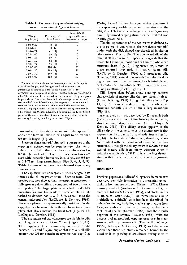
Table 1. Presence of asymmetrical cappingstructures in cilia of different lengths
Ciliarylength (/im)
0-OO-0-250-25-0-500-50-0-750-75-1-001-00-1-251-25-1-501-50-1-751-75-2-002-00-2-502-50-3-503-50-6-00
Percentage ofcilia with caps
0(12)0(8)
41 (17)56 (16)50(8)82 (11)85 (13)
100(8)100(12)100 (13)100 (19)
Percentage ofcapped cilia withasymmetrical caps
000000
18508369
100
The centre column shows the percentage of cilia with caps ateach ciliary length, and the right-hand column shows thepercentage of capped cilia that contain clear views of theasymmetrical arrangement of plates typical of fully grown Bombinacilia. The number of cilia scored for each measured length is givenin parenthesis. Since the plates lie in the same plane as the basalfeet attached to each basal body, the capping structures are onlycounted from thin sections of cilia in which the basal feet arevisible. Capping structures are seen with increasing frequency incilia greater than 0-5 fxm in length. The asymmetrically positionedplates in the caps, indicative of 'mature' caps are observed withincreasing frequency in cilia greater than 1-75 ^m.
proximal ends of central-pair microtubules appear toend at the terminal plate in cilia equal to or less than0-5 Jim in length (Fig. 2).
Electron-dense material similar in appearance to thecapping structures can be seen between the micro-tubule tips and the ciliary membrane in cilia as short as0-5 Jim (arrowhead in Fig. 3). These structures areseen with increasing frequency in cilia between 0-5 |Umand 1-75Jim long (arrowheads, Figs 3, 4, 5, 8, 9).Table 1 summarizes these data obtained from manythin sections.
The cap structure undergoes further changes in itsform as the cilium grows from 1-5 Jim to 6 Jim. Ourprevious studies showed that the capping structures infully grown palate cilia are composed of two differentsize plates. The large plate is attached to doubletmicrotubules nos 4-7 while the smaller plate is at-tached to doublet nos 1, 2, 3, 8, 9, as well as to thecentral microtubules (LeCluyse & Dentler, 1984).Since the plates are asymmetrically positioned in thecap, they can be seen only in those cilia sectioned in aplane that also contains the basal feet (Figs 10-16;LeCluyse & Dentler, 1984).
The asymmetrical cap structures are visible in ciliawith lengths between 1-75 and2-0jtm (Figs 9, 10, 11).The frequency of cap asymmetry increases in ciliabetween 1-75 and 2-5 Jim long so that virtually all ciliagreater than 2-5 Jim contain an asymmetrical cap (Figs
12-16; Table 1). Since the asymmetrical structure ofthe cap is only visible in certain orientations of thecilia, it is likely that all cilia longer than 2-2-5 Jim longhave fully formed capping structures identical to thosein fully grown cilia.
The first appearance of the two plates is defined bythe presence of amorphous electron-dense materialunderneath the disk-shaped cap described in shortercilia (arrows, Figs 9, 10). The downward tilt of thelower shelf relative to the upper shelf suggests that thelower shelf is not yet positioned within the whole capstructure (inset, Fig. 10). Plug structures, similar tothose reported previously in mature palate cilia(LeCluyse & Dentler, 1984) and protozoan cilia(Dentler, 1981), extend downwards from the develop-ing cap and insert into the lumen of each A-tubule andeach central-pair microtubule. The plug structures areas long as 50nm (insets, Figs 10, 11).
Cilia longer than 3-5jim show bending patternscharacteristic of mature cilia that are 'instantly fixed'(Omoto & Kung, 1980) during their ciliary beat (Figs14, 15, 16). Some cilia show tilting of the whole capstructure beneath the tip of the ciliary membrane(Fig. 15).
A ciliary crown, first described by Dirksen & Satir(1972), consists of rows of fine bristles above the capstructure and ciliary membrane (see LeCluyse &Dentler, 1984). The ciliary crown appears at theciliary tip at the same time as the asymmetry is firstapparent in the cap (small arrowheads, insets Figs 10,11, 14). The formation of the crown, therefore, occursconcomitant with the formation of a complete cappingstructure. Although the ciliary crown is reported at thetips of mature cilia from many different types ofepithelia (see Dentler, 1981), this is the first demon-stration that the crown hairs are present in growingcilia.
Discussion
Previous papers on studies of ciliogenesis in metazoansdescribed centriole formation in differentiating epi-thelium from mouse oviduct (Dirksen, 1971), Rhesusmonkey oviduct (Anderson & Brenner, 1971), rattrachea (Dirksen & Crocker, 1965), and chick trachea(Kalnins & Porter, 1969). The formation of cilia inmulticiliated epithelial cells has been described foronly a few tissues, including tracheal epithelium fromXenopus embryos (Steinman, 1968), tracheal epi-thelium of the rat (Sorokin, 1968), and the tubularnephron of the lamprey (Youson, 1982). With thediscovery of microtubule capping structures in meta-zoan as well as protozoan cilia (Dentler & LeCluyse,1982a; LeCluyse & Dentler, 1984) and the obser-vation that these structures remained bound to thedistal ends of growing microtubules during most of
Formation of microtubule caps 89

ciliary growth (Dentler, 1980), it was important toreexamine the process of ciliogenesis to determine thesite and timing of cap formation. Metazoan epithelialcilia are ideal for this study because their cappingstructures are prominent and well stained for examin-ation in thin sections.
The results reported here show that the cappingstructures are neither associated with the basal bodynor with the ciliary membrane during the initial stagesof attachment between these organelles. Shortly after
attachment of the basal body to the membrane, a massof amorphous granular material gradually collects atthe tips of the growing microtubules. When the ciliarymicrotubules are 0-75-l-Sfim long (Table 1), how-ever, a cap-like structure is seen within the amorphousmaterial. Similar to earlier observations of Xenopuscilia by Steinman (1968), the dense material inBombina palate cilia begins to differentiate into amultilayered cap. These layers eventually form thetwo plates of the asymmetrical cap previously shown
Figs 12-14. The tips of cilia show additional development of the cap. All Figs, X40000; insets, X75 600.Figs 12, 13. These cilia with lengths of 2-5 and 2-95 Jim, respectively, have a lower plate and plug extensions into
central-pair and outer doublet microtubules.Fig. 14. A cilium with a length of 347/im has both plates, and cap asymmetry is pointing to the right. Inset shows
ciliary crown (arrowhead) as well as cap asymmetry.
90 R. W. Portman et al.

both in Bombina and in Xenopus cilia (LeCluyse& Dentler, 1984; LeCluyse et al. 1985). The platesare linked to the microtubules by short plugs that
insert into the lumen of each A-tubule and centralmicrotubule. Together these structures form a cap-plug complex that binds to the microtubule tips
Figs 15-16. Cilia with lengths between 4-00 and 5-00/im. Both Figs, X40000; insets, X81900.Fig. 15. A cilium of 3-9/ttn shows more bending than shorter cilia. Inset shows a cap tilted upwards under the ciliary
membrane. Arrow points to lower plate.Fig. 16. A cilium of 5 1 ^m shows additional bending and a cap structure with the asymmetry pointing upwards. Inset
shows lower plate pointing to the right.
Formation of microtubule caps 91

(Dentler, 1980; LeCluyse & Dentler, 1984; Dentler &LeCluyse, 1982a; Dentler, 1984).
The assembly of cap-plug complex and asymmetricplacement of the plate structures characteristic of fullygrown palate cilia occur early in development when thecilia reach 1-75-2-00/im (Table 1), although well-formed caps can be found in cilia as short as 0-75 fim.Once the cap is formed, it remains connected to theends of microtubules throughout ciliary growth.These results correlate well with previous studies ofcapping structures in Chlamydomonas and Tetra-hymena cilia, in which the capping structures are fullyformed by the time the cilia are 3-4 (im long andremain attached to the microtubules throughout cili-ary growth (Dentler & Rosenbaum, 1977; Dentler,1980).
Although the results reported here reveal that thecapping structures are assembled during the initialstages of ciliogenesis, it remains unclear preciselywhen the components of the caps are first assembled.We do know that a basal body will not initiate ciliarymicrotubule assembly until it has attached to theplasma membrane. Isolated basal bodies and cen-trioles can initiate the assembly of single cytoplasmicmicrotubules in vitro (Snell et al. 1974; Rosenbaumet al. 1975; Gould & Borisy, 1977) and in vivo(Heideman et al. 1977), and single microtubules areoccasionally observed to be attached to basal body-likecentrioles in situ (Krishan & Buck, 1965). However,the microtubules initiated by isolated basal bodies orcentrioles were typical cytoplasmic microtubules andnot the doublet microtubules found in cilia andeukaryotic flagella. The nucleated assembly of ciliarymicrotubules may, therefore, be regulated by somefactor(s) associated with the docking of the basal bodyto the plasma membrane. The results of both in vivoand in vitro experiments suggest that initiation ofmicrotubule assembly may require the release of someinhibitory factor from the ends of basal body micro-tubules before the attachment of microtubule cappingstructures. One function of the proteins or otherfactors associated with the capping structures may beto release the postulated inhibitor and initiate micro-tubule assembly. Another function may be to directthe assembly of ciliary microtubules, since only single(cytoplasmic) microtubules are assembled in vivo(Krishan & Buck, 1965) or in vitro (Snell et al. 1974;Gould & Borisy, 1977) onto the ends of basal bodiesand centrioles that lack the capping structures. Theresults reported here show that the cap structures areformed within the first two micrometres of ciliarygrowth, but leave open the possibility that cappingproteins may join with the basal bodies prior to theformation of the prominent caps seen in thin-sectionedor negatively stained cilia. Although it is unlikely thatmorphological studies alone will reveal the initial
presence of capping structures or capping structureproteins, studies with antibodies directed against cap-ping structures may be useful in future studies. Recentprogress has been made in the isolation of cappingstructures (Suprenant & Dentler, 1986) and, hope-fully, antibodies against these structures will beobtained within the near future.
The axonemal microtubules of very short palatecilia appear more electron-opaque and less wellarranged than do microtubules of fully grown cilia inBombina. Some microtubules in the short cilia aredifficult to identify (see Figs 1,2). This problem hasbeen discussed previously in studies of flagellar devel-opment in the amoeboflagellate Naegleria and ciliarydevelopment in neuroepithelial cells of chick embryos(Dingle & Fulton, 1966; Sotelo & Trujillo-Cenoz,1958). The walls of axonemal microtubules becomemore electron-dense when the cilia reach a length atwhich the cap structure and a well-defined basal plateare formed. It may be that short microtubules areunstable but once the capping structures are as-sembled onto the ends of the microtubules, the tubulinwithin the microtubules may be modified to form amore stable polymer, perhaps, by acetylation as hasbeen shown in Chlamydomonas (L'Hernault & Rosen-baum, 1983; Piperno & Fuller, 1985). Alternatively,the capping structures may facilitate the attachment ofproteins (dynein, radial spoke proteins, etc.) to theciliary microtubules that will increase microtubulestability.
In addition to their attachment to the ciliary micro-tubules, the capping structures in many but not allepithelial cilia (Dentler & LeCluyse, 1982a) arelinked, through the membrane, to a complex ofextraciliary hairs, called the ciliary crown (Dirksen &Satir, 1972; Kuhn & Engleman, 1978; Dentler, 1981;LeCluyse & Dentler, 1984). Although Dirksen & Satir(1972) initially reported that the crown hairs appearedonly on fully grown cilia, the results reported hereshowed that the crown was formed at the same timeas the capping structure, since the crown and capwere found on virtually all cilia longer than 1-2 jum.The attachment of the cap to the membrane and to thecrown may help stabilize or position the cap at theciliary tip as the ciliary microtubules grow out to theirfinal lengths.
We thank Drs Kathy Suprenant and Paul Burton for theirhelpful criticism on the manuscript. We also thank ScottRobinson and Dr Sally Frost for providing us with Bombinaorientalis tadpoles and Ms Lorainne Hammer for technicalassistance. This work was supported by grant from theNational Institutes of Health (GM 32556).
92 R. W. Portman et al.

References
ANDERSON, R. G. W. & BRENNER, R. M. (1971). The
formation of basal bodies (centrioles) in the rhesusmonkey oviduct. J. Cell Biol. 50, 10-34.
DENTLER, W. L. (1980). Structures linking the tips ofciliary and flagellar microtubules to the membrane.J. Cell Sri. 42, 207-220.
DENTLER, W. L. (1981). Microtubule-membraneinteractions in cilia and flagella. Int. Rev. Cytol. 72,1-47.
DENTLER, W. L. (1984). Attachment of the cap to thecentral microtubules of Tetrahymena cilia. J. Cell Sri.66, 167-173.
DENTLER, W. L. (1986). Isolation of capped cilia frombovine trachea and the effect of caps on microtubuleassembly. J . Cell Biol. 103, 279a.
DENTLER, W. L. & LECLUYSE, E. L. (1982a).
Microtubule capping structures at the tips of trachealcilia: evidence for their firm attachment during ciliarybend formation and the restriction of microtubulesliding. Cell Motil. 2, 549-572.
DENTLER, W. L. & LECLUYSE, E. L. (19826). The effectsof structures attached to the tips of tracheal ciliarymicrotubules on the nucleation of microtubule assemblyin vitm. Cell Motil. (Suppl.) 1, 13-18.
DENTLER, W. L. & ROSENBAUM, J. L. (1977). Flagellarelongation and shortening in Chlamydomonas. III.Structures attached to the tips of flagellar microtubulesand their relationship to the directionality of flagellarmicrotubule assembly. J. Cell Biol. 74, 749-759.
DINGLE, A. D. & FULTON, C. (1966). Development of theflagellar apparatus of Naegleria. J. Cell Biol. 31, 43-54.
DIRKSEN, E. R. (1971). Centriole morphogenesis indeveloping ciliated epithelium of the mouse oviduct.J. Cell Biol. 51, 286-302.
DIRKSEN, E. R. & CROCKER, T. T. (1965). Centriolereplication in differentiating ciliated cells of mammalianrespiratory epithelium. An electron microscope study.J. Micmsc. 5, 629-644.
DIRKSEN, E. R. & SATIR, P. (1972). Ciliary activity in themouse oviduct as studied by transmission and scanningelectron microscopy. Tissue & Cell 4, 389-404.
GOULD, R. R. & BORISY, G. G. (1977). The pericentriolarmaterial in Chinese hamster ovary cells nucleatesmicrotubule formation. J. Cell Biol. 73, 601-615.
HEIDEMANN, S. R., SANDER, G. & KIRSCHNER, M. W.
(1977). Evidence for a functional role of RNA incentrioles. Cell 10, 337-350.
KALNINS, V. I. & PORTER, K. R. (1969). Centriolereplication during ciliogenesis in chick trachealepithelium. Z. Zellforsch. mikrosk. Anat. 100, 1-30.
KRISHAN, A. & BUCK, R. C. (1965). Structure of themitotic spindle in L-strain fibroblasts. J. Cell Biol. 24,433-444.
KUHN, C. & ENGLEMAN, W. (1978). The structure of thetips of mammalian respiratory cilia. Cell Tiss. Res. 186,491-498.
LECLUYSE, E. L. & DENTLER, W. L. (1984).
Asymmetrical microtubule capping structures in frogpalate cilia. J. Ultrastruct. Res. 86, 75-85.
LECLUYSE, E. L., FROST, S. K. & DENTLER, W. L.
(1985). Development and ciliation of the palate in twofrogs, Bombina and Xenopus; a comparative study.Tissue & Cell 17, 853-864.
L'HERNAULT, S. W. & ROSENBAUM, J. L. (1983).
Chlamydomonas a-tubulin is posttranslationally modifiedin the flagella during flagella assembly. J. Cell Biol. 97,258-263.
OMOTO, C. K. & KUNG, C. (1980). Rotation and twist ofthe central pair microtubules in the cilia of Paramecium.J. Cell Biol. 87, 33-46.
PIPERNO, G. & FULLER, M. T. (1985). Monoclonal
antibodies specific for an acetylated form of Q"-tubulinrecognize the antigen in cilia and flagella from a varietyof organisms. J . Cell Biol. 101, 2085-2094.
PrTELKA, D. R. (1974). Basal bodies and root structures.In Cilia and Flagella (ed. M. A. Sleigh), pp. 437-469.New York, London: Academic Press.
PORTMAN, R. W. & DENTLER, W. L. (1986). Development
of asymmetrical capping structures and orientation ofbasal bodies during ciliogenesis on palate epitheliumfrom Bombina orientalis. J. Cell Biol. 103, 279a.
ROSENBAUM, J. L., BINDER, L. I., GRANETT, S.,
DENTLER, W. L., SNELL, W., SLOBODA, R. & HAIMO,
L. (1975). Directionality and rate of assembly of chickbrain tubulin onto pieces of neurotubules, flagellaraxonemes, and basal bodies. Ann. N.Y. Acad. Sci. 253,147-177.
ROSENBAUM, J. L., MOULDER, J. E. & RINGO, D. L.
(1969). Flagellar elongation and shortening inChlamydomonas. The use of cycloheximide andcolchicine to study the synthesis and assembly offlagellar proteins. J. Cell Biol. 41, 600-619.
SNELL, W. J., DENTLER, W. L., HAIMO, L., BINDER, L. I.
& ROSENBAUM, J. L. (1974). Assembly of chick braintubulin onto isolated basal bodies of Chlamydomonasreinhardtii. Srience 185, 357-360.
SOROHN, S. (1968). Reconstruction of centriole formationand ciliogenesis in mammalian lungs. J. Cell Sri. 3,207-230.
SOTELLO, J. R. & TRUJILLO-CENOZ, O. (1958). Electron
microscope study on the epithelium of ciliarycomponents of the neural epithelium of the chickembryo. Z. Zellforsch. mikrosk. Anat. 49, 1-12.
SPURR, A. R. (1969). A low viscosity epoxy resinembedding medium for electron microscopy.J. Ultrastruct. Res. 26, 31-43.
STEINMAN, R. M. (1968). An electron microscope study ofciliogenesis in developing epidermis and trachea in theembryo of Xenopus laevis. Am. J. Anat. 122, 19-55.
SUPRENANT, K. A. & DENTLER, W. L. (1986). Differential
stability of microtubule-capping structures to Ca+ + andMg++.J. Cell Biol. 103, 133a.
TAYLOR, A. C. & KOLLROS, J. J. (1946). Stages in the
normal development of Rana pipiens larvae. Anat. Rec.94, 7-23.
Formation of microtubule caps 93

VENABLE, J. H. & COGGESHALL, R. (1965). A simplified YOUSON, J. H. (1982). Replication of basal bodies andlead citrate stain for use in electron microscopy. jf. Cell ciliogenesis in a ciliated epithelium of the lamprey. CellBiol. 25, 407-408. Tiss. Res. 223, 255-266.
WITMAN, G. B. (1975). The site of in vivo assembly offlagellar microtubules. Ann. N.Y. Acad. Sci. 253, {Received 26 September 1986 - Accepted 14 October 1986)178-191.
94 R. W. Portman et al.