Development of Glutamatergic Synaptic Transmission in Binaural … · 2019-12-21 · Development of...
17
doi:10.1152/jn.00468.2010 104:1774-1789, 2010. First published 28 July 2010; J Neurophysiol Jason Tait Sanchez, Yuan Wang, Edwin W Rubel and Andres Barria in Binaural Auditory Neurons Development of Glutamatergic Synaptic Transmission You might find this additional info useful... 92 articles, 37 of which can be accessed free at: This article cites http://jn.physiology.org/content/104/3/1774.full.html#ref-list-1 1 other HighWire hosted articles This article has been cited by [PDF] [Full Text] [Abstract] , February 23, 2011; 31 (8): 2983-2995. J. Neurosci. Polley Troy A. Hackett, Tania Rinaldi Barkat, Barbara M. J. O'Brien, Takao K. Hensch and Daniel B. Linking Topography to Tonotopy in the Mouse Auditory Thalamocortical Circuit including high resolution figures, can be found at: Updated information and services http://jn.physiology.org/content/104/3/1774.full.html can be found at: Journal of Neurophysiology about Additional material and information http://www.the-aps.org/publications/jn This information is current as of April 20, 2012. American Physiological Society. ISSN: 0022-3077, ESSN: 1522-1598. Visit our website at http://www.the-aps.org/. (monthly) by the American Physiological Society, 9650 Rockville Pike, Bethesda MD 20814-3991. Copyright © 2010 by the publishes original articles on the function of the nervous system. It is published 12 times a year Journal of Neurophysiology on April 20, 2012 jn.physiology.org Downloaded from
Transcript of Development of Glutamatergic Synaptic Transmission in Binaural … · 2019-12-21 · Development of...
doi:10.1152/jn.00468.2010 104:1774-1789, 2010. First published 28 July 2010;J NeurophysiolJason Tait Sanchez, Yuan Wang, Edwin W Rubel and Andres Barriain Binaural Auditory NeuronsDevelopment of Glutamatergic Synaptic Transmission
You might find this additional info useful...
92 articles, 37 of which can be accessed free at:This article cites http://jn.physiology.org/content/104/3/1774.full.html#ref-list-1
1 other HighWire hosted articlesThis article has been cited by
[PDF] [Full Text] [Abstract]
, February 23, 2011; 31 (8): 2983-2995.J. Neurosci.PolleyTroy A. Hackett, Tania Rinaldi Barkat, Barbara M. J. O'Brien, Takao K. Hensch and Daniel B.Linking Topography to Tonotopy in the Mouse Auditory Thalamocortical Circuit
including high resolution figures, can be found at:Updated information and services http://jn.physiology.org/content/104/3/1774.full.html
can be found at:Journal of Neurophysiologyabout Additional material and information http://www.the-aps.org/publications/jn
This information is current as of April 20, 2012.
American Physiological Society. ISSN: 0022-3077, ESSN: 1522-1598. Visit our website at http://www.the-aps.org/.(monthly) by the American Physiological Society, 9650 Rockville Pike, Bethesda MD 20814-3991. Copyright © 2010 by the
publishes original articles on the function of the nervous system. It is published 12 times a yearJournal of Neurophysiology
on April 20, 2012
jn.physiology.orgD
ownloaded from
Development of Glutamatergic Synaptic Transmission in BinauralAuditory Neurons
Jason Tait Sanchez,1,2 Yuan Wang,1,2 Edwin W Rubel,1–3 and Andres Barria1,3
1Virginia Merrill Bloedel Hearing Research Center and 2Departments of Otolaryngology—Head and Neck Surgery and 3Physiology andBiophysics, University of Washington, Seattle, Washington
Submitted 26 May 2010; accepted in final form 23 July 2010
Tait Sanchez J, Wang Y, Rubel EW, Barria A. Development ofglutamatergic synaptic transmission in binaural auditory neurons. JNeurophysiol 104: 1774–1789, 2010. First published July 28, 2010;doi:10.1152/jn.00468.2010. Glutamatergic synaptic transmission isessential for binaural auditory processing in birds and mammals.Using whole cell voltage clamp recordings, we characterized thedevelopment of synaptic ionotropic glutamate receptor (iGluR) func-tion from auditory neurons in the chick nucleus laminaris (NL), thefirst nucleus responsible for binaural processing. We show that syn-aptic transmission is mediated by AMPA- and N-methyl-D-aspartate(NMDA)-type glutamate receptors (AMPA-R and NMDA-R, respec-tively) when hearing is first emerging and dendritic morphology isbeing established across different sound frequency regions. Puffapplication of glutamate agonists at embryonic day 9 (E9) revealedthat both iGluRs are functionally present prior to synapse formation(E10). Between E11 and E19, the amplitude of isolated AMPA-Rcurrents from high-frequency (HF) neurons increased 14-fold. Asignificant increase in the frequency of spontaneous events is alsoobserved. Additionally, AMPA-R currents become faster and morerectifying, suggesting developmental changes in subunit composition.These developmental changes were similar in all tonotopic regionsexamined. However, mid- and low-frequency neurons exhibit fewerspontaneous events and evoked AMPA-R currents are smaller, slower,and less rectifying than currents from age-matched HF neurons. Theamplitude of isolated NMDA-R currents from HF neurons also increased,reaching a peak at E17 and declining sharply by E19, a trend consistentacross tonotopic regions. With age, NMDA-R kinetics become signifi-cantly faster, indicating a developmental switch in receptor subunitcomposition. Dramatic increases in the amplitude and speed of glutama-tergic synaptic transmission occurs in NL during embryonic develop-ment. These changes are first seen in HF neurons suggesting regulationby peripheral inputs and may be necessary to enhance coincidencedetection of binaural auditory information.
I N T R O D U C T I O N
In many brain regions, excitatory responses mediated bypostsynaptic ionotropic glutamate receptors (iGluRs) are re-quired for correct circuit formation and maturation (Espinosa etal. 2009; Hall and Ghosh 2008). During synaptic development,iGluRs often show remarkable modifications in their biophys-ical properties (Carmignoto and Vicini 1992; Esteban et al.2003; Kumar et al. 2002; Quinlan et al. 1999; Tovar andWestbrook 1999), which coincide with changes in synapticgrowth and pruning (Cline and Haas 2008) as well as alter-ations in dendritic structure and circuit refinement (Lichtmanand Colman 2000). Typically, the AMPA-type glutamate re-ceptor (AMPA-R) is necessary for rapid synaptic transmission,
whereas the N-methyl-D-aspartate (NMDA)-type glutamate re-ceptor (NMDA-R) mediates a slower excitatory responsethought to be critical for modulating synaptic function (Dingle-dine et al. 1999). Interestingly, little is known about thedevelopmental role of iGluRs in the avian binaural auditorysystem, a relatively simple circuit extensively studied withrespect to the development of structure and function.
In birds, nucleus laminaris (NL) is a third-order auditory brainstem structure responsible for binaural sound processing. The cellbody layer of NL neurons are arranged according to the optimalfrequency response of the neuron (i.e., tonotopic organization)and contain segregated dorsal and ventral aspiny dendrites thatexhibit a systematic gradient in dendritic length (Fig. 1A) that runsparallel to this tonotopic axis (Parks and Rubel 1978; Smith andRubel 1979). Functionally, mature NL neurons are highly special-ized for encoding temporal information of sound (Kuba et al.2006). These neurons possess a variety of physiological special-izations (Kuba et al. 2005) that facilitate coincidence detection(Kuba et al. 2002a,b, 2003) thought essential for binaural hearing(Hyson 2005; Young and Rubel 1983).
Soon after synapses form [�embryonic day 10 (E10)], excita-tory postsynaptic potentials (EPSPs) from NL neurons becomeextremely fast (Gao and Lu 2008) to enhance the ability of NLneurons to act as binaural coincident detectors (Raman et al. 1994;Reyes et al. 1996; Trussell 1997, 1999). This sharpening of theEPSP soon after synaptogenesis has been interpreted as a loss inthe NMDA-R mediated component (Gao and Lu 2008), consistentwith previous reports of the absence of NMDA-R responses laterin development (�E16) (Funabiki et al. 1998; Zhou and Parks1991). However, during this embryonic period of rich dendriticelaboration and circuit refinement, NMDA-Rs have clearly beenshown, in NL (Tang and Carr 2004). Moreover, differentNMDA-R subunits have specific temporal patterns of expression(Tang and Carr 2007), suggesting the presence of NMDA-Rs withdifferent biophysical properties during the developmental periodwhen the tonotopic gradient is being established.
In this report, we characterize the developmental profile andfunctional expression of iGluRs in NL neurons. We reportdramatic changes in both AMPA-R and NMDA-R responsesoccurring differentially across the tonotopic gradient during aperiod when synapses are forming, specializations and cir-cuitry are established, and hearing is emerging.
M E T H O D S
Slice preparation
Acute brain stem slices were prepared from White Leghorn chicken(Gallus domesticus) embryos at E9, E10, E11, E13, E15, E17, and E19
Address for reprint requests and other correspondence: A. Barria, Physiol-ogy and Biophysics, School of Medicine, University of Washington, Box357290, Seattle, WA 98195 (E-mail: [email protected]).
J Neurophysiol 104: 1774–1789, 2010.First published July 28, 2010; doi:10.1152/jn.00468.2010.
1774 0022-3077/10 Copyright © 2010 The American Physiological Society www.jn.org
on April 20, 2012
jn.physiology.orgD
ownloaded from
as described previously (Howard et al. 2007; Monsivais et al. 2000). Thebrain stem was dissected and isolated in ice-cold (�0°C) oxygenatedlow-Ca2� high-Mg2� modified artificial cerebral spinal fluid (ACSF)containing the following (in mM): 130 NaCl, 3 KCl, 1.25 NaH2PO4, 26NaHCO3, 4 MgCl2, 1 CaCl2, and 10 glucose. ACSF was continuouslybubbled with a mixture of 95% O2-5% CO2 (pH 7.4, osmolarity:295–310 mosM/l). The brain stem was blocked coronally, affixed to thestage of a vibratome slicing chamber (Technical Products International,St. Louis, MO) and submerged in the ice-cold ACSF. Bilaterally sym-metrical coronal slices were made (200–300 �m thick), and approxi-mately one to six slices (depending on age) containing NM and NL weretaken along the caudolateral to rostromedial axis, roughly representingthe low- to high-frequency regions of NL, respectively.
Slices were then collected in a holding chamber and allowed toequilibrate for 1 h at 36°C in normal ACSF containing the following (inmM): 130 NaCl, 3 KCl, 1.25 NaH2PO4, 26 NaHCO3, 1 MgCl2, 3 CaCl2,and 10 glucose. Normal ACSF was continuously bubbled with a mixtureof 95% O2-5% CO2 (pH 7.4, osmolarity: 295–310 mosM/l). Slices werethen allowed to cool to room temperature for 30 min before beingtransferred from the holding chamber to a 0.5 ml recording chambermounted on either a Zeiss Axioskop FS (Oberkochen, Germany) or anOlympus BX51W1 (Center Valley, PA) microscope for electrophysio-logical experiments. Microscopes were equipped with CCD cameras with�40 and �60 water-immersion objectives and infrared differential in-terference contrast optics. The recording chamber was superfused con-tinuously with room temperature (monitored at �21°-22°C), oxygenatednormal ACSF at a rate of 1.5–2 ml/min.
Whole cell electrophysiology
Patch pipettes were pulled to a tip diameter of 1–2 �m that hadresistances ranging from 3 to 6 M� when filled with a cesium-based
internal solution containing the following (in mM): 108 CsMeSO3, 5CsCl, 1 MgCl2, 15 phosphocreatine-Tris2, 8 BAPTA-Cs4, 10 HEPES,3 QX-314.Cl, 4 MgATP, and 0.4 Tris2GTP, pH adjusted to 7.3 withTrisOH. Voltage-clamp experiments were performed using either anAxoclamp 200B (Molecular Devices, Foster City, CA) or an AxonMulticlamp 700B amplifiers (Molecular Devices). For selected exper-iments, 2% neurobiotin was added to the internal solution for labelingof cells along the tonotopic axis (see following text). The Cs-basedinternal solution was used to block K� conductances and QX-314.Clwas used to block Na� conductances in an attempt to reduce space-clamp issues associated with dendritic filtering. Command voltagesduring experiments were corrected for a calculated liquid junctionpotential of 5 mV, and series resistance was compensated for by�80% in all voltage-clamp recordings. A small hyperpolarizing (�1to �5 mV, 100 ms) voltage command was presented at the beginningof each recorded trace to document and monitor whole cell parameters[resting membrane potential (RMP), cell membrane capacitance, se-ries resistance, and input resistance]. RMPs were measured immedi-ately after break-in to avoid cesium-induced depolarization. Neuronswere included in the data analysis only if they had RMPs between�50 and �65 mV and had series resistances �15 M�. Raw data werelow-pass filtered at 2 kHz and digitized at 10 kHz using a dataacquisition interface ITC-18 (Instrutech, Great Neck, NY) or a Digi-data 1440A (Molecular Devices).
Pipettes were visually guided to NL, and neurons were identifiedand distinguished from surrounding tissue based on cell morphology,known laminar structure, and location of the nucleus within the slice.After a G� seal was attained, membrane patches were ruptured andNL neurons were held in whole cell configuration at �60 mV forrecording of isolated AMPA-R mediated excitatory postsynaptic cur-rents (EPSCs) or at �40 mV for recording of isolated NMDA-Rmediated EPSCs. Isolated AMPA-R currents were recorded in the
BA
Cap
acita
nce
(pF)
Frequency Region
***
***
*
C
Age (Embryonic Days)
Cap
acita
nce
(pF)
LFMFHFNFR
D
Cap
acita
nce
(pF)E
Frequency Region
E13***
Max EPSC = -1.2 nAtau = 2.12 msCM = 43.3 pFRI = 0.4
E13HF-60 mVF
HF MF LF
E13HF
D
M
10 µm
50 µm
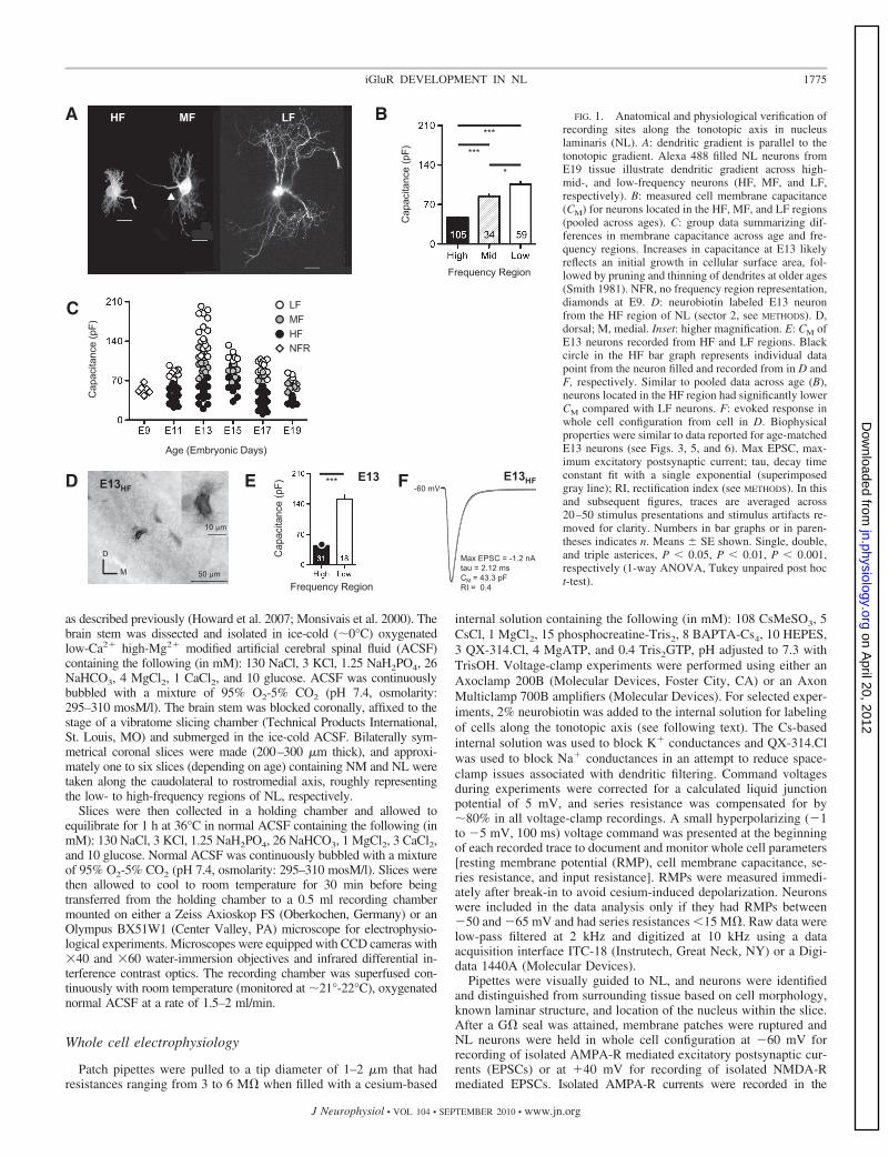
FIG. 1. Anatomical and physiological verification ofrecording sites along the tonotopic axis in nucleuslaminaris (NL). A: dendritic gradient is parallel to thetonotopic gradient. Alexa 488 filled NL neurons fromE19 tissue illustrate dendritic gradient across high-mid-, and low-frequency neurons (HF, MF, and LF,respectively). B: measured cell membrane capacitance(CM) for neurons located in the HF, MF, and LF regions(pooled across ages). C: group data summarizing dif-ferences in membrane capacitance across age and fre-quency regions. Increases in capacitance at E13 likelyreflects an initial growth in cellular surface area, fol-lowed by pruning and thinning of dendrites at older ages(Smith 1981). NFR, no frequency region representation,diamonds at E9. D: neurobiotin labeled E13 neuronfrom the HF region of NL (sector 2, see METHODS). D,dorsal; M, medial. Inset: higher magnification. E: CM ofE13 neurons recorded from HF and LF regions. Blackcircle in the HF bar graph represents individual datapoint from the neuron filled and recorded from in D andF, respectively. Similar to pooled data across age (B),neurons located in the HF region had significantly lowerCM compared with LF neurons. F: evoked response inwhole cell configuration from cell in D. Biophysicalproperties were similar to data reported for age-matchedE13 neurons (see Figs. 3, 5, and 6). Max EPSC, max-imum excitatory postsynaptic current; tau, decay timeconstant fit with a single exponential (superimposedgray line); RI, rectification index (see METHODS). In thisand subsequent figures, traces are averaged across20–50 stimulus presentations and stimulus artifacts re-moved for clarity. Numbers in bar graphs or in paren-theses indicates n. Means � SE shown. Single, double,and triple asterices, P � 0.05, P � 0.01, P � 0.001,respectively (1-way ANOVA, Tukey unpaired post hoct-test).
1775iGluR DEVELOPMENT IN NL
J Neurophysiol • VOL 104 • SEPTEMBER 2010 • www.jn.org
on April 20, 2012
jn.physiology.orgD
ownloaded from

presence of an NMDA-R blocker D-2-amino-5-phosphonopentanoic acid(D-APV, 100 �M), and isolated NMDA-R currents were recorded in thepresence of AMPA-R blockers 1,2,3,4-tetrahydro-6-nitro-2,3-dioxo-benzo-[f]quinoxaline-7-sulfonamide disodium salt hydrate (NBQX, 20 �M) or6,7-dinitroquinoxaline-2,3(1H,4H)-dione (DNQX, 50 �M). All experi-ments were conducted in the presence of GABAA-R blockers bicuculinemethiodide (BIC, 50 �M), SR-95331 (gabazine, 10 �M), or picrotoxin(PTX, 100 �M).
Extracellular synaptic stimulation was accomplished using a con-centric bipolar electrode (tip core diameter 200 �m, World Preci-sion Instruments, Sarasota, FL). Square electric pulses, 100 �s induration, were delivered by a stimulus isolator (1850A) and intervalgenerator (1830; WP Instruments, New Haven, CT) or by an Iso-fluxstimulator (AMPI; Jerusalem, Israel) and interval generator (S88;Grass, West Warwick, RI). Stimulating electrodes were placed ineither the ventral or dorsal neuropil region of NL, �30–50 �m fromrecorded NL neurons. No differences were observed in the size ofmaximum ESPC amplitudes or kinetics based on stimulating electrodeplacements (dorsal versus ventral). Stimulation current was adjustedfrom 0 to 200 �A in steps of 5–10 �A. Input-output functions werederived for each NL neuron and the stimulus intensity was adjusted toevoke the maximum EPSC, often resulting in a plateau EPSC re-sponse, in an attempt to recruit every and all-excitatory inputs fromone side of the brain. This stimulus intensity (mean 163 � 4.8 �A)never exceeded the output of the stimulus generator and was 20% lessthan the maximal intensity tolerated in this slice preparation withoutcausing hydrolysis or motion of the stimulating electrode tip.
The AMPA-R agonist 2-carboxy-3-carboxymethyl-4-isoprope-nylpyrrolidine (kainate, 100 �M) and the NMDA-R agonist (R)-2-(methylamino) succinic acid (NMDA, 500 �M) were puff appliedwith a picosprtizer (General Valve, Fairfield, NJ). Puff pipettes werepulled to a tip diameter of 3–6 �m and filled with ACSF containingeither kainate or NMDA. Puff pipettes were visually guided into closeproximity (�20–40 �m) of the neuron from which recordings werebeing made. Pressure pulses used (10–20 psi; 20–50 ms duration) didnot damage neurons or disrupt patch-pipette seals. Isolated AMPA-Rmediated miniature EPSCs (mEPSCs) were recorded with tetrodo-toxin (TTX, 1 �M) and D-APV added to the external bath ACSFsolution. For experiments where I-V relationships were constructedfor isolated AMPA-R current, N,N-bis(3-aminopropyl)-1,4-diami-nobutane, gerontine, musculamine, neuridine (spermine, 100 �M), apolyamine ion channel blocker for AMPA-Rs lacking the GluR2subunit, was added to the internal pipette solution to reduce dialyzingendogenous polyamines. To accurately report the NMDA-R decaytime constant (tau), a weighted tau was calculated as previouslydescribed (Rumbaugh and Vicini 1999) using the following formula:�W �F[IF/(IF � IS)] � �S[IS(IF � IS)], where I is the currentamplitude, IF and IS are the peak amplitudes of the fast and slowcomponents, respectively, and �F and �S are the respective timeconstants.
Co2� labeling of GluR2-lacking AMPA-Rs
Procedures for Co2� uptake studies were modified from previousreports (Estabel et al. 1999; Pruss et al. 1991; Zhou et al. 1995).Coronal slices (500-�m-thick) through NL were prepared from E11,E15, E19, and E21 embryos and P5 hatchlings as described in thepreceding text and collected in oxygenated ACSF. Slices were prein-cubated in 5 mM CoCl2 for 20 min before the addition of kainate (100�M; Sigma, St. Louis, MO) for another 20 min. For control cases,slices were either incubated in CoCl2 only or incubated in both CoCl2and Kainate and 50 �M DNQX. Slices were then washed in ACSF for5 min. Extracellular cobalt was removed by a 10-min incubation in 2mM EDTA to reduce background. Slices were washed twice in ACSFeach for 5 min before being developed in 1.2% (wt/vol) ammoniumsulfide for 10 min. After two 5-min washes in ACSF, slices were fixedin 4% paraformaldehyde in 0.1 M phosphate buffer for 2–5 h. Slices
were cryoprotected in 30% sucrose in 0.1 M phosphate buffer over-night and then resectioned at 30 �m on a freezing sliding microtome.Sections were mounted on gelatin-coated slides and then dehydrated,cleared, and coverslipped with DPX mounting medium (EMS, Hat-field, PA).
Digital images of selected sections with cobalt staining werecaptured with a Zeiss AxioPlan 2ie equipped with a Coolsnap HQmonochrome digital camera (Princeton Instruments, Trenton, NJ)through a No. 22 Wratten filter and collected in Slidebook (version4.0.2.8; Intelligent Imaging Innovations, Denver, CO). Image contrastand brightness adjustments were made in Adobe Photoshop (AdobeSystems, Mountain View, CA).
Anatomical verification of recorded neurons along thetonotopic gradient in NL
Characteristic frequencies (CFs) of NL neurons increase linearlyfrom the caudolateral to the rostromedial pole (Parks and Rubel 1975).To verify the anatomical region of NL recordings, one to six coronalslices containing the entire extent of NL were obtained from eachanimal. The number of slices was dependent on the age of the animal(see following text). The level of each slice was confirmed by thearrangement of NL and adjacent NM. Anatomical verification ofrecorded neurons along the tonotopic gradient in NL was determinedbased on previous studies (Parks and Rubel 1975, 1978; Smith andRubel 1979; Smith 1981). In addition to recording the rostrocaudalposition, each coronal slice was divided into one to four sectors,depending on their mediolateral position within NL (Kuba et al.2005). This procedure allowed us to define �11 sectors for NL in eachpreparation for animals E13–E19. Using this procedure, three CFregions were defined as follows: rostral sectors 1–4 as the high-frequency (HF) region, intermediate sectors 5–8 as the middle-frequency (MF) region, and caudal sectors 9–11 as the low-frequency(LF) region. This corresponds to predicted CFs of 2.5–3.3 kHz for theHF region, 1–2.5 kHz for the MF region, and 0.4–1 kHz for the LFregion, respectively (Kuba et al. 2005; Parks and Rubel 1975, 1978).However, due to the small size of embryonic brain stem tissue at E9, weobtained only one to two slices per animal, and no tonotopic informationwas gathered at this age. Similarly, only two slices were consistentlyobtained for E11 tissue. As a result, six sectors were used to define onlythe HF and LF tonotopic regions at E11. Rostral slices were divided intosectors 1–3 (HF regions) while caudal slices were divided into sectors4–6 (LF regions). In addition, cell membrane capacitance (CM) wasdetermined for each neuron within a given sector to further correlateneuron size with tonotopic location with the assumption that HF neuronswould have smaller CM compared with LF neurons based on dendriticsize. CM was calculated based on a steady-state current response elicitedby a small hyperpolarizing voltage command (�1 to �5 mV) after wholecell capacitance compensation. Neurons recorded from the LF region(sectors 9–11) of NL had significantly larger CM than MF and HFneurons (sectors 1–4, and 5–8, respectively, P � 0.05, Fig. 1B). Thisresult was consistent across developmental ages (Fig. 1C). To furtherconfirm recorded position from individual NL neurons, 2% neurobiotinwas included in the recording pipette solution to visualize dendritic lengthalong tonotopic regions of NL. Slices that contain labeled neurons werefixed by immersion in 4% paraformaldehyde in PB overnight andvisualized with 3-3=-diaminobenzidine (DAB). An example of anAMPA-R mediated EPSC recorded from a neurobiotin labeled E13 HFneuron (from sector 2) is shown in Fig. 1, D–F. The single cell body layerand neuropil region of the nucleus is identifiable and the short bitufteddendrites of the labeled NL neuron are visible (Fig. 1D). The measuredCM for this neuron was significantly smaller than LF neurons recordedfrom the same developmental age (Fig. 1E). Additionally, the physiologicresponse properties (i.e., maximum EPSC amplitude, decay tau, andrectification index) of this neuron are representative of an E13 HF neuron(Fig. 1F, compare with Figs. 3, 5, and 6).
1776 J. TAIT SANCHEZ, Y. WANG, E. W. RUBEL, AND A. BARRIA
J Neurophysiol • VOL 104 • SEPTEMBER 2010 • www.jn.org
on April 20, 2012
jn.physiology.orgD
ownloaded from

Data analysis
Recording protocols were written and run using either the Axo-graph acquisition and analysis software (version 4.5; Molecular De-vices) or the Clampex acquisition and Clampfit analysis software(version 10.1; Molecular Devices). mEPSCs were detected using atemplate, the kinetics (i.e., 10–90% rise-time and decay time con-stant) of which resembled that of a typical AMPA-R mediatedmEPSC. A detection threshold was set at 5 pA, and all events wereanalyzed using mini-analysis software (Synaptosoft, Fort Lee, NJ).Statistical analyses (ANOVAs, post hoc test, t-test, regression analy-sis) and graphing protocols were performed using Prism (GraphPadversions 5.0a, La Jolla, CA). The standard for significant differenceswas defined as P � 0.05. All graphic representations of data illustratemeans � SE. Tables represent means � SD. The number of obser-vations (n) contributing to each mean is reported in parentheses orinside bar graphs for each figure.
Reagents
All bath applied drugs were allowed to perfuse through the recordingchamber for �2 min before subsequent recordings. D-APV, NBQX,DNQX, kainate, NMDA, BIC, gabazine, spermine, and all other salts andchemicals were obtained from Sigma-Aldrich (St. Louis, MO). N,N,N,-rimethyl-5-[(tricyclo[3.3.1.13,7]dec-1-ylmethyl)amino]-1-pentanamini-umbromide hydrobromide (IEM-1460, 100 �M), (aR,bS)-a-(4-hydroxyphenyl)-b-methyl-4-(phenylmethyl)-1-piperidinepropanol mal-eate (Ro25-6981, 1 �M), and PTX were obtained from Tocris (Ellisville,MO). TTX and QX-314 were obtained from Alomone Labs (Jerusalem,Israel).
R E S U L T S
We characterized the biophysical properties of AMPA-Rand NMDA-R mediated EPSCs across developmental age andtonotopic regions in NL. A total of 208 NL neurons wererecorded from slice preparations taken at E9 (n 6), E10 (n 4), E11 (n 27), E13 (n 62), E15 (n 34), E17 (n 45),and E19 (n 30) chicken embryos. We describe dramaticchanges in synaptic responses mediated by iGluRs during atime period when hearing is first emerging and dendriticmorphology is being established across the tonotopic gradient.
Onset of iGluR responses
We observe physiologically functional iGluRs as early asNL neurons were reliably identifiable in brain stem slices (E9).Interestingly, the presence of iGluRs is evident as early as E7in nucleus magnocellularis (NM) (Diaz et al. 2009), suggestingthat iGluRs are present in both NM and NL prior to synapto-genesis. At E9 in NL, relative dendritic homogeneity is ob-served across different tonotopic regions (Smith and Rubel1979; Smith 1981) and postsynaptic action potentials are notevoked by stimulation of afferent inputs from NM axons(Jackson et al. 1982). Puff application of an NMDA-R agonist(NMDA, 500 �M), while voltage clamping NL neurons at �40mV, elicited a robust outward current that was reversiblyblocked by bath application of the NMDA-R antagonist D-APV(Fig. 2A1). Likewise, puff application of an AMPA-R agonist(kainate, 100 �M), while voltage clamping E9 NL neurons at�60 mV, elicited a strong inward current that was susceptibleto bath application of the AMPA-R antagonist NBQX (Fig.2A2). These robust iGluR currents to puff applications ofagonists were always observed, while electrical stimulation of
afferent excitatory inputs from NM never elicited an EPSC inall neurons tested at E9 (n 6), consistent with observationspreviously reported (Jackson et al. 1982). In contrast, synapti-cally evoked iGluR-mediated EPSCs were present �75% ofthe time at E10 (data not shown). These results indicate thatboth AMPA-Rs and NMDA-Rs are functionally present �1day before synaptic contacts are established.
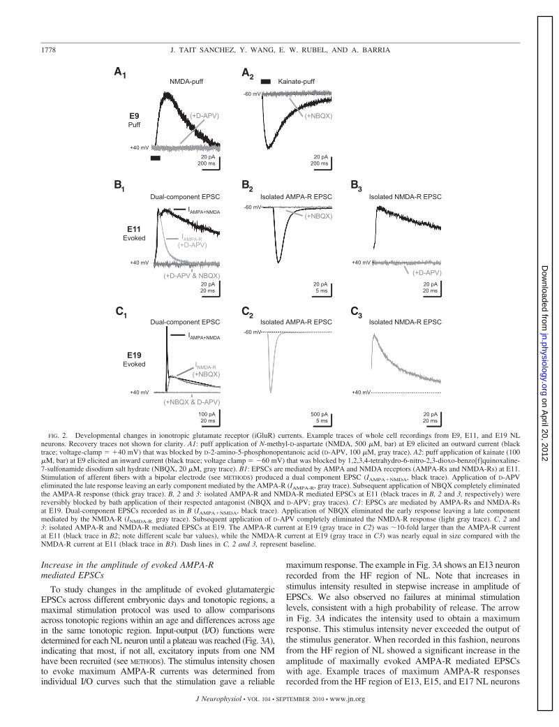
Changes in synaptic iGluR responses
At E11, �1 day after synaptogenesis, evoked EPSCs exhib-ited two clearly discernible peaks when the voltage of theneuron was held at �40 mV (Fig. 2B1). These dual-componentEPSCs contained both AMPA-R and NMDA-R mediated cur-rents that could be pharmacologically isolated. Bath applica-tion of the NMDA-R antagonist D-APV eliminated the slowerresponse leaving an early and faster component mediated bythe AMPA-R. Subsequent bath application of NBQX com-pletely eliminated the remaining AMPA-R response (Fig.2B1). The peak amplitude ratio of the slow EPSC component(NMDA-R mediated) to the fast EPSC component (AMPA-Rmediated) measured at �40 mV, resulted in an NMDA/AMPAratio close to 1 (0.96 � 0.04; n 4). It should be noted that inour experiments at E11, we never observed NMDA-R onlyEPSCs (i.e., silent synapses). This suggests that synaptogenesisin NL is initiated with both iGluRs present, similar to what hasbeen described for developing NM (Lu and Trussell 2007), andis in contrast to observations made at other developing centralsynapses (Durand et al. 1996; Isaac et al. 1997; Liao et al.1995; Rumpel et al. 2004).
The ratio of isolated NMDA-R responses recorded at �40 mVto isolated AMPA-R responses recorded at �60 mV (Fig. 2B, 2and 3) showed a similar value as the ratio of the two peaksobserved at �40 mV (0.94 � 0.08; n 11). These results suggestthat AMPA-Rs do not rectify at E11 and that NMDA-R mediatedcurrents are of similar magnitude as AMPA-R mediated currents.In contrast, at E19, when synaptic responses are considered to benearly mature (Kuba et al. 2002a), a significant difference in thecontribution of AMPA-Rs and NMDA-Rs is clearly observed.The NMDA/AMPA ratio recorded at �40 mV was 0.28 � 0.10,largely due to an increase in the AMPA-R component with littlechange in the NMDA-R response (Fig. 2C1; note different scalebar values). Moreover, the ratio of isolated NMDA-R responsesrecorded at �40 mV to isolated AMPA-R responses recorded at�60 mV was 0.04 � 0.03 due to an even larger AMPA-Rmediated current observed at �60 mV (Fig. 2C, 2 and 3). Thisdifference in the amplitude of the AMPA-R current at hyperpo-larized and depolarized membrane potentials suggest that at olderages the AMPA-R rectifies.
These data indicate that, initially, currents mediated byAMPA-Rs and NMDA-Rs are comparable (Fig. 2B, 1–3). How-ever, the contribution of each receptor changes dramaticallywithin a few days. The AMPA-R current increases significantly(Fig. 2, B2 and C2, note different scale bars), a classic indicator ofsynaptic maturity (Carmignoto and Vicini 1992; Hestrin 1992),while the NMDA-R current peaks at E17 (see Fig. 8), then isreduced at E19 to a similar level as E11 (Figs. 2, B3 and C3, and8). Specific changes in both AMPA-R and NMDA-R responsesacross age and tonotopic regions are discussed in greater detail next.
1777iGluR DEVELOPMENT IN NL
J Neurophysiol • VOL 104 • SEPTEMBER 2010 • www.jn.org
on April 20, 2012
jn.physiology.orgD
ownloaded from
Increase in the amplitude of evoked AMPA-Rmediated EPSCs
To study changes in the amplitude of evoked glutamatergicEPSCs across different embryonic days and tonotopic regions, amaximal stimulation protocol was used to allow comparisonsacross tonotopic regions within an age and differences across agein the same tonotopic region. Input-output (I/O) functions weredetermined for each NL neuron until a plateau was reached (Fig. 3A),indicating that most, if not all, excitatory inputs from one NMhave been recruited (see METHODS). The stimulus intensity chosento evoke maximum AMPA-R currents was determined fromindividual I/O curves such that the stimulation gave a reliable
maximum response. The example in Fig. 3A shows an E13 neuronrecorded from the HF region of NL. Note that increases instimulus intensity resulted in stepwise increase in amplitude ofEPSCs. We also observed no failures at minimal stimulationlevels, consistent with a high probability of release. The arrowin Fig. 3A indicates the intensity used to obtain a maximumresponse. This stimulus intensity never exceeded the output ofthe stimulus generator. When recorded in this fashion, neuronsfrom the HF region of NL showed a significant increase in theamplitude of maximally evoked AMPA-R mediated EPSCswith age. Example traces of maximum AMPA-R responsesrecorded from the HF region of E13, E15, and E17 NL neurons
(+NBQX)
Kainate-puff
20 pA200 ms
A2
(+NBQX)(+NBQX)
Isolated AMPA-R EPSC
20 pA5 ms
B2
Isolated AMPA-R EPSCC2
500 pA5 ms
(+NBQX)
A1
E9Puff
(+D-APV)
NMDA-puff
IAMPA+NMDA
20 pA20 ms
IAMPA-R (+D-APV)
B1
E11Evoked
(+D-APV & NBQX)
100 pA20 ms
(+NBQX & D-APV)
INMDA-R (+NBQX)
IAMPA+NMDA
C1
E19Evoked
20 pA200 ms
+40 mV
+40 mV
+40 mV
Dual-component EPSC Isolated NMDA-R EPSC
(+D-APV)20 pA20 ms
B3
+40 mV
Isolated NMDA-R EPSC
20 pA20 ms
C3
+40 mV
-60 mV
-60 mV
-60 mV
Dual-component EPSC
FIG. 2. Developmental changes in ionotropic glutamate receptor (iGluR) currents. Example traces of whole cell recordings from E9, E11, and E19 NLneurons. Recovery traces not shown for clarity. A1: puff application of N-methyl-D-aspartate (NMDA, 500 �M, bar) at E9 elicited an outward current (blacktrace; voltage-clamp �40 mV) that was blocked by D-2-amino-5-phosphonopentanoic acid (D-APV, 100 �M, gray trace). A2: puff application of kainate (100�M, bar) at E9 elicited an inward current (black trace; voltage clamp �60 mV) that was blocked by 1,2,3,4-tetrahydro-6-nitro-2,3-dioxo-benzo[f]quinoxaline-7-sulfonamide disodium salt hydrate (NBQX, 20 �M, gray trace). B1: EPSCs are mediated by AMPA and NMDA receptors (AMPA-Rs and NMDA-Rs) at E11.Stimulation of afferent fibers with a bipolar electrode (see METHODS) produced a dual component EPSC (IAMPA�NMDA, black trace). Application of D-APVeliminated the late response leaving an early component mediated by the AMPA-R (IAMPA-R, gray trace). Subsequent application of NBQX completely eliminatedthe AMPA-R response (thick gray trace). B, 2 and 3: isolated AMPA-R and NMDA-R mediated EPSCs at E11 (black traces in B, 2 and 3, respectively) werereversibly blocked by bath application of their respected antagonist (NBQX and D-APV; gray traces). C1: EPSCs are mediated by AMPA-Rs and NMDA-Rsat E19. Dual-component EPSCs recorded as in B (IAMPA�NMDA, black trace). Application of NBQX eliminated the early response leaving a late componentmediated by the NMDA-R (INMDA-R, gray trace). Subsequent application of D-APV completely eliminated the NMDA-R response (light gray trace). C, 2 and3: isolated AMPA-R and NMDA-R mediated EPSCs at E19. The AMPA-R current at E19 (gray trace in C2) was �10-fold larger than the AMPA-R currentat E11 (black trace in B2; note different scale bar values), while the NMDA-R current at E19 (gray trace in C3) was nearly equal in size compared with theNMDA-R current at E11 (black trace in B3). Dash lines in C, 2 and 3, represent baseline.
1778 J. TAIT SANCHEZ, Y. WANG, E. W. RUBEL, AND A. BARRIA
J Neurophysiol • VOL 104 • SEPTEMBER 2010 • www.jn.org
on April 20, 2012
jn.physiology.orgD
ownloaded from
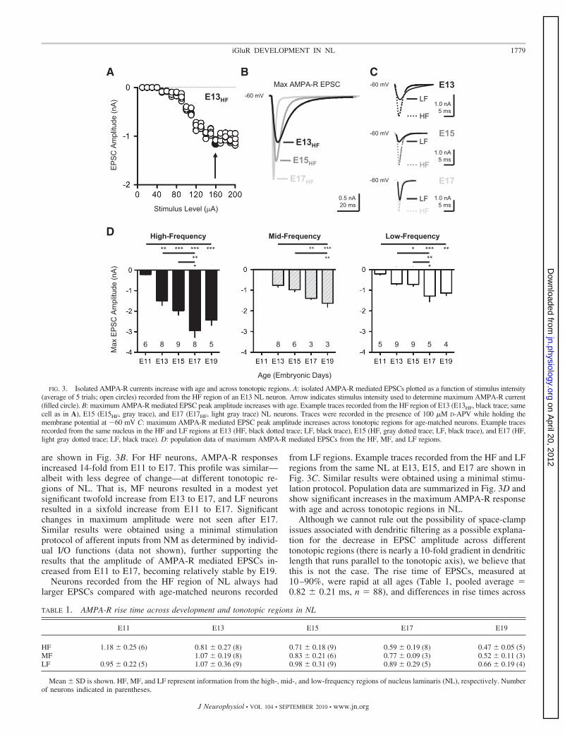
are shown in Fig. 3B. For HF neurons, AMPA-R responsesincreased 14-fold from E11 to E17. This profile was similar—albeit with less degree of change—at different tonotopic re-gions of NL. That is, MF neurons resulted in a modest yetsignificant twofold increase from E13 to E17, and LF neuronsresulted in a sixfold increase from E11 to E17. Significantchanges in maximum amplitude were not seen after E17.Similar results were obtained using a minimal stimulationprotocol of afferent inputs from NM as determined by individ-ual I/O functions (data not shown), further supporting theresults that the amplitude of AMPA-R mediated EPSCs in-creased from E11 to E17, becoming relatively stable by E19.
Neurons recorded from the HF region of NL always hadlarger EPSCs compared with age-matched neurons recorded
from LF regions. Example traces recorded from the HF and LFregions from the same NL at E13, E15, and E17 are shown inFig. 3C. Similar results were obtained using a minimal stimu-lation protocol. Population data are summarized in Fig. 3D andshow significant increases in the maximum AMPA-R responsewith age and across tonotopic regions in NL.
Although we cannot rule out the possibility of space-clampissues associated with dendritic filtering as a possible explana-tion for the decrease in EPSC amplitude across differenttonotopic regions (there is nearly a 10-fold gradient in dendriticlength that runs parallel to the tonotopic axis), we believe thatthis is not the case. The rise time of EPSCs, measured at10–90%, were rapid at all ages (Table 1, pooled average 0.82 � 0.21 ms, n 88), and differences in rise times across
TABLE 1. AMPA-R rise time across development and tonotopic regions in NL
E11 E13 E15 E17 E19
HF 1.18 � 0.25 (6) 0.81 � 0.27 (8) 0.71 � 0.18 (9) 0.59 � 0.19 (8) 0.47 � 0.05 (5)MF 1.07 � 0.19 (8) 0.83 � 0.21 (6) 0.77 � 0.09 (3) 0.52 � 0.11 (3)LF 0.95 � 0.22 (5) 1.07 � 0.36 (9) 0.98 � 0.31 (9) 0.89 � 0.29 (5) 0.66 � 0.19 (4)
Mean � SD is shown. HF, MF, and LF represent information from the high-, mid-, and low-frequency regions of nucleus laminaris (NL), respectively. Numberof neurons indicated in parentheses.
B
E13HF
E15HF
E17HF
Max AMPA-R EPSC
0.5 nA20 ms
1.0 nA5 ms
LF
LF
LF
HF
HF
HF
C
D
Max
EPS
C A
mpl
itude
(nA)
High-Frequency
** *** *** ******
6 8 58 9
Mid-Frequency
Age (Embryonic Days)
**** ***
3 38 6
Low-Frequency
* *** *****
5 5 49 9
A
EPSC
Am
plitu
de (n
A)
Stimulus Level (µA)
E13HF-60 mV
E13
-60 mV
-60 mV
-60 mV
E15
E17
1.0 nA5 ms
1.0 nA5 ms
FIG. 3. Isolated AMPA-R currents increase with age and across tonotopic regions. A: isolated AMPA-R mediated EPSCs plotted as a function of stimulus intensity(average of 5 trials; open circles) recorded from the HF region of an E13 NL neuron. Arrow indicates stimulus intensity used to determine maximum AMPA-R current(filled circle). B: maximum AMPA-R mediated EPSC peak amplitude increases with age. Example traces recorded from the HF region of E13 (E13HF, black trace; samecell as in A), E15 (E15HF, gray trace), and E17 (E17HF, light gray trace) NL neurons. Traces were recorded in the presence of 100 �M D-APV while holding themembrane potential at �60 mV C: maximum AMPA-R mediated EPSC peak amplitude increases across tonotopic regions for age-matched neurons. Example tracesrecorded from the same nucleus in the HF and LF regions at E13 (HF, black dotted trace; LF, black trace), E15 (HF, gray dotted trace; LF, black trace), and E17 (HF,light gray dotted trace; LF, black trace). D: population data of maximum AMPA-R mediated EPSCs from the HF, MF, and LF regions.
1779iGluR DEVELOPMENT IN NL
J Neurophysiol • VOL 104 • SEPTEMBER 2010 • www.jn.org
on April 20, 2012
jn.physiology.orgD
ownloaded from

tonotopic regions were not significant at any age. Moreover,there is no correlation in amplitude and rise time at any age(pooled correlation coefficient 0.24, n 88). In addition, weroutinely monitored the reversal potential of AMPA-R cur-rents. Only neurons with reversals potential near 0 mV wereused in the analysis. Further evidence against dendritic filteringis discussed in upcoming sections. It should also be noted thatwe were unable to reliably record MF neurons from E11 NLslices due to the relatively small nucleus compared with E13and older tissue. Therefore only neurons sampled from therostromedial and caudolateral regions of the nucleus were usedat E11, representing HF and LF neurons, respectively (seeMETHODS).
Increase in the frequency of spontaneous AMPA-R mediatedminiature EPSCs
The increase in amplitude of evoked AMPA-R mediatedEPSCs suggests several possibilities: an increase in the numberof synaptic contacts, an increase in density of synaptic AMPA-Rs, or changes that would affect release probability, i.e., anincrease in presynaptic Ca2� channels. To test some of thesepossibilities, we recorded spontaneous miniature AMPA-Rmediated EPSCs (mEPSCs) in the presence of the Na2�-channel blocker TTX and the NMDA-R antagonist D-APV. Alarge increase in the frequency of mEPSCs is observed throughdevelopment (Fig. 4 and Table 2), suggesting that an increasein synaptic contacts account for the increase in evoked EPSCsobserved (Hsia et al. 1998; Petralia et al. 1999). Example tracesare shown in Fig. 4A from E13 and E17 NL neurons recordedfrom the HF and LF regions. The inter-event interval signifi-cantly decreased with age and across tonotopic regions, indi-cating an increase in the frequency of spontaneous events. Thehighest frequency was observed for E17 neurons from the HFregion (Fig. 4, A and B). As expected for an increase infrequency, the total number of events increased with age andacross different tonotopic regions, with E17 neurons from theHF region having the most events per neuron (Fig. 4C andTable 2). Thus the overall mEPSC frequency increased �10-fold from E13 to E17, a slightly smaller increase than that ofthe maximum amplitude of the evoked EPSC for the same ageperiod.
The mEPSCs 10–90% rise times were quite rapid anddifferences in rise times across age and tonotopic regions werenot significant (Fig. 4D and Table 2, P 0.89). Surprisingly,differences in decay tau across age and tonotopic regions werealso not significant despite a twofold increase in decay tauacross the same developmental time period of evoked EPSCs(Fig. 4D, P 0.57, see next section). There was no correlationbetween mEPSC amplitude and rise time at any age (pooledcorrelation coefficient 0.04), suggesting that dendrites didnot compromise the control of membrane voltages despitedifferences in dendritic length across tonotopic regions of NL.In addition, the amplitude of mEPSCs did not significantlychange across ages (Fig. 4D and Table 2, P 0.92), and thepopulation data showed stable amplitude histograms across ageand tonotopic region (Fig. 4C).
To probe for changes in release probability, we evokedpairs of EPSCs at 100 ms apart. Strong paired pulse synapticdepression was observed as early as E13 (Fig. 4E) andchanges in the paired-pulse ratio (2nd EPSC/1st EPSC) did
not decrease with age (Fig. 4, E and F), suggesting that theprobability of release remains constant throughout this de-velopmental period. In addition, we observed a low coeffi-cient of variation (CV) in the amplitude of evoked EPSCsthat remained constant across development. The data indi-cate that no changes in the probability of release occurduring this development period.
Although we cannot rule out an increase in receptor density,the large increase in mEPSC frequency is sufficient to accountfor the large increase in AMPA-R synaptic transmission. Ourresults suggest that older neurons located in the HF region
B
E13HF
E17HF
E13LF
E17LF
10 pA100 ms
A
D
5 pA1 ms
-60 mV
E17HF
E17LF
E13HF
E13LF
-60 mV
-60 mV
-60 mV
Cum
ulat
ive
Prob
abilit
y
Even
ts (p
er 4
00 s
wee
ps)
mEPSC Amplitude (pA)
mEPSC Inter-Event Interval (ms)
E17HF
E17LF
E13HF
E13LF
E17HF
E17LF
E13HF
E13LF
-60 mV
FE
Paire
d-Pu
lse
Rat
io
Age (Embryonic Days)
-60 mV -60 mV
200 pA50 ms
200 pA50 ms
E17HFE13HF
CIsolated AMPA-R mEPSC
FIG. 4. Frequency of isolated AMPA-R miniature EPSCs (mEPSCs) in-crease with age and across tonotopic regions. A: representative AMPA-Rmediated mEPSCs recorded from E17 and E13 NL neurons located in the HFregion (E17HF, red trace; E13HF, black trace) and LF region (E17LF, pink trace;E13LF, gray trace) of the same NL. Spontaneous mEPSCs were recorded in thepresence of 50 �M TTX and 100 �M D-APV. B: cumulative probability plotshows a significant increase in the inter-event interval of mEPSCs across ageand tonotopic region. C: population data from the HF and LF regions of E17and E13 NL neurons. Histogram shows similar amplitude distribution acrossage and tonotopic region. n 8 neurons for E17HF (red bars; total events 2992); 5 neurons for E17LF (white bars; total events 878), 12 neurons forE13HF (black bars; total events 1452), and 4 neurons for E13LF (gray bars;total events 303). D: average traces from E17HF (red solid line, events 374), E17LF (red dotted line, events 176), E13HF (black solid line, events 121), and E13LF (black dotted line, events 76) show similar AMPA-Rmediated mEPSC amplitudes and kinetics (same neurons in A). E: paired pulserecordings (100 ms interval) from the HF region of E13 (left, black trace) andE17 (right, red trace) NL neurons. F: population data showing paired pulseratios (pulse 2EPSC/pulse 1EPSC) for HF neurons recorded at E13 (black bar)and E17 (red bar).
1780 J. TAIT SANCHEZ, Y. WANG, E. W. RUBEL, AND A. BARRIA
J Neurophysiol • VOL 104 • SEPTEMBER 2010 • www.jn.org
on April 20, 2012
jn.physiology.orgD
ownloaded from
contain more synaptic contacts than younger neurons located inthe LF region of NL despite HF neurons being considerablysmaller than LF neurons.
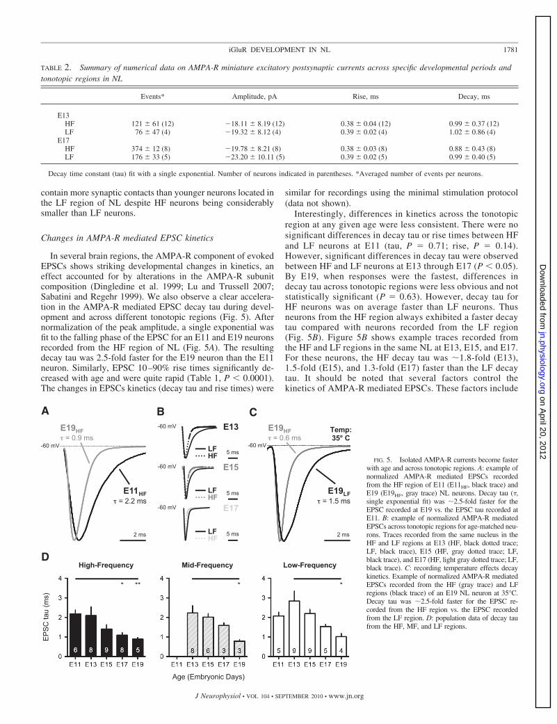
Changes in AMPA-R mediated EPSC kinetics
In several brain regions, the AMPA-R component of evokedEPSCs shows striking developmental changes in kinetics, aneffect accounted for by alterations in the AMPA-R subunitcomposition (Dingledine et al. 1999; Lu and Trussell 2007;Sabatini and Regehr 1999). We also observe a clear accelera-tion in the AMPA-R mediated EPSC decay tau during devel-opment and across different tonotopic regions (Fig. 5). Afternormalization of the peak amplitude, a single exponential wasfit to the falling phase of the EPSC for an E11 and E19 neuronsrecorded from the HF region of NL (Fig. 5A). The resultingdecay tau was 2.5-fold faster for the E19 neuron than the E11neuron. Similarly, EPSC 10–90% rise times significantly de-creased with age and were quite rapid (Table 1, P � 0.0001).The changes in EPSCs kinetics (decay tau and rise times) were
similar for recordings using the minimal stimulation protocol(data not shown).
Interestingly, differences in kinetics across the tonotopicregion at any given age were less consistent. There were nosignificant differences in decay tau or rise times between HFand LF neurons at E11 (tau, P 0.71; rise, P 0.14).However, significant differences in decay tau were observedbetween HF and LF neurons at E13 through E17 (P � 0.05).By E19, when responses were the fastest, differences indecay tau across tonotopic regions were less obvious and notstatistically significant (P 0.63). However, decay tau forHF neurons was on average faster than LF neurons. Thusneurons from the HF region always exhibited a faster decaytau compared with neurons recorded from the LF region(Fig. 5B). Figure 5B shows example traces recorded fromthe HF and LF regions in the same NL at E13, E15, and E17.For these neurons, the HF decay tau was �1.8-fold (E13),1.5-fold (E15), and 1.3-fold (E17) faster than the LF decaytau. It should be noted that several factors control thekinetics of AMPA-R mediated EPSCs. These factors include
TABLE 2. Summary of numerical data on AMPA-R miniature excitatory postsynaptic currents across specific developmental periods andtonotopic regions in NL
Events* Amplitude, pA Rise, ms Decay, ms
E13HF 121 � 61 (12) �18.11 � 8.19 (12) 0.38 � 0.04 (12) 0.99 � 0.37 (12)LF 76 � 47 (4) �19.32 � 8.12 (4) 0.39 � 0.02 (4) 1.02 � 0.86 (4)
E17HF 374 � 12 (8) �19.78 � 8.21 (8) 0.38 � 0.03 (8) 0.88 � 0.43 (8)LF 176 � 33 (5) �23.20 � 10.11 (5) 0.39 � 0.02 (5) 0.99 � 0.40 (5)
Decay time constant (tau) fit with a single exponential. Number of neurons indicated in parentheses. *Averaged number of events per neurons.
High-Frequency Mid-Frequency Low-FrequencyD
EPSC
tau
(ms)
Age (Embryonic Days)
* ** * *
AE19HF = 0.9 ms
2 ms
E11HF = 2.2 ms
-60 mV
B
5 ms
5 ms
5 ms
HF
LF
HFLF
HF
LF
-60 mV
-60 mV
-60 mV
E13
E15
E17
E19HF = 0.6 ms
C
2 ms
E19LF = 1.5 ms
-60 mV
Temp:35 C
FIG. 5. Isolated AMPA-R currents become fasterwith age and across tonotopic regions. A: example ofnormalized AMPA-R mediated EPSCs recordedfrom the HF region of E11 (E11HF, black trace) andE19 (E19HF, gray trace) NL neurons. Decay tau (�,single exponential fit) was �2.5-fold faster for theEPSC recorded at E19 vs. the EPSC tau recorded atE11. B: example of normalized AMPA-R mediatedEPSCs across tonotopic regions for age-matched neu-rons. Traces recorded from the same nucleus in theHF and LF regions at E13 (HF, black dotted trace;LF, black trace), E15 (HF, gray dotted trace; LF,black trace), and E17 (HF, light gray dotted trace; LF,black trace). C: recording temperature effects decaykinetics. Example of normalized AMPA-R mediatedEPSCs recorded from the HF (gray trace) and LFregions (black trace) of an E19 NL neuron at 35°C.Decay tau was �2.5-fold faster for the EPSC re-corded from the HF region vs. the EPSC recordedfrom the LF region. D: population data of decay taufrom the HF, MF, and LF regions.
1781iGluR DEVELOPMENT IN NL
J Neurophysiol • VOL 104 • SEPTEMBER 2010 • www.jn.org
on April 20, 2012
jn.physiology.orgD
ownloaded from

dendritic filtering, glutamate clearance, and receptor subunitcomposition. The lack of differences in the kinetics ofspontaneous mEPSCs across age and tonotopic region (seeFig. 4D) suggests that factors like glutamate clearance(Diamond and Jahr 1997) and dendritic filtering (Magee andCook 2000; Williams and Mitchell 2008) play a major roleonly in evoked responses. Previous reports show cleardifferences in the kinetics of AMPA-R mediated currentsbetween HF and LF neurons (Kuba et al. 2005; Slee et al.2010). This is likely the result of more physiological tem-peratures used in the aforementioned studies. We minimizedthe influence of glutamate dynamics in the synaptic cleft anddendritic filtering by recording EPSCs at room temperature(�21–22°C) to study subunit composition of iGluRs basedon their electrophysiological properties. Control experi-ments at 35°C indeed show larger differences in kineticsacross tonotopic regions at older ages (E19; Fig. 5C).Despite the small changes we observe in decay tau, EPSCrise times across tonotopic regions between E11 and E19were stable and not significantly different (Table 1). Popu-lation data are summarized in Fig. 5D showing changes inthe evoked AMPA-R mediated EPSC decay tau across ageand tonotopic regions in NL. The increase in speed ofAMPA-R mediated responses suggests that subunit compo-
sition is changing. We next investigated whether the subunitcomposition of AMPA-Rs changes across age and tonotopicregions in NL.
Increases in AMPA-R mediated inward rectification
AMPA-Rs containing the GluR2 subunit exhibit slowerEPSC kinetics than AMPA-Rs lacking the GluR2 subunit(Lawrence and Trussell 2000; Raman and Trussell 1992;Raman et al. 1994). Also GluR2 lacking AMPA-Rs arepermeable to Ca2� and other divalent ions (Hollmann et al.1991) and are inwardly rectifying (Williams 1997) due tovoltage-dependent blockade by intracellular polyamines(Donevan and Rogawski 1995). We recorded current-volt-age (I-V) relationships mediated by AMPA-Rs across ageand tonotopic regions to test the hypothesis that the inwardrectification and the rapid kinetics of AMPA-R mediatedEPSCs observed in older animals are due to a developmentalswitch in GluR2 content.
We constructed I-V curves with 100 �M spermine addedto the internal patch pipette solution. Spermine is a poly-amine that blocks the ion channel of GluR2-lackingAMPA-Rs in a voltage dependent manner (Bowie andMayer 1995). Insets in Fig. 6, A and B, show recordings of
AE13HF
I/Im
ax
E17HF
C
D
Rec
tific
atio
n In
dex
VHold (mV)
200 pA10 ms
100 pA10 ms
*** ***** ***
High-Frequency
Age (Embryonic Days)
Mid-Frequency
* ***
Low-Frequency
BE13HF
E15HF
E17HF
FIG. 6. Increase in rectification of AMPA-R currents with age and across tonotopic regions. A and B: I-V functions recorded from the HF region of E13 (A)and E17 (B) NL neurons. Insets: isolated AMPA-R mediated EPSCs recorded at different holding voltages (see METHODS) with 100 �M spermine added to pipetteinternal solution (note different scale bars for insets). C: population I-V functions constructed from NL neurons recorded in the HF region at E13 (HF, black line),E15 (HF, gray line), and E17 (HF, light gray line). I-V plots show a near linear function at E13, changing to an inwardly rectifying I-V function at E17.D: population data of rectification index (IAMPA-R �40 mV/IAMPA-R �60 mV) from the HF, MF, and LF regions.
1782 J. TAIT SANCHEZ, Y. WANG, E. W. RUBEL, AND A. BARRIA
J Neurophysiol • VOL 104 • SEPTEMBER 2010 • www.jn.org
on April 20, 2012
jn.physiology.orgD
ownloaded from
isolated AMPA-R mediated EPSCs from the HF region ofE13 and E17 NL neurons at various holding potentials, fromwhich we constructed normalized I-V curves (Fig. 6, A andB). At E13, the I-V relationship was nearly linear, but atE17, there was a strong reduction in the outward flow ofcurrent mediated by isolated AMPA-Rs, consistent withinward rectification. Population I-V functions recorded fromthe HF region of E13, E15, and E17 NL neurons are overlaidin Fig. 6C and show a near linear function at E13, changingto an inwardly rectifying I/V relationship at E17. The extentof rectification is summarized in Fig. 6D by using a recti-fication index (RI IAMPA-R �40 mV/IAMPA-R �60 mV) asan indication of AMPA-Rs lacking GluR2 subunit (Bowieand Mayer 1995; Donevan and Rogawski 1995). Populationdata show a significant increase in AMPA-R mediated rec-tification with increasing age at every tonotopic region (Fig.6D). The rectification index for HF neurons at E11 was 0.62� 0.19 and was 0.11 � 0.07 at E19 (P � 0.0001). Thisprofile was similar for MF and LF neurons (P � 0.01 and P� 0.002, respectively). These results suggest a dramaticdecrease in GluR2-containing AMPA-Rs between E11 andE19. This developmental change in AMPA-R subunit com-position could also contribute to the changes in kinetics andamplitude of AMPA-R mediated EPSCs observed duringthis period of development. However, the lack of consistentdifferences across tonotopic regions at a given age suggests
that other mechanisms could contribute to the differencesobserved in AMPA-R mediated EPSCs kinetics as well.
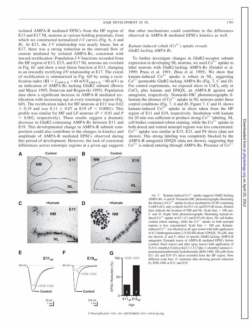
Kainate-induced cobalt (Co2�) uptake revealsGluR2-lacking AMPA-Rs
To further investigate changes in GluR2-receptor subunitexpression in developing NL neurons, we used Co2� uptake tolabel neurons with GluR2-lacking AMPA-Rs (Estabel et al.1999; Pruss et al. 1991; Zhou et al. 1995). We show thatkainate-induced Co2� uptake is robust in NL, suggestingCa2�-permeable GluR2-lacking AMPA-Rs (Fig. 7, C and D).For control experiments, we exposed slices to CoCl2 only orCoCl2 plus kainate and DNQX, an AMPA-R agonist andantagonist, respectively. Nomarski-DIC photomicrographs il-lustrate the absence of Co2� uptake in NL neurons under thesecontrol conditions (Fig. 7, A and B). Figure 7, C and D, showskainate-induced Co2� uptake in slices taken from the HFregion of E11 and E19, respectively. Incubation with kainatefor 20 min was sufficient to produce strong Co2� labeling. NLcell bodies contained robust staining, while the Co2� uptake inboth dorsal and ventral neuropil regions was less concentrated.Co2� uptake was similar at E15, E21, and P4 slices (data notshown). This strong labeling was completely blocked by theAMPA-R antagonist DNQX (data not shown), suggesting thatCo2� is indeed entering through AMPA-Rs. Presence of Co2�
50 pA1 ms
250 pA1 ms
E FE19HFE11HF
Age
+IEM-1460
EPSC
Red
uctio
n (%
)
G
E11NM
NL
BA
DCE19
NM
NL
Control
+Kainate
+IEM-1460Control
+IEM-1460
Control
-60 mV-60 mV
NL
E19
Control
NM
E11NM
NL
+Kainate
FIG. 7. Kainate-induced Co2� uptake suggests GluR2-lackingAMPA-Rs. A and B: Nomarski-DIC photomicrographs illustratingthe absence of Co2� uptake in slices incubated in ACSF containing5 mM CoCl2 only (control) for E11 (A) and E19 (B) tissue. Dashedlines indicate the location of NM and NL. Scale bars 100 �m.C and D: bright field photomicrographs illustrating kainate-in-duced Co2� uptake in E11 (C) and E19 (D) slices. NL cell bodiescontain robust staining, while the Co2� uptake in both neuropilregions is less concentrated. Scale bars 100 �m. Kainate-induced Co2� was blocked at all ages tested with bath applicationof 6,7-dinitroquinoxaline-2,3(1H,4H)-dione (DNQX, 50 �M, datanot shown). E and F: effect of specific GluR2-lacking AMPA-Rantagonist. Example traces of AMPA-R mediated EPSCs before(control, black traces) and after (gray traces) bath application ofN,N,N,-rimethyl-5-[(tricyclo[3.3.1.13,7]dec-1-ylmethyl )amino]-1-pentanaminiumbromide hydrobromide (IEM-1460, 100 �M) fromE11 (E) and E19 (F) slices recorded from the HF region. Notedifferent scale bars. G: summary data showing percent reductionby IEM-1460 at E11 and E19.
1783iGluR DEVELOPMENT IN NL
J Neurophysiol • VOL 104 • SEPTEMBER 2010 • www.jn.org
on April 20, 2012
jn.physiology.orgD
ownloaded from

uptake throughout NL suggests that neurons express Ca2�-permeable GluR2-lacking AMPA-Rs early and late in devel-opment and across different tonotopic regions of NL. A gra-dient in Co2� uptake across different tonotopic regions was notconsistently observed, which may be due to inadequate sensi-tivity of the method (see next section).
The strong Co2� uptake at E11 was somewhat surprisingbased on the electrophysiology data. The EPSC decay tau wasrelatively slow for E11 neurons recorded from the HF regions(tau 2.17 � 0.47, n 6, see Fig. 5), and the rectificationindex showed near linearity (RI 0.62 � 0.19, n 6, see Fig.6), both of which are considered reliable biophysical markersthat indicate GluR2-containing AMPA-Rs. However, it shouldbe noted that exposure to CoCl2 and kainate for time periods�10 min produced results with highly variable staining pat-terns, whereas treatments �15 min always resulted in robuststaining consistent with a saturation effect. An interpretation ofthese data, consistent with other developmental research (Pel-legrini-Giampietro et al. 1997), is that NL neurons contain bothGluR2-containing and -lacking AMPA-Rs early in develop-ment and exclusively or primarily GluR2-lacking AMPA-Rsby E17-19 and that differential expression of each type isindistinguishable with the Co2�-uptake technique.
To better resolve this issue, we bath applied a dicationicadamantine derivative (IEM-1460, 100 �M), a highly specificantagonist to the GluR2-lacking AMPA-R, to determine therelative contribution of this Ca2� permeant AMPA-R to theoverall current response when NL neurons were voltage-clamped at �60 mV. Representative traces recorded from theHF region of E11 and E19 NL neurons are shown in Fig. 7, Eand F, respectively. For the E11 neuron, the control EPSC usingthe maximum stimulus protocol was �350 pA but was reduced to�275 pA (21% reduction) when IEM-1460 was bath applied for15 min (Fig. 7E). On average, the EPSC decay tau was slightlyfaster in the control responses than after treatment with the drug(controltau 2.14 � 0.41 ms; drugtau 2.37 � 0.57 ms). Thesedata indicate a small contribution of GluR2 lacking AMPA-Rs to
the total AMPA-R mediated EPSC at E11. Thus the kainateinduced Co2� uptake observed at E11 is likely due to the contri-bution of a small percentage of GluR2 lacking AMPA-Rs. Incontrast, at E19, the control maximum EPSC was �1,700 pA butwas reduced considerably to �320 pA (81% reduction) when theGluR2-lacking AMPA-Rs were blocked (Fig. 7F). On average,the EPSC decay time constant of the control responses wasconsiderably faster than after application of the drug (controltau 0.88 � 0.05; drugtau 1.07 � 0.16). These data indicate that themajority of the current at E19 is mediated by AMPA-Rs lackingthe GluR2 subunit. Population data showing the percent of EPSCreduction following blockade of GluR2-lacking AMPA-Rs aresummarized in Fig. 7G.
Changes in the amplitude of evoked NMDA-Rmediated EPSCs
Pharmacologically isolated NMDA-R mediated EPSCs wererecorded while holding the membrane potential at �40 mV. Amaximal stimulation protocol was used to examine changes inthe amplitude of evoked EPSCs with development and acrossdifferent tonotopic regions. I/O functions were determined foreach NL neurons in an attempt to recruit most, if not all,excitatory inputs from the ipsilateral NM. The stimulus inten-sity used to evoke maximum NMDA-R currents was deter-mined from individual I/O functions and an example is shownin Fig. 8A. NMDA-R currents increased with increases instimulus intensity with few failures at minimal stimulationconsistent with a high probability of release. The stimulusintensity used to determine a maximum response was chosenwhen the evoked EPSC reached a plateau response (arrow inFig. 8A). This stimulus intensity never exceeded the output ofthe stimulus generator.
NL neurons showed dramatic changes with age in theamplitude of maximally evoked NMDA-R mediated EPSCs.Representative traces from the HF region of E13, E15, E17,and E19 NL neurons are shown in Fig. 8B. For HF neurons,
A
EPSC
Am
plitu
de (n
A)
B
E13HF
E15HF
E17HF
+40 mV
0.1 nA40 ms
E15HF
C
Max
EPS
C A
mpl
itude
(nA)
Age (Embryonic Days)
** ******
*****
**
Stimulus Level (µA)
* **
**
E19HF
*
Max NMDA-R EPSC
High-Frequency Mid-Frequency Low-Frequency
FIG. 8. Changes in isolated NMDA-R currents during develop-ment and across tonotopic regions. A: isolated NMDA-R mediatedEPSCs plotted as a function of stimulus intensity (average of 5 trials;open circles) recorded from the HF region of an E15 NL neuron(same cell in B). Arrow indicates stimulus intensity used to deter-mine maximum NMDA-R current (filled circle). B: maximumNMDA-R mediated EPSC peak amplitudes changes with age. Rep-resentative recordings from the HF regions of E13 (black trace) E15(dark gray trace), E17 (gray trace), and E19 (light gray trace) NLneurons. Responses were recorded in the presence of 20 �M NBQXwhile holding the membrane potential at �40 mV. Dash linerepresents the baseline. C: population data of maximum NMDA-Rmediated EPSCs from the HF, MF, and LF regions.
1784 J. TAIT SANCHEZ, Y. WANG, E. W. RUBEL, AND A. BARRIA
J Neurophysiol • VOL 104 • SEPTEMBER 2010 • www.jn.org
on April 20, 2012
jn.physiology.orgD
ownloaded from
NMDA-R responses increased approximately sevenfold fromE11 to E17, followed by a sharp and significant decline inamplitude at E19 (Fig. 8, B and C) to values similar to thoseobserved at E11 (Ell trace not shown in B for clarity). In fact,the average NMDA-R currents recorded at E11 and E19 werenot significantly different from one another (P 0.72). Thisprofile was similar in each tonotopic region of NL. MF neuronsshowed a modest, yet significant 2.5-fold increase from E13 toE17 (Fig. 8C, middle) and LF neurons resulted in a ninefoldincrease from E11 to E17 (C, right). These results were alsoobserved using a minimal stimulation protocol of afferentinputs from NM as determined by individual I/O functions(data not shown). No significant differences in the maximumamplitude were observed across different tonotopic regions atany particular age regardless of the difference in dendriticelaboration, supporting the idea that space clamp issues acrossdifferent tonotopic regions did not affect the amplitude ofEPSCs.
Changes in NMDA-R mediated EPSC kinetics
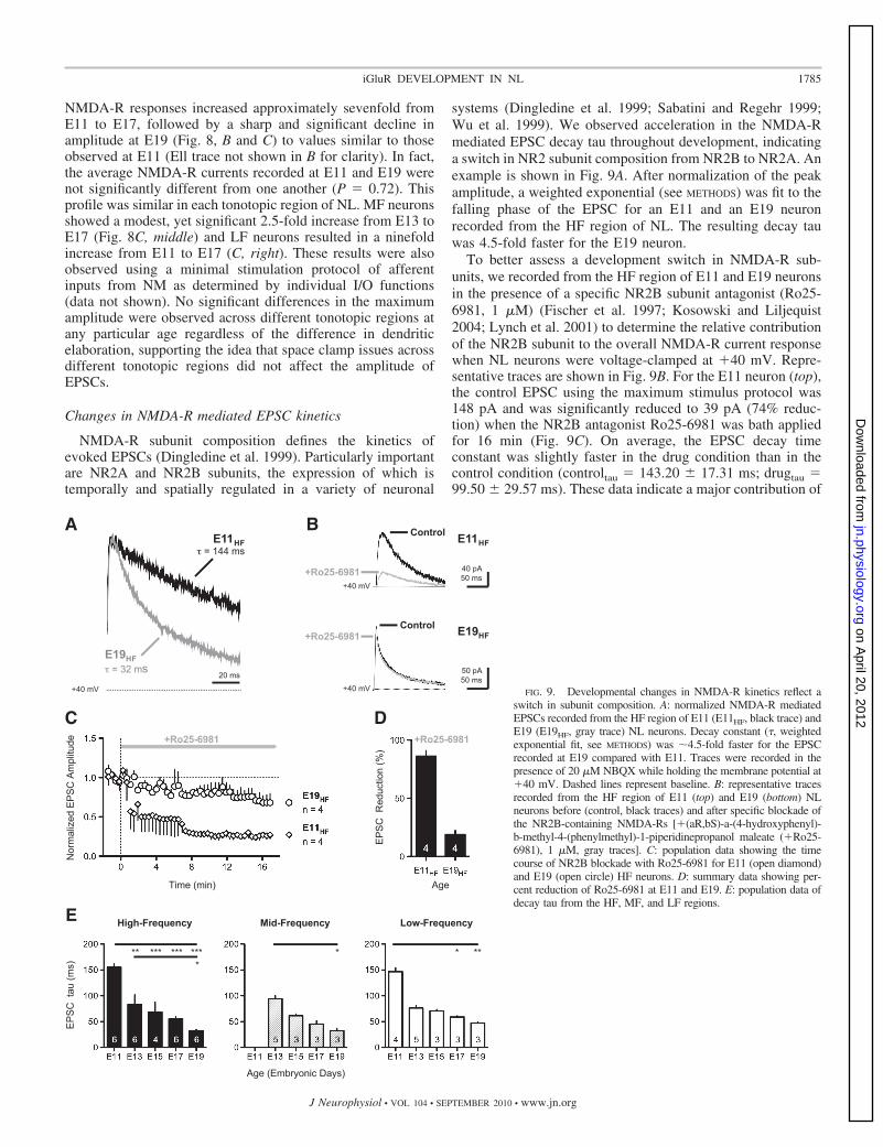
NMDA-R subunit composition defines the kinetics ofevoked EPSCs (Dingledine et al. 1999). Particularly importantare NR2A and NR2B subunits, the expression of which istemporally and spatially regulated in a variety of neuronal
systems (Dingledine et al. 1999; Sabatini and Regehr 1999;Wu et al. 1999). We observed acceleration in the NMDA-Rmediated EPSC decay tau throughout development, indicatinga switch in NR2 subunit composition from NR2B to NR2A. Anexample is shown in Fig. 9A. After normalization of the peakamplitude, a weighted exponential (see METHODS) was fit to thefalling phase of the EPSC for an E11 and an E19 neuronrecorded from the HF region of NL. The resulting decay tauwas 4.5-fold faster for the E19 neuron.
To better assess a development switch in NMDA-R sub-units, we recorded from the HF region of E11 and E19 neuronsin the presence of a specific NR2B subunit antagonist (Ro25-6981, 1 �M) (Fischer et al. 1997; Kosowski and Liljequist2004; Lynch et al. 2001) to determine the relative contributionof the NR2B subunit to the overall NMDA-R current responsewhen NL neurons were voltage-clamped at �40 mV. Repre-sentative traces are shown in Fig. 9B. For the E11 neuron (top),the control EPSC using the maximum stimulus protocol was148 pA and was significantly reduced to 39 pA (74% reduc-tion) when the NR2B antagonist Ro25-6981 was bath appliedfor 16 min (Fig. 9C). On average, the EPSC decay timeconstant was slightly faster in the drug condition than in thecontrol condition (controltau 143.20 � 17.31 ms; drugtau 99.50 � 29.57 ms). These data indicate a major contribution of
E
EPSC
tau
(ms)
Age (Embryonic Days)
High-Frequency Low-FrequencyMid-Frequency
*** ***** ****
* * **
A
+40 mV
20 ms
E11HF = 144 ms
E19HF = 32 ms
B
50 pA50 ms
40 pA50 ms
E11HF
E19HF
Control
Control
+Ro25-6981
+Ro25-6981
C D
AgeTime (min)
Nor
mal
ized
EPS
C A
mpl
itude
EPSC
Red
uctio
n (%
)
+Ro25-6981+Ro25-6981
+40 mV
+40 mV FIG. 9. Developmental changes in NMDA-R kinetics reflect aswitch in subunit composition. A: normalized NMDA-R mediatedEPSCs recorded from the HF region of E11 (E11HF, black trace) andE19 (E19HF, gray trace) NL neurons. Decay constant (�, weightedexponential fit, see METHODS) was �4.5-fold faster for the EPSCrecorded at E19 compared with E11. Traces were recorded in thepresence of 20 �M NBQX while holding the membrane potential at�40 mV. Dashed lines represent baseline. B: representative tracesrecorded from the HF region of E11 (top) and E19 (bottom) NLneurons before (control, black traces) and after specific blockade ofthe NR2B-containing NMDA-Rs [�(aR,bS)-a-(4-hydroxyphenyl)-b-methyl-4-(phenylmethyl)-1-piperidinepropanol maleate (�Ro25-6981), 1 �M, gray traces]. C: population data showing the timecourse of NR2B blockade with Ro25-6981 for E11 (open diamond)and E19 (open circle) HF neurons. D: summary data showing per-cent reduction of Ro25-6981 at E11 and E19. E: population data ofdecay tau from the HF, MF, and LF regions.
1785iGluR DEVELOPMENT IN NL
J Neurophysiol • VOL 104 • SEPTEMBER 2010 • www.jn.org
on April 20, 2012
jn.physiology.orgD
ownloaded from

the NR2B subunit to the NMDA-R mediated EPSC at E11. Incontrast, at E19, the control maximum EPSC was 145 pA butwas minimally reduced to 118 pA (�19% reduction) whenNR2B-containing NMDA-Rs were blocked for 16 min (Fig. 9,B, bottom, and C). On average, the EPSC decay tau of thecontrol responses was not different after application of the drug(controltau 39.17 � 6.39; drugtau 34.16 � 4.79), indicat-ing that the majority of the current at E19 is mediated byNMDA-Rs lacking the NR2B subunit. Population data show-ing the percent of EPSC reduction following blockade ofNR2B-containing NMDA-Rs are summarized in Fig. 9D.
This acceleration of NMDA-R responses was similar andsignificant across age for all tonotopic regions, with HF neu-rons resulting in a 4.7-fold increase (Fig. 9E, left), MF neuronsincreasing threefold from E13 to E19 (Fig. 9E, middle), and LFneurons increasing 3.1-fold from E11 to E19 (Fig. 9E, right).The acceleration in the kinetics of NMDA-R dependent EPSCswas similar for recordings using the minimal stimulation pro-tocol (data not shown). In contrast, differences in kineticsacross tonotopic region for age-matched neurons were notapparent, suggesting that unlike AMPA-R development,NMDA-Rs are not differentially regulated tonotopically. Theseresults further argue against space-clamp issues associated withdendritic filtering. Population data summarized in Fig. 9E,along with specific blockade of the NR2B subunit summarizedin B–D, strongly suggest a developmental switch in NMDA-Rsubunit composition from NR2B- to NR2A-containing recep-tors in NL neurons during this period of development.
D I S C U S S I O N
In several brain regions, synaptic expression of AMPA-Rsand NMDA-Rs change throughout development, fine-tuningsynaptic properties, and facilitating proper dendritic and neu-ronal circuit maturation (Cline and Haas 2008; Haas et al.2006). Synaptic maturation increases the AMPA/NMDA ratio(Hall and Ghosh 2008; Kasanetz and Manzoni 2009; Rumpel etal. 2004) and changes the subunit composition of NMDA-Rs,altering integration of presynaptic inputs, Ca2� influx into thepostsynaptic neuron, and the ability of the synapse to undergoplasticity (Barria and Malinow 2005; Carmignoto and Vicini1992; Crair and Malenka 1995; Hestrin 1992; Lu et al. 2001;Philpot et al. 2001).
In NL, the first neuronal circuit that integrates binaural infor-mation from both ears in the avian brain stem, AMPA-R currentsare critical in shaping extremely rapid EPSPs, a physiologicnecessity for binaural auditory processing (Trussell 1997). Therole of NMDA-Rs is less clear. It has been suggested thatresponses mediated by NMDA-Rs disappear shortly after synap-togenesis, contributing to the sharpening of EPSPs (Gao and Lu2008; Kuba et al. 2002a). However, robust expression levels ofNMDA-Rs are evident into adulthood (Tang and Carr 2004,2007).
Our results indicate that NL synapses undergo majorpostsynaptic changes in the strength and subunit compositionof AMPA-Rs and NMDA-Rs prior to hatching. These dramaticchanges correlate to a developmental time period when thecharacteristic morphology of NL neurons and circuitry arebeing established, the tonotopic gradient is being defined, andhearing is emerging. These changes seem to occur initially inthe HF region of NL, a pattern that mimics receptor develop-
ment and other morphological properties in NM and NL,suggesting that the development of glutamatergic synaptictransmission is regulated by the activity of their sensory inputs.These developmental changes increase the amplitude andspeed of glutamatergic synaptic transmission, likely enhancingcoincidence detection of binaural auditory information.
Development of synaptic AMPA-Rs in NL
We observe significant age and tonotopic dependence in theamplitude and kinetics of EPSCs mediated by AMPA-Rs inNL. These results reflect increases in synaptic contacts andchanges in receptor subunit composition. Similar changes as afunction of age have been reported in different relay synapsessuch as the calyx of Held synapse in the mouse (Joshi andWang 2002), the endbulb of Held synapse in the rat (Belling-ham et al. 1998), and the endbulb synapse of the chick NM (Luand Trussell 2007).
The increase in AMPA-R responses can be attributed toeither an upregulation of presynaptic voltage-gated Ca2� chan-nels resulting in an increase in release probability (Brenowitzand Trussell 2001; Kasanetz and Manzoni 2009), an increase inthe number of postsynaptic AMPA-Rs (Elias et al. 2006, 2008;Turrigiano 2008), or an increase in the number of synapticcontacts innervating the postsynaptic neuron (Brewer et al.2009; Hackett et al. 1982). We observe no changes throughoutdevelopment in the amplitude of mEPSCs and no changes inthe probability of release. In addition, the stepwise increase ofAMPA-R mediated responses observed in the input/outputcurves and the lack of differences in the rise time of EPSCsacross different ages suggest no differences in the synchroni-zation of transmitter release during development. Thus it isunlikely that presynaptic changes account for the large increasein AMPA-R mediated transmission observed. The increase inmEPSCs frequency suggests that the increase in the amplitudeof AMPA-R responses is due to an increase in the number ofsynaptic contacts (Hsia et al. 1998; Petralia et al. 1999),although it does not rule out changes in receptor density.Interestingly, the increase in AMPA-R responses is first appar-ent in the HF region, conforming to the gradient in synapticdevelopment first reported by Jackson et al. (1982) and similarto NM development (Lu and Trussell 2007). Neurons locatedin the HF region of NL had larger amplitude EPSCs comparedwith the MF and LF regions, suggesting that afferent inputsfrom NM make more synaptic contacts with HF neuronsdespite considerable size differences in dendritic arborizationcompared with LF neurons (Smith and Rubel 1979) (see alsoFig. 1). We cannot rule out the contribution of differentialfiltering due to dendritic length on the peak amplitude ofAMPA-R mediated EPSCs; however, we did not observetonotopic changes in the peak amplitude of NMDA-R mediatedresponses (discussed in the next section). Thus our resultsindicate that the increase in AMPA-R mediated responses isdue primarily to increases in synaptic contacts. We also reportthat AMPA-Rs in NL undergo a switch in subunit composition,from a predominantly small conductance channel at E11 (i.e.,containing the GluR2 receptor subunit), to a large conductancechannel at E19 (i.e., lacking the GluR2 receptor subunit),which may further contribute to an increase in the amplitude ofAMPA-R currents (Conrad et al. 2008; Cull-Candy et al. 2006;Dingledine et al. 1999).
1786 J. TAIT SANCHEZ, Y. WANG, E. W. RUBEL, AND A. BARRIA
J Neurophysiol • VOL 104 • SEPTEMBER 2010 • www.jn.org
on April 20, 2012
jn.physiology.orgD
ownloaded from

Inward rectification, Co2� uptake, and pharmacologicalblockade indicate a small proportion of GluR2-lackingAMPA-Rs at E11 that increases dramatically with age. How-ever, unlike increases in peak amplitude of AMPA-R mediatedEPSC, inward rectification did not appear to emerge differen-tially across the tonotopic gradient. It should also be noted thatour finding of late tissue (�E19) Co2� uptake is in contrast tothose reported by Zhou et al. (1995). We suggest that thefailure to observe Co2� uptake in their study of late embryoswas due to the lack of adequate tissue penetration in the intactbrain stem preparation.
We also observe acceleration in the decay kinetics ofAMPA-R responses with age. Channel gating, desensitization,subunit composition, dynamics of glutamate in the synapticcleft, and passive properties of dendrites can influence thedecay kinetics of AMPA-R mediated EPSCs (Parks 2000;Trussell 1998). We recorded EPSCs at room temperature tominimize dendritic filtering and the influence of glutamatedynamics in the synaptic cleft, two factors highly dependent ontemperature (Tzingounis and Wadiche 2007). Our data suggestthat the subunit composition changes in a direction that in-creases the kinetics of AMPA-R responses (i.e., from predom-inately GluR2 containing to GluR2-lacking receptors). There-fore it is reasonable to expect that the stabilization of responsekinetics we observe occur because the change in AMPA-Rsubunit composition is near complete at older ages. Our data atE19 show minimal differences in response kinetics betweenHF and LF neurons, suggesting maximal replacement ofAMPA-R subunits across the tonotopic gradient prior to hatch-ing. Consequently, if the composition of receptors becomeshomogenous across tonotopic regions, factors like glutamateclearance and dendritic filtering become dominant in definingthe kinetics of EPSCs. It is clear from previous studies in oldertissue and at more physiological temperatures, that dendriticfiltering plays a major role in defining tonotopic differences inkinetics of AMPA-R mediated EPSCs (Kuba et al. 2005; Sleeet al. 2010).
NL neurons have both intrinsic and synaptic properties thatare extremely brief to accommodate rapid transmission (Parks2000; Trussell 1997, 1999). We propose that a developmentalincrease in GluR2-lacking AMPA-Rs helps sharpen the EPSPtime course, thus minimizing distortion, a requirement forbinaural auditory processing (Otis et al. 1995; Raman et al.1994). Additionally, an increase in Ca2� permeability, due toGluR2-lacking AMPA-R receptors, could be necessary fordendritic remodeling and expression of synaptically regulatedextrusion mechanisms (Wang and Rubel 2008). However,Ca2� -permeable AMPA-Rs could be a consequence of theauditory system requiring fast AMPA-Rs (Parks 2000; Trussell1999), and the reported Ca2� influx through these receptors isrelatively low (Otis et al. 1995). Further work needs to beperformed to elucidate the functional role of increased Ca2�
permeability due to GluR2-lacking AMPA-Rs in the auditorysystem.
Development of synaptic NMDA-Rs in NL
As soon as synapses are established in NL (�E10), currentresponses are mediated by both AMPA-Rs and NMDA-Rswith a ratio close to one. Due to the large increase in AMPA-Rtransmission, it is easy to underestimate the contribution of
NMDA-Rs in later stages of development (Funabiki et al.1998; Zhou and Parks 1991). Similarly to AMPA-Rs, weobserve an initial increase in synaptic NMDA-R responses,which peak at E17, followed by a dramatic and significantdecrease by E19 to values similar to E11. Subunit compositionof NMDA-Rs alters the kinetics of NMDA-R mediated EPSCs(Dingledine et al. 1999). We observe dramatic changes inisolated NMDA-R decay kinetics from �150 ms decay timeconstant at E11 to �30 ms at E19. These results, along withour specific blockade of NR2B-containing receptors, indicate aswitch in subunit composition from NR2B-containing recep-tors early in synaptic development to NR2A-containing recep-tors later in development, as shown for other developingcentral synapses (Carmignoto and Vicini 1992; Flint et al.1997; Tovar and Westbrook 1999). This developmental switchin subunit composition accompanies significant changes inchannel kinetics, which plays key roles in regulating Ca2�
influx, organizing functional circuits, regulating dendriticgrowth, and refining topographic projections (Cline 2001;Ruthazer and Cline 2004; Sin et al. 2002).
This pattern of development for NMDA-Rs is different fromNM neurons where no developmental change in NMDA-Rsubunit composition is observed and synaptic NMDA-R re-sponses disappear by E19 (Lu and Trussell 2007). This differ-ence in the development of NMDA-Rs highlights the diverseroles of each synapse in the transmission and processing ofauditory information. It also suggests that NMDA-Rs couldplay a role in the precise timing and processing of binauralinformation by contributing to firing probability, responselatency, and spike jitter (Pliss et al. 2009; Sanchez et al. 2007).In addition, NMDA-Rs could be critical for synaptic stability,dendritic maintenance, and circuit refinement (Gillespie et al.2005).
Functional role of NMDA-Rs in NL
Little is known about the functional role of NMDA-Rs inNL. Both light and electron microscopy studies in NL haveshown the presence of NMDA-R subunits during developmentand into adulthood in a pattern of expression consistent withour functional studies (Tang and Carr 2004, 2007). In view ofthe critical role of NMDA-Rs in development, and plasticity ofmany immature synapses (Rajan and Cline 1998; Rajan et al.1999), it is tempting to speculate that the transient but dramaticincrease of NMDA-R function during synaptic formation inNL may provide the appropriate Ca2� signaling for incorpo-ration, stabilization, and subunit re-arrangement of AMPA-Rsin the postsynaptic density and/or other specializations such asthe emergence of channel specializations important for binau-ral processing and the dendritic gradient. Downregulation ofsynaptic NMDA-Rs could stabilize the system as well asprevent Ca2� overloading due to high rates of spontaneous andacoustically driven activity (Born et al. 1991).
The current study describes dramatic changes in glutama-tergic synaptic transmission in NL during embryonic develop-ment and provides a unique platform to evaluate relationshipsbetween the developmental expression of synaptic receptorsand other structure/function specializations of a well-definedneuronal circuit.
1787iGluR DEVELOPMENT IN NL
J Neurophysiol • VOL 104 • SEPTEMBER 2010 • www.jn.org
on April 20, 2012
jn.physiology.orgD
ownloaded from
A C K N O W L E D G M E N T S
We thank current members of the Rubel and Barria labs for helpfuldiscussion of the data and manuscript. We also thank the critiques of threereviewers for improving the manuscript.
G R A N T S
This work was supported by National Institute on Deafness and OtherCommunications Disorders Research Grant R01 DC-003829 and NationalResearch Service Award F32 DC-010307.
D I S C L O S U R E S
No conflicts of interest, financial or otherwise, are declared by the author(s).
R E F E R E N C E S
Barria A, Malinow R. NMDA receptor subunit composition controls synapticplasticity by regulating binding to CaMKII. Neuron 48: 289–301, 2005.
Bellingham MC, Lim R, Walmsley B. Developmental changes in EPSCquantal size and quantal content at a central glutamatergic synapse in rat. JPhysiol 511: 861–869, 1998.
Born DE, Durham D, Rubel EW. Afferent influences on brainstem auditorynuclei of the chick: nucleus magnocellularis neuronal activity followingcochlea removal. Brain Res 557: 37–47, 1991.
Bowie D, Mayer ML. Inward rectification of both AMPA and kainate subtypeglutamate receptors generated by polyamine-mediated ion channel block.Neuron 15: 453–462, 1995.
Brenowitz S, Trussell LO. Minimizing synaptic depression by control ofrelease probability. J Neurosci 21: 1857–1867, 2001.
Brewer GJ, Boehler MD, Pearson RA, DeMaris AA, Ide AN, Wheeler BC.Neuron network activity scales exponentially with synapse density. J NeuralEng 6: 1–12, 2009.
Carmignoto G, Vicini S. Activity-dependent decrease in NMDA receptorresponses during development of the visual cortex. Science 258: 1007–1011,1992.
Cline HT. Dendritic arbor development and synaptogenesis. Curr OpinNeurobiol 11: 118–126, 2001.
Cline H, Haas K. The regulation of dendritic arbor development and plasticityby glutamatergic synaptic input: a review of the synaptotrophic hypothesis.J Physiol 586: 1509–1517, 2008.
Conrad KL, Tseng KY, Uejima JL, Reimers JM, Heng LJ, Shaham Y,Marinelli M, Wolf ME. Formation of accumbens GluR2-lacking AMPAreceptors mediates incubation of cocaine craving. Nature 454: 118–121,2008.
Crair MC, Malenka RC. A critical period for long-term potentiation atthalamocortical synapses. Nature 375: 325–328, 1995.
Cull-Candy S, Kelly L, Farrant M. Regulation of Ca2�-permeable AMPAreceptors: synaptic plasticity and beyond. Curr Opin Neurobiol 16: 288–297, 2006.
Diamond JS, Jahr CE. Transporters buffer synaptically released glutamate ona submillisecond time scale. J Neurosci 17: 4672–4687, 1997.
Diaz C, Martinez-Galan JR, Juiz JM. Development of glutamate receptorsin auditory neurons from long-term organotypic cultures of the embryonicchick hindbrain. Eur J Neurosci 29: 213–230, 2009.
Dingledine R, Borges K, Bowie D, Traynelis SF. The glutamate receptor ionchannels. Pharmacol Rev 51: 7–61, 1999.
Donevan SD, Rogawski MA. Intracellular polyamines mediate inward rectifica-tion of Ca(2�)-permeable alpha-amino-3-hydroxy-5-methyl-4-isoxazolepropi-onic acid receptors. Proc Natl Acad Sci USA 92: 9298–9302, 1995.
Durand GM, Kovalchuk Y, Konnerth A. Long-term potentiation and func-tional synapse induction in developing hippocampus. Nature 381: 71–75,1996.
Elias GM, Elias LA, Apostolides PF, Kriegstein AR, Nicoll RA. Differentialtrafficking of AMPA and NMDA receptors by SAP102 and PSD-95 under-lies synapse development. Proc Natl Acad Sci USA 105: 20953–20958,2008.
Elias GM, Funke L, Stein V, Grant SG, Bredt DS, Nicoll RA. Synapse-specific and developmentally regulated targeting of AMPA receptors by afamily of MAGUK scaffolding proteins. Neuron 52: 307–320, 2006.
Espinosa JS, Wheeler DG, Tsien RW, Luo L. Uncoupling dendrite growthand patterning: single-cell knockout analysis of NMDA receptor 2B. Neuron62: 205–217, 2009.
Estabel J, Konig N, Exbrayat JM. AMPA/kainate receptors permeable todivalent cations in amphibian central nervous system. Life Sci 64: 607–616,1999.
Esteban JA, Shi SH, Wilson C, Nuriya M, Huganir RL, Malinow R. PKAphosphorylation of AMPA receptor subunits controls synaptic traffickingunderlying plasticity. Nat Neurosci 6: 136–143, 2003.
Fischer G, Mutel V, Trube G, Malherbe P, Kew JN, Mohacsi E, Heitz MP,Kemp JA. Ro 25–6981, a highly potent and selective blocker of N-methyl-d-aspartate receptors containing the NR2B subunit. Characterization invitro. J Pharmacol Exp Ther 283: 1285–1292, 1997.
Flint AC, Maisch US, Weishaupt JH, Kriegstein AR, Monyer H. NR2Asubunit expression shortens NMDA receptor synaptic currents in developingneocortex. J Neurosci 17: 2469–2476, 1997.
Funabiki K, Koyano K, Ohmori H. The role of GABAergic inputs forcoincidence detection in the neurons of nucleus laminaris of the chick. JPhysiol 508: 851–869, 1998.
Gao H, Lu Y. Early development of intrinsic and synaptic properties ofchicken nucleus laminaris neurons. Neuroscience 153: 131–143, 2008.
Gillespie DC, Kim G, Kandler K. Inhibitory synapses in the developingauditory system are glutamatergic. Nat Neurosci 8: 332–338, 2005.
Haas K, Li J, Cline HT. AMPA receptors regulate experience-dependentdendritic arbor growth in vivo. Proc Natl Acad Sci USA 103: 12127–12131,2006.
Hackett JT, Jackson H, Rubel EW. Synaptic excitation of the second andthird order auditory neurons in the avian brain stem. Neuroscience 7:1455–1469, 1982.
Hall BJ, Ghosh A. Regulation of AMPA receptor recruitment at developingsynapses. Trends Neurosci 31: 82–89, 2008.
Hestrin S. Developmental regulation of NMDA receptor-mediated synapticcurrents at a central synapse. Nature 357: 686–689, 1992.
Hollmann M, Hartley M, Heinemann S. Ca2� permeability of KA-AMPA–gated glutamate receptor channels depends on subunit composition. Science252: 851–853, 1991.
Howard MA, Burger RM, Rubel EW. A developmental switch to GABAergicinhibition dependent on increases in Kv1-type K� currents. J Neurosci 27:2112–2123, 2007.
Hsia AY, Malenka RC, Nicoll RA. Development of excitatory circuitry in thehippocampus. J Neurophysiol 79: 2013–2024, 1998.
Hyson RL. The analysis of interaural time differences in the chick brain stem.Physiol Behav 86: 297–305, 2005.
Isaac JT, Crair MC, Nicoll RA, Malenka RC. Silent synapses duringdevelopment of thalamocortical inputs. Neuron 18: 269–280, 1997.
Jackson H, Hackett JT, Rubel EW. Organization and development of brainstem auditory nuclei in the chick: ontogeny of postsynaptic responses. JComp Neurol 210: 80–86, 1982.
Joshi I, Wang LY. Developmental profiles of glutamate receptors and syn-aptic transmission at a single synapse in the mouse auditory brain stem. JPhysiol 540: 861–873, 2002.
Kasanetz F, Manzoni OJ. Maturation of excitatory synaptic transmission ofthe rat nucleus accumbens from juvenile to adult. J Neurophysiol 101:2516–2527, 2009.
Kosowski AR, Liljequist S. The NR2B-selective N-methyl-d-aspartate recep-tor antagonist Ro 25–6981 [(�/�)-(R*,S*)-alpha-(4-hydroxyphenyl)-beta-methyl-4-(phenylmethyl)-1-pipe ridine propanol] potentiates the effect ofnicotine on locomotor activity and dopamine release in the nucleus accum-bens. J Pharmacol Exp Ther 311: 560–567, 2004.
Kuba H, Ishii TM, Ohmori H. Axonal site of spike initiation enhancesauditory coincidence detection. Nature 444: 1069–1072, 2006.
Kuba H, Koyano K, Ohmori H. Development of membrane conductanceimproves coincidence detection in the nucleus laminaris of the chicken. JPhysiol 540: 529–542, 2002a.
Kuba H, Koyano K, Ohmori H. Synaptic depression improves coincidencedetection in the nucleus laminaris in brain stem slices of the chick embryo.Eur J Neurosci 15: 984–990, 2002b.
Kuba H, Yamada R, Fukui I, Ohmori H. Tonotopic specialization ofauditory coincidence detection in nucleus laminaris of the chick. J Neurosci25: 1924–1934, 2005.
Kuba H, Yamada R, Ohmori H. Evaluation of the limiting acuity ofcoincidence detection in nucleus laminaris of the chicken. J Physiol 552:611–620, 2003.
Kumar SS, Bacci A, Kharazia V, Huguenard JR. A developmental switchof AMPA receptor subunits in neocortical pyramidal neurons. J Neurosci22: 3005–3015, 2002.
1788 J. TAIT SANCHEZ, Y. WANG, E. W. RUBEL, AND A. BARRIA
J Neurophysiol • VOL 104 • SEPTEMBER 2010 • www.jn.org
on April 20, 2012
jn.physiology.orgD
ownloaded from
Lawrence JJ, Trussell LO. Long-term specification of AMPA receptorproperties after synapse formation. J Neurosci 20: 4864–4870, 2000.
Liao D, Hessler NA, Malinow R. Activation of postsynaptically silentsynapses during pairing-induced LTP in CA1 region of hippocampal slice.Nature 375: 400–404, 1995.
Lichtman JW, Colman H. Synapse elimination and indelible memory.Neuron 25: 269–278, 2000.
Lu HC, Gonzalez E, Crair MC. Barrel cortex critical period plasticity isindependent of changes in NMDA receptor subunit composition. Neuron 32:619–634, 2001.
Lu T, Trussell LO. Development and elimination of endbulb synapses in thechick cochlear nucleus. J Neurosci 27: 808–817, 2007.
Lynch DR, Shim SS, Seifert KM, Kurapathi S, Mutel V, Gallagher MJ,Guttmann RP. Pharmacological characterization of interactions of RO25–6981 with the NR2B (epsilon2) subunit. Eur J Pharmacol 416: 185–195, 2001.
Magee JC, Cook EP. Somatic EPSP amplitude is independent of synapselocation in hippocampal pyramidal neurons. Nat Neurosci 3: 895–903, 2000.
Monsivais P, Yang L, Rubel EW. GABAergic inhibition in nucleus magno-cellularis: implications for phase locking in the avian auditory brain stem. JNeurosci 20: 2954–2963, 2000.
Otis TS, Raman IM, Trussell LO. AMPA receptors with high Ca2� perme-ability mediate synaptic transmission in the avian auditory pathway. JPhysiol 482: 309–315, 1995.
Parks TN. The AMPA receptors of auditory neurons. Hear Res 147: 77–91,2000.
Parks TN, Rubel EW. Organization and development of brain stem auditorynuclei of the chicken: organization of projections from n. magnocellularis ton. laminaris. J Comp Neurol 164: 435–448, 1975.
Parks TN, Rubel EW. Organization and development of the brain stemauditory nuclei of the chicken: primary afferent projections. J Comp Neurol180: 439–448, 1978.
Pellegrini-Giampietro DE, Gorter JA, Bennett MV, Zukin RS. The GluR2(GluR-B) hypothesis: Ca(2�)-permeable AMPA receptors in neurologicaldisorders. Trends Neurosci 20: 464–470, 1997.
Petralia RS, Esteban JA, Wang YX, Partridge JG, Zhao HM, WentholdRJ, Malinow R. Selective acquisition of AMPA receptors over postnataldevelopment suggests a molecular basis for silent synapses. Nat Neurosci 2:31–36, 1999.
Philpot BD, Sekhar AK, Shouval HZ, Bear MF. Visual experience anddeprivation bidirectionally modify the composition and function of NMDAreceptors in visual cortex. Neuron 29: 157–169, 2001.
Pliss L, Yang H, Xu-Friedman MA. Context-dependent effects of NMDAreceptors on precise timing information at the endbulb of held in thecochlear nucleus. J Neurophysiol 102: 2627–2637, 2009.
Pruss RM, Akeson RL, Racke MM, Wilburn JL. Agonist-activated cobaltuptake identifies divalent cation-permeable kainate receptors on neurons andglial cells. Neuron 7: 509–518, 1991.
Quinlan EM, Olstein DH, Bear MF. Bidirectional, experience-dependentregulation of N-methyl-d-aspartate receptor subunit composition in the ratvisual cortex during postnatal development. Proc Natl Acad Sci USA 96:12876–12880, 1999.
Rajan I, Cline HT. Glutamate receptor activity is required for normaldevelopment of tectal cell dendrites in vivo. J Neurosci 18: 7836–7846,1998.
Rajan I, Witte S, Cline HT. NMDA receptor activity stabilizes presynapticretinotectal axons and postsynaptic optic tectal cell dendrites in vivo. JNeurobiol 38: 357–368, 1999.
Raman IM, Trussell LO. The kinetics of the response to glutamate andkainate in neurons of the avian cochlear nucleus. Neuron 9: 173–186, 1992.
Raman IM, Zhang S, Trussell LO. Pathway-specific variants of AMPAreceptors and their contribution to neuronal signaling. J Neurosci 14:4998–5010, 1994.
Reyes AD, Rubel EW, Spain WJ. In vitro analysis of optimal stimuli forphase-locking and time-delayed modulation of firing in avian nucleuslaminaris neurons. J Neurosci 16: 993–1007, 1996.
Rumbaugh G, Vicini S. Distinct synaptic and extrasynaptic NMDA receptorsin developing cerebellar granule neurons. J Neurosci 19: 10603–10610,1999.
Rumpel S, Kattenstroth G, Gottmann K. Silent synapses in the immaturevisual cortex: layer-specific developmental regulation. J Neurophysiol 91:1097–1101, 2004.
Ruthazer ES, Cline HT. Insights into activity-dependent map formation fromthe retinotectal system: a middle-of-the-brain perspective. J Neurobiol 59:134–146, 2004.
Sabatini BL, Regehr WG. Timing of synaptic transmission. Annu Rev Physiol61: 521–542, 1999.
Sanchez JT, Gans D, Wenstrup JJ. Contribution of NMDA and AMPAreceptors to temporal patterning of auditory responses in the inferior col-liculus. J Neurosci 27: 1954–1963, 2007.
Sin WC, Haas K, Ruthazer ES, Cline HT. Dendrite growth increased byvisual activity requires NMDA receptor and Rho GTPases. Nature 419:475–480, 2002.
Slee SJ, Higgs MH, Fairhall AL, Spain WJ. Tonotopic tuning in a soundlocalization circuit. J Neurophysiol 103: 2857–2875.
Smith DJ, Rubel EW. Organization and development of brain stem auditorynuclei of the chicken: dendritic gradients in nucleus laminaris. J CompNeurol 186: 213–239, 1979.
Smith ZD. Organization and development of brain stem auditory nuclei of thechicken: dendritic development in N. laminaris. J Comp Neurol 203:309–333, 1981.
Tang YZ, Carr CE. Development of NMDA R1 expression in chickenauditory brain stem. Hear Res 191: 79–89, 2004.
Tang YZ, Carr CE. Development of N-methyl-d-aspartate receptor subunitsin avian auditory brain stem. J Comp Neurol 502: 400–413, 2007.
Tovar KR, Westbrook GL. The incorporation of NMDA receptors with adistinct subunit composition at nascent hippocampal synapses in vitro. JNeurosci 19: 4180–4188, 1999.
Trussell L. Control of time course of glutamatergic synaptic currents. ProgBrain Res 116: 59–69, 1998.
Trussell LO. Cellular mechanisms for preservation of timing in centralauditory pathways. Curr Opin Neurobiol 7: 487–492, 1997.
Trussell LO. Synaptic mechanisms for coding timing in auditory neurons.Annu Rev Physiol 61: 477–496, 1999.
Turrigiano GG. The self-tuning neuron: synaptic scaling of excitatory syn-apses. Cell 135: 422–435, 2008.
Tzingounis AV, Wadiche JI. Glutamate transporters: confining runawayexcitation by shaping synaptic transmission. Nat Rev Neurosci 8: 935–947,2007.
Wang Y, Rubel EW. Rapid regulation of microtubule-associated protein 2 indendrites of nucleus laminaris of the chick following deprivation of afferentactivity. Neuroscience 154: 381–389, 2008.
Williams K. Interactions of polyamines with ion channels. Biochem J 325:289–297, 1997.
Williams SR, Mitchell SJ. Direct measurement of somatic voltage clamperrors in central neurons. Nat Neurosci 11: 790–798, 2008.
Wu GY, Zou DJ, Rajan I, Cline H. Dendritic dynamics in vivo changeduring neuronal maturation. J Neurosci 19: 4472–4483, 1999.
Young SR, Rubel EW. Frequency-specific projections of individual neuronsin chick brain stem auditory nuclei. J Neurosci 3: 1373–1378, 1983.
Zhou N, Parks TN. Pharmacology of excitatory amino acid neurotransmissionin nucleus laminaris of the chick. Hear Res 52: 195–200, 1991.
Zhou N, Taylor DA, Parks TN. Cobalt-permeable non-NMDA receptors indeveloping chick brain stem auditory nuclei. Neuroreport 6: 2273–2276,1995.
1789iGluR DEVELOPMENT IN NL
J Neurophysiol • VOL 104 • SEPTEMBER 2010 • www.jn.org
on April 20, 2012
jn.physiology.orgD
ownloaded from


![Dopamine Neurons Make Glutamatergic Synapses In Vitronals, identified by orthograde [3H]leucine transport, that have asymmetric synaptic specializations (associated with excitatory](https://static.fdocuments.us/doc/165x107/607d3f4db379062c8c35bc4d/dopamine-neurons-make-glutamatergic-synapses-in-vitro-nals-identiied-by-orthograde.jpg)