Development of a speciesspecific coproantigen ELISA for...
6
Development of a species-specific coproantigen ELISA for human taenia solium taeniasis Guezala, MC, Rodriguez, S, Zamora, H, Garcia, HH, Gonzalez, AE, Tembo, A, Allan, JC and Craig, PS Title Development of a species-specific coproantigen ELISA for human taenia solium taeniasis Authors Guezala, MC, Rodriguez, S, Zamora, H, Garcia, HH, Gonzalez, AE, Tembo, A, Allan, JC and Craig, PS Type Article URL This version is available at: http://usir.salford.ac.uk/19240/ Published Date 2009 USIR is a digital collection of the research output of the University of Salford. Where copyright permits, full text material held in the repository is made freely available online and can be read, downloaded and copied for non-commercial private study or research purposes. Please check the manuscript for any further copyright restrictions. For more information, including our policy and submission procedure, please contact the Repository Team at: [email protected] .
Transcript of Development of a speciesspecific coproantigen ELISA for...

Development of a speciesspecific coproantigen ELISA for human taenia
solium taeniasisGuezala, MC, Rodriguez, S, Zamora, H, Garcia, HH, Gonzalez, AE, Tembo, A,
Allan, JC and Craig, PS
Title Development of a speciesspecific coproantigen ELISA for human taenia solium taeniasis
Authors Guezala, MC, Rodriguez, S, Zamora, H, Garcia, HH, Gonzalez, AE, Tembo, A, Allan, JC and Craig, PS
Type Article
URL This version is available at: http://usir.salford.ac.uk/19240/
Published Date 2009
USIR is a digital collection of the research output of the University of Salford. Where copyright permits, full text material held in the repository is made freely available online and can be read, downloaded and copied for noncommercial private study or research purposes. Please check the manuscript for any further copyright restrictions.
For more information, including our policy and submission procedure, pleasecontact the Repository Team at: [email protected].
Am. J. Trop. Med. Hyg., 81(3), 2009, pp. 433–437Copyright © 2009 by The American Society of Tropical Medicine and Hygiene
433
* Address correspondence to Philip S. Craig, School of Environmental and Life Sciences, Peel Building, The Crescent, Salford M54WT, UK. E-mail: [email protected]
Development of a Species-Specific Coproantigen ELISA for Human Taenia solium Taeniasis
Maria-Claudia Guezala , Silvia Rodriguez , Humberto Zamora , Hector H. Garcia , Armando E. Gonzalez , Alice Tembo , James C. Allan , and Philip S. Craig *
Universidad Nacional Mayor de San Marcos, School of Veterinary Medicine, Lima, Peru; Instituto de Ciencias Neurológicas, Cysticercosis Unit, Lima, Peru; Cysticercosis Working Group in Peru, Lima, Peru; Universidad Peruana Cayetano Heredia, Department of Microbiology,
School of Sciences, Lima, Perú; Cestode Zoonoses Research Group, Biomedical Sciences Research Institute and School of Environment and Life Sciences, University of Salford, Greater Manchester, United Kingdom
Abstract. Taenia solium causes human neurocysticercosis and is endemic in underdeveloped countries where back-yard pig keeping is common. Microscopic fecal diagnostic methods for human T. solium taeniasis are not very sensi-tive, and Taenia saginata and Taenia solium eggs are indistinguishable under the light microscope. Coproantigen (CoAg) ELISA methods are very sensitive, but currently only genus ( Taenia ) specific. This paper describes the development of a highly species-specific coproantigen ELISA test to detect T. solium intestinal taeniasis. Sensitivity was maintained using a capture antibody of rabbit IgG against T. solium adult whole worm somatic extract, whereas species specificity was achieved by utilization of an enzyme-conjugated rabbit IgG against T. solium adult excretory-secretory (ES) antigen. A known panel of positive and negative human fecal samples was tested with this hybrid sandwich ELISA. The ELISA test gave 100% specificity and 96.4% sensitivity for T. solium tapeworm carriers ( N = 28), with a J index of 0.96. This sim-ple ELISA incorporating anti-adult somatic and anti-adult ES antibodies provides the first potentially species-specific coproantigen test for human T. solium taeniasis.
INTRODUCTION
Human taeniasis caused by Taenia solium (the pork tape-worm) is a potentially dangerous zoonosis that increases the risk of the tapeworm carrier and its contacts to acquire cystic-ercosis or neurocysticercosis. Humans are the only host of the adult stage of the tapeworm. When humans, however, become accidentally infected with T. solium eggs, usually because of a lack of good hygiene, development of the intermediate larval stage (cysticercus) of the parasite may occur. T. solium cystic-ercosis is a common parasitosis of the central nervous system, usually causing neurocysticercosis. This serious illness can cause severe neurologic damage and in some cases be fatal. 1 T. solium is endemic in areas of developing countries with poor sanitation and reliance on free-range porcine husbandry. Backyard free-ranging pigs gain access to infected human feces, allowing the completion of the parasite life cycle.
Diagnostic methods for human taeniasis traditionally depend on microscopic examination of the stool sample to search for eggs, but this copro-parasitologic approach is not very sensitive, and the eggs of T. saginata (human beef tape-worm) and T. solium are indistinguishable under the light microscope. 2 Histologic identification by uterine branch stain-ing of proglottids requires intact mature proglottids, which may not always be available. 3 Development of polymerase chain reaction (PCR) for amplification of Taenia spp. DNA is a species-specific method but requires sophisticated equip-ment and is not practical for mass-screening surveys. 4
The detection of parasite-specific antigens released in the feces of Taenia spp. carriers (coproantigens) has improved diagnostic potential and is currently the most reliable approach for surveillance of human taeniasis. 5 The ELISA test for coproantigen detection (ELISA-CoAg) developed by Allan and others 6,7 has proven very useful for diagnosis of human taeniasis, including T. solium taeniasis, but the test is genus specific rather than species specific, giving cross-reactions
with T. saginata and T. asiatica infections. 8–10 This ELISA test used hyperimmune rabbit IgG (polyclonal antibodies) raised against Taenia adult tapeworm derived crude somatic extract (known as WWE for whole worm extract), but nevertheless, was able to detect very small amounts of coproantigen present in human fecal samples. 7,11 Epidemiologic studies showed that this genus-specific ELISA test could detect up to 2.6 times more human tapeworm carriers than microscopy alone. 12,13 Coproantigens are also produced by both mature and imma-ture tapeworms, allowing their identification at early stages 7,14 and from the first week after infection in an experimental T. solium –hamster model. 15 Once the tapeworm carrier has been successfully treated, the patient becomes negative for coproantigens at ~5 days after treatment. 7,13 Lack of species specificity for the ELISA-CoAg, however, remains a problem in areas where T. saginata (and/or T. asiatica ) is co-endemic. 7,10,16 This seems to be caused by crude WWE antigens being shared by members of the Taenia genus. There is some evidence, how-ever, that ES antigens of canid Taenia species were potentially more specific than somatic antigens. 17,18 The high sensitivity (> 95%) of the ELISA-CoAg test based on anti- T. solium WWE antibodies makes it a very useful mass screening test, 5 but the combined use of anti- T. solium WWE antibodies and anti- T. solium ES antibodies could provide greater specificity for the coproantigen ELISA.
Therefore, in this study, an ELISA for T. solium coproan-tigen detection was developed based on rabbit IgG raised against T. solium WWE as a capture antibody in combination with an enzyme-conjugated rabbit IgG raised against T. solium adult ES antigens. The sensitivity and specificity values were calculated, and the test performance was evaluated using a defined panel of human fecal samples. Also, the capacity of potential coproantigen detection for the new test was assessed using serial dilutions of antigen preparations in human feces.
MATERIALS AND METHODS
Fecal samples. For the initial assay standardization process, positive and negative pools of fecal supernatants were used. The positive pools were produced by mixing positive fecal samples
434 GUEZALA AND OTHERS
of confirmed T. solium carriers. The original samples were all positive to the following: standard WWE-ELISA-CoAg test and microscopy ( Taenia egg positive), and the patients had confirmed release of T. solium proglottids and/or scolices. The pools were prepared by mixing the positive fecal samples with 10% phosphate-buffered saline (PBS)-formaldehyde at a rate of 1:5. The pools used were kindly provided by the Parasitic Diseases of the Nervous System Research Unit, INCN/UPCH (Lima, Peru).
For the sensitivity and specificity calculation, a panel of 94 human fecal samples was tested. The panel comprised 28 con-firmed T. solium positive samples, 33 confirmed T. saginata samples, 6 Taenia asiatica samples, and 27 confirmed Taenia spp.-negative samples including 7 non-endemic negative con-trols, as well as 20 samples positive only to other parasite infections including Ancylostoma duodenale, Ascaris lumbri-coides, Blastocystis hominis, Chilomastix mesnili, Endolimax nana, Entamoeba coli, Enterobius vermicularis, Giardia lam-blia, Hymenolepis nana, Iodamoeba butschlii, Strongyloides stercoralis, and Trichuris trichiura . The T. solium – and T. saginata –positive fecal samples were obtained from endemic regions of Peru. Species were confirmed by PCR 3,9 and/or staining and counting of uterine branches of proglot-tids eliminated by the patients. 3,19 The T. asiatica fecal samples were obtained from China and Indonesia and were confirmed by PCR. 20 Fecal samples from patients with other parasites were obtained from different parts of Peru considered of low risk for T. solium , including Lima. The negative controls used were from fecal samples obtained from persons residing in an area of Peru (Iquitos) non-endemic for cysticercosis.
Individual fecal samples were processed by mixing the fecal material in a 1:5 proportion with PBS-formalin 5%. The sam-ples were mixed using a vortex to form a slurry and centri-fuged at 3,000 g for 30 minutes at 25°C. The supernatant was recovered, aliquoted in 1.5-mL vials, and stored at 4°C until used. On the day of use, vials containing fecal supernatants were vortex mixed and re-centrifuged at 3,000 g for 15 minutes before use.
Taenia solium extracts / antigens. Antigen preparations were obtained by in vitro maintenance of immature adult T. solium tapeworms obtained by experimentally infecting golden hamsters ( Mesocricetus aureatus ). 7,14 Hamsters were each infected with four T. solium cysts obtained from naturally infected pigs and killed 32 days later. Immature tapeworms were harvested from hamster intestines, washed in sterile PBS, and maintained in vitro in protein-free culture media supplemented with 2 mmol/L glutamine, 1 mmol/L sodium pyruvate, 0.007% NaHCO 3 , 1% glucose, and antibiotics 21,22 for collection of ES antigens, and the proglottids were also processed to obtain WWE. 6,7 The T. solium antigen extracts were also used to prepare dilution curves to determine the antigen detection capabilities of the ELISA. The starting con-centration of both extracts (WWE and ES) was 1.25 μg/mL, which were serially diluted to obtain concentration curves. A pool of negative human feces was used to dilute the antigen preparations. This pool came from Taenia negative persons from an endemic area of Peru (Tumbes).
Anti- T. solium antibodies. Hyperimmune rabbit IgG was raised against T. solium adult WWE and separately against T. solium adult ES antigen. 7 In brief, rabbits were immunized with T. solium WWE obtained from specimens grown in immunosuppressed golden hamsters according to Allan and
others. 7 with 0.5 mL antigen plus an equal volume of Freund complete adjuvant. Boosts were given at 10- and 14-day intervals with the same amount of antigen in incomplete adjuvant. The IgG of hyperimmune rabbit serum fraction was purified using Protein A–coupled sepharose. 7 Protein A–purified IgG was dialyzed in excess PBS (pH 7.2) at 4°C and re-concentrated by vacuum dialysis. The hyperimmune IgG was divided into two equal portions, one of which was conjugated to horseradish peroxidase following the method of Wilson and Nakane. 23 Pure IgG or peroxidase-conjugated IgG was aliquoted and stored at −80°C until further use. The protein concentration of antigen extracts (WWE, ES) in the polyclonal antibody preparations was estimated by a micro-Bradford protein detection assay. 24 The values obtained were of 1.78 and 1.76 mg/mL for WWE and ES antibodies, respectively.
Taenia solium coproantigen ELISA. Two coproantigen ELISAs were assessed. The first was the already established genus ( Taenia )-specific ELISA of Allan and others, 7,13 which used capture and conjugated polyclonal antibodies against adult WWE of T. solium (WWE–ELISA). The second was a novel “hybrid” coproantigen ELISA that incorporated anti- T. solium WWE capture antibodies and anti- T. solium adult ES conjugated antibodies (WWE-ES ELISA). For the hybrid WWE-ES ELISA, the test protocol was as follows: Immulon 4 HBX microtiter plate (Dynex) coated with a 1:4,000 dilution (0.445 μg/mL) of capture antibody (anti-WWE) at 100 μL/well, diluted in bicarbonate coating buffer (pH 9.6) (Sigma, U.K.). Sealed plates were incubated at 4°C overnight. Plates were washed three times with 200 μL of washing buffer com posed of PBS (pH 7.3 ± 0.2; 0.15 mol/L; OXOID) plus 0.1% C58 H114 O26 polyoxyethylene 20 sorbitan monolaurate (Tween-20; Mallinckrodt), leaving wash buffer on for 30- to 60-second intervals each time. A blocking buffer composed of PBS plus 0.3% Tween-20 was added at 100 μL/well to all wells including blanks, re-sealed, and left 1 hour at room temperature on an orbital shaker. Heat-inactivated fetal bovine serum (HIFBS; Gibco) was added (60 μL/well) to all microtiter wells including blanks, followed by 40 μL of fecal supernatant sample to wells in duplicate. For the blank wells, 40 μL of the sample diluent (PBS-formalin 5%) was added. Plates were re-sealed and incubated for 1 hour at room temperature on an orbital shaker. Plates were washed as above. Peroxidase-conjugated anti- T. solium ES antibodies were added at a 1:4,000 dilution (0.44 μg/mL) in blocking buffer plus 15% HIFBS (100 μL/well) to all wells except the blank. Plates were re-sealed and incubated for 1 hour at room temperature on an orbital shaker. Plate contents were discarded, and the plate was washed as above. The substrate TMB (3,3′,5,5′ tetramethylbenzidine, Sureblue reserve; KPL) was added at room temperature to all wells including blanks and the plates foil wrapped and incubated on the shaker for 5 minutes to develop. The OD values of well reactants were read after 5 minutes at a wavelength of 650 nm.
Positive-negative cut-off determination. For comparison purposes, two cut-off values were determined for the test. One was calculated as the average of the OD values of the negative fecal samples plus 3 SD, and the other cut-off value was calculated using the Youden ( J ) index as an indicator of the test performance and optimal cut-off. 25 The Youden index, or J index, is a statistical tool to assess the performance of a given test, reducing it to a single number value. It is especially useful when introducing modifications into a diagnostic test. The best

435SPECIES-SPECIFIC COPROANTIGEN ELISA FOR T. SOLIUM
cut-off point was established by choosing the optimal J index that indicated the best sensitivity and specificity combination. The results were processed using Microsoft Excel software. The average results of the OD values were sorted along with its real status of infection (positive or negative to T. solium ) and the sensitivity, specificity, and J index were calculated for each OD value as if it were used as a cut-off value.
Sensitivity was calculated by dividing the number of true infected samples that gave a positive result in the test by the total number of true infected samples. Specificity was calcu-lated by dividing the number of true negative samples that gave negative test results by the total number of true negative samples. The J index was calculated by the following formula:
J = ( S + E )-1
Where S is the sensitivity and E is the specificity. A J index was calculated for every OD value and the OD value with the best index was chosen as a cut-off.
RESULTS
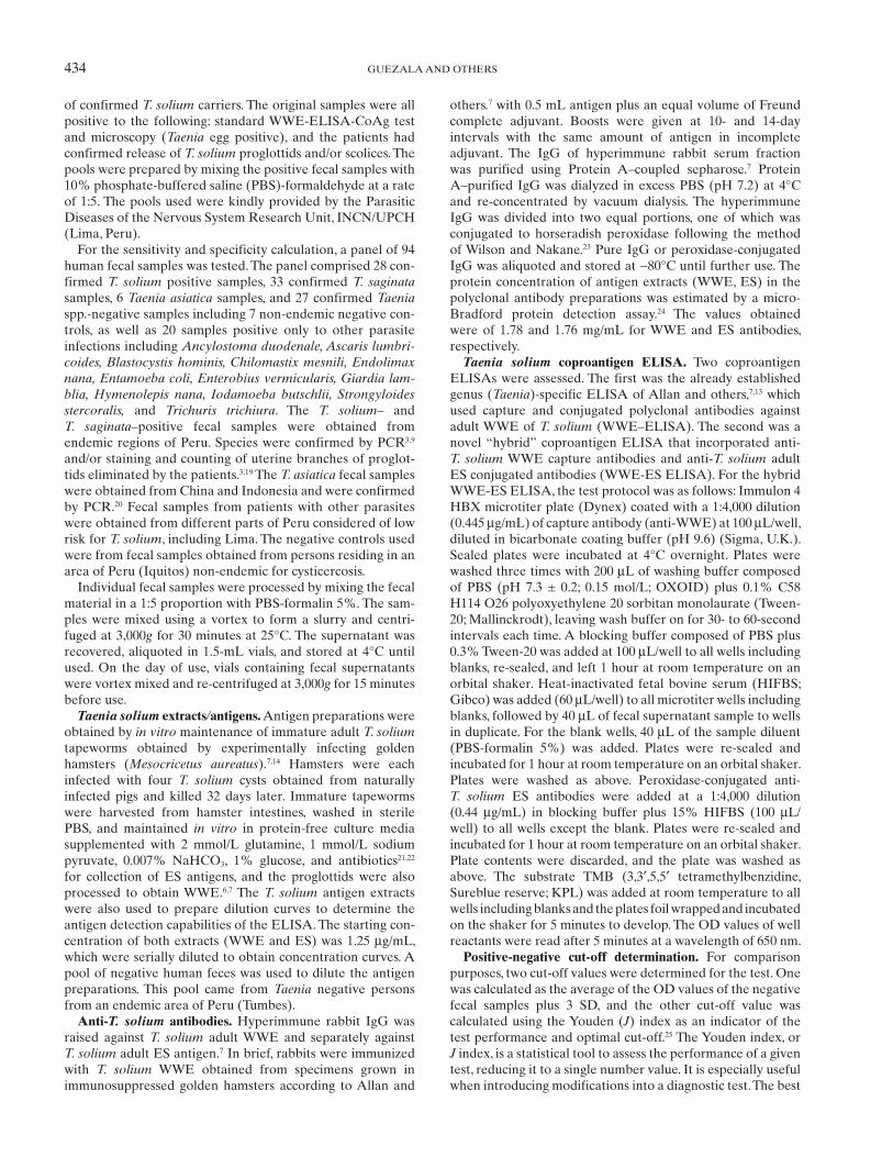
When testing a known panel of human T. solium fecal samples from taeniasis patients, heterologous gut parasitic infections, and normal controls, the new hybrid WWE-ES coproantigen ELISA was 100% specific and 96.4% (27/28) sensitive for detection of T. solium taeniasis ( Figure 1 ). There were no cross-reactions with fecal samples from T. saginata ( N = 33) or T. asiatica ( N = 6) carriers. Similarly, fecal sam-ples for other parasite species ( N = 20) were also negative and remained under the ELISA cut-off value.
The cut-off value was determined by two means: 1) by aver-aging the negative samples OD values plus 3 SD and 2) by assessing the value with the best J index. The cut-off values thus calculated were 0.108 by averaging the negative samples OD values plus 3 SD and 0.104 with a J index of 0.964. Both cut-off values were similar ( Figure 1 ).
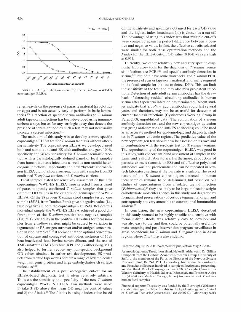
To estimate the detection sensitivity of the T. solium WWE-ES coproantigen ELISA, ES or WWE T. solium adult extracts were titrated into normal human feces. Using the J index determined cut-off of 0.104, the minimum detectable amounts of spiked antigen the test detected in human feces was 0.6 ng/mL of adult ES antigen and 19.5 ng/mL of adult WWE antigen ( Figure 2 ).
The performance of the new WWE-ES ELISA for T. solium coproantigen detection was also compared with the standard WWE-ELISA. 7 For the new test, the specificity was 100% and the sensitivity was 96.4%. The specificity and sensitivity of the standard WWE-ELISA for T. solium was 45.5% and 92%, respectively, with the same panel of human fecal samples. Notably, cross-reaction occurred in the latter test with 97% (32/33) of T. saginata taeniasis and 67% (4/6) of T. asiatica tae-niasis cases but none of the heterologous non- Taenia infection controls (data not shown).
DISCUSSION
The accurate detection of Taenia solium tapeworm carriers is crucial for epidemiologic studies as well as surveillance and intervention programs. 1 The development of an ELISA for T. solium coproantigen detection significantly improved the diagnosis of human taeniasis 7,16 but was genus specific rather than species specific. 5 Before coproantigen tests, microscopy for detection of Taenia eggs in fecal samples, or the recov-ery and identification of tapeworm proglottids, were the only means to diagnose this gastrointestinal infection. Microscopy lacked sensitivity, because the detection of eggs in samples depended on the onset of the patent period of the parasite, and also Taenia eggs may only be shed intermittently. Furthermore, the species-specific parasitologic identification of the tape-worm depends on the recovery of non-degraded proglottids, which is not always possible. 5,19,26 The use of PCR to amplify DNA from proglottids recovered from feces or directly from eggs in a stool sample can provide species specificity but again
Figure 1 . Specificity of the WWE-ES coproantigen ELISA for T. solium carriers ( N = 28) versus fecal samples from T. saginata ( N = 33), T. asiatica ( N = 6), and a range of non- Taenia gut infections or normal controls ( N = 27). PC, positive control.
436 GUEZALA AND OTHERS
relies heavily on the presence of parasite material (proglottids or eggs) and is not actually easy to perform in basic labora-tories. 9,20 Detection of specific serum antibodies to T. solium adult tapeworm infections has been developed using immuno-sorbent assays, but as for any serologic assay that detects the presence of serum antibodies, such a test may not necessarily indicate a current infection. 21,22
The main aim of this study was to develop a more specific coproantigen ELISA test for T. solium taeniasis without affect-ing sensitivity. The coproantigen ELISA we developed used both anti-somatic and anti-ES adult antibodies and gave 100% specificity and 96.4% sensitivity for T. solium taeniasis detec-tion with a parasitologically defined panel of fecal samples from human taeniasis infections as well as non-taeniid heter-ologous infections. Importantly, the new “hybrid” coproanti-gen ELISA did not show cross-reactions with samples from 33 confirmed T. saginata carriers or 6 T. asiatica carriers.
Fecal samples tested for sensitivity assessment of this new coproantigen WWE-ES ELISA were selected from a panel of parasitologically confirmed T. solium samples that gave different OD values in the established genus-specific WWE-ELISA. Of the 28 proven T. solium stool samples tested, one sample (55351, from Tumbes, Peru) gave a negative value (i.e., false negative) in both the coproantigen ELISAs. Besides this individual sample, the WWE-ES ELISA achieved a good dif-ferentiation of the T. solium positive and negative samples ( Figure 1 ). Variability in the positive OD values for fecal sam-ples from T. solium carriers might be caused by variation in tegumental or ES antigen turnover and/or antigen concentra-tion in stool samples. 15,17 It seemed that the optimal concentra-tion of capture and conjugated antibodies, inclusion of 15% heat-inactivated fetal bovine serum diluent, and the use of TMB substrate (TMB Sureblue; KPL Inc., Gaithersburg, MD) also helped to further reduce any non-specific background OD values obtained in earlier test developments. ES prod-ucts from taeniid tapeworms contain a range of low molecular weight antigenic proteins and large carbohydrate-rich surface molecules. 5,22
The establishment of a positive-negative cut-off for an ELISA-based diagnostic test is often relatively arbitrary. To assess the sensitivity and specificity of the new T. solium coproantigen WWE-ES ELISA, two methods were used: 1) take 3 SD above the mean OD negative control values and 2) the J index. 25 The J index is a single index value based
on the sensitivity and specificity obtained for each OD value and the highest index (maximum 1.0) is chosen as a cut-off. The advantage of using this index was that multiple cut-offs were compared against a perfect difference between a posi-tive and negative value. In fact, the effective cut-offs selected were similar for both these optimization methods, and the J index for the ELISA cut-off OD value (0.104) was very high at 0.964.
Currently, two other relatively new and very specific diag-nostic laboratory tools for the diagnosis of T. solium taenia-sis infections are PCR, 3,20 and specific antibody detection in serum, 21,22 but both have some drawbacks. For T. solium PCR, the presence of eggs or tapeworm material is normally required in the fecal sample for the test to detect DNA. This can limit the sensitivity of the test and may also miss pre-patent infec-tions. Detection of anti-adult serum antibodies has the draw-back of detecting residual circulating antibodies in human serum after tapeworm infection has terminated. Recent stud-ies indicate that T. solium adult antibodies could last several years, and therefore, may not be as useful for detection of current taeniasis infections (Cysticercosis Working Group in Peru, 2008, unpublished data). The combination of a serum antibody detection test and the new coproantigen detection test (using anti-somatic and anti-ES antibodies) could be used as an accurate method for epidemiologic and diagnostic stud-ies in T. solium –endemic regions. The predictive value of the new coproantigen test should now be assessed on its own and in combination with the serologic test for T. solium taeniasis. The reproducibility of the coproantigen ELISA was good in this study, with concordant blind assessment of samples in the Lima and Salford laboratories. Furthermore, production of parasite extracts (somatic or ES) and of effective polyclonal antibodies was not problematic and can be repeated in low-tech laboratory settings if the parasite is available. The exact nature of the T. solium coproantigens detected in human fecal samples remains to be determined, but based on our studies of coproantigens from a related taeniid infection ( Echinococcus ), 27 they are likely to be large molecular weight carbohydrate molecules (hence, in this study, not degraded by formalin stool preservation) of cestode tegumental origin and consequently not very amenable to conventional immunoblot analysis. 27
In conclusion, the new coproantigen ELISA described in this study seemed to be highly specific and sensitive with formalin-fixed stools, was relatively easy to develop, and was also easy to use, and thus, could be potentially useful for mass screening and post-intervention program surveillance in areas co-endemic for T. solium and T. saginata and in Asian regions where T. asiatica is also endemic.
Received August 18, 2008. Accepted for publication May 23, 2009.
Acknowledgments: The authors thank Helen Bradshaw and Dr. Gillian Campbell from the Cestode Zoonoses Research Group, University of Salford, the members of the Parasitic Diseases of the Nervous System Research Unit, INCN/UPCH Laboratory, for invaluable assistance, and Peruvian colleagues involved in sample collection and processing. We also thank Drs. Li Tiaoying (Sichuan CDC Chengdu, China), Toni Wandra (Ministry of Health, Jakarta, Indonesia), and Professor Akira Ito (Asahikawa Medical College, Japan) for provision of T. asiatica human fecal samples.
Financial support: This study was funded by the Burroughs Wellcome collaborative grant (“New Insights in the Epidemiology and Control of T. solium Taeniasis/Cysticercosis,” c.c. 8000742). Laboratory work
Figure 2 . Antigen dilution curve for the T. solium WWE-ES coproantigen ELISA.
437SPECIES-SPECIFIC COPROANTIGEN ELISA FOR T. SOLIUM
and research were undertaken at the Biomedical Sciences Research Institute, University of Salford, Salford, UK, and at the Parasitic Diseases of the Nervous System Research Unit, INCN/UPCH, Lima, Peru. Partial support was also provided by Grant 23981 from the Bill and Melinda Gates Foundation.
Authors’ addresses : Maria-Claudia Guezala, Humberto Zamora, and Armando E. Gonzalez, Department of Public Health, School of Veterinary Medicine, Universidad Nacional de San Marcos, Lima, Peru. Silvia Rodriguez and Hector H. Garcia, Department of Microbiology, Universidad Peruana Cayetano Heredia, and Cysticercosis Unit, Instituto de Ciencias Neurologicas, Lima, Peru. Alice Tembo, James C. Allan, and Philip S. Craig, Cestode Zoonoses Research Group, Parasitology and Disease Centre, School of Environment and Life Sciences, University of Salford, Manchester M54WT, UK.
REFERENCES
1. Garcia HH, Martinez M, Gilman RH, Herrera G, Tsang VCW, Pilcher JB, Diaz F, Verastegui M, Gallo C, Porras M, Alvarado, M, Naranjo J, Miranda E, and the Cysticercosis Working Groupin Perú, 1991. Diagnosis of cysticercosis in endemic regions. Lancet 338: 549–551.
2. Schantz PM, Sarti E, 1989. Diagnostic methods and epidemiologi-cal surveillance of Taenia solium infection. Acta Leiden 57: 153–163.
3. Mayta H, Talley A, Gilman RH, Jimenez J, Verastegui M, Ruiz M, Garcia HH, Gonzalez AE, 2000. Differentiating Taenia solium and Taenia saginata infections by simple hematoxylin-eosin staining and PCR-restriction enzyme analysis. J Clin Microbiol 38: 133–137.
4. Pawlowski ZS, Allan JC, Meinardi H, 2005. Control measures for taeniosis and cysticercosis. Murrell KD, ed. WHO/FAO/OIE Guidelines for the Surveillance, Prevention and Control of Taeniosis/Cysticercosis. Paris: OIE, 73–92.
5. Allan JC, Craig PS, 2006. Coproantigens in taeniasis and echi-nococcosis. Parasitol Int 55: S75–S80.
6. Allan JC, Craig PS, 1989. Coproantigens in gut tapeworms infec-tions: Hymenolepis diminuta in rats. Parasitol Res 76: 68–73.
7. Allan JC, Avila G, Garcia Noval J, Flisser A, Craig PS, 1990. Immunodiagnosis of taeniasis by coproantigens detection. Parasitology 101: 473–477.
8. Allan JC, Wilkins P, Tsang VWC, Craig PS, 2003. Immunodiagnos-tic tools for taeniasis. Acta Trop 87: 87–93.
9. Ito A, Craig P, 2003. Immunodiagnostic and molecular approaches for the detection of taeniid cestode infections. Trends Parasitol 19: 377–381.
10. Wandra T, Margono SS, Gafar MS, Saragih JM, Sutisna P, Sudewi AA, Depary AA, Yulfi H, Darlan DM, Okamoto M, Sato MO, Sako Y, Nakao M, Nakaya K, Craig PS, Ito A, 2007. Current situation of taeniasis and cysticercosis in Indonesia. Trop Med Health 35: 323–328.
11. Machnicka B, Dziemian E, Zwierz C, 1996. Factors conditioning detection of Taenia saginata antigens in feces. Appl Parasitol 37: 99–105.
12. Allan JC, Mencos F, Garcia Noval J, Sarti E, Flisser A, Wang Y, Liu D, Craig PS, 1993. Dipstick dot ELISA for the detection of coproantigens in humans. Parasitology 107: 79–85.
13. Allan JC, Velasquez-Tohom M, Torres-Alvarez R, Yurrita P, Garcia-Noval J, 1996a. Field trial of the coproantigen-based diagnosis of Taenia solium taeniasis by enzyme-linked immu-nosorbent assay. Am J Trop Med Hyg 54: 352–356.
14. Maravilla P, Avila G, Cabrera V, Aguilar L, Flisser A, 1998. Comparative development of Taenia solium in experimental models. J Parasitol 84: 882–886.
15. Avila G, Benitez M, Aguilar-Vega L, Flisser A, 2003. Kinetics of Taenia solium antibodies and antigens in experimental taenio-sis. Parasitol Res 89: 284–289.
16. Fraser A, Craig PS, 1997. Detection of gastrointestinal helminthic infections using coproantigens and molecular approaches. J Helminthol 71: 103–107.
17. Deplazes P, Gottstein B, Stingelin Y, Eckert J, 1990. Detection of Taenia hydatigena coproantigens by ELISA in dogs. Vet Parasitol 36: 91–103.
18. Deplazes P, Gottstein B, Eckert J, Jenkins DJ, Ewald D, Jimenez-Palacios S, 1992. Detection of Echinococcus coproantigens by enzyme-linked immunosorbent assay in dogs, dingoes and foxes. Parasitol Res 78: 303–308.
19. Jeri C, Gilman RH, Lescano AG, Mayta H, Ramirez ME, Gonzalez AE, Nazerali R, Garcia HH, 2004. Species identification after treatment for human taeniasis. Lancet 363: 949–959.
20. Yamasaki H, Allan JC, Sato MO, Nakao M, Sako Y, Nakaya K, Qiu D, Mamuti W, Craig PS, Ito A, 2004. DNA differential diag-nosis of taeniasis and cysticercosis by multiplex PCR. J Clin Microbiol 42: 548–553.
21. Wilkins PP, Allan JC, Verastegui M, Acosta M, Eason AG, Garcia HH, Gonzalez AE, Gilman RH, Tsang VWC, 1999. Development of a serologic assay to detect Taenia solium taeniasis. Am J Trop Med Hyg 60: 199–204.
22. Levine MZ, Calderon JC, Wilkins PP, Lane WS, Asara JM, Hancock K, Gonzalez AE, Garcia HH, Gilman RH, Tsang VCW, 2004. Characterization, cloning, and expression of two diagnostic antigens for Taenia solium tapeworm infection. J Parasitol 90: 631–638.
23. Wilson MB, Nakane PK, 1978. Recent developments in the perio-date method of conjugating horseradish peroxidase (HRPO) to antibodies. Knapp W, Holubar K, Wicks G, eds. Immuno-fluorescence and Related Staining Techniques. Amsterdam: Elsevier, 215–224.
24. Bradford MM, 1976. A rapid and sensitive method for the quanti-zation of microgram quantities of protein utilizing the principle of protein–dye binding. Anal Biochem 72: 248–254.
25. Youden WJ, 1950. Index for rating diagnostic tests. Cancer 3 : 32–35.
26. Allan JC, Velasquez-Tohom M, Garcia-Noval J, Torres-Alvarez R, Yurrita P, Fletes C, de Mata F, Soto de Alfaro H, Craig PS, 1996. Epidemiology of intestinal taeniasis in four rural Guatemalan communities. Ann Trop Med Parasitol 90: 157–165.
27. Elayoubi FA, Craig PS, 2004. Echinococcus granulosus coproanti-gens: chromatographic fractionation and characterization. Parasitology 128: 1–11.