Development of a PCR Assay to detect Papillomavirus ... · Development of a PCR Assay to detect...
11
RESEARCH ARTICLE Open Access Development of a PCR Assay to detect Papillomavirus Infection in the Snow Leopard Katherine Mitsouras 1,2 , Erica A Faulhaber 3 , Gordon Hui 3 , Janis O Joslin 3 , Curtis Eng 4 , Margaret C Barr 3 and Kristopher JL Irizarry 2,3* Abstract Background: Papillomaviruses (PVs) are a group of small, non-encapsulated, species-specific DNA viruses that have been detected in a variety of mammalian and avian species including humans, canines and felines. PVs cause lesions in the skin and mucous membranes of the host and after persistent infection, a subset of PVs can cause tumors such as cervical malignancies and head and neck squamous cell carcinoma in humans. PVs from several species have been isolated and their genomes have been sequenced, thereby increasing our understanding of the mechanism of viral oncogenesis and allowing for the development of molecular assays for the detection of PV infection. In humans, molecular testing for PV DNA is used to identify patients with persistent infections at risk for developing cervical cancer. In felids, PVs have been isolated and sequenced from oral papillomatous lesions of several wild species including bobcats, Asian lions and snow leopards. Since a number of wild felids are endangered, PV associated disease is a concern and there is a need for molecular tools that can be used to further study papillomavirus in these species. Results: We used the sequence of the snow leopard papillomavirus UuPV1 to develop a PCR strategy to amplify viral DNA from samples obtained from captive animals. We designed primer pairs that flank the E6 and E7 viral oncogenes and amplify two DNA fragments encompassing these genes. We detected viral DNA for E6 and E7 in genomic DNA isolated from saliva, but not in paired blood samples from snow leopards. We verified the identity of these PCR products by restriction digest and DNA sequencing. The sequences of the PCR products were 100% identical to the published UuPV1 genome sequence. Conclusions: We developed a PCR assay to detect papillomavirus in snow leopards and amplified viral DNA encompassing the E6 and E7 oncogenes specifically in the saliva of animals. This assay could be utilized for the molecular investigation of papillomavirus in snow leopards using saliva, thereby allowing the detection of the virus in the anatomical site where oral papillomatous lesions develop during later stages of infection and disease development. Background Papillomaviruses (PVs) are a group of small, non-encap- sulated epitheliotropic DNA viruses. PVs infect basal keratinocytes and cause benign proliferative lesions, termed papillomas, on the surface of cutaneous and mucosal tissues [1]. A subset of PVs are associated with the development of epithelial malignancies, such as cer- vical carcinomas, oral squamous cell carcinomas (OSCCs) and head and neck squamous cell carcinomas (HNSCCs) in humans [2,3]. The genomes of PVs are circular and double-stranded and contain up to 8 viral genes that are classified as early (E) or late (L) based on their temporal pattern of expression. Early genes encode regulatory proteins that function in viral replication (E1 and E2), viral shedding (E4), or transformation (E5, E6 and E7) [4], whereas late genes (L1 and L2) encode viral capsid proteins (Figure 1a) [1,5]. PVs are strictly spe- cies-specific and have been identified in a wide variety of vertebrates, including mammals, birds and recently reptiles [6,7]. Genome sequencing of several PVs and nucleotide sequence comparisons led to the develop- ment of a PV classification scheme based on L1 * Correspondence: [email protected] 2 The Applied Genomics Center, Graduate College of Biomedical Sciences, Western University of Health Sciences, Pomona, CA, USA Full list of author information is available at the end of the article Mitsouras et al. BMC Veterinary Research 2011, 7:38 http://www.biomedcentral.com/1746-6148/7/38 © 2011 Mitsouras et al; licensee BioMed Central Ltd. This is an Open Access article distributed under the terms of the Creative Commons Attribution License (http://creativecommons.org/licenses/by/2.0), which permits unrestricted use, distribution, and reproduction in any medium, provided the original work is properly cited.
Transcript of Development of a PCR Assay to detect Papillomavirus ... · Development of a PCR Assay to detect...

RESEARCH ARTICLE Open Access
Development of a PCR Assay to detectPapillomavirus Infection in the Snow LeopardKatherine Mitsouras1,2, Erica A Faulhaber3, Gordon Hui3, Janis O Joslin3, Curtis Eng4, Margaret C Barr3 andKristopher JL Irizarry2,3*
Abstract
Background: Papillomaviruses (PVs) are a group of small, non-encapsulated, species-specific DNA viruses that havebeen detected in a variety of mammalian and avian species including humans, canines and felines. PVs causelesions in the skin and mucous membranes of the host and after persistent infection, a subset of PVs can causetumors such as cervical malignancies and head and neck squamous cell carcinoma in humans. PVs from severalspecies have been isolated and their genomes have been sequenced, thereby increasing our understanding of themechanism of viral oncogenesis and allowing for the development of molecular assays for the detection of PVinfection. In humans, molecular testing for PV DNA is used to identify patients with persistent infections at risk fordeveloping cervical cancer. In felids, PVs have been isolated and sequenced from oral papillomatous lesions ofseveral wild species including bobcats, Asian lions and snow leopards. Since a number of wild felids areendangered, PV associated disease is a concern and there is a need for molecular tools that can be used to furtherstudy papillomavirus in these species.
Results: We used the sequence of the snow leopard papillomavirus UuPV1 to develop a PCR strategy to amplifyviral DNA from samples obtained from captive animals. We designed primer pairs that flank the E6 and E7 viraloncogenes and amplify two DNA fragments encompassing these genes. We detected viral DNA for E6 and E7 ingenomic DNA isolated from saliva, but not in paired blood samples from snow leopards. We verified the identity ofthese PCR products by restriction digest and DNA sequencing. The sequences of the PCR products were 100%identical to the published UuPV1 genome sequence.
Conclusions: We developed a PCR assay to detect papillomavirus in snow leopards and amplified viral DNAencompassing the E6 and E7 oncogenes specifically in the saliva of animals. This assay could be utilized for themolecular investigation of papillomavirus in snow leopards using saliva, thereby allowing the detection of the virusin the anatomical site where oral papillomatous lesions develop during later stages of infection and diseasedevelopment.
BackgroundPapillomaviruses (PVs) are a group of small, non-encap-sulated epitheliotropic DNA viruses. PVs infect basalkeratinocytes and cause benign proliferative lesions,termed papillomas, on the surface of cutaneous andmucosal tissues [1]. A subset of PVs are associated withthe development of epithelial malignancies, such as cer-vical carcinomas, oral squamous cell carcinomas(OSCCs) and head and neck squamous cell carcinomas
(HNSCCs) in humans [2,3]. The genomes of PVs arecircular and double-stranded and contain up to 8 viralgenes that are classified as early (E) or late (L) based ontheir temporal pattern of expression. Early genes encoderegulatory proteins that function in viral replication (E1and E2), viral shedding (E4), or transformation (E5, E6and E7) [4], whereas late genes (L1 and L2) encode viralcapsid proteins (Figure 1a) [1,5]. PVs are strictly spe-cies-specific and have been identified in a wide varietyof vertebrates, including mammals, birds and recentlyreptiles [6,7]. Genome sequencing of several PVs andnucleotide sequence comparisons led to the develop-ment of a PV classification scheme based on L1
* Correspondence: [email protected] Applied Genomics Center, Graduate College of Biomedical Sciences,Western University of Health Sciences, Pomona, CA, USAFull list of author information is available at the end of the article
Mitsouras et al. BMC Veterinary Research 2011, 7:38http://www.biomedcentral.com/1746-6148/7/38
© 2011 Mitsouras et al; licensee BioMed Central Ltd. This is an Open Access article distributed under the terms of the CreativeCommons Attribution License (http://creativecommons.org/licenses/by/2.0), which permits unrestricted use, distribution, andreproduction in any medium, provided the original work is properly cited.

sequence identity and helped elucidate the evolutionaryrelationships among PVs isolated from a variety of spe-cies [7-9].Due to their known association with human neopla-
sias, human papillomaviruses (HPVs) have been exten-sively studied, thereby increasing our understanding ofthe molecular mechanisms of viral oncogenesis andrevealing additional factors that contribute to the devel-opment of malignancies. The finding that immune-defi-cient (HIV positive) or immune-suppressed (renalallograft recipients) individuals harbor higher numbersof high-risk PVs underscores the importance of immunefunction in the reactivation of latent infection and possi-bly in HPV induced oncogenesis [10-12].Feline-specific PVs belong to the lambda-papilloma-
virus genus and consist of mucosal and cutaneous types[8]. In the domestic cat (Felis domesticus), PV infection
has been associated with a number of skin lesions suchas squamous cell carcinoma, bowenoid in-situ carci-noma, viral plaques and sarcoids [13-16]. PV associateddisease is of particular interest in wild endangered felids,where these viruses have been isolated from oral papillo-matous lesions in the Florida panther (Puma concolorcoryi; PcPV1), bobcat (Lynx rufus; LrPV1), Asian lion(Panthera leo persica; PlpPV1), snow leopard (Unciauncia; UuPV1) and the clouded leopard (Neofelis nebu-losa) [9]. Sequencing and nucleotide comparisons of theLrPV1, PcPV1, PlpPV1 and UuPV1 genomes providedinsight into their genomic organization, their evolution-ary relationships and the mechanism of PV-host co-spe-ciation [9]. Sequence comparisons of UuPV1 genes tothose of PVs in the alpha and beta genera revealed thatUuPV1 is more similar to beta-PVs (for example HPV-5) which are associated with latent infections that
UuPV1E4
8078/ 1 2,000 4,000 6,000 8,000
E6 E7 E1E2 L2
L1NCR2 URRURR
OncogenesisReplicationTranscriptionCapsid proteins
Cis-regulatory
A.
B.E6 E7URR E1
E6 PCR (694 bp)
E7 PCR (472 bp)
Figure 1 Snow leopard PV1 genomic map and PCR strategy for detection of viral DNA. (a) Schematic representation of the linear Uu PV1genome. The numbers indicate the genomic nucleotide positions. The individual open reading frames (ORFs) of the early (E) and late (L)genomic regions are depicted as shaded arrows, and are color-coded according to the known function of the corresponding gene product. Thetwo cis-regulatory regions are depicted as open rectangles and consist of the upstream regulatory region (URR) and the second non-codingregion (NCR2). (b) Schematic representation of the genomic targets for PCR-based detection of viral DNA. The primers used for amplification ofthe E6 and E7 genes are depicted as arrows, and the resulting amplification products as shaded rectangles. The size of each amplificationproduct is indicated.
Mitsouras et al. BMC Veterinary Research 2011, 7:38http://www.biomedcentral.com/1746-6148/7/38
Page 2 of 11

become reactivated under immunosuppression, than tohigh-risk alpha PVs (for example HPV-16), which areassociated with malignancies [9].Until recently, diagnosis of PV infection in snow leo-
pards and other wild felids has depended upon observa-tion and histopathology of papillomatous lesions. Oralpapillomas in snow leopards are commonly seen underthe tongue and appear as small, pale nodules (Figure 2).Very little is known about snow leopard immuneresponses to PV infection, and relatively few moleculartools are available to facilitate their study. Interpretationof serological tests for papillomavirus antibodies can beproblematic. As with HPV infections, positive antibodytests in snow leopards probably indicate exposure butnot necessarily current infection; PV antibodies may bepresent for several months after the virus has beencleared. False negative results are also possible becauseseroconversion to the major coat protein, L1, may bedelayed for as much as 18 months after initiation of per-sistent infections, or may not occur at all [17,18].Epithelial infections with PVs are rarely accompanied byinflammation, the viruses are non-lytic, viral capsid pro-teins are differentially expressed in keratinocytes and atvery low levels in less mature cells, and the basal laminaof the epithelium remains intact, effectively allowingPVs to replicate without stimulating innate or adaptiveimmune responses [18,19]. Additionally, PVs can evadeimmune responses by establishing latent persistentinfections, integrating into the host cell genome, or initi-ating a multitude of other defense mechanisms [18,20].
For endangered captive species such as the snow leo-pard, immune function is an important trait. In order tobetter understand how individual variation in snow leo-pard immune function relates to papillomavirus infec-tion, appropriate molecular tools must be developed tostudy UuPV1 infection and its impact on the health andsurvival of endangered felids. In the current study, wereport the development of a PCR-based assay thatdetects the E6 and E7 viral oncogenes in the saliva ofsnow leopards. This assay can be utilized as a novelmolecular tool to investigate the mechanisms underlyinginfection and the development and progression of PVinduced disease in this endangered species.
ResultsDesign of a PCR strategy to amplify papillomavirus DNAWe used the published genome sequence of UuPV1(Figure 1a) [9] to design PCR primers that amplify viralDNA (Figure 1b). The E6 and E7 genes present idealtargets for viral DNA detection for a number of reasons.First, E6 and E7 are the key genes involved in the inter-action of papillomavirus with host cell cycle pathwaysand are also the target of PCR-based assays that detectHPV-16 DNA in head and neck squamous cell carci-noma (HNSCC) [21], suggesting that E6 and E7 can besuccessfully amplified from clinical samples. Addition-ally, even in cases when PV integrates into the host gen-ome and a number of viral genes are lost, E6 and E7may be the only genes that remain intact [20]. There-fore, a PCR strategy focusing on E6 and E7 maximizesthe probability of identifying viral DNA in a variety ofsnow leopard specimens corresponding to differentstages of disease progression. We designed two sets ofprimers that amplify the full-length E6 and E7 genes astwo separate PCR products (Figure 1b). The two primerpairs are positioned such that the E6 and E7 PCR pro-ducts overlap by 95 nucleotides, which allows us to con-firm DNA sequence information from two independentPCR products as an additional internal control. The E6primers anneal at genomic positions 12 (left primer)and 705 (right primer), and amplify a 694-bp fragmentencompassing the entire E6 coding region (which spansgenomic positions 31-447), and the first 267 bp of theE7 gene (located at genomic positions 444-728). The E7primers anneal at genomic positions 327 (left primer)and 800 (right primer) and produce a 472-bp fragment,that contains the last 120 bp of the E6 gene and theentire E7 coding region.
PCR amplification of viral DNA from snow leopard salivaBecause papillomatous lesions occur within the snowleopard oral cavity [9,22], we wanted to determinewhether saliva represents an appropriate biological spe-cimen for the detection of papillomavirus DNA in
Figure 2 Oral papillomatous lesions in a snow leopard. The palenodules on the bottom of the tongue are papillomas and areindicated by the black circle. The papillomatous lesions are arrangedin a ring. This circular arrangement is likely the result of an earlierbiopsy in this animal. During the biopsy, viral particles seeded thesurrounding skin and caused additional papillomas to form in acircle around the original site of ablation.
Mitsouras et al. BMC Veterinary Research 2011, 7:38http://www.biomedcentral.com/1746-6148/7/38
Page 3 of 11

asymptomatic animals. Additionally, since saliva collec-tion can be performed in a non-invasive manner, beha-vioral modification techniques can be used to trainanimals to calmly accept an oral swab without the needfor chemical restraint. In this fashion, we were able toobtain saliva samples from snow leopards in their enclo-sure, thus minimizing anxiety to the animals and with-out introducing them into stress-provoking situations(Figure 3a). This is particularly important for captivespecies that are not routinely subjected to chemicalrestraint, and are only anesthetized for scheduled physi-cal exams every year or every two or three years. Inthese cases, saliva represents a more easily and fre-quently accessible biological specimen than blood.A positive result in a PCR-based assay would indicate
presence of the virus in the oral cavity of the animal, asopposed to a serological test which can indicate expo-sure, but not necessarily current infection. We obtained
matched saliva and blood samples from three snow leo-pards that did not have oral papillomatous lesions: twoadult females that were approximately 18 years old (SL1 and SL 3), and a 7-week old female cub (SL 2; Figure3b). We used the E6 and E7 primer pairs in amplifica-tion reactions using DNA purified from the pairedblood and saliva samples (Figure 4a and 4b). Weobtained amplification products of the expected size forboth E6 (Figure 4a, lanes 3 and 7) and E7 (Figure 4b,lanes 3 and 7) using saliva from the two adult snow leo-pards (SL 1 and SL 3) but not from the cub (Figure 4a,lane 5; Figure 4b lane 5). Additionally, we were unableto detect viral DNA for E6 and E7 in DNA purifiedfrom the matched blood samples of the two adult snowleopards (Figure 4a, lanes 2 and 6; Figure 4b, lanes 2and 6). This was not due to the absence of DNA in anyof the saliva or blood samples, since we successfullyamplified a 503-bp product corresponding to the coding
Figure 3 Collection of saliva samples from a captive snow leopard using the Oragene •ANIMAL kit. (a) Saliva was collected from a 2-yearold female snow leopard in its enclosure using the Oragene •ANIMAL kit. The sample was collected by the handler of the animal through thecage using two Oragene •ANIMAL sponges, and was stabilized in the Oragene •ANIMAL solution. (b) Saliva collection using the Oragene•ANIMAL kit from a 7-week old female cub during a routine veterinary exam.
Mitsouras et al. BMC Veterinary Research 2011, 7:38http://www.biomedcentral.com/1746-6148/7/38
Page 4 of 11
region of the snow leopard transferrin receptor (TfR) asa positive control (Figure 4c, lanes 1-6). Additional sal-iva samples obtained from two snow leopards at anotherfacility were negative for E6 and E7 DNA (data notshown).
Validation of the E6 and E7 PCR products by restrictiondigest and DNA sequencingSince restriction digests provide a fast and cost-effec-tive method to rapidly screen PCR products, we devel-oped a restriction endonuclease digestion assay of the
amplification products to confirm that they correspondto the E6 and E7 viral genes. The restriction enzymeTaqI cleaves the E6 PCR product twice, producingthree fragments of 288, 279 and 127 bp (Figure 5a, toppanel). The E7 PCR product contains three RsaIrestriction sites, yielding two large fragments (263 and144 bp) and two smaller fragments (36 and 29 bp; Fig-ure 5b, top panel). Digestion of the E6 PCR products
Figure 4 Amplification of the UuPV1 E6 and E7 genes frompaired saliva and blood samples. 50 ng of genomic DNA isolatedusing the Oragene•ANIMAL kit (An), or paired blood samples (Bl)from three snow leopards (SL 1, 2 and 3) were used for PCRamplification. Amplification products were resolved by agarose gelelectrophoresis and visualized by staining with SYBR® Green. (a)Amplification of a 694-bp fragment encompassing the E6 viraloncogene from saliva (lanes 3, 5 and 7) and blood (lanes 2, 4 and6). An amplification control with no DNA was performed in parallel(lane 1). (b) Amplification of a 472-bp DNA fragment encompassingthe E7 viral oncogene from paired saliva (lanes 3, 5 and 7) andblood samples (Bl; lanes 2, 4 and 6). Lane 1 shows a controlamplification reaction with no DNA. (c) Control amplificationreactions of a 503-bp fragment in the coding region the snowleopard Transferrin Receptor (TfR) gene, using paired saliva (An;lanes 2, 4 and 6) and blood samples (Bl; lanes 1, 3 and 5). Thenegative amplification control with no DNA is shown in lane 7.
Figure 5 Validation of the E6 and E7 amplification products byrestriction enzyme digestion. (a) Top panel: Schematicrepresentation of the 694-bp E6 amplification product indicating thepositions of the two TaqI cleavage sites, and the sizes of theresulting fragments (288, 279 and127 bp). Bottom panel: 50 ng ofgenomic DNA isolated from two snow leopards (SL 1 and SL 3)using the Oragene•ANIMAL kit were amplified using the E6 primerpair. Half of each PCR reaction was incubated with the TaqIrestriction endonuclease. Restriction products (lanes 2 and 4), alongwith an aliquot of the original PCR reactions (lanes 1 and 3) wereresolved by agarose gel electrophoresis and visualized by stainingwith SYBR® Green. (b) Top panel: Schematic representation of 472-bp E7 amplification product indicating the positions of the threeRsaI cleavage sites, and the sizes of the resulting restriction products(263, 144, 36 and 29 bp). Bottom panel: 50 ng of genomic DNAisolated from two snow leopards (SL 1 and SL 3) using theOragene•ANIMAL kit were amplified using the E7 primer pair. Half ofeach PCR reaction was incubated with the TaqI restrictionendonuclease. Restriction products (lanes 2 and 4), along with analiquot of the original PCR reactions (lanes 1 and 3) were resolvedby agarose gel electrophoresis and visualized by staining withSYBR® Green.
Mitsouras et al. BMC Veterinary Research 2011, 7:38http://www.biomedcentral.com/1746-6148/7/38
Page 5 of 11
obtained from the saliva of the two older snow leo-pards (SL 1 and SL 3) produced a doublet correspond-ing to the 288 and 279-bp fragments and a single 127-bp fragment (Figure 5a, bottom panel, lanes 2 and 4).Similarly, incubation of the E7 amplification productsfrom these animals with RsaI yielded two fragments ofthe expected size (Figure 5b, bottom panels, lanes 2and 4).As an independent measure to confirm their identity,
we gel purified the E6 and E7 PCR products from thetwo animals and subjected them to bi-directional DNAsequencing. The sequencing results were assembled intocontigs and aligned to the consensus genome sequenceof UuPV1 (Figure 6). The multiple sequence alignmentin Figure 6 shows that the sequence of the E6 and E7amplification products obtained from saliva is 100%identical to the published UuPV1 genome sequence forE6 and E7.These two validation methods independently confirm
the identity of the E6 and E7 amplicons. Althoughamplicon sequencing is preferable, it is can be moreexpensive, time-consuming and requires significantamounts of purified PCR products, which might notalways be feasible. In such cases, restriction digests pro-vide an accurate, simple and cost-effective method torapidly screen even large numbers of samples. Thechoice of two distinct validation methods in our assayprovides flexibility and makes it amenable to perform ina variety of laboratory settings.
DiscussionWe developed a molecular assay that detects papilloma-virus DNA in snow leopard saliva. Our method is similarto PCR-based molecular assays designed to detect differenttypes of human papillomavirus (HPV) in a variety of clini-cal samples [21,23]. Specifically, presence of HPV-16 inhead and neck squamous cell carcinoma (HNSCC) biop-sies is determined by amplification of the E6 and E7 genes[21]. Similarly, E6 amplification is used to detect the pre-sence of high-risk HPV subtypes in human cutaneous neo-plasms and in cervical smears [23-25]. In some cases of PVintegration into host genetic material, E6 and E7 may bethe only genes that remain intact [20], thus making themideal choices for PCR-based detection of viral DNA.The detection of UuPV1 DNA in the saliva of two
older snow leopards parallels results from HPV studiesthat have demonstrated that HPV DNA can be ampli-fied from oral rinses and saliva samples from HNSCCpatients and HIV positive individuals [10,26-28].Because PVs are epitheliotropic, detection of PV DNAin blood samples from infected snow leopards is unlikely[18]; therefore, the absence of viral DNA in blood isconsistent with an expected PV replication pattern inmucosal cells with no concurrent viremia.
The utilization of a PCR-based method to detect viralDNA in saliva presents a number of advantages. First,saliva collection is a non-invasive sampling method,thereby circumventing the requirement for anesthesiaprior to obtaining a blood sample in captive wild felids.This is particularly important for captive species thatare not routinely subjected to chemical restraint, andmay only be anesthetized for scheduled physical exams.In these cases, saliva represents a more easily and fre-quently accessible biological specimen than blood. Anadditional advantage of testing saliva is that behavioralmodification techniques can be used to train snow leo-pards and other felids to calmly accept an oral swab forDNA collection without chemical restraint. In fact, wewere able to collect saliva samples from snow leopardsin their enclosure, with minimal disturbance to the ani-mal. Subsequently, animals testing positive for viralDNA can then be scheduled for a more thorough oralexamination under sedation. The use of saliva alsoallows for detection of the virus in the anatomical sitewhere papillomatous lesions develop during later stagesof disease progression. Additionally, saliva is extremelystable, and more easily preserved than serum samples,thereby facilitating collection and shipping of samplesfrom different sites. In testing for HPV, it has beendemonstrated that PCR-based methods have increasedsensitivity as compared to serotesting [29], and canaccommodate even lower quality samples, such as archi-val samples and paraffin-embedded specimens [23]. Incontrast to seropositivity which can indicate a presentand ongoing infection or a past infection that has beencleared, a positive PCR test indicates a current infectionin the oral cavity of the animal. Therefore, PCR-basedtesting of snow leopard saliva can complement serotest-ing and help provide a more accurate picture of an ani-mal’s disease status.PCR-based detection of papillomavirus E6 and E7
DNA in the saliva of snow leopards may have utility indiagnosing infection in animals that have no obviousoral papillomatous lesions, confirming infection in catswith oral papillomas, and detecting infection in animalsthat have not yet seroconverted. Due to its non-invasive-ness and ease of implementation, PCR-based testing ofsaliva can be incorporated into the routine and regularscreening of captive snow leopards, which would greatlyfacilitate the collection of epidemiological data onUuPV1 infections. Additionally, since papillomatouslesions have been reported to progress to SCC in snowleopards [9,22], our assay can also be used to determinewhether viral DNA is present in biopsy samples of SCCsto explore the relationship between UuPV1 infectionand neoplasia and elucidate the molecular mechanismsunderlying the development and progression of PVinduced disease.
Mitsouras et al. BMC Veterinary Research 2011, 7:38http://www.biomedcentral.com/1746-6148/7/38
Page 6 of 11
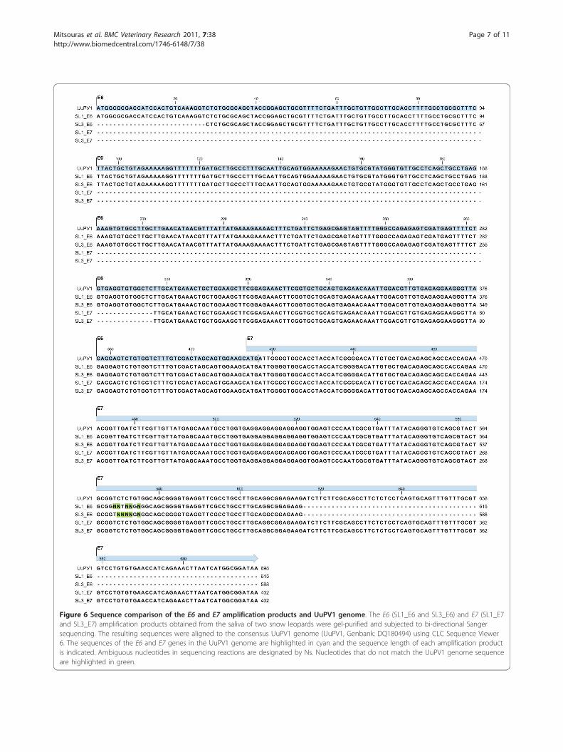
Figure 6 Sequence comparison of the E6 and E7 amplification products and UuPV1 genome. The E6 (SL1_E6 and SL3_E6) and E7 (SL1_E7and SL3_E7) amplification products obtained from the saliva of two snow leopards were gel-purified and subjected to bi-directional Sangersequencing. The resulting sequences were aligned to the consensus UuPV1 genome (UuPV1, Genbank: DQ180494) using CLC Sequence Viewer6. The sequences of the E6 and E7 genes in the UuPV1 genome are highlighted in cyan and the sequence length of each amplification productis indicated. Ambiguous nucleotides in sequencing reactions are designated by Ns. Nucleotides that do not match the UuPV1 genome sequenceare highlighted in green.
Mitsouras et al. BMC Veterinary Research 2011, 7:38http://www.biomedcentral.com/1746-6148/7/38
Page 7 of 11
The finding that UuPV1 DNA is present in the salivaof two adult snow leopards that have no current ordocumented lesions is intriguing. The most likely expla-nation for our finding is supported by the advanced ageof these animals and the fact that saliva was collectedfollowing euthanasia due to progressive illness. Papillo-maviruses can establish latent or low-level inapparentinfections, remaining undetected for long periods oftime [1], and these cats may have been experiencingreactivation of a previously controlled PV infection. It isalso possible, although unlikely, that these animals hadrecently become infected, and PV induced disease wasnot manifested at the time of death.One of the goals of conservation biology is to main-
tain genetically diverse captive populations of endan-gered species. In relatively small populations, like the150-200 captive snow leopards in North America, it iscritical to develop functional assays for assessing indivi-dual differences in health and fitness. One such differ-ence is an animal’s susceptibility to PV infection and tothe development of PV associated disease. In the currentstudy, we demonstrate that viral DNA is present in thesaliva of clinically normal animals. Therefore, our assaycan be used to classify clinically normal snow leopardsinto PV-positive and PV-negative populations, which,along with clinical data, can be studied to identify bothgenetic and environmental factors underlying suscept-ibility to viral infection and to the development and pro-gression of disease. Similar studies in humans haveidentified specific variants in immune genes as well asin genes that interact with viral proteins and demon-strated that these variants are associated with an indivi-dual’s risk for viral persistence and with the subsequentdevelopment of disease [30,31].The conservation of endangered captive species
requires maintaining the genetic diversity of the popula-tion. When captive populations suffer from inbreedingdepression, fitness decreases and mortality rates mayincrease along with decreased reproductive success andimpaired immune function [32,33]. Genetic studies insmall populations of endangered species are challengingbecause standard approaches like large-scale geneticassociation studies are not possible. Instead, geneticapproaches must focus on individuals and place theemphasis on developing effective means of characteriz-ing phenotypes which can be used to study phenotypicvariation across the captive population. In the case ofthe snow leopard, the well-documented infection of ani-mals with papillomavirus [22] offers an opportunity todevelop robust phenotyping methods for assessingimmune function within the captive population. Unfor-tunately, relatively little is known about snow leopardsusceptibility and resistance to papillomavirus infectionand disease progression. In order to better understand
the potential phenotypic variation underlying suscept-ibility and resistance to viral infection, it is critical thatmolecular tools be developed that can allow investiga-tors to begin characterizing how phenotypes associatedwith papilomavirus infection and disease progressionvary across closely and distantly related individuals. ThePCR assay we describe is a first step in this direction.This assay can be used to (1) monitor the exposure ofindividual snow leopards to papillomavirus, (2) accu-rately identify which snow leopards are positive andwhich are negative for papillomavirus, (3) characterizethe relationship between papillomavirus exposure andthe development of oral papillomatous lesions, (4) inves-tigate the relationship between exposure and infectionwhen uninfected snow leopards are housed with infectedsnow leopards and, (5) determine the timeline underly-ing subclinical infection and presentation of clinicalsigns. As more snow leopards participate in these stu-dies, distributions describing these traits for the entirecaptive population can be produced. Subsequently, indi-viduals exhibiting phenotypes at the tails of the distribu-tions can be identified and further studies aimed atcharacterizing the genetic basis of snow leopard immu-nity in captivity can be performed.
ConclusionsWe developed a novel PCR strategy to detect papilloma-virus in the snow leopard. Using this assay, we success-fully amplified the E6 and E7 viral oncogenes in DNApurified from saliva of two snow leopards. DNA sequen-cing verified that the amplified fragments indeed repre-sent the E6 and E7 genes of UuPV1. In addition, wedemonstrated that viral DNA cannot be detected inpaired blood samples from these animals, which is con-sistent with the mechanism of papillomavirus infectionand viral lifecycle in the host. Taken together with thenon-invasiveness, and ease of collection relative toblood, our results further underscore the utility of salivaas a suitable clinical specimen for the detection of papil-lomavirus in snow leopards. Our findings allow for thedevelopment of a molecular tool to elucidate themechanisms underlying the development and progres-sion of PV induced disease in this endangered species.
MethodsSample collection and purificationWe obtained paired saliva and blood samples from 3captive snow leopards housed in North American zoos:two female snow leopards that were approximately 18years old, and a 7-week old female cub. None of theseanimals had oral papillomatous lesions. We additionallycollected saliva samples from a 2-year old female and a2-year old male snow leopard in their enclosures with-out the use of anesthesia. Collection protocols were
Mitsouras et al. BMC Veterinary Research 2011, 7:38http://www.biomedcentral.com/1746-6148/7/38
Page 8 of 11

approved by the Western University Institutional Ani-mal and Care Use Committee. Blood was drawn intoPAXgene Blood DNA tubes (Qiagen, Valencia, CA,USA) and genomic DNA was isolated using the PAX-gene Blood DNA kit (Qiagen, Valencia, CA, USA). Sal-iva was collected using Oragene•ANIMAL kits (DNAGenotek, Ontario, Canada) and DNA was purified fromthe entire saliva sample using the manufacturer’s proto-col [34]. All DNA samples were quantitated using aNanovue spectrophotometer (GE LifeSciences, Piscat-away, NJ, USA). The purity of each DNA sample wasassessed using the A260/A280 ratio.
Primer designThe published UuPV1 genome sequence [Genbank:DQ180494] was used to design PCR primers thatamplify DNA fragments encompassing the entire E6 andE7 genes as shown in Figure 1(b). Primers weredesigned using the freely available Primer3 softwarepackage [35], and tested by In-silico PCR [36] to assesswhether they non-specifically amplify feline genomicsequences. Primer sequences are as follows: E6-Forward5’-AGTGACTCGGAGGGCATTC-3’, E6-Reverse 5’-GATGGTTCACACAGGACACG-3’, E7-Forward 5’-TTGCATGAAACTGCTGGAAG-3’, E7-Reverse 5’-GGTTCGTCATCATCGCTACA-3’. The feline transfer-rin receptor (TfR) mRNA sequence [Genbank:NM_001009312] was used to design PCR primers thatamplify a 503-bp fragment in the coding region of thegene. Primers were designed as described above andused for cross-species amplification of the correspond-ing region of the snow leopard transferrin receptorgene. Primer sequences are as follows: TfR-Forward 5’-TTTCTTGATATTTGAGTTCATTGTTT-3’, TfR-Reverse 5’-AGTAACTGTCGCTGCTTTACTGT-3’
PCR amplificationDNA extracted from the 3 sets of matched saliva andblood samples was used for amplification of the E6and E7 genes of the snow leopard PV1. A 694-bp frag-ment encompassing the E6 gene was amplified usingthe E6-Forward and E6-Reverse primers. Reactionswere performed in a 50 microliter volume using 50 nggenomic DNA, 0.2 μM each primer, 0.125 mM dNTPs,1.5 mM MgCl2, 1X GeneAmp® PCR Gold Buffer and2.5 U Amplitaq Gold® DNA Polymerase (Applied Bio-systems, Foster City, CA, USA) in a Veriti™ 96-wellthermal cycler (Applied Biosystems, Foster City, CA,USA) using the following conditions: 95°C for 10 min,10 cycles of 95°C 30 sec, 1 min annealing with a start-ing temperature of 63°C decreasing by 0.5°C per cycledown to 58.5°C and 72°C 1 min, followed by an addi-tional 25 cycles of 95°C 30 sec, 58°C 1 min, 72°C 1min and a final extension for 10 min at 72°C. A 472-
bp fragment encompassing the E7 gene was amplifiedusing the E7-Forward and E7-Reverse primers. Ampli-fication reactions were performed in a 50 microlitervolume using 50 ng genomic DNA, 0.2 μM each pri-mer, 0.125 mM dNTPs, 2.5 mM MgCl2, 1X Gen-eAmp® PCR Gold Buffer and 2.5 U Amplitaq Gold®
DNA Polymerase (Applied Biosystems, Foster City,CA, USA) in a Veriti™ 96-well thermal cycler (AppliedBiosystems, Foster City, CA, USA) using the followingconditions: 95°C for 10 min, 12 cycles of 95°C 30 sec,1 min annealing with a starting temperature of 61°Cdecreasing by 0.5°C per cycle down to 55.5°C and 72°C1 min, followed by an additional 25 cycles of 95°C 30sec, 55°C 1 min, 72°C 1 min and a final extension for10 min at 72°C. 25 microliters of each PCR reactionwere resolved on a 1.5% agarose/1X TBE gel stainedwith SYBR® Green (Invitrogen, Carlsbad, CA, USA).All PCR reactions were repeated at least twice in inde-pendent experiments.A 503-bp fragment in the coding region of the snow
leopard transferrin receptor gene was amplified usingthe TfR-Forward and TfR-Reverse primers. Reactionswere performed in a 50 microliter volume using 50 nggenomic DNA, 0.4 μM each primer, 0.25 mM dNTPs,2.5 mM MgCl2 and 2.5 U Taq DNA Polymerase (Qia-gen, Valencia, CA, USA) in a Veriti™ 96-well thermalcycler (Applied Biosystems, Foster City, CA, USA) usingthe following conditions: 95°C for 10 min, 30 cycles of95°C 30 sec, 54°C 1 min, 72°C 1 min and a final exten-sion for 10 min at 72°C. 15 microliters of each PCRreaction were resolved on a 1.5% agarose/1X TBE gelstained with SYBR® Green (Invitrogen, Carlsbad, CA,USA). All PCR reactions were repeated at least twice inindependent experiments.
Restriction analysisThe identity of the E6 and E7 amplification productswas validated by restriction enzyme digest. 25 microli-ters of the E6 amplification products obtained from twosnow leopard saliva samples were incubated with 15 UTaqI (Fermentas, Glen Burnie, MD, USA) in a 30microliter reaction at 65°C for 2 hours. Digestion pro-ducts were resolved on a 2.5% agarose/1X TBE gelstained with SYBR® Green (Invitrogen, Carlsbad, CA,USA). 25 microliters of each E7 amplification productwere digested with 15 U RsaI (Fermentas, Glen Burnie,MD, USA) in a 30 microliter reaction at 37°C for 2hours. Digestion products were resolved on a 2.5% agar-ose/1X TBE gel with SYBR® Green (Invitrogen, Carls-bad, CA, USA).
DNA sequencingThe E6 and E7 amplification products obtained fromtwo snow leopard saliva samples were purified using the
Mitsouras et al. BMC Veterinary Research 2011, 7:38http://www.biomedcentral.com/1746-6148/7/38
Page 9 of 11
Qiaex II Gel Extraction kit (Qiagen, Valencia, CA, USA)and subjected to bi-directional DNA sequencing usingthe E6 and E7 PCR primer pairs respectively. Sequen-cing was performed at the UCLA Sequencing Core (LosAngeles, CA, USA). The forward and reverse sequencesof each amplification product were assembled into a sin-gle contig, and aligned to the consensus UuPV1 genomesequence [Genbank: DQ180494] using the CLCSequence Viewer 6 software package (CLC Bio, Cam-bridge, MA, USA).
List of abbreviationsBp: Basepair; DNA: Deoxyribonucleic acid; HPV: Human papillomavirus;HNSCC: Head and neck squamous cell carcinoma; HIV: Humanimmunodeficiency virus; OSCC: Oral squamous cell carcinoma; ORF: Openreading frame; PV: Papillomavirus; PCR: Polymerase chain reaction; TfR:Transferrin receptor.
AcknowledgementsThe authors wish to acknowledge Ellen McLean and Daria Vasilitsova at DNAGenotek Inc for generously providing Oragene•ANIMAL kits, Dr JamesWheeler, Dr Amy Ekerberg, Terry K. Lincoln and the staff at the Dakota Zoo,Dr Rhonda Aliah and the staff at the Tautphaus Park Zoo for allowing theircaptive snow leopards to participate in this study, Joe Maynard and the staffat the Exotic Feline Breeding Compound for collecting saliva samples,allowing us to document the procedure and allowing their snow leopardsto participate in the study. The authors also wish to thank Dr Nissar Darmaniat Western University of Health Sciences for his generous support of travelto collect samples, and Lauren Kreisberg for facilitating access to samples.The authors wish to thank Dr Uma Dandekar at the UCLA Sequencing Corefor sequencing, technical support and feedback. This study was supportedby a National Leadership Collaborative Planning Grant (LG-54-09-0068-09)from the Institute of Museum and Library Services (JJ, MCB and KJLI), by anacademic gift from DNA Genotek Inc (KM) and by a Merial VeterinaryStudent Summer Scholar Award (GH).
Author details1College of Osteopathic Medicine of the Pacific, Western University ofHealth, Sciences, Pomona, CA, USA. 2The Applied Genomics Center, GraduateCollege of Biomedical Sciences, Western University of Health Sciences,Pomona, CA, USA. 3College of Veterinary Medicine, Western University ofHealth Sciences, Pomona, CA, USA. 4Los Angeles Zoo and Botanical Gardens,5333 Zoo Drive, Los Angeles, CA, USA.
Authors’ contributionsKM conceived of the study, participated in study design, assisted with thecollection of saliva and blood samples, purified saliva and blood samples,performed PCR amplification, restriction digests and gel purification of E6and E7 amplicons for sequencing, performed data analysis, prepared figuresand drafted the manuscript. EAF participated in the design of the PCRamplification strategy, assisted with PCR amplification and drafted themanuscript. GH performed computational analysis of papillomavirus-hostinteractions to specifically select E6 and E7 as suitable targets for PCR-baseddetection of viral DNA. JOJ assisted with the clinical interpretation of oralpapillomatous lesions in snow leopards. CE participated in study design andprovided clinical interpretation/relevance of experimental results. MCBobtained IACUC approval, coordinated data collection, provided access tosnow leopard saliva and blood samples, assisted with data interpretationand drafted the manuscript. KJLI conceived of the study, designed the study,coordinated data collection, designed the PCR amplification strategy,performed data analysis and drafted the manuscript. All authors have readand approved the manuscript.
Received: 27 January 2011 Accepted: 18 July 2011Published: 18 July 2011
References1. Doorbar J: The papillomavirus life cycle. J Clin Virol 2005, 32(Suppl 1):
S7-15.2. Marur S, D’Souza G, Westra WH, Forastiere AA: HPV-associated head and
neck cancer: a virus-related cancer epidemic. Lancet Oncol 2010,11(8):781-789.
3. Ibeanu OA: Molecular pathogenesis of cervical cancer. Cancer Biol Ther2011, 11(3).
4. Hamid NA, Brown C, Gaston K: The regulation of cell proliferation by thepapillomavirus early proteins. Cell Mol Life Sci 2009, 66(10):1700-1717.
5. Longworth MS, Laimins LA: Pathogenesis of human papillomaviruses indifferentiating epithelia. Microbiol Mol Biol Rev 2004, 68(2):362-372.
6. Herbst LH, Lenz J, Van Doorslaer K, Chen Z, Stacy BA, Wellehan JF Jr,Manire CA, Burk RD: Genomic characterization of two novel reptilianpapillomaviruses, Chelonia mydas papillomavirus 1 and Caretta carettapapillomavirus 1. Virology 2009, 383(1):131-135.
7. Shah SD, Doorbar J, Goldstein RA: Analysis of host-parasite incongruencein papillomavirus evolution using importance sampling. Mol Biol Evol2010, 27(6):1301-1314.
8. de Villiers EM, Fauquet C, Broker TR, Bernard HU, zur Hausen H:Classification of papillomaviruses. Virology 2004, 324(1):17-27.
9. Rector A, Lemey P, Tachezy R, Mostmans S, Ghim SJ, Van Doorslaer K,Roelke M, Bush M, Montali RJ, Joslin J, Burk RD, Jenson AB, Sundberg JP,Shapiro B, Van Ranst M: Ancient papillomavirus-host co-speciation inFelidae. Genome Biol 2007, 8(4):R57.
10. Adamopoulou M, Vairaktaris E, Panis V, Nkenke E, Neukam FW, Yapijakis C:HPV detection rate in saliva may depend on the immune systemefficiency. In Vivo 2008, 22(5):599-602.
11. de Villiers EM: Human papillomavirus infections in skin cancers. BiomedPharmacother 1998, 52(1):26-33.
12. de Villiers EM, Lavergne D, McLaren K, Benton EC: Prevailingpapillomavirus types in non-melanoma carcinomas of the skin in renalallograft recipients. Int J Cancer 1997, 73(3):356-361.
13. Munday JS, Knight CG, French AF: Evaluation of feline oral squamous cellcarcinomas for p16(CDKN2A) protein immunoreactivity and thepresence of papillomaviral DNA. Res Vet Sci 2010.
14. Munday JS, Dunowska M, De Grey S: Detection of two differentpapillomaviruses within a feline cutaneous squamous cell carcinoma:case report and review of the literature. N Z Vet J 2009, 57(4):248-251.
15. Munday JS, Willis KA, Kiupel M, Hill FI, Dunowska M: Amplification of threedifferent papillomaviral DNA sequences from a cat with viral plaques.Vet Dermatol 2008, 19(6):400-404.
16. Munday JS, Kiupel M, French AF, Howe L: Amplification of papillomaviralDNA sequences from a high proportion of feline cutaneous in situ andinvasive squamous cell carcinomas using a nested polymerase chainreaction. Vet Dermatol 2008, 19(5):259-263.
17. Carter JJ, Koutsky LA, Wipf GC, Christensen ND, Lee SK, Kuypers J, Kiviat N,Galloway DA: The natural history of human papillomavirus type 16capsid antibodies among a cohort of university women. J Infect Dis 1996,174(5):927-936.
18. Stanley MA: Immune responses to human papilloma viruses. Indian J MedRes 2009, 130(3):266-276.
19. Frazer IH, Thomas R, Zhou J, Leggatt GR, Dunn L, McMillan N, Tindle RW,Filgueira L, Manders P, Barnard P, Sharkey M: Potential strategies utilisedby papillomavirus to evade host immunity. Immunol Rev 1999,168:131-142.
20. Chow LT, Broker TR, Steinberg BM: The natural history of humanpapillomavirus infections of the mucosal epithelia. APMIS 118(6-7):422-449.
21. Capone RB, Pai SI, Koch WM, Gillison ML, Danish HN, Westra WH, Daniel R,Shah KV, Sidransky D: Detection and quantitation of humanpapillomavirus (HPV) DNA in the sera of patients with HPV-associatedhead and neck squamous cell carcinoma. Clin Cancer Res 2000,6(11):4171-4175.
22. Joslin JOGM, Collins D, Kamaka E, Sinabaldi K, Meleo K, Montali R,Sundberg JP, Jenson AB, Ghim SJ, Davidow B, Hargis AM, West K, Clark T,Haines D: Viral papilloma and squamous cell carcinomas in snowleopards (Uncia uncia). merican Association of Zoo Veterinarians (AAZV) andInternational Association for Aquatic Animal Medicine (IAAAM) JointConference: 2000; New Orleans 2000, 155-158.
Mitsouras et al. BMC Veterinary Research 2011, 7:38http://www.biomedcentral.com/1746-6148/7/38
Page 10 of 11
23. Resnick RM, Cornelissen MT, Wright DK, Eichinger GH, Fox HS, terSchegget J, Manos MM: Detection and typing of human papillomavirusin archival cervical cancer specimens by DNA amplification withconsensus primers. J Natl Cancer Inst 1990, 82(18):1477-1484.
24. Stark LA, Arends MJ, McLaren KM, Benton EC, Shahidullah H, Hunter JA,Bird CC: Prevalence of human papillomavirus DNA in cutaneousneoplasms from renal allograft recipients supports a possible viral rolein tumour promotion. Br J Cancer 1994, 69(2):222-229.
25. Lungu O, Sun XW, Wright TC Jr, Ferenczy A, Richart RM, Silverstein S: Apolymerase chain reaction-enzyme-linked immunosorbent assay methodfor detecting human papillomavirus in cervical carcinomas and high-grade cervical cancer precursors. Obstet Gynecol 1995, 85(3):337-342.
26. Smith EM, Ritchie JM, Summersgill KF, Hoffman HT, Wang DH, Haugen TH,Turek LP: Human papillomavirus in oral exfoliated cells and risk of headand neck cancer. J Natl Cancer Inst 2004, 96(6):449-455.
27. D’Souza G, Sugar E, Ruby W, Gravitt P, Gillison M: Analysis of the effect ofDNA purification on detection of human papillomavirus in oral rinsesamples by PCR. J Clin Microbiol 2005, 43(11):5526-5535.
28. Agrawal Y, Koch WM, Xiao W, Westra WH, Trivett AL, Symer DE, Gillison ML:Oral human papillomavirus infection before and after treatment forhuman papillomavirus 16-positive and human papillomavirus 16-negative head and neck squamous cell carcinoma. Clin Cancer Res 2008,14(21):7143-7150.
29. Wieland U, Pfister H: Molecular diagnosis of persistent human papillomavirus infections. Intervirology 1996, 39(3):145-157.
30. Wang SS, Bratti MC, Rodriguez AC, Herrero R, Burk RD, Porras C, Gonzalez P,Sherman ME, Wacholder S, Lan ZE, Schiffman M, Chanock SJ, Hildesheim A:Common variants in immune and DNA repair genes and risk for humanpapillomavirus persistence and progression to cervical cancer. J Infect Dis2009, 199(1):20-30.
31. Wang SS, Gonzalez P, Yu K, Porras C, Li Q, Safaeian M, Rodriguez AC,Sherman ME, Bratti C, Schiffman M, Wacholder S, Burk RD, Herrero R,Chanock SJ, Hildesheim A: Common genetic variants and risk for HPVpersistence and progression to cervical cancer. PLoS One 2010, 5(1):e8667.
32. Fredrickson RJ, Siminski P, Woolf M, Hedrick PW: Genetic rescue andinbreeding depression in Mexican wolves. Proc Biol Sci 2007,274(1623):2365-2371.
33. Charlesworth D, Willis JH: The genetics of inbreeding depression. Nat RevGenet 2009, 10(11):783-796.
34. Laboratory Protocol for Manual Purification of DNA from 250 mL ofOrageneANIMAL/saliva. [http://www.dnagenotek.com/pdf_files/PD-PR-095%20Issue%202.1%20Laboratory%20Protocol.pdf].
35. Rozen S, Skaletsky H: Primer3 on the WWW for general users and forbiologist programmers. Methods Mol Biol 2000, 132:365-386.
36. UCSC In-Silico PCR. [http://genome.ucsc.edu/cgi-bin/hgPcr?command=start].
doi:10.1186/1746-6148-7-38Cite this article as: Mitsouras et al.: Development of a PCR Assay todetect Papillomavirus Infection in the Snow Leopard. BMC VeterinaryResearch 2011 7:38.
Submit your next manuscript to BioMed Centraland take full advantage of:
• Convenient online submission
• Thorough peer review
• No space constraints or color figure charges
• Immediate publication on acceptance
• Inclusion in PubMed, CAS, Scopus and Google Scholar
• Research which is freely available for redistribution
Submit your manuscript at www.biomedcentral.com/submit
Mitsouras et al. BMC Veterinary Research 2011, 7:38http://www.biomedcentral.com/1746-6148/7/38
Page 11 of 11