Development and validation of a new method for locating ...
8
RESEARCH Open Access Development and validation of a new method for locating patella sensory nerves for the treatment of inferior and superior knee pain Emily Hu 1* , Jessica Preciado 1 , Vinod Dasa 2 and Jason Mussell 3 Abstract Background: Radiofrequency ablation and percutaneous cryoneurolysis to relieve knee pain requires treating large areas to ensure coverage due to high variability in the sensory innervation of the knee and limitations of current methods for defining treatment targets. This study sought to define and validate a new treatment approach targeting the major sensory nerves of the superior patella and expand upon previous work to define a more efficient treatment approach targeting the sensory nerves of the inferior patella. Methods: Transcutaneous electrical nerve stimulation and ultrasound were used to evaluate the location and relationship of the cutaneous nerves to the superior and inferior aspects of the knee in 25 healthy volunteers. Using information derived from these evaluations, investigators defined new linear target treatment areas, or treatment lines, using anatomical landmarks, which were validated against locations of sensory nerves through cadaveric dissection of 15 fresh specimens. Results: The proposed treatment lines captured the vast majority of nerve branching variations during cadaveric validation. Conclusion: This study defined treatment lines, identifiable using only anatomical landmarks, which effectively target the nerves responsible for superior and inferior knee pain and reduce the total treatment area and procedure time when administering treatments such as radiofrequency ablation and cryoneurolysis. Keywords: Anterior knee pain; Infrapatellar nerves; Saphenous nerve; Femoral cutaneous nerves; Cryoneurolysis Background Nerve-targeting treatments for knee pain, such as radio- frequency ablation and percutaneous cryoneurolysis, re- quire treating large areas to ensure coverage of the target nerve(s), due to high variability in the sensory in- nervation patterns of the knee and limited descriptions of these variations in the literature (Horner & Dellon 1994). Methods of localizing the nerves that contribute to patellar pain include ultrasound imaging and transcu- taneous electrical nerve stimulation (TENS). Because the nerves that innervate the patella are normally small (0.5-3 mm in diameter), sonographic visualization can be difficult and time consuming (Gray 2012; Scott 2012). TENS requires searching the entire surface area of the anterior thigh using the TENS wand, which is time consuming as well and may be uncomfortable for the patient. A method of localizing the sensory nerves of the superior and inferior patella using only anatom- ical landmarks would improve treatment efficiency and tolerability. Currently, there is no validated anatomical treatment approach that targets the branches of the anterior femoral cutaneous nerve (AFCN) and the lateral fem- oral cutaneous nerve (LFCN), which innervate the su- perior aspect of the patella and the surrounding tissue (Scott 2012). * Correspondence: [email protected] 1 myoscience, Inc., 46400 Fremont Blvd, Fremont, CA 94538, USA Full list of author information is available at the end of the article © 2015 Hu et al. Open Access This article is distributed under the terms of the Creative Commons Attribution 4.0 International License (http://creativecommons.org/licenses/by/4.0/), which permits unrestricted use, distribution, and reproduction in any medium, provided you give appropriate credit to the original author(s) and the source, provide a link to the Creative Commons license, and indicate if changes were made. Hu et al. Journal of Experimental Orthopaedics (2015) 2:16 DOI 10.1186/s40634-015-0032-2
Transcript of Development and validation of a new method for locating ...
Hu et al. Journal of Experimental Orthopaedics (2015) 2:16 DOI 10.1186/s40634-015-0032-2
RESEARCH Open Access
Development and validation of a newmethod for locating patella sensory nervesfor the treatment of inferior and superiorknee pain
Emily Hu1*, Jessica Preciado1, Vinod Dasa2 and Jason Mussell3Abstract
Background: Radiofrequency ablation and percutaneous cryoneurolysis to relieve knee pain requires treating largeareas to ensure coverage due to high variability in the sensory innervation of the knee and limitations of currentmethods for defining treatment targets. This study sought to define and validate a new treatment approach targetingthe major sensory nerves of the superior patella and expand upon previous work to define a more efficient treatmentapproach targeting the sensory nerves of the inferior patella.
Methods: Transcutaneous electrical nerve stimulation and ultrasound were used to evaluate the location andrelationship of the cutaneous nerves to the superior and inferior aspects of the knee in 25 healthy volunteers. Usinginformation derived from these evaluations, investigators defined new linear target treatment areas, or treatment lines,using anatomical landmarks, which were validated against locations of sensory nerves through cadaveric dissection of15 fresh specimens.
Results: The proposed treatment lines captured the vast majority of nerve branching variations during cadavericvalidation.
Conclusion: This study defined treatment lines, identifiable using only anatomical landmarks, which effectively targetthe nerves responsible for superior and inferior knee pain and reduce the total treatment area and procedure timewhen administering treatments such as radiofrequency ablation and cryoneurolysis.
Keywords: Anterior knee pain; Infrapatellar nerves; Saphenous nerve; Femoral cutaneous nerves; Cryoneurolysis
BackgroundNerve-targeting treatments for knee pain, such as radio-frequency ablation and percutaneous cryoneurolysis, re-quire treating large areas to ensure coverage of thetarget nerve(s), due to high variability in the sensory in-nervation patterns of the knee and limited descriptionsof these variations in the literature (Horner & Dellon1994). Methods of localizing the nerves that contributeto patellar pain include ultrasound imaging and transcu-taneous electrical nerve stimulation (TENS). Becausethe nerves that innervate the patella are normally small(0.5-3 mm in diameter), sonographic visualization can
* Correspondence: [email protected], Inc., 46400 Fremont Blvd, Fremont, CA 94538, USAFull list of author information is available at the end of the article
© 2015 Hu et al. Open Access This article isInternational License (http://creativecommoreproduction in any medium, provided youlink to the Creative Commons license, and i
be difficult and time consuming (Gray 2012; Scott2012). TENS requires searching the entire surface areaof the anterior thigh using the TENS wand, which istime consuming as well and may be uncomfortable forthe patient. A method of localizing the sensory nervesof the superior and inferior patella using only anatom-ical landmarks would improve treatment efficiency andtolerability.Currently, there is no validated anatomical treatment
approach that targets the branches of the anteriorfemoral cutaneous nerve (AFCN) and the lateral fem-oral cutaneous nerve (LFCN), which innervate the su-perior aspect of the patella and the surrounding tissue(Scott 2012).
distributed under the terms of the Creative Commons Attribution 4.0ns.org/licenses/by/4.0/), which permits unrestricted use, distribution, andgive appropriate credit to the original author(s) and the source, provide andicate if changes were made.
Hu et al. Journal of Experimental Orthopaedics (2015) 2:16 Page 2 of 8
A previous anatomical study in five embalmed cadaversmapped the course of the infrapatellar saphenous nerve(ISN) and medial cutaneous femoral nerve (MFCN),which are responsible for sensory innervation of theinferior aspect of the patella and the surrounding tissue(Le Corroller et al. 2011). Investigators found that theinfrapatellar branches of the ISN and the MFCN are lo-cated within the area between 55 mm from the medialborder of the patella and 44 mm from the medial borderof the patellar tendon (Le Corroller et al. 2011). A “treat-ment box” which targets the branches of ISN and MFCNresponsible for inferior patella pain may be defined usingthese anatomical landmarks as the medial and lateral bor-ders, respectively, and the midline of the patella and topof the tibial tubercle as the superior and inferior borders,respectively. When utilizing this approach to located andtreat these nerve branches, the entire length of the treat-ment box must be treated. This is a large area that in-cludes locations that do not contain nerves of interest,increasing operator time and patient discomfort.The present study was designed to 1) investigate the lo-
cations of the sensory nerves that innervate the superiorand inferior patella; 2) using only bony landmarks, definea treatment approach which encompasses all variations ofbranching patterns of the AFCN, LFCN, MFCN, and ISNas identified using TENS and ultrasound; and 3) validatethe accuracy of the treatment approach using cadavericdissections. This study aims to break new ground by de-veloping the first validated methodology for targeting thenerves that innervate the superior patella. In addition, thisstudy will expand upon previous work targeting the nervesthat innervate the inferior patella by defining a more effi-cient treatment approach.
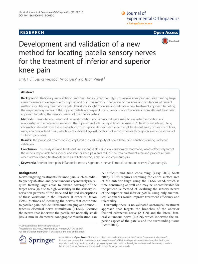
Fig. 1 AFCN and LFCN localization and measurement method. A vertical lithe center of the patella to the top of the femoral head at the iliac creaselocated using TENS. Black dots on thigh indicate nerve locations (b). For eameasured, denoted by the red dotted lines. A line is drawn from the nerveand the angle between this line and the vertical center line is measured (c
MethodsTENS and ultrasound nerve location study in volunteersThe study protocol was approved by an ethics committeeand informed consent was obtained from all individualparticipants included in the study. All procedures per-formed in studies involving human participants were inaccordance with the ethical standards of the institutionaland/or national research committee and with the 1964Helsinki declaration and its later amendments or compar-able ethical standards. Eligible subjects were healthy adultsaged 18–60 years with no history of surgery, trauma, or al-tered anatomy in the measurement areas.All subjects were examined in the supine position with
the knees extended using the SonoSite M-Turbo® Ultra-sound System with a 14 MHz linear transducer and theBraun Stimuplex® HNS 12. The ultrasound transducerwas placed in the transverse plane with a short axis viewof the nerve and held in minimal contact with the skin toensure that the skin above the nerve was not compressedduring measurement. Branches of nerves were identifiedon the ultrasound as small echogenic honeycomb-shapedstructures under the fat layers and on top of the musclefascia. Nerve branches were located and measured on oneor both knees.
Innervation of the superior kneeTo locate the AFCN and LFCN, a vertical line bisectingthe anterior thigh along the sagittal plane was drawnfrom the center of the patella to the top of the femoralhead at the iliac crease (Fig. 1). The vertical line and thewidth of the patella were measured. The surface of thethigh was traced with the TENS wand with an initial set-ting of 1.3 mA, 1.0 ms, and 2.0 Hz. The TENS current
ne bisecting the anterior thigh along the sagittal plane is drawn from(a). AFCN and LFCN branches which innervate the superior patella arech nerve location, the distance to and along the vertical center line islocation to the center of the knee, denoted by the blue dotted line,)
Hu et al. Journal of Experimental Orthopaedics (2015) 2:16 Page 3 of 8
was increased in increments of 0.2 mA until the subjectreported a sensation in or near the medial portion of thesuperior edge of the patella. Areas of maximal TENS re-sponse for the AFCN branches were located using subjectfeedback and recorded. These locations were retraced withincrementally lower currents to minimize the radius ofstimulation and further specify the nerve location, untilpain or sensation in the superior knee was no longer feltby the subject. The final nerve location was marked; thisprocess was repeated for all areas of TENS response forthe superior knee. The distance from each nerve locationto the vertical center line was measured. A line was drawnfrom each nerve location to the center of the knee toform an angle with the vertical center line of the thigh,which was measured.The area between two nerve locations was then traced
with the TENS wand in order to discern which nerve lo-cations arose from the same branch. If the subject re-ported identical sensations when the wand was passedbetween the nerve locations as when the wand was dir-ectly on the nerve locations, it was assumed that the lo-cations were part of the same nerve branch. All nervelocations were confirmed and nerve depths measuredusing ultrasound. .
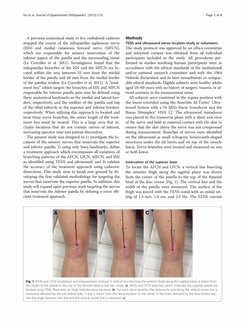
Innervation of the inferior kneeTo locate the infrapatellar nerve branches innervatingthe inferior patella, a treatment box (Fig. 2) was drawnon the knee using the anatomical landmarks describedby Le Corroller et al., and its dimensions measured
Fig. 2 Infrapatellar nerve “treatment box” targeting the ISN and MFCNas defined by Le Corroller. The medial and lateral borders of “treatmentbox” occur 55 mm medial from the medial border of the patella and44 mm medial from the medial border of the patellar tendon,respectively. The midline of patella serves as the superior border,and the top of the tibial tubercle the inferior border. Treatmentbox is not drawn to scale
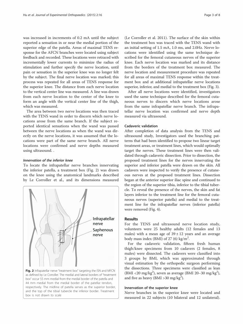
(Le Corroller et al. 2011). The surface of the skin withinthe treatment box was traced with the TENS wand withan initial setting of 1.5 mA, 1.0 ms, and 2.0Hz. Nerve lo-cations were identified using the same technique de-scribed for the femoral cutaneous nerves of the superiorknee. Each nerve location was marked and its distancefrom the borders of the treatment box measured. Thenerve location and measurement procedure was repeatedfor all areas of maximal TENS response within the treat-ment box and at additional infrapatellar nerve locationssuperior, inferior, and medial to the treatment box (Fig. 3).After all nerve locations were identified, investigators
used the same technique described for the femoral cuta-neous nerves to discern which nerve locations arosefrom the same infrapatellar nerve branch. The infrapa-tellar nerve location was confirmed and nerve depthmeasured via ultrasound.
Cadaveric validationAfter completion of data analysis from the TENS andultrasound study, Investigators used the branching pat-terns that had been identified to propose two linear targettreatment areas, or treatment lines, which would optimallytarget the nerves. These treatment lines were then vali-dated through cadaveric dissection. Prior to dissection, theproposed treatment lines for the nerves innervating thesuperior and inferior patella were drawn on the skin. Allcadavers were inspected to verify the presence of cutane-ous nerves at the proposed treatment lines. Dissectionbegan at the anterior superior iliac spine and continued tothe region of the superior tibia, inferior to the tibial tuber-cle. To reveal the presence of the nerves, the skin and fatlayers inferior to the treatment line for the femoral cuta-neous nerves (superior patella) and medial to the treat-ment line for the infrapatellar nerves (inferior patella)were removed (Fig. 4).
ResultsFor the TENS and ultrasound nerve location study,volunteers were 25 healthy adults (12 females and 13males) with a mean age of 39 ± 12 years and an averagebody mass index (BMI) of 27 (6) kg/m2.For the cadaveric validation, fifteen fresh human
thigh/knee specimens from 10 cadavers (2 females, 8males) were dissected. The cadavers were classified into3 groups by BMI, which was approximated throughvisual estimation by the orthopedic surgeon performingthe dissections. Three specimens were classified as lean(BMI <20 mg/kg2), seven as average (BMI 20–30 mg/kg2),and five as heavy (BMI >30 mg/kg2).
Innervation of the superior kneeNerve branches in the superior knee were located andmeasured in 22 subjects (10 bilateral and 12 unilateral).
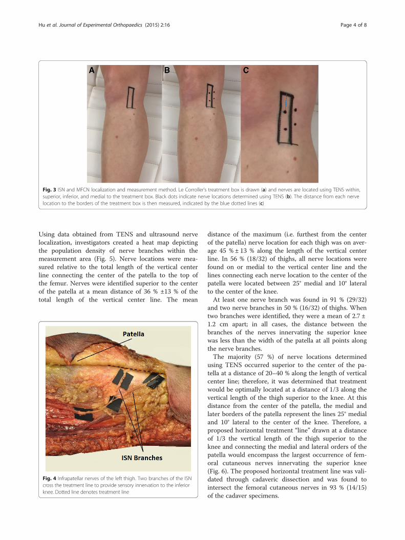
Fig. 3 ISN and MFCN localization and measurement method. Le Corroller’s treatment box is drawn (a) and nerves are located using TENS within,superior, inferior, and medial to the treatment box. Black dots indicate nerve locations determined using TENS (b). The distance from each nervelocation to the borders of the treatment box is then measured, indicated by the blue dotted lines (c)
Hu et al. Journal of Experimental Orthopaedics (2015) 2:16 Page 4 of 8
Using data obtained from TENS and ultrasound nervelocalization, investigators created a heat map depictingthe population density of nerve branches within themeasurement area (Fig. 5). Nerve locations were mea-sured relative to the total length of the vertical centerline connecting the center of the patella to the top ofthe femur. Nerves were identified superior to the centerof the patella at a mean distance of 36 % ±13 % of thetotal length of the vertical center line. The mean
Fig. 4 Infrapatellar nerves of the left thigh. Two branches of the ISNcross the treatment line to provide sensory innervation to the inferiorknee. Dotted line denotes treatment line
distance of the maximum (i.e. furthest from the centerof the patella) nerve location for each thigh was on aver-age 45 % ± 13 % along the length of the vertical centerline. In 56 % (18/32) of thighs, all nerve locations werefound on or medial to the vertical center line and thelines connecting each nerve location to the center of thepatella were located between 25° medial and 10° lateralto the center of the knee.At least one nerve branch was found in 91 % (29/32)
and two nerve branches in 50 % (16/32) of thighs. Whentwo branches were identified, they were a mean of 2.7 ±1.2 cm apart; in all cases, the distance between thebranches of the nerves innervating the superior kneewas less than the width of the patella at all points alongthe nerve branches.The majority (57 %) of nerve locations determined
using TENS occurred superior to the center of the pa-tella at a distance of 20–40 % along the length of verticalcenter line; therefore, it was determined that treatmentwould be optimally located at a distance of 1/3 along thevertical length of the thigh superior to the knee. At thisdistance from the center of the patella, the medial andlater borders of the patella represent the lines 25° medialand 10° lateral to the center of the knee. Therefore, aproposed horizontal treatment “line” drawn at a distanceof 1/3 the vertical length of the thigh superior to theknee and connecting the medial and lateral orders of thepatella would encompass the largest occurrence of fem-oral cutaneous nerves innervating the superior knee(Fig. 6). The proposed horizontal treatment line was vali-dated through cadaveric dissection and was found tointersect the femoral cutaneous nerves in 93 % (14/15)of the cadaver specimens.

Fig. 5 Heat map of the occurrence of the branches of the femoral cutaneous nerve using the left leg where x = 0, y = 0 is the center of the patella.The x-axis represents the angle from the nerve branch to the vertical midline of the patella; the y-axis represents the percent distance of the nervebranch along the thigh; and the z-axis indicates the number of nerve branches occurring at each location. The majority of nerve locations determinedusing TENS occur at the light blue and green regions of the map. The dotted black line denotes the proposed treatment line
Hu et al. Journal of Experimental Orthopaedics (2015) 2:16 Page 5 of 8
Innervation of the inferior kneeNerve branches in the inferior knee were located andmeasured in 24 subjects (19 bilateral and 5 unilateral).Knees were assessed for the number of branches innerv-ating the inferior knee located within the measurementarea; there were 14 % (6/43) of knees with one branch,72 % (31/43) with two branches, 7 % (3/43) with threebranches, and 7 % (3/43) for whom no branch patternscould be discerned within the measurement area. Most(83 %; 15/18) subjects had the same number of nervebranches innervating the inferior knee on both lower ex-tremities; there was one subject for whom nerve loca-tions were identified but branching patterns could notbe confirmed. Although it is possible that the remaining17 % of subjects had an asymmetrical number ofbranches, it is more likely that additional infrapatellar
branches of the MFCN or ISN could not be detected byTENS due to their small size. For many subjects, includ-ing those with a symmetrical number of nerve brancheson both extremities, nerve locations were not necessarilyidentical on each side. In 37 % (7/19) of assessed bilat-eral knees, there was a 20 % or greater difference in thedistance from the superior border of the treatment boxdefined using Le Corroller’s methodology for one ormore branches of the infrapatellar nerve between thesubject’s right and left lower extremity.For the 24 subjects (43 knees) measured, the most su-
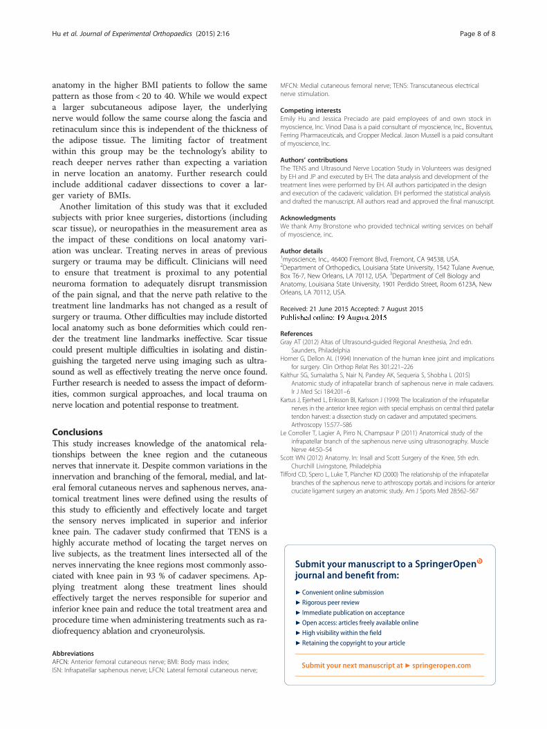
perior infrapatellar nerve branch was located an averagedistance of 45 % ± 30 % of the length of the treatmentbox inferior to the superior border of the treatment box(Fig. 7). The most inferior infrapatellar nerve branch waslocated an average distance of 15 % ± 25 % of the length
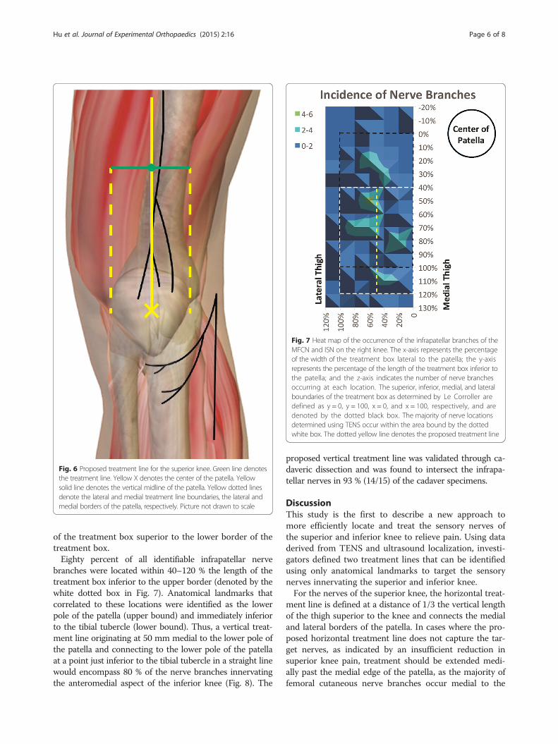
Fig. 6 Proposed treatment line for the superior knee. Green line denotesthe treatment line. Yellow X denotes the center of the patella. Yellowsolid line denotes the vertical midline of the patella. Yellow dotted linesdenote the lateral and medial treatment line boundaries, the lateral andmedial borders of the patella, respectively. Picture not drawn to scale
Fig. 7 Heat map of the occurrence of the infrapatellar branches of theMFCN and ISN on the right knee. The x-axis represents the percentageof the width of the treatment box lateral to the patella; the y-axisrepresents the percentage of the length of the treatment box inferior tothe patella; and the z-axis indicates the number of nerve branchesoccurring at each location. The superior, inferior, medial, and lateralboundaries of the treatment box as determined by Le Corroller aredefined as y = 0, y = 100, x = 0, and x = 100, respectively, and aredenoted by the dotted black box. The majority of nerve locationsdetermined using TENS occur within the area bound by the dottedwhite box. The dotted yellow line denotes the proposed treatment line
Hu et al. Journal of Experimental Orthopaedics (2015) 2:16 Page 6 of 8
of the treatment box superior to the lower border of thetreatment box.Eighty percent of all identifiable infrapatellar nerve
branches were located within 40–120 % the length of thetreatment box inferior to the upper border (denoted by thewhite dotted box in Fig. 7). Anatomical landmarks thatcorrelated to these locations were identified as the lowerpole of the patella (upper bound) and immediately inferiorto the tibial tubercle (lower bound). Thus, a vertical treat-ment line originating at 50 mm medial to the lower pole ofthe patella and connecting to the lower pole of the patellaat a point just inferior to the tibial tubercle in a straight linewould encompass 80 % of the nerve branches innervatingthe anteromedial aspect of the inferior knee (Fig. 8). The
proposed vertical treatment line was validated through ca-daveric dissection and was found to intersect the infrapa-tellar nerves in 93 % (14/15) of the cadaver specimens.
DiscussionThis study is the first to describe a new approach tomore efficiently locate and treat the sensory nerves ofthe superior and inferior knee to relieve pain. Using dataderived from TENS and ultrasound localization, investi-gators defined two treatment lines that can be identifiedusing only anatomical landmarks to target the sensorynerves innervating the superior and inferior knee.For the nerves of the superior knee, the horizontal treat-
ment line is defined at a distance of 1/3 the vertical lengthof the thigh superior to the knee and connects the medialand lateral borders of the patella. In cases where the pro-posed horizontal treatment line does not capture the tar-get nerves, as indicated by an insufficient reduction insuperior knee pain, treatment should be extended medi-ally past the medial edge of the patella, as the majority offemoral cutaneous nerve branches occur medial to the
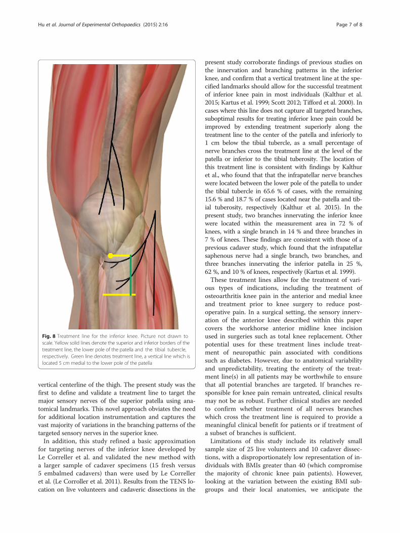
Fig. 8 Treatment line for the inferior knee. Picture not drawn toscale. Yellow solid lines denote the superior and inferior borders of thetreatment line, the lower pole of the patella and the tibial tubercle,respectively. Green line denotes treatment line, a vertical line which islocated 5 cm medial to the lower pole of the patella
Hu et al. Journal of Experimental Orthopaedics (2015) 2:16 Page 7 of 8
vertical centerline of the thigh. The present study was thefirst to define and validate a treatment line to target themajor sensory nerves of the superior patella using ana-tomical landmarks. This novel approach obviates the needfor additional location instrumentation and captures thevast majority of variations in the branching patterns of thetargeted sensory nerves in the superior knee.In addition, this study refined a basic approximation
for targeting nerves of the inferior knee developed byLe Correller et al. and validated the new method witha larger sample of cadaver specimens (15 fresh versus5 embalmed cadavers) than were used by Le Correlleret al. (Le Corroller et al. 2011). Results from the TENS lo-cation on live volunteers and cadaveric dissections in the
present study corroborate findings of previous studies onthe innervation and branching patterns in the inferiorknee, and confirm that a vertical treatment line at the spe-cified landmarks should allow for the successful treatmentof inferior knee pain in most individuals (Kalthur et al.2015; Kartus et al. 1999; Scott 2012; Tifford et al. 2000). Incases where this line does not capture all targeted branches,suboptimal results for treating inferior knee pain could beimproved by extending treatment superiorly along thetreatment line to the center of the patella and inferiorly to1 cm below the tibial tubercle, as a small percentage ofnerve branches cross the treatment line at the level of thepatella or inferior to the tibial tuberosity. The location ofthis treatment line is consistent with findings by Kalthuret al., who found that that the infrapatellar nerve brancheswere located between the lower pole of the patella to underthe tibial tubercle in 65.6 % of cases, with the remaining15.6 % and 18.7 % of cases located near the patella and tib-ial tuberosity, respectively (Kalthur et al. 2015). In thepresent study, two branches innervating the inferior kneewere located within the measurement area in 72 % ofknees, with a single branch in 14 % and three branches in7 % of knees. These findings are consistent with those of aprevious cadaver study, which found that the infrapatellarsaphenous nerve had a single branch, two branches, andthree branches innervating the inferior patella in 25 %,62 %, and 10 % of knees, respectively (Kartus et al. 1999).These treatment lines allow for the treatment of vari-
ous types of indications, including the treatment ofosteoarthritis knee pain in the anterior and medial kneeand treatment prior to knee surgery to reduce post-operative pain. In a surgical setting, the sensory innerv-ation of the anterior knee described within this papercovers the workhorse anterior midline knee incisionused in surgeries such as total knee replacement. Otherpotential uses for these treatment lines include treat-ment of neuropathic pain associated with conditionssuch as diabetes. However, due to anatomical variabilityand unpredictability, treating the entirety of the treat-ment line(s) in all patients may be worthwhile to ensurethat all potential branches are targeted. If branches re-sponsible for knee pain remain untreated, clinical resultsmay not be as robust. Further clinical studies are neededto confirm whether treatment of all nerves brancheswhich cross the treatment line is required to provide ameaningful clinical benefit for patients or if treatment ofa subset of branches is sufficient.Limitations of this study include its relatively small
sample size of 25 live volunteers and 10 cadaver dissec-tions, with a disproportionately low representation of in-dividuals with BMIs greater than 40 (which compromisethe majority of chronic knee pain patients). However,looking at the variation between the existing BMI sub-groups and their local anatomies, we anticipate the
Hu et al. Journal of Experimental Orthopaedics (2015) 2:16 Page 8 of 8
anatomy in the higher BMI patients to follow the samepattern as those from < 20 to 40. While we would expecta larger subcutaneous adipose layer, the underlyingnerve would follow the same course along the fascia andretinaculum since this is independent of the thickness ofthe adipose tissue. The limiting factor of treatmentwithin this group may be the technology’s ability toreach deeper nerves rather than expecting a variationin nerve location an anatomy. Further research couldinclude additional cadaver dissections to cover a lar-ger variety of BMIs.Another limitation of this study was that it excluded
subjects with prior knee surgeries, distortions (includingscar tissue), or neuropathies in the measurement area asthe impact of these conditions on local anatomy vari-ation was unclear. Treating nerves in areas of previoussurgery or trauma may be difficult. Clinicians will needto ensure that treatment is proximal to any potentialneuroma formation to adequately disrupt transmissionof the pain signal, and that the nerve path relative to thetreatment line landmarks has not changed as a result ofsurgery or trauma. Other difficulties may include distortedlocal anatomy such as bone deformities which could ren-der the treatment line landmarks ineffective. Scar tissuecould present multiple difficulties in isolating and distin-guishing the targeted nerve using imaging such as ultra-sound as well as effectively treating the nerve once found.Further research is needed to assess the impact of deform-ities, common surgical approaches, and local trauma onnerve location and potential response to treatment.
ConclusionsThis study increases knowledge of the anatomical rela-tionships between the knee region and the cutaneousnerves that innervate it. Despite common variations in theinnervation and branching of the femoral, medial, and lat-eral femoral cutaneous nerves and saphenous nerves, ana-tomical treatment lines were defined using the results ofthis study to efficiently and effectively locate and targetthe sensory nerves implicated in superior and inferiorknee pain. The cadaver study confirmed that TENS is ahighly accurate method of locating the target nerves onlive subjects, as the treatment lines intersected all of thenerves innervating the knee regions most commonly asso-ciated with knee pain in 93 % of cadaver specimens. Ap-plying treatment along these treatment lines shouldeffectively target the nerves responsible for superior andinferior knee pain and reduce the total treatment area andprocedure time when administering treatments such as ra-diofrequency ablation and cryoneurolysis.
AbbreviationsAFCN: Anterior femoral cutaneous nerve; BMI: Body mass index;ISN: Infrapatellar saphenous nerve; LFCN: Lateral femoral cutaneous nerve;
MFCN: Medial cutaneous femoral nerve; TENS: Transcutaneous electricalnerve stimulation.
Competing interestsEmily Hu and Jessica Preciado are paid employees of and own stock inmyoscience, Inc. Vinod Dasa is a paid consultant of myoscience, Inc., Bioventus,Ferring Pharmaceuticals, and Cropper Medical. Jason Mussell is a paid consultantof myoscience, Inc.
Authors’ contributionsThe TENS and Ultrasound Nerve Location Study in Volunteers was designedby EH and JP and executed by EH. The data analysis and development of thetreatment lines were performed by EH. All authors participated in the designand execution of the cadaveric validation. EH performed the statistical analysisand drafted the manuscript. All authors read and approved the final manuscript.
AcknowledgmentsWe thank Amy Bronstone who provided technical writing services on behalfof myoscience, inc.
Author details1myoscience, Inc., 46400 Fremont Blvd, Fremont, CA 94538, USA.2Department of Orthopedics, Louisiana State University, 1542 Tulane Avenue,Box T6-7, New Orleans, LA 70112, USA. 3Department of Cell Biology andAnatomy, Louisiana State University, 1901 Perdido Street, Room 6123A, NewOrleans, LA 70112, USA.
Received: 21 June 2015 Accepted: 7 August 2015
ReferencesGray AT (2012) Altas of Ultrasound-guided Regional Anesthesia, 2nd edn.
Saunders, PhiladelphiaHorner G, Dellon AL (1994) Innervation of the human knee joint and implications
for surgery. Clin Orthop Relat Res 301:221–226Kalthur SG, Sumalatha S, Nair N, Pandey AK, Sequeria S, Shobha L (2015)
Anatomic study of infrapatellar branch of saphenous nerve in male cadavers.Ir J Med Sci 184:201–6
Kartus J, Ejerhed L, Eriksson BI, Karlsson J (1999) The localization of the infrapatellarnerves in the anterior knee region with special emphasis on central third patellartendon harvest: a dissection study on cadaver and amputated specimens.Arthroscopy 15:577–586
Le Corroller T, Lagier A, Pirro N, Champsaur P (2011) Anatomical study of theinfrapatellar branch of the saphenous nerve using ultrasonography. MuscleNerve 44:50–54
Scott WN (2012) Anatomy. In: Insall and Scott Surgery of the Knee, 5th edn.Churchill Livingstone, Philadelphia
Tifford CD, Spero L, Luke T, Plancher KD (2000) The relationship of the infrapatellarbranches of the saphenous nerve to arthroscopy portals and incisions for anteriorcruciate ligament surgery an anatomic study. Am J Sports Med 28:562–567
Submit your manuscript to a journal and benefi t from:
7 Convenient online submission
7 Rigorous peer review
7 Immediate publication on acceptance
7 Open access: articles freely available online
7 High visibility within the fi eld
7 Retaining the copyright to your article
Submit your next manuscript at 7 springeropen.com