Development and biocompatibility of a novel corrodible...
14
Development and biocompatibility of a novel corrodible fluoride-coated magnesium-calcium alloy with improved degradation kinetics and adequate mechanical properties for cardiovascular applications Andreas Drynda, 1 Thomas Hassel, 2 Rene ´ Hoehn, 1 Angela Perz, 1 Friedrich-Wilhelm Bach, 2 Matthias Peuster 1 1 Department of Pediatric Cardiology and Intensive Care, Children’s Hospital, University of Rostock, Rembrandtstrabe 16/17, 18057 Rostock, Germany 2 Institute of Materials Science, Leibniz University Hannover, An der Universita ¨t 2, 30823 Garbsen, Germany Received 8 April 2008; revised 13 March 2009; accepted 5 May 2009 Published online 3 August 2009 in Wiley InterScience (www.interscience.wiley.com). DOI: 10.1002/jbm.a.32582 Abstract: Recently, corrodible magnesium-based alloys have been introduced for use as cardiovascular stents and orthopedic implants. However, rapid corrosion rates have raised questions about their biocompatibility. Therefore, we developed a binary fluoride-coated magnesium-calcium alloy with improved degradation kinetics. Biocompatibility of the alloys was evaluated with metabolic assays (colori- metric WST-1 test). Furthermore, five different probes of magnesium-calcium alloys (MgCa 0.4, 0.6, 0.8, 1.2, and 2.0 wt %) were cocultivated with human smooth muscle cells and endothelial cells. To investigate the decomposition kinetics in a physiological environment the alloys were used untreated and fluoride coated (MgF 2 ). Mg and Ca decreased the metabolic activity in vascular cells dose- dependently, with cytotoxic effects only at unphysiological concentrations. Uncoated magnesium alloys showed signs of decomposition after a short incubation time of 24 h in contrast to MgF 2 coated alloys. After 10 days smooth mus- cle and endothelial cells around the alloys were still alive, whereas colonization of the surfaces was only observed for smooth muscle cells. The fluoride-coated MgCa alloys exhibited good results concerning mechanical properties, degradation kinetics, and biocompatibility in vitro. We con- clude that a binary fluoride magnesium-calcium alloy is a promising candidate for the production of cardiovascular stents. Ó 2009 Wiley Periodicals, Inc. J Biomed Mater Res 93A: 763–775, 2010 Key words: intravascular stent; biocorrosion; magnesium; calcium; fluorine-fluoride INTRODUCTION The implantation of metallic stents has become a standard procedure in the treatment of vascular obstructions in patients with congenital heart defects and coronary artery disease. 1–4 None of these metal- lic biomaterials can be considered ‘‘permanent.’’ Cor- rosion products were detected in the surrounding tissue of a multitude of implant materials, including stainless steel and nitinol. 5–9 Endovascular coils produced from tungsten which were implanted for the therapeutic occlusion of unwanted vascular con- nections showed complete degradation after implan- tation. 10,11 Based on these observations, corrosion of stents produced from pure iron was introduced as a novel principle for the generation of biodegradable cardiovascular implants. 12 Subsequently, industrial magnesium-based standard alloys (AZ 31, AZ 91) were used for orthopedic biodegradable implants. 13 These alloys showed an undesired rapid degradation rate. 14 AE21 was the first magnesium alloy used for stent applications. Although a favorable biocompati- bility with little neointimal proliferation and a low thrombogenicity was observed, magnesium stents produced from AE21 degraded too rapidly and AE21 was abandoned for use in medical implants. 15 Subsequently, a commercially available magnesium alloy (WE43) containing zirconium, and various amounts of rare earths (yttrium, neodymium, ytter- bium, erbium, dysprosium, and gadolinium) was used for the production of a degradable cardiovascu- lar stent. 16 Little is known about the biocompatibility of rare earth metals, however, recently published data have shown that rare earth metals exhibit no adverse effects on smooth muscle cells. In contrast, genes associated with inflammation were upregu- lated at high concentrations. 17 Correspondence to: M. Peuster; e-mail: matthias.peuster@ med.uni-rostock.de Ó 2009 Wiley Periodicals, Inc.
Transcript of Development and biocompatibility of a novel corrodible...

Development and biocompatibility of a novel corrodiblefluoride-coated magnesium-calcium alloy with improveddegradation kinetics and adequate mechanical propertiesfor cardiovascular applications
Andreas Drynda,1 Thomas Hassel,2 Rene Hoehn,1 Angela Perz,1 Friedrich-Wilhelm Bach,2 Matthias Peuster11Department of Pediatric Cardiology and Intensive Care, Children’s Hospital, University of Rostock,Rembrandtstrabe 16/17, 18057 Rostock, Germany2Institute of Materials Science, Leibniz University Hannover, An der Universitat 2, 30823 Garbsen, Germany
Received 8 April 2008; revised 13 March 2009; accepted 5 May 2009Published online 3 August 2009 in Wiley InterScience (www.interscience.wiley.com). DOI: 10.1002/jbm.a.32582
Abstract: Recently, corrodible magnesium-based alloyshave been introduced for use as cardiovascular stents andorthopedic implants. However, rapid corrosion rates haveraised questions about their biocompatibility. Therefore,we developed a binary fluoride-coated magnesium-calciumalloy with improved degradation kinetics. Biocompatibilityof the alloys was evaluated with metabolic assays (colori-metric WST-1 test). Furthermore, five different probes ofmagnesium-calcium alloys (MgCa 0.4, 0.6, 0.8, 1.2, and 2.0wt %) were cocultivated with human smooth muscle cellsand endothelial cells. To investigate the decompositionkinetics in a physiological environment the alloys wereused untreated and fluoride coated (MgF2). Mg and Cadecreased the metabolic activity in vascular cells dose-dependently, with cytotoxic effects only at unphysiological
concentrations. Uncoated magnesium alloys showed signsof decomposition after a short incubation time of 24 h incontrast to MgF2 coated alloys. After 10 days smooth mus-cle and endothelial cells around the alloys were still alive,whereas colonization of the surfaces was only observed forsmooth muscle cells. The fluoride-coated MgCa alloysexhibited good results concerning mechanical properties,degradation kinetics, and biocompatibility in vitro. We con-clude that a binary fluoride magnesium-calcium alloy is apromising candidate for the production of cardiovascularstents. � 2009 Wiley Periodicals, Inc. J Biomed Mater Res93A: 763–775, 2010
Key words: intravascular stent; biocorrosion; magnesium;calcium; fluorine-fluoride
INTRODUCTION
The implantation of metallic stents has become astandard procedure in the treatment of vascularobstructions in patients with congenital heart defectsand coronary artery disease.1–4 None of these metal-lic biomaterials can be considered ‘‘permanent.’’ Cor-rosion products were detected in the surroundingtissue of a multitude of implant materials, includingstainless steel and nitinol.5–9 Endovascular coilsproduced from tungsten which were implanted forthe therapeutic occlusion of unwanted vascular con-nections showed complete degradation after implan-tation.10,11 Based on these observations, corrosion ofstents produced from pure iron was introduced as anovel principle for the generation of biodegradable
cardiovascular implants.12 Subsequently, industrialmagnesium-based standard alloys (AZ 31, AZ 91)were used for orthopedic biodegradable implants.13
These alloys showed an undesired rapid degradationrate.14 AE21 was the first magnesium alloy used forstent applications. Although a favorable biocompati-bility with little neointimal proliferation and a lowthrombogenicity was observed, magnesium stentsproduced from AE21 degraded too rapidly andAE21 was abandoned for use in medical implants.15
Subsequently, a commercially available magnesiumalloy (WE43) containing zirconium, and variousamounts of rare earths (yttrium, neodymium, ytter-bium, erbium, dysprosium, and gadolinium) wasused for the production of a degradable cardiovascu-lar stent.16 Little is known about the biocompatibilityof rare earth metals, however, recently publisheddata have shown that rare earth metals exhibit noadverse effects on smooth muscle cells. In contrast,genes associated with inflammation were upregu-lated at high concentrations.17
Correspondence to: M. Peuster; e-mail: [email protected]
� 2009 Wiley Periodicals, Inc.
Waksman et al. reported that the neointimal areain porcine arteries was significantly reduced afterimplantation of magnesium stents produced fromWE43, however, luminal diameter of the vessel wasnot increased if compared with ‘‘permanent’’ controlstents.18 It can therefore be speculated, that theincrease in vessel wall diameter, mostly attributableto an increase in media thickness, is a result of therapid accumulation of degradation products andassociated biological reaction of the vessel to hydro-gen formation, local rise in pH and the associatedcalcification within the vessel.
Therefore, to develop a magnesium-based alloywith decreased degradation rates, suitable mechani-cal properties and biocompatible alloy components,we invented a novel binary MgCa alloy system.Fluoride-coating was added to further decreasecorrosion by formation of an MgF2 layer.
MATERIALS AND METHODS
Alloy development of the new binarysystem MgCa
All investigated alloys were produced in the light metalfoundry (Institute of Materials Science, Leibniz Universityof Hannover). Pure magnesium (Norsk Hydro, Norway99.8 wt % þ 0.2 wt % impurities like Al, Mn, Fe, Zn, andothers) and MgCa30 prealloy (-CALTM Timminco Metals,Canada 70 wt % Mg þ 30 wt % Ca) were used to produceMgCa alloys with different calcium concentrations. Themould was accomplished by casting the melt from a steelpot at a temperature of 7508C into a cylindrical steelmould which was preheated up to a constant temperature
of 4508C. The mould was protected by an Ar-CO2 gasmixture (82 vol % Argon and 18 vol % carbon dioxide)and stirred for 45 min by an agitator.
Preparation of the bending bars and the celltest specimens
The resulting cylindrical casting bars with a diameter of140 mm and a length of 450 mm were worked to extrusionstuds (diameter 5 120 mm; length 5 300 mm) by externalshape cutting. In a circulating air oven, the studs werehomogenized at a temperature of 3508C for 24 h without aprotecting gas. At a 10 MN extrusion press, the studs weredirectly extruded to a rod of 16 mm in diameter. From thismaterial the specimens for the tensile test (see ‘‘Magnesiumfluoride coating of the alloys’’ section), the corrosioninvestigations (see ‘‘Magnesium fluoride coating of the alloysand Characterization of the alloys and the coating system’’sections) and the biocompatibility tests were produced (see‘‘Determination of metabolic competence, Cell colonizationon magnesium alloy probes, and Detection of cell viabilityon colonized magnesium-calcium alloy probes’’ sections).
Bending bars (50 3 4 3 4 mm) were made from theextruded material by milling. The bars were wet groundwith ethanol and abrasive paper with a granulation of 2500.
The test specimens for the cell culture experiments wereproduced from five different alloys (Table I). All testedalloys belong to the group of wrought alloys because oftheir good mechanical properties. The alloy probes wereproduced by contour turning, rinsed in ethanol and air-dried at 808C for 5 min. The uncoated specimens wereused in this condition.
Magnesium fluoride coating of the alloys
The coating process was initiated by boiling the speci-mens (bars or specimens for cell culture experiments) in
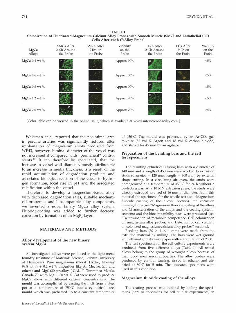
TABLE IColonization of Fluorinated-Magnesium-Calcium Alloy Probes with Smooth Muscle (SMC) and Endothelial (EC)
Cells After 240 h (P:Alloy Probe)
MgCaAlloys
SMCs After240h Aroundthe Probe
SMCs After240h onthe Probe
Viabilityon theProbe
ECs After240h Aroundthe Probe
ECs After240h onthe Probe
Viabilityon theProbe
MgCa 0.4 wt % Approx 90% <5%
MgCa 0.6 wt % Approx 80% <5%
MgCa 0.8 wt % Approx 90% <5%
MgCa 1.2 wt % Approx 70% <5%
MgCa 2.0 wt % Approx 70% <5%
[Color table can be viewed in the online issue, which is available at www.interscience.wiley.com.]
764 DRYNDA ET AL.
Journal of Biomedical Materials Research Part A

sodium hydroxide solution (cNaOH 5 200 g/L; t 5 3 h) toestablish a thick Mg(OH)2 layer. Thereafter, the bars wereimmersed into hydrofluoric acid (HF 40%; t 5 96 h) toconvert the Mg(OH)2 layer into MgF2 (Mg(OH)2 þ 2HF ?MgF2 þ 2H2O). To avoid inhomogeneous layers the speci-mens were slowly moved within the liquids during thecoating procedure. Scanning electron microscopy (SEM)confirmed the generation of an MgF2 layer with a thick-ness of 10–20 lm covering the MgCa alloy (Fig. 1, leftside).
The composition of the layers was investigated byEDX-line scans using SEM. Therefore, the cross-sectionof the specimen was analyzed to the calcium, magne-sium, oxygen, and the fluoride X-ray signal (Fig. 1, rightside). The element allocation shows that MgF2 coats thesurface by the conversion of the Mg(OH)2 by thefluoride.
Characterization of the alloys and thecoating system
The mechanical properties of the produced magnesium-calcium alloys were investigated by a standard tensile testaccording to DIN EN 10002. Therefore, the specimenswere produced according to the DIN 50125 (shape B) byturning. Fifteen tensile test specimens of each alloy compo-sition were tested to confirm the results statistically.
The protective function of the MgF2 layer was investi-gated by a four-point bending test combined with an elec-trochemical measurement. The equipment consisted of thefollowing components:
� Four-point loading gadget with an integrated gal-vanic cell (Pt; Mg/MgF2; Ag/AgCl [3 mol KCl;DV(NHE) 5 2207 mV]) (self-construction)
� Instron material testing facility (Model 1251) (Instron,Buckinghamshire, England)
� Potentiostat (Wenking Model MP 81; Bank-Elektro-nik, Goettingen, Germany)
The four-point-loading gadget with an integrated gal-vanic cell was assembled between the chuck heads of thetesting device. The galvanic cell (PMMA), two cylindricalelectrolyte reservoirs were interconnected with a smalltube, serving as specimen holder. The specimen was gluedcrossways in an integrated flute into the tube (POP-mixpolymer; WEICON GmbH & Co. KG, Munster, Germany).The glue sealed the cell to exclude leakage interfering withthe corrosion measurements. The potentiostat controlledthe cell current if a potential difference occurred and gaveindications of the layer destruction and the repassivationprocesses on the surface. In the potentiostatic mode theopen circuit potential (OCPmagnesium fluoride) was deter-mined and permanently adjusted. The automatic OPC-con-trol of the potentiostat implemented a zero-current intervalinto the cell. With the destruction of the MgF2 coating theOCPmagnesium fluoride changed to the value of the bulkmaterial (OCPmagnesium). This potential difference wasregulated by the potentiostat with an adequate cell currentback to the previously definite adjusted value of theOCPmagnesium fluoride. The initiation of the potential adjust-
ment by the cell current indicated the beginning of thelayer destruction. The cell current (I), the force (F), and thedisplacement (DL) were measured in relation to time.Results were recorded and plotted. To initiate an adequatecell current for all measurements an electrolyte consistingof 5% NaCl (Merck, Germany) in deionized water wasused. The decoupling and isolation of the specimen fromthe testing facility the loading gadget and the force trans-mission was implemented with Al2O3-rolls (cylindricalwith a very low-electric conductivity).
Corrosion of the alloys
Electrochemical properties of the different alloys weredetermined to measure the open circuit potential and thecorrosion current density. All electrochemical measure-ments were performed in a three electrode standard cell(filled with 0.5, 0.9, 2.5, or 5.0% NaCl (w/v), 378C) andconnected to an electrochemical workstation (IM6, ZahnerElektrik, Germany). Specimens were embedded in epoxyresin and ground with SiC-paper (300/600/800/1200/2000) and then polished with an alcoholic diamond sus-pension to a maximum roughness of 1 lm before theywere attached to the specimen holder. To avoid the alkali-zation of the electrolyte during the Mg corrosion by theformation of hydrogen gas and hydroxide ions the electro-lyte was buffered with a tris[2-amino-2-(hydroxymethyl)-propan-1,3-diol] solution to a constant pH value of 7.4(physiologically pH value). A platinum electrode was usedas a counter electrode. The reference potential wasobtained by use of a silver/silver chloride electrode in sat-urated potassium chloride (SE11, Sensortechnik MeinsbergGmbH, Germany) installed with a Hubber Luggin capil-lary separating the solute Mg2þ ions from the referenceelectrode.
To compare the corrosion rates of the different MgCaalloys, the amounts of gas formation (H2) were investi-gated. Therefore cylindrical specimens (d 5 16 mm; h 5 5mm) were immersed in 15 L bowls filled with an isotonicNaCl concentration of 0.9% (buffered at pH 5 7.4, 378C).The physiological chloride concentration was used to eval-uate the alloys corrosion kinetics in an isotonic solutionanalogous to the body environment. Above the specimen,a solution filled container was arranged to catch the aris-ing gas. The electrolyte composition was kept at constantlevels by gentle mixing of the solution. The gas formationwas detected automatically by capture of digitalphotographics set at an interval of 15 min.
Isolation and cultivation of primary cells
Primary human vascular smooth muscle cells (SMCs)and endothelial cells (ECs) were isolated from umbilicalveins according to standard protocols.19 The purity of theSMCs and ECs was assessed by immunohistochemistry,using a primary mouse anti-human antibody (DakoM0851, Dako, Hamburg, Germany) and a secondary dye-labeled goat anti-mouse antibody (Alexa 488, Invitrogen,Karlsruhe, Germany) for SMCs, respectively a primary rab-bit anti-human ‘‘von Willebrand’’ factor antibody (DAKO
NOVEL CORRODIBLE FLUORIDE-COATED MAGNESIUM-CALCIUM ALLOY 765
Journal of Biomedical Materials Research Part A

A0082) and a secondary dye-labeled anti-rabbit antibody(DAKO F0205) for ECs. The cells from several individualswere pooled to account for unavoidable variations in geno-type and phenotype. Reproducibility was ensured by stor-ing cell culture aliquots in liquid nitrogen until use. Afterthawing the SMCs were cultivated in SmGM medium(SmGM-2-Bulletkit, orderID#CC-3182, Lonza, Switzerland),containing 5% fetal bovine serum (FBS), gentamicin sul-fate, amphotericin B, human fibroblast growth factor-b(hFGF-b), and human epidermal growth factor (hEGF).ECs were cultivated in EGM medium (EGM Bulletkit,orderID#CC-3162, Lonza, Switzerland), containing 2% FBS,gentamicin sulfate, amphotericin B, hydrocortisone, hEGF,vascular endothelial growth factor (vEGF), hFGF-b, R3-insulin-like-growth-factor-1 (R3-IGF-1), ascorbic acid, andheparin at 378C in a humidified 5% CO2 atmosphere. Forcell culture experiments the SMCs and ECs were used upto passage 10. For cultivation of EC the cell culture flaskwere coated with 1% gelatine (w/v) (Sigma, Munich,Germany) in PBS (Sigma).
For cell culture experiments, the cells were seeded at adensity of 5 3 103 in 100 lL medium per well of a 96-wellmicrotiter plate (Corning, Netherlands) and incubated for24 h under standard conditions.
Determination of metabolic competence
The alloy components were added in form of solublesalts. Magnesium was used as magnesium sulfate heptahy-drate (MgSO4 3 7H2O; Merck, Darmstadt, Germany),Calcium was used as calcium chloride (CaCl2; Merck,Darmstadt, Germany). Metabolic competence of the cellsexposed to various concentrations of calcium and magne-sium salts was measured using the WST-1 test (Roche,Germany orderID#1644807). The principle of this testdepends on the cleavage of the tetrazolium salt (4-[3-(4-iodophenyl)-2-(4-nitrophenyl)-2H-5-tetrazolio]-1,3-benzenedisulfonate) to formazan, by mitochondrial dehydroge-nases. The augmentation of enzyme activity leads to anincrease in the amount of formazan dye formed, whichdirectly correlates to the number of metabolically activecells in the culture. After 24, 72, 144, and 240 h the cellculture medium was removed and replaced by 100 lLmedium containing 10% of the WST-1 reagent. After an
incubation period of 2 h (378C, humidified 5% CO2 atmos-phere), the optical density (OD) at 440 nm was determinedrelative to a reference OD at 650 nm using an ELISAmultiplate reader (Sunrise, Tecan). The background wasadjusted using 100 lL of each medium, containing 10%WST-1 without cells.
Cell colonization on magnesium-calcium alloy probes
Five different (uncoated and fluoride coated) magne-sium alloy probes (diameter 10 mm) were fixed with par-affine on six-well cell culture plates. Ethanol (70% (v/v))for disinfection of the probes was added until the surfacesof the probes were covered. After 1 h, the alcohol wasremoved and the plates were dried on air (10 min). Subse-quently, the probes were covered with fetal calf serum andincubated for 30 min. After removal of the serum, theplates were again dried on air. 1 3 105 cells in a small vol-ume were dropped to the surface of the magnesium alloysafterward the plates were filled with the appropriate cellsuspensions and incubated for 24, 72, and 240 h.
Detection of cell viability on colonizedmagnesium-calcium alloy probes
After an incubation time of 10 days, the cell culturemedium was removed. After washing twice with PBS, the
Figure 1. SEM cross-section of the magnesium fluoridelayer on magnesium (left side), EDX-mapping of the alloycomponents, and MgF2 (right side). [Color figure can beviewed in the online issue, which is available at www.interscience.wiley.com.]
Figure 2. Mechanical properties of a new alloying system(MgCa) which is exclusively composed of the mineralnutrients magnesium and calcium. [Color figure can beviewed in the online issue, which is available at www.interscience.wiley.com.]
766 DRYNDA ET AL.
Journal of Biomedical Materials Research Part A

viability of SMCs and ECs on the alloy probes was deter-mined using a live/dead, viability/cytotoxicity assay(Molecular probes, L-3234, Eugene, OR). The principle ofthe assay is that calcein diffuses through cell membranesand reacts with intracellular esterase to produce greenfluorescence (living cells), whereas ethidium homodimerdiffuses only through damaged cell membranes and bindsto nucleic acids to produce red fluorescence (dead cells).The assay was performed according to the manufacturer’sinstructions.
Analysis of data
The experiments for the determination of metabolicactivity were performed as quintuplicates. From five inde-pendent readings, the median was calculated with thecorresponding standard deviation. The significance of met-abolic activity data was analyzed using the one-sample,double-sided student’s t-test. The p-values were calculatedfor each data point. The p-values < 0.05 were consideredto be significant.
The tensile tests were implemented by the investigationof 15 specimens of each alloy composition. To confirm theresults statistically, the median of the 15 values was calcu-lated with the corresponding standard deviation.
The current density was determined by geometricalanalysis of the Tafel plots into the measured current den-sity potential curves of the electrochemical measurements.
RESULTS
Evaluation of the optimum calcium content in thebinary MgCa alloys by mechanical testing andcorrosion measurements
Alloying of low-calcium amounts, up to 4 wt %,led to an increase of the tensile strength up to �210–240 MPa compared with extruded pure magnesium(Rm < 200 MPa). The results show that with increas-ing concentrations of calcium the tensile strengthalso steadily increases (Fig. 2). For low alloyed com-positions, the 0.2 elastic limit is about 80 MPa lowerthan the tensile strength. This explains the relativehigh plasticity (Fig. 2). The difference of the tensilestrength (Rm) and the elastic limit Rp0.2 decreases upto 40 MPa for an increasing amount of calcium.From an amount of 2 wt % of calcium no more sig-nificant increase of the yield strength was observed.The workability also decreased for an increasingamount of calcium. Regarding to direct extrusion ofmagnesium, elevation of force is increased signifi-cantly for increasing amounts of calcium. The proc-essing of alloys containing >4 wt % of calcium canonly be processed by indirect extrusion because thecontainer friction can be neglected as a limiting fac-tor. For increased amounts of calcium, an increasedeutectic phase with a melting temperature of 516.58C
was observed. This can lead to an increased amountof hot cracks if the temperature of deformation is thesame as the melting temperature of the eutectic.
The electrochemical investigations demonstratedthe inhibitory effects of increasing calcium concen-trations within the alloy on its corrosion rate. Atdifferent chloride ion concentrations, the pure mag-nesium showed a higher corrosion current densityand it decreased at calcium contents of 0.4–0.8 wt %to a minimum (Fig. 3). At calcium concentrationsbeyond 0.8 wt %, the corrosion current densityincreased again. The application of the fluoride layerled to a better electric insulation of the surface bythe high impedance of the salt and shows theprotecting character of the material. The current den-sity decreased to values lower than 150 lA/cm2 andno further dependence on the Ca-content had beenobvious.
The structural constitution of the producedMgCa alloys is marked to the formation of eutectic(a-phase and Mg2Ca). The distribution of the eutec-tic strongly depends on the extrusion process. Onthe longitudinal micrograph of the same alloy, aclear formation of bands in the extrusion directionoccurs [Fig. 4(b)]. The micrograph of the cross-section shows the homogeneous distributed eutecticat an MgCa 0.4 alloy [Fig. 4(a)]. The increasedcalcium content leads to an increased part ofeutectic and forms a relative inhomogeneousmicrostructure.
Figure 3. Corrosion current densities at the OCPcalculated by the Tafel-analysis of the current density—potential—curves of different MgCa alloys at differentchloride concentrations.
NOVEL CORRODIBLE FLUORIDE-COATED MAGNESIUM-CALCIUM ALLOY 767
Journal of Biomedical Materials Research Part A

The longitudinal micrograph [Fig. 4(d)] clearlyshows these inhomogeneities by extreme formation ofeutectic bands. The shape of the eutectic is very longstretched and the bands are parallel to each other.Therefore, the material should show a characteristicanisotropy which has to minimize a special heat treat-ment. The cross-section [Fig. 4(c)] shows the lamellarshape of the eutectic of the MgCa2.0 alloy.
The grain size of the different MgCa alloys in themicrostructure of the extruded material shows nofundamental grain refining at increasing calciumcontent. The refining influence of calcium only existsin casted microstructures of such alloys. The recrys-tallization process and the grain deformation during
the extrusion lead to an alloy independent grain sizewhich ranged from 5 to 30 lm (Fig. 5). At highstrains caused by deformation, such as the extrusionof tubes with a following mandrel, the cross- andthe longitudinal-section cannot be distinguished bythe grain size and the shape. The characteristic ofsuch a microstructure is the high amount of twin-ning which can be easily removed by stress freeannealing.
The electrochemical results were confirmed by thequalitative assessment of hydrogen formation duringthe corrosion of the alloy probes in sodium chloridesolutions. The uncoated pure magnesium rapidlycorroded after immersion, with a strong gas forma-
Figure 4. Microstructure (precipitations) of MgCa 0.4 wt % (cross a; longitudinal b) and MgCa 2.0 wt % (cross c; longitudi-nal d) etched with 2% nitric acid. [Color figure can be viewed in the online issue, which is available at www.interscience.wiley.com.]
Figure 5. Grain size of a extruded MgCa 0.4 wt % (left) and MgCa 2.0 wt % (right) alloy (picronitric acid). [Color figurecan be viewed in the online issue, which is available at www.interscience.wiley.com.]
768 DRYNDA ET AL.
Journal of Biomedical Materials Research Part A

tion. Figure 6 shows the long-term plot (1000 h) ofthese investigations. The decreased corrosion rate atsmall amounts of calcium (0.4–2.0 wt %) and thecomparison between coated and uncoated surfaces isclearly demonstrated. Of all examined alloys, theslowest corrosion was observed by adding Ca con-tents from 0.6 to 0.8 wt %. At higher Ca concentra-tions, the hydrogen formation increased again as anindicator of corrosion. The fluoridation of the surfacewith MgF2 layer measuring 15–20 lm delayed theinitiation of the corrosion processes. In fluorinatedalloy probes, no gas formation was detected withinthe first 8–40 h.
To characterize the stability of the coating on theMgCa alloys during deformations, the results of thecorrelated bending-corrosion-test with an uncoatedspecimen are shown in Figure 7 (top) in reference tothe coated material. The bending force increased upto a value of 630 N and implemented a bar dilata-tion of 2.2 mm. After finishing of bending (DL 5 0),the force decreased due to the material relaxation.The material relaxation decreased the force to 380 N.The result of the current signal measurement was aconstant signal with no increase or decrease duringthe bending test and shows that no insolating sur-face layers exist. Compared with Figure 7 (top), themeasurement illustrated in Figure 7 (middle) showsa very different current signal. The load up to 300 Nlead to an increase of the cell current. The increaseof the force to 450 N induced the voltage control ofthe potentiostat and current was supplied into thecell. The rising of the current at maximum force was�12 mA. The interruption of bending test decreasedthe cell current clearly and the current decreased tothe initial value during 70 s. The relaxation of themagnesium is also visible. A cyclic iteration of thebending test on a coated specimen is shown inFigure 7 (bottom). The repeated bending resulted ina cyclic current signal of the potentiostat. The cur-rent at the twice bended specimen also arose and
decreased twice in correlation to the bending force.At first, the bending force measured 350 N. A stopfor 100 s decreased this value of the current from 15to 0 mA. The force decreased through the relaxationprocess to 240 N. During the second bending of thespecimen, the current increased again to 5 mA anddecreased after the interruption to 0 mA. The secondload amounted also 350 N. The relaxation of thematerial after the second bending was the same likein the first bending step.
Using the geometric base of the deflection line cal-culation, the force initiation during the bending andthe description of the bending moment for the four-point bending test, the surface tension of the layer fail-ure was calculated. The measured deflection of z 5 2mm caused a longitudinal elongation on the surfaceof 0.5–1%. From the measured forces, a surface normal
Figure 6. Measurement of the hydrogen formation onpure magnesium and MgCa—alloys (duration time 1000 h).
Figure 7. Four-point bending corrosion test of MgF2coated magnesium specimen (top: uncoated specimen;middle: MgF2 coated static test; bottom: MgF2 coated cyclictest.
NOVEL CORRODIBLE FLUORIDE-COATED MAGNESIUM-CALCIUM ALLOY 769
Journal of Biomedical Materials Research Part A

tension r between 73 and 98 N/mm2 was calculated.After the test, we were able to demonstrate an abun-dance of corrosive attacks in the contact area to theelectrolyte. Cracks and surface damages indicated thecorrosive influence of the bending load and the plasticdeformation of the material. Corrosion products weredeposited on the surface and the crack widths meas-ured from 1 to 10 lm. At the ground of the crack nofluorine, but higher amounts of oxygen, were detectedby EDX-mappings.
As shown in Figure 1, the MgF2 coating was wellconnected to the metal layer. The element distribu-tion obtained by EDX-scans indicates a zoningmechanism. The gradient of Mg distribution is anindicator for the change over of the layer. Duringthe coating procedure, Mg was transformed toMg(OH)2 and subsequently to MgF2. The signal forCa indicates that the small amounts of calcium donot influence the zoning process. The maximum sig-nal for oxygen explains that the pretreatment of theMgCa leads in a first step to Mg(OH)2 layer which isthen transformed to MgF2 on substitution of hydrox-ide ions through fluoride ions (indicated by thefluorine signal). The fluoridization occurs from theoutside to the inside depending on the immersiontime in hydrofluoric acid.
Evaluation of the biocompatibility ofthe alloy components
In a first set of cell culture experiments, we ana-lyzed the influence of magnesium and calcium ions(see ‘‘Isolation and cultivation of primary cells’’ sec-tion) with regard to their influence toward the prolif-eration of human smooth muscle and endothelialcells. Figure 8(a) displays the metabolic activity ofsmooth muscle cells after stimulation with magne-sium sulfate in a concentration range from 0.1 to5 mg/mL. Up to 1 mg/mL only slight alterations inmetabolic activity were observed, higher concentra-tions led to a significant (p < 0.05) downregulationto baseline levels indicating cytotoxic effects at aconcentration of 5 mg/mL. The incubation of endo-thelial cells with magnesium sulfate concentrationsgreater than 1 mg/mL led to a significant (p < 0.05)decrease of metabolic activity as indicated in Figure8(b). A concentration of 5 mg/mL exhibited strong
Figure 8. (a) Human smooth muscle cells (5 3 103 each)were incubated with Mg(II) ions (magnesium sulfate hep-tahydrate) in a concentration range from 0.1 to 5 mg/mL(4–206 mM). The metabolic activity was measured in atime period from 24 to 240 h using WST-1. The data werecalculated as percentage related to unstimulated cells. Allindicated data points are mean values of five independentmeasurements. The p-values of each data point are listedlater. The p-values < 0.05 were considered to be significant(one sample, double sided t-test). (b) Human endothelialcells (5 3 103 each) were incubated with Mg(II) ions (mag-nesium sulfate heptahydrate) in a concentration rangefrom 0.1 to 5 mg/mL (4–206 mM). The metabolic activitywas measured in a time period from 24 to 240 h usingWST-1. The data were calculated as percentage related tounstimulated cells. All indicated data points are mean val-ues of five independent measurements. The p-values ofeach data point are listed later. The p-values < 0.05 wereconsidered to be significant (one sample, double sidedt-test).
770 DRYNDA ET AL.
Journal of Biomedical Materials Research Part A

cytotoxicity for all indicated time points. Incubationof smooth muscle cells with calcium [Fig. 9(a)] led toan almost linear, significant (p < 0.05) decrease ofmetabolic activity in a concentration range from 0.1to 5 mg/mL. Concentrations >5 mg/mL exhibitedalso significant cytotoxic effects.
As shown in Figure 9(b), the sensitivity of endo-thelial cells toward calcium is considerably largercompared with smooth muscle cells. For incubationtimes >24 h already concentrations larger than1 mg/mL led to a significant (p < 0.05) downregula-tion to baseline levels.
Figure 9. (a) Human smooth muscle cells (5 3 103) wereincubated with Ca(II) ions (calcium chloride) in a concen-tration range from 0.1 to 10 mg/mL (2.5–250 mM). Themetabolic activity was measured in a time period from 24to 240 h using WST-1. The data were calculated as percent-age related to unstimulated cells. All indicated data pointsare mean values of five independent measurements. Thep-values of each data point are listed later. The p-values <0.05 were considered to be significant (one sample, doublesided t-test). (b) Human endothelial cells (5 3 103 each)were incubated with Ca(II) ions (calcium chloride) in aconcentration range from 0.1 to 10 mg/mL (2.5–250 mM).The metabolic activity was measured in a time periodfrom 24 to 240 h using WST-1. The data were calculated aspercentage related to unstimulated cells. All indicated datapoints are mean values of five independent measurements.The p-values of each data point are listed later. Thep-values < 0.05 were considered to be significant (one sam-ple, double sided t-test).
Figure 10. Human smooth muscle cells were incubatedwith the magnesium-based alloy MgCa 0.8, uncoated(a) and fluoride coated (b). The picture shows the hydro-gen gas formation as an indication for the decompositionprocess after 24 h. The cell culture medium used for theexperiment represents normal physiological conditions.[Color figure can be viewed in the online issue, which isavailable at www.interscience.wiley.com.]
NOVEL CORRODIBLE FLUORIDE-COATED MAGNESIUM-CALCIUM ALLOY 771
Journal of Biomedical Materials Research Part A

Cell colonization on magnesium alloy probes
In a second set of cell culture experiments, we ana-lyzed the capability of smooth muscle and endothelialcells to adhere on different magnesium-calcium alloyprobes with regard to alloy decomposition and cellviability.
The cell colonization experiments were performedusing five different magnesium-calcium alloy probeswith Ca concentrations of 0.4, 0.6, 0.8, 1.2, and 2 wt% according to the experimental proceduredescribed in ‘‘Magnesium fluoride coating of thealloys’’ section. After a time period of 24 h all testeduncoated alloy probes exhibited a strong bubble andfoam generation accompanied by discoloration. Thisis exemplarily shown in Figure 10(a) for the alloyMgCa0.8 incubated with smooth muscle cells. Aftera time period of 72 h, there was no cell colonizationon all tested alloys.
An identical experimental setup was used to inves-tigate cell colonization on the five different MgF2coated alloys (‘‘Preparation of the bending bars andthe cell test specimens’’ section). After incubation for24 h no bubble or foam generation was observed, asshown for MgCa0.8 in Figure 10(b). In contrast to theuncoated alloys, after 240 h the fluorinated alloyswere colonized with cells (Table I). A live/dead assaywas performed for each alloy as well as for the cellsaround the alloy probe to determine the cell viabilityafter 10 days of incubation. Smooth muscle cells aswell as endothelial cells around the alloy probes werestill alive (>95%) (Table I, columns 2 and 5).The colo-nization of the alloy probes with living smooth musclecells was between 70 and 90%, whereas on the alloyprobes incubated with endothelial cells an abundanceof dead cells was recognized. The percentage ofadherent living cells was below 5% (Table I, columns3 and 6).
As a sign of decreased corrosion rates, bubble for-mation was moderate and all coated alloys showedonly minor accumulation of corrosion products onthe surface when compared with uncoated alloys.
DISCUSSION
Cardiovascular stents produced from magnesium-based alloys have already entered clinical trials inpatients with peripheral arterial obstructions14 andcoronary artery disease.20 However, results ofrecently published clinical data have demonstrated,that currently available magnesium-based coronarystents have major limitations. After stent implanta-tion for atheromatous obstruction of coronaryvessels, it has been demonstrated, that the loss ofluminal diameter of the vessel treated with a magne-
sium stent was equivalent to the luminal diameterafter bare metal stenting and inferior to drug elutingstents.20 Besides an ‘‘in stent neointimal prolifera-tion’’ another entity—so far unknown after stentimplantation—the ‘‘ex-stent restenosis’’ with thicken-ing of the media of the stented vessel was observed.This led to reduced luminal area contributing to anincreased maze rate after implantation of a magne-sium stent if compared with bare metal stents anddrug-eluting stents.20 Calcifications of the treatedvessels were observed accompanied by medialdestruction. Unexplained swelling of the vessel wallcontributed to a net loss of luminal area.18
Possible factors contributing to the describedadverse effects are:
1. Too rapid degradation rate of the alloysexceeding the achievable clearance rate of thealloy components with:a. Rapid release and accumulation of hydro-
gen due to the corrosion of magnesiumleading to vascular damage.
b. Rapid accumulation of alkaline ions due tothe corrosion of the magnesium alloy lead-ing to vascular damage.
c. Toxic effects of alloy components. The per-centage of rare earths metals in the WE43alloy varies between 2.4 and 4.4%, the min-imal content of zirconium is at least 0.4%(Elektron data sheet for WE4321). Therelevance of these ‘‘additional elements’’ interms of biocompatibility and toxicity isnot yet clear.17,22,23
2. Inferior mechanical properties or inadequatestent design contributing to early stent fatiguefractures leading to mechanical negative inter-ference with the vessel wall.
To address the aforementioned limitations of cur-rently available magnesium-based alloys, this studywas designed to produce a magnesium-based alloyand characterize magnesium fluoride covered binarymagnesium-calcium alloys with different amounts ofcalcium in the alloy. The investigations of the MgCaalloys show a good workability and adequatemechanical properties at a calcium concentration<1.5 wt %. The increase in calcium contentdecreased the plasticity of the alloys but the extru-sion quality is good. No hot crack occurred duringthe extrusion to an amount of 4 wt % calcium andthe rod surface was bright and plain. The polariza-tion curves showed a minimum current density atCa contents of 0.6–0.8 wt %.
From a material scientific view, the production ofmedical implants, like stents, can be realized withthese alloy systems due to the fact that MgCa alloyswith a Ca content <1 wt % are easily castable, mach-
772 DRYNDA ET AL.
Journal of Biomedical Materials Research Part A

inable and deformable. Therefore, the product prop-erties are easy to reproduce. The production of stent-tubes by metal forming processes like drawing ispossible because of the sufficient ductility which isrelated to the small grain size.24
Corrosion of the MgCa system
The NaCl electrolyte system with different concen-trations was selected to compare the materials andits corrosion behavior in vitro. The low impedancewith higher amounts of chloride of the electrolyteoffers reproducible results with tight tolerances. Thediscussion of the electrochemical results in the viewof the corrosion behavior will be more complicatedwith the use of serum or blood plasma because ofthe poor stability of these solutions.
The alloy system used in our study showed prom-ising approaches to reach improved mechanical anddegradation properties in comparison with the alloyWE43. On the magnesium rich site of this system,the maximum solubility of calcium in the magne-sium lattice is about 0.8 wt % at room temperature.Low alloyed MgCa-systems consist of a solid solu-tion (a-phase—magnesium with interstitial calcium)as well as an eutectic one (a-phase þ Mg2Ca). Froman amount of 0.2 wt % calcium can be distinctlyseen in metallographic micrographs. The addition ofsmall amounts of calcium increases the corrosion sta-bility and has a positive influence on the graingrowth. In contrast, calcium concentrations exceed-ing the maximal solubility decrease the corrosionstability because of the formation of an intermetallicMg2Ca phase on the surface. This phase leads to ahigher corrosion rate by formation of micro-galvaniccells between Mg2Ca and the a-phase.
With this very simple binary alloying system, it ispossible to reach adequate mechanical properties toevaluate these materials. The corrosion rate can beoptimized by modification of the amount of calciumin the alloy. It has been shown, that decreasing thecorrosion rate improves mechanical properties.25–27
The addition of small amounts of calcium leads toa minimization of the gas formation during the cor-rosion of the implant, avoiding embolism and theformation of gas cavities.
Surface coating of the alloy with MgF2
The fluoride coating of the MgCa alloys dependson the formation of a hydroxide layer of a definedthickness (depending on NaOH concentration, tem-perature, and time). The thickness of this layer deter-mines the thickness of the fluoride layer because thehydroxide is converted to fluoride by immersion in
hydrofluoric acid. The thickness of the fluoride layervaries between 0.5 and 20 lm. At MgF2 coatingsexceeding 20 lm of thickness the formation of capil-lary branched cracks over the whole surface wereobserved.
By adding a surface coating with MgF2 adecreased degradation rate was accomplished. Thedegradation of stents produced from an MgF2 coatedalloy can be divided into two phases: a first ‘‘phaseof function,’’ when the stent is stable and supportsthe vessel mechanically followed by a second ‘‘phaseof degradation.’’ Optimally, the second phase startswhen the main physiological functions of the vesselwall are restored by remodeling and formation ofthe neointimal and endothelial layer. In support ofthis concept, we were able to demonstrate ex vivo,that an MgF2 layer on the surface of the alloyprotects the underlying alloy from early corrosion.
The creation of the protective layer results from asimple chemical conversion of the natural MgO/Mg(OH)2 surface layer to the MgF2 layer by immer-sion with hydrofluoric acid. The decreased electricalconductivity of the fluoride insulates the centralmagnesium/calcium bulk from the electrolyte andthereby decreases the corrosive attack of chlorideions. Therefore, the corrosion of the magnesium/calcium bulk cannot start before the protective coat-ing has dissolved. The solubility of magnesium fluo-ride in aqueous physiological solutions is very low(0.13 g/L) and prevents a fast ion leakage. The layerthickness by the NaOH pretreatments of the magne-sium specimen amounts �20 lm. For this reason, itmight be possible to adjust the degradation rate ofan implant by modification of the thickness of theprotective MgF2 layer.
It has been shown that the deformation of thecoated material does not result in a decline of degra-dation characteristics, although crimping and dilata-tion lead to partial deterioration of the layer. Theformation of insoluble Mg(OH)2 in the crack groundoccurs very fast, resulting in a good insulation of themetal from the electrolyte.
During the bending test an effective repassivationof the MgF2 layer was demonstrated. Relaxation ofthe bulk material at constant loads and the reductionof corrosion aiding internal mechanical stress werethought to be the reason for this observation. Fur-thermore, the natural passivation process in thecrack grounds takes place by the formation of anMg(OH)2 layer. Because of the small width of thecracks (crack width < 10 lm) and their length (cracklength < 250 lm) big tensile stress cannot be builtup like in plane Mg(OH)2 surface areas. Therefore inthe crack grounds, a dense Mg(OH)2 filling occurswhich acts as a protective insulation of the alloyfrom the electrolyte. Cyclic deformations of MgF2layers showed a cyclic repassivation. The deforma-
NOVEL CORRODIBLE FLUORIDE-COATED MAGNESIUM-CALCIUM ALLOY 773
Journal of Biomedical Materials Research Part A

tion of such layers does not lead to an uncontrollablecorrosion of the magnesium bulk.
Biocompatibility of the magnesium-calcium alloy
Magnesium and calcium are essential elements forthe human organism. The daily recommended intakefor Ca is 80–100 mg/day and for Mg 75–80 mg/dayfor children up to an age of 2 years.28 For adults, therecommended daily intake is �1000 mg/day. Whenadministered parenterally, no adverse effects at theinjection site were observed.
Magnesium is the major component (at least 98 wt%) of all tested alloys, is predominantly stored inbone and the intracellular compartments of muscleand soft tissues; <1% of total body magnesium iscirculating in the blood.29 Magnesium homeostasisdepends on the balance between intestinal absorp-tion and renal excretion.30 In this study, five differ-ent magnesium-calcium alloys were tested uncoatedas well as MgF2 coated for their biocompatibilitywith regard to vascular cells. Furthermore, theeffects of Mg and Ca salts have been tested onhuman vascular cells in a cell culture model.
It has been shown that currently used magnesiumstents for cardiovascular applications can be safelydegraded within 4 months.20 Assuming a linearrelease of alloy components, a uncoated new magne-sium-calcium stent (Mg:Ca: 98:2 (w/w)) with a massof 10 mg would lead to a liberation of �82 lg Mgand 2 lg Ca per day. An MgF2 layer would furtherdecelerate this degradation kinetics. Our cell culturedata indicate, that calculated magnesium and cal-cium concentrations resulting from the degradationof a cardiovascular stent do not lead to an increasedproliferation of smooth muscle cells. Furthermore,no toxic or other adverse effects were observed forthese concentrations.
Biocompatibility of the MgF2 coating
Because of the low solubility of MgF2 toxic effectsrelated to the decomposition of the MgF2 layer arenot expected, because the amounts of MgF2 on thealloy probes used in this study are very small (�1.3mg on an alloy surface with a diameter of 5 mm).The average daily intake of dietary fluoride byyoung children is �0.5 mg or 0.04–0.07 mg/kg perday.31 Therefore, fluoride concentrations caused byMgF2 decomposition should have no systemiceffects. If the average decomposition time of theMgF2 coated alloys is estimated with considerable>4 months, major local toxic effects in vivo are alsounlikely. Even though we cannot exclude themtotally, due to local accumulation leading to
increased concentrations within the vessel wall, thistheoretical assessment was confirmed by cell culturedata. It seems that the MgF2 layer of the five fluori-nated alloys has different effects on smooth muscleand endothelial cells. All five alloys were signifi-cantly colonized with smooth muscle cells after 10days (at least 100 cells per alloy surface (�20 mm2)),whereas for endothelial cells no significant coloniza-tion was observed (<5%). Probably, the cells diedbecause the alloy surface was not sufficient for opti-mal adherence. As mentioned in ‘‘Isolation and culti-vation of primary cells’’ section, endothelial cellsneed more specialized surfaces for optimal growingthan smooth muscle cells. Toxic influences of degra-dation products can be excluded because both celltypes appeared visible nearby the alloy probes.
In contrast to in vivo conditions, our closed experi-mental cell culture model is not able to remove corro-sion products from the implantation site. Therefore,further animal experiments with fluorinated MgCaalloys are ongoing to elucidate their biocompatibilityin vivo.
CONCLUSION
Results of our study suggest that the developedMgF2 coated magnesium-calcium alloys may servefor the production of biodegradable corrosive cardio-vascular stents. In comparison with currently avail-able magnesium-based alloys, they exhibit improvedmechanical features, decreased degradation kineticsand a good biocompatibility toward vascular cells.Experiments with the most promising fluoride-coated alloys are under way to elucidate mechanicalfeatures, degradation and biocompatibility in anin vivo animal model.
References
1. Sigwart U, Puel J, Mirkovitch V, Joffre F, Kappenberger L.Intravascular stents to prevent occlusion and restenosis aftertransluminal angioplasty. N Engl J Med 1987;316:701–706.
2. Mullins CE, O’Laughlin MP, Vick GW III, Mayer DC, MyersTJ, Kearney DL, Schatz RA, Palmaz JC. Implantation of bal-loon-expandable intravascular grafts by catheterization inpulmonary arteries and systemic veins. Circulation 1988;77:188–199.
3. O’Laughlin MP, Perry SB, Lock JE, Mullins CE. Use of endo-vascular stents in congenital heart disease. Circulation1991;83:1923–1939.
4. Okubo M, Benson LN. Intravascular and intracardiac stentsused in congenital heart disease. Curr Opin Cardiol 2001;16:84–91.
5. Ryan MP, Williams DE, Chater RJ, Hutton BM, McPhail DS.Why stainless steel corrodes. Nature 2002;415:770–774.
6. Reclaru L, Lerf R, Eschler PY, Meyer JM. Corrosion behaviorof a welded stainless-steel orthopedic implant. Biomaterials2001;22:269–279.
774 DRYNDA ET AL.
Journal of Biomedical Materials Research Part A

7. Kraft CN, Diedrich O, Burian B, Schmitt O, Wimmer MA.Microvascular response of striated muscle to metal debris.A comparative in vivo study with titanium and stainlesssteel. J Bone Joint Surg Br 2003;85:133–141.
8. Carroll WM, Kelly MJ. Corrosion behavior of nitinol wires inbody fluid environments. J Biomed Mater Res A 2003;67:1123–1130.
9. Heintz C, Riepe G, Birken L, Kaiser E, Chakfe N, Morlock M,Delling G, Imig H. Corroded nitinol wires in explanted aorticendografts: An important mechanism of failure? J EndovascTher 2001;8:248–253.
10. Peuster M, Fink C, von Schnakenburg C, Hausdorf G. Disso-lution of tungsten coils does not produce systemic toxicity,but leads to elevated levels of tungsten in the serum andrecanalization of the previously occluded vessel. CardiolYoung 2002;12:229–235.
11. Peuster M, Fink C, Wohlsein P, Bruegmann M, Gunther A,Kaese V, Niemeyer M, Haferkamp H, Schnakenburg C.Degradation of tungsten coils implanted into the subcla-vian artery of New Zealand white rabbits is not associatedwith local or systemic toxicity. Biomaterials 2003;24:393–399.
12. Peuster M, Hesse C, Schloo T, Fink C, Beerbaum P, vonSchnakenburg C. Long-term biocompatibility of a corrodibleperipheral iron stent in the porcine descending aorta. Bioma-terials 2006;27:4955–4962.
13. Williams D. New interests in magnesium. Med Device Tech-nol 2006;17:9–10.
14. Witte F, Kaese V, Haferkamp H, Switzer E, Meyer-Linden-berg A, Wirth CJ, Windhagen H. In vivo corrosion of fourmagnesium alloys and the associated bone response. Bioma-terials 2005;26:3557–3563.
15. Di Mario C, Griffiths H, Goktekin O, Peeters N, Verbist J,Bosiers M, Deloose K, Heublein B, Rohde R, Kasese V, IlsleyC, Erbel R. Drug-eluting bioabsorbable magnesium stent.J Interv Cardiol 2004;17:391–395.
16. Peeters P, Bosiers M, Verbist J, Deloose K, Heublein B. Pre-liminary results after application of absorbable metal stentsin patients with critical limb ischemia. J Endovasc Ther2005;12:1–5.
17. Drynda A, Deinet N, Braun N, Peuster M. Rare earth metalsused in biodegradable magnesium-based stents do not inter-fere with proliferation of smooth muscle cells but do inducethe upregulation of inflammatory genes. J Biomed Mater ResA 2009;91:360–369.
18. Waksman R, Pakala R, Kuchulakanti PK, Baffour R, HellingaD, Seabron R, Tio FO, Wittchow E, Hartwig S, Harder C,Rohde R, Heublein B, Andreae A, Waldmann KH, HaverichA. Safety and efficacy of bioabsorbable magnesium alloy
stents in porcine coronary arteries. Catheter CardiovascInterv 2006;68:607–617.
19. Jaffe EA, Nachman RL, Becker CG, Minick CR. Culture ofhuman endothelial cells derived from umbilical veins. Identi-fication by morphologic and immunologic criteria. J ClinInvest 1973;52:2745–2756.
20. Erbel R, Di Mario C, Bartunek J, Bonnier J, de Bruyne B,Eberli FR, Erne P, Haude M, Heublein B, Horrigan M, IlsleyC, Bose D, Koolen J, Luscher TF, Weissmann N, Waksman R.Temporary scaffolding of coronary arteries with bioabsorb-able magnesium stents: A prospective, non-randomised mul-ticentre trial. Lancet 2007;369: 1869–1875.
21. Magnesium Elektron, Datasheet: 478, May 2006, MagnesiumElektron, UK. Available at: http://www.magnesium-elektron.com/data/downloads/DS478WroughtWE43.pdf.
22. Hirano S, Suzuki KT. Exposure, metabolism, and toxicity ofrare earths and related compounds. Environ Health Perspect1996;104(Suppl 1):85–95.
23. Nakamura Y, Tsumura Y, Tonogai Y, Shibata T, Ito Y. Differ-ences in behavior among the chlorides of seven rare earthelements administered intravenously to rats. Fundam ApplToxicol 1997;37:106–116.
24. Hassel T. Beitrag zur Entwicklung bioresorbierbarer implantat-materialien auf magnesium-basis, Dissertation LU Hannover:Berichte aus dem IW Band 01/2008. ISBN 978-3-939026-90-7.
25. Krause C, Bormann D, Hassel T, Bach FW. Mechanical prop-erties of degradable magnesium implants in dependence ofthe implantation duration. Magnesium technology in theglobal age. In: 45th Annual Conference of Metallurgists ofCIM, QC, Canada, October 1–4, 2006. p 329–343.
26. Hassel T, Bach FW, Golovko A, Krause C. Investigation ofthe mechanical properties and the corrosion behaviour of lowalloyed magnesium calcium alloys for use as absorbable bio-material in the implant technique. Magnesium technology inthe global age. In: 45th Annual Conference of Metallurgists ofCIM, QC, Canada, October 1–4, 2006. p 359–370.
27. Hermawan H, Moravej M, Dube D, Fiset M, Mantovani D.Degradation behaviour of metallic biomaterials for degrad-able stents. Adv Mater Res 2007;15–17:113–118.
28. Abrams SA, Atkinson SA. Calcium, magnesium, phosphorusand vitamin D fortification of complementary foods. J Nutr2003;133:2994S–2999S.
29. Elin RJ. Magnesium: The fifth but forgotten electrolyte. Am JClin Pathol 1994;102:616–622.
30. Konrad M, Weber S. Recent advances in molecular geneticsof hereditary magnesium-losing disorders. J Am Soc Nephrol2003;14:249–260.
31. Whitford GM. Intake and metabolism of fluoride. Adv DentRes 1994;8:5–14.
NOVEL CORRODIBLE FLUORIDE-COATED MAGNESIUM-CALCIUM ALLOY 775
Journal of Biomedical Materials Research Part A

本文献由“学霸图书馆-文献云下载”收集自网络,仅供学习交流使用。
学霸图书馆(www.xuebalib.com)是一个“整合众多图书馆数据库资源,
提供一站式文献检索和下载服务”的24 小时在线不限IP
图书馆。
图书馆致力于便利、促进学习与科研,提供最强文献下载服务。
图书馆导航:
图书馆首页 文献云下载 图书馆入口 外文数据库大全 疑难文献辅助工具