Developing inner ear sensory neurons require TrkB and TrkC
11
INTRODUCTION The Trk family of protein tyrosine kinase receptors mediate the biological effects of the nerve growth factor (NGF) family of neurotrophins (Meakin and Shooter, 1992; Glass and Yan- copoulos, 1993). The trkA and trkC genes encode high-affinity receptors for NGF (Kaplan et al., 1991; Klein et al., 1991a) and neurotrophin-3 (NT-3; Lamballe et al., 1991), respectively. The product of the trkB gene serves as the receptor for two related neurotrophins, brain derived neurotrophic factor (BDNF; Klein et al., 1991b; Soppet et al., 1991; Squinto et al., 1991) and neurotrophin 4 (NT-4; Berkemeier et al., 1991; Klein et al., 1992; Ip et al., 1992). TrkB also binds NT-3, although the extent to which this occurs in vivo is controver- sial (Ip et al., 1993). In addition to the Trks, all neurotrophins bind to another cell surface receptor, known as p75 or the low affinity NGF receptor (LNGFR) whose role may be to modulate Trk signalling (Meakin and Shooter, 1992; Glass and Yancopoulos, 1993; Davies et al., 1994; Hantzopoulos et al., 1994; Verdi et al., 1994). Cranial sensory neurons have been especially useful for studying the specificity and time-course of the survival- promoting effects of neurotrophins (Davies, 1987; Vogel and Davies, 1991; Davies, 1994). The cochleovestibular nerve (cranial nerve VIII) contains afferent fibres whose targets are the sensory epithelia of the inner ear. Cochlear fibres commence in the organ of Corti, and vestibular fibres originate in the utricular and saccular maculae as well as the ampullary cristae of the semicircular canals. The somata of all of these afferent fibres are located in the cochlear and vestibular ganglia. Most of the cochlear and vestibular neurons are bipolar, with a peripheral process contacting the hair cells in their respective sensory epithelia, and a central process that projects in a specific topographical fashion to the cochlear and vestibular nuclei of the medulla (Spoendlin, 1988). TrkB, TrkC as well as LNGFR are expressed in the cochlear and vestibu- lar ganglion before and during innervation of their target fields, whereas TrkA is expressed only transiently and at a low level (Von Bartheld, 1991; Represa et al., 1991; Ernfors et al., 1992; Tessarollo et al., 1993; Pirvola et al., 1994; Schecterson and Bothwell, 1994; Vazquez et al., 1994). BDNF and NT-3, but not NT-4 or NGF, are detected in the sensory epithelia (Pirvola et al., 1992, 1994; Schecterson and Bothwell, 1994). In explant cultures, exogenous BDNF and NT-4 promote neurite outgrowth from the cochlear and vestibular ganglion neurons, while NT-3 is mainly effective on cochlear neurons (Pirvola et al., 1994; Vazquez et al., 1994). In dissociated neuron cultures, BDNF and NT-3 promote survival of cochlear and vestibular neurons (Davies et al., 1986; Pirvola et al., 1994; Vazquez et al., 1994). Results of the effectiveness of NGF in bioassays are controversial (Lefebvre et al., 1990, 1991; Pirvola et al., 1992, 1994). Based on spatiotemporal expression patterns and bio- 3381 Development 121, 3381-3391 (1995) Printed in Great Britain © The Company of Biologists Limited 1995 The trkB and trkC genes are expressed during the formation of the vestibular and auditory system. To elucidate the function of trkB and trkC during this process, we have analysed mice carrying a germline mutation in the tyrosine kinase catalytic domain of these genes. Neu- roanatomical analysis of homozygous mutant mice revealed neuronal deficiencies in the vestibular and cochlear ganglia. In trkB (-/-) animals vestibular neurons and a subset of cochlear neurons responsible for the inner- vation of outer hair cells were drastically reduced. The peripheral targets of the respective neurons showed severe innervation defects. A comparative analysis of ganglia from trkC (-/-) mutants revealed a moderate reduction of vestibular neurons and a specific loss of cochlear neurons innervating inner hair cells. No nerve fibres were detected in the sensory epithelium containing inner hair cells. A developmental study of trkB (-/-) and trkC (-/-) mice showed that some vestibular and cochlear fibres initially reached their peripheral targets but failed to maintain innervation and degenerated. TrkB and TrkC receptors are therefore required for the survival of specific neuronal populations and the maintenance of target innervation in the peripheral sensory system of the inner ear. Key words: trkB, trkC, mouse mutant, inner ear, vestibular ganglion, cochlear ganglion, hair cell, target innervation SUMMARY Developing inner ear sensory neurons require TrkB and TrkC receptors for innervation of their peripheral targets Thomas Schimmang 1 , Liliana Minichiello 3 , Esther Vazquez 1 , Isabel San Jose 2 , Fernando Giraldez 1 , Rüdiger Klein 3 and Juan Represa 2 1 Departamento Bioquimica, Biologia Molecular y Fisiologia y 2 Departamento Anatomia, Facultad de Medicina, Universidad de Valladolid, CSIC, 47005 Valladolid, Spain 3 Differentiation Programme, European Molecular Biology Laboratory, 69117 Heidelberg, Germany
Transcript of Developing inner ear sensory neurons require TrkB and TrkC

3381Development 121, 3381-3391 (1995)Printed in Great Britain © The Company of Biologists Limited 1995
Developing inner ear sensory neurons require TrkB and TrkC receptors for
innervation of their peripheral targets
Thomas Schimmang1, Liliana Minichiello3, Esther Vazquez1, Isabel San Jose2, Fernando Giraldez1,Rüdiger Klein3 and Juan Represa2
1Departamento Bioquimica, Biologia Molecular y Fisiologia y 2Departamento Anatomia, Facultad de Medicina, Universidad deValladolid, CSIC, 47005 Valladolid, Spain3Differentiation Programme, European Molecular Biology Laboratory, 69117 Heidelberg, Germany
The trkB and trkC genes are expressed during theformation of the vestibular and auditory system. Toelucidate the function of trkB and trkC during this process,we have analysed mice carrying a germline mutation in thetyrosine kinase catalytic domain of these genes. Neu-roanatomical analysis of homozygous mutant micerevealed neuronal deficiencies in the vestibular andcochlear ganglia. In trkB (−/−) animals vestibular neuronsand a subset of cochlear neurons responsible for the inner-vation of outer hair cells were drastically reduced. Theperipheral targets of the respective neurons showed severeinnervation defects. A comparative analysis of ganglia fromtrkC (−/−) mutants revealed a moderate reduction of
vestibular neurons and a specific loss of cochlear neuronsinnervating inner hair cells. No nerve fibres were detectedin the sensory epithelium containing inner hair cells.
A developmental study of trkB (−/−) and trkC (−/−) miceshowed that some vestibular and cochlear fibres initiallyreached their peripheral targets but failed to maintaininnervation and degenerated. TrkB and TrkC receptorsare therefore required for the survival of specific neuronalpopulations and the maintenance of target innervation inthe peripheral sensory system of the inner ear.
Key words: trkB, trkC, mouse mutant, inner ear, vestibular ganglion,cochlear ganglion, hair cell, target innervation
SUMMARY
INTRODUCTION
The Trk family of protein tyrosine kinase receptors mediate thebiological effects of the nerve growth factor (NGF) family ofneurotrophins (Meakin and Shooter, 1992; Glass and Yan-copoulos, 1993). The trkA and trkC genes encode high-affinityreceptors for NGF (Kaplan et al., 1991; Klein et al., 1991a)and neurotrophin-3 (NT-3; Lamballe et al., 1991), respectively.The product of the trkB gene serves as the receptor for tworelated neurotrophins, brain derived neurotrophic factor(BDNF; Klein et al., 1991b; Soppet et al., 1991; Squinto et al.,1991) and neurotrophin 4 (NT-4; Berkemeier et al., 1991;Klein et al., 1992; Ip et al., 1992). TrkB also binds NT-3,although the extent to which this occurs in vivo is controver-sial (Ip et al., 1993). In addition to the Trks, all neurotrophinsbind to another cell surface receptor, known as p75 or the lowaffinity NGF receptor (LNGFR) whose role may be tomodulate Trk signalling (Meakin and Shooter, 1992; Glass andYancopoulos, 1993; Davies et al., 1994; Hantzopoulos et al.,1994; Verdi et al., 1994).
Cranial sensory neurons have been especially useful forstudying the specificity and time-course of the survival-promoting effects of neurotrophins (Davies, 1987; Vogel andDavies, 1991; Davies, 1994). The cochleovestibular nerve(cranial nerve VIII) contains afferent fibres whose targets arethe sensory epithelia of the inner ear. Cochlear fibres
commence in the organ of Corti, and vestibular fibres originatein the utricular and saccular maculae as well as the ampullarycristae of the semicircular canals. The somata of all of theseafferent fibres are located in the cochlear and vestibularganglia. Most of the cochlear and vestibular neurons arebipolar, with a peripheral process contacting the hair cells intheir respective sensory epithelia, and a central process thatprojects in a specific topographical fashion to the cochlear andvestibular nuclei of the medulla (Spoendlin, 1988). TrkB, TrkCas well as LNGFR are expressed in the cochlear and vestibu-lar ganglion before and during innervation of their target fields,whereas TrkA is expressed only transiently and at a low level(Von Bartheld, 1991; Represa et al., 1991; Ernfors et al., 1992;Tessarollo et al., 1993; Pirvola et al., 1994; Schecterson andBothwell, 1994; Vazquez et al., 1994). BDNF and NT-3, butnot NT-4 or NGF, are detected in the sensory epithelia (Pirvolaet al., 1992, 1994; Schecterson and Bothwell, 1994). In explantcultures, exogenous BDNF and NT-4 promote neuriteoutgrowth from the cochlear and vestibular ganglion neurons,while NT-3 is mainly effective on cochlear neurons (Pirvola etal., 1994; Vazquez et al., 1994). In dissociated neuron cultures,BDNF and NT-3 promote survival of cochlear and vestibularneurons (Davies et al., 1986; Pirvola et al., 1994; Vazquez etal., 1994). Results of the effectiveness of NGF in bioassays arecontroversial (Lefebvre et al., 1990, 1991; Pirvola et al., 1992,1994). Based on spatiotemporal expression patterns and bio-

3382 T. Schimmang and others
Table 1. Volumes of vestibular and cochlear ganglia intrkB mutant mice
Age Ganglion wild-type trkB (−/−) Reduction (%)
14.5 p.c. cochlear 17.6±0.7 (4) 17.3±0.1 (4) not significantvestibular 13.2±0.4 (4) 12.9±0.2 (4) not significant
18.5 p.c. cochlear 28.9±0.8 (4) 27.3±0.5 (4) 6*vestibular 19.3±0.5 (4) 11.9±0.3 (4) 38**
P1 cochlear 26.8±0.3 (4) 23.9±0.4 (4) 11**vestibular 17.6±0.2 (4) 7.5±0.3 (4) 57**
The volume in 106 µm3 (mean ± s.e.m.) of ganglia were calculated in thedevelopmental stages indicated. The sampling number is shown inparentheses. Student’s two-tailed t-test determined significance levels.*P<0.005, **P<0.00005.
Fig. 1. Cell size histograms of cochlear ganglion neurons of wild-type (+/+), trkB (−/−) (A) and trkC (−/−) (B) mice. The mean numberof cells falling into each size category is expressed as a percentage oftotal cells in wild-type ganglia. (A) Only small sized (<15 µm)neurons are reduced in trkB (−/−) mutant mice. (B) TrkC (−/−)animals specifically lack large sized (>20 µm) neurons compared towild-type (+/+) mice.
logical effects, BDNF and NT-3, rather than NGF and NT-4,are considered to be responsible for the survival and neuriteoutgrowth of cochlear and vestibular neurons, acting via theirhigh-affinity receptors TrkB and TrkC, respectively.
Recently, the phenotypes of germline-targeted mutant micelacking functional neurotrophins and their receptors haveallowed the identification of the specific neuron populationsthat are dependent on neurotrophin action during development(Barbacid, 1994; Klein, 1994a; Snider, 1994). Consistent withthe NT-3 expression pattern and in vitro experiments, NT-3 (−/−) mutant mice show reductions in the cochlear and vestibu-lar ganglion (Fariñas et al., 1994). Mice carrying an inactiveform of trkC are available, but have not been analysed for innerear defects (Klein et al., 1994b). BDNF (−/−) mutant mice havesevere deficiencies in coordination and balance, suggestingabnormalities in the functioning of the vestibular system(Ernfors et al., 1994a; Jones et al., 1994). Vestibular ganglianeurons are drastically depleted and the vestibular fibres failto innervate the sensory epithelia in postnatal animals.Together with the known expression pattern of BDNF and itseffects in in vitro systems, the results suggest that most of thevestibular ganglion cells require BDNF for survival. Incontrast, the cochlear ganglion, the cochlea and the innerva-tion of inner and outer hair cells appear to be unaffected inBDNF (−/−) mice (Ernfors et al., 1994a).
In the present study, we have analysed the vestibular andauditory system in mice mutant for the TrkB and TrkCreceptor, respectively (Klein et al., 1993, 1994b). Our resultsreveal that TrkB and TrkC are required for the survival ofspecific neuronal populations and the innervation of their
Table 2. Neuron cell numbers in the vestibular andcochlear ganglia of trkB mutant mice
Age Ganglion wild-type trkB (−/−) Reduction (%)
14.5 p.c. cochlear 4570±260 (4) 4477±165 (4) not significantvestibular 3411±170 (4) 3366±184 (4) not significant
18.5 p.c. cochlear 8001±259 (4) 7652±133 (4) not significantvestibular 5330±189 (4) 3343±101 (4) 37**
P1 cochlear 7950±236 (8) 6765±119 (4) 15*vestibular 4819±115 (8) 2128±113 (4) 56**
Ganglia were dissected and sectioned at 8 µm thickness. Neurons werecounted in every sixth section. Values were not corrected for split nucleoli.Mean number of neurons (± s.e.m.) are listed and the sampling number isshown in parentheses. Differences were tested using a two-tailed Student’s t-test. *P<0.01, **P<0.00005.
peripheral targets. A developmental study of trkB (−/−) andtrkC (−/−) mice demonstrates that these neurons do not dependon functional Trk receptors for target encounter of peripheralnerve processes, but rather that they are required for neuronalsurvival and maintenance of target innervation.
MATERIALS AND METHODS
Genotyping of animalsTrkB and TrkC mutant mice were generated by breeding heterozy-gous mutant mice kept on a mixed 129/sv × C57Bl/6 background.Genomic DNA was extracted from tail biopsies of mouse embryosusing standard procedures (Laird et al., 1991). The trkB genotype wasdetermined by PCR amplification (94°C, 1 minute; 65°C, 2 minutes;72°C, 3 minutes for 40 cycles) using a common 5′ primer (5′-TCGCGTAAAGACGGAACATGATCC-3′) and either a 3′ primer

3383Inner ear defects of trkB and trkC mutant mice
for the wild-type allele (5′-AGACCATGATGAGTGGGTCGCC-3′)or a 3′ primer from the pgk-1 promoter of the neo cassette (5´-GAT-GTGGAATGTGTGCGAGGCC-3′). The trkC genotype was deter-mined using a common 5′ primer (5′-CTGAAGTCACTGGCTA-GAGTCTGGG-3′) and either a 3′ primer for the wild-type allele(5′-GTCCCATCTTGCTTACCCTGAGG-3′) or a 3′ primer from thepgk-1 promoter of the neo cassette (5′-CCAGCCTCTGAGCCCA-GAAAGC-3′). PCR amplified DNA was analyzed on a 1.5% agarosegel.
Histological analysisEntire temporal bone primordia were dissected and fixed in 4%paraformaldehyde in 0.1 M sodium phosphate (pH.7.2) for between6-12 hours, dehydrated in ethanol, embedded in paraffin, seriallysectioned at 8 µm and stained with 0.1% cresyl violet. The volume ofeach ganglion was measured using a morphometric analysis program(VIDS Image Analyzer, Synoptics Ltd, Cambridge). For counts ofcochlear and vestibular neuron nuclei were counted from cresyl violetand hematoxilyn and eosin-stained sections. Neurons were identifiedby their size and distinct nucleus. No correction was made for splitnucleoli. Nuclei were counted at 200× magnification in 4 randomlychosen fields in every section of 8 µm thickness. Up to about 80 fieldsper ganglion in sections that were 40 µm apart were analysed. No cor-rections were used because neuronal size (max. 25 µm) was alwayssmaller than the intersection interval, making it impossible to countthe same neuron twice. The cell size histograms were generated fromcomputer aided drawings of neuronal profiles from cochlear gangliaof P1 mice. At least 500 profiles were drawn and the profiles werecorrected as described by Bolender (1983). Immunohistochemistry
Fig. 2. Abnormalities in the vestibular and cochlear ganglia of trkB (−/−and trkB (−/−) mice (B,D,F,H,J). The vestibular (VG) and cochlear gang(C,D and G,H, respectively). Note the reduced cell density and enlargedmagnification of C,D,G and H, respectively. Arrows in F point to cells win B corresponds to 450 µm (A,B); 100 µm (C,D,G,H); 20 µm (E,F,I,J).
was carried out using the ABC method (Vectastain Kit, Vector Lab-oratories) on sections cut at 25 µm on a cryostat or on paraffin sections(8 µm). Sections were incubated in TBS solution (50 mM Tris-HClbuffer (pH 7.5) containing 0.1% sheep serum, 0.1% bovine serumalbumin, 0.1% Triton X-100), quenched in 3% H2O2, blocked withserum and left overnight at 4°C in TBS solution containing 2-4 µg/mlof a mouse anti-200K neurofilament monoclonal antibody(Boehringer, Mannheim). After incubation with an biotinylatedantibody and the ABC reagent, peroxidase was reacted with 0.05%diaminobenzidine tetrahydrochloride and 0.003% hydrogen peroxide.
Transmission electron microscopyMembranous labyrinths were isolated and fixed for 2 hours at 4°C in2% glutaraldehyde, rinsed twice for 15 minutes in 0.1 M cacodylatebuffer, postfixed in 2% osmium tetroxide for 30 minutes, dehydratedin a graded series of acetone and embedded in plastic. Transverse thinsections were taken at various levels, stained with uranyl acetate andlead citrate and examined under a JEOL 1200EX electron microscopeat 80 kV.
RESULTS
Homozygous trkB (−/−) mutant mice exhibit cell lossin vestibular and cochlear gangliaTo elucidate the role of TrkB in the auditory and vestibularsystem, we studied mice carrying a trkB locus specificallytargeted within its protein tyrosine kinase sequences (Klein et
) mice. Sections through the inner ear of wild-type (+/+) (A,C,E,G,I)lion (CG, arrows) are indicated in A and B and are shown in detail extracellular spaces in trkB (−/−) mutant animals. (E,F,I,J) High powerith nuclei surrounded by a very small amount of cytoplasm. Scale bar

3384 T. Schimmang and others
k of trkB leads to deficiencies of innervation in the vestibular sensoryections of P1 wild-type (+/+) (A,C,E,G) and trkB (−/−) (B,D,F,H)stained with a monoclonal antibody directed against phosphorylated neurofilaments. The utricular macula (A-D) and the ampullary crista
al semicircular canal (E-H) are shown. (A,C,E,G) Wild-type animalsnormal innervation pattern. (C,G) High power magnifications of theithelium show fibres entering the epithelium and innervation of hairrve chalicle typical of the innervation of type I hair cells is indicated byB,D,F,H) Note the overall low immunoreactivity in mutant trkB (−/−)mpared with wild-type mice. (D) In the utricular macula nerve fibresed the base of the hair cells, but are degenerating and retracting froms. (H) No innervation is observed in the sensory epithelia of thecrista. Scale bar in A corresponds to 250 µm (A,B,E,F); 50 µm (G,H);
).
al., 1993). To examine defects in vestibular and cochlearganglia, their volumes were calculated and neurons werecounted on serial sections. At day 14.5 post coitum (p.c.), mostcochlear and vestibular neurons have become postmitotic(Ruben, 1967). No differences in sizes andneuronal profiles of the ganglia were detected inwild-type or homozygous trkB (−/−) mutant mice(Tables 1 and 2). However, at day 18.5 p.c.vestibular ganglia of trkB (−/−) mice were smallerthan those of wild-type animals and the numbersof neurons were reduced. Also, the volume of thecochlear ganglion of trkB (−/−) animals was foundto be reduced to a small but significant extent. Atpostnatal day 1 (P1) neuronal defects had becomemore dramatic in the vestibular ganglion. Theganglion had shrunk to 43% of its volume and hadundergone a neuronal cell loss of 56% whencompared to wild-type animals. Neuronal cellcounts and volume calculations of the cochlearganglion revealed a 15% and 11% reduction,respectively. Cell size histograms of the cochlearganglion showed that the modest reduction ofcells in trkB (−/−) mutant mice was due to the loss(77% reduction) of small sized (<15 µm) type IIneurons, whereas large sized (>20 µm) type Ineurons were unaffected (Fig. 1). Type I and typeII neuronal cells are responsible for the innerva-tion of inner and outer hair cells in the sensoryepithelium of the cochlea, respectively(Spoendlin, 1969; Kiang et al., 1982; Sobkowicz,1992).
Cresyl violet staining of sections from trkB(−/−) mice at P1 revealed areas of reduced celldensity and large extracellular spaces in thevestibular ganglion (Fig. 2D) in addition to itssmaller size compared to wild-type mice (Fig.2C). At the cellular level, nuclei surrounded by avery small amount of cytoplasm were moreabundant in the ganglia of trkB (−/−) mutants (Fig.2F) than in wild-type animals (Fig. 2E). A similarsituation, although to a lesser extent, was alsoobserved in the cochlear ganglion (Fig. 2G-J).These results indicate that the TrkB signallingpathway is not required for the initial phase of cellproliferation and ganglion formation. The TrkBreceptor is, however, critically important forsurvival of the majority of vestibular and a smallsubset of cochlear ganglion neurons during devel-opment.
Neonatal trkB mutant mice lack vestibularfibres and exhibit selective loss of outerhair cell innervation in the cochleaTo study target innervation of the vestibular andcochlear neurons, an antibody (RT97) directedagainst phosphorylated epitopes on neurofila-ments was used (Wood and Anderton, 1981). Inthe inner ear of P1 wild-type mice, RT97immunoreactive fibres contacted the utricularmacula and the ampullary crista of the semicircu-lar canals (Fig. 3A,E). Fibre bundles reached and
Fig. 3. Lacepithelia. Smice were epitopes onof the laterrevealed a sensory epcells. A nean arrow. (animals cohave reachtheir targetampullary 30 µm (C,D
entered the sensory epithelium, where they innervated haircells. At the cellular level, innervation of type II hair cells andnerve chalicles characteristic of type I hair cells were found(Fig. 3C,G). TrkB (−/−) mutant mice revealed less immuno-

3385Inner ear defects of trkB and trkC mutant mice
ts fail to innervate the outer hair cells of the cochlea. Longitudinalial part of the cochlea of P1 wild-type (+/+) (A,C) and trkB (−/−)ve been stained with an antibody raised against neurofilaments. nerve fibres enter the sensory epithelium and innervate inner (A) ande fibres (arrows) and the sensory epithelium (arrowheads) are) The innervation pattern of inner hair cells of trkB (−/−) mutant (D) The sensory epithelium containing outer hair cells is generallycale bar in A corresponds to 75 µm in A-D.
reactivity of neurofilaments. In the utricular macula, vestibu-lar fibres were poorly stained and appeared fractionated anddegenerate (Fig. 3B). The vestibular nerve terminals hadreached the sensory epithelium, but appeared in the process ofretraction from their targets (Fig. 3D). In the ampullary cristaof trkB (−/−) mice, no innervation was visible, as revealed bythe absence of immunoreactivity for neurofilaments (Fig.3F,H).
We had observed a selective loss of small type II neurons inthe cochlear ganglion of trkB (−/−) mutant mice. Lack of theseneurons is expected to cause innervation defects of their cor-responding peripheral sense organs, the outer hair cells. Inwild-type mice, sections at the level of the outer hair cellsrevealed nerve fibres, underneath thesensory epithelium, which entered theepithelium and innervated hair cells(Fig. 4C). In trkB (−/−) mice thesensory epithelium was generallydevoid of innervation (Fig. 4D). At afew sites, retracting neurites withresidual synaptic contacts weredetected by the neurofilamentantibody. We also examined the inner-vation of inner hair cells. In both,wild-type and trkB (−/−) mutantanimals thick fibres were entering thesensory epithelium and innervatedhair cells (Fig. 4A,B).
To characterize further the innerva-tion defects of neonatal trkB (−/−)animals at the ultrastructure level weanalysed ultrathin sections usingelectron microscopy. Wild-type miceshowed the normal pattern of synap-togenesis and myelination of the innerear of newborns (Pujol and Sans,1986; Dechesne et al., 1987). At thejunction between nerve endings andhair cells high-density synaptic platesand membrane vesicles were detected,while in trkB (−/−) mutant mice thesewere absent or dramatically reduced(Fig. 5A,B). In wild-type specimenneurites were myelinated and associ-ated with Schwann cells, whereassections of trkB (−/−) animalsrevealed degenerating fibres, emptyperineural sheets and no evidence formyelination (Fig. 5C,D).
TrkC mutant mice exhibitspecific defects in the cochlearganglion and innervation ofinner hair cellsIn addition to TrkB, the TrkC receptoris expressed during ganglionformation and target innervation of theinner ear (Ernfors et al., 1992; Tes-sarollo et al., 1993). To analyse itsfunctional role during this process weexamined mice in which sequences
Fig. 4. TrkB (−/−) mutansections through the med(B,D) animals, which ha(A,C) In wild-type mice,outer hair cells (C). Nervindicated in A and B. (Banimals appears normal.devoid of nerve fibres. S
encoding the TrkC protein kinase had been inactivated by genetargeting (Klein et al., 1994b). Neuronal counts of cochlearganglia showed that compared to wild-type animals the numberof neurons was reduced by 51% in P1 trkC (−/−) mice (Table3). Cell size histograms of the cochlear ganglion showed thatthe reduction of cells in trkC (−/−) mutant mice was due to theloss (62% reduction) of large sized (>20 µm) type I neurons(Fig. 1B). Cresyl violet stained sections from trkC (−/−)mutants also revealed that the cochlear ganglion specificallylacks large type I neurons, which are responsible for the inner-vation of inner hair cells in the sensory epithelium (Fig.6A,B,E,F). Upon further histological examination, theformation and differentiation of the cochlear sensory epi-

3386 T. Schimmang and others
ts of trkB (−/−) mice at the ultrastructural level. Transverse sectionscula (A,B) and the vestibular nerve (C,D) of a P1 wild-type (+/+)utant (B,D). (A) In wild-type mice nerve endings (asterisk) contact thehe sensory epithelium and form synaptic plates (arrowheads). Ated by arrows. (B) trkB (−/−) animals lack membrane vesicles and plates at their nerve endings (asterisk). (C) Neurites (asterisks) ofsociated with Schwann cells (arrow) and myelin (arrowheads).ed degenerating nerve fibres (asterisk), empty perineural sheets
tion. Magnification in A,B (12000×), in C,D (7500×).
thelium of trkC (−/−) mice was found to be incomplete (Fig.6B,D). The epithelium was thinner and less stratified comparedto wild-type mice (Fig. 6A,C). Innervation of the sensoryepithelium was again examined by using a neurofilamentantibody, as described above. In wild-type animals nerve fibresreached and entered the sensory epithelium containing innerand outer hair cells (Fig. 7A,C). Inner hair cells of trkC (−/−)mice were devoid of innervation, whereas outer hair cells wereproperly innervated (Fig. 7B,D).
The vestibular ganglion of trkC (−/−) mice had undergone amoderate but significant reduction in neuronal cell number(16% reduction), when compared to wild-type animals (Table3). Histological examination of the vestibular ganglia, thevestibular sensory epithelia and their innervation patternsrevealed no apparent abnormalities intrkC (−/−) mutant mice (data notshown).
Affected nerve fibres in micecarrying a disrupted trkB or trkCallele are capable of targetinnervationWe were interested to know whether thesevere reduction or absence of innerva-tion in the vestibular and/or cochlearsystem of trkB (−/−) and trkC (−/−)mutant mice was caused by a failure ofnerve fibres to reach their targets or byexcessive cell death after they havecontacted the sensory epithelia. Ultra-structural studies of the mouse duringdevelopment of vestibular and cochlearhair cells and their afferent innervationhave shown that most of the afferentcontacts and early synapses are set upfrom day 14 p.c to day 18 p.c. (Anniko,1983). Wild-type mice developed thinfibres, characteristic of this develop-mental stage, which innervated sensorycells of the utricular macula, theampullary crista and at the level of theouter and inner hair cells of the cochlea(Figs 8A,C,E,G,I and 7E), respectively.Similarly, examination of trkB (−/−)and trkC (−/−) animals revealed thepresence of vestibular fibres andcochlear nerve fibers contacting thebase of the sensory epithelia, althoughthey showed less immunoreactivity andsome regressing nerve terminals (Figs8B,D,F,H,J and 7F). However,compared with the mutant phenotypesobserved in the utricular macula (Fig.3D) and in the outer hair cells of thecochlea (Fig. 4D) of P1 trkB (−/−)mice, more and better developed nervefibres were detected. In the sensoryepithelium of the ampullary crista of P1trkB (−/−) animals and the inner haircells of P1 trkC (−/−) mutants no inner-vation was observed (Figs 3H and 7B).
Fig. 5. Innervation defecthrough the utricular ma(A,C) and a trkB (−/−) mbase of the hair cells in tsynaptic vesicle is indicacontain residual synapticwild-type animals are as(D) trkB (−/−) mice show(arrows) and no myelina
These results show that some inner ear sensory neurons of trkB(−/−) and trkC (−/−) mutant mice are initially capable of con-tacting their peripheral targets. Lacking neurotrophic signals,nerve fibres fail to maintain their innervation with the sensoryepithelium and are retracted, paralleling an abnormal patternof cell death in the vestibular and cochlear ganglion.
DISCUSSION
Survival of vestibular neurons is mainly dependenton functional TrkB receptorsThe present study shows that, at P1, trkB (−/−) mutant vestibu-lar ganglia reveal a dramatic reduction in neuron numbers
3387Inner ear defects of trkB and trkC mutant mice
Table 3. Neuron cell numbers of vestibular and cochlearganglia of trkC mutant mice
Ganglion wild-type trkC (−/−) Reduction (%)
P1 cochlear 7950±236 (8) 3901±157 (6) 51vestibular 4819±115 (8) 4059±76 (6) 16
Ganglia from P1 mice were dissected and sectioned at 8 µm thickness.Neurons were counted in every sixth section. Values were not corrected forsplit nucleoli. Mean number of neurons (± s.e.m.) are listed and the samplingnumber is shown in parentheses. Differences were tested using a two-tailedStudent’s t-test. P<0.0002.
(56%) and ganglion volume (57%). In comparison, trkC (−/−)mutant ganglia show a much less severe, yet significantreduction in vestibular neurons (16%). The more severe defi-
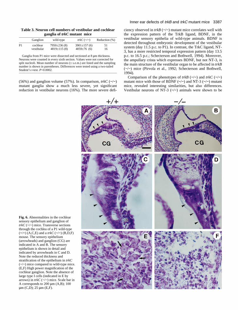
Fig. 6. Abnormalities in the cochlearsensory epithelium and ganglion oftrkC (−/−) mice. Transverse sectionsthrough the cochlea of a P1 wild-type(+/+) (A,C,E) and a trkC (−/−) (B,D,F)mouse. The sensory epithelium(arrowheads) and ganglion (CG) areindicated in A and B. The sensoryepithelium is shown in detail andindicated by arrowheads in C and D.Note the reduced thickness andstratification of the epithelium in trkC(−/−) mice compared to wild-type mice.(E,F) High power magnification of thecochlear ganglion. Note the absence oflarge type I cells (indicated in E byarrows) in trkC (−/−) mice. Scale bar inA corresponds to 200 µm (A,B); 100µm (C,D); 25 µm (E,F).
ciency observed in trkB (−/−) mutant mice correlates well withthe expression pattern of the TrkB ligand, BDNF, in thevestibular sensory epithelia of wild-type animals. BDNF isdetected throughout embryonic development of the vestibularsystem (day 11.5 p.c. to P1). In contrast, the TrkC ligand, NT-3, has a more restricted temporal expression pattern (day 13.5p.c. to 16.5 p.c.; Schecterson and Bothwell, 1994). Moreover,the ampullary crista which expresses BDNF, but not NT-3, isthe main structure of the vestibular organ to be affected in trkB(−/−) mice (Pirvola et al., 1992; Schecterson and Bothwell,1994).
Comparison of the phenotypes of trkB (−/−) and trkC (−/−)mutant mice with those of BDNF (−/−) and NT-3 (−/−) mutantmice, revealed interesting similarities, but also differences.Vestibular neurons of NT-3 (−/−) animals were shown to be

3388 T. Schimmang and others
vation of trkC (−/−) mutants. Longitudinal sections at the level of innerr hair cells (C,D) of wild-type (+/+) (A,C,E) and trkC (−/−) mice (B,D,F).ensory epithelium has been examined using a neurofilament antibody.pe animals, nerve fibres reach and enter the sensory epithelium forming) and spiral (C) nerve fiber systems. (B) The inner hair cells of P1 trkCid of innervation. Nerve fibres in A (arrows) and the sensory epitheliumeads) are indicated. (D) The innervation pattern of the outer hair cells ofappears normal. A nerve fibre entering the sensory epithelium (arrow)basal membrane (arrowheads) are indicated in C and D. In both, wild-t trkC (−/−) (F) mice of day 16.5 p.c. nerve fibres (arrows) are detectedhin the sensory epithelium (arrowheads) at the level of the inner hair corresponds to 75 µm (A,B,E,F); 50 µm (C,D).
reduced by 23% in newborns (Fariñas et al., 1994) This corre-lates very well with the observed 16% reduction of vestibularneurons in trkC (−/−) mice, indicating that the main NT-3receptor in this sensory organ is indeedgp145TrkC. NT-3 (−/−) and trkC (−/−)mutant mice are characterized by abnormalmovements and postures which has beenexplained by the deficiency of propriocep-tive neurons (Ernfors et al., 1994b; Fariñaset al., 1994; Klein et al., 1994b). Theobserved loss of vestibular neuronspossibly contributes to the abnormal behav-ioural phenotype of the mutant animals.
There is a clear difference between thereduction in the volume of the vestibularganglion and the number of vestibularneurons surviving in trkB (−/−) mice (57%and 44%, respectively; this study) andBDNF (−/−) mice (87% and 18%, respec-tively; Jones et al., 1994; Ernfors et al.,1994a). One possible explanation could bethat at least in one study (Ernfors et al.,1994a) vestibular neuron numbers weredetermined in juvenile animals (postnataldays P14 to P16) rather than in neonates.More vestibular neurons may undergo celldeath in trkB (−/−) mice during subsequentpostnatal days. A further, although lesslikely explanation may be that BDNFwould be able to exert its function in thevestibular system via an as yet unidentifiedreceptor, distinct from TrkB. This wouldresult in a higher percentage of neuronalcell death in BDNF (−/−) mutantscompared with trkB (−/−) mice. Alterna-tively, the lack of functional TrkB receptorsmay cause a compensatory up-regulation ofTrkC receptor expression in vestibularneurons which then could be rescued by thepresence of NT-3 in their target fields. Oncequantitative PCR-based expression assaysfor the TrkC receptor have been developed,it will be interesting to analyze thesurviving vestibular neurons in trkB (−/−)mice for the expression of TrkC receptors.It is however, conceivable that vestibularneurons in trkB (−/−) mice up-regulatecomponents of other, as yet undefined, sig-nalling pathways than the NGF-like neu-rotrophins. It will therefore be crucial togenerate double trkB/trkC mutant mice toanalyze the interaction between TrkB andTrkC signalling pathways and to identifypossible neurotrophin-independent survivalsignals for sensory neurons.
Inner and outer hair cells of thecochlea depend on the function ofTrkC and TrkB, respectively Recent experiments suggest that BDNFand NT-3 acting via their high-affinity
Fig. 7. Target inner(A,B,E,F) and outeInnervation of the s(A,C) In P1 wild-tythe typical radial (A(−/−) mice are devoin A and B (arrowhP1 trkC (−/−) mice and the underlying type (E) and mutanunderneath and witcells. Scale bar in A
receptors TrkB and TrkC are the dominant biologically activeneurotrophins in the cochlea (Ernfors et al., 1992; Pirvola etal., 1992; Avila et al., 1993; Tessarollo, 1993; Pirvola et al.,

3389Inner ear defects of trkB and trkC mutant mice
Fig. 8. Target innervation of trkB (−/−) mutants at day 18.5 p.c. Sections ofthe utricular macula (A-D), the ampullary crista (E-H) and the sensoryepithelium of the cochlea containing outer hair cells (I,J) of wild-type (+/+)(A,C,E,G,I) and trkB (−/−) (B,D,F,H,J) mice of day 18.5 p.c. have beenstained with a neurofilament antibody. In both, wild-type and trkB (−/−)animals nerve fibres are detected in the vestibular and cochlear system.High power magnifications of the sensory epithelia reveal that as in wild-type mice (C,G,I) nerve fibres reach and enter the epithelia in trkB (−/−)mutants (D,H,J). (J1) Typical image of an outer hair cell being innervatedby a nerve fibre. The synaptic contact is indicated by an arrow. Scale bar inA corresponds to 250 µm (A,B,E,F); 50 µm (G,H,I,J2); 30 µm (C,D); 10 µm (J1).
1994; Schecterson and Bothwell, 1994; Vazquez et al.,1994). Our analysis of the cochlear system of trkC(−/−) mutant mice shows that the number of neuronsin the cochlear ganglion is reduced by 51%, due to theloss of large size type I neurons, which innervate innerhair cells with afferent fibres. TrkC (−/−) mice alsorevealed developmental defects in the cochlearsensory epithelium. The incomplete formation anddifferentiation of the epithelium may be a consequenceof the innervation deficiences (Speidel, 1947, 1948;Guth, 1969).
TrkB (−/−) mutants specifically lack small size typeII neurons which are responsible for the afferent inner-vation of the outer hair cells of the cochlea andtherefore give a mirror image of the phenotype of trkC(−/−) mutants. In this context it will be important toconfirm trkB and trkC expression in type II and type Ineurons, respectively. Since no innervation of outerhair cells was detected in trkB (−/−) mice at P1,efferent fibres, which originate in the superior olivarycomplex and have reached the cochlea by P1 musthave also been affected (Sobkowicz, 1992). Therefore,the modulation and feedback system of hearing, whichcontrols the quality of hearing is most likely lost intrkB (−/−) mice.
While in the cochlear ganglion TrkB and TrkCreceptors have been detected (Ernfors et al., 1992; Tes-sarollo et al., 1993; Pirvola et al., 1994; Schectersonand Bothwell, 1994; Vazquez et al., 1994) inner andouter hair cells express both BDNF and NT-3 duringauditory system development (Pirvola et al., 1992;Wheeler et al., 1994). NT-3 (−/−) mutant mice lack85% of their cochlear neurons (Fariñas et al., 1994).The neuronal population affected has not been char-acterized. The fact that NT-3 (−/−) mutant mice loosemore neurons than trkC (−/−) mice may be explainedby the ability of NT-3 to exert its function via the TrkBreceptor (Ip et al., 1993). The extra population ofneurons surviving in trkC (−/−) animals may berescued by this mechanism. Interestingly, the entirecochlear system, including cochlear ganglion neuronsand the innervation pattern of outer hair cells are phe-notypically normal in BDNF (−/−) mutants (Ernfors etal., 1994a). Considering the lack of the second TrkBligand, NT-4 (Pirvola et al., 1992, 1994; Schectersonand Bothwell, 1994), these data suggest that NT-3,which has been shown to bind the TrkB receptor (Ipet al., 1993), may compensate for the loss of BDNF.Finally, phenotypic differences found between ligandand receptor knockouts may also be explained by thepresence of non-catalytic isoforms of trkB and trkC intrkB (−/−) and trkC (−/−) mice, whose functionsremain unclear (Klein et al., 1990; Middlemas et al.,1991; Tsoulfas et al., 1993; Valenzuela et al., 1993).
TrkB and TrkC are dispensable for targetencounter but essential for maintenance oftarget innervationThe time course of TrkB, TrkC, BDNF and NT3expression in vivo and BDNF and NT3 response invitro is tightly correlated with the development of
3390 T. Schimmang and others
inner ear afferent innervation, including the appearance andstabilization of presynaptic equipment (Pirvola et al., 1992,1994; Tessarollo et al., 1993; Schecterson and Bothwell, 1994;Vazquez et al., 1994). This suggests a functionally relevantneurotrophin action during all these processes. Our resultsshow that in the absence of a functional TrkB or TrkC receptor,nerve fibres can contact their peripheral targets. During laterdevelopment neurite lysis and atrophy as well as synaptic dis-assembling is observed, leading to the observed neuronal celldeath in vestibular and cochlear ganglia. Alternatively, loss ofTrk signaling may trigger apoptosis which then leads to neu-rodegeneration. These data are consistent with current viewsabout the relationship of target encounter to cell death sayingthat cranial sensory neurons survive independently of neu-rotrophins when their nerve fibres are growing and acquireneurotrophin dependence close to the time their neurites reachtheir targets (Davies and Lumsden, 1984; Vogel and Davies,1991). Consequently, ganglion formation and afferent inner-vation may depend on the action of other yet unidentifiedgrowth factors in vivo.
In the inner ear ganglia of wild-type mice, trkB and trkCexpression are maintained during postnatal development andadulthood (Ylikoski et al., 1993), possibly indicating apermanent role for innervation maintenance and/or neuronalsurvival. Consistent with this hypothesis, BDNF and NT-3 areexpressed in the target fields of the ganglion cells duringpostnatal life (Ylikoski et al., 1993). Finally, neurotrophinshave recently been found to regulate, among other properties,the synthesis of neuropeptides and the activity of voltage gatedchannels, the latter being crucial for neuronal survival (Ghoshet al., 1994; Snider et al., 1994). It will therefore be importantto investigate, if the absence of functional TrkB receptors cancause additional phenotypic abnormalities in neurons.
We would like to thank Carmen Valero and the electronmicroscopy facilities of the University of Valladolid (Laborotoriotécnicas instrumentales) for technical assistance. We thank AlunDavies for advice and comments on the manuscript. T.S. and L.M.are holders of an EMBO and an A.I.R.C. fellowship, respectively.This research was supported by a grant from Dirección General deCiencia y Tecnologiá (PB92/0621) to F. G. and Fondo de Investiga-ciones Sanitarias (94/1405) to J. R.
REFERENCES
Anniko, M. (1983). Embryonic development of vestibular sense organs andtheir innervation. In Development of Auditory and Vestibular Systems, vol 2.,(ed. R. Romand), pp. 375-423. Amsterdam: Elsevier.
Avila, M. A., Varela-Nieto, I., Romero, G., Mato, J. M. Giraldez, F., VanDe Water, T. R. and Represa, J. (1993). Brain-derived neurotrophic factorand neurotrophin-3 support the survival and neuritogenesis response ofdeveloping cochleovestibular ganglion neurons. Dev. Biol. 159, 266-275.
Barbacid, M. (1994). The Trk family of neurotrophin receptors. J. Neurobiol.25, 1386-1403.
Berkemeier, L. R., Winslow, J. W., Kaplan, D. R., Nikolics, K., Goeddel, D.V. and Rosenthal, A. (1991). Neurotrophin-5: a novel neurotrophic factorthat activates trk and trkB. Neuron 7, 857-866.
Bolender, R. P. (1983). Methods for decreasing the statistical variance ofstereological estimates. Anatomical Record 207, 89-106.
Davies, A. M. and Lumsden, A. G. S. (1984). Relation of target encounter andneuronal death to nerve growth factor responsiveness in the mousedeveloping mouse trigeminal ganglion. J. Comp. Neurol. 223, 124-137.
Davies, A. M., Thoenen, H. and Barde, Y.-A. (1986). The response of chick
sensory neurons to brain-derived neurotrophic factor. J. Neurosci. 6, 1897-1904.
Davies, A. M. (1987). Molecular and cellular aspects of patterning sensoryneurone connections in the vertebrate nervous system. Development 101,185-208.
Davies, A. M. (1994) The role of neurotrophins in the developing nervoussystem. J. Neurobiol. 25, 1334-1348.
Davies, A. M., Lee, K.-F. and Jaenisch, R. (1994). p75-deficient trigeminalsensory neurons have an altered response to NGF but not to otherneurotrophins. Neuron 11, 565-574.
Dechesne, C. J., Desmandryl, G. and Dememes, D. (1987). Myelination ofthe mouse vestibular ganglion. Acta Otolaryngol. (Stockh.) 101, 11-18.
Ernfors, P., Merlio, J.-P. and Persson, H. (1992). Cells expressing mRNA forneurotrophins and their receptors during embryonic rat development. Eur. J.Neurosci. 4, 1140-1158.
Ernfors, P. Lee, K.-F. and Jaenisch, R. (1994a). Mice lacking brain-derivedneurotrophic factor develop with sensory deficits. Nature 368, 147-150.
Ernfors, P., Lee, K.-F., Kucera, J. and Jaenisch, R. (1994b). Lack ofneurotrophin-3 leads to deficiencies in the peripheral nervous system and lossof limb proprioceptive afferents. Cell 77, 503-512.
Fariñas, I. Jones, K. R., Backus, C., Wang, X.-Y. and Reichardt, L. F.(1994). Severe sensory and sympathetic deficits in mice lackingneurotrophin-3. Nature 369, 658-661.
Glass, D. J. and Yancopoulos, G. D. (1993). The neurotrophins and theirreceptors. Trends Cell Biol. 3, 262-268.
Ghosh, A., Carnahan, J. and Greenberg, M. E. (1994). Requirement forBDNF in activity-dependent survival of cortical neurons. Science 263, 1618-1623.
Guth, L. (1969). Trophic effects of vertebrate neurons. Neurosci. Res. Prog.Bull. 7, 1-73.
Hantzopoulos, P. A., Suri, C., Glass, D. J., Goldfarb, M. P. andYancopoulos, G. D. (1994). The low affinity NGF receptor, p75, cancollaborate with each of the Trks to potentiate functional responses to theneurotrophins. Neuron 13, 187-201.
Ip, N. Y., Ibáñez, C. F., Nye, S. H., McClain, J., Jones, P. F., Gies, D. R.,Belluscio, L., LeBeau, M. M., Espinosa, R., Squinto, S. P., Persson, H.and Yancopoulos, G. D. (1992). Mammalian neurotrophin-4: structure,chromosomal location, tissue distribution, and receptor specificity. Proc.Natl. Acad. Sci. USA 89, 3060-3064.
Ip, Y. I., Stitt, T. N., Tapley, P., Klein, R., Glass, D. J., Fandl, J., Greene, L.A., Barbacid, M. and Yancopoulos, D. (1993). Similarities and differencesin the way neurotrophins interact with the Trk receptors in neuronal andnonneuronal cells. Neuron 10, 137-149.
Jones, K. R., Fariñas, I., Backus, C. and Reichardt, L. F. (1994). Targeteddisruption of the BDNF gene perturbs brain and sensory neuron developmentbut not motor neuron development. Cell 76, 989-999.
Kaplan, D. R., Martin-Zanca, D. and Parada, L. F. (1991). Tyrosinephosphorylation and tyrosine kinase activity of the trk proto-oncogeneproduct by NGF, Nature 350, 158-160.
Kiang, N. Y. S., Rho, J. M., Northrop, C. C., Liebermann, M. C. andRyngo, D. R. (1982). Hair cell innervation by spiral ganglion cells in adultcats. Science 217, 175-177.
Klein, R., Conway, D., Parada, L.F., and Barbacid, M. (1990). The trkBtyrosine kinase gene codes for a second neurogenic receptor that lacks thecatalytic kinase domain. Cell 61, 647-656.
Klein, R., Jing, S., Nanduri, V., O’Rourke, E. and Barbacid, M. (1991a).The trk proto-oncogene encodes a receptor for nerve growth factor. Cell 65,189-197.
Klein, R., Jing, S., Nanduri, V., Jing, S., Lamballe, F., Tapley, P., Bryant,S., Cordon-Cardo, C., Jones, K. R., Reichardt, L. F. and Barbacid, M.(1991b). The trkB tyrosine protein kinase is a receptor for brain-derivedneurotrophic factor and neurotrophin-3. Cell 66, 395-403.
Klein, R., Lamballe, F., Bryant, S. and Barbacid, M. (1992). The trkBtyrosine kinase is a receptor for neurotrophin-4. Neuron 8, 947-956.
Klein, R., Smeyne, R. J., Wurst, W., Long, L. K., Auerbach, B. A. Joyner,A. L. and Barbacid, M. (1993). Targeted disruption of the trkB neurotrophinreceptor gene results in nervous system lesions and neonatal death. Cell 75,113-122.
Klein, R. (1994a). Role of neurotrophins in mouse neural development. FASEBJ. 8, 738-744.
Klein, R., Silos-Santiago, I. Smeyne, R. J., Lira, S. A., Brambilla, R.,Bryant, S., Zhang, L., Snider, W. D. and Barbacid M. (1994b). Disruptionof the neurotrophin-3 receptor gene trkC eliminates Ia muscle afferents andresults in abnormal movements. Nature 368, 249-251.

3391Inner ear defects of trkB and trkC mutant mice
Laird, P. W., Zijderweld, A., Linders, K., Rudnicki, M. A., Jaenisch, R.and Berns, A. (1991). Simplified mammalian DNA isolation procedure.Nucl. Acids Res. 19, 4293.
Lamballe, F., Klein, R. and Barbacid, M. (1991). trkC, a new member of thetrk family of tyrosine protein kinases is a receptor for neurotrophin-3. Cell66, 967-979.
Lefebvre, P. P., Leprince, P., Weber, T., Rigo, J.-M., Delree and Moonen,G. (1990). Neuronotrophic effect of developing otic vesicle on cochleo-vestibular neurons: evidence for nerve growth factor involvement. Brain Res.507, 254-260.
Lefebvre, P. P., Van De Water, T. R., Represa, J., Liu, W., Bernd, P.,Modlin, S., Moonen, G. and Mayer, M. B. (1991). Temporal pattern ofnerve growth factor (NGF) binding in vivo and the in vitro effects of NGF oncultures of developing auditory and vestibular neurons. Acta Otolaryngol.(Stockh.) 111, 304-311.
Meakin, S. O. and Shooter, E. M. (1992). The nerve growth factor family ofreceptors. Trends Neurosci. 15, 323-331.
Middlemas, D.S., Lindberg, R.A., and Hunter, T. (1991). trkB, a neuralreceptor protein-tyrosine kinase: evidence for a full-length and two truncatedreceptors. Mol. Cell. Biol. 11, 143-153.
Pirvola, U., Ylikoski, J., Palgi, J., Lehtonen, E., Arumäe, U. and Saarma,M. (1992). Brain-derived neurotrophic and neurotrophin-3 mRNAs in theperipheral target fields of developing inner ear ganglia. Proc. Natl. Acad. Sci.USA 89, 9915-9919.
Pirvola, U., Arumäe, U., Moshynakov, M., Palgi, J., Saarma, M. andYlikoski, J. (1994). Coordinated expression and function of neurotrophinsand their receptors in the rat inner ear during target innervation. Hearing Res.75, 131-144.
Pujol, R and Sans, A. (1986). Synaptogenesis in the cochlear and vesibularreceptors. In Advances in Neural and Behavioural Development (ed. R.Aslin), pp. 1-18. Norwood, New York: Ablex Press.
Represa, J., Van De Water, T. R. and Bernd, P. (1991). Temporal pattern ofnerve growth factor receptor expression in developing cochlear andvestibular ganglia in quail and mouse. Anat. Embryol. 184, 421-432.
Ruben, R. J. (1967). Development of the inner ear of the mouse. ActaOtolaryngol. (Stockh.) Suppl. 220, 1-44.
Schecterson, L. C. and Bothwell, M. (1994). Neurotrophin and neurotrophinreceptor mRNA expression in developing inner ear. Hearing Res. 73, 92-100.
Snider, W. D. (1994). Functions of the neurotrophins during nervous systemdevelopment: What the knockouts are teaching us. Cell 77, 627-638.
Sobkowicz, H. M. (1992). The development of innervation in the organ ofCorti. In Development of Auditory and Vestibular Systems, vol.2, (ed. R.Romand), pp. 59-95. Amsterdam: Elsevier.
Soppet, D., Escandon, E., Maragos, J., Middlemas, D. S., Reid, S. W., Blair,J., Burton, L. E., Stanton, B. R., Kaplan, D. R., Hunter, T., Nikolics, K.and Parada, L. F. (1991). The neurotrophic factors brain-derivedneurotrophic factor and neurotropin-3 are ligands for the trkB tyrosine kinasereceptor. Cell 65, 895-903.
Speidel, C. C. (1947). Correlated studies of sense organs and nerves of thelateral line in living frog tadpoles. I. Regeneration of denervated organs. J.Comp. Neurol. 87, 29-55.
Speidel, C. C. (1948). Correlated studies of sense organs and nerves of thelateral line in living frog tadpoles. II. The trophic influence of specific nervesupply as revealed by prolonged observations of denervated and reinnervatedorgans. Am. J. Anat. 82, 227-320.
Spoendlin, H. (1969). Hair cell innervation in adult cats. Acta Otolaryngol.(Stockh.) 67, 239-243.
Spoendlin, H. (1988). Neural anatomy of the inner ear. In Physiology of the Ear(eds. Jahn, A. F. and Santos-Sachi, J.), pp. 201-219. New York: Raven Press.
Squinto, S. P., Stitt, T. N., Aldrich, T. H., Davis, S., Bianco, S. M.,Radziejewski, C., Glass, D. J., Masiakowski, P., Firth, M. E., Valenzuela,D. M., DiStefano, P. S. and Yancopoulos, G. D. (1991). trkB encodes afunctional receptor for brain-derived neurotrophic factor and neuotrophin-3but not nerve growth factor. Cell 65, 885-893.
Tessarollo, L., Tsoulfas, P., Martin-Zanca, D., Gilbert, D. J., Jenkins, N. A.,Copeland, N. G., and Parada, L. F. (1993). trkC, a receptor forneurotrophin-3, is widely expressed in the developing nervous system and innon-neuronal tissues. Development 118, 463-475.
Tsoulfas, P., Soppet, D., Escandon, E., Tessarollo, L., Mendoza, R. J.,Rosenthal, A., Nikolics, K., and Parada, L. F. (1993). The rat trkC locusencodes multiple neurogenic receptors that exhibit differential response toneurotophin-3 in PC12 cells. Neuron 10, 975-990.
Valenzuela, D., Maisonpierre, P., Glass, D., Rojas, E., Nunez, L., Kong, Y.,Gies, D., Stitt, T., Ip, N., and Yancopoulos, G. (1993). Alternative forms ofrat trkC with different functional capabilities. Neuron 10, 963-974.
Vazquez, E., Van De Water, T. R., Del Valle, M., Vega, J. A., Staecker, H.,Giraldez, F. and Represa, J. (1994). Pattern of trkB protein-likeimmunoreactivity in vivo and the in vitro effects of brain-derivedneurotrophic factor (BDNF) on developing cochlear and vestibular neurons.Anat. Embryol. 189, 157-167.
Verdi, J. M., Birren, S. J., Ibañez, C. F., Persson, H., Kaplan, D. R.,Benedetti, M., Chao, M. V. and Anderson, D. J. (1994). p75LNGFR
regulates Trk signal transduction and NGF-induced neuronal differentiationin MAH cells. Neuron 12, 733-745.
Vogel, K. S. and Davies, A. M. (1991). The duration of neurotrophic factorindependence in early sensory neurons is matched to the time course of targetfield innervation. Neuron 7, 819-830.
Von Bartheld, C. S., Patterson, S. L., Hener, J. G., Wheeler, E. F.,Bothwell, M. and Rubel, E. (1991). Expression of nerve growth factor(NGF) receptors in the developing inner ear of chicken and rat. Development113, 455-470.
Wheeler, E. F., Bothwell, M., Schecterson, L. C. and Von Bartheld, C. S.(1994). Expression of BDNF and NT-3 mRNA in hair cells of the organ ofCorti: Quantitative analysis in developing rats. Hearing Res. 73, 46-56.
Wood, J. N. and Anderton, B. H. (1981). Monoclonal antibodies tomammalian neurofilaments. Biosci. Rep. 1, 263-268.
Ylikoski, J., Pirvola, U., Moshnyakov, M., Palgi, J., Arumäe, U. andSaarma, M. (1993). Expression patterns of neurotrophin and their receptormRNAs in the rat inner ear. Hearing Res. 65, 69-78.
(Accepted 19 June 1995)