
Determination of Gangliosides in Human Cerebrospinal Fluid ...
10
Trbojevic-Cepe and Kracun: Gangliosides in cerebrospinal fluid 863 J. Clin. Chem. Clin. Biochem. Vol. 28, 1990, pp. 863-872 © 1990 Walter de Gruyter & Co. Berlin · New York Determination of Gangliosides in Human Cerebrospinal Fluid by High-Performance Thin-Layer Chromatography and Direct Densitometry By Milica Trbojevic-Cepe and Ivica Kracun Institute of Clinical Laboratory Diagnostics, Zagreb University School of Medicine, Clinical Hospital Centre, Zagreb, Yugoslavia (Received January 4/July 16, 1990) Summary: A method for the separation and quantification of a complex ganglioside mixture from a clinically available amount (5 ml) of human cerebrospinal fluid (CSF) is described. After reduction of the CSF volume by ultrafiltration, gangliosides are extracted with methanol/chloroform, then separated and quantified by high performance thin layer chromatography (HPTLC) and direct densitometry. For purification of crude gan- glioside extract, the method of choice was microdialysis against water. Recovery for the present method including all methodological steps was 78%. No delective loss of gangliosides was demonstrated. The CSF ganglioside pattern from 'normal' CSF samples resembles that of brain gangliosides, particularly cerebellum gangliosides. Based on Chromatographie comparison with standards, the percentages of lipid- bound NeuAc-positive fractions were: GMI = II 3 NeuAc-GgOse 4 Cer (3%), G D3 - II 3 NeuAc 2 -Lac-Cer (3%), G Dla = IV 3 NeuAc,II 3 NeuAc-GgOse 4 Cer(15%), Xi (3%), G Dlb = II 3 (NeuAc) 2 -GgOse 4 Cer(16%), X 2 (3%), G T ib = IV 3 NeuAc,II 3 NeuAc 2 -GgOse 4 -Cer (41%), and G Qlb = IV 3 NeuAc 2 -,II 3 NeuAc 2 -GgOse 4 -Cer(16%). The total ganglioside content varied between 616 —944 μ§/1. Within-run and between-run assay precision (relative standard deviation) for 'normal' pooled CSF ranged from 0.04 to 0.12 for the predominant CSF ganglioside fractions (G D i a , G Dlb , G T n» G Qlb ), and from 0.13 to 0.23 for the less pronounced fractions (G M i, G D3 ). Introduction , ,^ „ „ , , , . , , , . onstrated (3). So far, only a few published studies The content of lipids in cerebrospinal fluid (CSF) is have shown that the ganglioside profile of CSF more very low (1). Gangliosides 1 ) are sialic acid-containing closely resembles that of brain tissue than that of glycosphingolipids displaying their highest concentra- plasma (4 —6). However, due to methodological dif- tions in nervous tissue (2). Their accumulation in CSF ficulties, the gangliosides in normal human CSF were in lipid storage diseases (gangliosidosis) has been dem- not clearly identified in these previous reports. Quan- tification of gangliosides in the picomolar range using *) See page 864. scanning densitometry on high-performance thin- J. Clin. Chem. Clin. Biochem. / Vol. 28, 1990 / No. 11
Transcript of Determination of Gangliosides in Human Cerebrospinal Fluid ...

Trbojevic-Cepe and Kracun: Gangliosides in cerebrospinal fluid 863
J. Clin. Chem. Clin. Biochem.Vol. 28, 1990, pp. 863-872© 1990 Walter de Gruyter & Co.
Berlin · New York
Determination of Gangliosides in Human Cerebrospinal Fluidby High-Performance Thin-Layer Chromatographyand Direct Densitometry
By Milica Trbojevic-Cepe and Ivica Kracun
Institute of Clinical Laboratory Diagnostics, Zagreb University School of Medicine, Clinical Hospital Centre,Zagreb, Yugoslavia
(Received January 4/July 16, 1990)
Summary: A method for the separation and quantification of a complex ganglioside mixture from a clinicallyavailable amount (5 ml) of human cerebrospinal fluid (CSF) is described. After reduction of the CSF volumeby ultrafiltration, gangliosides are extracted with methanol/chloroform, then separated and quantified by highperformance thin layer chromatography (HPTLC) and direct densitometry. For purification of crude gan-glioside extract, the method of choice was microdialysis against water. Recovery for the present methodincluding all methodological steps was 78%. No delective loss of gangliosides was demonstrated.
The CSF ganglioside pattern from 'normal' CSF samples resembles that of brain gangliosides, particularlycerebellum gangliosides. Based on Chromatographie comparison with standards, the percentages of lipid-bound NeuAc-positive fractions were:
GMI = II3NeuAc-GgOse4Cer (3%),GD3 - II3NeuAc2-Lac-Cer (3%),GDla = IV3NeuAc,II3NeuAc-GgOse4Cer(15%),Xi (3%),GDlb = II3(NeuAc)2-GgOse4Cer(16%),X2 (3%),GTib = IV3NeuAc,II3NeuAc2-GgOse4-Cer (41%), andGQlb = IV3NeuAc2-,II3NeuAc2-GgOse4-Cer(16%).
The total ganglioside content varied between 616 — 944 μ§/1.
Within-run and between-run assay precision (relative standard deviation) for 'normal' pooled CSF rangedfrom 0.04 to 0.12 for the predominant CSF ganglioside fractions (GDia, GDlb, GTn» GQlb), and from 0.13 to0.23 for the less pronounced fractions (GMi, GD3).
Introduction , ,^ „ „ , , , . , , , .onstrated (3). So far, only a few published studiesThe content of lipids in cerebrospinal fluid (CSF) is have shown that the ganglioside profile of CSF morevery low (1). Gangliosides1) are sialic acid-containing closely resembles that of brain tissue than that ofglycosphingolipids displaying their highest concentra- plasma (4 — 6). However, due to methodological dif-tions in nervous tissue (2). Their accumulation in CSF ficulties, the gangliosides in normal human CSF werein lipid storage diseases (gangliosidosis) has been dem- not clearly identified in these previous reports. Quan-
tification of gangliosides in the picomolar range using*) See page 864. scanning densitometry on high-performance thin-
J. Clin. Chem. Clin. Biochem. / Vol. 28, 1990 / No. 11

864 Trbojevic-Cepe and Kracun: Gangliosides in cerebrospinal fluid
layer chromatography (HPTLC) has been reported(7, 8). The aim of this study was to present a methodsuitable for simultaneous profile analysis and gan-glioside quantification from clinically availableamounts of CSF (5 ml). The method consisted ofganglioside extraction, separation and quantification,employing HPTLC on silica gel and scanning densi-tometry.
Materials and MethodsAll reagents were of reagent-grade quality. Solvents were redis-tilled before used. A commercially available apparatus fromSartorius GmbH (G ttingen, FRG) and collodium bags SM13200, were used for ultraflltration. Sephadex G-25 fine waspurchased from Pharmacia Fine Chemicals (Uppsala, Sweden).Standard dialysis membranes from Technicon Chemicals Co.(Orcq, Belgium) were used for dialysis. High-performance thin-layer Chromatographie plates (HPTLC) silica gel 60 (10 χ 10cm) were purchased from Merck (Darmstadt, FRG). The Cron-assial ganglioside mixture from Fidia (Abano Terme, Italy),diluted 1:10 with water (0.5 g per litre of gangliosides) wasused as a standard for the ganglioside HPTLC determination.Coronassial contains: GMi (21%), GDla (40%), GDlb (16%) andG™ (19%). Radiolabelled [3H]GDla (1 g/1) was a generous giftfrom Dr. G. Schwarzmann, Inst. Organ. Chem. Biochem., Uni-versity of Bonn (14000 counts/min -5 μg GDia). GM2 ganglioside(bovine) was purchased from Calbiochem. A LKB 2202 Ul-trascan Laser Densitometer and a 2220 Recording Integrator(Bromma, Sweden) were used for densitometric analysis.
Lumbar CSFs were obtained from adult patients and childrenwith various neurological diseases. CSFs from adults free ofany signs of acute inflammatory process of the CNS, tumours,demyelination, intracranial haemorrhage or congenital errorsof lipid metabolism, were used as 'normals'. After cytologicaland biochemical examinations, CSF samples were centrifuged
Designation according to IUPAC-IUB Recommendations(20).Ganglioside abbreviations follow the nomenclature systemof L. Svennerholm (19):
Svennerholm (19)* Lipid Document (20)**
II3NeuAc-LacCerII3NeuAc-GgOse4CerIV3NeuAc,II3NeuAc-GgOse4CerII3(NeuAc)2-GgOse4CerIV3NeuAc,II3(NeuAc)2-GgOse4CerIV3(NeuAc)2II3(NeuAc)2-GgOse4Cer
GDlb
GTibGQlb
* G = ganglioside; M = monosialo; D = disialo; T =trisialo. Arabic numerals indicate sequence of migrationin thin-layer chromatograms (19).
** Neu Ac = 7V-Acetylneuraminic acid; Lac = lactose;GgOse4 = gangliotetraose =Gal( l -3)GalNAc( l -4)Gal( l -4)GlcCer; Cer =Cer amide.The location of the NeuAc residue is indicated by aRoman numeral designating the position of the mono-saccharide residue in the parent oligosaccharide (countingfrom the ceramide end) to which the NeuAc residue isattached, with an Arabic numeral superscript indicatingthe position within that residue to which the NeuAc isattached (20).
before ganglioside extraction (2000 g, 10 minutes). Pooled 'nor-mal' CSFs were used for method evaluation and statisticalanalysis. Recovery was measured in two ways, i. e. either withCSF itself using a radiolabelled tracer ([3H]GDla, 5 μg/5 mlCSF), or by processing the Cronassial ganglioside mixture inH2O or artificial CSF (7 μg/5 ml CSF art.). Artificial CSFconsisted of CSF ultrafiltrate (5 ml) and serum (20 μΐ). Assayprecision was evaluated by analysing pooled CSF (or Cronassialadded to artificial CSF) twelve times on the same day and ondifferent days in a period of four mounths. Linearity of therelationship between densitometric responses (total area) andganglioside content was determined by spotting increasingamounts of gangliosides from diluted Cronassial on HPTLCplates.
U l t r a f i l t r a t i o n (vacuum dialysis)
Native CSF samples (5 ml) were reduced to about 0.5 ml byultraflltration against water, the semipermeable membranerinsed with 0.250 ml of 0.077 mol/1 saline and the volume ofthe CSF concentrate adjusted to 0.750 ml.
CSF ganglioside ext rac t ion procedure
Gangliosides were extracted from CSF by a slightly modifiedmethod of Svennerholm & Fredman (9). In place of the waterphase, a reduced volume of CSF (concentrate) was used. Theratio chloroform-methanol-CSF was 1:2:0.75 by volume.Briefly, 2 ml of methanol were added to 0.75 ml of CSF con-centrate; after mixing on a cyclomixer, 1 ml of chloroform wasadded and mixed again. The gangliosides were separated fromother lipids by phase partition, after the addition of 0.65 ml ofwater. The two phases were separated by centrifugation (15 — 20min, 3000 g), and the upper phase was set aside. For second-phase partition of the lower phase, 0.380 ml of methanol and0.250 ml of water were added. The two upper phases werecombined and evaporated to dryness (37 °C).
Al t e rna t ive procedure for CSF ganglioside extraction
Gangliosides were directly extracted from 6 ml of native CSF,using a solvent ratio of chloroform-methanol-CSF of 8:16:6by volume. For phase partition, 5.2 ml of water were added.After overnight separation in a glass funnel, the upper phasewas set aside. For second-phase partition of the lower phase,3 ml of methanol and 2 ml of water were added. The two upperphases were combined and evaporated to dryness (37 °C).
Puri f icat ion of crude gangl ioside extract by microdialysis
Crude ganglioside extract was dissolved in a few microlitres ofchloroform-methanol-water (60:30:4.5, by volume) and quan-titatively transferred to a microcup (0.1 ml) for dialysis. Afterevaporation of the solvent to dryness, the ganglioside residuewas dissolved in 100 μΐ of water and the microcup covered witha dialysis membrane. After dialysis against water for about fourhours, the dialysis membrane was pierced with the plastic tipof a 100 μΐ micropipette, and the ganglioside water extractquantitatively transferred to a conical tube and evaporated todryness.
Al t e rna t ive procedure for pur i f ica t ion of crude gan-glioside extract by gel f i l t ra t ion on Sephadex G-25.A modified method of Wells and Dittmer (10)
Crude ganglioside extract was dissolved in chloroform-metha-nol-water (60:30:4.5, by volume) and applied to a SephadexG-25 column (110 χ 5mm, 1.4 ml bed volume, resin mixed
J. Clin. Chem. Clin. Biochem. / Vol. 28,1990 / No. 11

Trbojevic-Cepe and Kracun: Gangliosides in cerebrospinal fluid 865
with the same solvent mixture). The gangliosides were elutedfrom the column with 6 — 7 ml of the same solvent mixture.The column was then eluted with about 1 ml of the secondsolvent mixture of chloroform-methanol-water (20:20:7.5, byvolume), and the combined eluates were evaporated to dryness.
High per formance t h i n - l a y e r ch roma tog raphy(HPTLC)
Gangliosides were separated using the ascending "sandwich"HPTLC technique (11). Two plastic 1 mm-thick strips wereused as gaskets. Before use, HPTLC plates were pre-washedwith chloroform-methanol (1:1, by volume) and activated at110 °C during 60 min. The ganglioside extract was dissolved ina few microlitres of chloroform-methanol-water (60:30:4.5, byvolume) and quantitatively transferred onto a HPTLC plate.The samples were applied as a line. After formation of a"sandwich" the following solvent system was used for Chro-matographie separation of gangliosides: chloroform-methanol-12 mmol/1 MgCl2 in H2O- 13.3 mol/1 NH3 (60:40:9:0.5, byvolume). Chromatography was performed at 17 —18 °C usinga freshly prepared solvent system. Development was stoppedwhen the solvent front reached 1 — 1.5 cm from the upper edgeof the plate. After drying the HPTLC plate, the same procedurewas repeated with a fresh solvent mixture.
Densi tometr ic scanning and quan t i f i ca t ion of CSFgangliosides
For densitometric quantification, three standards of 5, 10 and15 μΐ of diluted Cronassial (corresponding to 2.5, 5 and 7.5 μgof gangliosides, respectively) were spotted on the same HPTLCplate. Gangliosides were visualized on the plate (as lipid-boundNeuAc) by spraying with resorcinol-HCl reagent diluted withan equal volume of water (12), and the HPTLC plate washeated as suggested by Ando et al. (13). Total amounts of CSFgangliosides were calculated from a calibration curve plottedfrom the densitometric response (total area) as a function ofthe amount of gangliosides ^g) in standards (Cronassial). Thecontent of individual ganglioside was calculated from the cali-bration curve based on an appropriate densitometric response(area) or from the relative percentage multiplied by the totalcontent of gangliosides.
Results and Discussion
Procedure of CSF ganglioside extraction
Reduction of the CSF and solvent volumes increasedthe recovery and reproducibility of the extractionmethod (tab. 1). Ultrafiltration did not result in eitherany major loss of total gangliosides (tab. 1) or anyselective loss (tab. 2), although the molecules involvedwere those of low molecular mass. The procedure isbased on the assumption that CSF gangliosides areassociated with proteins. Albumin is known tostrongly bind to gangliosides (14). Moreover, a rapidprocedure of CSF volume reduction causes an abruptincrease in ganglioside concentration, thereby trigger-ing the formation of micelles of higher molecular mass(2).Ultrafiltration also removes large quantities of salts(especially divalent cations) from the CSF before ex-
traction of the gangliosides (CSF contains about 270mmol/1 salts and 300 nmol/1 gangliosides). A tendencyof gangliosides to accumulate into a 'ganglioside-protein-divalent cation complex' at the interface inthe biphasic solvent system has been described (15).
We also investigated whether this ganglioside isolationprocedure, based on the partitioning method, is ap-plicable in the present CNS pathology where less polargangliosides (i.e. GM2 gangliosidosis) are elevated.About 75% of GM2 was recovered in the upper organicphase after adding GM2 ganglioside to a CSF sample(fig. 3e).
In pathological CSF samples with high protein con-centrations, the concentration of polysialoganglio-sides, particularly GQlb, is decreased (tab. 3), but asimilar effect was also demonstrated 'in vitro' byadding serum to CSF samples (tab. 3, fig. 1). Adramatic decrease in the concentration of polysialo-gangliosides (GQlb about 75%, GTlb and GDib about50%) was also observed after the addition of crudeCSF ganglioside extract to serum, as determined byganglioside extraction and HPTLC (figs. 3g, f)· Incontrast, a parallel experiment using isolated ganglio-sides of CSF and serum gave a strictly additiveHPTLC analysis upon mixing (fig. 1). This meansthat high protein concentrations markedly interferewith the extraction of gangliosides. The action ofserum sialidase as a possible cause of the decrease ofganglioside values was ruled out by replacing serumwith pure albumin solution (fig. 1). Therefore, for theanalysis of pathological CSFs with high protein con-centration should be reduced less and the volume oforganic solvents proportionally increased, allowingefficient protein precipitation and ganglioside extrac-tion. The question remains, however, as to the natureof the real ganglioside pattern in serum or in othersamples with high protein concentration.
Purif icat ion of crude CSF ganglioside ex-tract
Microdialysis is a very simple and reproduciblemethod for crude CSF ganglioside extract purifica-tion, with good recovery (98%) and no selective lossof gangliosides (tab. 1). In microdialysis, the CSFganglioside extracts were dissolved in the least pos-sible amount of water (up to 0.1 ml), thus achievinga ganglioside concentration closely approaching thecritical micellar concentrations (2). Besides, the shorttime needed for dialysis (about 4 hours) also avoidsany major loss of gangliosides. Such a brief dialysisis possible because the CSF samples are desalinatedduring the preceding procedure of ultrafiltration.
J. Clin. Chem. Clin. Biochem. / Vol. 28,1990 / No. 11
866 Trbojevic-Cepe and Kracun: Gangliosides in cerebrospinal fluid
f"oo*cdo*5h
meth
odol
o]
•sSc
βoυSox>C
1L«
T3Gcd
£T
S
'3ccd.2&
-s§G
"cd
i<υ
'ΟΛ
Ga"
UH
*«
*3
ab. 1
. Pr
oced
ural
sche
di
H
«υO
M»5oω
CU
OJ
,XiL-
"cd
1g
U
S1osex0)_>"cd|<
ωSO8aesoδ
OH
Λ ^
OET1 .
3O)cd§v«,
°
0
<υ00
cn
GQ00
iin s!S
.
Q~bOOO=S._j_|
*or^ v^S
WO
II
Q"
iir> s^
G"I I0Λ
00 OO" * +1Ό
^
COuS+1uSt—
•
+1mCN
-Λ
C rtG W8 ?c c=5 Co|t
11 +cd Ov°2 tu "5 oo -u«a +ο Ό ^^g 3 t
•G cd 00Q U
ONτ-ί+1CN[~^00
,«__,
!2-hlcnON00
5 m
l CS
F1
0.75
ml
(con
cent
rate
)
. U
ltraf
lltra
tion
(vac
uum
dia
lysi
sag
ains
t w
ater
)
^
cl|•2 ^cd α
cC/3 §cd QJ
ϊί+1
CNCN
+10000
0
CN
+1IO0000
0 GG f ^ -2
° -. ου
to fi^ζ_^ VO "*-*
i0 8^
G ^ ·Ξ αε ο ^ _-cS Η- &. g
cd
. G
angl
iosi
de e
xtra
ctio
rw
ith c
hlor
ofor
m-m
eth
nol-H
2O(S
venn
erho
lm &
Fre
d-m
an (
1980
)
CN
INcn+1
co00
^-flCN
S'
cd -Λ -^
fi Λέ ^ " /-s£ ^ c ^"
"B + £ «n0 0 ^ ^^ ^ ΐ ε +
CN ° + O 0
ΟθοΞ^
1=0^0cd O H£J Ό >—'
oo S c E
Γ~-^+1f-~ION
^3
+1r-H
OOON
><
•89,'s ffioUSS)5<L) G
T3 '-
Dia
lysis
of
cru
tract
dis
solv
ed
(-1ω"cd
(Λ
'cds1!.22 Ο
ITQ cnen
(U
(D,£3
_G
I1
Dou
ble
deve
lo
C/D
. H
PTLC
"sa
ndw
ich"
a
• f
Ο _£*£ πo <«jLs
US-°° i ^τ ΐ εf- CN ^T-i -r-< Ο
si κ +3 g z <*0/3 J3 — H H"
Ip:> i ^os£££,
cend
ing
met
hod
c_o"cdt-l
JD
c S
1 i§ «fa
Prof
ile d
eter
mi
Qua
ntifi
catio
ncu
rve
"7?
. La
ser
dens
itom
etry
(gan
glio
side
s w
ere
de-
tect
ed b
y th
e re
sorc
imH
CI
met
hod,
And
o, e
tal
. 197
8)
m
CNvd-hi,_,t^;SO
vq
+1CNCOΌ
T-l
CN+1CNvit-~
0
CN+1VO00
. A
ll st
eps
incl
uded
^o
6<υr-cΟ
cT3
i•5•gcd
*3I fclac|£•8-80 S
Ό §•ο σG .
'S)J cH gh£ ·— <
8.£> IO
<N
tS2 O G• r* χ '-ζ^ S G§ *cd 0
°"&Sc^oo .2v— ' <u "cdο·5§
. »ΤΗ* O C00 Λ -Μ ·£
.S 'q *^3 ^" PH .2^ >·>isl^o S '-^
'"S /-*N ω §
1|1?O O ^ S(D U^ [τ ( ^^
^Sitε ^ ΐ
o "*"* ^~* ·*-·"*"* ^ j· η-J
*^3 Ό Τ3 .«
|·3·3·&rt S3 S «I * * i.S .SS g ^ SCO Cd r h»
c α
£§<§£"° o M^
ea Λ " w "
J. Clin. Chem. Clin. Biochem. / Vol. 28,1990 / No. 11

Trbojevic-Cepe and Kracun: Gangliosides in cerebrospinal fluid 867
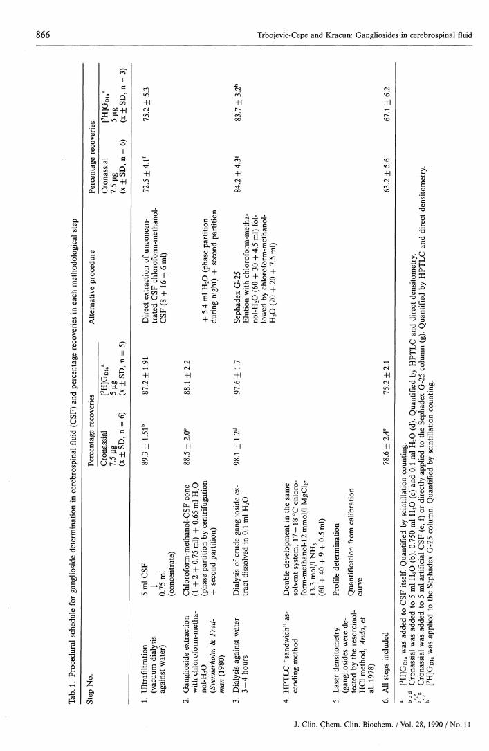
Tab. 2. Precision of ganglioside determination in cerebrospinal fluid (CSF)*
GD3GDla
Xl
GDlb
Cronassial**Within-run(n = 12)HPTLCPercentagedistribution(%, mean ± SD)
17.8 ± 1.5
40.9 ± 1.8
15.1 ± 1.3
20.7 ± 1.6
Pooled CSF (5 ml)Between-run(n = 12)HPTLCPercentagedistribution(%, mean ± SD)
17.2 ±1.8
40.2 ± 2.1
15.5 ± 1.6
21.2 ±1.8
Within-run(n = 12)HPTLCPercentagedistribution(%, mean ± SD)
3.2 + 0.43.5 ± 0.4
14.8 ± 12.9 ± 0.4
15.8 ±1.33.5 ± 0.4
40.7 + 1.715.6 + 1.5
Amount ofgangliosidesfog/I, mean + SD)
2 5 + 42 8 + 4
117+ 823+ 3
125 + 102 8 + 3
323 + 14124+ 12
Between-run(n = 12)HPTLCPercentagedistribution(%, mean + SD)
3.0 + 0.73.3 + 0.5
14.3 + 1.22.7 + 0.7
16.1 + 1.52.9 + 0.7
41.5 + 2.116.2 + 1.9
Amount ofgangliosidesfog/I, mean + SD)
24+ 52 7 + 4
116+ 1022+ 5
130 + 122 3 + 5
336 + 18131 + 15
Amount of total CSF gangliosidescalculated from calibration curve
793 + 44 810 + 70
* All methodological steps are included (ultrafiltration, extraction, dialysis and HPTLC)** Cronassial added to artificial CSF was used (see Material and Methods)
Purification of crude ganglioside extract by gel filtra-tion on Sephadex G-25 columns is a methodologicallymore complex and less reproducible method (tab. 1).Use of the original procedure of Wells & Dittmer (10)resulted in delay of trisialo- and tetrasialogangliosideson Sephadex G-25 (and the absence of GQ2b from theHPTLC-CSF ganglioside profile). Our modificationof this method by changing the eluent polarity in thelast milliliter results in the elution of GQib and GTibgangliosides (fig. 2).
HPTLC and quant i f icat ion by scanning den-sitometry
A high quality chromatogram is a prerequisite forganglioside quantification by densitometry. Using theHCl-resorcinol method for detection of lipid-boundNeuAc, individual fractions containing about 60 ngof ganglioside can be detected on HPTLC, if appliedas a 6-mm line (fig. 3).
Linearity of the relationship between the densitome-tric detector response (total area) and gangliosidecontent in standards (Cronassial) was demonstratedup to 10 g of gangliosides. A calibration curve forquantification of total CSF gangliosides was designedwithin this range.
Mullin et al. (7, 8) have reported that gangliosidesafter HPTLC and resorcinol staining produce differ-ent densitometric detector responses per mol ofNeuAc (different calibration curves), due to differ-ences in band geometry following migration. In our"sandwich" HPTLC technique, a lower Chromato-graphie temperature (17-18°C) without previous
saturation of the TLC chamber with solvent vapour,and linear application of the sample resulted in gan-glioside bands of nearly identical size and shape, thusminimizing the factors that might interfere whenquantitating individual gangliosides within a mixture.
Recovery and precision of CSF gangliosidedetermination
Our results revealed an overall loss of about 25% ofgangliosides during various methodological steps (tab.1). No selective loss of gangliosides was demonstrated(tab. 2). Assay precision (relative standard deviation),within-run and between-run, ranged from 0.04 to 0.12for major fractions (GQib, GTib, GDib, Goia), and from0.13 to 0.23 for minor fractions (GMi, GD3) (tab. 2).
CSF ganglioside pattern (HPTLC)
Lipid-bound NeuAc-positive fractions in 'normal'CSF samples were demonstrated in the following re-gions as defined by standards (%, mean): GMi (3.4%),GD3 (3.6%), GDla (14.6%), X, (2.5%), GDlb (15.6%),X2 (3.8%), GTib (40.6%), and GQlb (15.9%) (tab. 3).
The CSF ganglioside profile resembled that of braingangliosides, particularly cerebellum gangliosides,with GTib as a predominant fraction (16). Our resultsalso showed GDla to.be higher in infancy (tab. 3),which correlated well with the results obtained onbrain tissue (17). Higher proportions of GM3 and GD3in pathological CSF ganglioside patterns correlatedwith the blood-brain barrier dysfunction (CSF hae-morrhages, tumour processes), and suggested an in-creased transport of serum gangliosides into CSF.
J. Clin. Chem. Clin. Biochem. / Vol. 28,1990 / No. 11

868 Trbojevic-Cepe and Kracun: Gangliosides in cerebrospinal fluid
O5I05
H313ο'5b"οsG<υgΟ
.S
8Ό'Β
C'5<ο£D<D
W.
<4-ιΟΟ5<υ*Ο
1ΉοοCd
CO
.0£
x-s
13(-4
Ίϋ2οΙs•s<L>
_GsIο
ϊ.W)s
PH
8. s«— « 53-2 "Χ ί^£ g. 3
O5<U
._
111I-H ei) v3?
2O
i
noO
S
X
ΛQo
>— /
X(N
JO
c?
vo0
+1CNCO
CN00
-Ho00
1
vo
+1,-j.•
+1VOCO
+1
5
VO
+1mCN
2+1vqin
+100CO
CN+1vq
00
+1ONwo
0
I IαO5
3T3&
13o
^
Tf O VO CN OO ^cooo in co τ— ι · co ο ο \Γ~~ · r -~co^ ·^ ·O C N O ^ i N ^ O O O O O ^ O O O O
o o o o o o o o o-^t o o v o oOs-»-i V O C N O C N O O O N O O O N « n v o - r - ivo oo vo t^~- oo ON ^^ vo Γ*·» *n vo CN CO ON-r-l Kn T-l <*- \
| C O | Γ — C O O O C N ·«— ι C N C N I I I )l ^-ι 1 ·«-! CN Tf t^· 1 1 1 1
1 v+ T^- T*
CN ^
m ^ - m m i n ^ - O N i n corf ^ ^ ^ ^
C O C N i n i n i n ^ f c s l C N C N r f < N c N < N
vo^ t v o v o r - c N O j m m v o r - m v o
coTf I l l l l l ^ j - t^ T f « n » n t ^
? in - « r - i O O C N V O c O T t ΟΓ"- O O O ·<— ιco ^ t c o c o c N c N T f c o T f ^ t · ^ · ^ ·
vooo i n c N O v o i n i coo ^ t c o o o c N
0)
I 0 *«J£ % ωΓ2 'S 0° .22
ί-* ·α ο u <u < cd C^ cd cd '55^ CO i / - \ O 5 O 5 O 5 O 5 s~*> D ^ -^ *gj\ 'S Q
"g g, | ^3. "ι "3. 3. g %·ό .2 "3 |Si al -B° S §12 -s § l ' l s^i l l Ι ΐ ί Η ί ϊ 11 i|ll
esCN
CO
vo
1
"*
s
CO
^
00CO
0
PH00UΌJID
Paed
iatri
c po
c
EL,00Uo(L)
cd05cd
so*
J. Clin. Chem. Clin. Biochem. / Vol. 28, 1990 / No. 11

Trbojevic-Cepe and Kracun: Gangliosides in cerebrospinal fluid 869
11s£-isi"άΓ
σ>Is*<Ε CD CD CD CD CD CD CD ΓΝ CD CC·
*
oj CD ΓΝ in co in CD ro er»ΓΟ 00 ΓΟ ΓΟ
ro rx « D σ·ι ro · ro ro ro ro. -r-« cu -tr vo co CD OJ ^r in atCS' G5 C3? CD CS> '
Oh*r\ cpCD CD·+· +
J Lin Φ~* CDro CDOJ CD
OJ -I I I I<r et
L O
o5 o8o3
fc00U
0)fe
ts2
i SE
#CNJOJCO 00
N co COΟ
CC CD CD CD CD CD CD CD CD CD CE1 CD CC"UJ Γ·") φ CD CD C*> «S> CD <3? Γ^*> "> CD CD
ojr\ro rocu-«, , , , γ . M
CU (\j Γ-.»vσ Ρ δο ο σ
IO0)O O
0 0 20 0 0
l/> CM CJO t" 00
CC CD ΓΝ CD CD _ CD CD CD CD ' ) CD CD1 CD CD
10 10 ro w
s?
r\ CDCC» CD-h +J Lin CD
ΓΟ CD
ro
(Oσ>ΓΝ CC'OS' CD
LCDCTCD
5 CD
LOJ
»— ro in co σϊ ίπ r in CD co ^ o? CD »-« L oCD CD «3? CD *-ί -*<**ί r-· oj cxj ro ro cc o• • • • v » v _ j j
o o*~ ο ο ο ο o 2 5c
εΐ§g a
-£ gΛ -^
s!IS1S -3-iJa2 »
) CD CD CD CD CD <ι ro vJ VD OJ
rN J - T f v O o j ^ c r i C V l r o .c r i ^ - ^ c o r N C O ^ r N v o r o CO^f
V
coH- rf CO CD CO ΙΠ VjOι CO CD ΓΟ CO> CDJ -
CD CD i - ^HI »H CO CU C ΓΟ ΓΌV V
• O C C D T s C D C D c D C D c D C u
:8J iΙΩ
rocor\"a-crirNtst-<") ro in · -^ »-
; ro in ΓΛ o^ VH ro \o CDCD CD CD CD *~4 *-·*-< OJ
δ δ S 2o o o o
ΓΝ CDCD CD
OJ *-*
ΓΝ CDCD CD
ΓΝ CD^ CD^ CD
I I I ICC C£
<c o<E
-J U.CC
•5-
l « sS Λ Sfepu « gs 1^o S c^s aHIO ^"^
.s-g^β «·2O c 'S)
'O O GC o 00
il00E
J. Clin. Chem. Clin. Biochem. / Vol. 28,1990 / No. 11

870 Trbojevic-Cepe and Kracun: Gangliosides in cerebrospinal fluid
CSF Brain
GM,
Fig. 2. HPTLC patterns of gangliosides from CSF and brainafter purification of the crude ganglioside extract by themethod of Wells & Dittmer with Sephadex G-25 (A)and after our modification of this method (C) by chang-ing effluent polarity in the last milliliter (B) (see Mate-rials and Methods). Note a delay in the elution oftrisialo- and tetrasialo-gangliosides on Sephadex G-25columns when using the original method of Wells &Dittmer. The ganglioside profile from CSF obtained byour modification did not differ from the profile pro-duced by microdialysis (see fig. 2). Ganglioside frac-tions on HPTLC were visualized by spraying with re-sorcinol/HCl reagent.
Fig. 3. HPTLC patterns of gangliosides from 5 ml CSF (0,5 ml CSF + 5 μg GM2 (e) and after addition of pureganglioside extract from 5 ml CSF to 1 ml serum (g, h).Note loss of polysialogangliosides after extraction ofsamples with high protein content. Crude gangliosideextracts were purified by microdialysis. Gangliosidefractions on HPTLC were visualized by spraying withresorcinol/HCl reagent.a, b, c, d: 2.5, 5 and 7.5 μg of Cronassial gangliosides,and 5 μg GM2 standard, respectively.
Bernheimer (4), Leeden & Yu (5), and Nagai et al. (6)were the first to show that the TLC profile of CSFgangliosides is similar to that of the brain. Naturally,because of methodologic difficulties, these early chro-matograms were of poor quality, or lacking certainfractions.
In contrast to these results, Hirabayashi et al. (18)have shown that GMi and GDia gangliosides predom-inate in CSF, using a very sensitive method of enzyme-immunostaining on the TLC plate. The reason forthe absence of the polysialogangliosides from the CSFprofile, may have been an inadequate procedure ofganglioside extraction (chloroform/methanol alone,without aqueous phase), leading to the loss of polargangliosides (9); or sialidase may have acted differ-entially on particular types of gangliosides and con-verted them into the corresponding asialolipids.
Comparison of brain and CSF ganglioside patterns
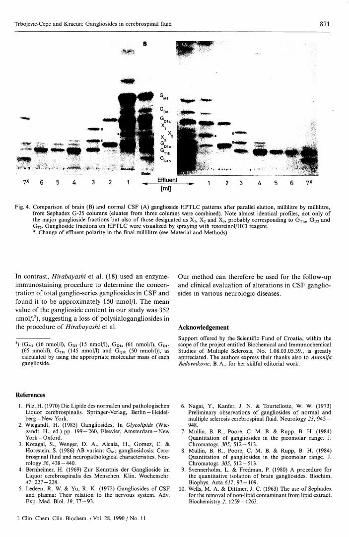
Parallel elution of the gangliosides of pooled 'normal"CSF and of brain tissue from Sephadex G-25 columnsshowed almost identical compositions, not only of themajor ganglioside fractions, but also of the fractionsdesignated as X,, X2 and X3. When a more polarsolvent system for HPTLC was used, chloroform-methanol-12 mmol/1 MgCl2 in H2O- 13.3 mol/1 NH3
(56:44:9:0.5, by volume), the fractions designatedas X did not overlap with the major gangliosidefractions and migrated between GDia and GDib (fig.4). Unidentified Xi, X2 and X3 could be detected inthe composition of both brain and CSF gangliosidesand probably corresponded to GTia (Xi), GD2 (X2)and GT3 (X3).
The data presented suggest that CSF contains gan-gliosides that are nearly identical with typical braingangliosides. Therefore, CSF gangliosides are prob-ably mostly secreted from brain cells, which appearsto be supported by the appearance of GM2 gangliosidein the CSF of patients with GM2 gangliosidosis (3).
Content of CSF gangliosides in 'normal 'adults
Our results on the total content of gangliosides in ten'normal' CSFs, i. e. 780 ± 82 μ§/1 of gangliosides(tab. 3), is comparable to that reported by Ledeen &Yu (466 — 1130 μg/l) (5). The presence of contaminants(probably protein-bound NeuAc) might have beenresponsible for the higher ganglioside value in normalpooled CSF (1320 μg/l) reported by Nagai et al. (6).Our method eliminates this effect, as the gangliosideextract is first chromatographed on HPTLC, thenlipid-bound NeuAc is determined by densitometry.
J. Clin. Chem. Clin. Biochem. / Vol. 28,1990 / No. 11
Trbojevic-Cepe and Kracun: Gangliosides in cerebrospinal fluid 871
B
7X
Fig. 4. Comparison of brain (B) and normal CSF (A) ganglioside HPTLC patterns after parallel elution, millilitre by m lilitre,from Sephadex G-25 columns (eluates from three columns were combined). Note almost identical profiles, not only ofthe major ganglioside fractions but also of those designated as Xl5 X2 and X3, probably corresponding to GTia, GD2 andGTS- Ganglioside fractions on HPTLC were visualized by spraying with resorcinol/HCl reagent.* Change of effluent polarity in the final millilitre (see Material and Methods)
In contrast, Hirabayashi et al. (18) used an enzyme-immunostaining procedure to determine the concen-tration of total ganglio-series gangliosides in CSF andfound it to be approximately 150 nmol/1. The meanvalue of the ganglioside content in our study was 352nmol/12), suggesting a loss of polysialogangliosides inthe procedure of Hirabayashi et al.
2) {GMi (16 nmol/1), GD3 (15 nmol/1), GDla (61 nmol/1), GDlb(65 nmol/1), Gm (145 nmol/1) and GQlb (50 nmol/1)}, ascalculated by using the appropriate molecular mass of eachganglioside.
Our method can therefore be used for the follow-upand clinical evaluation of alterations in CSF ganglio-sides in various neurologic diseases.
Acknowledgement
Support offered by the Scientific Fund of Croatia, within thescope of the project entitled Biochemical and ImmunochemicalStudies of Multiple Sclerosis, No. 1.08.03.05.39., is greatlyappreciated. The authors express their thanks also to AntonijaRedovnikovic, Β. Α., for her skilful editorial work.
References
1. Pilz, H. (1970) Die Lipide des normalen und pathologischenLiquor cerebrospinalis. Springer-Verlag, Berlin — Heidel-berg—New York.
2. Wiegandt, H. (1985) Gangliosides, In Glycolipids (Wie-gandt, H., ed.) pp. 199 — 260, Eisevier, Amsterdam—NewYork-Oxford.
3. Kotagal, S., Wenger, D. A., Alcala, H., Gomez, C. &Horestein, S. (1986) AB variant GM2 gangliosidosis: Cere-brospinal fluid and neuropathological characteristics. Neu-rology 36, 438-440.
4. Bernheimer, H. (1969) Zur Kenntnis der Ganglioside imLiquor cerebrospinalis des Menschen. Klin. Wochenschr.47, 227-228.
5. Ledeen, R. W. & Yu, R. K. (1972) Gangliosides of CSFand plasma: Their relation to the nervous system. Adv.Exp. Med. Biol. 19, 77-93.
6. Nagai, Y, Kanfer, J. N. & Tourtellotte, W. W. (1973)Preliminary observations of gangliosides of normal andmultiple sclerosis cerebrospinal fluid. Neurology 23, 945 —948.
7. Mullin, B. R., Poore, C. M. B. & Rupp, B. H. (1984)Quantitation of gangliosides in the picomolar range. J.Chromatogr. 305, 512-513.
8. Mullin, B. R., Poore, C. M. B. & Rupp, B. H. (1984)Quantitation of gangliosides in the picomolar range. J.Chromatogr. 305, 512-513.
9. Svennerholm, L. & Fredman, P. (1980) A procedure forthe quantitative isolation of brain gangliosides. Biochim.Biophys. Acta 617, 97-109.
10. Wells, M. A. & Dittmer, J. C. (1963) The use of Sephadexfor the removal of non-lipid contaminant from lipid extract.Biochemistry 2, 1259-1263.
J. Clin. Chem. Clin. Biochem. / Vol. 28, 1990 / No. 11

872 Trbojevic-Cepe and Kracun: Gangliosides in cerebrospinal fluid
11. Stahl, E. (1969) In: Thin-layer Chromatography (Stahl, E.,ed.) pp. 52 — 85, Springer-Verlag, Berlin —Heidelberg—NewYork.
12. Svennerholm, L. (1957) Quantitative estimation of sialicacid. Biochim. Biophys. Acta 24, 604—611.
13. Ando, S., Chang, N. & Yu, R. K. (1978) High performancethin-layer chromatography and densitometric determina-tion of brain ganglioside composition of several species.Anal. Biochem. 89, 437-450.
14. Tomasi, M., Roda, L. G., Ausiello, C., D'Angelo, G., Ve-nerando, B., Ghidoni, R., Sonnino, S. & Tettamanti, G.(1980) Interaction of GMi ganglioside with bovine serumalbumin, formation and isolation of multiple complexes.Eur. J. Biochem. Ill, 315-324.
15. Hayashi, K. & Katagiri, A. (1974) Studies on the interactionbetween gangliosides, protein and divalent cations.Biochim. Biophys. Acta 357, 107-117.
16. Kracun, L, Rosner, H., Cosovic, C. & Stavljenic, A. (1984)Topographical atlas of the gangliosides of the adult humanbrain. J. Neurochem. 43, 979-989.
17. Kracun, I., Rosner, H. & Cosovic, C. (1986) In: Gangliosidesand Neuronal Plasticity (Tettamanti, G., Ledeen, R. W.,Sandhoff, K., Nagai, Y. & Toffano, G., eds.) pp. 67-76,Fidia Research Series, Liviana Press — Springer Verlag, Pa-dova — Berlin — Heidelberg—New York—Tokyo.
18. Hirabayashi, Y, Koketsu, K., Higashi, H., Suzuki, Y.,Matsumoto, M., Sugimoto, M. & Ogawa, T. (1986) Sensi-tive enzymeimmunostaining and densitometric determina-tion of ganglioseries gangliosides on thin-layer plate: pmoldetection of gangliosides in cerebrospinal fluid. Biochim.Biophys. Acta 876, 178-182.
19. Svennerholm, L. (1963) Chromatographie separation ofhuman brain gangliosides. J. Neurochem. 10, 613 — 623.
20. IUPAC-IUB Commission on Biochemical Nomenclature(CBN) (1977) The Nomenclature of Lipids-Recommenda-tions, 1976. Eur. J. Biochem. 79, 11-21.
Milica Trbojevic, PhDInstitute of Clinical Laboratory DiagnosticsZagreb University School of MedicineClinical Hospital CentreKispaticeva 12YU-41000 Zagreb
J. Clin. Chem. Clin. Biochem. / Vol. 28, 1990 / No. 11


![Research Paper Determination of cerebrospinal fluid ... · nonsurgical traumatic (80%), surgical (16%) and non-traumatic (4%) modalities [1-3]. CSF has functions related to brain](https://static.fdocuments.us/doc/165x107/5e196547b3ed4a5e342642ea/research-paper-determination-of-cerebrospinal-fluid-nonsurgical-traumatic-80.jpg)


