Determination of ABO Blood Groups and Rhesus factorin ...
67
ﺑﺴﻢ ﷲ اﻟﺮﺣﻤﻦ اﻟﺮﺣﯿﻢSudan University of Science and Technology College of Graduated Studies Determination of ABO Blood Groups and Rhesus factorin Sudanese Patientswith Cardiovascular Diseases ﺗﺤﺪﯾﺪ اﻟﺰﻣﺮ اﻟﻮظﯿﻔﯿﺔ ﻟﻠﻔﺼﺎﯾﻞ اﻟﺪﻣﻮﯾﺔ واﻟﻌﺎﻣﻞ اﻟﺮﯾﺼﻲ ﻟﺪى ا ﻟﺴﻮداﻧﯿﯿﻦ اﻟﻤﺼﺎﺑﯿﻦ ﺑﺄﻣﺮاض اﻟﻘﻠﺐ واﻷوﻋﯿﺔ اﻟﺪﻣﻮﯾﺔA Dissertation submitted in partial Fulfillment of Requirements of M.Sc degree in Medical Laboratory Science (Hematology and Immunohematology) By: Reela AbdElmageed AbdElraheem (B. Sc.Medical laboratory science honor degree) (SUST, 2012) Supervisor: Dr: KhaldaMirghaniHamza 2014
Transcript of Determination of ABO Blood Groups and Rhesus factorin ...
بسم الله الرحمن الرحیم
Sudan University of Science and Technology
College of Graduated Studies
Determination of ABO Blood Groups and Rhesus factorin Sudanese Patientswith Cardiovascular Diseases
لسودانیین للفصایل الدمویة والعامل الریصي لدى اتحدید الزمر الوظیفیة المصابین بأمراض القلب والأوعیة الدمویة
A Dissertation submitted in partial Fulfillment of Requirements of M.Sc degree in Medical Laboratory Science
(Hematology and Immunohematology)
By:
Reela AbdElmageed AbdElraheem
(B. Sc.Medical laboratory science honor degree)
(SUST, 2012)
Supervisor:
Dr: KhaldaMirghaniHamza
2014

I
ةـــــالآی
:قال تعالى
صدق االله العظيم
)19(سورة النمل الآية

II
Dedication
To…My beloved and blessed mother who gives meaning and light to my
life.
To…My wonderful beautiful half (my husband) for his support and
encouragement.
To…My wonderful supervisorDr.KhaldaMeirghani, who helped
whenever I needed her.
To…our teacher's very special friends and colleagues who were integral
part of our support group.
Special dedication to all patients from whom samples were collected.
I dedicate this work
.Reela

III
ACKNOWLEDGMENT
First of all my thanks to Allah for giving me assistance, health and patience
to complete this work.
I would like to express my deep thanks to my supervisor Dr.
KhaldaMeirghani for her expertise, support and endless valuable advice.
Thanks for all the staff of the hematology department for their valuable
assistance and encouragement throughout the research.
Best thanks all persons who helped me in my research especially Whagdy,
Halah and Zeinab.
Thanks so much to you all
Reela.
IV
Abstract
Many reports have appeared in recent years suggesting an association between blood groups and various manifestations of heart diseases.
This a descriptive analytical study aimed to determine frequency of blood group and Rhesus factor of patients with cardiovascular diseases in Khartoum state during the period from May to August 2014.
Seventy patients with different types of cardiovascular diseases attended Khartoum State Hospital was enrolled in this study. An informed consent was obtained from each patient before blood sample collection.ABO and Rh factors were performed by slide techniques using specific anti-sera. The data were analyzed by SPSS computer program. The results showed that most common blood group in patients with cardiovascular was O followed by A, least frequent was AB.
Majority (91.4%) of patients with cardiovascular were Rh positive.
Most common blood group in males was O (54.5%) and least frequent was AB (6.1%), and most common blood group in females was O (45.9%) followed by A (40.6%) and least frequent was AB (2.7%), and most common cardiovascular disease in females compared to males and most common disease was found in elder patients with age between (51-85years).
Most common cardiovascular diseases were ischemic heart disease followed by heart failure and least frequent was vavular heart disease, and most common disease in males was ischemic heart disease (33.3%) and least frequent was permanent pacemaker (0.0%) , also most common disease in females was ischemic heart disease (27.0%) and least frequent was valvular heart disease, and most common disease in patients with age between (23-50) was dilated cardiomyopathy (33.3) and most frequent disease in patients more than 50 years was ischemic heart disease (34.7%).
Finally, the results of the present study showed that there is no association of ABO blood group and Rh factor with cardiovascular diseases. Although the frequent of ischemic heart disease (IHD) was higher in O group, the difference was statistically insignificant.
V
المستخلص أظھرت إرتباط بین فصائل الدم والأنواع المختلفة لأمراض القلب والأوعیة عدة تقاریر أجریت حدیثا
. الدمویة
وصفیة تحلیلیة ھدفت لتحدید تكرار زمر الدم في المرضى المصابین بأمراض القلب ھذه دراسة
سبعین من المصابین . 2014والأوعیة الدمویة في ولایة الخرطوم في الفترة من مایو إلى أغسطس
البراءة الأخلاقیة أخذت من . بمختلف أنواع امراض القلب والأوعیة الدمویة ضمنوا في ھذه الدراسة
.على حدة قبل أخذ العینة منھ كل مریض
إختبارات زمر الدم أجریت بإستخدام طریقة الشرائح بإسخدام أمصال مضادة بعد ذلك حللت البیانات
بإستخدام نظام الحزم الإحصائیة للمجتمع حیثوجد أن أكثر زمر الدم انتشارا في مرضى القلب
.ABواقلھ ھو Aیتبع بـ Oوالأوعیة الدمویة ھو
.ابین بأمراض والأوعیة الدمویة كانوا موجبي العامل الریصيأكثر المص
في الرجال ھو الـ ، كما أن أكثر زمر الدم ABوأقلھ ھو الـ Oوجد أن أكثر زمر الدم إنتشارا
في النساء ھو الـ ABوأقلھ ھو الـ Aیتبع بـ Oإنتشارا
في النساء من الرجال و - 51(في المرضى ذوي الأعمار بین وجد أن أكثر أمراض القلب انتشارا
ووجد أن أكثر أنواع أمراض القلب والأوعیة الدمویة انتشارا ) 50-8(أكثر من المرضى بین ) 85
).VHD(والأقل انتشارا ھومرض القلب الصمامي )IHD(ھو مرض القلب الإحتباسي
.)IHD(أكثر أمراض القلب انتشارا في الرجال ھو مرض القلب الإحتباسي وأن
النتائج الحالیة لھذه الدراسة أظھرت عدم وجود إرتباط بین فصائل الدم والعامل الریصي وأمراض
.القلب والأوعیة الدمویة
إلا أن الإختلاف Oبالرغم من أن تكرار حدوث مرض القلب الاحتباسي أعلى في فصیلة الدم
لیس لھ دلالة . إحصائیا

VI
List of contents
Contents Page I الآیــةDedication II Acknowledgement III Abstract (English) IV V المستخلصList of Contents VI List of Tables IX List of abbreviations X
Chapter One Introduction and Literature review
1.1 Introduction 1 1.2 Literature Review 3
1.2.1 Blood Physiology 3 1.2.1.1 Blood group system 4
1.2.1.1.1 History of ABO discoveries 5 1.2.1.1.2 The importance of ABO blood group system 5 1.2.1.1.3 Classifications of ABO blood group system 6 1.2.1.1.4 Antigens of ABO blood group system 7 1.2.1.1.5 Antibodies of ABO blood group system 8 1.2.1.1.6 Sub groups of A 9 1.2.1.1.7 ABH secretor status 9 1.2.1.2 The Rhesus system 9
1.2.1.2.1 Discovery of Rh system 10 1.2.1.2.2 The nomenclature of Rh blood group system 11 1.2.1.2.3 The fisher- Race nomenclature 11 1.2.1.2.4 The Weiner nomenclature 12 1.2.1.2.5 Rh blood group system antigens and antibodies 12 1.2.1.2.6 Other Rh blood group system antigens 13 1.2.1.2.7 Rh phenotypes 13
1.2.2 Heart structure and function 13 1.2.2.1 Cardiovascular diseases 13

VII
1.2.2.2 Cardiovascular diseases types and risk factors 15 1.2.2.2.1 Coronary artery disease 16 1.2.2.2.2 Prosthetic heart valves 16 1.2.2.2.3 Heart Failure 17 1.2.2.2.4 Congenital Heart disease 18 1.2.2.2.5 Ischemic heart disease 18 1.2.2.2.6 Myocardial infarction 19 1.2.2.2.7 Valvular heart disease 20 1.2.2.2.8 Dilated cardiomyopathy 20 1.2.2.2.9 Permanent Pacemakers 21
1.2.3 Previous Studies 21 1.3 Rationale 22 1.4 Objectives 23
1.4.1 General objectives 24 1.4.2 Specific objectives 24
Chapter Two Materials and Methods
2.1 Study design 25 2.2 Study population 25 2.3 Inclusion criteria 25 2.4 Exclusion criteria 25 2.5 Data collection 25 2.6 Ethicalconsideration 26 2.7 Materials 26 2.8 Methods 26
2.8.1 Sampling 26 2.8.2 ABO slide agglutination test 27
2.8.2.1 Principle 27 2.8.2.2 Procedure 27 2.8.2.3 Interpretation 27 2.8.2.4 Controls 27 2.8.3 Rh (D) red blood cell typing 28
2.8.3.1 Principle 28 2.8.4 Du method (the indirect anti globulin) 28

VIII
2.8.4.1 Principle 29 2.8.4.2 The technique of Du method 29 2.8.4.3 Requirements 29 2.8.4.4 Interpretation 30
Chapter Three Results
3. Results 31 Chapter Four
Discussion, Conclusion and Recommendation 4.1 Discussion 36 4.2 Conclusion 38 4.3 Recommendations 39 References 40 Appendices 43-50

IX
List of Tables
Table No
Title Page No
(1-1) Blood group system recognized by ISBT 2 (1-2) The ABO blood group system 6 (1-3) The ABO blood group system 7 (1-4) The most common Rh genotypes 10 (3-1) Distribution of study group according to gender 32 (3-2) Distribution of study group according to age 32 (3-3) Frequency of different types of cardiovascular diseases 33 (3-4) Frequency of ABO among study groups 33 (3-5) Distribution of ABO blood group according to gender 33 (3-6) Frequency of RH group in study groups 33 (3-7) Distribution of different types of cardiovascular diseases
according to gender 33
(3-8) Distribution of different types of cardiovascular diseases according to age group
34
(3-9) Distribution of ABO blood groups among different types of cardiovascular diseases
34
(3-10) Distribution of Rh blood group among different types of cardiovascular diseases
35

X
List of Abbreviations
CAD Coronary Artery Disease
CDC Centers of Disease Control and Prevention
CHD Congestive heart disease
CHF Congestive heart Failure
CVD Cardiovascular disease
DCM Dilated Cardiomyopathy
HIV Human immunodeficiency viral
HF Heart Failure
IgGImunoglobulin G
IgMImunoglobulin M
IHD Ischemic heart disease
ISBT International Society of Blood transfusion
K2EDTA Potassium Ethylene Diamine tetra Acetic Acid
MI Myocardial Infarction
PPMPermanent Pacemaker
Rh Rhesus
VHD Valvular heart disease
SPSS Statistical Package of Social Science

Chapter One
Introduction and Literature Review

1
Chapter One
1. Introduction and Literature review 1.1. Introduction
Since the discovery of the ABO system in 1900, a multitude of blood group
antigens have been identified and many different styles of terminology have
been used. The International Society of blood transfusion (ISBT) recognizes
285 blood group antigens; 245 of these are classified into one of 29 blood
group system. Forty years later, both Landsteiner and Wiener discovered
Rh(D)antigen (Garratty, 2000;Mollison, 1994).
The genes of ABO and Rh(D) are located on chromosome 9 and 1
respectively. The bombardment of the red blood cells with A and /or B
antigens occur as a consequence of the action of the glycosyltransferases
enzymes that add specific sugars to the precursor substance (John, 1996).
The cardio vascular system includes the heart and the blood vessels. A
functional cardio vascular system is vital for survival, because without blood
circulation, the tissues lack oxygen and nutrients, and wastes accumulate.
Under such conditions, the cells soon begin irreversible, which quickly leads
to death. Cardio vascular disease (also called heart disease) is a class of
disease that involves the heart, the blood vessels (arteries, capillaries and
veins) or both. This study aimed to determine the frequency of ABO blood
groups and Rhesus factor of cardio vascular patients and to correlate
association of blood group with cardio vascular diseases. Results of this
study may be useful in determines which blood group system is more
susceptible to cardiovascular diseases(Bridget, 2010).

2
Table (1.1) BloodGroup System Recognized by ISBT (Hoff Brand et al.,
2000)
System number System name conventional
System symbol ISBT
Chromosomal location
Gene(s)
001 ABO ABO gq34.1 – q34.2 ABO
002 MNS MNS 4q28-q31 GYPA GYPB
003 P P1 22q11.2-qter P 004 Rh RH 1p36.2-p34 PHD
RHCE 005 Lutheran Lu 19q12-q13 LU 006 Kell KEL 7q33 KEL 007 Lewis LE 19p13.3 FUT3 008 Duffy Fy 1q22-q23 FY 009 Kidd JK 18q11-q12 HUT11 010 Diego DI 17q12-q21 SLC4A1 011 Yt YT 7q22 ACHE 012 Xg XG Xp22.32 XG 013 Scianna SC 1p36.2-p22.1 SC 014 Dombrock DO 12p13.2-p12.1 GO 015 Colton CO 7p14 AQP1 016 LW LW 19p13.2-cen LW 017 Chid/Rogers CH/RG 6p21.3 C4A,C4P 018 H H 19p13 FUT1 019 Kx XK Xp21.1 XK 020 Gerbich GE 2q14-q21 GYPC 021 Cromer CROM 1q32 GAF 022 Knops KN 1q32 CR1 023 Indian IN 11p13 CD44 024 Ok OK 19pter –p13-2 OK 025 MER2 RAPH 11p15 MER2
International Society of Blood Transfusion (ISBT)
3
1.2. Literature Review
1.2.1. Blood physiology
Word blood is related to blowan means bloom or flourish. Blood is a vital
fluid of our body and as such the life line of human body. It is a red colored
viscid fluid slightly salty in taste. Blood is alkaline in reaction PH 7.4 and
specific gravity ranges from 1.052 to 1.060. In adult human, blood volume
ranges between 4.5 to 6.0 liters and is approximately about one thirteenth of
adult human body weight. Temperature of circulating blood is 37 ◌.7 C.
Blood has two main components cells and plasma. Cells consist of 40 to
45% of the total amount of blood and plasma consists of 55 to 60% of total
amount of blood. Cells are the formed elements and are of three types red
cells (erythrocyte), white cells (leucocytes) and platelet (thrombocyte) and
each has its own characteristic (Talib, 1995).
Blood is a fluid which is continuously on movement. This process of
movement of blood is known as circulation and the system that sustains the
process is known as circulatory system and Dr. William Harvey was first
person to describe circulation in 1616. It consists of mainly the heart and
blood vessels viz. arteries and vein _ cardio vascular system. The whole
system works under supreme control of nervous system and cardio _
respiratory centre is believed to be located in medulla oblongata or brain
stem. The circulatory activity is also indirectly influenced by some
hormones produced in our body. So it is a co-operative function of nervous
and humeral mechanism that co-ordinate various activities of heart and
blood vessels ensuring proper supply of blood in every corner of our body
there by feeding the vital organs and tissues with oxygen , nutrient and

4
other essential substances . Blood vessels are of three types – arteries,
capillaries and veins. Arteries are vessel that carries blood away from the
heart and are having thicker walls consisting tunica adventitia, media and
intima from outside in ward respectively composed smaller arterioles tissue
and endothelium. Artery gradual becomes smaller arterioles and then
capillary as they move away from heart. Capillaries are very small, capillary
wall consists single layer of endothelium through which inter change
between blood and tissue fluid takes place. As blood flow through capillary
wall into the tissue the arterial blood changes into venous blood after
exchange of substance between blood and tissues. This changed blood drains
into vein which is brought back to heart and thence to the lung for
purification. So veins are also part of circulatory system but unlike arteries
all the three coats of venous walls contains less elastic and less muscle tissue
and so the veins are less resilient and easily collapses, it also consists of
valves to allow uni directional flow of blood towards heart, valves prevents
back flow of blood (Talib, 1995).
1.2.1.1. Blood group system
The clinical importance of a blood group system in blood transfusion lies in
the frequency of its antibodies and in the possibility that such as antibodies
will destroy in compatible cells in vivo. A B O system was the first to be
recognized and remains the most important in transfusion and trance
plantation (histo - blood group system). The reason for this is that almost
everybody over the age of about 6months has clinically significant anti – A
and anti –B in his or her serum if they lack the corresponding antigens on
their red cells bence transfusions given without regard to ABO groups would

5
result in incompatibility patient will has in a vivo adverse hemolytic
reactions (Hoff Brand et al., 2000) .
1.2.1.1.1. History of ABO discoveries
The two most significant blood group systems were discovered by Karl
Landsteiner during early experiments with blood transfusion: the ABO
group in 1901. Landsteiner and in Co-operation with Alexander S. Wiener
and Rhesus group. (Landsteiner et al., 1940) Development of the coombs
test in 1945, (Coombs et al., 1945) the advent of transfusion medicine, and
the understanding of hemolytic disease of the new born, led to discovery of
more blood groups, and now 30 human blood group systems are recognized
by the international society of blood transfusion (ISBT), (Lewis et al., 1990)
and across the 30 blood groups, over 600 different blood group antigens
have been found, (Lewis et al., 1991) many of these are very rare or are
mainly found in certain ethnic groups. Landsteiner was in advertently the
first individual to perform forward and reserve grouping. The forward
grouping is defined as using known sources of reagent anti-sera to detect
antigens on an individual red cells .Reverse grouping is defined as using
reagent cells with known ABO antigens and testing the serum of patient for
ABO group anti bodies (Denis and Hameening,1998).
1.2.1.1.2. The importance of ABO blood group system
The clinical significance of different red cell anti bodies depends partly on
their destructive capacity importance in blood transfusion practice owing to
its great Varity. Conversely, ABO and D anti bodies are by far the most
significant, due to their high frequency and destructive capacity (Hoff Brand
et al., 2000).

6
The O gene is a morph and does not transform the H-substance. Although
there are six possible genotypes, the absence of a specific anti-O prevents
the serological recognition of more than four phenotypes. The two major sub
groups of A (A1 and A2).A2 cells react more weakly than A1 cells with anti-
A and patients who are A2B can be wrongly grouped as B. A, B and H
antigens are present in most body cells including white cells and platelets. In
80% of the population who possess secretor genes, these antigens are also
found in soluble forms in secretions and body fluids (e.g. plasma, saliva,
semen and sweat).Naturally occuring anti bodies to A and/ or B antigens are
found in the plasma of subjects whose red cells lack the corresponding
antigens (Hoff Brand et al., 2000).
Table (1-2) The ABO Blood Group System. (Hoff brand et al.,
2006)
Phenotype Genotype Antigens Naturally
occurring anti
bodies
Frequency
(UR) (%)
O OO O Anti-A, Anti-B 46
A AA or AO A Anti-B 42
B BB or BO B Anti-A 9
AB AB AB None 3
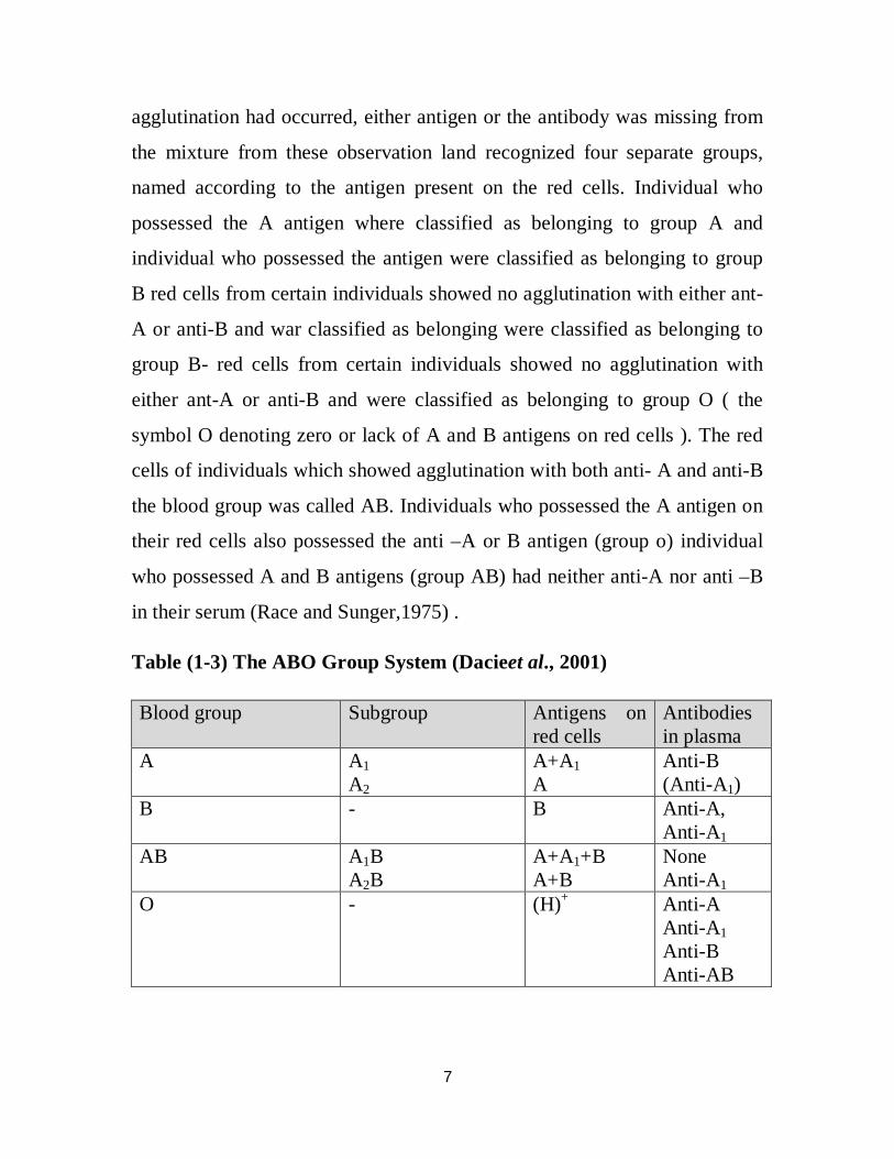
1.2.1.1.3. Classification of ABO blood group system
Classification of ABO blood groups was based on the redization hat
agglutination had occurred because the red cells possessed an antigen and
corresponding specific antibody was present in the serum, when no
7
agglutination had occurred, either antigen or the antibody was missing from
the mixture from these observation land recognized four separate groups,
named according to the antigen present on the red cells. Individual who
possessed the A antigen where classified as belonging to group A and
individual who possessed the antigen were classified as belonging to group
B red cells from certain individuals showed no agglutination with either ant-
A or anti-B and war classified as belonging were classified as belonging to
group B- red cells from certain individuals showed no agglutination with
either ant-A or anti-B and were classified as belonging to group O ( the
symbol O denoting zero or lack of A and B antigens on red cells ). The red
cells of individuals which showed agglutination with both anti- A and anti-B
the blood group was called AB. Individuals who possessed the A antigen on
their red cells also possessed the anti –A or B antigen (group o) individual
who possessed A and B antigens (group AB) had neither anti-A nor anti –B
in their serum (Race and Sunger,1975) .
Table (1-3) The ABO Group System (Dacieet al., 2001)
Blood group Subgroup Antigens on red cells
Antibodies in plasma
A A1 A2
A+A1 A
Anti-B (Anti-A1)
B - B Anti-A, Anti-A1
AB A1B A2B
A+A1+B A+B
None Anti-A1
O - (H)+ Anti-A Anti-A1 Anti-B Anti-AB
8
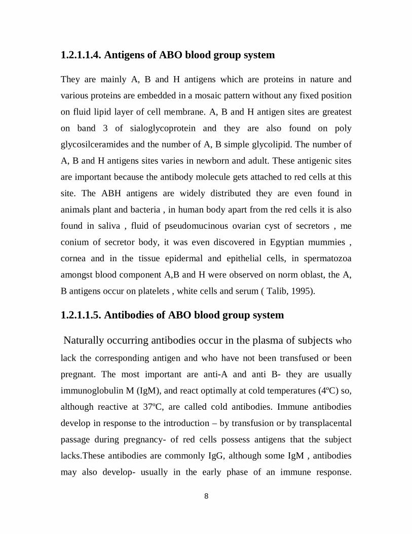
1.2.1.1.4. Antigens of ABO blood group system
They are mainly A, B and H antigens which are proteins in nature and
various proteins are embedded in a mosaic pattern without any fixed position
on fluid lipid layer of cell membrane. A, B and H antigen sites are greatest
on band 3 of sialoglycoprotein and they are also found on poly
glycosilceramides and the number of A, B simple glycolipid. The number of
A, B and H antigens sites varies in newborn and adult. These antigenic sites
are important because the antibody molecule gets attached to red cells at this
site. The ABH antigens are widely distributed they are even found in
animals plant and bacteria , in human body apart from the red cells it is also
found in saliva , fluid of pseudomucinous ovarian cyst of secretors , me
conium of secretor body, it was even discovered in Egyptian mummies ,
cornea and in the tissue epidermal and epithelial cells, in spermatozoa
amongst blood component A,B and H were observed on norm oblast, the A,
B antigens occur on platelets , white cells and serum ( Talib, 1995).
1.2.1.1.5. Antibodies of ABO blood group system
Naturally occurring antibodies occur in the plasma of subjects who
lack the corresponding antigen and who have not been transfused or been
pregnant. The most important are anti-A and anti B- they are usually
immunoglobulin M (IgM), and react optimally at cold temperatures (4ºC) so,
although reactive at 37ºC, are called cold antibodies. Immune antibodies
develop in response to the introduction – by transfusion or by transplacental
passage during pregnancy- of red cells possess antigens that the subject
lacks.These antibodies are commonly IgG, although some IgM , antibodies
may also develop- usually in the early phase of an immune response.

9
Immune antibodies react optimally at 37ºC (worm antibodies). Only IgG
antibodies are cable of transplacental passage from mother to fetus. The
most important immune antibody is Rh antibody, anti-D (Hoff Brand etal.,
2000).
1.2.1.1.6Sub groups of A
In addition to the common phenotypes A1 and A2 numerous phenotypes with
weak expression of A on the red cells have been found and multitude of
names has been adopted. Most of these phenotypes can be fitted into the
following categories: A3, Ax, Am, Ay and Ae . The serological
characteristics of these phenotypes results from inheritance of a rare allele at
the ABO locus, which can be detected when parried with our B, but not with
A1 or A2 (Daniels et al., 2002) .
1.2.1.1.7. ABH secretor status
A bout 80% of the UK populations are ABH secretors as they have H
antigen plus A or B according to their ABO genotype, in a water – soluble
form in their body secretions. The remaining 20% are non – secretors and
have no secreted ABH antigens, regardless of ABO phenotype (Hoff Brand
et al., 2000).
1.2.1.2. The Rhesus System
The Rhesus system is the second most clinically important and complex
blood group system. It consists of 50 different antigens, but only 5 antigens
D, C, c, E and e are inherited in various combinations and account for most
of the Rh-related problems encountered in practice. The Rh antigen with the
strongest antigenicity is the Rh (D) antigen. As simple rule, it can be noted
10
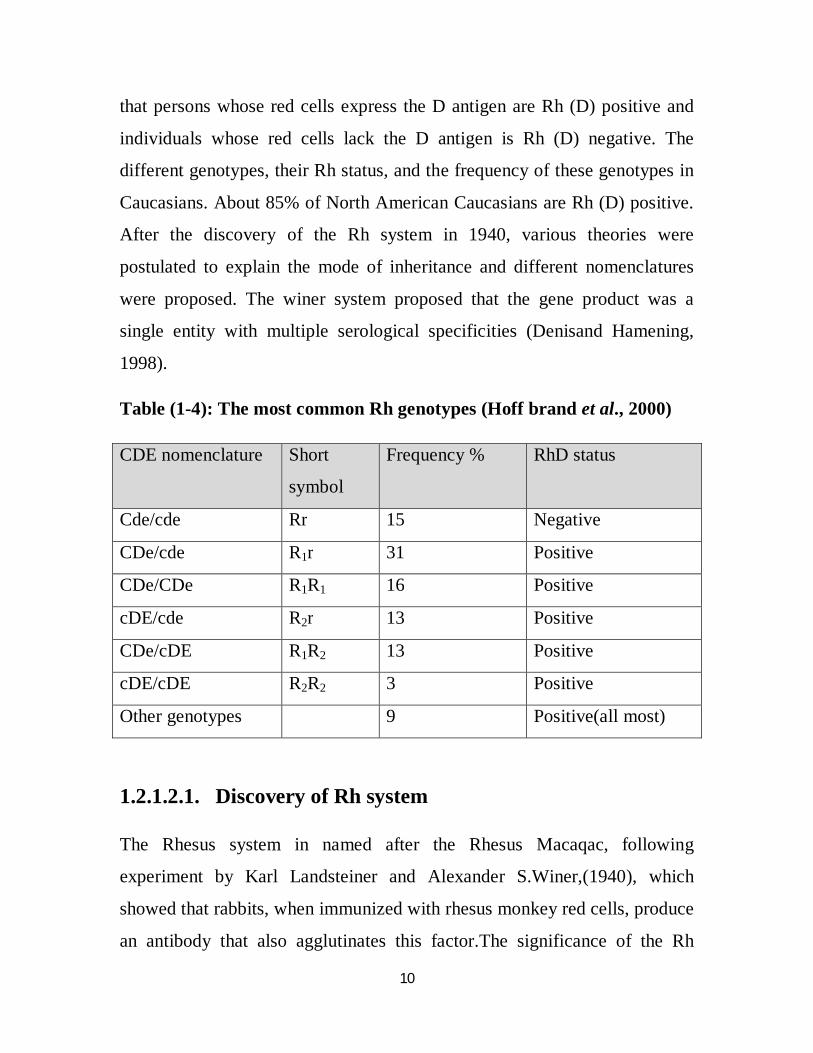
that persons whose red cells express the D antigen are Rh (D) positive and
individuals whose red cells lack the D antigen is Rh (D) negative. The
different genotypes, their Rh status, and the frequency of these genotypes in
Caucasians. About 85% of North American Caucasians are Rh (D) positive.
After the discovery of the Rh system in 1940, various theories were
postulated to explain the mode of inheritance and different nomenclatures
were proposed. The winer system proposed that the gene product was a
single entity with multiple serological specificities (Denisand Hamening,
1998).
Table (1-4): The most common Rh genotypes (Hoff brand et al., 2000)
CDE nomenclature Short
symbol
Frequency % RhD status
Cde/cde Rr 15 Negative
CDe/cde R1r 31 Positive
CDe/CDe R1R1 16 Positive
cDE/cde R2r 13 Positive
CDe/cDE R1R2 13 Positive
cDE/cDE R2R2 3 Positive
Other genotypes 9 Positive(all most)
1.2.1.2.1. Discovery of Rh system
The Rhesus system in named after the Rhesus Macaqac, following
experiment by Karl Landsteiner and Alexander S.Winer,(1940), which
showed that rabbits, when immunized with rhesus monkey red cells, produce
an antibody that also agglutinates this factor.The significance of the Rh

11
factor was soon realized.Dr. Philip Levine working at the Newark Beth
Israel Medical center made a connection between the Rh factor and the
incidence of erythroblastosis, and Weiner realized adverse reactions from
the Rh factor. Weiner then pioneered the exchange transfusion technique
saved the lives of many thousands of infants before intrauterine transfusion
was invented which enabled much more severely affected fetuses to be
successfully treated (Denisand Hamening, 1998).
1.2.1.2.2. The nomenclature of Rh blood group system
Several nomenclatures can be used to describe Rh genes and antigens.
Fisher- Race nomenclature, which uses CDE terminology, more commonly
is used for Antigens; Weiner nomenclature, which uses Rh designations, is
favored for haplotypes and gene complexes. An individual who inherits (ce
gene) from one parent and (D and cegnes) from the other parent expresses
DC, c and e antigens on his or her erythrocyte (Hoffbrandet al., 2000).
1.2.1.2.3. The Fisher- Race nomenclature
Fisher- Race theory states that there are three closely linked loci, each with
primary set of allelic genes D and d, C, c, E and e. these three loci are to be
closely linked that crossing over occurs only very rarely,and the three Rh
genes are inherited as a complex. The Rh genes complex was assumed to
possess closely linked genes, which could be assembled in eight different
ways: CDE, CDe, cDE, Cde, Cde, cDe, cdE, cde. Inter on nomenclature, the
Rh antigens are therefore named: C, D, E, c, d, e. the antigen (d) and its
corresponding antibody has never been discovered and is through not be
exists. The symbol (d) is used to donate the absence of D antigen. All
individuals who lack the D- antigen are Rh negative. Regardless of whether
12
the C or E or both are present, the most frequent genotype among D-
negative individuals is cde.The theory of Fisher- Race was confirmed when
the two unknown reactions CDE were shown to be as predicated by Murray
and Cowkers in 1945 and when anti e was discovered by Mourant in same
year. The only weakness in theory therefore was failure to find the expected
antigen- d.Other antigens since found be a part of Rh system have been
classified using the same basic principle (Nevillenet al., 1994).
1.2.1.2.4. The Weiner nomenclature
Weiner visualized multiple allele determining its won particular antigen. The
antigen comprises multiple factors depending on which genes are present
and are recognized by which ever factors are detectable. The two genes (i.e.
one paternal and one maternal),have been alike (homozygous) or different
(heterozygous). Therefore, multiple allele are called R1, R2, RO, r, r, r , RZ,
RY.Rh- antigens are called Rho, h1, RH, rh. In simple terms, for example
the Rh1 gene produces a complex antigen on the red cell that made up of at
least three factors: Rh, Rho, rh (Nevillenetal., 1994).
1.2.1.2.5. Rh blood group system antigens and antibodies
Rh antigen is aprotein surrounded by lipid, Rh activity is not lost when lipid
is extracted from red cells membranes (the lipid doesn’t carry the antigenic
determination but may be essential for confirmation of the determinants)
(Nevillenet al.,1994).
In the Rhesus blood group system, naturally occurring Rhesus antibodies are
not found in the serum of individuals lacking the corresponding Rhesus
antigens. Rhesus antibodies are formed by immunization. The most
13
important Rhesus antibodies is Anti- D, which can be formed when a Rh
negative individual is transfused Rh positive blood or when a Rh negative
woman becomes pregnant with a Rh positive infant and the red cells of the
baby pass into her circulation particularly at the time of delivery, stimulating
the production of anti- D antibody. Such circulating anti- D will not become
immediately harmful unless the individual receives a transfusion of Rh
positive blood, in such a situation the donor’s D antigen red cells will be
hemolyzed by the anti- D (Nevillenet al., 1994).
1.2.1.2.6. Other Rh blood group system antigens
Currently, 50 antigens have been described in the Rh group system, D, C, c,
E and e antigens are the most important ones. The other antigens are much
less frequently encountered or are rarely clinically significant. Each is given
a number (Mark, 2005).
1.2.1.2.7. Rh Phenotypes
The completeness with which the Rh phenotype can be determined depends
on the anti sera available; if anti- c is available but not anti- C, samples can
be classified as c positive (i.e. cc or Cc) and c negative (i.e. CC). If anti- C is
also available, Cc can be distinguished from cc. If a sample is tested with
anti- D, anti- C, anti-c and anti- E and gives positive reactions with all for
anti sera: the phenotype is written DCcE.Red cells that fail to react with
aanti-D are described as dd. Mountran’notation is occasionally misleading
for example , although anegative reaction with anti-E usually implies that
the cells are ee (Mollison, 1997) .
14
1.2.2. Heart structure and function
The heart is one of the most important organs in the entire human body. It is
really nothing more than a pump, composed of muscle which pumps blood
throughout the body, beating approximately 72 times per minute of our lives.
The heart pumps the blood, which carries all the vital materials which help
our bodies function and removes the waste products that we do not need. For
example, the brain requires oxygen and glucose, which, if not received
continuously, will cause it to lose consciousness. Muscles need oxygen
glucose and amino acids, as well as the proper ratio of sodium, calcium and
potassium salts in order to contract normally. The glands need sufficient
supplies of raw materials from which to manufacture the specific secretions.
If the heart ever ceases to pump blood the body begins to shut down and
after very short period of time will die.The walls of the heart are made up of
three layers, while the cavity is divided into four parts, three are two upper
chambers, called the right and the left atria, and two lower chambers, called
the right and the left ventricles. The right atrium, as it is called, receives
blood from the upper and lower body through the superior vena cava and the
inferior vena cava, respectively, and from the heart muscle itself through the
coronary sinus. The right atrium is the larger of the two atria, having very
thin walls. The right atrium opens into the right ventricle through the right
atrioventicular valve (tricuspid), which only allows the blood to flow from
the atria into the ventricle, but not in the reserve direction. The right
ventricle pumps the blood to the lungs to be reoxygenated. The left atrium
receives blood from the lungs via the four pulmonary veins. It is smaller
than the right atrium, but has thicker walls. The valve between the left
atrium and the left ventricle, the left atrioventicular valve (bicuspid), is

15
smaller than the tricuspid. It opens into the left ventricle and again is a one
way valve. The left ventricle pumps the blood throughout the body. It is the
Aorta the largest artery in the body, which originates from the left
ventricle.The heart works as a pump moving blood around in our bodies to
nourish every cell. Used blood, that is blood that has already been to the
cells and has given up its nutrients to them, is drawn from the body by the
right half of the heart, and then sent to lungs to be reoxygenated. Blood that
has been reoxygenated by the lungs is drawn into the left side of the heart
and then pumped into the blood stream. It is the atria that draw the blood
from the lungs and body, and the ventricles that pump it to the lungs and
body. The output of each ventricle per peat is about 70 ml, or about 2 table
spoons. In a trained athlete this amount is about double, with the average
heart rate of 72 beats per minute the heart will pump about 5 liters per
ventricle, or about 10 liters total per minute. This is called the cardiac
output. In a trained athlete the total cardiac output is about 20 liters (Bridget,
2012)
1.2.2.1. Cardiovascular diseases
Cardiovascular disease (CVD) is the leading cause of death inthe world and
accounts for well over one million deaths each year in the United States. Of
the more than two million deaths in the United States in 1998, CVD was
listed as the primary or contributing cause in 70% of cases.1 According to
theCenters of Disease Control and Prevention (CDC) and the National
Health and Nutrition Examination Survey III, the probability at birth of
dying from CVD is 47%, compared to22% from cancer, 2% from diabetes,
and less than 1% from human immunodeficiency virus (HIV) disease. The
largest proportion of this high mortality is attributed to coronaryartery
16
disease (CAD) or coronary heart disease (CHD), whichwas the primary
contributing cause of death in 459,841Americans in 1998. (Dallas, 2001)
CVD includes hypertension, coronary artery disease(CAD), congestive heart
failure (CHF), congenital cardiovasculardefects, and stroke. Although these
diseases are associated with a high mortality,the associated morbidity affects
all walks of life and has agreat impact on the quality of life of affected
individuals (Bridget, 2010).
1.2.2.2. Types of Cardiovascular diseases and risk factors
There are different types of cardiovascular diseases include: Coronary
artery disease ,cardiomyopathy- diseases of cardiac muscle, hypertensive
heart disease, heart failure, inflammatory heart disease, valvular heart
disease ,cerebrovascular disease ,peripheral arterial disease ,congenital
heart disease ,and rheumatic heart disease .Evidence suggests anumber of
risk factors for heart diseases ;age,gender ,high blood
pressure,hyperlipidemia,diabetes mellitus, tobacco smoking, processed
meat consumption, family history,obesity, lack of physical activity,
psychosocial factors, and air pollution (Bridget, 2010).
1.2.2.2.1 Coronary artery disease
Coronary artery disease (CAD) accounts for approximately 30to 50% of all
cases of CVD. It is estimated that 12,400,000 .Americans alive today have
already suffered a myocardial infarction (MI) or experienced angina
pectoris (chest pain). Atherosclerosis, the most common cause of CAD,
results froma wide variety of pathologic processes that interact with
anddisrupt the vascular endothelium. The result is plaque formation,with
the compromise of effective arterial luminalarea. In the coronary
circulation, this process may cause achronic reduction in coronary blood

17
flow and ensuingmyocardial ischemia or it may cause acute plaque rupture
with intracoronary thrombus formation and subsequent MI.Atherosclerosis
may affect any vascular bed, including thecoronary, cerebral, renal,
mesenteric, and peripheral vascularsystems. When end-organ blood flow is
compromised, theresulting ischemia can cause subsequent organ
dysfunction.The sudden rupture of an atherosclerotic plaque, with
ensuingintracoronary thrombus formation that acutely reducescoronary
blood flow, causes the acute coronary syndromes.This results in
myocardial ischemia and subsequent infarctionif there is a prolonged and
severe reduction in blood flow.Acute coronary syndromes represent a
continuous spectrumof disease ranging from unstable angina to non-Q-
wavemyocardial infarction to acute Q-wave myocardial infarction.If the
intraluminal thrombus following acute plaque ruptureis not completely
occlusive, the corresponding clinical presentationis that of unstable angina
(Yeghiazarians Y., et al 2000).
1.2.2.2.2. Prosthetic Heart Valves
There are numerous types and models of prosthetic heartvalves, each with
their own characteristics. These valves areeither mechanical or
bioprosthetic. The mechanical valves,which are classified according to
their structure, include thecaged-ball (Starr-Edwards) valve, the single
tilting-disk(Björk-Shiley) valve, and bileaflet tilting-disk valves (ie, St.
Jude, Edwards-MIRA).Bioprosthetic valves are either (1)heterografts made
from porcine or bovine tissue or (2)homografts from preserved human
aortic valves. Patientswith mechanical valves are placed on anticoagulation
therapytypically warfarin) to prevent thromboembolism,
according to the type of their replacement valve. The thrombogenic

18
potential is high for caged-ball valves, moderate forsingle tilting-disk valves,
and low for bileaflet tilting-diskvalves. In patients with mechanical valves,
the risk of systemicembolization is approximately 4% per patient per
yearwithout anticoagulation, 2.2% with aspirin therapy, and 0.7to 1.0% with
warfarin therapy. Patients with mitral valveprostheses are at approximately
twice the risk of those with aortic valve prostheses (Cannegeister SC., et al
1995).
1.2.2.2.3. Heart failure
Congestive heart failure (CHF) is the common end point of many forms of
heart disease; it is a pathologic state in which impaired cardiac function
renders the heart unable to maintain output sufficient for the metabolic
requirements of the body. CHF is characterized by diminished cardiac output
(forward failure), accumulation of blood in the venous system (backward
failure), or both. The major causes of left-sided failure are ischemic heart
disease, hypertension, aortic and mitral valve disease, and myocardial
disease. Right-sided heart failure is most commonly caused by left-sided
failure .Pure right-sided heart failure can be caused by tricuspid or
pulmonary valvular disease, or by intrinsic pulmonary or pulmonary
vasculature disease causing functional right ventricular outflow obstruction
(Mitchell R N ., et al 2005).
1.2.2.2.4. Congenital Heart Disease
Congenital heart disease refers to cardiac or great vessel abnormalities that
are present at birth; most are attributable to faulty embryogenesis during
gestational weeks 3 through 8, when major cardiovascular structures
develop. Congenital heart disease likely has strong developmental basis;
multifactorial genetic , environmental, and maternal factors probably

19
account for the majority of cases, Well-defined genetic or environmental
influences are thus far identifiable in 10% of cases; trisomy 21is the most
common known genetic cause, and congenital rubella infection or teratogens
are common environmental factors (Mitchell ., et al 2005).
1.2.2.2.5. Ischemic Heart Disease
Ischemic heart disease(IHD) comprises a group of closely related syndromes
resulting from ischemia-essentially a mismatch between cardiac demand and
vascular supply of oxygenated blood .In most cases ,ischemia not only
causes oxygen insufficiency(hypoxia, anoxia),but also reduces nutrient
availability and metabolic removal. Ischemia can be cause by: reduced
coronary blood flow due to some combination of coronary atherosclerosis,
vasospasm, and thrombosis, or caused by increased myocardial demand and
hypoxia due to diminished oxygen transport. There are four overlapping
ischemic syndrome s, differing in severity and rate of onset:
o Myocardial infarction (MI) is the most important form of IHD; MI
occurs when duration and severity of ischemia is sufficient to cause
death of heart muscle.
o Angina pectoris is characterized by paroxysmal substernal pain .Three
patterns of angina are recognized based on the nature of the
provocation and severity of the pain: stable angina, prinzmetal angina
and unstable angina.
o Chronic ischemic heart disease is seen typically in elderly patients
with moderate to severe multivessel coronary atherosclerosis who
develop CHF.
20
o Sudden cardiac death is defined as unexpected cardiac death within 1
hour of symptom onset (Mitchell.et al., 2005).
1.2.2.2.6. Myocardial Infarction (MI)
There are two types of MI, with different morphology, pathogenesis and
clinical significance: transmural infarct is an MI involving the full thickness
of the ventricular wall; it is usually caused by severe coronary
atherosclerosis, with acute plaque rupture superimposed occlusive
thrombosis, and subendocardial infarct is typically limited to the inner one
third of the ventricular wall; it is caused by increased cardiac demand in the
setting of limiting supply due to fixed atherosclerotic disease; alternatively,
subendothelial infarction can occur in an evolving transmural infarct when
the coronary obstruction is relieved in sufficient time to prevent transmural
necrosis. Complications of an MI depend on the size and location of injury,
as well as functional myocardial reserves. Overall mortality rate in the first
year after MI is 30.0% and thereafter 5.0% to 10.0% per year. Typical
complications include: Arrhythmias, CHF, Cardiogenic shock, Ventricular
rupture, Papillary muscle infarction with or without rupture, Fibrinous
pericarditis is common 2 to 3 days after MI, Mural thrombosis adjacent to a
noncontractile area and repetitive infarction (Mitchell et al., 2005).
1.2.2.2.7.Valvular Heart Disease (VHD)
Valvular heart disease in adults is typically caused by degeneration,
immunologic inflammatory processes or infection. The failure of
compensatory hypertrophy mechanisms is heralded by angina, syncope or
CHF. With onset of such symptoms, and if left untreated, there is a 50.0%

21
risk of death within 2 to 5 years; urgent surgical valve replacement is clearly
indicated (Mitchell et al., 2005).
Although myocardial dysfunction can occur secondary to ischemic, valvular,
hypertensive, or other heart diseases, the term myocardial disease implies
principal cardiac dysfunction. When the abnormality is primary in and
localized to the myocardium, the condition is called cardiomyopathy.
Cardiomyopathy is not synonymous with CHF; the latter represents a
consequence of many forms of cardiac disease. Cardiomyopathy is divided
into three main categories: dilated, hypertrophic, and restrictive (Mitchell et
al., 2005).
1.2.2.2.8. Dilated Cardiomyopathy (DCM)
Characterized by gradual four- chamber hypertrophy and dilation. Dilated
cardiomyopathy (DCM) can occur at any age as slow, progressive CHF.
Only 25.0% of patients survive more than five years. Although the cause is
frequently unknown (idiopathic DCM), certain pathologic mechanisms may
contribute: genetic defect, alcohol toxicity, peripartum cardiomyopathy and
postviral myocarditis (Mitchell et al., 2005).
1.2.2.2.9. PERMANENT PACEMAKERS
Permanent cardiac pacing is used in a wide variety of cardiac conditions,
including symptomatic heart block and bradycardia, brady-tachy syndrome,
carotid hypersensitivity, neurocardiogenic syncope, heart failure, and
hypertrophic cardiomyopathy. Single (typically ventricular) or dual
chamber(atrial and ventricular) models are typically employed. Guidelines
for the implantation of cardiac pacemakers have been established by the
American College of Cardiology and the American Heart Association joint
22
task force on the basis of available evidence in the medical literature
(Gregoratos G., et al 1998).
1.2.3. Previous Studies
Many reports have appeared in recent years showing an association between
blood groups and cardiovascular diseases. Sheikh MK et al., (2009)
investigated association between blood group B and myocardial infarction in
Malaysia. In Bangladesh, Biswas J et al., et al (2008) showed the prevalence
of coronary Artery Disease (CAD) was higher in blood group O than other
blood groups. Allen and Dawson (1968) , Havlic RJ, et al (1969),Rosenberg
L, et al (1983) and Wazirali H, et al (2005) all reported higher risk of
Ischemic heart disease with blood group A as compared to group O.

23
1.3. Rationale
Various studies have been done trying to co-relate the ABO blood groups
with diseases e.g. peptic ulcer, duodenal ulcer, pernicious anemia, gall
stones, carcinoma of the stomach and ischemic heart disease (IHD) etc. But
very few studies have been done in Sudan, to relate cardiovascular disease
with ABO blood group and Rhesus factor. Since few published data
regarding this subject is available, the results of this study may add new
results for early prevention of cardiovascular diseases according to ABO and
Rh blood group systems.

24
1.4.Objectives
1.4.1.General objective
To determine frequency of ABO and Rh blood group systems of Sudanese
patients with cardiovascular diseases.
1.4.2. Spesific objectives
1. To determine frequency of ABO and Rh blood groups according to
age and gender.
2. To determine frequency of ABO and Rh blood groups of patients with
cardiovascular diseases.
3. To determine frequency of ABO and Rh blood groups according to
types of cardiovascular disease.
4. To correlate association of ABO and Rh blood groups with types of
cardiovascular diseases.

Chapter Two
Material and Methods
25
Chapter Tow
2. Material and Methods
2.1. Study design
This is a descriptive analytical study conducted in Khartoum State during the
period of May to August 2014 in Sudanese patients with cardiovascular
diseases to determine frequency of ABO and Rh blood groups in Sudanese
patients and to correlate its association between ABO and Rh blood groups
with cardio vascular diseases.
2.2. Study population
Seventy (70) patients with cardiovascular diseases with different age group
both males and females were included.
2.3. Inclusion criteria
Patients who were diagnosed in different age group with cardio vascular
diseases in Khartoum State hospitalswere included in this study.
2.4. Exclusion criteria
Healthy individuals or patients with other diseases (not cardiac diseases)
were excluded from this study.
2.5. Data collection
Data were collected using self administered pre- coded questionnaires from
a field survey. The questionnaires were specifically designed to obtain
26
informations about sex, Age, type of cardio vascular disease and presence of
other diseases.
2.6Ethical consideration
Participant was informed in their simple language about the research and its
benefits method of sample collection, and the approval consent was taken.
2.7. Materials
General Equipment and reagents:
- Syringe
- Cotton and gloves
- 70% alcohol
- EDTA containers
- Slides
- Antibody A
- Antibody B
- Applicator sticks
- Pipettes
2.8. Methods
2.8.1 Sample collection
Tow point five ml venous blood was drawn after make sterilization by
70% alcohol use 20 or 21 G needle with limited occlusion of the arm by
the tourniquet. The blood was collected in K2 EDTA (Potassium
Ethylene Di amine Tetra Acetic) and mix gently (Kathenet al., 1998).

27
2.8.2. ABO slide agglutination test
2.8.2.1 Principle
When red cells were mixed with various reagents of antisera (soluble
antibody), agglutination occurred on the slides containing cells positive
(possessing the antigen) for the corresponding antigen. No agglutination
occurred in the red cells did not contain the corresponding antigen
(Walker etal., 1999).
2.8.2.2 Procedure
1. On the section of slide labeled anti- A one drop of antibody A was
placed.
2. On the section of slide labeled anti- B one drop of antibody B was
placed.
3. One drop of cells was placed in each antibody containing circle.
4. Mentioned solution was mixed carefully with a separate applicator
stick.
5. The slide slowly was tilted for one minute, then agglutination was
observed.
6. Result was recorded.
2.8.2.3. Interpretation
Agglutination (clumping) of the red blood cells is positive. No
agglutination is negative- It's critical to read the results immediately as
false positive can occur when the mixture begins to dry on the slide.

28
2.8.2.4. Controls
Known positive (+ve) and negative (-ve) (RBcs positive and ned negative
for A, B antigen) were included in accordance with the relevant guide
lines of quality assurance.
2.8.3. Rh (D) red blood cell typing
2.8.3.1. Principle
Rh (D) typing is based on the principle of agglutination. Normal human
red blood cells prossessing antigen will clump in the presence of
antibody directed toward the antigens.
Agglutination of patient or control red blood cells with anti- D serum and
no agglutination with the control reagent is a positive test result, which
indicates the presence of the D antigen on the red blood cells. Absence of
agglutination is a negative test result, which indicates the D antigen is not
demonstrable.
If Rh typing is negative, Du typing is automatically performed.
2.8.4. Du Method (The indirect anti globulin)
2.8.4.1. Principle
The indirect anti globulin test is used for the detection of antibodies that
may cause red cell sensitization in vitro. If both IgG antibodies and the
corresponding antigens are present in serum, red cell mixture incubation
will cause the antibody to attach antigenic receptor on red cell.

29
2.8.4.2. The technique of Du method
1- Two drop of mixture (IgG and IgM) anti- D was placed in 10×75mm
test tube.
2- One drop of washed 5% suspension of the test cell was added.
3- Mix well, and the tube was incubated at 37 ◌C for 15 minutes in LISS.
4- After incubation, the mixture was centerfuged and then he result was
red and recorded.
5- The mixture was washed 3-4 times in large volume of saline, and then
each wash was decanted completely.
6- Two drops of anti globulin reagent was added, mixed well and
incubated for 4-5 minutes at room temperature.
7- The mixture was center fuged at 3400rpm for 15 seconds.
8- The final results were read and recorded (Walker etal., 1999).
2.8.4.3. Requirements
- Test tubes
- Water bath at 37 ◌C
- Anti- D sera
- Coomb's sera
- Pasteur pipette
- Microscope
- Bench centrifuge

30
2.8.4.4. Interpretation
Agglutination in test sample and negative reaction in control sample
shows a positive test and the sample are labeled Rh (d) positive.

Chapter Three
Results

31
Chapter Three
3. Results
This was a descriptive analytical study aimed to determine frequency of
ABO and Rh blood groups in Sudanese patients and to correlate their
association with cardiovascular diseases, this study done on seventy patients
33 (47.1%) males and 37 (52.9%) females.
The results of this study showed that cardiovascular diseases have been
found in females more than males table (3- 1), and also found in elder
patients with age between (51- 85 years) table (3- 2).
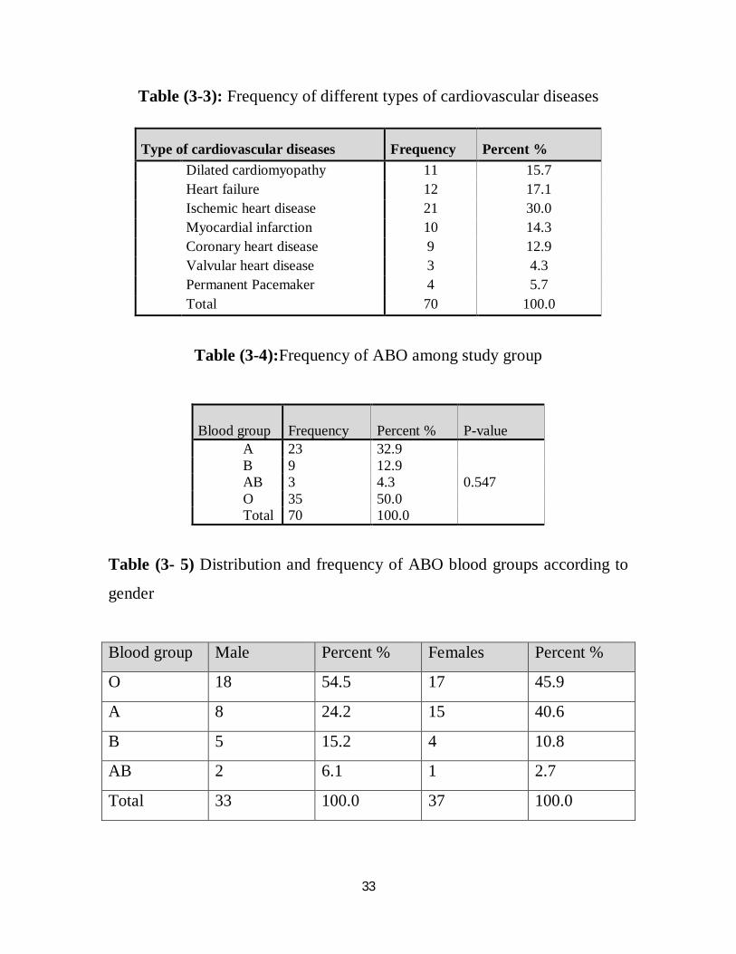
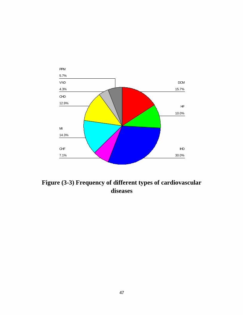
Most common type of cardiovascular disease was ischemic heart disease
(30.0%) followed by heart failure (17.1%) and least frequent was valvular
heart disease as showed in table (3- 3).
According to table (3- 4) found that most frequent blood group in study was
O (50.0%) followed by A (32.9%) and least frequent was AB (4.3%).
According to the results showed in table (3- 5) most frequent blood group in
males was O (54.5%) followed by A (24.2%) and least frequent was AB
(6.1%) and most frequent blood group in females was O (45.9%) followed
by A (40.6%) and least frequent was AB (2.7%).
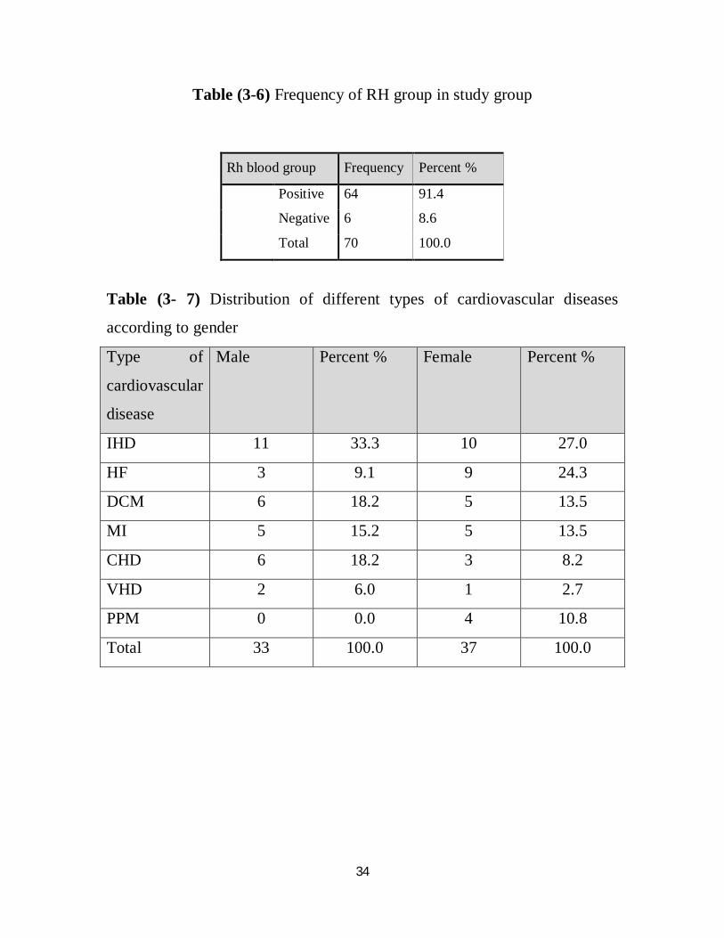
The majority of patients with cardiovascular diseases were Rh positive
(91.4) which showed in table (3- 6).
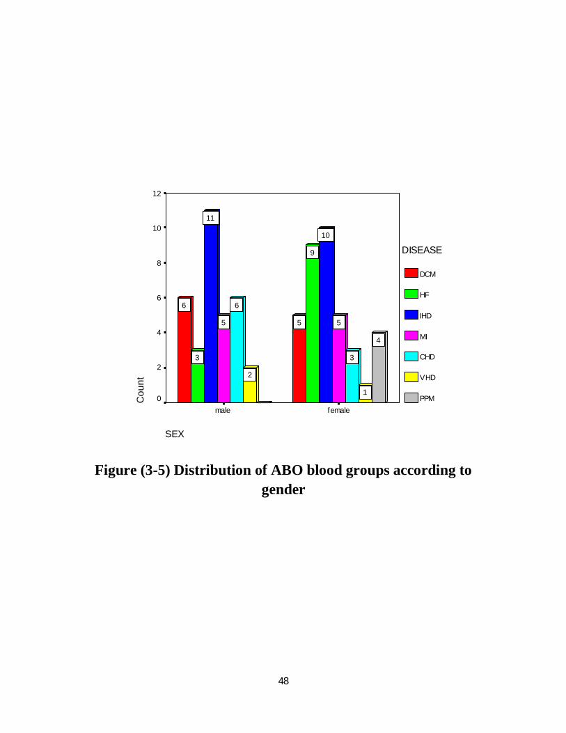
Most common type of cardiovascular disease in females was IHD (27.0%)
followed by HF (24.3%) and least frequent was VHD (2.7%), table (3- 7).
Most common type of cardiovascular disease in males was IHD (33.3%)
followed by DCM and CHD (18.2) and least frequent was PPM (0.0%),
table (3- 7).

32
Most common type of cardiovascular disease in elder patients with age
between (51- 85years) was IHD (34.7%) followed by CHD (19.6%) and
least frequent was VHD (0.0%), most common type of cardiovascular
diseases in patients with age between (8- 50 years) was DCM (33.3)
followed by IHD (20.8%) and least frequent was CHD and PPM (0.0%),
table (3- 8).
Most common disease in blood group O was IHD and in group A was MI,
table (3- 9).
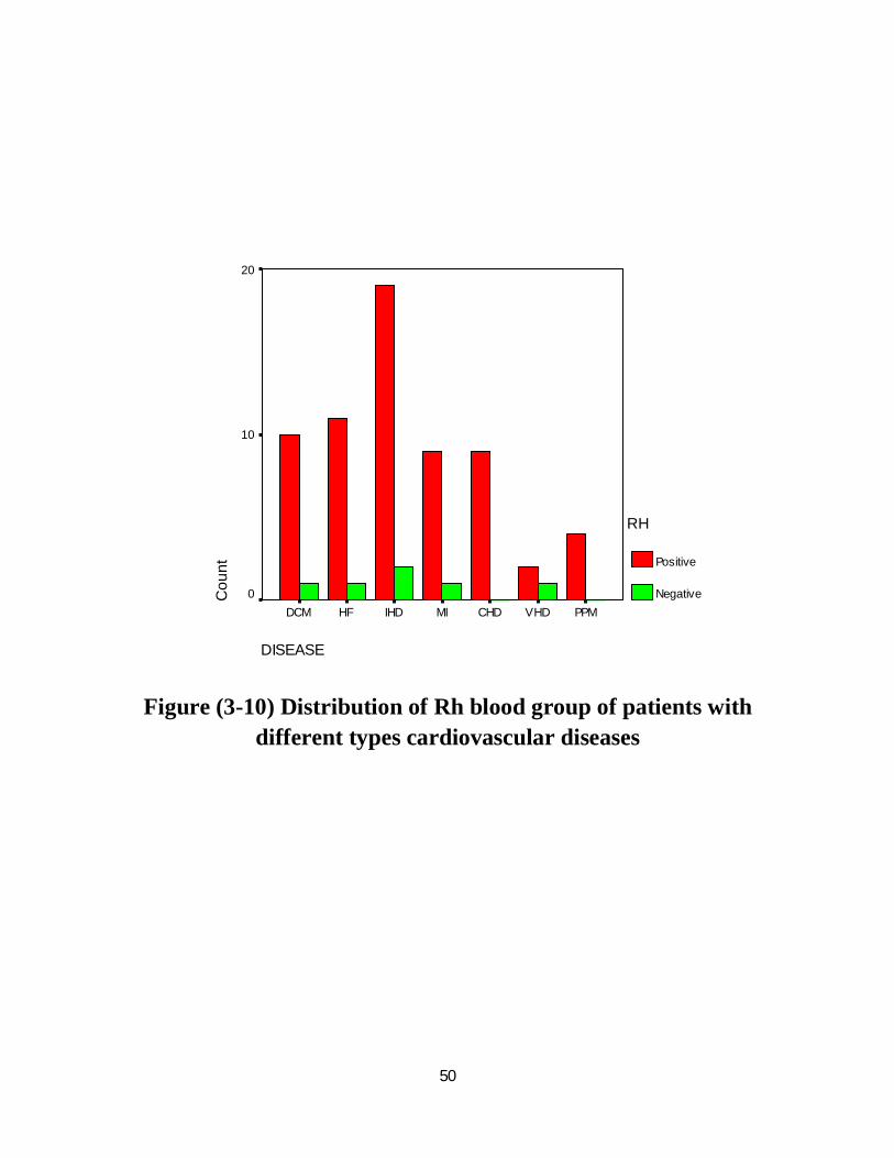
According to table (3- 10), majority of patients with different types of
cardiovascular diseases were Rh positive.
Table (3-1) Distribution of study group according to gender
Gender Frequency Percent %
male 33 47.1
female 37 52.9
Total 70 100.0
Table (3-2) Distribution of study group according to age group
Age group Frequency Percent %
8-50 24 34.3
51-85 46 65.7
Total 70 100.0
33
Table (3-3): Frequency of different types of cardiovascular diseases
Type of cardiovascular diseases Frequency Percent % Dilated cardiomyopathy 11 15.7 Heart failure 12 17.1 Ischemic heart disease 21 30.0 Myocardial infarction 10 14.3 Coronary heart disease 9 12.9 Valvular heart disease 3 4.3 Permanent Pacemaker 4 5.7 Total 70 100.0
Table (3-4):Frequency of ABO among study group
Blood group Frequency Percent % P-value
A 23 32.9 B 9 12.9 AB 3 4.3 0.547 O 35 50.0 Total 70 100.0
Table (3- 5) Distribution and frequency of ABO blood groups according to
gender
Blood group Male Percent % Females Percent %
O 18 54.5 17 45.9
A 8 24.2 15 40.6
B 5 15.2 4 10.8
AB 2 6.1 1 2.7
Total 33 100.0 37 100.0
34
Table (3-6) Frequency of RH group in study group
Rh blood group Frequency Percent %
Positive 64 91.4
Negative 6 8.6
Total 70 100.0
Table (3- 7) Distribution of different types of cardiovascular diseases
according to gender
Type of
cardiovascular
disease
Male Percent % Female Percent %
IHD 11 33.3 10 27.0
HF 3 9.1 9 24.3
DCM 6 18.2 5 13.5
MI 5 15.2 5 13.5
CHD 6 18.2 3 8.2
VHD 2 6.0 1 2.7
PPM 0 0.0 4 10.8
Total 33 100.0 37 100.0

35
Table (3-8) Distribution of different types of cardiovascular diseases according to Age group
Type of Cardiovascular disease
Age(8- 50) Percent % Age(51- 85) Percent %
IHD 5 20.8 16 34.7 HF 4 16.7 8 17.4 DCM 8 33.3 3 6.6 MI 4 16.7 6 13.0 CHD 0 0.0 9 19.6 VHD 3 12.5 0 0.0 PPM 0 0.0 4 8.7 Total 24 100.0 46 100.0
Table (3-9) Distribution of ABO blood groups of patients with different
types of cardiovascular diseases
Blood group DCM HF IHD CHF MI CHD VHD PPM NO
Percent
%
A 2 2 6 1 7 1 2 2 23 32.9
B 2 1 2 1 0 1 1 1 9 12.9
AB 1 0 1 0 1 0 0 0 3 4.3
O 6 4 12 3 2 7 0 1 35 50.0
Total 11 7 21 5 10 9 3 4 70 100.0
Table (3-10) Distribution of Rh blood group of patients with different types of cardiovascular diseases
Type of cardiovascular diseases RH Total
Percent %
Positive Negative DCM 10 1 11 15.7 HF 11 1 12 17.1 IHD 19 2 21 30.0 MI 9 1 10 14.3 CHD 9 0 9 12.9 VHD 2 1 3 4.3 PPM 4 0 4 5.7 Total 64 6 70 100.0

Chapter Four
Discussion, Conclusion and Recommendations

36
Chapter Four
4. Discussion, Conclusion, and Recommendation
4.1. Discussion
This is a descriptive analytical study was conducted in Khartoum State
during the period from May to August 2014 to determine frequency of ABO
and Rh blood groups in Sudanese patients and to correlate their association
with cardiovascular diseases.
Seventy samples were collected from patients with cardiovascular diseases
33 (47.1%) males and 37(52.9%) females.
Most common cardiovascular disease was Ischemic heart disease followed
by heart failure and least frequent was vavular heart disease.
Most common cardiovascular disease in females was ischemic heart disease
followed by heart failure and least frequent was valvular heart disease.
Most common disease in males was Ischemic heart disease and the least
frequent was Permanent pacemaker. According to these results ischemic
heart disease most common in males as compared to females.
Most common blood group in males was O followed by A and least frequent
was AB.
Most common blood group in females was O followed by A and least
frequent was AB.
Most common Rh blood group in males was Rh (D) positive and most
common Rh blood group in females also was Rh (D) positive.
37
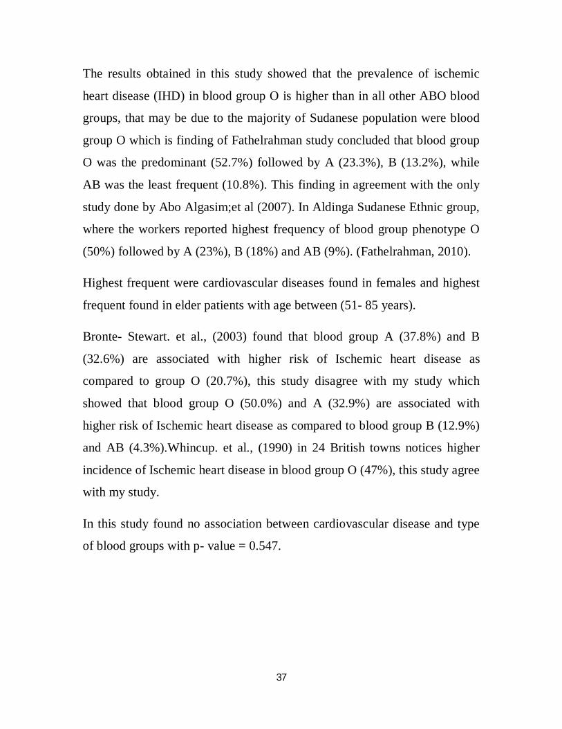
The results obtained in this study showed that the prevalence of ischemic
heart disease (IHD) in blood group O is higher than in all other ABO blood
groups, that may be due to the majority of Sudanese population were blood
group O which is finding of Fathelrahman study concluded that blood group
O was the predominant (52.7%) followed by A (23.3%), B (13.2%), while
AB was the least frequent (10.8%). This finding in agreement with the only
study done by Abo Algasim;et al (2007). In Aldinga Sudanese Ethnic group,
where the workers reported highest frequency of blood group phenotype O
(50%) followed by A (23%), B (18%) and AB (9%). (Fathelrahman, 2010).
Highest frequent were cardiovascular diseases found in females and highest
frequent found in elder patients with age between (51- 85 years).
Bronte- Stewart. et al., (2003) found that blood group A (37.8%) and B
(32.6%) are associated with higher risk of Ischemic heart disease as
compared to group O (20.7%), this study disagree with my study which
showed that blood group O (50.0%) and A (32.9%) are associated with
higher risk of Ischemic heart disease as compared to blood group B (12.9%)
and AB (4.3%).Whincup. et al., (1990) in 24 British towns notices higher
incidence of Ischemic heart disease in blood group O (47%), this study agree
with my study.
In this study found no association between cardiovascular disease and type
of blood groups with p- value = 0.547.

38
4.2. Conclusion
- There is no association between cardiovascular diseases and type of
blood groups with p- value = 0.547.
- Rh positive was higher frequent than Rh negative.
- Most common cardiovascular disease in males was Ischemic heart
disease and most common cardiovascular disease in females also was
Ischemic heart disease.
- O blood group was more frequent among patients with cardiovascular
diseases followed by A blood group and least frequent was AB.
- Cardiovascular diseases were more common among females regard to
males.
- From the results of this study may conclude that most common type of
cardiovascular disease was Ischemic heart disease, followed by Heart
Failure and least frequent disease was valvular heart disease.
39
4.3. Recommendations
1. ABO and Rh blood grouping should be done as routine investigations
for patients with cardiovascular diseases.
2. Other studies should be conducted with using of advance techniques
to confirm the results.
3. Other studies should be conducted with using of gel technique.
4. According to the results of this study, individuals with blood group O
and A should aware of preventive measures against cardiovascular
disease including diet and physical exercise.
5. Further study is required to give baseline data regarding distribution
of ABO of patients with cardiovascular diseases.
6. Further investigations in other regional settings with much larger
population may elucidate these findings.

The References

40
References
Abo Algasim EI, Malik H,andTarq E (2007).Equencies of ABO, Rh-D and Kell Blood Group Antigens in Dinka Sudanese Ethnic Group,SUST;01-12.
Allen T.M and Dawson A.A (1968).ABO blood groups and ischemic heart disease in men.Br Heart J; 30:377-382.
Biswas J, Islam MA, RudraS,Haque MA, Bhuiyan ZR, Husain M and Mamun AA,(2008). Relationship between blood groups and coronary artery disease.Mymensingh Med J; 17(2 suppl):S22-7.
Bridget B. K; (2010). Promoting Cardiovascular Health in the developing world; A Critical challenge to achieve Global Health.Washington, D.C; National Academies.ISBNO - 309- 14774 - 3.
Bronte-Stewart B, Botha MC, and Kru LH.(2003).ABO blood groups in relation to ischemic heart disease.Br Med J;1:1646-50.
Cannegeister SC,Rosendall FR and BrietE.(1994).Thromboembolic and bleeding complications with mechanical heart valve prostheses.Circulation;89:635.
Coombs R.R,Mollrant AE, Race RR. (1945). A new test for the detection of weak and incomplete Rh agglutination.Brit J EXP Path; 26:255- 66.
Dacie J.V and Lewis .S.H. (2001).Practical hematology. 9thed, Churchill living stone.
Dallas T.X, Hyman D.J, and PavlikV.N(2001). Characteristics of patients with un controlled hypertention in the United States N EnglJMed ;345:479.
Denis M., and Hamening, M.T (1998). Modern blood banking and transfusion practice, Third Edition, New Delhi, India, 5; 86 -130.
Ernest Beulter., (2001). William’s hematology, 6th ed. San Francisco: 1840 -53.
Fathelrahman H M. (2010). Frequency of ABO, sub group ABO and Rh(D) blood groups in Major Sudanese Ethnic Groups. Pak J Med Res; 49.1.

41
Gregoratos G, Cheitlin MD, and Conill A.(1998).Guidelines for implantation of cardiac pacemakers and antiarrythmia devices. Circulation; 97:1325.
Garraty G,Dzik W, Issitt PD, Lublin DM, Reid ME, Zelinski T. (2000).Terminology for blood group antigens and genes, historical origins and guideline in the new millennium.Transfusion; 40: 471- 89.
Havlic RJ, FeineibM, and Garrison RJ. (1969). Blood groups and coronary heart disease. Lancet; 2:269-70.
Hoff Brand, A.V., Smi cell, Lewis, Edward, G.D., Tuddewhan. (2000). Post graduate Heamtology; 4thed, British Library, London, UK.
Hoff Brand, A.V. Pettit. J.E.M.D. Otago. M.D, (2006).Essential Hematology. Black well.
John DR, (1996). Technical Manual of American Association of Blood Banks.Blood groups and genetics.12th ed. USA; 95: 373- 87.
Kathen, E, B., Barbara, E.D., LinconP.J..(1988). Blood group serology.6th edition, London, UK, 3; 39: 88.
Lewis S.M.,Anstee, D.J., Bird, G.W.G, (1990). Party blood group terminology from ISBT working P ortyon terminology for red cells surface antigens. Vox sang 58- 158.
Lewis S.M., Bain, B, J., Bates, I. (1991), Practical Hematology 7th ed. Churchill living stone, 19: 430.
Landsteiner K. Weiner AS. (1940).Anagglutinable factor in human blood recognized by immune sera for rhesus blood. ProcsocExpBiol Med; 43: 223- 224.
Mark E.B , (2005). Technical Manual, 15th ed. Bethesda; 1:563-95.
Mitchell RN, Kumar V, Abbas AK and Fausto N.(2005).PATHOLOGIC BASIS OF DISEASE,7th edition;12:288-316.
42
Mollison PL. (1994). The genetic basis of the Rh blood group system.Transfusion; 34: 549- 41.
Nevillen, J., Bryant, A.R.T., F. A. (1994). An Introduction to Immunohematology, 3rd edition. Canada.
Race PR.And Sunger R., (1975). Blood groups in man, 6th ed. San Francisco: 1840- 53.
Rosenberg L, Miller DR,and Kawfman DW.(1983). Myocardial infarction in women under 50 years of age.JAMA; 250(20):2801-6.
Sheikh MK, Mazura BL, Yusoff NM and Knight A.(2009). Association of ABO blood group B with MI. J collphysicianssurg Pak; 19 (8):514-517.
Talib.V.H, (1995). Blood Banking and Transfusion Medicine, First Edition. New Delhi.
Walker, R.H., Hoppe, P.A., Judd, W.J., (1999)., Technical Manual, Third Edition, American Association of blood banks, Arlington, 197; 223.
WhincupPH,CookDG,Philips AN and Shaper AG.(1990).ABO blood group and Ischemic heart disease in British men.BMJ;300(6741):1679-1682.
Yeghiazarian Y, Braunstein J B, and Askari A (2000). Medical progress: un stable angina pectoris, N Engl J Med; 342:101.

Appendices

43
Sudan University of Science and Technology
College of Graduate Studies
Medical Laboratory Science
Hematology department
Questionnaire about ABO blood group and Rh factor among patients with cardiovascular diseases
Sample No : …………………..
Age : ………………………………..
Gender : ……………………………
Type of cardiovascular disease : ……………………….
Laboratory Investigation :
Blood Group : …………………………………………..
Rh factor : ………………………………………………
Date : / /
Signature : ………

44
مبسم الله الرحمن الرحی
جامعة السودان للعلوم والتكنلوجیا
مختبرات طبیة -برنامج الماجستیر -كلیة الدراسات العلیا
تخصص علم الدم ومبحث المناعة
اقرار موافقة
:.....................................................................الاسم
من الورید بواسطة حقنة طعن وذلك بعد مسح مل (2.5)سوف یتم أخذ عینة من الدمكل الأدوات المستخدمة لأخذ العینة معقمة ومتبع فیھا . مكان أخذ العینة بواسطة المطھر
.وسائل السلامة المعملیة
.وأنا أقر بأن ھذه العینات سوف یتم تحلیلھا فقط لغرض البحث
.أوافق أنا المذكورأعلاه بأخذ عینة لإجراء الدراسة
:.............................................الإسم
:.........................................الإمضاء

45
Master sheet
Number Sex Age Disease ABO group RH group Other diseases Age group
1 Female 44 DCM A Positive No 23-50
2 Male 24 DCM O Positive No 23-50 3 Female 52 DCM B Positive Yes 51-85 4 Female 34 DCM A Positive No 23-50 5 Male 80 HF O Positive Yes 51-85 6 Female 60 DCM O Negative No 51-85 7 Female 60 HF O Positive No 51-85 8 Female 60 HF A Positive Yes 51-85 9 Female 60 IHD O Positive Yes 51-85
10 Female 46 HF B Positive Yes 23-50 11 Female 50 IHD O Positive Yes 23-50 12 Female 71 HF O Negative No 51-85 13 Female 60 IHD O Positive Yes 51-85 14 Female 35 VHD A Positive No 23-50 15 Female 43 MI A Positive Yes 23-50 16 Female 59 MI A Positive No 51-85 17 Female 48 IHD B Positive Yes 23-50 18 Female 62 IHD O Positive Yes 51-85 19 Male 54 MI A Positive No 51-85 20 Male 52 MI O Positive Yes 51-85 21 Female 51 IHD A Positive Yes 51-85 22 Male 60 IHD O Positive Yes 51-85 23 Female 62 IHD O Positive No 51-85 24 Male 70 IHD A Positive Yes 51-85 25 Female 49 MI A Positive No 23-50 26 Male 44 IHD O Positive Yes 23-50 27 Male 60 IHD B Positive Yes 51-85 28 Female 40 MI A Positive No 23-50 29 Male 25 VHD A Positive No 23-50 30 Male 65 IHD O Positive Yes 51-85 31 Male 80 DCM B Positive Yes 51-85 32 Male 78 CHD O Positive Yes 51-85 33 Male 53 CHD B Positive No 51-85 34 Male 72 CHD O Positive No 51-85 35 Male 69 MI A Positive No 51-85 36 Male 29 IHD O Positive No 23-50 37 Female 75 CHD O Positive No 51-85 38 Female 60 CHD A Positive No 51-85 39 Male 67 IHD A Negative No 51-85 40 Male 72 HF A Positive Yes 51-85 41 Male 78 CHD O Positive No 51-85 42 Male 48 DCM O Positive Yes 23-50 43 Female 75 HF O Positive No 51-85 44 Male 60 HF O Positive No 51-85

46
45 Female 60 IHD O Positive Yes 51-85 46 Male 48 DCM O Positive No 23-50 47 Female 70 IHD AB Positive No 51-85 48 Male 68 IHD A Positive Yes 51-85 49 Female 39 MI A Positive Yes 23-50 50 Female 42 DCM O Positive No 23-50 51 Female 23 HF A Positive Yes 23-50 52 Female 45 HF O Positive No 23-50 53 Male 67 MI AB Positive Yes 51-85 54 Female 63 IHD A Positive No 51-85 55 Female 75 HF O Positive Yes 51-85 56 Female 65 PPM B Positive Yes 51-85 57 Male 30 DCM AB Positive No 23-50 58 Mal 43 HF B Positive Yes 23-50 59 Male 56 IHD A Negative Yes 51-85 60 Male 52 IHD O Positive Yes 51-85 61 Male 70 CHD O Positive No 51-85 62 Male 80 CHD O Positive No 51-85 63 Female 60 CHD O Positive No 51-85 64 Male 23 VHD B Negative No 23-50 65 Female 84 PPM A Positive No 51-85 66 Female 60 PPM A Positive No 51-85 67 Female 43 DCM O Positive No 23-50 68 Female 57 PPM O Positive No 51-85 69 Male 62 MI O Negative Yes 51-85 70 Male 49 IHD O Positive Yes 23-50
47
5.7%
4.3%
12.9%
14.3%
7.1% 30.0%
10.0%
15.7%
PPM
VhD
CHD
MI
CHF IHD
HF
DCM
Figure (3-3) Frequency of different types of cardiovascular diseases
48
SEX
femalemale
Cou
nt
12
10
8
6
4
2
0
DISEASE
DCM
HF
IHD
MI
CHD
VHD
PPM
4
1
2
3
6
55
10
11
9
3
5
6
Figure (3-5) Distribution of ABO blood groups according to gender

49
DISEASE
PPMVHDCHDMIIHDHFDCM
Cou
nt14
12
10
8
6
4
2
0
ABO
A
B
AB
O
Figure (3-9) Distribution of ABO blood group of patients with different types of cardiovascular diseases
50
DISEASE
PPMVHDCHDMIIHDHFDCM
Cou
nt
20
10
0
RH
Positive
Negative
Figure (3-10) Distribution of Rh blood group of patients with different types cardiovascular diseases