DetectionoftheIntercellularAdhesionGeneCluster( )and ... ·...
7
INFECTION AND IMMUNITY, 0019-9567/97/$04.0010 Mar. 1997, p. 890–896 Vol. 65, No. 3 Copyright q 1997, American Society for Microbiology Detection of the Intercellular Adhesion Gene Cluster (ica) and Phase Variation in Staphylococcus epidermidis Blood Culture Strains and Mucosal Isolates WILMA ZIEBUHR, 1,2 CHRISTINE HEILMANN, 3 FRIEDRICH GO ¨ TZ, 3 PETER MEYER, 2 ² KLAUS WILMS, 2 EBERHARD STRAUBE, 4 AND JO ¨ RG HACKER 1 * Institut fu ¨r Molekulare Infektionsbiologie 1 and Medizinische Poliklinik der Universita ¨t, 2 D-97070 Wu ¨rzburg, Mikrobielle Genetik, D-72076 Tu ¨bingen, 3 and Institut fu ¨r Medizinische Mikrobiologie, D-07740 Jena, 4 Germany Received 26 August 1996/Returned for modification 15 October 1996/Accepted 16 December 1996 Staphylococcus epidermidis is a common cause of catheter-associated infections and septicemia in immuno- compromised patients. To answer the question whether S. epidermidis skin isolates differ from isolates causing septicemic diseases, 51 strains obtained from blood cultures, 1 strain from shunt-associated meningitis, and 36 saprophytic isolates were characterized. The study demonstrates that most of the blood culture strains formed a multilayered biofilm on plastic material, whereas skin and mucosal isolates did not. Moreover, biofilm-producing strains were found to generate large bacterial autoaggregates in liquid culture. Autoaggre- gation and biofilm formation on polymer surfaces was associated with the presence of a DNA sequence encoding an intercellular adhesion gene cluster (ica) that mediates the production of a polysaccharide intercellular adhesin. The presence of the intercellular adhesion genes in blood culture isolates was also found to be correlated with the exhibition of black colonies on Congo red agar, whereas the adhesin-negative strains formed red colonies. Upon subcultivation on Congo red agar, the black colony forms of the blood culture strains exhibited red colony variants which were biofilm and autoaggregation negative and occurred at a frequency of 10 25 . The DNA analysis of these S. epidermidis variants by pulsed-field gel electrophoresis and Southern hybridization with an ica-specific gene probe revealed no detectable difference between the black and red colony types. Moreover, after repeated passage, the phenotype of the parent strain could be restored. Therefore, these colony forms were regarded as phase variants. This phenotypic change was observed exclu- sively in adhesin-positive clinical isolates and not in adhesin-negative saprophytic strains of S. epidermidis. Coagulase-negative staphylococci, e.g., Staphylococcus epi- dermidis, are known to be part of the normal skin and mucosal microflora. In recent years, however, S. epidermidis emerged as a common cause of nosocomial infections in immunocompro- mised patients (23, 33). Septicemia caused by S. epidermidis is often associated with the use of intravenous catheters and other medical devices. An additional problem of these infec- tious diseases is the increasing resistance of staphylococci to oxacillin and other antibiotics and the spread of multiresistant isolates within the hospital environment (6). There is a strong correlation between the use of indwelling medical devices and the emergence of staphylococcal infections. However, the in- formation on the microbial mechanisms and factors contribut- ing to the virulence of S. epidermidis is limited. It has been proposed that the staphylococcus adherence to polymer surfaces contributes considerably to the pathogenesis of polymer-associated infections (7, 31, 32). Recent data sug- gest that the bacterial biofilm is produced in a two-step man- ner, i.e., the initial bacterial monolayer is converted to a typical biofilm consisting of bacteria and an extracellular slime sub- stance (20). Both polysaccharide adhesins (10, 27, 28, 29, 40) and protein components (39) that are involved in initial ad- herence as well as in biofilm accumulation have been de- scribed. Recently, mutants with altered initial adherence and autoaggregation properties have been generated by chemical and transposon mutagenesis (20, 35). The data obtained from these experiments indicate that initial adherence and biofilm formation by intercellular adhesion and slime production are distinct events. In the initial binding of S. epidermidis, a 60-kDa surface-associated protein is involved (20), whereas cell aggre- gation and biofilm accumulation were found to be mediated by the products of a gene locus comprising three intercellular adhesion genes (icaABC) which are organized in an operon structure (21). It was shown that the ica-encoded genes lead to the biosynthesis of the polysaccharide intercellular adhesin (PIA) (21), which contains N-acetylglucosamine as a major component and is involved in the accumulative stage of biofilm formation. Another important property of staphylococci is their capac- ity to change specific phenotypic features rapidly. Several stud- ies showed that adherence, slime production, and resistance to antibiotics undergo changes and differ among variants of the same parent strain (2, 3, 9, 11–13, 30). In this context, it has been suggested that phase variation may represent a virulence factor contributing to bacterial survival and growth under changing environmental conditions. The question of whether differences between S. epidermidis strains causing infections and those isolated from healthy skin and mucosal flora exist is of central importance to the under- standing of S. epidermidis pathogenesis. To address this ques- tion, we compared, in this study, strains isolated from patients with plastic material-associated infections with isolates from skin and mucosa. Our data indicate that S. epidermidis strains from clinical material differ from saprophytic strains in terms of the presence of the ica gene cluster as well as in the capac- * Corresponding author. Mailing address: Institut fu ¨r Molekulare Infektionsbiologie, Ro ¨ntgenring 11, 97070 Wu ¨rzburg, Germany. Phone: 0049 931 312 575. Fax: 0049931 571954. E-mail:j.hacker@rzbox .uni-wuerzburg.de. ² Present address: Sentralsjukehuset Rogaland, N-4003 Stavanger, Norway. 890 on February 2, 2019 by guest http://iai.asm.org/ Downloaded from
Transcript of DetectionoftheIntercellularAdhesionGeneCluster( )and ... ·...

INFECTION AND IMMUNITY,0019-9567/97/$04.0010
Mar. 1997, p. 890–896 Vol. 65, No. 3
Copyright q 1997, American Society for Microbiology
Detection of the Intercellular Adhesion Gene Cluster (ica) andPhase Variation in Staphylococcus epidermidis Blood Culture
Strains and Mucosal IsolatesWILMA ZIEBUHR,1,2 CHRISTINE HEILMANN,3 FRIEDRICH GOTZ,3 PETER MEYER,2†
KLAUS WILMS,2 EBERHARD STRAUBE,4 AND JORG HACKER1*
Institut fur Molekulare Infektionsbiologie1 and Medizinische Poliklinik der Universitat,2 D-97070 Wurzburg, Mikrobielle Genetik,D-72076 Tubingen,3 and Institut fur Medizinische Mikrobiologie, D-07740 Jena,4 Germany
Received 26 August 1996/Returned for modification 15 October 1996/Accepted 16 December 1996
Staphylococcus epidermidis is a common cause of catheter-associated infections and septicemia in immuno-compromised patients. To answer the question whether S. epidermidis skin isolates differ from isolates causingsepticemic diseases, 51 strains obtained from blood cultures, 1 strain from shunt-associated meningitis, and36 saprophytic isolates were characterized. The study demonstrates that most of the blood culture strainsformed a multilayered biofilm on plastic material, whereas skin and mucosal isolates did not. Moreover,biofilm-producing strains were found to generate large bacterial autoaggregates in liquid culture. Autoaggre-gation and biofilm formation on polymer surfaces was associated with the presence of a DNA sequenceencoding an intercellular adhesion gene cluster (ica) that mediates the production of a polysaccharideintercellular adhesin. The presence of the intercellular adhesion genes in blood culture isolates was also foundto be correlated with the exhibition of black colonies on Congo red agar, whereas the adhesin-negative strainsformed red colonies. Upon subcultivation on Congo red agar, the black colony forms of the blood culturestrains exhibited red colony variants which were biofilm and autoaggregation negative and occurred at afrequency of 1025. The DNA analysis of these S. epidermidis variants by pulsed-field gel electrophoresis andSouthern hybridization with an ica-specific gene probe revealed no detectable difference between the black andred colony types. Moreover, after repeated passage, the phenotype of the parent strain could be restored.Therefore, these colony forms were regarded as phase variants. This phenotypic change was observed exclu-sively in adhesin-positive clinical isolates and not in adhesin-negative saprophytic strains of S. epidermidis.
Coagulase-negative staphylococci, e.g., Staphylococcus epi-dermidis, are known to be part of the normal skin and mucosalmicroflora. In recent years, however, S. epidermidis emerged asa common cause of nosocomial infections in immunocompro-mised patients (23, 33). Septicemia caused by S. epidermidis isoften associated with the use of intravenous catheters andother medical devices. An additional problem of these infec-tious diseases is the increasing resistance of staphylococci tooxacillin and other antibiotics and the spread of multiresistantisolates within the hospital environment (6). There is a strongcorrelation between the use of indwelling medical devices andthe emergence of staphylococcal infections. However, the in-formation on the microbial mechanisms and factors contribut-ing to the virulence of S. epidermidis is limited.It has been proposed that the staphylococcus adherence to
polymer surfaces contributes considerably to the pathogenesisof polymer-associated infections (7, 31, 32). Recent data sug-gest that the bacterial biofilm is produced in a two-step man-ner, i.e., the initial bacterial monolayer is converted to a typicalbiofilm consisting of bacteria and an extracellular slime sub-stance (20). Both polysaccharide adhesins (10, 27, 28, 29, 40)and protein components (39) that are involved in initial ad-herence as well as in biofilm accumulation have been de-scribed. Recently, mutants with altered initial adherence and
autoaggregation properties have been generated by chemicaland transposon mutagenesis (20, 35). The data obtained fromthese experiments indicate that initial adherence and biofilmformation by intercellular adhesion and slime production aredistinct events. In the initial binding of S. epidermidis, a 60-kDasurface-associated protein is involved (20), whereas cell aggre-gation and biofilm accumulation were found to be mediated bythe products of a gene locus comprising three intercellularadhesion genes (icaABC) which are organized in an operonstructure (21). It was shown that the ica-encoded genes lead tothe biosynthesis of the polysaccharide intercellular adhesin(PIA) (21), which contains N-acetylglucosamine as a majorcomponent and is involved in the accumulative stage of biofilmformation.Another important property of staphylococci is their capac-
ity to change specific phenotypic features rapidly. Several stud-ies showed that adherence, slime production, and resistance toantibiotics undergo changes and differ among variants of thesame parent strain (2, 3, 9, 11–13, 30). In this context, it hasbeen suggested that phase variation may represent a virulencefactor contributing to bacterial survival and growth underchanging environmental conditions.The question of whether differences between S. epidermidis
strains causing infections and those isolated from healthy skinand mucosal flora exist is of central importance to the under-standing of S. epidermidis pathogenesis. To address this ques-tion, we compared, in this study, strains isolated from patientswith plastic material-associated infections with isolates fromskin and mucosa. Our data indicate that S. epidermidis strainsfrom clinical material differ from saprophytic strains in termsof the presence of the ica gene cluster as well as in the capac-
* Corresponding author. Mailing address: Institut fur MolekulareInfektionsbiologie, Rontgenring 11, 97070 Wurzburg, Germany.Phone: 0049 931 312 575. Fax: 0049931 571954. E-mail:[email protected].† Present address: Sentralsjukehuset Rogaland, N-4003 Stavanger,
Norway.
890
on February 2, 2019 by guest
http://iai.asm.org/
Dow
nloaded from

ities for phase variation, polymer adherence, autoaggregation,and colony morphology on Congo red agar (CRA).
MATERIALS AND METHODS
Bacterial strains. Fifty-one S. epidermidis strains obtained from a blood cul-ture strain collection, one S. epidermidis strain isolated from cerebrospinal fluid,and 36 skin and mucosal isolates from healthy volunteers were investigated. Skinand mucosal strains were isolated by swabbing either the anterior nares or thenondominating hand of the test person, respectively. Staphylococci were identi-fied by colonial appearance on whole blood agar (5% defibrinated human eryth-rocytes) and Gram’s stain.Biotyping and antibiotic susceptibility. Species determination and biotyping
were performed by use of a numeric profile based upon 20 biochemical reactionsas described in the manufacturer’s instructions (API-Staph; BioMerieux, Nurtin-gen, Germany). Antibiotic susceptibility was tested by the standard agar diffusionmethod on Mueller-Hinton agar plates supplemented with 5% sodium chloride.Colony morphology and detection of phenotypic variants. Colony morphology
and its phenotypic change were studied on CRA, which was prepared by adding0.8 g of Congo red (Serva, Heidelberg, Germany) and 36 g of saccharose (Roth,Karlsruhe, Germany), both of which had been previously autoclaved separately,to 1 liter of brain heart infusion agar (Oxoid, Basingstoke, England) (14). Plateswere incubated for 24 h at 378C and subsequently overnight at room tempera-ture. Colony morphology was examined with a plate microscope. The variationrate was determined by counting the different colony forms in relation to thetotal number of CFU.Initial adherence on polymer surfaces. The assay for the determination of the
initial adherence was essentially performed as described previously (20). Briefly,bacterial cultures were grown in Trypticase soy broth (TSB) at 378C to the earlystationary phase. Cultures were diluted with sterile phosphate-buffered saline(PBS) to an optical density at 600 nm (OD600) of 0.1. A polystyrene petri dish(Sarstedt, Nurnbrecht, Germany) was filled with an aliquot of 10 ml and incu-bated at 378C for 30 min. After extensive washing of the dishes with PBS, theadhered cells were visualized by microscopy, photographed, and counted.Biofilm formation and autoaggregation. Quantitative estimations of biofilm
formation were performed by the method of Christensen et al. (8) with thefollowing modification. TSB with the usual glucose supplementation (Difco Lab-oratories, Detroit, Mich.) was used exclusively. For each strain, the test wasperformed eight times in a tissue culture plate (Greiner, Nurtingen, Germany).Following washing and fixation as described previously (8), adherent biofilm wasstained for 10 min with 0.4% crystal violet solution. The absorbance at 490 nmwas determined. The well-characterized biofilm-producing strain S. epidermidisRP62A (ATCC 35984) and the biofilm-negative Staphylococcus carnosus TM 300strain were used as positive and negative controls, respectively. An OD490 of.0.120 was regarded as biofilm positive.For autoaggregation screening, strains were cultivated in tissue culture plates
overnight at 378C in TSB and examined by light microscopy.Detection of PIA by immunofluorescence microscopy. Immunofluorescence
microscopy was performed essentially as described previously (27), with minormodifications. Cultures were grown in TSB overnight at 378C. Aliquots of 20 mlwere applied to slides and air dried. Following fixation by heat, the slides wereincubated with PIA-specific antiserum diluted 1:100 in PBS for 45 min at roomtemperature. The PIA-specific antiserum had been absorbed before with anica-negative S. epidermidis strain as described previously (27). Thereafter, theslides were washed three times with PBS and incubated with Texas red-conju-gated goat anti-rabbit immunoglobulin G antibody (Dianova, Hamburg, Ger-many) diluted 1:150 in PBS for 45 min at room temperature. The slides werewashed three times in PBS and once in distilled water. After air drying, the slideswere embedded in Citifluor and viewed with a fluorescence microscope (Zeiss,Oberkochen, Germany).Scanning electron microscopy. For scanning electron microscopy, staphylo-
cocci were grown overnight in TSB medium on polystyrene chamber slides at378C. After decanting the medium, the slides were washed three times with PBS,dried at 428C for 36 h in a desiccator, mounted on aluminum stubs, and shad-
owed with gold. For visualization, a scanning electron microscope (Zeiss DSM962) was used at 15 kV.Stability of biofilm-negative variants. To test the stability of nonadhering S.
epidermidis variants, single colonies were picked and incubated in fresh sterileTSB in a tissue culture flask (Greiner) at 378C. After 24 h, the medium wasreplaced. This procedure was repeated until a biofilm of adhering bacteriabecame visible on the bottom of the flask (maximum of 15 days).Pulsed-field gel electrophoresis. DNA isolation from S. epidermidis and
pulsed-field gel electrophoresis were performed as described previously (15, 25).Isolation of chromosomal DNA and restriction endonuclease cleavage. Bac-
terial strains were grown to the mid-log phase in Luria-Bertani broth (Difco)supplemented with 1% glycine at 378C. Bacteria were harvested by centrifugationof 1.5 ml of the culture. The bacterial pellet was resuspended in 100 ml of buffercontaining 25% sucrose, 10 mM Tris-HCl (pH 7.5), and 1 mg of lysostaphin(Sigma, Deisenhofen, Germany) per ml and incubated for 10 min at 378C. Afterlysis of the bacterial cells, the chromosomal DNA was isolated by standardprocedures (34).After restriction endonuclease digestion, the DNA was analyzed by agarose
gel electrophoresis (1% agarose in Tris-phosphate-EDTA).Southern hybridization. Gels were blotted onto nylon membranes (Hybond-
N1; Amersham Life Science, Little Chalfont, England) by standard methods(34).For hybridization, DNA of the plasmid pCN27 (21) was digested with EcoRI.
The 2.6-kb EcoRI DNA fragment encoding icaA and the truncated icaB of theintercellular adhesin gene cluster (ica) of S. epidermidis RP62A was isolated bystandard protocols (34). Probe labeling and hybridization were performed withthe nonradioactive ECL direct nucleic acid labeling and detection system (Am-ersham Life Science). The hybridization buffer contained 1 M NaCl, and washingsteps were performed at 558C in 13 SSC (0.15 M NaCl plus 0.015 M sodiumcitrate) containing 0.4% sodium dodecyl sulfate.Northern (RNA) blot analysis of ica expression. For RNA extraction, the
RNagents total RNA isolation system (Promega, Madison, Wis.) was used.Bacterial cells were grown in TSB to an OD600 of 2.0. Bacterial cells (1010) werethen pelleted, resuspended in 400 ml of diethylpyrocarbonate-treated water, andincubated with 100 ml of lysostaphin (1 mg/ml; Sigma) for 5 min at 378C. Thefollowing RNA extraction and purification procedure was performed as recom-mended by the manufacturer.Following DNase I digestion (fast-performance liquid chromatography-pure
DNase I, RNase free; Pharmacia, Uppsala, Sweden), 30 mg of RNA was loadedonto 1% agarose gels containing 5% formaldehyde in 13 MOPS (morpho-linepropanesulfonic acid) running buffer. Gel electrophoresis was performed at5 V/cm for 3 h. The gel was blotted onto a nylon membrane (Hybond-N1;Amersham Life Science) and hybridized with an ECL-labeled (Amersham LifeScience) ica-specific DNA probe.
RESULTS
Biofilm formation and autoaggregation. The 88 S. epidermi-dis strains, i.e., 52 clinical isolates and 36 strains from bodysurfaces, were tested for their capacity to form biofilms onplastic material by a quantitative adherence assay (8). Thereby,a significant difference between the blood culture isolates andsaprophytic strains could be observed. Among the former, 87%(45 of 52) formed biofilms on plastic, whereas only 11% (4 of36) of the skin isolates produced a detectable biofilm on tissueculture plates.The strains were screened for autoaggregation in TSB by
light microscopy. In 79% (41 of 52) of the clinical isolates butonly in 6% (2 of 36) of the skin and mucosal strains, largebacterial aggregates were detected. The data are summarizedin Table 1.
TABLE 1. Properties of S. epidermidis blood culture and saprophytic isolatesa
Type of isolates (n)
No. (%) of isolates
Biofilmpositive
Biofilmnegative
Autoaggregatedin TSB
With icagenes
Colony morphology on CRA
Black Red
Blood culture (52) 45b (87) 7c (13) 41 (79) 44 (85) 45 (87) 7 (13)Saprophytic (36) 4d (11) 32c (89) 2 (6) 2 (6) 2 (6) 34 (94)
a Biofilm formation was detected by the quantitative adherence assay described in Materials and Methods. Autoaggregation was detected by Southern hybridization.b This total number included 3 isolates with an OD490 of .0.12 but ,0.60, 30 isolates with an OD490 of .0.60 but ,2.00, and 12 isolates with an OD490 of .2.00.c All of these isolates had an OD490 of ,0.12.d This total number included three isolates with an OD490 of .0.12 but ,0.60 and one isolate with an OD490 of .0.60.
VOL. 65, 1997 S. EPIDERMIDIS INTERCELLULAR ADHESION 891
on February 2, 2019 by guest
http://iai.asm.org/
Dow
nloaded from

Distribution of the intercellular adhesion-specific DNA se-quence (ica). Recent data of Heilmann and coworkers (21)provided evidence that gene products of the ica locus play acrucial role in biofilm formation of S. epidermidis. To obtaininformation on the distribution of the ica gene cluster amongS. epidermidis strains from several sources, the isolates weretested for the presence of the gene cluster by Southern hybrid-ization of chromosomal DNA.Following blotting onto nylon membranes, the chromosomal
DNA was hybridized with an ica-specific gene probe. As dem-onstrated in Table 1, the gene was present in 85% (44 of 52) ofthe clinical S. epidermidis strains but only in 6% (2 of 36) of thesaprophytic isolates. Thus, it appears that the presence of theica gene is a common feature of S. epidermidis obtained fromplastic material-associated infections but not of saprophyticisolates.Correlation of colony morphology, autoaggregation, biofilm
formation, and ica gene cluster presence. After incubation ofstaphylococcal strains on CRA plates, two major colony typescould be distinguished by the color of their colonies. Nearly87% (45 of 52) of the S. epidermidis blood culture isolates grewon CRA as black colonies, whereas only 6% (2 of 36) of thesaprophytic strains formed black colonies.Of the 88 S. epidermidis strains investigated, 49 strains, i.e.,
45 from blood cultures and 4 from skin and mucosa, werefound to be biofilm positive in the quantitative adherenceassay. In 93% (46 of 49) of the biofilm-positive S. epidermidisstrains, the ica gene cluster was present. In contrast, this DNAsequence was not detectable in biofilm-negative isolates.Evaluation of all data on colony morphology, adherence,
autoaggregation, and detection of the ica genes revealed astrong correlation between these features. Biofilm formationand autoaggregation were associated with the intercellular ad-hesion genes and the black colony type. This pattern was pre-dominant in clinical isolates, whereas red colonies were morefrequently observed in the nonadhering, adhesin-negativestrains obtained from skin or mucosa.Detection and characterization of phase variants. After
plating a single, black colony of a blood culture strain ontoCRA again, black colonies and a few red colonies emerged.The incidence of the emergence of the red colony type wasapproximately 1025. In the case of the saprophytic strains, nochange of colony morphology could be observed, even afterrepeated passage of single colonies on CRA plates.To decide whether the red colonies of clinical isolates rep-
resent contaminations or variants from the originally blackparental strains, the variants were subcultivated and the re-striction length polymorphism was tested by pulsed-field gelelectrophoresis. These experiments revealed no detectable dif-ferences between the black and red colony forms in thegenomic pattern (Fig. 1). However, the red variants were al-tered in their ability to form biofilms on polymer surfaces (Fig.2B). To test the stability of the biofilm-negative variants, asingle red colony of S. epidermidis 161 was picked for serialpassage in TSB as described in Materials and Methods. Thisexperiment revealed that the biofilm-negative phenotype ofthe variant is very stable. Only after ninefold serial passage ofthe nonadhering variant in TSB could the biofilm-producingphenotype be restored (Fig. 2B, strain 161/2).Interestingly, the phenotypically different variants appeared
to be identical on the DNA level as judged by pulsed-field gelelectrophoresis. Therefore, we decided to analyze the basis ofthe phenotypic variation in more detail. To this end, the bio-film formation of S. epidermidis 161 and that of its biofilm-negative variant, strain 161/1, and its biofilm-positive revertant,strain 161/2, were investigated.
First, the initial adherence to polymer surfaces was studied.Initial adherence of staphylococci to polymers is mediated by asurface-associated, 60-kDa protein (20). The variants did notshow any difference in their initial binding to a polymer surface(Fig. 2A). Consequently, it is most likely that an alteration ofthe second stage of biofilm formation is responsible for thebiofilm-negative phenotype.Second, the production of the extracellular slime substance
(PIA) was investigated by immunofluorescence. PIA is a hex-osamine-rich polysaccharide mediating the intercellular adher-ence of bacterial cells, and it was shown to be essentiallyinvolved in biofilm accumulation (28). It is synthesized by apolysaccharide synthetase which is presumably encoded byicaA (21). Immunofluorescence microscopy revealed clear ev-idence that the biofilm-negative variant does not produce PIA.In contrast, the biofilm-positive parent strain and the revertantwere found to synthesize PIA (Fig. 2D). Since it had beenshown that PIA production is necessary for autoaggregationand biofilm formation, the biofilm-negative phenotype of thevariant can be explained by the absent PIA production. More-over, immunofluorescence analysis showed that the PIA ismainly synthesized by those bacterial cells that are organized inlarge cell clusters, whereas bacteria that remain in suspensionexhibit only a diminished adhesin production (Fig. 2C and D).To get insight into the molecular mechanisms causing the
biofilm-negative phenotype, Southern and Northern blot ex-periments were performed. Southern hybridization of chromo-somal DNA with the ica-specific gene probe revealed that thegenetic information is still present within the variants (Fig. 3).Therefore, the absence of PIA production cannot be caused bya complete deletion of the ica gene cluster.The transcription of the ica operon of S. epidermidis 161 and
that of its biofilm-negative variant, strain 161/1, were investi-gated by Northern blot analysis. Hybridization of total RNAwith an ica-specific probe revealed that the transcription of theica gene cluster is reduced in the PIA-negative phase variant(Fig. 4).
DISCUSSION
S. epidermidis is part of the epidermal microflora of healthyhumans. However, during the last two decades, it has beendemonstrated conclusively that this species and other coagu-lase-negative staphylococci are important pathogens causing
FIG. 1. Analysis of SmaI-digested genomic DNA from S. epidermidis 161 bypulsed-field gel electrophoresis. Lanes: 1, S. epidermidis 161; 2, biofilm-negativephase variant 161/1; 3, biofilm-forming revertant 161/2.
892 ZIEBUHR ET AL. INFECT. IMMUN.
on February 2, 2019 by guest
http://iai.asm.org/
Dow
nloaded from
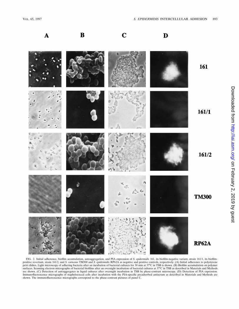
FIG. 2. Initial adherence, biofilm accumulation, autoaggregation, and PIA expression of S. epidermidis 161, its biofilm-negative variant, strain 161/1, its biofilm-positive revertant, strain 161/2, and S. carnosus TM300 and S. epidermidis RP62A as negative and positive controls, respectively. (A) Initial adherence to polystyrenepetri dishes. Light microscopy of adhering bacteria after an incubation of bacterial cultures for 30 min at 378C in TSB is shown. (B) Biofilm accumulation on polymersurfaces. Scanning electron micrographs of bacterial biofilms after an overnight incubation of bacterial cultures at 378C in TSB as described in Materials and Methodsare shown. (C) Detection of autoaggregates in liquid cultures after overnight incubation in TSB by phase-contrast microscopy. (D) Detection of PIA expression.Immunofluorescence micrographs of staphylococcal cells after incubation with the PIA-specific preadsorbed antiserum as described in Materials and Methods areshown. The immunofluorescence micrographs correspond to the phase-contrast pictures of panel C.
VOL. 65, 1997 S. EPIDERMIDIS INTERCELLULAR ADHESION 893
on February 2, 2019 by guest
http://iai.asm.org/
Dow
nloaded from

many nosocomial infections. Often, a correlation of infectionwith the use of medical devices can be observed. In the courseof many studies, it became clear that the capacity of staphylo-cocci to adhere to and grow on plastic surfaces is a crucial step
in the pathogenesis of polymer-associated infections (7, 31,32). Recently, several components that appear to be involvedin adherence and slime production of staphylococci have beendescribed (10, 20, 21, 27–29, 35, 39, 40). However, it remainsunclear whether these factors are, indeed, virulence factors orsimply common features of saprophytic staphylococci. It isimportant, therefore, to compare the properties of infectiousagents and strains of the normal microflora.In this study, we have compared S. epidermidis strains ob-
tained from polymer-associated septicemic diseases with iso-lates obtained from skin and mucosa of healthy volunteers.The data clearly indicate genotypic and phenotypic differencesamong these groups of strains. First, the coding sequence of anintercellular adhesion gene cluster was found to be present in85% (44 of 52) of S. epidermidis blood culture isolates com-pared to only 6% (2 of 36) of saprophytic strains. The twoadhesin-positive strains that were found among the sapro-phytic isolates had been obtained from nasal swabs of twoemployees of an oncological ward, suggesting that this genemight be more prevalent in S. epidermidis strains isolated fromhospital communities than in isolates obtained from normalhealthy individuals.Second, striking differences in terms of biofilm production
and colony morphology on CRA between strains from differ-ent sources have been observed. In the quantitative adherenceassay, 87% (45 of 52) of the blood culture isolates but only11% (4 of 36) of the saprophytic strains adhered to plasticmaterial. On CRA, the adhering strains formed black colonieswhereas nonadhering strains developed red colony forms.Moreover, in liquid culture (TSB medium), the biofilm-pro-ducing isolates accumulated in bacterial aggregates that weredetectable by light microscopy. Biofilm-negative strains, how-ever, formed homogeneous suspensions in TSB medium. Astrong correlation was found between biofilm production,
FIG. 3. Southern analysis of S. epidermidis 161. The chromosomal DNA wasdigested with BamHI and hybridized with an ica-specific probe as described inMaterials and Methods. Lanes: 1, biofilm-forming S. epidermidis 161; 2, nonad-hering phase variant 161/1; 3, biofilm-forming revertant 161/2; 4, S. carnosus TM300 (negative control); 5, S. epidermidis RP62A (positive control).
FIG. 4. Investigation of ica transcription by Northern blot analysis. RNAs of the biofilm-positive parent strain S. epidermidis 161 and its biofilm-negative variant,161/1, were isolated and hybridized with the ica-specific probe at different ODs of the bacterial growth curve. S. epidermidis RP62A and S. carnosus TM 300 are positiveand negative controls, respectively.
894 ZIEBUHR ET AL. INFECT. IMMUN.
on February 2, 2019 by guest
http://iai.asm.org/
Dow
nloaded from

black colony type on CRA, autoaggregation, and the presenceof the intercellular adhesion genes.The ica-specific gene probe used for Southern hybridization
originated from the slime-producing strain S. epidermidis RP62A (21). The probe is specific for the ica operon coding forgene products required for the bacterial cell-to-cell contactand biofilm formation. The operon has been shown to containat least three open reading frames. icaA encodes an enzymethat is anchored in the cytoplasmic membrane and probablyacts as a polysaccharide synthetase mediating the synthesis ofthe PIA. A second open reading frame, icaB, which is locateddownstream of icaA, has been identified. It contains the infor-mation for an extracellular protein whose function is still un-known. The third open reading frame, icaC, encodes an inte-gral membrane protein which is assumed to exert a receptorfunction for polysaccharide antigens. It is important to notethat the simultaneous expression of these three staphylococcusgene products is required for PIA synthesis and the subsequentbiofilm formation on polymer surfaces and autoaggregation inliquid cultures.In biofilm-producing S. epidermidis strains, the expression of
the intercellular adhesion genes (ica) was found to undergo aphase variation that correlates with a change of colony mor-phology on CRA and an altered biofilm formation. Biofilm-negative variants were unable to produce the PIA whose syn-thesis is mediated by the ica gene locus expression products.Genome analysis by pulsed-field gel electrophoresis andSouthern hybridization revealed no detectable difference be-tween the variants. Moreover, the biofilm-producing pheno-type could be restored after repeated passages of a nonadher-ing single colony. These data lead us to conclude that thephenotypic change is not caused by a deletion of the adhesingene. Northern blot analysis showed that the biofilm-negativephenotype of the variant is due to a reduced ica gene tran-scription. The molecular mechanisms underlying the regula-tion of ica transcription remain to be elucidated.Nonadhering, red colony variants of S. epidermidis RP62A
on CRA have also been described by Mempel et al. (30). Thesevariants showed a remarkable increase of their methicillin sus-ceptibility due to a lack of transcription of the methicillinresistance gene mecA. In the present study, 77% of the clinicalS. epidermidis strains were resistant to oxacillin whereas only8% of the saprophytic isolates were resistant. These data sup-port the assumption that the mecA gene is more prevalent inthe hospital environment. We also observed changes in theoxacillin susceptibilities of phenotypic variants (data notshown), supporting the suggestion of Christensen et al. (11)that heteroresistance to b-lactam antibiotics is a common fea-ture of clinical staphylococcal isolates.Our data on the distribution of the intercellular adhesion
genes among clinical isolates suggest that the correspondinggene products could be considered a possible virulence factorin S. epidermidis. The intercellular adhesion gene expressionwas found to undergo a phase variation. Phenotypic changes ofsurface structures are common in many pathogenic bacteria.They have been observed in Escherichia coli adhesins (1, 4, 5,16, 17, 26), in Salmonella typhimurium flagellum expression(36, 43), in antigenic and phase variation of Neisseria adhesins(18, 22, 24, 37, 38), and also inHaemophilus influenzae (41) andpneumococci (42). These studies have demonstrated that theheterogeneous expression of adhesins is important for the ad-aptation of bacteria to changing growth conditions and for theevasion of the host immune system. The phenotypic variationof the intercellular adhesin and the underlying genetic mech-anisms are important scientific subjects. But clearly, more in-formation is required to elucidate the role of the intercellular
adhesin and its phenotypic variation in the pathogenesis ofpolymer-associated infections.
ACKNOWLEDGMENTS
We thank Hilde Merkert for technical assistance in the preparationof the scanning electron and immunofluorescence micrographs andInge Muhldorfer for critical reading of the manuscript. We also thankMatthias Trautmann of the Institut fur Medizinische Mikrobiologieder Universitat Ulm for providing some of the clinical S. epidermidisisolates and Dietrich Mack of the Institut fur Medizinische Mikrobi-ologie und Immunologie der Universitat Hamburg for the PIA-specificantiserum.This work was supported by a grant from the Deutsche Forschungs-
gemeinschaft (Graduiertenkolleg Infektiologie).
REFERENCES
1. Abraham, J. M., C. S. Freitag, J. R. Clements, and B. I. Eisenstein. 1985. Aninvertible element of DNA controls phase variation of type 1 fimbriae ofEscherichia coli. Proc. Natl. Acad. Sci. USA 82:5724–5727.
2. Baddour, L. M., L. P. Barker, G. D. Christensen, J. T. Parisi, and W. A.Simpson. 1990. Phenotypic variation of Staphylococcus epidermidis in infec-tion of transvenous endocardial pacemaker electrodes. J. Clin. Microbiol.28:676–696.
3. Baselga, R., I. Albizu, M. de La Cruz, E. del Cacho, M. Barberan, and B.Amorena. 1993. Phase variation of slime production in Staphylococcus au-reus: implications in colonization and virulence. Infect. Immun. 61:4857–4862.
4. Blyn, L., B. A. Braaten, C. A. White-Ziegler, D. H. Rolfson, and D. A. Low.1989. Phase-variation of pyelonephritis-associated pili in Escherichia coli:evidence for transcriptional regulation. EMBO J. 8:613–620.
5. Braaten, B. A., X. Nou, L. S. Kaltenbach, and D. Low. 1994. Methylationpatterns in pap regulatory DNA control pyelonephritis-associated pili phasevariation in E. coli. Cell 76:577–588.
6. Chambers, H. F. 1988. Methicillin-resistant staphylococci. Clin. Microbiol.Rev. 1:173–186.
7. Christensen, G. D., W. A. Simpson, A. L. Bisno, and E. H. Beachey. 1982.Adherence of slime-producing Staphylococcus epidermidis to smooth sur-faces. Infect. Immun. 37:318–326.
8. Christensen, G. D., W. A. Simpson, J. J. Younger, L. M. Baddour, F. F.Barrett, D. M. Melton, and E. H. Beachey. 1985. Adherence of coagulase-negative staphylococci to plastic tissue culture plates: a quantitative modelfor the adherence of staphylococci to medical devices. J. Clin. Microbiol.22:996–1006.
9. Christensen, G. D., L. M. Baddour, and W. A. Simoson. 1987. Phenotypicvariation of Staphylococcus epidermidis slime production in vitro and in vivo.Infect. Immun. 55:2870–2877.
10. Christensen, G. D., L. P. Barker, T. P. Mawhinney, L. M. Baddour, andW. A. Simpson. 1990. Identification of an antigenic marker of slime produc-tion for Staphylococcus epidermidis. Infect. Immun. 58:2906–2911.
11. Christensen, G. D., L. M. Baddour, B. M. Madison, J. T. Parisi, S. N.Abraham, D. L. Hasty, J. H. Lowrance, J. A. Josephs, and W. A. Simpson.1990. Colonial morphology of staphylococci on memphis agar: phase varia-tion of slime production, resistance to b-lactam antibiotics, and virulence.J. Infect. Dis. 161:1153–1169.
12. Deighton, M., S. Pearson, J. Capstick, D. Spelman, and R. Borland. 1992.Phenotypic variation of Staphylococcus epidermidis isolated from a patientwith native valve endocarditis. J. Clin. Microbiol. 30:2385–2390.
13. Deighton, M. A., J. Capstick, and R. Borland. 1992. A study of phenotypicvariation of Staphylococcus epidermidis using Congo red agar. Epidemiol.Infect. 109:423–432.
14. Freeman, D. J., F. R. Falkiner, and C. T. Keane. 1989. New method fordetection slime production by coagulase-negative staphylococci. J. Clin.Pathol. 42:872–874.
15. Goering, R. V., and A. Winters. 1992. Rapid method for epidemiologicalevaluation of gram-positive cocci by field inversion gel electrophoresis.J. Clin. Microbiol. 30:577–580.
16. Hacker, J. 1990. Genetic determinants coding for fimbriae and adhesins ofextraintestinal Escherichia coli. Curr. Top. Microbiol. Immunol. 151:1–27.
17. Hacker, J., L. Bender, M. Ott, J. Wingender, B. Lund, R. Marre, and W.Goebel. 1990. Deletions of chromosomal regions coding for fimbriae andhemolysins occur in vitro and in vivo in various extraintestinal Escherichiacoli isolates. Microb. Pathog. 8:213–225.
18. Haas, R., and T. F. Meyer. 1986. The repertoire of silent pilus genes inNeisseria gonorrhoeae: evidence for gene conversion. Cell 44:107–115.
19. Haas, R., S. Veit, and T. F. Meyer. 1992. Silent pilin genes of Neisseriagonorrhoeae MS11 and the occurrence of related hypervariant sequencesamong other gonococcal isolates. Mol. Microbiol. 6:197–208.
20. Heilmann, C., C. Gerke, F. Perdreau-Remington, and F. Gotz. 1996. Char-acterization of Tn917 insertion mutants of Staphylococcus epidermidis af-
VOL. 65, 1997 S. EPIDERMIDIS INTERCELLULAR ADHESION 895
on February 2, 2019 by guest
http://iai.asm.org/
Dow
nloaded from

fected in biofilm formation. Infect. Immun. 64:277–282.21. Heilmann, C., O. Schweitzer, C. Gerke, N. Vanittanakom, D. Mack, and F.
Gotz. 1996. Molecular basis of intercellular adhesion in the biofilm-formingStaphylococcus epidermidis. Mol. Microbiol. 20:1083–1091.
22. James, J. F., and J. Swanson. 1978. Studies on gonococcus infection. XIII.Occurrence of color/opacity colonial variants in clinical cultures. Infect.Immun. 19:332–340.
23. Kloos, W. E., and T. L. Bannerman. 1994. Update on clinical significance ofcoagulase-negative staphylococci. Clin. Microbiol. Rev. 7:117–140.
24. Koomey, J. M., E. C. Gotschlich, K. Robbins, S. Bergstrom, and J. Swanson.1987. Effects of recA mutations on pilus antigenic variation and phase tran-sitions in Neisseria gonorrhoeae. Genetics 117:391–398.
25. Linhardt, F., W. Ziebuhr, P. Meyer, W. Witte, and J. Hacker. 1992. Pulsed-field gel electrophoresis of genomic restriction fragments as a tool for theepidemiological analysis of Staphylococcus aureus and coagulase-negativestaphylococci. FEMS Microbiol. Lett. 95:181–186.
26. Low, D., E. N. Robinson, Z. A. McGee, and S. Falkow. 1987. The frequencyof expression of pyelonephritis-associated pili is under regulatory control.Mol. Microbiol. 1:335–346.
27. Mack, D., N. Siemssen, and R. Laufs. 1992. Parallel induction of adherenceand a polysaccharide antigen specific for plastic-adherent Staphylococcusepidermidis: evidence for functional relation to intercellular adhesion. Infect.Immun. 60:2048–2057.
28. Mack, D., M. Nedelmann, A. Krokotsch, A. Schwarzkopf, J. Heesemann, andR. Laufs. 1994. Characterization of transposon mutants of biofilm-producingStaphylococcus epidermidis impaired in the accumulative phase of biofilmproduction: genetic identification of a hexosamine-containing polysaccharideinterecellular adhesin. Infect. Immun. 62:3244–3253.
29. Mack, D., M. Nedelmann, A. Krokotsch, A. Schwarzkopf, J. Heesemann, andR. Laufs. 1996. The intercellular adhesin involved in biofilm accumulation ofStaphylococcus epidermidis is a linear b-1,6-linked glucosaminoglycan: puri-fication and structural analysis. J. Bacteriol. 178:175–183.
30. Mempel, M., H. Feucht, W. Ziebuhr, M. Endres, R. Laufs, and L. Gruter.1994. Lack of mecA transcription in slime-negative phase variants of methi-cillin-resistant Staphylococcus epidermidis. Antimicrob. Agents Chemother.38:1251–1255.
31. Peters, G. 1986. Adherence of Staphylococcus epidermidis to plastic devices.J. Med. Microbiol. 22:252–253.
32. Peters, G., R. Locci, and G. Pulverer. 1982. Adherence and growth ofcoagulase-negative staphylococci on surfaces of intravenous catheters. J. In-fect. Dis. 146:479–482.
33. Pfaller, M. A., and L. A. Herwaldt. 1988. Laboratory, clinical and epidemi-ological aspects of coagulase-negative staphylococci. Clin. Microbiol. Rev.1:281–299.
34. Sambrook, J., E. F. Fritsch, and T. Maniatis. 1989. Molecular cloning: alaboratory manual, 2nd ed. Cold Spring Harbor Laboratory Press, ColdSpring Harbor, N.Y.
35. Schumacher-Perdreau, F., C. Heilmann, G. Peters, F. Gotz, and G. Pulverer.1994. Comparative analysis of a biofilm-forming Staphylococcus epidermidisstrain and its adhesion-positive, accumulation-negative mutant M7. FEMSMicrobiol. Lett. 117:71–78.
36. Silverman, M., and M. Simon. 1980. Phase variation: genetic analysis ofswitching mutants. Cell 19:845–851.
37. Stern, A., M. Brown, P. Nickel, and T. F. Meyer. 1986. Opacity genes inNeisseria gonorrhoeae: control of phase and antigenic variation. Cell 47:61–71.
38. Stern, A., and T. F. Meyer. 1987. Common mechanism controlling phase andantigenic variation in pathogenic Neisseria. Mol. Microbiol. 1:5–12.
39. Timmerman, C. P., A. Fleer, J. M. Besnier, L. DeGraaf, F. Cremers, and J.Verhoef. 1991. Characterization of a proteinaceous adhesin of Staphylococ-cus epidermidis which mediates attachment to polystyrene. Infect. Immun.59:4187–4192.
40. Tojo, M., N. Yamashita, D. Goldman, and G. B. Pier. 1988. Isolation andcharacterization of a capsular polysaccharide adhesin from Staphylococcusepidermidis. J. Infect. Dis. 157:713–722.
41. Weiser, J. N. 1993. Relationship between colony morphology and life cycle ofHaemophilus influenzae: the contribution of lipopolysaccharide phase varia-tion to pathogenesis. J. Infect. Dis. 168:672–680.
42. Weiser, J. N., R. Austrian, P. K. Sreenivasan, and H. R. Masure. 1994. Phasevariation in pneumococcal opacity: relationship between colonial morphol-ogy and nasopharyngeal colonization. Infect. Immun. 62:2582–2589.
43. Zeig, J., M. Silverman, H. Hilmen, and M. Simon. 1977. Recombinantswitching for gene expression. Science 196:170–175.
Editor: V. A. Fischetti
896 ZIEBUHR ET AL. INFECT. IMMUN.
on February 2, 2019 by guest
http://iai.asm.org/
Dow
nloaded from