Detection of the Typical Pulse Condition on Cun-Guan-Chi...
7
Sensors & Transducers, Vol. 165, Issue 2, February 2014, pp. 46-52 46 S S S e e e n n n s s s o o o r r r s s s & & & T T T r r r a a a n n n s s s d d d u u u c c c e e e r r r s s s © 2014 by IFSA Publishing, S. L. http://www.sensorsportal.com Detection of the Typical Pulse Condition on Cun-Guan-Chi Based on Image Sensor Aihua ZHANG, Liming YANG and Hongzhi DANG College of Electrical and Information Engineering, Lanzhou University of Technology, Lanzhou, 730050, China Tel.: 0931-2973726, fax: 0931-2973902 E-mail: [email protected] Received: 11 November 2013 /Accepted: 28 January 2014 /Published: 28 February 2014 Abstract: In order to simulate the diagnosis by feeling the pulse with Traditional Chinese Medicine, a device based on CCD was designed to detect the pulse image of Cun-Guan-Chi. Using the MM-3 pulse model as experimental subject, the synchronous pulse image data of some typical pulse condition were collected by this device on Cun-Guan-Chi. The typical pulses include the normal pulse, the slippery pulse, the slow pulse and the soft pulse. According to the lens imaging principle, the pulse waves were extracted by using the area method, then the 3D pulse condition image was restructured and some features were extracted including the period, the frequency, the width, and the length. The slippery pulse data of pregnant women were collected by this device, and the pulse images were analyzed. The results are consistent based on comparing the features of the slippery pulse model with the slippery pulse of pregnant women. This study overcame shortages of the existing detection device such as the few detecting parts and the limited information, and more comprehensive 3D pulse condition information could be obtained. This work laid a foundation for realizing the objective diagnosis and revealing the comprehensive information of the pulse. Copyright © 2014 IFSA Publishing, S. L. Keywords: Pulse detection, Image sensor, Pulse-taking pressure, Pulse waveform. 1. Introduction In Traditional Chinese Medicine, the pulse is considered as having three divisions: Cun, Guan and Chi. Traditional Chinese Pulse Diagnosis (TCPD) is the most distinct one of the four kinds of Traditional Chinese Medical Diagnoses. Doctors of Traditional Chinese Medicine get disease information such as location, nature and state by feeling the pulse. However, doctors always rely on subjective feeling to get patients pulse condition information, which is blended with many subjective factors, such as the doctor's discriminate experience, feeling difference and so on. This gives teaching and clinical diagnosis many difficulties and differences, and restricts the development of TCPD. So it is significant to promote the development of objective Pulse Diagnosis. Pulse condition information detection devices are the basis for obtaining pulse information. It is also the priority work for objective Pulse Diagnosis. At present, the probes used in pulse condition information detection devices are mainly single probe, double probes and three probes etc. [1-3]. The single probe is used most commonly, but it is just suitable for collecting one part information. Although the double probes can collect two parts information, the information obtained is one-dimensional signal. Some researchers have engaged in study of three probes or multi drop probes devices. The detection system of pulse condition on Cun-Guan-Chi has been Article number P_1875
Transcript of Detection of the Typical Pulse Condition on Cun-Guan-Chi...

Sensors & Transducers, Vol. 165, Issue 2, February 2014, pp. 46-52
46
SSSeeennnsssooorrrsss &&& TTTrrraaannnsssddduuuccceeerrrsss
© 2014 by IFSA Publishing, S. L. http://www.sensorsportal.com
Detection of the Typical Pulse Condition on Cun-Guan-Chi Based on Image Sensor
Aihua ZHANG, Liming YANG and Hongzhi DANG
College of Electrical and Information Engineering, Lanzhou University of Technology, Lanzhou, 730050, China
Tel.: 0931-2973726, fax: 0931-2973902 E-mail: [email protected]
Received: 11 November 2013 /Accepted: 28 January 2014 /Published: 28 February 2014 Abstract: In order to simulate the diagnosis by feeling the pulse with Traditional Chinese Medicine, a device based on CCD was designed to detect the pulse image of Cun-Guan-Chi. Using the MM-3 pulse model as experimental subject, the synchronous pulse image data of some typical pulse condition were collected by this device on Cun-Guan-Chi. The typical pulses include the normal pulse, the slippery pulse, the slow pulse and the soft pulse. According to the lens imaging principle, the pulse waves were extracted by using the area method, then the 3D pulse condition image was restructured and some features were extracted including the period, the frequency, the width, and the length. The slippery pulse data of pregnant women were collected by this device, and the pulse images were analyzed. The results are consistent based on comparing the features of the slippery pulse model with the slippery pulse of pregnant women. This study overcame shortages of the existing detection device such as the few detecting parts and the limited information, and more comprehensive 3D pulse condition information could be obtained. This work laid a foundation for realizing the objective diagnosis and revealing the comprehensive information of the pulse. Copyright © 2014 IFSA Publishing, S. L. Keywords: Pulse detection, Image sensor, Pulse-taking pressure, Pulse waveform. 1. Introduction
In Traditional Chinese Medicine, the pulse is considered as having three divisions: Cun, Guan and Chi. Traditional Chinese Pulse Diagnosis (TCPD) is the most distinct one of the four kinds of Traditional Chinese Medical Diagnoses. Doctors of Traditional Chinese Medicine get disease information such as location, nature and state by feeling the pulse. However, doctors always rely on subjective feeling to get patients pulse condition information, which is blended with many subjective factors, such as the doctor's discriminate experience, feeling difference and so on. This gives teaching and clinical diagnosis many difficulties and differences, and restricts the
development of TCPD. So it is significant to promote the development of objective Pulse Diagnosis.
Pulse condition information detection devices are the basis for obtaining pulse information. It is also the priority work for objective Pulse Diagnosis.
At present, the probes used in pulse condition information detection devices are mainly single probe, double probes and three probes etc. [1-3]. The single probe is used most commonly, but it is just suitable for collecting one part information. Although the double probes can collect two parts information, the information obtained is one-dimensional signal. Some researchers have engaged in study of three probes or multi drop probes devices. The detection system of pulse condition on Cun-Guan-Chi has been
Article number P_1875

Sensors & Transducers, Vol. 165, Issue 2, February 2014, pp. 46-52
47
developed by Shanghai University of Traditional Chinese Medicine [4]. The PVDF multi-point pulse transducer has been developed by Professor Jin [5]. The number of testing parts on these devices is increased, but the output of each probe is still one-dimentional signal.
The sensors used in pulse condition detection devices are mainly pressure sensor, infrared light electricity sensor, microphones and Doppler technology. Researchers have developed various pulse condition detection devices with different sensors such as HMX-3C Pulse Transducer, MX-5 Sphygmograph, MX-811 Sphygmograph, ZM-III Intelligent Sphygmograph, ZM-IIIC Intelligent Sphygmograph, MXY-II Sphygmograph, CYX-10T Pulse Figure Sensor, DDMX-100 Sphygmograph, ZMH-I TCM Pulse Condition Sensor, BYS-14 ECG Pulse Condition Instrument and so on [6]. The sensors used in these pulse condition detection devices above are piezoresistive or piezoelectric pressure sensor. Shu has used wrist strap pressure pulse sensor to record 13 kinds of pulse condition waveforms [7]. Xu used pressure pulse sensor to collect pulse waveforms, and tried to extract some features such as the pulse position, the pulse potential, the pulse width, the pulse-taking pressure and the pulse frequency [8]. Japanese researchers have developed a kind of pulse sensor by using the ceramic type pressure sensor. This type of sensor is suitable for various pressure levels of floating, medium and sinking [9]. An American medical doctor called John H. Laub has developed a noninvasive pulse wave recorder, whose pressure sensors are fixed to the front of the gloves side by side on index, middle finger and ring finger respectively. When the sensors are pressed on Cun-Guan-Chi parts, the pulse condition waveforms of three parts are recorded simultaneously. The feeling pressure can be also shown at the same time [10]. In 2001, a pulse measurement device was produced by Seiko Epson Company, which could measure the radial pulse of human body by using the piezoelectric element as the sensor. Researchers have also applied PVDF pulse transducer to obtain driver's pulse signals in pilot-vehicle safety dynamic assessment system, and have evaluated the fatigue degree of pilot with the pulse rate [11]. Some researchers have adopted photoelectric pulse sensor to acquire pulse signals. This is different from TCPD method totally [12]. One scholar has made the semiconductor strain gauge stick onto fingers of surgery rubber glove, which is treated as the testing and recording device of the arterial waves [13]. Some scholars have developed a non-contact phototubes volume pulse instrument which can also detect some pulse information [14]. Wang has used microphone pulse sensor to get information of the normal pulse, the slippery pulse and string pulse [15].
Many researchers have fully realized the importance of the objective detection for the pulse condition. However, these detection devices are restricted by the sensor type or the number of test
sites. They can just acquire one-dimension or one part pulse information. Therefore, the collected information can not reflect every element of the pulse condition, which has restricted the development and application of objective TCPD [16, 17].
In order to simulate the diagnosis by feeling the pulse with Traditional Chinese Medicine, a device based on CCD image sensor and three probes is designed to detect the pulse condition image on Cun-Guan-Chi. The MM-3 Pulse Model which can simulate human common pulse condition is used as the experimental subject. The synchronous pulse condition image data of some typical pulse conditions on Cun-Guan-Chi parts are collected by this device. The typical pulses include the normal pulse, the slippery pulse, the slow pulse and the soft pulse. According to the lens imaging principle, the pulse waves are extracted by using the area method. Then the 3D-pulse condition configuration is restructured and some features are extracted including the period, the frequency, the width, and the length. Some of these features such as the length and the width can not be obtained by one-dimensional signal, but these features have important reference value for the disease diagnosis. The slippery pulse data of pregnant women are collected by this device. The results are consistent by comparing the features of the slippery pulse model with those of pregnant women. This study has overcome the shortages such as the few detecting parts and the limited information of the existing detection devices. This work has laid a foundation for realizing the objective diagnosis and revealing the comprehensive information of the pulse. 2. Material and Method 2.1. The Device for Detecting Pulse Condition
on Cun-Guan-Chi 2.1.1. Image Sensor and Three Probes
With the certain pulse diagnosis pressure, the arbitrary multi-point dynamic images on the diagnosis parts are detected by CCD image sensor with three probes developed by this research. The three-dimensional pulse condition information can be acquired from the dynamic image. But the traditional pulse sensor such as pressure sensor, infrared light electricity sensor, microphone and Doppler technologies can not get the three-dimensional pulse condition information [18].
One CCD image sensor is placed above three probes vertically. When pulse beat, the dynamic image is acquired. This makes the silicon thin film image on three probes appear in its field clearly. At every instant, the film image of three probes is recorded in the same frame image, which has realized the synchronous collection of pulse condition information on Cun-Guan-Chi. Three same size
Sensors & Transducers, Vol. 165, Issue 2, February 2014, pp. 46-52
48
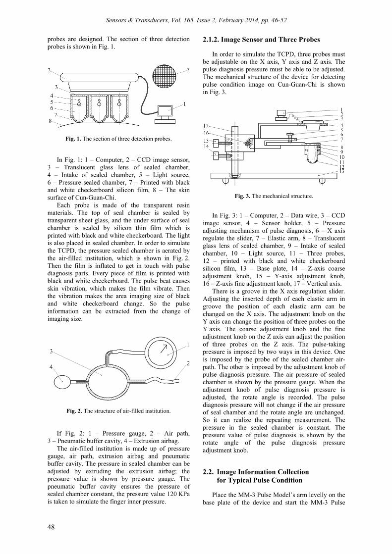
probes are designed. The section of three detection probes is shown in Fig. 1.
Fig. 1. The section of three detection probes.
In Fig. 1: 1 – Computer, 2 – CCD image sensor, 3 – Translucent glass lens of sealed chamber, 4 – Intake of sealed chamber, 5 – Light source, 6 – Pressure sealed chamber, 7 – Printed with black and white checkerboard silicon film, 8 – The skin surface of Cun-Guan-Chi.
Each probe is made of the transparent resin materials. The top of seal chamber is sealed by transparent sheet glass, and the under surface of seal chamber is sealed by silicon thin film which is printed with black and white checkerboard. The light is also placed in sealed chamber. In order to simulate the TCPD, the pressure sealed chamber is aerated by the air-filled institution, which is shown in Fig. 2. Then the film is inflated to get in touch with pulse diagnosis parts. Every piece of film is printed with black and white checkerboard. The pulse beat causes skin vibration, which makes the film vibrate. Then the vibration makes the area imaging size of black and white checkerboard change. So the pulse information can be extracted from the change of imaging size.
Fig. 2. The structure of air-filled institution.
If Fig. 2: 1 – Pressure gauge, 2 – Air path, 3 – Pneumatic buffer cavity, 4 – Extrusion airbag.
The air-filled institution is made up of pressure gauge, air path, extrusion airbag and pneumatic buffer cavity. The pressure in sealed chamber can be adjusted by extruding the extrusion airbag; the pressure value is shown by pressure gauge. The pneumatic buffer cavity ensures the pressure of sealed chamber constant, the pressure value 120 KPa is taken to simulate the finger inner pressure.
2.1.2. Image Sensor and Three Probes
In order to simulate the TCPD, three probes must be adjustable on the X axis, Y axis and Z axis. The pulse diagnosis pressure must be able to be adjusted. The mechanical structure of the device for detecting pulse condition image on Cun-Guan-Chi is shown in Fig. 3.
Fig. 3. The mechanical structure.
In Fig. 3: 1 – Computer, 2 – Data wire, 3 – CCD image sensor, 4 – Sensor holder, 5 – Pressure adjusting mechanism of pulse diagnosis, 6 – X axis regulate the slider, 7 – Elastic arm, 8 – Translucent glass lens of sealed chamber, 9 – Intake of sealed chamber, 10 – Light source, 11 – Three probes, 12 – printed with black and white checkerboard silicon film, 13 – Base plate, 14 – Z-axis coarse adjustment knob, 15 – Y-axis adjustment knob, 16 – Z-axis fine adjustment knob, 17 – Vertical axis.
There is a groove in the X axis regulation slider. Adjusting the inserted depth of each elastic arm in groove the position of each elastic arm can be changed on the X axis. The adjustment knob on the Y axis can change the position of three probes on the Y axis. The coarse adjustment knob and the fine adjustment knob on the Z axis can adjust the position of three probes on the Z axis. The pulse-taking pressure is imposed by two ways in this device. One is imposed by the probe of the sealed chamber air-path. The other is imposed by the adjustment knob of pulse diagnosis pressure. The air pressure of sealed chamber is shown by the pressure gauge. When the adjustment knob of pulse diagnosis pressure is adjusted, the rotate angle is recorded. The pulse diagnosis pressure will not change if the air pressure of seal chamber and the rotate angle are unchanged. So it can realize the repeating measurement. The pressure in the sealed chamber is constant. The pressure value of pulse diagnosis is shown by the rotate angle of the pulse diagnosis pressure adjustment knob. 2.2. Image Information Collection
for Typical Pulse Condition
Place the MM-3 Pulse Model’s arm levelly on the base plate of the device and start the MM-3 Pulse

Sensors & Transducers, Vol. 165, Issue 2, February 2014, pp. 46-52
49
Model. Then the air-filled institution is aerated by the extrusion airbag until the pressure value of each sealed chamber comes to 120 KPa. Imposing a smaller pulse diagnosis pressure and keeping the pulse diagnosis pressure unchanged, the probe position of Cun-Guan-Chi is adjusted in the regional area by X axis regulate the slider, Y-axis adjustment knob, Z-axis fine adjustment knob, Z-axis coarse adjustment knob. Observing the image of three film patterns in the field of image sensor, the position of each probe is adjusted to make the most intense changing area of imaging size locate at their imaging regional center area respectively. Then, changing the pulse diagnosis pressure, the changing degree of the film imaging size will also be changed, which can be observed from the image sensor view filed. When the pulse diagnosis pressure is a certain value, the arterial blood vessel wall pressure and the pulse diagnosis pressure is in balance state. At this time, the changing degree of the imaging size is the strongest. Adjusting the LED lamp brightness and CCD focal length, the imaging of silicon film on three probes appears clearly in the image sensor view field. The image and dynamic change of the tested parts on skin surface are recorded by the changed imaging size of silicon film. The 60th frame image of the slow pulse is shown in Fig. 4. Three black and white checkerboard areas correspond with Cun-Guan-Chi of MM-3 Pulse Model respectively from left to right.
Fig. 4. The 60th frame image of the slow pulse.
2.3. Dynamic Image Analysis for Pulse Condition
2.3.1. Pulse Waveform Obtain Principle
According to the lens imaging principle, if the relative position between CCD and lens is fixed, the object is allowed to move in range of filed depth. The imaging size will change as the objects move. Based on analyzing the changes of film checkerboard size (that is area) in the image sequences, the displacement information on vascular radial surface can be extracted. The imaging principle is shown in Fig. 5.
In Fig. 5, L is lens, a, b and c are three planes which are perpendicular to the lens axis. c is imaging plane. a and b are imaging objects plane. M is the distance from a to L, m is the distance of surface displacement from a to b. d is a linear type imaging object in plane a.
Fig. 5. The imaging principle of the lens.
Due to the invariability of objects distance, image distance and focal distance, when d moves or rotates in the plane of a, the length of s which is the image of d in plane c is not variable. The length of s will change only when d leaves from plane a, and runs parallel motion relative to L, or rotates relative to plane a. When d leaves from plane a and moves to plane b that is parallel to plane a, it forms p. At the same time, its image is z in the plane of c. The linear type object of the tiny grid in the thin film can be diagonal or the perimeter of the grid. Then, the following equation can be obtained.
×
d
Sm = M 1-
S
, (1)
where S and Sd are the initial acreage value and deformation acreage value of the black and white checkerboard image respectively [19]. 2.3.2. 3D Pulse Image Reconstruction
Eight areas of each image frame on Cun-Guan-Chi part are selected respectively on horizontal and vertical axis in the same pulse condition. After the values of 64 areas on each image frame for every part are calculated by using Eq. (1), the surface displacements that represent the pulse amplitude are obtained. 64 area values in the same frame of one part are sequenced and 3-spline interpolation is used, then 3D pulse image of one frame is conducted. In the same way, 3D pulse image of three parts are obtained. So the 3D image of pulse condition under the same pulse condition on Cun-Guan-Chi can be drawn. The 3D pulse images are shown from Fig. 6(a) to Fig. 6(d). The three columnar bumps along Y axis are 3D configuration on Cun part, Guan part and Chi part respectively. The 3D configuration of slippery pulse for a pregnant woman is shown in Fig. 6(e). In Fig. 6 X is Pulse Length, Y is Pulse Width, Z is Pulse Amplitude.

Sensors & Transducers, Vol. 165, Issue 2, February 2014, pp. 46-52
50
Fig. 6. The 3D configuration: (a) the normal pulse, (b) the slippery pulse, (c) the slow pulse, (d) the soft pulse,
(e) the pregnant woman slippery pulse. 2.3.3. Pulse Length and Pulse Width
In the 3D pulse image, X axis is the axis of pulse length, Y axis is the axis of pulse width, Z axis is the axis of pulse amplitude that is vascular radial surface displacement. The 3D pulse condition of eight continuous cycles is analyzed on every part of Cun-Guan-Chi, and the averages of eight cycle pulse length and pulse width are treated as the characteristic parameters. The pulse length average and the pulse width average of four pulse condition for MM-3 Pulse Model on Cun-Guan-Chi are shown in Table 1. The pulse length average and the pulse width average of the slippery pulse condition for four pregnant women are shown in Table 2. The unit is mm. Table 1. The four pulses condition’s pulse length and pulse
width of MM-3 pulse model.
Pulse condition
Part Pulse length
average Pulse width
average
Normal pulse Cun 2.164 2.344 Guan 2.648 2.421 Chi 2.077 2.361
Slippery pulse
Cun 2.347 2.177 Guan 2.970 2.670 Chi 2.170 2.117
Slow pulse Cun 2.278 2.386 Guan 3.087 3.495 Chi 2.192 2.299
Soft pulse Cun 2.409 2.172 Guan 2.613 2.214 Chi 2.389 2.188
Table 2. Pulse length and pulse width for the pregnant women.
Pregnant woman
Part Pulse length
average Pulse width
average
First Cun 2.050 1.842 Guan 2.146 1.984 Chi 2.015 1.976
Second Cun 1.682 1.739 Guan 2.527 2.081 Chi 1.661 1.552
Third Cun 2.212 1.951 Guan 2.672 1.993 Chi 2.073 1.861
Fourth Cun 2.123 1.916 Guan 2.335 2.108 Chi 2.028 1.858
2.3.4. Pulse Cycle and Pulse Frequency
Pulse cycle refers to the amount of time that the pulse beats once, and the unit is second. Frequency refers to the number of beats that pulse beats per second. The frequency and the cycle of MM-3 Pulse Model of each pulse condition are shown in Table 3. The frequency and the cycle of four pregnant woman slippery pulses of each part are shown in Table 4.
Table 3. The frequency and cycle of MM-3 pulse model each part of each pulse condition.
Pulse condition Part Cycle Frequency
Normal pulse Cun 0.799 1.251 Guan 0.798 1.253 Chi 0.808 1.237
Slippery pulse Cun 0.755 1.325 Guan 0.766 1.306 Chi 0.797 1.255
Slow pulse Cun 1.112 0.899 Guan 1.075 0.930 Chi 1.134 0.882
Soft pulse Cun 0.896 1.116 Guan 0.897 1.114 Chi 0.901 1.110
Table 4. The frequency and cycle of four pregnant woman
slippery pulse each part.
Pregnant woman Part Cycle Frequency
First Cun 0.757 1.320 Guan 0.757 1.320 Chi 0.756 1.323
Second Cun 0.775 1.290 Guan 0.759 1.317 Chi 0.745 1.343
Third Cun 0.757 1.320 Guan 0.757 1.320 Chi 0.756 1.323
Fourth Cun 0.731 1.369 Guan 0.747 1.339 Chi 0.743 1.346

Sensors & Transducers, Vol. 165, Issue 2, February 2014, pp. 46-52
51
3. Analysis and Discussion
It can be seen from the 3D pulse images, whether the subject is the MM-3 Pulse Model or the pregnant woman, the pulse amplitude on Guan part is bigger than that on Cun part. The pulse amplitude on Chi part is smaller than that on Cun part. Table 1 and Table 2 show that the pulse length average and the pulse width average on Guan part is the biggest, the pulse length average and pulse width average on Chi part is the smallest, and both of them on Cun part are between the Guan part and the Chi part. These results are consistent with the clinical diagnosis results of TCPD [20-21]. From Table 3 and Table 4, we can get that the frequency of slippery pulse is the fastest, the slow pulse frequency is the slowest and the value is less than 1 Hz. The normal pulse frequency is slower than the slippery pulse frequency, and the soft pulse frequency is slower than the normal pulse frequency. The two-dimensional power spectrum of each part has shown that, for the same pulse condition, the Guan part pulse energy is the largest, Chi part pulse energy is the least. Namely, the pulse amplitude on Guan part is the largest and pulse amplitude on Chi part is the least in the time-domain. Those are consistent with the description of TCPD theory and the results have important reference value for the pathologic study [16].
4. Conclusions
In this paper, a device based on CCD image sensor and three probes is designed to detect the pulse condition image of Cun-Guan-Chi. The MM-3 Pulse Model is used as experimental subject, the synchronous pulse image data of some typical pulse are collected by this device on Cun-Guan-Chi. And the slippery pulse data of pregnant women are collected with this device too. Comparing the features of the slippery pulse model with the slippery pulse of pregnant women, the results are consistent. These results show that this device has certain validity and value of clinical applications. Compared with the traditional device, the detection of the typical pulse condition on Cun-Guan-Chi based on image sensor can test more parts and have realized the collection of pulse synchronous on Cun-Guan-Chi. Furthermore, the pulse-taking pressure is adjustable, so it can simulate TCPD well and acquire more information. The 3D information and the more comprehensive pulse condition characteristic can be obtained. Although this device can get the 3D pulse information image and dynamic of skin surface on Cun-Guan-Chi, the analysis is needed to make further to get more pulse information, which can promote the development of the objective pulse diagnoses.
Acknowledgements
This work was supported by the National Natural Science Foundation of China under Grant 81360229.
References [1]. H. X. Yan, Y. Q. Wang, F. F. Li. Study progress of
the TCM pulse transducer, Acta Universitatis Traditionis Medicalis Sinensis Pharmacologiaeque Shanghai, Vol. 19, Issue 1, 2005, pp. 62-64.
[2]. J. J. Di, S. Chen, X. M Wang. Study on the traditional Chinese medicine pulse-taking machine, Biomedical Engineering and Clinical Medicine, Vol. 12, Issue 6, 2008, pp. 503-506.
[3]. X. Gu, F. Wang. Study on development of pulse-taking sensor in traditional Chinese medicine, Journal of TCM University of Hunan, Vol. 26, Issue 2, 2006, pp. 51-52.
[4]. W. C. Tang, R. Li. Research on the Cun-Guan-Chi pulse detecting System, Chinese Journal of Medical Instrumentation, Vol. 39, Issue 8, 2005, pp. 164-166.
[5]. G. C Jin, M. Yu, N. K. Bao. Research of multi-point pulse wave computer measurement system using PVDF, Journal of Tsinghua University (Science and Technology), Vol. 39, Issue 8, 1999, pp. 117-120.
[6]. G. J. Tian. To discuss the modern study of pulses diagnosis, Journal of Henan University of Chinese Medicine, Vol. 23, Issue 5, 2008, pp. 9-11.
[7]. J. J. Shu, Y. Sun. Developing classification indices for Chinese pulse diagnosis, Complementary Therapies in Medicine, Vol. 15, Issue 3, 2007, pp. 190-198.
[8]. L. Sh. Xu, Q.-H. Max, Ch. Shi, et al. Quantitative analyses of pulse images in Traditional Chinese Medicine, Medical Acupuncture, Vol. 20, Issue 3, 2008, pp. 175-189.
[9]. P. Okada. The Clinical Practical of Oriental medicine pulse diagnose, Foreign Medical-Volume of Chinese Medicine, Vol. 2, 1989, pp. 28-30.
[10]. J. H. Laub. A new non-invasive Pulse Wave recording instrument for acupuncture clinic, American Journal of Acupuncture, Vol. 11, Issue 3, 1983, pp. 255-258.
[11]. Y. Lin, H. Leng, G. Yang, et al. An intelligent noninvasive sensor for driver pulse wave measurement, IEEE Sensors Journal, Vol. 7, Issue 5, 2007, pp. 790-799.
[12]. S. R. Alty, N. Angarita-Jaimes, S. C. Millasseau, and P. J. Chowienczyk. Predicting arterial stiffness from the digital volume pulse waveform, IEEE Transactions on Biomedical Engineering, Vol. 54, Issue 12, 2007, pp. 2268-2275.
[13]. D. Shanren, K. Macro. Touch detection sphygmograph-according to finger touch and pressure felt visualization, in Proceedings of the Foreign Medical-Volume of Chinese Medicine, Vol. 3, 1991, pp. 13-14.
[14]. R. Fujita. The six positioning of using ultrasound diagnosis instrument, in Proceedings of the Foreign Medical-Volume of Chinese Medicine, Vol. 1, 1986, pp. 12-14.
[15]. B. H. Wang, J. L. Xiang, Y. Yang, et al. Evaluation of the transfer function of human pulse system based on signal detection, Chinese Science Bulletin, Vol. 44, Issue 17, 1999, pp. 1566-1571.
[16]. Zh. F. Fei, Modern Chinese medicine diagnosis, People's Medical Publishing House, 2003.
[17]. B. Yang, X. Niu, Y. L. Wang. The Research progress of diagnosis instrument and analysis method, Journal of Beijing University of TCM, Vol. 23, Issue 6, 2000, pp. 68-70.
[18]. A. H. Zhang, Y. Y. Zhou, L. Zhu. Development of pulse acquisition device based on image, Chinese

Sensors & Transducers, Vol. 165, Issue 2, February 2014, pp. 46-52
52
Journal of Sensors and Actuators, Vol. 19, Issue 4, 2006, pp. 1261-1263.
[19]. A. H. Zhang, W. G. Guo, Y. P. Li. Multi-points pulse information acquisition and 3-D reconstruction based on grid-net images. Chinese Journal of Medical Instrumentation, Vol. 32, Issue 3, 2008, pp. 179-182.
[20]. C. Y. Liu, J. J. Wang, C. W. Tang. The quantitative discussion of the length information of Traditional
Chinese Pulse, Exposition Collected Papers of Traditional Medicine International Technology, 2009, pp. 67–71.
[21]. P. H. Tsui, L. Y. Lin, C. C. Chang, et al. Arterial pulse waveform analysis by the probability distribution of amplitude, Physiological Measurement, Vol. 28, Issue 8, 2007, pp. 803-812.
___________________
2014 Copyright ©, International Frequency Sensor Association (IFSA) Publishing, S. L. All rights reserved. (http://www.sensorsportal.com)