Detection of rotavirus and coronavirus shedding in two beef cow
5
Detection of rotavirus and coronavirus shedding in two beef cow herds in Idaho Marie S. Bulgin, Alton C.S. Ward, Dannie P. Barrett and V. Michael Lane Abstract Fecal samples were taken at the time of pregnancy examinations and at parturition from two beef herds. They were also taken from sick calves at the onset of disease, and from 25% of the healthy calves at 15 days of age. All fecal samples were examined by electron microscopy for viruses. Four cows in herd A were detected excreting corona- virus, one at the time of the pregnancy examinations and three at parturition. The first cow was removed from the herd and the others calved at the end of the season. There were no sick calves. No cows in herd B were detected excreting virus at the time of pregnancy checks, but fourteen coronavirus and two rotavirus carrier cows were found at parturi- tion. All but two calves sampled had large numbers of virus particles in their feces. Clinical illness was associated with dams shedding virus and with nightly low temperatures. Resum6 Excr6tlon de rotavirus et de coronavirus dans deux 6levages de bovins de boucherlo dans I'lIdaho Des echantillons de feces furent preleves lors de l'examen de gestation et A la parturition dans deux elevages de bovins de boucherie. D'autres echantillons furent aussi preleves chez des veaux malades, au tout debut des symptomes ainsi que chez 25% des veaux en sante a l'Age de 15 jours. Tous les echantillons ont ete examines par microscopie electronique pour la detection de particules virales. Dans le troupeau A, une vache excretait le corona- virus au moment de l'examen de gestation, tandis que quatre autres furent identifiees au moment de la par- turition. La premiere identifiee fut retiree du troupeau tandis que les autres ont mis bas normalement a la fin de la saison, sans qu'aucun veau ne devienne malade. Aucune des vaches de l'elevage B n'a et identifiie positive au moment de l'examen de gestation tandis que 14 porteuses du coronavirus et deux porteuses du rotavirus le furent a la parturition. Tous les veaux, sauf deux, excretaient un grand nombre de virus dans les feces. Les signes cliniques de maladie chez les veaux furent associds a l'excrdtion virale par les mires ainsi qu'a des temperatures nocturnes basses. Can Vet J 1989; 30: 235-239 University of Idaho, Caine Veterinary Teaching Center, WOI Regional Program, Route 8, Box 267, Caldwell, Idaho 83605. Published with the approval of the Director of the Idaho Agricultural Experiment Station, Moscow, Idaho as Research Paper No. 8684. Supported, in part, by funds from USDA/CSRS/SEA Sec- tion 1433 Formula Funds grant #623-2161-36100-FY 1985-86 (728-K281), and research funds from Idaho Agricultural Experiment Station, University of Idaho. Introduction According to agricultural statistics, 72,000 calves (10% of all calves born in Idaho) died in 1984 and represented a loss of over $3,000,000 to the livestock industry of the state (1). This does not include cost of extra man- hours for treatment, medications or weight losses. Coronavirus has been one of the most significant agents related to mortality of calves in Idaho (2). In 1978-80 this virus was associated with two-thirds of the incidence of disease in beef calves and one-third in dairy calves under one month of age as diagnosed at the hospital of the Veterinary Teaching Center, University of Idaho (2). In our experience, control of coronavirus-related disease by use of a commercially available modified- live-virus vaccine used either orally in newborn calves or intramuscularly in preparturient cows has not proved beneficial. This is not surprising because concentra- tions of coronavirus-specific antibody in colostrum and milk from vaccinated dams were not found to be significantly elevated over those of nonvaccinated cows (3,4). There is little information concerning the stability of coronavirus in the environment. However, hypothe- sizing that coronavirus, like rotavirus, is stable in feces for up to nine months (5) and may remain viable in the ground from year to year, calving areas have, in some instances, been moved to virgin ground and increased in size (6). Changes of this type have not con- sistently reduced the morbidity from this disease. There remains the possibility (7-10) that persistent carrier cows may initially contaminate calving grounds or their calves and thus perpetuate the infection year after year. Studies done with swine suggest that shedding of rotavirus by pregnant sows may be influenced by hormonal levels near parturition as is the shedding of parasitic ova during the same period (5). Shedding of rotavirus by cows with diarrheic calves has also been reported (7). A study in Colorado suggested that dairy cows shed coronavirus particularly during the winter months (8) and other evidence suggested that persis- tent infection with coronavirus may be very common in cows (9,10). This project was undertaken to deter- mine if beef cows from herds which had experienced rotavirus and coronavirus associated neonatal diarrhea in past years shed rotavirus and/or coronavirus during the pre and periparturient period and if so, whether it was related to clinical disease in the calf of the shedder. Materials and methods Animals - Two beef herds in which severe neonatal diarrhea had occurred for at least four years were chosen for this study. Herd A consisted of a cross-bred beef herd of 200 individually identified cows and 30 heifers. These cows grazed on crested wheat grass range during summer months and were fed predom- Can Vet J Volume 30, March 1989 235
Transcript of Detection of rotavirus and coronavirus shedding in two beef cow

Detection of rotavirus and coronavirus sheddingin two beef cow herds in Idaho
Marie S. Bulgin, Alton C.S. Ward, Dannie P. Barrett and V. Michael Lane
AbstractFecal samples were taken at the time of pregnancyexaminations and at parturition from two beef herds.They were also taken from sick calves at the onset ofdisease, and from 25% of the healthy calves at 15 daysof age. All fecal samples were examined by electronmicroscopy for viruses.Four cows in herd A were detected excreting corona-
virus, one at the time of the pregnancy examinationsand three at parturition. The first cow was removedfrom the herd and the others calved at the end of theseason. There were no sick calves.No cows in herd B were detected excreting virus at
the time of pregnancy checks, but fourteen coronavirusand two rotavirus carrier cows were found at parturi-tion. All but two calves sampled had large numbersof virus particles in their feces. Clinical illness wasassociated with dams shedding virus and with nightlylow temperatures.
Resum6Excr6tlon de rotavirus et de coronavirus dansdeux 6levages de bovins de boucherlo dansI'lIdahoDes echantillons de feces furent preleves lors del'examen de gestation et A la parturition dans deuxelevages de bovins de boucherie. D'autres echantillonsfurent aussi preleves chez des veaux malades, au toutdebut des symptomes ainsi que chez 25% des veauxen sante a l'Age de 15 jours. Tous les echantillons ontete examines par microscopie electronique pour ladetection de particules virales.Dans le troupeau A, une vache excretait le corona-
virus au moment de l'examen de gestation, tandis quequatre autres furent identifiees au moment de la par-turition. La premiere identifiee fut retiree du troupeautandis que les autres ont mis bas normalement a la finde la saison, sans qu'aucun veau ne devienne malade.Aucune des vaches de l'elevage B n'a et identifiie
positive au moment de l'examen de gestation tandisque 14 porteuses du coronavirus et deux porteuses durotavirus le furent a la parturition. Tous les veaux, saufdeux, excretaient un grand nombre de virus dans lesfeces. Les signes cliniques de maladie chez les veauxfurent associds a l'excrdtion virale par les mires ainsiqu'a des temperatures nocturnes basses.
Can Vet J 1989; 30: 235-239
University of Idaho, Caine Veterinary Teaching Center, WOIRegional Program, Route 8, Box 267, Caldwell, Idaho83605.Published with the approval of the Director of the IdahoAgricultural Experiment Station, Moscow, Idaho asResearch Paper No. 8684.Supported, in part, by funds from USDA/CSRS/SEA Sec-tion 1433 Formula Funds grant #623-2161-36100-FY 1985-86(728-K281), and research funds from Idaho AgriculturalExperiment Station, University of Idaho.
IntroductionAccording to agricultural statistics, 72,000 calves (10%of all calves born in Idaho) died in 1984 and representeda loss of over $3,000,000 to the livestock industry ofthe state (1). This does not include cost of extra man-hours for treatment, medications or weight losses.Coronavirus has been one of the most significantagents related to mortality of calves in Idaho (2). In1978-80 this virus was associated with two-thirds ofthe incidence of disease in beef calves and one-thirdin dairy calves under one month of age as diagnosedat the hospital of the Veterinary Teaching Center,University of Idaho (2).
In our experience, control of coronavirus-relateddisease by use of a commercially available modified-live-virus vaccine used either orally in newborn calvesor intramuscularly in preparturient cows has not provedbeneficial. This is not surprising because concentra-tions of coronavirus-specific antibody in colostrumand milk from vaccinated dams were not found to besignificantly elevated over those of nonvaccinatedcows (3,4).
There is little information concerning the stabilityof coronavirus in the environment. However, hypothe-sizing that coronavirus, like rotavirus, is stable in fecesfor up to nine months (5) and may remain viable inthe ground from year to year, calving areas have, insome instances, been moved to virgin ground andincreased in size (6). Changes of this type have not con-sistently reduced the morbidity from this disease. Thereremains the possibility (7-10) that persistent carriercows may initially contaminate calving grounds ortheir calves and thus perpetuate the infection year afteryear. Studies done with swine suggest that sheddingof rotavirus by pregnant sows may be influenced byhormonal levels near parturition as is the shedding ofparasitic ova during the same period (5). Shedding ofrotavirus by cows with diarrheic calves has also beenreported (7). A study in Colorado suggested that dairycows shed coronavirus particularly during the wintermonths (8) and other evidence suggested that persis-tent infection with coronavirus may be very commonin cows (9,10). This project was undertaken to deter-mine if beef cows from herds which had experiencedrotavirus and coronavirus associated neonatal diarrheain past years shed rotavirus and/or coronavirus duringthe pre and periparturient period and if so, whetherit was related to clinical disease in the calf of theshedder.
Materials and methodsAnimals - Two beef herds in which severe neonataldiarrhea had occurred for at least four years werechosen for this study. Herd A consisted of a cross-bredbeef herd of 200 individually identified cows and30 heifers. These cows grazed on crested wheat grassrange during summer months and were fed predom-
Can Vet J Volume 30, March 1989 235

inantly alfalfa hay during the winter. Generally theycalved during a 60-90 day period beginning approxi-mately the first of March. Heifers were not separatedfrom the cows for breeding or calving. Calving usu-ally took place on one of several 40 acre alfalfa fieldswhere they had been fed during the winter. The herdremained together before, during and after calving.This herd had also been vaccinated with modified-liverota-coronavirus combined with a killed Escherichiacoli bacterin (Scour Guard III, Norden, Lincoln,Nebraska 68501) for each of the past two years.Neonatal death losses were at their highest the springof 1984 with 70% morbidity and a mortality of 50%7o.Coronavirus had been the only known pathogen foundin affected calves each of the past three years.Herd B consisted of 117 Shorthorn cows and
36 heifers, individually identified by eartag. Manage-ment was essentially the same as herd A. The cows hadalso been vaccinated with a modified-live rota-coronavirus vaccine combined with a killed E. colibacterin (Scour Guard III, Norden, Lincoln, Nebraska68501). A modified-live rota-corona vaccine (CalfGuard, Norden, Lincoln, Nebraska 68501) had beengiven to the calves orally at birth the two previousyears. During that four-year period the morbidity rateof neonatal diarrhea ranged from 35-500o and themortality rate from 10-35 07. Four to six calves fromeach herd had been examined each year and the onlyknown enteropathogens found in these calves werecoronavirus and occasionally rotavirus as diagnosedby electron microscopy (EM). Rota-coronavirus vac-cine was not administered to either herd during theyear of the study.
Sample collection Fecal samples for examinationby EM were taken from all cows in both herds at thetime of pregnancy examinations approximately fourmonths before calving time. Plastic sleeves were notdiscarded between sampling of cows, although residualfeces were washed off the glove after each sample wastaken. Cows that were found to be positive for eitherrota or coronavirus were removed from the herd whenwinter feeding began. At calving, a second fecal samplewas taken from 100 of the cows in each herd. Calveswere not sampled in herd A because there was noclinical disease. Feces from 23 of the 28 diarrheic calvesfrom herd B were sampled on the first day of diarrhea.In addition, feces were collected for EM from 24 clin-ically normal 15-day-old calves from herd B. Fecalsamples were placed in Whirlpak bags (Nasco-West,1524 Princeton Ave, Modesto, California 95352)identified with the number of the calf and date, refrig-erated, and mailed within 24 hours of sampling.Samples were 24-72 h in transit, and were processedwithin 24 h of receipt by the laboratory.
Electron microscopy Fecal samples were processedaccording to standard procedures for the detection ofviruses by negative staining (11,12). Two to 3 g of fecalmaterial diluted in 12.5 mL of distilled water weremixed well and centrifuged slowly to separate theheavier sediment. The supernatant was then centrifugedat 100,000 g for concentration of all viruses present.
Figure 1. Particles identified as coronavirus. Approximately80,000 magnification.
~~~~~. '....'.'. .:.4. 'z j.
Figure 2. Particles identified as rotavirus. Approximately80,000 magnification.
The resultant pellet was suspended in distilled watercontaining 0.207o phosphotungstic acid and 0.05%7obovine serum albumin and sprayed on carbon coated,Formvar-covered, 200 mesh, copper grids by use ofglass nebulizers. The grids were read at 32,000 magni-fication in a Zeiss EM IOA transmission electronmicroscope at 60 kV. Positive samples were quantitatedby recording as follows: rare, which indicated an occa-sional particle seen; 1+, 0-1 particle per field (ppf);2+, 2-5 ppf; 3+, 5-10 ppf, and 4+, > l0ppf.Particles resembling those in Figure 1 were identifiedas coronavirus and those in Figure 2 as rotavirus. Ifviral particles other than rota- and coronavirus werepresent, they were also noted.
Samples from both herds were also examined forenterotoxigenic E. coli, by use of K99 precipitation test(Coli-Test, Molecular Genetics, Inc. 10320 Bren Rd.East, Minnetonka, Minnesota 55343) and infantmice inoculation (14), for Cryptosporidia using acid-fast stained smears (15), and for Salmonella andCampylobacter by culture using standard laboratorymethods (16).
236 Can Vet J Volume 30, March236 Can Vet J Volume 30, March 1989
~~~~~~~~~~~~~~~~~~~~~~~~~~~~~~~~~~~~~~. .... . $s ........... . w... ;ee*..:: :.... ...:.. ... ::.::.:. :':.::.::!':
Cows : . (:54%):: 4^*.......~~~~~~~~~~~~~~~~~~~~~~~~~~~~~. .... . .... ... . :.: ... : .:The calving procedure new to both ranches was set
up as follows: A designated calving area which hadnot housed cattle for at least a year was chosen. Thisarea, approximately eight hectares, was maintainedfree of cows until calving was imminent. Cows expectedto calve within a seven to ten day period were broughtto the calving ground. Each cow-calf pair was thenmoved as soon as possible into one of several areas,each filled sequentially, ranging in size from 4 to16 hectares holding no more than ten cows/hectare.Calves which had clinical signs of disease (depressionand/or diarrhea) were moved immediately, with thedam, from the cow-calf area into a hospital pen. Inthis pen there was a shelter equipped with heat lampsavailable only to the calves. If the calf recovered, itand the dam were moved into a recovery area separatefrom the cow-calf area.
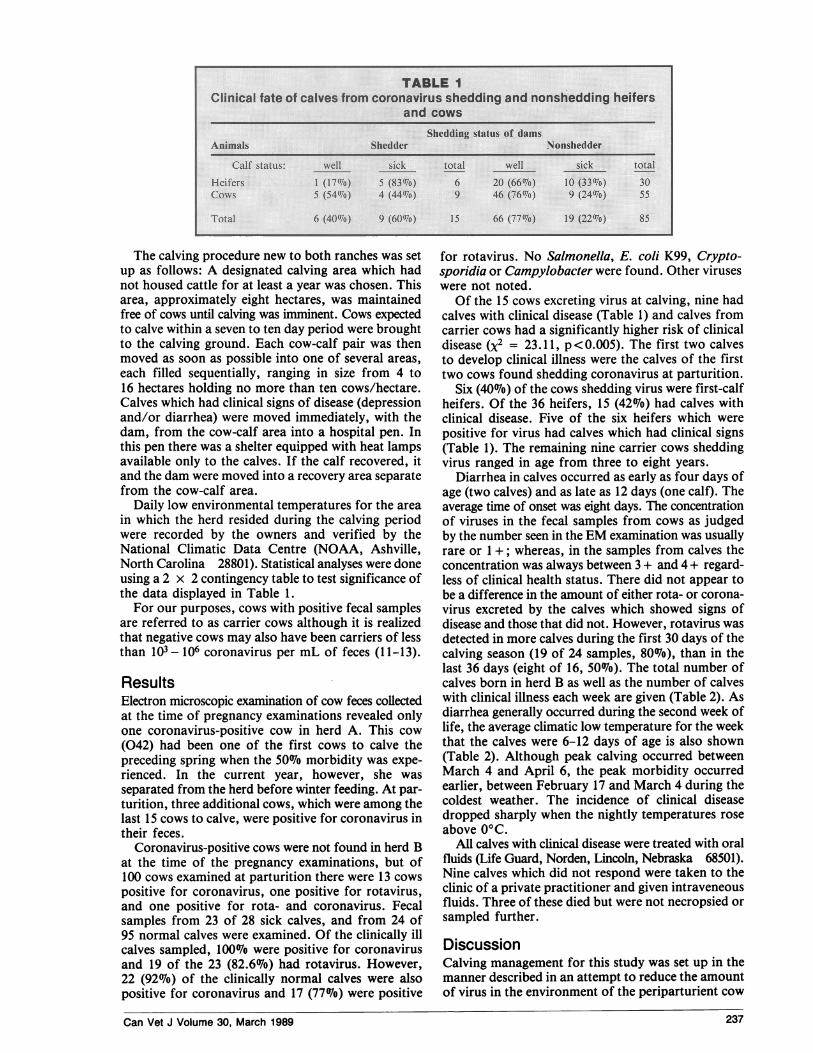
Daily low environmental temperatures for the areain which the herd resided during the calving periodwere recorded by the owners and verified by theNational Climatic Data Centre (NOAA, Ashville,North Carolina 28801). Statistical analyses were doneusing a 2 x 2 contingency table to test significance ofthe data displayed in Table 1.
For our purposes, cows with positive fecal samplesare referred to as carrier cows although it is realizedthat negative cows may also have been carriers of lessthan 103- 106 coronavirus per mL of feces (11-13).
ResultsElectron microscopic examination of cow feces collectedat the time of pregnancy examinations revealed onlyone coronavirus-positive cow in herd A. This cow(042) had been one of the first cows to calve thepreceding spring when the 500o morbidity was expe-rienced. In the current year, however, she wasseparated from the herd before winter feeding. At par-turition, three additional cows, which were among thelast 15 cows to calve, were positive for coronavirus intheir feces.
Coronavirus-positive cows were not found in herd Bat the time of the pregnancy examinations, but of100 cows examined at parturition there were 13 cowspositive for coronavirus, one positive for rotavirus,and one positive for rota- and coronavirus. Fecalsamples from 23 of 28 sick calves, and from 24 of95 normal calves were examined. Of the clinically illcalves sampled, 100% were positive for coronavirusand 19 of the 23 (82.6%o) had rotavirus. However,22 (92%7o) of the clinically normal calves were alsopositive for coronavirus and 17 (77%) were positive
for rotavirus. No Salmonella, E. coli K99, Crypto-sporidia or Campylobacter were found. Other viruseswere not noted.Of the 15 cows excreting virus at calving, nine had
calves with clinical disease (Table 1) and calves fromcarrier cows had a significantly higher risk of clinicaldisease (X2 = 23.11, p<0.005). The first two calvesto develop clinical illness were the calves of the firsttwo cows found shedding coronavirus at parturition.
Six (40/o) of the cows shedding virus were first-calfheifers. Of the 36 heifers, 15 (42%) had calves withclinical disease. Five of the six heifers which werepositive for virus had calves which had clinical signs(Table 1). The remaining nine carrier cows sheddingvirus ranged in age from three to eight years.
Diarrhea in calves occurred as early as four days ofage (two calves) and as late as 12 days (one calf). Theaverage time of onset was eight days. The concentrationof viruses in the fecal samples from cows as judgedby the number seen in the EM examination was usuallyrare or 1+; whereas, in the samples from calves theconcentration was always between 3 + and 4 + regard-less of clinical health status. There did not appear tobe a difference in the amount of either rota- or corona-virus excreted by the calves which showed signs ofdisease and those that did not. However, rotavirus wasdetected in more calves during the first 30 days of thecalving season (19 of 24 samples, 80%), than in thelast 36 days (eight of 16, 50%). The total number ofcalves born in herd B as well as the number of calveswith clinical illness each week are given (Table 2). Asdiarrhea generally occurred during the second week oflife, the average climatic low temperature for the weekthat the calves were 6-12 days of age is also shown(Table 2). Although peak calving occurred betweenMarch 4 and April 6, the peak morbidity occurredearlier, between February 17 and March 4 during thecoldest weather. The incidence of clinical diseasedropped sharply when the nightly temperatures roseabove 0°C.
All calves with clinical disease were treated with oralfluids (Life Guard, Norden, Lincoln, Nebraska 68501).Nine calves which did not respond were taken to theclinic of a private practitioner and given intraveneousfluids. Three of these died but were not necropsied orsampled further.
DiscussionCalving management for this study was set up in themanner described in an attempt to reduce the amountof virus in the environment of the periparturient cow
Can Vet J Volume 30, March 1989 237

and the newborn calf. Removal of sick calves fromthe cow-calf herd was an attempt to reduce the con-centration of virus in the environment of subsequentcalves. This type of calving management did appearto aid in the control of neonatal enteric disease in thesetwo herds. The removal of the carrier cow in herd Aand the fortuitous calving of the other three at the endof the calving season certainly seemed to influence thedisease problem in that herd. It is unfortunate thatsamples from calves were not taken from herd A asit would have been helpful to know if the virus wasactually eliminated from the environment in that herd.In herd B the 23% morbidity was the lowest this herdhad experienced since 1978.The fact that the first two calves in herd B to show
clinical signs were the offspring of the first two carriercows to calve suggests the importance of the carriercow in initiating a disease outbreak. The calves ofcarrier cows had a 600/o chance of developing clinicalillness whereas those of noncarriers had only a 22%chance (Table 1). In addition, calves born before thecow-calf area was contaminated, i.e. before the carriercows calved, remained healthy even though exposedlater. Furthermore, one would have expected manymore calves of both carriers and noncarriers to showdisease as the calving season progressed, particularlysince the cow-calf area was probably becomingprogressively more contaminated. However, an increasein diarrhea was not observed (Table 2). Actually 6807o(19) of the sick calves were seen in the first half of theseason when the majority (10) of the carrier cowscalved.
Calving grounds would become rapidly and heavilycontaminated with virus after the onset of calving if,as in herd B, 98% of the calves were incubating andexcreting great quantities of virus. The fact that onlysome calves became ill, although viruses werepresumably multiplying at a great rate in the intestinalcells of the unaffected calves also, makes it apparentthat neonatal enteric disease results from the effect ofmultiple circumstances. The presence of a pathogenis merely one of several necessary requisites for clinicaldisease.One factor contributing to clinical disease associated
with coronavirus in the calf may well be time of expo-sure, which is suggested by the fact that the first twosick calves were the offspring of the first two carrier
cows to calve. In experimental enteropathogenic E. coliinfections, exposure to the organism before colostralintake resulted in disease despite the fact that colostrumcontaining specific antibodies against E. coli was
ingested soon after inoculation of pathogenic organisms(17). The exposure of the calf of a carrier cow tocoronavirus could possibly occur before (in utero),during (coming in contact with the virus as it camethrough the birth canal), or soon after birth, beforeor during suckling. Although all of the calves bornearlier were shedding large amounts of virus at 15 daysof age, it is reasonable to assume that they becameinfected with the viruses after the first two carrier cowsand their calves were moved into the cow-calf area.Calves of the carrier cows could have been sheddingcoronavirus and exposing the older calves (which werethen two to eight days old) as early as 24 h after theirbirth (18). Furthermore, since there was no uniformtime of moving calves from the calving ground to thecow-calf area, calves could have been moved at anytime during their first 24 h, even before they hadsuckled. It could be speculated that such calves ofnegative dams may have been exposed to virus in thethen contaminated cow-calf area within several hoursof birth, or were actually calves of carrier cows whichwere not identified by the relatively insensitive EMexaminations.Another factor affecting clinical disease in these
calves seemed to be age of the dam. First calf heifersappeared to be real liabilities in herd B. Not only were40% carriers, but calves of the negative heifers alsoappeared to be more susceptible to disease (Table 1)than calves from older cows. Heifer colostrum is, nodoubt, of lesser amount and quality. It was also possiblethat some of the negative heifers were actually carriersand were not identified due to the insensitivity of theEM examination. Furthermore the heifers were never
vaccinated against rotavirus and coronavirus whereasthe cows had been in past years. Vaccination did appearto reduce shedding of coronavirus by dairy cowsstudied in Colorado (8). However, how long vaccina-tion would have an effect upon shedding of virus isunknown.The study in Colorado also showed an increase in
the shedding of coronavirus at parturition (8). Parturi-tion has been reported to influence the shedding ofother viruses (5), and is perhaps associated with corti-
Can Vet J Volume 30, March 1989
D.............................[w...........-.....~~~~~~~~~~~~~~~~~~~~~~~~~~~~~~~~~~~~~~~~~~~~~~~~~~~~~~~~~~~~~~~.......
thosi ~~~~Mwrww..7.. 14.a.. of WAg
Date Wek Cd. bo-t No..,:. (') Lot....e.m.p.. setep. 0.
.......Feb17-23 2 6 4(66) -6.IFr24-M0r2 3 5 3(60) -10ar3 4 5 (237Mr0-1.6 5 34 7(1 -3
.1Marl7-I 6 14320 -4Mar24-30 7 14 3lr3l-Ap6 8 r 5 2(13 +3
Ap....r.7 -13 15 0 (0) +3Apr 14-20 10 15 2(13) -2~~~~~~~~~~~~~~~~~~~~~~~~~~~.
238

;O...
dila.rrhea I".n...lv....fromroronsvvssh.dlg:and ........ :
Haty2 (20/) 50(89.3%o)Total calved .10C .. 56..: :Abov 0C arrlic 1(0) 27)
Heathy 4.(0%) 27: (93%).Total caled 5 29
costeroid increase in the blood of the dam. Theincreased level of corticosteroid in the blood of thenewborn may also influence the clinical manifestationof disease by affecting the susceptibility of the calf toearly exposure to the virus.The shedding of coronavirus by dairy cows in
Colorado peaked during the winter months and was notdetected during summer months of July-September(8). Colorado, like Idaho, is well known for cold,stressful winters. In our study, 60070 of the carrierswere found during the period of below-freezingtemperatures. Cold weather appeared to affect clinicalmanifestation of disease in the calves as well. Thepercentage of calves clinically affected decreased as thenightly low temperature rose, although numbers ofcalves born and exposed were increasing. The numberof calves born during the first three weeks was toosmall to show a statistically significant relationshipwith the cold temperatures, however, 80%o of the calvesof carrier cows born before the nightly temperaturerose above freezing showed clinical disease. Evencalves of the nonshedder cows had a higher risk of con-tracting disease during the coldest period (Table 3).The use ofEM to identify carrier animals is probably
not a practical method of attempting to control viralneonatal disease of calves, particularly since the lowlevel of shedding at time of pregnancy examination(summer and fall) yields too few viral particles fordetection. However, since this study suggests thatremoval of carrier animals may be an efficient methodof controlling this economically important disease,tests on fecal material utilizing serological techniquessuch as an enzyme-linked immunosorbent assay(ELISA) could be investigated for such a purpose.Such tests would have the advantage of being inexpen-sive for the examination of many samples and has theprobability of being much more sensitive. cvJ
References1. Idaho Agricultural Statistics, U.S. Dept. of Agriculture and
Idaho Dept. of Agriculture Crop and Livestock Reporting Ser-vice, Boise, Idaho, 1984: 124.
2. Bulgin MS, Anderson BC, Ward ACS, Evermann JF. Infectiousagents associated with neonatal calf disease in southwesternIdaho and eastern Oregon. J Am Vet Med Assoc 1982; 180:1222-1226.
3. Myers LL, Snodgrass DF. Colostral and milk antibody titer incows vaccinated with a modified-live rotavirus-coronavirusvaccine. J Am Vet Med Assoc 1982; 181: 486-488.
4. Waltner-Toews D, Martin SW, Meek AH, McMillan I, CrouchCF. A field trial to evaluate the efficacy of a combined rotavirus-coronavirus/Escherichia coli vaccine in dairy cattle. Can J CompMed 1985; 49: 1-9.
5. Benfield DA, Stotz I, Moore R, McAdarach JP. Shedding ofrotavirus in feces of sows before and after farrowing. J ClinMicrobiol 1982; 16: 186-190.
6. Radostits OM, Acres SD. The prevention and control ofepidemics of acute undifferentiated diarrhea of beef calves inwestern Canada. Can Vet J 1980; 21: 243-249.
7. Woods GN, Bridger JC. Viral enteritis of calves. Vet Rec 1982;96: 85-88.
8. Collins JK, Riegel CA, Olson JD, Fountain A. Shedding ofenteric coronavirus in adult cattle. Am J Vet Res 1987; 48:361-365.
9. Crouch CF, Acres SD. Prevalence of rotavirus and coronavirusantigens in feces of normal cows. Can J Comp Med 1984; 48:340-342.
10. Crouch CF, Ohman HB, Watts TC, Babiuk LA. Chronicshedding of bovine enteric coronavirus antigen-antibodycomplex by clinically normal cows. J Gen Virol 1985; 66:1489-1500.
11. England JJ, Reed DE. Negative contrast electron microscopictechniques for diagnosis of viruses of veterinary importance.Cornell Vet 1980; 70: 125-136.
12. Horne RW. Electron microscopy of isolated virus particles andtheir components. In: Maramorosch K, Koprowski H, eds.Methods in Virology, Vol. III. New York: Academic Press,1967: 521-574.
13. England JJ, Frye CS, Enright EA. Negative contrast electronmicroscopic diagnosis of viruses of neonatal calf diarrhea.Cornell Vet 1976; 66: 172-182.
14. Dean AG, Ching Y, Williams RG, Harden LB. Test forEscherichia coli enterotoxin using infant mice: Application ina study of diarrhea in children in Honolulu. J Infect Dis 1972;125: 407-411.
15. Anderson BC. Quick and easy diagnosis of cryptosporidiosisand Johne's disease. Vet Med 1985; 80: 87-89.
16. Firehammer BD, Myers LL. Campylobacterfetus subspjejuni:Its possible significance in enteric disease of calves and lambs.Am J Vet Res 1981; 42: 918-922.
17. Logan DF, Pearson GR, McNulty MS. Studies on the immunityof the calf to colibacillosis-VII: The experimental reproductionof enteric colibacillosis in colostrum-fed calves. Vet Rec 1977;101: 443-446.
18. Mebus DA, Stair EL, Rhodes MD, Twiehaus ML. Pathologyof neonatal calf diarrhea induced by a coronavirus-like agent.Vet Pathol 1973; 10: 45-64.
Can Vet J Volume 30, March 1989 239