Detection of recombinant human EPO administered to horses using MAIIA lateral flow isoform test
10
ORIGINAL PAPER Detection of recombinant human EPO administered to horses using MAIIA lateral flow isoform test Maria Lönnberg & Ulf Bondesson & Florence Cormant & Patrice Garcia & Yves Bonnaire & Jan Carlsson & Marie-Agnes Popot & Niclas Rollborn & Kristina Råsbo & Ludovic Bailly-Chouriberry Received: 18 December 2011 / Revised: 9 March 2012 / Accepted: 22 March 2012 / Published online: 18 April 2012 # Springer-Verlag 2012 Abstract Doping of horses with recombinant human eryth- ropoietin (rHuEPO) to illegally enhance their endurance capacity in horseracing has been reported during the last years. This leads to increased blood viscosity which can result in sudden death and is of concern for the horse welfare. Additionally, the horse can start production of rHuEPO antibodies, which cross-reacts with endogenous equine EPO and can lead to severe anaemia and even death. In this study, a novel micro-chromatographic method, EPO WGA MAIIA, has been tested for the capability in plasma and urine samples to detect administration of erythropoiesis- stimulating agents, like the rHuEPO glycoprotein varieties Eprex and Aranesp, to horses. After administration of 40 IU Eprex kg -1 day -1 to seven horses during 6 days, the pres- ence of Eprex in horse plasma was detected up to 2–5 days after last injection. In urine samples collected from two horses, Eprex was detected up to 3 days. A single injection of Aranesp (0.39 μg/kg) was detected up to 9 days in plasma and up to 8 days, the last day of testing, in the urine sample. The LC-FAIMS-MS/MS system, with 1 day reporting time, confirmed the presence of Eprex up to 1 day after last injection for six out of seven horses and the presence of Aranesp up to 5 days after last injection in plasma samples. The MAIIA system showed to be a promising tool with high sensitivity and extremely short reporting time (1 h). Keywords Aranesp . EPO doping control . Eprex . Equine EPO . Micro-chromatography . WGA Introduction The human 30-kDa glycoprotein erythropoietin (HuEPO), produced by human adults mainly in the renal cortex, is essential for red blood cell production. For healthy individ- uals, a decrease in haemoglobin concentration directly increases the production rate of erythropoietin (EPO), as recently reviewed [ 1]. The human recombinant EPO (rHuEPO) has successfully been synthesised in mammal cells since the late 1980s [2] and has been administered to millions of anaemic patients with chronic kidney disease, AIDS and cancer. It has been speculated for horses, due to the large red blood cell storage capacity in the spleen [3], that rHuEPO doping will not enhance their exercise performance. This was tested by administration in healthy horses of rHuEPO, with 50 IU/kg injected three times a week during 2 weeks, which increased the number of red blood cells and improved the oxygenation of main muscles compared with the control group [4]. A substantial 19 % increase in maximal aerobic M. Lönnberg (*) : K. Råsbo Department of Chemistry-Biomedical Center, Uppsala University, Box 599, 751 24 Uppsala, Sweden e-mail: [email protected] U. Bondesson Department of Chemistry, Environment and Feed Hygiene, The National Veterinary Institute (SVA), Uppsala, and Division of Analytical Pharmaceutical Chemistry, Biomedical Center, Uppsala University, 751 23 Uppsala, Sweden F. Cormant : P. Garcia : Y. Bonnaire : M.-A. Popot : L. Bailly-Chouriberry L.C.H., Laboratoire des Courses Hippiques, 15 rue de Paradis, 91370 Verrières le Buisson, France J. Carlsson : N. Rollborn MAIIA Diagnostics, 751 38 Uppsala, Sweden Anal Bioanal Chem (2012) 403:1619–1628 DOI 10.1007/s00216-012-5972-0
Transcript of Detection of recombinant human EPO administered to horses using MAIIA lateral flow isoform test
ORIGINAL PAPER
Detection of recombinant human EPO administered to horsesusing MAIIA lateral flow isoform test
Maria Lönnberg & Ulf Bondesson & Florence Cormant &Patrice Garcia & Yves Bonnaire & Jan Carlsson &
Marie-Agnes Popot & Niclas Rollborn & Kristina Råsbo &
Ludovic Bailly-Chouriberry
Received: 18 December 2011 /Revised: 9 March 2012 /Accepted: 22 March 2012 /Published online: 18 April 2012# Springer-Verlag 2012
Abstract Doping of horses with recombinant human eryth-ropoietin (rHuEPO) to illegally enhance their endurancecapacity in horseracing has been reported during the lastyears. This leads to increased blood viscosity which canresult in sudden death and is of concern for the horsewelfare. Additionally, the horse can start production ofrHuEPO antibodies, which cross-reacts with endogenousequine EPO and can lead to severe anaemia and even death.In this study, a novel micro-chromatographic method, EPOWGA MAIIA, has been tested for the capability in plasmaand urine samples to detect administration of erythropoiesis-stimulating agents, like the rHuEPO glycoprotein varietiesEprex and Aranesp, to horses. After administration of 40 IUEprex kg−1 day−1 to seven horses during 6 days, the pres-ence of Eprex in horse plasma was detected up to 2–5 daysafter last injection. In urine samples collected from two
horses, Eprex was detected up to 3 days. A single injectionof Aranesp (0.39 μg/kg) was detected up to 9 days in plasmaand up to 8 days, the last day of testing, in the urine sample.The LC-FAIMS-MS/MS system, with 1 day reporting time,confirmed the presence of Eprex up to 1 day after lastinjection for six out of seven horses and the presence ofAranesp up to 5 days after last injection in plasma samples.The MAIIA system showed to be a promising tool with highsensitivity and extremely short reporting time (1 h).
Keywords Aranesp . EPO doping control . Eprex . EquineEPO .Micro-chromatography .WGA
Introduction
The human 30-kDa glycoprotein erythropoietin (HuEPO),produced by human adults mainly in the renal cortex, isessential for red blood cell production. For healthy individ-uals, a decrease in haemoglobin concentration directlyincreases the production rate of erythropoietin (EPO), asrecently reviewed [1]. The human recombinant EPO(rHuEPO) has successfully been synthesised in mammalcells since the late 1980s [2] and has been administered tomillions of anaemic patients with chronic kidney disease,AIDS and cancer.
It has been speculated for horses, due to the large redblood cell storage capacity in the spleen [3], that rHuEPOdoping will not enhance their exercise performance. Thiswas tested by administration in healthy horses of rHuEPO,with 50 IU/kg injected three times a week during 2 weeks,which increased the number of red blood cells and improvedthe oxygenation of main muscles compared with the controlgroup [4]. A substantial 19 % increase in maximal aerobic
M. Lönnberg (*) :K. RåsboDepartment of Chemistry-Biomedical Center, Uppsala University,Box 599, 751 24 Uppsala, Swedene-mail: [email protected]
U. BondessonDepartment of Chemistry, Environment and Feed Hygiene,The National Veterinary Institute (SVA), Uppsala, and Division ofAnalytical Pharmaceutical Chemistry, Biomedical Center,Uppsala University,751 23 Uppsala, Sweden
F. Cormant : P. Garcia :Y. Bonnaire :M.-A. Popot :L. Bailly-ChouriberryL.C.H., Laboratoire des Courses Hippiques,15 rue de Paradis,91370 Verrières le Buisson, France
J. Carlsson :N. RollbornMAIIA Diagnostics,751 38 Uppsala, Sweden
Anal Bioanal Chem (2012) 403:1619–1628DOI 10.1007/s00216-012-5972-0
capacity was shown 1 week after treatment. A higher num-ber of red blood cells leads to increased blood viscosity,which may result in cerebral thromboembolism, as forhumans [5]. Moreover, horses treated with rHuEPO havedeveloped immune response, with production of anti-humanEPO antibodies cross-reacting with equine EPO (eEPO),and obtained severe anaemia [6]. The use of rHuEPO andits analogues is prohibited in the majority of human andanimal sports.
In humans and horses, only minute blood concentrationsof rHuEPO, in the nanogrammes per litre range, is requiredto increase erythropoiesis. The clearance from blood is rapidand shows large variations between individuals. For epoetinalpha, one type of rHuEPO, the biological half-lives were19.4±10.7 h after subcutaneous (s.c.) and 6.8±2.7 h afterintravenous (i.v.) injections to humans [7]. Moreover, thevarieties of protein-based erythropoiesis stimulating agents(ESAs) [8,9] are increasing rapidly, with up to 80 biosimilarepoetin products available in 2009 [2], which makes thedoping control for humans even more difficult [10]. TheEPO variants on the market are differing in biological ac-tivity and structure, e.g. glycosylation, due to cell line ex-pression and details in the manufacturing process, likeculture conditions and purification methods. EPO-resembling recombinant protein drugs with longer biologi-cal half-lives, like Aranesp with a molecular weight of37 kDa and a biological half-life of 26 h (i.v.) [11], andMircera, a pegylated rHuEPO, with a molecular weight of60 kDa and a biological half-life of 134 h (i.v.) [12], areefficient ESAs. They can be used with less frequent injec-tion regimes, which facilitates for the patients.
For doping control of rHuEPO administered to humans,the only difference between exogenous and endogenousEPO is the composition of the three N-linked and one O-linked oligosaccharide complex, constituting 40 % of theprotein molecular weight. The main methodologies, as re-cently reviewed [13], combine electrophoretic or chromato-graphic separation of EPO glycoforms, with sensitive anti-EPO antibody based detection methods. The methods oftenrequire an additional purification and/or concentration stepbefore urine and plasma samples analysis [14,15]. Endoge-nous and recombinant HuEPO glycosylation can be distin-guished by differences in charge [16,17], isoelectric point[18–20], molecular mass [21–23] or interaction with lectins,e.g. wheat germ agglutinin (WGA) [24,25] for the glyco-proteins. The methods differ in how well they distinguish acertain type of glycosylation and how much EPO is requiredfor analysis.
For EPO doping control in horses, a rapid screeningmethod for rHuEPO using a human EPO immunoassayswith low reactivity for eEPO compared with HuEPO hasbeen used [26] to select samples to be confirmed with themore resource demanding methods. Exogenous rHuEPOs
and analogues present in equine samples have been detectedby isoelectric focusing (IEF) with double-immunoblotting[26], due to their differences in oligosaccharide composi-tion. Equine and human EPO have 84 % identity in theamino acid structure [27] which has been utilised for LC-MS/MS analyses of peptide sequences specific for HuEPOappearing after trypsin digestion of EPO [28–32]. However,if recombinant eEPO will appear at the market MS basedmethods cannot be used for its detection, but methodsshowing differences in glycosylation can still be used.
MAIIA is a novel analytical micro-chromatographic sys-tem [33] for separation and detection of low abundant pro-tein isoforms with good resolution [34] and high sensitivity[35]. The lateral flow based micro-column has beenequipped with ligands suitable for ion-exchange chromatog-raphy [36,37] and with lectin ligands, like WGA [25], foraffinity chromatography. The MAIIA (EPO WGA MAIIA)method, using WGA as chromatographic ligand and EPOdetection was recently found to distinguish ten differentrHuEPOs and analogues from human endogenous EPO[25]. It was also shown, when testing samples from a majorsport competition, that the use of the MAIIA test revealed6 % of the athletes as positive for EPO doping while theaccredited IEF method for human athletes found no positivesamples.
In this study, the MAIIA method is evaluated to detectthe daily s.c. administration of 40 IU/kg of Eprex during sixconsecutive days and the single administration of 0.39 μg/kgof Aranesp to horses.
Materials and methods
Sample collection from horses administered with rHuEPO(Eprex) and analogues (Aranesp)
The details for the administration series performed at Labo-ratoire des Courses Hippiques (L.C.H.; Verrières le Buisson,France) have recently been presented [30]. Briefly, eightthoroughbred horses were administered subcutaneously inthe neck. Seven horses (H579 to H585) received 40 IU kgbodyweight−1 day−1 of Eprex (epoetin alpha) during 6 days,and horse H626 received a single dose of 0.39 μg/kg ofAranesp (darbepoetin alpha). Heparin plasma samples wereobtained before, during and after the end of administrationsas indicated in Fig. 1. During the 6 days with Eprex injec-tions, samples were collected 24 h after previous injection.Urine samples were also collected from three horses (H579,H580 and H626). All samples were stored at −20 °C untiltheir analysis. The study was led in agreement with animalwelfare rules at the administration and sampling Centre ofthe Fédération Nationale des Courses Françaises (FNCF).
1620 M. Lönnberg et al.
Reference urine and plasma samples used for EPO WGAMAIIA
As reference plasma samples, one of the pre-administrationsamples, collected 2 days prior to the administration, wasselected from the eight thoroughbred horses (eight samples)and ten samples were leftovers from routine analysis (post-racing). For the urine analysis, six reference samples wereused, three samples were collected before injection and threesamples were leftovers from routine analysis.
Affinity purification of EPO from biological samplesas a pre-step for EPO WGA MAIIA analysis
EPO Purification Kit, Art. No. 0250, was obtained fromMAIIA Diagnostics (Uppsala, Sweden). EPO from 0.5 mLequine plasma samples (for 20 % of the samples 0.47 to0.06 mL was used) or 20-mL equine urine samples waspurified according to the instructions from the producer,using the recommended addition of detergent and bovineserum albumin to the reagents. Purified EPO was finallyobtained in a volume of 220 μL for equine urine. EPO fromthe plasma samples was obtained in an eluate volume of55 μL, except for the high EPO concentration samplescollected during 1 to 3 days after first injection of Eprex,for which 220-μL eluate was collected. EPO QuantificationKit Art. no. 0100 (MAIIA Diagnostics), was used for thedetermination of EPO concentration in the eluate. The EPO
affinity purification recovery was measured and found to bein accordance with recent evaluations [14,15] showing 75and 55 % recovery for NeoRecormon® (Roche GmBH,Mannheim, Germany) added to buffer and equine plasma,respectively, and 39 % for Aranesp (Thousands Oak, CA,USA) added to equine plasma.
EPO WGA MAIIA isoform analysis
A MAIIA kit (EPO WGA MAIIA prototype kit, MAIIADiagnostics), containing MAIIA lateral flow strips andreagents for performing WGA lectin chromatography andEPO immunoassay, was used with EPO affinity purifiedsamples in accordance with the instructions from the sup-plier. The procedure and scanner equipment has been de-scribed recently [25] together with the standardisationregime. The MAIIA lateral flow strip contains both aWGA zone (lectin affinity) and downstream this, an anti-EPO zone (capturing antibody for immunoassay). Briefly,after immersing the strip in a well containing the sample, allEPO is bound in the first tenth of the 8-mm long WGAzone. Then the strip is moved to a second well for desorp-tion of EPO, from its WGA interaction, by a WGA compet-ing sugar derivate, N-acetylglucosamine (GlcNAc). At leasttwo strips are used to obtain total (using high concentrationof GlcNAc, 100 mM) and retarded (using low concentrationof GlcNAc) desorption of EPO for each sample. The mem-brane lot and the selected GlcNAc concentrations were: (a)
1
10
100
1000
0
10
20
30
40
50
60
70
80
90
100
110
-6 -4 -2 0 2 4 6 8 10 12 14
To
tal E
PO
co
nce
ntr
atio
n,
"ng
Hu
EP
O/L
"
EP
O W
GA
MA
IIA, P
MI
SD
Days after final injection with Eprex
EPREXinjection
EPO WGA MAIIA
Injection day
Total "EPO" concentration
1
10
100
1000
10000
0
10
20
30
40
50
60
70
80
90
100
110
-2 -1 0 1 2 3 4 5 6 7 8 9 10 11
To
tal E
PO
co
nce
ntr
atio
n, "
ng
Hu
EP
O/L
"
EP
O W
GA
MA
IIA,
PM
I
Days after injection with Aranesp
ARANESPinjection
EPO WGA MAIIA
Injection day
Total "EPO" concentration
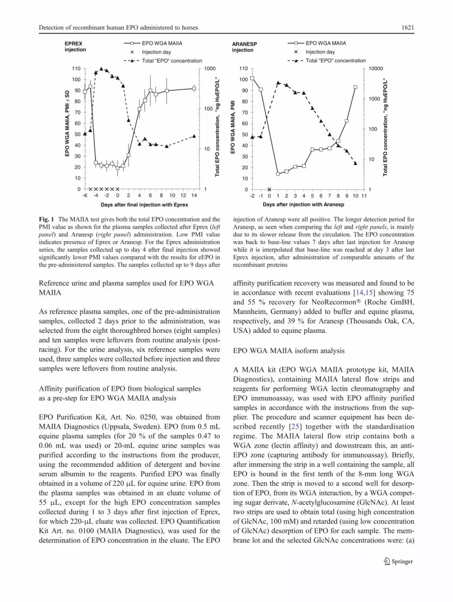
Fig. 1 The MAIIA test gives both the total EPO concentration and thePMI value as shown for the plasma samples collected after Eprex (leftpanel) and Aranesp (right panel) administration. Low PMI valueindicates presence of Eprex or Aranesp. For the Eprex administrationseries, the samples collected up to day 4 after final injection showedsignificantly lower PMI values compared with the results for eEPO inthe pre-administered samples. The samples collected up to 9 days after
injection of Aranesp were all positive. The longer detection period forAranesp, as seen when comparing the left and right panels, is mainlydue to its slower release from the circulation. The EPO concentrationwas back to base-line values 7 days after last injection for Aranespwhile it is interpolated that base-line was reached at day 3 after lastEprex injection, after administration of comparable amounts of therecombinant proteins
Detection of recombinant human EPO administered to horses 1621
results in Table 1, L100112—3 mM GlcNAC, (b) plasmaruns, L091118—2 mM GlcNAc, and (c) urine runs,L090108—10mMGlcNAc. Desorption from theWGA bind-ing starts the migration of EPO and the most rapidly migratingones can pass the WGA zone and be captured in the subse-quent anti-EPO zone on the strip. Desorption is interruptedafter 5 min by removing the WGA zone by cutting. EPObound to the anti-EPO zone is reacted with anti-EPO boundto carbon black nano-string, and the obtained grey to blacksignal intensity is quantified with an image scanner.
In this study, 25 μL of affinity-purified EPO sample wasapplied to each strip. For each sample, four strips wereutilised since duplicates were used for determination of theamount of EPO that had passed the WGA zone during the5 min of total and retarded desorption. By using the appro-priate low GlcNAc concentration for retarded desorption, aminor part of rHuEPO (about 13–31 %), and most of en-dogenous eEPO (>75 %), were allowed to migrate into theanti-EPO zone. The amount of EPO reaching the anti-EPOzone was calculated using a standard curve of NeoRecor-mon (3 to 1,000 ng/L). Stored signal values for the standardcurve were used for runs performed with the same batch ofreagents. The MAIIA value characterises the WGA interac-tion with the EPO glycosylation by the unit percentage ofmigrated isoforms (PMI), [EPO amount released duringretarded conditions using low GlcNAc concentration]/[totalamount of EPO released using 100 mM GlcNAc]×100. Ineach of the 11 plasma runs, two control preparations were
included, an affinity purified equine plasma and a bufferpreparation with NeoRecormon. The test procedure tookabout 30 min for processing 56 strips.
Concentration determination using the EPO WGA MAIIA
EPO concentration in the eluates from the affinity purifica-tion was determined from the MAIIA strips used with100 mM GlcNAc in the desorption step. The concentrationof EPO in the plasma samples was estimated by adjustingfor applied sample volume in the affinity purification step,and correcting for the 55 and 39 % recovery obtained for thecontrols with NeoRecormon and Aranesp applied to equineplasma. The variation in recovery was recently estimated to15 % in coefficient of variation for the affinity purificationof NeoRecormon added to buffer [15]. The concentration ofthe EPO analogue Aranesp was underestimated with 18 %when using NeoRecormon as standard, as reported earlierfor the antibodies used in the sandwich immunoassay part ofthe test [35]. The EPO concentration of samples containingeEPO might also be underestimated as the used anti-EPOantibodies were obtained by injection with HuEPO, and nopurified eEPO preparation was available for standardisation.
Enzyme immunoassay of EPO in horse plasma samples
An ELISA for HuEPO, Quantikine EPO, was purchasedfrom R&D Systems (Minneapolis, MN, USA) and used at
Table 1 Different types of EPOs and their MAIIA PMI values
Buffer prep Plasma Urine Plasma runs(2 mM GlcNAc)
Urine runs(10 mM GlcNAc)
n PMI n PMI n PMI n PMI n PMI
Comparison of PMI values (mean±SD) for a variety of EPOs (3 mM GlcNAc)a
rHuEPO
NeoRecormon 4 8.0 (±0.7)
rHuEPO analogues
Aranesp 2 3.0 (±0.7)
Mircera 3 57.6 (±6.9)
Endogenous EPO
Human EPO 2 25.7 (±3.6) 3 14.1 (±2.8)
Equine EPO 3 87.9 (±6.9) 3 57.1 (±3.2)
Optimised WGA interaction for the studyb
Equine EPO 26 90.0 (±7.3) 6 77.0 (±2.5)
NeoRecormon in buffer 11 13.4 (±2.0) 1 31
a An example of WGA interaction optimised (3 mM GlcNAc) to measure both the EPO variety with the strongest (Aranesp, only 3.0 % passed theWGA zone) and with the weakest binding (purified eEPO from plasma, 87.9 % passed the WGA zone)b The WGA interaction optimised for this study to allow almost all equine EPO to migrate through the WGA zone. Urine eEPO migrated slowerthrough the WGA zone than plasma eEPO when compared in Table 1a, and a reduced WGA reactivity was required to obtain higher relativepassage of eEPO in urine. The use of 2- and 10-mM GlcNAc for plasma runs and urine runs, respectively, is indicated by the increased migration ofthe control NeoRecormon in urine runs to 31 % migrating compared with 13.4 % for the plasma runs
1622 M. Lönnberg et al.
L.C.H. for selecting horse plasma samples above 170 ng/L(5 pM) for further LC-MS/MS analysis. The conversionfactor used to obtain units to mass for the rHuEPO standardwas 1 IU08.4 ng.
Mass spectrometry analysis, LC-FAIMS-MS/MS
The method [30], developed and performed at L.C.H., com-bines a rapid pre-analytical purification and concentrationdevice with detection using LC-high-field asymmetricwaveform ion mobility spectrometry (FAIMS)-MS/MS.EPO was purified and concentrated from 4-mL plasmasamples or 10-mL urine samples with the EPO Purificationkit (MAIIA Diagnostics) further elaborated [30] to be usedprior to MS analysis. The target peptides for human EPOwere T6 and T17 as previously described [28] with a reten-tion time of 9.9 and 9.0 min, respectively. Each peptideshowed four SRM transitions and a signal-to-noise ratioabove 3. Performances were validated with blank equineplasma and urine samples, and the same samples spikedwith Aranesp. The validated limit of confirmation was de-fined at 250 ng Aranesp/L and the lower limit of detectionwas found at 100 ng/L [30]. In the validation, Aranesp wasselected as reference as this molecule was less efficientlycaptured than NeoRecormon in the affinity purification pro-cess. In the present study, a sample was identified as positivewhen Association of Official Racing Chemists (AORCs)minimum criteria for MS identification of small moleculeswere fulfilled.
Statistics
Values are means±1 standard deviation (SD). Differencesbetween results for samples collected before and after injec-tion were examined by paired t test (SigmaPlot 12, SystatSoftware, San Jose, CA, USA) and statistical significancewas accepted at p<0.05. For the MAIIA test, the one-tailed99.9 % confidence limit (CL) were calculated from the meanresult for the reference samples and results outside CL weretermed positive.
Results
MAIIA test optimisation of the percentage of EPOmigrating through the WGA zone
The interaction strength with WGA for different types ofEPO related molecules has recently been shown to be con-siderable different [25]. The resolution between two types ofEPO populations can be increased by optimising the con-centration of GlcNAc used for competitive desorption ofEPO [25]. However, as the binding strength of the WGA
ligand bound to the membrane also can vary, the selectedconcentration of GlcNAc has to be optimised for eachmembrane lot. In Table 1, the target for the optimisationwas to compare the WGA interaction from the EPO-likemolecule with the strongest interaction (Aranesp) to theEPO population with the weakest interaction (equine EPOin plasma). Aranesp has increased WGA interaction due tothe two additional carbohydrate structures; only 3 % (03.0PMI) was passing the WGA zone. Equine EPO bound withconsiderable less strength, having values of 57.1 and 87.9PMI in urine and plasma, respectively, compared with Neo-Recormon with 8.0 PMI. Human endogenous EPO in urineand plasma showed values of 14.1 and 25.7 PMI, respec-tively. Both for humans and horses, the oligosaccharides onEPO in urine showed more structures interacting withWGA, with lower PMI values for urine compared withplasma EPOs.
In the present administration study, a slightly differentWGA interaction strength was selected, specific for plasmaand for urine runs, to allow almost all eEPO in each run tomigrate through the WGA zone. For the plasma runs, 90 %(90.0 PMI) of eEPO were allowed to pass the WGA zone,which was comparable to the interaction obtained for theresults presented in Table 1. For the urine run, 77 % of eEPOwas allowed to pass the zone. Compared with the resultsfound in Table 1, 57.1 %, the WGA interaction was reducedfor the urine run to obtain better resolution from recombi-nant EPO and Aranesp. The results for NeoRecormon, with13.4 and 31 PMI for the plasma and the urine runs, respec-tively, makes it possible to compare that different interactionstrengths has been used.
For the administration series, the rHuEPO type Eprexwas injected to the horses. This EPO variety has recentlybeen compared with NeoRecormon and showed comparableWGA interaction with values of 25.1 and 30.0 PMI,respectively [25].
MAIIA results for the Eprex and Aranesp administrationseries
The PMI values obtained by MAIIA analysis of equineplasma during the administration of Eprex and Aranesp areshown in Fig. 1. Included in the figure is also the total EPOconcentration in plasma, although both eEPO and Aranespare likely to be underestimated when using rHuEPO forstandardisation. The low PMI values for EPO in the samplesduring the administration of Eprex indicate strongly thepresence of exogenous EPO glycoforms in the equine plas-ma. The results for horse plasma samples collected 1 dayafter injection (n047) showed a highly significant (p<0.001) reduction in PMI values to 21.8±4.8 PMI after Eprexinjections, compared with 88.7±7.2 PMI for eEPO presentin the eight samples collected 2 days prior to the initial
Detection of recombinant human EPO administered to horses 1623
injection. Samples collected 2 (p<0.001) and 4 days (p00.019) after last injection of Eprex from seven horses werealso significantly different from the samples collected beforeinjection. No plasma sample was collected 3 days afterinjection. The samples collected 5 days after last injectionand later on could not be differentiated from eEPO. Thesamples from horse 581, collected during the administrationperiod after the last three injections, showed considerablelower EPO concentrations compared with samples fromother horses, which was also confirmed with the EPOELISA immunoassay. The PMI values were not as low asfor the other horses, although still clearly aberrant fromeEPO.
For the diagnostics application, to settle if a single sampleis aberrant, 18 plasma reference samples were tested and amean value of 88.0±7.1 PMI was obtained. There was nosignificant difference between the ten left-over samples (87.2±7.7 PMI) and the eight pre-administration samples (89.0±6.7 PMI) used as reference samples. The one-tailed 99.9%CLwas 65.9 PMI. Samples with PMI values outside 99.9 % CLwere regarded as positive and the results are shown in Table 2.All plasma samples (n07) collected 2 days after injectionwere positive. The samples from horse H579, collected 4and 5 days after injection, were also positive, hence 14.3 %of the horses were positive on days 4 and 5. For the Aranespinjection of one horse, see Table 3, the PMI valueswere positive up to 9 days after the single injection.
For urine samples, the six reference samples showeda mean value of 77.0±2.5 PMI, and the 99.9 % CL was69.0 PMI. The urine samples collected up to 3 daysafter last injection with Eprex (n02), and up to 8 daysafter the single injection with Aranesp (n01), were
clearly positive. No urine samples were tested afterthese collections days.
EPO concentration in plasma sample determined with EPOWGA MAIIA
The estimated EPO concentration in equine plasma shownin Fig. 1 for seven horses injected with 40 IU (0.34 μg)Eprex/kg and one horse injected with 0.39 μg Aranesp/kgbodyweight reveal the quite different elimination pattern forEprex and the more glycosylated Aranesp.
For samples collected 1 day after injection with Eprex,the EPO concentration was 891±179 ng/L (n021) after thefirst three injections, and 522±208 ng/L (n021) after thethree following injections. The samples collected 1 day afterthe last injection was 375±165 ng/L (n07) and after 2 days52±26 ng/L (n07), a decline to 10 % when adjusted for themean baseline values at 13 ng/L for these horses. For thehorse injected with a single dose of Aranesp, the samplecollected 1 day after injection showed a value at 3.400 ng/L,which declined to 2.800 ng/L (84%) after further 1 day.Between days 3 (1562 ng/L) and 4 (487 ng/L), the concen-tration of Aranesp declined to 33 %, when correcting for thebaseline value at 10 ng/L for this horse. The concentrationwas back to baseline 3–4 and 7–8 days after injection withEprex and Aranesp, respectively, while still the PMI valueswere aberrant.
It was also found that the estimated concentration ofeEPO was much lower in the samples collected after injec-tion than before. The eEPO concentration was reduced to52 % (p00.026) for samples collected at days 10–14 after
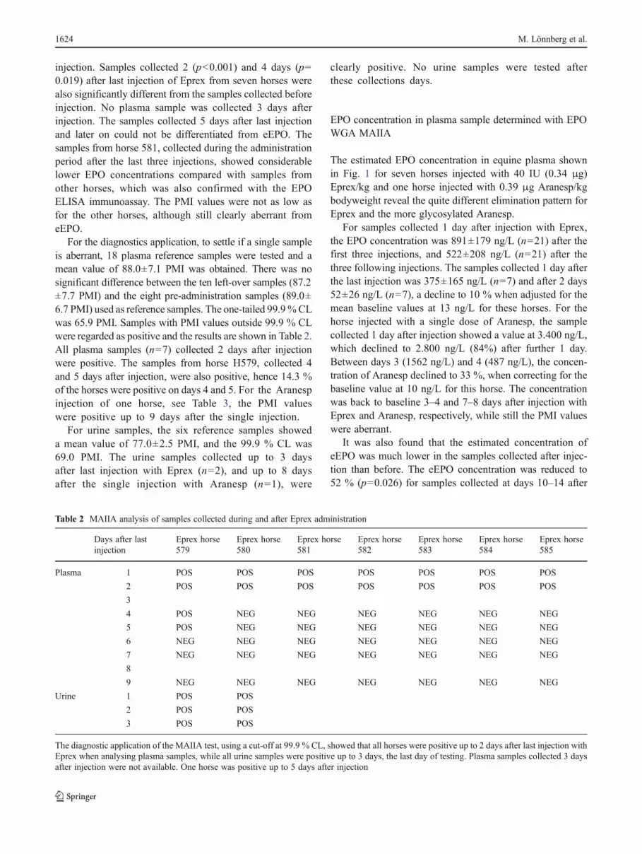
Table 2 MAIIA analysis of samples collected during and after Eprex administration
Days after lastinjection
Eprex horse579
Eprex horse580
Eprex horse581
Eprex horse582
Eprex horse583
Eprex horse584
Eprex horse585
Plasma 1 POS POS POS POS POS POS POS
2 POS POS POS POS POS POS POS
3
4 POS NEG NEG NEG NEG NEG NEG
5 POS NEG NEG NEG NEG NEG NEG
6 NEG NEG NEG NEG NEG NEG NEG
7 NEG NEG NEG NEG NEG NEG NEG
8
9 NEG NEG NEG NEG NEG NEG NEG
Urine 1 POS POS
2 POS POS
3 POS POS
The diagnostic application of the MAIIA test, using a cut-off at 99.9 % CL, showed that all horses were positive up to 2 days after last injection withEprex when analysing plasma samples, while all urine samples were positive up to 3 days, the last day of testing. Plasma samples collected 3 daysafter injection were not available. One horse was positive up to 5 days after injection
1624 M. Lönnberg et al.
the initial injection (5 to 9 days after last injection) com-pared with the pre-administration concentration.
Imprecision for EPO WGA MAIIA
The immunoassay measurement of the affinity purified plas-ma samples included in the injection study tested in desorp-tion mode with low and high GlcNAc concentration showeda median coefficient of variation (CV) of 5.7 (n0163; mean,27 ng/L) and 3.9 % (n0163; mean, 97 ng/L), respectively,between the duplicates. The mean inter-assay CVs for thePMI values were 10.5 and 15.2 % for the controls at 74.4±7.9 PMI (an affinity-purified plasma eEPO) and 13.4±2.0PMI for NeoRecormon in buffer, when measured in dupli-cate at 11 different plasma runs.
LC-FAIMS-MS/MS results
In order to confirm that the results of the administrationstudy was in accordance with other studies analysed withmass spectrometry, liquid chromatography-ion-mobilityMS/MS [30] was performed on some selected samples.Samples having a plasma concentration above 170 ng/L,as tested with ELISA, were analysed. This limit was arealistic target concentration in regards of the validated(n010) limit of identification obtained at 250 ng/L and thelower limit of detection (LLOD) at 100 ng/L (detectable butwas not able to validate at n010) [30] when using 4-mLplasma or 10-mL urine sample. In addition, one of the twopre-administration plasma samples was tested for all sevenhorses and found to be negative. Two pre-administered urinesamples for horse H579 and H626 were also negative withthe method.
The samples from six horses collected 1 day after the lastinjection, with an Eprex concentration of 218–353 ng/L(ELISA), were all positive. H579, having only one targetpeptide positive when analysed in plasma, was signed aspositive in regards of the minimum AORCs requirementswhile the urine sample, collected the same day, was clearlypositive. For the remaining horse injected with Eprex, H581,the sample collected 1 day after last injection was notanalysed as the EPO concentration was only 97 ng/L. Theurine sample from H580, 1 day after injection, was nottested although the plasma concentration showed that itwas measurable.
For the horse injected with Aranesp, H626, the samplescollected 4 and 5 days after injection were positive (176–294 ng/L) while the urine sample collected 6 days afterinjection was negative as expected due to its low plasmaEPO concentration (75.6 ng/L) that day.
Discussion
Detecting administration of rHuEPO
For MAIIA, all seven horses were positive both 1 and 2 daysafter last s.c. injection of Eprex, 40 IU/kg, and one of thehorses were positive at 4 and 5 days after last injection whenanalysing the plasma samples. Urine samples from horse 579and 580, collected 3 days after last injection, were all positivewhile blood samples were not collected that day. It is likelythat the MAIIA test can detect the presence of Eprex up to3 days after last injection for all horses. LC-FAIMS-MS/MSidentified the presence of Eprex for six of the seven horses inthe plasma samples collected 1 day after last injection.
Both MAIIA and LC-FAIMS-MS/MS used the same typeof EPO purification test prior to the analysis, althoughfurther elaborated for use prior to mass spectrometry. Forthe MAIIA test, 0.5-mL plasma was sufficient for identifi-cation of Eprex even though, 2–5 days after injection, someof the samples were as low as 20 ng/L. LC-FAIMS-MS/MSrequired 4 mL of plasma, and only samples above 170 ng/Lwas selected for analysis due to the confirmation limitvalidated at 250 ng/L with LLOD at 100 ng/L.
Other laboratories have presented studies involving fewerhorses and different administration regimes (differing inamount of ESA, s.c. or i.v. administration, single or severaldoses), which make comparisons difficult. The accreditedEPO doping method for human athletes based on isoelectricfocusing IEF, showed that s.c. administration of 36 IUEprex/kg bodyweight was not detectable in equine urinefrom the three horses after 48 h from last injection [26].By LC-MS/MS (limit of identification at 200 ng/L), thesingle i.v. injection of 8.4 and 34 IU/kg of epoetin alpha,to one horse, could be identified in plasma up to 24- and 48-
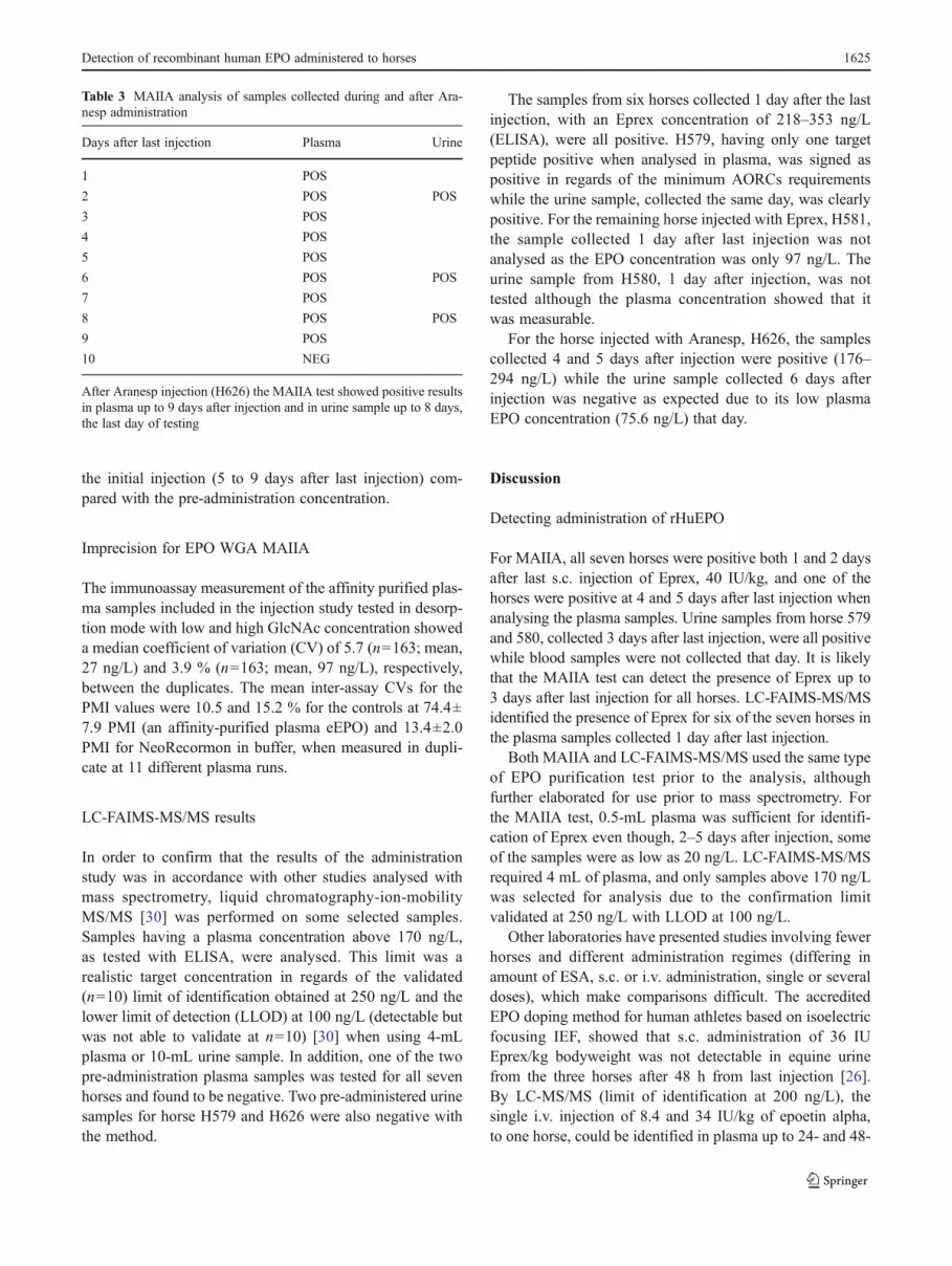
Table 3 MAIIA analysis of samples collected during and after Ara-nesp administration
Days after last injection Plasma Urine
1 POS
2 POS POS
3 POS
4 POS
5 POS
6 POS POS
7 POS
8 POS POS
9 POS
10 NEG
After Aranesp injection (H626) the MAIIA test showed positive resultsin plasma up to 9 days after injection and in urine sample up to 8 days,the last day of testing
Detection of recombinant human EPO administered to horses 1625
h post-administration, respectively [28]. The results for theMAIIA test, with detection up to 2–5 days after s.c. admin-istration of 40 IU/kg of Eprex, seem very competitive topreviously presented methods for doping control of horses,although enhanced exercise performance have been found1 week after injection [4]. Additional out-of-competitiontesting will hence be valuable for anti-doping purpose.
Detecting administration of Aranesp
After the single s.c. administration 0.39 μg Aranesp/kg, toone horse, positive plasma samples were found up to 9 daysafter last injection for MAIIA. In the urine samples, Aranespwas detectable up to 8 days after last injection, the last dayof testing, with MAIIA. The LC-FAIMS-MS/MS methodconfirmed the presence of Aranesp in plasma up to 5 daysafter last injection.
Other studies using the IEF method showed that a singles.c. administration of 0.37 μg/kg of Aranesp to one horsewas detectable in equine urine up to 5 days post-administration [26]. In a study using LC-MS/MS, an identifi-cation limit of 100 ng/L was obtained for Aranesp and an i.v.dose of 0.37 μg/kg was identified 168 h (7 days) post-administration for one horse [29]. Another study using LC-MS/MS detected the injection of 100 μg Aranesp to one horseup to 4 days [38] while the long-reacting pegylated epoetinbeta (Mircera) was found up to 120 h (5 days) after s.c.injection of 100 μg into three horses. The MAIIA methodseems to be very competitive also for Aranesp detection withdetection up to 9 days post-drug administration.
Suspected anti-HuEPO antibody production
The estimated concentration of eEPO with the MAIIA testwas significantly lower in samples collected about 1 weekafter ceasing administration (13.7 ng/L) compared with pre-administration samples (26.5 ng/L). This might depend onproduction of equine anti-HuEPO antibodies, which caninterfere by shielding the EPO epitope for the capturingantibody in the affinity purification system, or disturb theinteraction with one or two of the antibodies in the immu-noassay, resulting in lower amount of EPO captured ordetected. Formation of anti-HuEPO antibodies is also likelyto be the reason why the estimation of the Eprex concentra-tion in samples collected 1 day after injection was lowerafter the last three injections (522 ng/L) compared with afterthe first three injections (891 ng/L).
Equine anti-HuEPO is of analytical interest as the anti-bodies remain in the circulation far longer than rHuEPO. Itis also of methodological interest; if available, it will beadvantageous to make sure that the mouse monoclonal anti-HuEPO antibodies on the affinity matrix, or in the
immunoassay, do not cross-react with the same EPO epitopeas the dominating equine polyclonal anti-HuEPO antibodies.
Doping test
Doping control is traditionally performed by mass spectrom-etry which is the best technology for molecular character-isation of small drugs through specific and characteristicfragmentation pathways. However, for large molecules suchas proteins (insulin, growth hormone, EPO and IGF-1), forwhich the amino acid sequence of the recombinant andendogenous forms are identical, other or additional analyt-ical tools would be needed to reveal differences. The re-quirement of unique MS/MS mass spectral data for equineforensic drug testing [29] limits the detectability for proteinsused for doping, like rHuEPO, for which the MAIIA test canshow aberrant EPO forms in samples containing severaltimes lower EPO concentration. Presence of rhEPO wasfound in samples with an EPO concentration at 20 ng/L inthis study when using 0.5-mL plasma samples. Recent stud-ies have shown that the presence of rhEPO was detected inhuman samples at such low EPO concentration as 0.2 ng/Lwhen using 20–30 mL of urine [25]. Moreover, with theadvent of equine recombinant EPO, both the presently usedscreening method and the MS/MS methods will fail todistinguish recombinant eEPO forms from endogenouseEPO.
Besides high sensitivity and specificity for the dopingsubstance, the potential to set up high-throughput analysis(including sample pre-treatment) is of high importance. Thiswill reduce the analysis cost for the final customer andenable more samples to be analysed. More samples analysedwith a highly sensitive doping test will increase the risk foridentification of doping and thus hampering it. Even thoughLC-MS/MS is a rapid detection technique, the pre-analyticalhandling takes considerable resources using traditionallyaffinity purification methods, denaturation and trypsin di-gestion steps, followed by liquid chromatography separa-tion. The replacement of the conventional immunoaffinityprocess required for biological samples based on, e.g. beads[28,29], by the easy-to-use kit with the anti-EPO monolith[30], reduce both the hands-on-time and total time consid-erably. Besides, using the disposable anti-EPO monolithinstead of cleaning and re-using the anti-EPO matrix willomit the risk for carry-over between doping samples.
A doping test performed with the MAIIA test, includingrapid affinity purification using the disposable anti-EPOmonolith, takes about one hour totally to proceed whenusing 0.5 mL of equine plasma. Further development ofboth the affinity purification and the MAIIA test seems tobe possible to enable the use of suitable equipment for large-scale analysis.
1626 M. Lönnberg et al.
Conclusions
The MAIIA test is a promising tool exhibiting high sensitivityand short reporting time (1 h) for equine doping analysis ofESAs in urine or plasma samples. Introduction of such easy-to-use test will most likely increase the analysis frequency,which will be valuable for horse welfare.
Acknowledgements The authors thank Maria Andrén, Malin Drevin,Mikael Lönnberg and Trikien Quach for technical assistance, and theSwedish Foundation for Equine Research, Stockholm, Sweden, andMAIIA Diagnostics, Uppsala, Sweden, for support. The authors areindebted to Dr. Jean-Jacques Garin, veterinary surgeon at FNCF, to thehorse farm manager in Coye la Forêt and to the staff who participated indrug administration, sampling and horse care.
References
1. Jelkmann W (2011) Regulation of erythropoietin production. JPhysiol 589(Pt 6):1251–1258
2. Macdougall IC, Ashenden M (2009) Current and upcomingerythropoiesis-stimulating agents, iron products, and other novelanemia medications. Adv Chron Kidney Dis 16(2):117–130
3. Tablin F, Weiss L (1983) The equine spleen: an electron micro-scopic analysis. Am J Anat 166(4):393–416
4. McKeever KH, Agans JM, Geiser S, Lorimer PJ, Maylin GA(2006) Low dose exogenous erythropoietin elicits an ergogeniceffect in standardbred horses. Equine Vet J Suppl 36:233–238
5. Shaskey DJ, Green GA (2000) Sports haematology. Sports Med 29(1):27–38
6. Piercy RJ, Swardson CJ, Hinchcliff KW (1998) Erythroid hypo-plasia and anemia following administration of recombinant humanerythropoietin to two horses. J Am Vet Med Assoc 212(2):244–247
7. Halstenson CE, Macres M, Katz SA, Schnieders JR, Watanabe M,Sobota JT, Abraham PA (1991) Comparative pharmacokineticsand pharmacodynamics of epoetin alfa and epoetin beta. ClinPharmacol Ther 50(6):702–712
8. Franz SE (2009) Erythropoiesis-stimulating agents: development,detection and dangers. Drug Test Anal 1(6):245–249
9. Park SS, Park J, Ko J, Chen L, Meriage D, Crouse-Zeineddini J,Wong W, Kerwin BA (2009) Biochemical assessment of erythro-poietin products from Asia versus US epoetin alfa manufacturedby Amgen. J Pharm Sci 98(5):1688–1699
10. Jelkmann W (2007) Novel erythropoietic agents: a threat to sports-manship. Medicina Sportiva 11(2):32–42
11. Egrie JC, Browne JK (2001) Development and characterization ofnovel erythropoiesis stimulating protein (NESP). Br J Cancer 84(Suppl 1):3–10
12. Macdougall IC, Robson R, Opatrna S, Liogier X, Pannier A,Jordan P, Dougherty FC, Reigner B (2006) Pharmacokinetics andpharmacodynamics of intravenous and subcutaneous continuouserythropoietin receptor activator (C.E.R.A.) in patients with chron-ic kidney disease. Clin J Am Soc Nephrol 1(6):1211–1215
13. Reichel C (2011) Recent developments in doping testing for eryth-ropoietin. Anal Bioanal Chem 401(2):463–481
14. Dehnes Y, Lamon S, Lonnberg M (2010) Erythropoietin (EPO)immunoaffinity columns—a powerful tool for purifying EPO andits recombinant analogues. J Pharm Biomed Anal 53(4):1028–1032
15. Lonnberg M, Dehnes Y, Drevin M, Garle M, Lamon S,Leuenberger N, Quach T, Carlsson J (2010) Rapid affinity
purification of erythropoietin from biological samples using dis-posable monoliths. J Chromatogr A 1217(45):7031–7037
16. Wide L, Bengtsson C (1990) Molecular charge heterogeneity ofhuman serum erythropoietin. Br J Haematol 76(1):121–127
17. Wide L, Bengtsson C, Berglund B, Ekblom B (1995) Detection inblood and urine of recombinant erythropoietin administered tohealthy men. Med Sci Sports Exerc 27(11):1569–1576
18. Lasne F (2001) Double-blotting: a solution to the problem of non-specific binding of secondary antibodies in immunoblotting pro-cedures. J Immunol Methods 253(1–2):125–131
19. Lasne F, de Ceaurriz J (2000) Recombinant erythropoietin in urine.Nature 405(6787):635
20. Lasne F, Martin L, Crepin N, de Ceaurriz J (2002) Detection ofisoelectric profiles of erythropoietin in urine: differentiation ofnatural and administered recombinant hormones. Anal Biochem311(2):119–126
21. Kohler M, Ayotte C, Desharnais P, Flenker U, Lüdke S, Thevis M,Völker-Schänzer E, Schänzer W (2008) Discrimination of recom-binant and endogenous urinary erythropoietin by calculating rela-tive mobility values from SDS gels. Int J Sports Med 29(01):1–6
22. Reichel C, Kulovics R, Jordan V, Watzinger M, Geisendorfer T(2009) SDS-PAGE of recombinant and endogenous erythropoie-tins: benefits and limitations of the method for application indoping control. Drug Test Anal 1(1):43–50
23. Reichel C, Abzieher F, Geisendorfer T (2009) SARCOSYL-PAGE: a new method for the detection of MIRCERA- and EPO-doping in blood. Drug Test Anal 1(11–12):494–504
24. Franco Fraguas L, Carlsson J, Lönnberg M (2008) Lectin affinitychromatography as a tool to differentiate endogenous and recom-binant erythropoietins. J Chromatogr A 1212(1–2):82–88
25. Lonnberg M, Andren M, Birgegard G, Drevin M, Garle M,Carlsson J (2012) Rapid detection of erythropoiesis-stimulatingagents in urine and serum. Anal Biochem 420(2):101–114
26. Lasne F, Popot MA, Varlet-Marie E, Martin L, Martin JA,Bonnaire Y, Audran M, de Ceaurriz J (2005) Detection of recom-binant epoetin and darbepoetin alpha after subcutaneous adminis-tration in the horse. J Anal Toxicol 29(8):835–837
27. Sato F, Yamashita S, Kugo T, Hasegawa T, Mitsui I, Kijima-Suda I(2004) Nucleotide sequence of equine erythropoietin and charac-terization of region-specific antibodies. Am J Vet Res 65(1):15–19
28. Guan F, Uboh CE, Soma LR, Birks E, Chen J, Mitchell J, You Y,Rudy J, Xu F, Li X, Mbuy G (2007) LC-MS/MS method forconfirmation of recombinant human erythropoietin and darbepoe-tin alpha in equine plasma. Anal Chem 79(12):4627–4635
29. Guan F, Uboh CE, Soma LR, Birks E, Chen J, You Y, Rudy J, Li X(2008) Differentiation and identification of recombinant humanerythropoietin and darbepoetin Alfa in equine plasma by LC-MS/MS for doping control. Anal Chem 80(10):3811–3817
30. Bailly-Chouriberry L, Cormant F, Garcia P, Lönnberg M, SzwandtS, Bondesson U, Popot MA, Bonnaire Y (2012) New analyticalmethod based on anti-EPO monolith column for the rHuEPOpurification in horse plasma and urine samples. Analyst,DOI:10.1039/C2AN15662H
31. Yu NH, Ho EN, Wan TS, Wong AS (2010) Doping control anal-ysis of recombinant human erythropoietin, darbepoetin alfa andmethoxy polyethylene glycol-epoetin beta in equine plasma bynano-liquid chromatography-tandem mass spectrometry. AnalBioanal Chem 396(7):2513–2521
32. Scarth JP, Seibert C, Brown PR, Teale P, Beamon GJ, Pearce CM,Sams RA (2011) UPLC-MS/MS method for the identification ofrecombinant erythropoietin analogues in horse plasma and urine.Chromatographia 74(7–8):593–608
33. Lönnberg M (2002) Membrane-assisted isoform immunoassay:separation and determination of protein isoforms. Acta Universi-tatis Upsaliensis Comprehensive Summaries of Uppsala Disserta-tions, Faculty of Science and Technology, 691 pp
Detection of recombinant human EPO administered to horses 1627
34. Lonnberg M, Carlsson J (2001) Chromatographic performance of athin microporous bed of nitrocellulose. J Chromatogr B 763(1–2):107–120
35. Lönnberg M, Drevin M, Carlsson J (2008) Ultra-sensitive immu-nochromatographic assay for quantitative determination of eryth-ropoietin. J Immunol Methods 339(2):236–244
36. Lonnberg M, Carlsson J (2000) Membrane assisted isoform im-munoassay. A rapid method for the separation and determination
of protein isoforms in an integrated immunoassay. J ImmunolMethods 246(1–2):25–36
37. Lonnberg M, Carlsson J (2006) Lab-on-a-chip technology fordetermination of protein isoform profiles. J Chromatogr A 1127(1–2):175–182
38. Chang Y, Maylin GM, Matsumoto G, Neades SM, Catlin DH(2011) Screen and confirmation of PEG-epoetin beta in equineplasma. Drug Test Anal 3(1):68–73
1628 M. Lönnberg et al.