Detection of Gliotoxin in Patients with Pulmonary … of Babylon University/Pure and Applied...
12
Journal of Babylon University/Pure and Applied Sciences/ No.(2)/ Vol.(25): 2017 639 Detection of Gliotoxin in Patients with Pulmonary Aspergillosis Baheeja A.Hmood Al-Qadssiyah university -College of Nursing [email protected] Abstract Objective: This study was designed to detection of gliotoxin in patients with pulmonary Aspergillosis . Method: A total of 100 samples (sputum and blood) were selected from 100 outpatients who attended to Al-Qadissiyia Centre of Tuberculosis and Chest Diseases, , during the period from 2014 to May 2015. All patients had clinical manifestation in addition to 100 sample (sputum and blood) as control group. Results: All patients infected with fungi and three different genus isolated from sputum of patients ,these genus are Aspergillus sp. 60 Isolates( 60%)with three species were A.fumigatus (50%) A.flavus(33.3%) and A.niger (16.6% )Cryptococcus neoformans (20%) Penicillum sp.(14%) and Rhizopus sp.(6%). Seventy nine (79%) from patients had gliotoxin distributed in their sputum and serum by ratio (71%) and 5 (5%) had GT in their serum while 3 (3%) of them had GT in their sputum.. So this toxin was found in50(5%) in control group distributed in sputum and serum by (40%) , 6(6%)in sputum and 4(4%)in serum. The concentration of GT in sputum of patients was (40-63μg/kg) and in sputum of control group was (16-23μg/kg) and in serum of patients was (33-47μg/kg) and (10-21μg/kg) in serum of control group. and all A.fumigatus isolates 30 (100 %)were have gliz gene. Conclusion: Several species of Aspergillus and other fungi as well as possibly yeast, produced gliotoxin both in vitro and in vivo. More attention should be paid to this mycotoxin because of its multi-faceted toxic properties. Keyword: Aspergillosis ,Gliotoxin, Aspergillus fumigatus. صة الخر الرئويلفطابين بداء المصا المرضى ايوتوكسين لدىجلتحري عن سم الل اسة ل صممت ىذه الدر. اسة جمعضمنت الدر ت100 ن الى مركز الصدرية اجعية من المرضى المر عينلفترة من اذارلقادسية لفظة ا في محا والتدرن الرئوي2014 ية اذار ولغا2015 فة الى جمعضاض سريرية با اون من اعر يعاننوالمرضى كا ان جميع ا علما100 سيطرة . عينة كمجموعةت من القشعلفطرياس من اجناث ا وتم عزل ثون بالفطرياتلمرضى مصاب اسة ان جميع ائج الدر نتا اظيرت الفطريا ساد فيAspergillus sp. نسبة ب60 ث انواع ىي وظم ث% Aspergillus fumigatus نسبة ب50 و% A.flavus ( نسبة ب33.3% ) وA.niger ( نسبة ب16.6% وخميرة) Cryptococcus neoformans نسبة ب20 والفطر% Penicillium sp نسبة ب14 ن ثم الفطروم% Rhizopus نسبة ب6 .% نسبةن في القشع والدم بجليوتوكسيون لسم ال حاملنوالمصابون كائة من المرضى ا وسبعون بالما تسع71 اذ ان% 5 نوانيم كا م% ذا السم في الدم ون لي حاملي3 ( 3 ذلك وجد ىذا السم فيسم في القشع كلين ل حاملنوا كا)% 5 نسب السيطرة توزع ب من مجموعة% 40 في% القشع والدم و6 في القش% ع و4 في الدم .% ( بينلمصابينن في قشع المرضى اجليوتوكسي اوح تركيز سم ال تر40 - 63 ( ومنن وزن الجسم م ام /كغممايكروغر) 16 - 32 ( حين بلغ تركيزه في السيطرة مجموعة ام /كغم في قشعمايكروغر) 33 - 47 ( مجموعة المرضى ومن ام /كغم في دممايكروغر)% 10 - 21 م في دم ام /كغممايكروغر) ت الفطر عزلسيطرة .وكانت جميع جموعة اA.fumigatus ( 100 ة على جين حاوي)% gliz . ض انواع الفطر ىناك بعAspergillus نتباهن ويجب اجليوتوكسيج سم النتابيلة على القا الخمائر ليافة الى اضاخرى بات ا وفطرياى الجسم .ددة عل متع ات لما لو من تاثير ىذا السم الى الكملمفتاحيةت ا ما: يوتوكسين ،الفطرجل سم الAspergillus fumigatus ر الرئويلفطا وا.
Transcript of Detection of Gliotoxin in Patients with Pulmonary … of Babylon University/Pure and Applied...

Journal of Babylon University/Pure and Applied Sciences/ No.(2)/ Vol.(25): 2017
639
Detection of Gliotoxin in Patients with Pulmonary Aspergillosis
Baheeja A.Hmood
Al-Qadssiyah university -College of Nursing
Abstract Objective: This study was designed to detection of gliotoxin in patients with pulmonary Aspergillosis .
Method: A total of 100 samples (sputum and blood) were selected from 100 outpatients who attended to
Al-Qadissiyia Centre of Tuberculosis and Chest Diseases, , during the period from 2014 to May 2015. All
patients had clinical manifestation in addition to 100 sample (sputum and blood) as control group. Results: All patients infected with fungi and three different genus isolated from sputum of patients ,these
genus are Aspergillus sp. 60 Isolates( 60%)with three species were A.fumigatus (50%) A.flavus(33.3%) and
A.niger (16.6% )Cryptococcus neoformans (20%) Penicillum sp.(14%) and Rhizopus sp.(6%). Seventy
nine (79%) from patients had gliotoxin distributed in their sputum and serum by ratio (71%) and 5 (5%)
had GT in their serum while 3 (3%) of them had GT in their sputum.. So this toxin was found in50(5%) in
control group distributed in sputum and serum by (40%) , 6(6%)in sputum and 4(4%)in serum.
The concentration of GT in sputum of patients was (40-63µg/kg) and in sputum of control group was
(16-23µg/kg) and in serum of patients was (33-47µg/kg) and (10-21µg/kg) in serum of control group. and
all A.fumigatus isolates 30 (100 %)were have gliz gene.
Conclusion: Several species of Aspergillus and other fungi as well as possibly yeast, produced gliotoxin
both in vitro and in vivo. More attention should be paid to this mycotoxin because of its multi-faceted
toxic properties.
Keyword: Aspergillosis ,Gliotoxin, Aspergillus fumigatus.
الخالصة . صممت ىذه الدراسة للتحري عن سم الجليوتوكسين لدى المرضى المصابين بداء الفطار الرئوي
والتدرن الرئوي في محافظة القادسية للفترة من اذار عينة من المرضى المراجعين الى مركز الصدرية 100تضمنت الدراسة جمع عينة كمجموعة سيطرة . 100علما ان جميع المرضى كانوا يعانون من اعراض سريرية باالضافة الى جمع 2015ولغاية اذار 2014
ساد فييا الفطر اظيرت نتائج الدراسة ان جميع المرضى مصابون بالفطريات وتم عزل ثالث اجناس من الفطريات من القشع Aspergillus sp. وظم ثالث انواع ىي 60بنسبة %Aspergillus fumigatus و50بنسبة %A.flavus ( 33.3بنسبة% )
%ومن ثم الفطر 14بنسبة Penicillium sp%والفطر 20بنسبة Cryptococcus neoformans( وخميرة%16.6بنسبة ) A.nigerوRhizopus 6بنسبة .%
% منيم كانوا 5% اذ ان 71تسع وسبعون بالمائة من المرضى المصابون كانوا حاملون لسم الجليوتوكسين في القشع والدم بنسبة % في 40% من مجموعة السيطرة توزع بنسب 5%( كانوا حاملين للسم في القشع كذلك وجد ىذا السم في 3) 3حاملين ليذا السم في الدم و
% في الدم . 4ع و% في القش6القشع والدم و-16(مايكروغرام /كغم من وزن الجسم ومن) 63-40تراوح تركيز سم الجليوتوكسين في قشع المرضى المصابين بين )
-10%(مايكروغرام /كغم في دم مجموعة المرضى ومن )47-33(مايكروغرام /كغم في قشع مجموعة السيطرة في حين بلغ تركيزه )32 . gliz%( حاوية على جين 100) A.fumigatusجموعة السيطرة .وكانت جميع عزالت الفطر (مايكروغرام /كغم في دم م21وفطريات اخرى باالضافة الى الخمائر ليا القابيلة على انتاج سم الجليوتوكسين ويجب االنتباه Aspergillusىناك بعض انواع الفطر
الى ىذا السم لما لو من تاثيرات متعددة على الجسم . .والفطار الرئوي Aspergillus fumigatusسم الجليوتوكسين ،الفطر :مات المفتاحيةالكم
Journal of Babylon University/Pure and Applied Sciences/ No.(2)/ Vol.(25): 2017
640
Introduction Pulmomary Aspergillosis (PA) is the most common mycotic infection of the
respiratory tract caused by Aspergillus, a common mold (fungus) that lives indoors and
outdoors. Most people breathe in Aspergillus spores every day without getting sick.
However, people with weakened immune systems or lung diseases are at a higher risk of
developing health problems due to Aspergillus. (Smith and Denning, 2011) The two
major agents causing aspergillosis are A.fumagatus and A. flavus as show in more studies
(Kradin and Mark, 2008) .
Asergillus fumigatus is an ubiquitous saprophytic fungus which plays an important
role in recycling environmental carbon and nitrogen ,but also it may be an opportunistic
pathogen ,Human constantly inhale high amounts of conidia from this fungus ,which may
affected their respiratory tract after long exposure (Latge , 1999). A. fumigatus consider
the leading cause of mold infections worldwide, is an opportunistic pathogen that causes
severe problems in immune-compromised populations,These populations include: AIDS
patients, cancer patients receiving chemotherapy, solid organ transplant/skin graft
patients and victims of chronic granulomatous disease (Brand, 2012). A.fumigatus is also
capable of producing secondary metabolites ,which can be harmful, One of the most
studied secondary metabolites produced by A. fumigatus is gliotoxin, which is also
produced by several other Aspergillus species, Trichoderma species, and Penicillium
species (Kwon-Chang and Sugui, 2009). Gliotoxin is a member of the
epidithiodioxopiperazine (ETP) class of toxins, which are characterized by a disulfide
bridge across a piperazine ring with low molecular weight (326 Da) (Scharf et al., 2012,
Cramer et al., 2006). The oxidized form of gliotoxin travels into host immu ne cells
where it is able to affect cellular functions essential to the immune response. These
include impediment of phagocytosis and NF-κB activation, as well as induction of
apoptosis (Waring et al., 1994; Yoshida et al., 2000). As with other secondary
metabolites, most of the genes responsible for the production and transport of gliotoxin
exist within a gene cluster. The gliotoxin biosynthesis cluster was first identified based on
its homology to the sirodesmin PL biosynthesis gene cluster in the ascomycete
Leptosphaeria maculans (Fox and Howlett, 2008) Within this cluster lies a Zn2Cys6
binuclear finger transcription factor, GliZ, thought to be responsible for general gliotoxin
induction and regulation. Indeed, over-expression of gliZ leads to an increase in gliotoxin
production and deletion of gliZ results in a loss in gliotoxin production. (Bok et al.,2006).
Biosynthetic ETPs are derived fromat least one aromatic amino acid. GTX is derived
from phenylalanine and serine as precursor amino acids .Secondary metabolites of fungi
that have more than one amino acid are generallyproduced by non-ribosomal peptide
synthetases. The complete genome sequence of A. fumigatus showed that the non-
ribosomal peptide synthetase enzymes that synthesise GTX usually have genes clustered
in the genome (Gardiner et al., 2004). Several genes (gli genes) have been identified
related to the biosynthesis of GTX (Balibar and Walsh, 2006) including gliZ,
atranscriptional regulator of GTX production that encodes the Zn(II) 2Cys6 binuclear
transcription factor. Substitution of the gliZ gene with a marker gene caused no
detectable GTX biosynthesis and failure to express the other gli cluster genes
(Schrettle et al., 2010). Additionally mutation in the gliP gene resulted in failure to make
GTX (Bok et al., 2006). These genes are activated when secondary metabolism
Journal of Babylon University/Pure and Applied Sciences/ No.(2)/ Vol.(25): 2017
641
commences in fungi (Rementeria et al., 2005). Lea A is a methyltransferase that possibly
is involved in regulation of these genes (Stack et al., 2007).
However ,studies focusing on the relation between mycotoxins and its pathogenesis
have been limited and significance of mycotoxins in the virulence of Aspergillus
fumigatus has not yet been demonstrated in Iraq .
So, until recently the relationship between mycotoxins and the pathogenicity of the fungi
that produce them has received little attention ,therefore this study was designed to
detection of gliotoxin in patients with pulmonary Aspergillosis .
Material s and methods A total of 100 samples (sputum and blood) were selected from 100 outpatients who
attended to Al-Qadissiyia Centre of Tuberculosis and Chest Diseases, during the period from
2014 to May 2015. All patients had clinical manifestation, signs such as fever, weight loss,
cough, anorexia, and some of them with bloody sputum., and clinical examination by a
specialist clinician. Those patients already diagnosed as not tuberculosis patients and did
not respond to treatment.
Diagnosis was established by clinical picture, chest X-ray examination . Samples of
blood and sputum were taken from all patients. So The healthy person 100 were also studied
as control as clinical examination showed by clinician specialist, sputum and blood samples
were taken from each subject.
Sputum sampling (Ellis, 2004).
Patients were advised to wash their mouths with antiseptic mouth wash and then three
times with water. To obtain a sputum sample, the patient was given a labeled sputum
container and was asked to :
- Take a deep breath.
- Open the container ,bring it close to the mouth and bring the sputum out into it.
- Not to put saliva or nasal excretion into the container .
- Not to have sputum in the mouth but immediately spit into the container.
- Close the container.
Processing of sputum samples
The sputum samples were decontaminated and digested by treatment with an equal
volume of sputolysin/sodium hydroxide (4%) for 30 minutes at room temperature with
rocking. After neutralization with 10 ml of PBS (pH 7.4), the mixture was centrifuged at
3000 rpm for 30 minutes. the supernatant put in sterile tube and added to it equal amount
chloroform ,after shaking the content of tube will be separated into two portion ,one portion
chloroform layer which contain toxin and another potion was discharge the sediment was
obtained, and was inoculated in Sabouraud Dextrose Agar and incubated at 25C˚ for one
weeks for mycotic examination, The isolated fungus was suspended in Lacto phenol and
examined microscopically by slide (Kubica et al.,1993).
Blood samples
Four ml of blood were collected by vein puncture into two sterile test tubes, in one of them
3ml of blood were putted and left for about 2-4 hours, then the upper layer (serum) was
collected in clean test tube and add to it equal amount chloroform after shaking the content of
tube will be separated into two portion one portion chloroform layer which contain toxin and
another portion was discharge ,the tube which contain chloroform layer stored at -20˚C until
use.
Journal of Babylon University/Pure and Applied Sciences/ No.(2)/ Vol.(25): 2017
642
Determination of Gliotoxin in serum and sputum by Thin Layer Chromatography (TLC)
By capillary tube 10 µl from chloroform layer which separated from each sputum
and serum of patients in above step was took and spotted on the TLC plate(Silica gel G60.Sigma Aldrich Z) adjacent to 1 µl of standard GT which dissolved in 200 mixture of
dichloromethane :methanol (97:3 v/v) were also spotted on the same plate , and the
gliotoxin were separated by TLC method using chloroform :methanol (70:30 v/v) as the
developing solvent, TLC plate was dried after the end of development and front was
marked . Location of GT spot on TLC plates was accomplished by illumination under
shortwave UV light (254 nm) and by comparing the sample spots to the standard
GT(same shape, retention factors (Rf and color) (Vander Merwe et al.,1965).
Standard curve of Gliotoxin
Standard curve was prepared by measuring the absorbance of the following prepared
pure standard GT concentration (10, 20, 40,80,160 and 320 µg/ml) .Each of standard
concentration was plotted against its absorbance values and a linear standard curve was
achieved from which the GT quantity of any sample was determined according to its
absorbance in comparison with that of standard curve values.
Detection of gliz gene in A.fumigatus isolates
1- Isolation of Fungal DNA
Template DNA was extracted from fungal mycelia according to( Lee etal.,1998) as
follows: fungal mycelia grown in Potato Dextrose Broth (PDB) under stationery
conditions for 21 days was harvested by filtration. The mycelium was washed twice with
phosphate buffered saline (137 mM NaCl, 2.7 mM KCl, 10 mM Na2HPO4, 2 mM
KH2PO4, pH 7.4) followed by centrifugation. The mycelium was transferred to a mortar
and ground well. Freshly prepared, sterile Lysis buffer (50 mM Tris, 150 mM EDTA, 1%
(w/v) SDS, pH 8.0) was added to the pulverized mycelia and incubated at 65°C for 1 h .
The suspension was centrifuged and supernatant was then extracted twice with phenol:
chloroform: isoamylalcohol (25:24:1) and the aqueous layer was washed twice with
chloroform and then precipitated with two volumes of isopropanol. The precipitate was
re suspended in 200 μl of TE buffer (10 mM Tris-Cl, 1.0 mM EDTA, pH 8.0.
2-Polymerase chain reaction (PCR)
The primer GZ5( R 5´GGAGAGAATTCATTTAACCTTCTATCGCAG3´,GZR5
F5´AGTGACCGACCGTCCAAGAACCGTAG3´ ) were synthesized by American
Geneoids Company. The PCR conditions were optimized by varying the concentration of
above primer, the number of units of Taq polymerase and annealing temperature of the
reaction. The PCR reaction mixture (25 μL) contained 100 ng of genomic DNA,
deoxyribonucleoside triphosphates at 0.025 n mol each, primer at 4 n mol each and
reaction buffer. Each reaction mixture was heated to 95°C for 10 min before adding 0.3
units of Taq DNA polymerase. Amplification conditions used consisted of 4 min at 94°C
followed by 35 cycles at 94°C for 30 sec, 50°C for 45 sec, 72°C for 75 sec. The reaction
was completed with incubation for 10 min at 72°C. PCR products were analyzed by
electrophoresis in a 1% a garose gel in TAE buffer. Ethidium bromide (0.5 μg μL-1
)
stained gels were visualized under UV light with Digital camera (Sambrook et al.,1989).

Journal of Babylon University/Pure and Applied Sciences/ No.(2)/ Vol.(25): 2017
643
Results and Discussion All cases studied where didn't respond to anti tuberculosis treatment and didn't
isolated any bacterial agents from pulmonary infection ,therefore this study attention in
isolated the fungal etiological agents of pulmonary infection thus, the results of study
showed that all patients infected with fungi and three different genus isolated from
sputum of patients ,these genus are Aspergillus sp. 60 Isolates( 60%)with three species
were A.fumigatus (50%), A.flavus (33.3%) and A.niger (16.6%) Cryptococcus
neoformans (20%) Penicillum sp.(14%) and Rhizopus sp.(6%). While didn’t isolate any
fungus from blood of patients and blood and sputum of control group. Table (1).
The predominant fungal species in this study was A. fumigatus (50 %). This results
agree with (Al-Taee, 2009; Ellis, 1994) whom found A.fumigatus was the most common
cause pulmonary mycotic infection .This fungus responsible for causes especially lung
diseases . the size of the spores (around 3µm in diameter) which are present ubiquitously
in the air considers the major factor in determining the pathogenicity of this fungus.
(Waring et al., 1994).
Mullins et al., (1976) have shown that the inhalation of A.fumigatus spores which
present in present in the air lead to precipitate in lung as he shows that in lung in
necropsy. So ,this fungus can produce toxins such as gliotoxin which is an important
factor in helping the fungus mycelium to grow in tissue such as lung . Aspergillus fumigatus is a saprotroph widespread in nature commonly found in
soil and decaying organic compounds. this fungus cause invasive in the lung of
immunocompromised patients and responsible for morbidity and mortality in above
individuals (Kupfahl et al., 2006). So A. fumigatus can cause allergic bronchopulmonary
aspergilosis and allergic reactions in immunocompetent hosts. (Latge, 1999).
Aspergillus fumigatus produce mycotoxins ,(secondary metaboites) one importants of
these mycotoxins is gliotoxin which is affect on host deffenses through suppresser of
immune system. (Kwon-Chung and Sugui ,2009). Gliotoxin production by A. fumigatus
isolates can vary from isolat to another (Denning , 1998; Lewis et al., 2005).
Table (1) Number of mycotic isolates and their percentage throughout the study
Specimens Type of
specimens
Agents of mycotic infection No. of
isolates
%
Patients group (100)
Sputum
Aspergillus
A.fumigatus
A.flavus
A.niger
60/100
30/60
20/60
10/60
60
50
33.33
16.66
Cryptococcus neoformans 20/100 20
Penicillium sp. 14/100 14
Rhizopus sp. 6/100 6
Control group(100)
Blood No growth
Sputum No growth
Blood No growth
Total 100
Journal of Babylon University/Pure and Applied Sciences/ No.(2)/ Vol.(25): 2017
644
Detection of gliotoxin in serum and sputum of study groups
In the solvent system chloroform and methanol (70:30V/V), gliotoxin had an Rf of
0.97 and visualized as orange /brown spots under UV light.
Seventy nine (79%) from patients had gliotoxin distributed in their sputum and
serum by ratio (71%) and 5 (5%) had GT in their serum while 3 (3%) of them had GT in
their sputum.. So this toxin was found in50(5%) in control group distributed in sputum
and serum by (40%) , 6(6%)in sputum and 4(4%)in serum. table (2).
There are no similar study for detection of gliotoxin in human cases so that we can
compare our results with it.
Table (2) Distribution of GT in serum and sputum of patients and control group
Study groups Serum Sputum Sputum +serum Total
Patients 5(5%) 3(3%) 71(71%) 79(79%)
Control 4(4%) 6(6%) 40(40%) 50(50%)
Gliotoxin effects on the function of leukocytes by inhibiting migration and
production of superoxide and causes apoptosis in macrophages. So Gliotoxin inhibition
of NF-κB thus disrupts the proinflammatory response and play an important role in the
establishment and development of an infection with A. fumigatus . We research about
studies which designed to the detection of GT in vivo ,and found only few cases of
gliotoxin being detected in infected tissues have been reported such as Bauer et al.
(1989) who detected of gliotoxin in Cows, Richard et al.,(1998) designed in animal
model by using Turkeys and detected detectable level of gliotoxin in the poults of
animals infected with A. fumagatus. Reves et al., (2004) found gliotoxin in the bodies of
larvae of Galleria mellonella, which expermintally infected , so (Lwies et al.,2005)
showed a significant amount of gliotoxin in the sera of mice infected with A. fumagatus.
Sutton et al.,(1994) referred to treatment of Rats with a one injection of a sub lethal
dose of gliotoxin was suitable to make above animals critical to infection and ensuing
death, after challenge with A. fumigatus spores .So showed if infected animals with the
strain that non-gliotoxin producing ,these animal survived safely longer than the animals
infected with strains that produced gliotoxin..
After injection of animals with sub lethal dose of GT and Study of the morphology of
cells of the thymus, spleen, and mesenteric lymph nodes by electron microscopy and
agarose gel electrophoresis of DNA from these organs showed that the gliotoxin induced
apoptosis in macrophage in vivo. Thus, gliotoxin has immunosuppressive effectiveness
in vivo and may be play a critical role in the pathogenicity of A.fumagatus.
(Sutton et al., 1996)
Production of gliotoxin was found to be based on the oxygen concentration of the
environmental , the lung considers as first target for infection by A.fumigatus ,the most
well-aerated organ ,in this meaning ,found high level of O2 in the lung supply an best
environment to production of gliotoxin by A.fumigatus .SO it is able dramatically to alter
function of lung cell such as attachment of epithelial cells and fibroblasts as well as
inhibitory phagocytosis by macrophages ;so others critical functions of the individuals
immune defense are also impaired by gliotoxin ,including induction of cytotoxic and all
reactive T .cell (Reijula et al., 1992). Eichner et al., (1986) referred to a small

Journal of Babylon University/Pure and Applied Sciences/ No.(2)/ Vol.(25): 2017
645
molecular weight (`10 KD) that released from isolate of A.fumigatus within minutes of
deposition in the right place of lung ,which is capable of inhibiting the oxidative burst of
macrophage.
Gliotoxin stimulate apoptosis in cellular components of immune system which
are monocytes and dendrite cells, leading to the inhibition of cellular immune
responses.( Suen etal.,2001) studied of the capability of gliotoxin to cause apoptosis in
polymorph nuclear leukocytes (PMN) and refers to gliotoxin was effect on neutrophil
functions, which include phagocytic function, degranulation, myeloperoxidase activity,
and the production of reactive oxygen species (ROS). So GT contains an
epipolythiodioxopiperazine (ETP) ring that is consider to be take part in redox reactions.
The sensitive oxygen radicals yield interaction with DNA to form hydroxylated and
other foreign DNA components .( Golden et al.,1998).
Different histological changes found in tissues of lung included necrosis in lung
alveoli with inflammatory cells infiltration and abuses formation in addition to thickness
in lung alveolar sac wall and bronchioles with hemorrhage. ( Korbel et al.,1993).
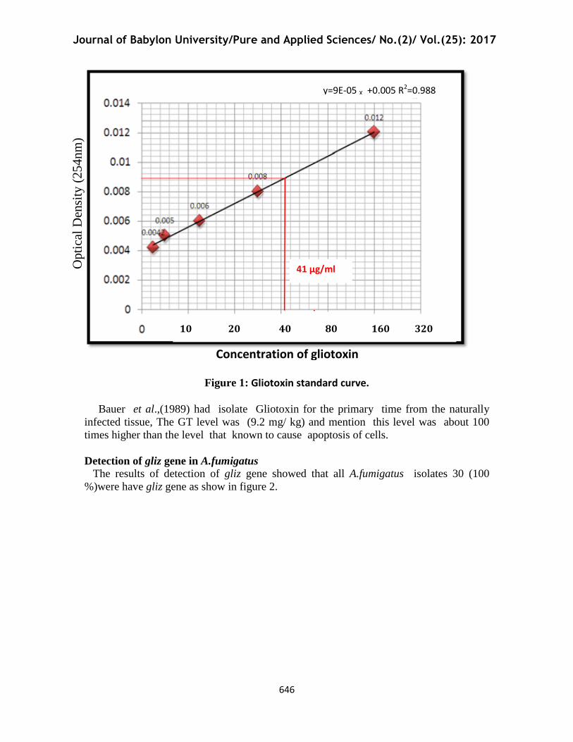
Quantification of Gliotoxin
Spectrophotometric method was used for quantification of GT in Serum and sputum
of study groups after its extraction, isolation and identification by TLC method.
The following standard curve was plotted for different standard GT prepared
concentrations that used for quantification of GT in different samples.
The concentration of GT in sputum of patients was (41-63µg/kg) and in sputum
of control group was (16-23µg/kg) and in serum of patients was (33-47µg/kg) and (10-
21µg/kg) in serum of control group .(table 3,figure1)
Table (3) Concentration of GT in serum and sputum of patients and control group
Study groups
Concentration of GT(µg/kg) in
Serum Sputum
Patients 33-47 41-63
Control 10-21 16-23
Journal of Babylon University/Pure and Applied Sciences/ No.(2)/ Vol.(25): 2017
646
Figure 1: Gliotoxin standard curve.
Bauer et al.,(1989) had isolate Gliotoxin for the primary time from the naturally
infected tissue, The GT level was (9.2 mg/ kg) and mention this level was about 100
times higher than the level that known to cause apoptosis of cells.
Detection of gliz gene in A.fumigatus
The results of detection of gliz gene showed that all A.fumigatus isolates 30 (100
%)were have gliz gene as show in figure 2.
10 20 40 80 160 320
Opti
cal
Den
sity
(25
4n
m)
y=9E-05 X +0.005 R2=0.988
41 µg/ml
Concentration of gliotoxin

Journal of Babylon University/Pure and Applied Sciences/ No.(2)/ Vol.(25): 2017
647
Figure (2) Electrophoresis of the amplified product of gliz in a 1 % agarose gel. Lane M,
1500bp DNA ladder; lanes 1 to 15, A.fumigatus isolates that contain gliz gene in size
(600 bp) .
gliZ, a transcription factor which regulates gliotoxin biosynthesis, encoding a Zn(11)2
cys6 protein and located within the gliotoxin gene cluster(gli cluster) and is likely to be
contributory in gliotoxin production (Gardiner and Howlett,2005) .Deletion of gliZ result in
inability of A.fumigatus to produce GT and loss of gene expression of other gli cluster genes
(Bok etal.,2006).
gliZ gene involved in metabolic pathways related to virulence of A .fumigatus appeared to
be Up-regulated at 37C ْ when above fungus had high ability to grow in this temperature,
this finding is critical factors to confirm the found of GT in human body who infected with A
.fumigatus where human body temperature 37C˚. (Rementeria etal.,2005 ).
Sugui et al.,(2008) referred to gliZ is a positive regulator of gliP gene (which is a
nonribosomal peptide synthase act on catalyses the first step of gliotoxin biosynthesis) and
the over regulation of gliZ inhance the ability of fungus to produce GT ,So deletion of gliZ
lead to loss the ability of fungus to produce of above toxin.
The study of Scharf et al., (2012) which amid to Biosynthesis and function of gliotoxin in
Aspergillus fumigatus and detection the role of GT in pathogencity of this fungus reached
to the culture supernatant of Wild-type of A.fumagatus enhanced apoptosis process in
neutrophils of treated animals ,while supernatant of gliZ deletion mutants didn't induce
apoptosis in neutrophils of another group of animals ,this result confirms an important role
of GT in increase pathogenicity of A. fumigatus . However, loss of gliotoxin resulted in
decreased toxicity as measured either by mast cell degranulation (Cramer etal.,2006) or
macrophage/T-cell viability (Lewis etal.,2005), thus leading to speculation that this
metabolite can play a role in disease development. Here, cytotoxicity assays with
polymorphonuclear leukocytes (PMNs) support a role for gliotoxin in apoptotic but not
gliz(600bp)
Journal of Babylon University/Pure and Applied Sciences/ No.(2)/ Vol.(25): 2017
648
necrotic cell death. Taken together, they posit that gliotoxin is one factor that can be
involved in disease development and that its effects may not be readily measured by the
current animal model systems.
TCGGN3CCGA is the region which considers as A DNA binding site and has been
proposed for GliZ , but has not been experimentally certain. This site is found within the
promoter region of every gene within the gliotoxin cluster, Gliotoxin itself positively
regulates expression of the genes within the gliotoxin cluster, (Gradiner and Howlett,2005).
In conclusion, More attention should be paid to Aspergillus fumigatus which had
ability to produced gliotoxin that have multi toxic properties.
References Al-Taee, Orass Madhi Shaheed(2009) Using The PCR in Comparison With Other Tests
in The Diagnosis of Pulmonary TB Associated with Mycotic Infections. A thesis
of master,AL-Qadissiyiah university ,Medicine college .
Balibar ,C.J and Walsh ,C.T (2006) Gliz, a multimodular nonribosomal peptide
synthetase in Aspergillus fumigatus, makes the diketopiperazine scaffold of
gliotoxin. Biochemistry. 19;45(50):15029-38.
Bauer, J.; Gareis, M.;Bott, A.; Gedek, B.(1989) .Isolation of a mycotoxin (gliotoxin)
from a bovine udder infected with Aspergillus fumigatus. J. Med .Vet
.Mycol.;27(1):45-50.
Bok, J.W.;Chung, D.; Balajee, S.A.; Marr, K.A.; Andes, D. et al. (2006) gliz, a
transcriptional regulator of gliotoxin biosynthesis, contributes to Aspergillus
fumigatus virulence. Infect Immun 74: 6761–6768.
Brand, A. (2012) Hyphal growth in human fungal pathogens and its role in virulence. Int
J Microbiol 2012: 517529.
Cramer, R.A.; Gamcsik, M.P.; Brooking, R.M.; Najvar, L.K., Kirkpatrick, W.R., et al.
(2006) Disruption of a nonribosomal peptide synthetase in Aspergillus fumigatus
eliminates gliotoxin production. Eukaryot Cell 5: 972–980.
Denning, D. W. (1998). Invasive aspergillosis. Clin Infect Dis 26, 781–805
Eichner, R.D.; Al Salami, M.;Wood ,P.R.; Müllbacher ,A. (1986)The effect of gliotoxin
upon macrophage function. Int. J. Immunopharmacol.;8(7):789–797.
Ellis, D. H.(1994).Clinical Mycology .The Humman’s Opportunistic Mycoses.
Gillingham Printers Ltd., Australia . Pp. 166
Fox, E.M.; Howlett, B.J. (2008) Biosynthetic gene clusters for
epipolythiodioxopiperazines in filamentous fungi. Mycol Res 112: 162–169.
Gardiner, D. M. and B. J. Howlett. (2005). Bioinformatic and expression analysis of the
putative gliotoxin biosynthetic gene cluster of Aspergillus fumigatus. FEMS
Microbiol. Lett. 248:241-248.
Gardiner, D.M.; Cozijnsen ,A.J; Wilson, L.M.; Pedras, M.S.; Howlett, B.J. (2004) The
sirodesmin biosynthetic gene cluster of the plant pathogenic fungus Leptosphaeria
maculans. Mol Microbiol 53: 1307–1318.
Golden,M.C. ;Hahm, S.J; Elessar,R.E; Saksonov. J .;J Steinberg,.(1998) DNA damage by
gliotoxin from Aspergillus fumigatus. An occupational and environmental
propagule: adduct detection as measured by 32P DNA radiolabelling and two-
dimensional thin-layer chromatography. PMID MycosesVolume 41, Issue 3-4,
pages 97–104.
Journal of Babylon University/Pure and Applied Sciences/ No.(2)/ Vol.(25): 2017
649
Korbel ,R.;Bauer ,J.; Gedek, B.; Tierarztl Prax. (1993) Pathologico-anatomic and
mycotoxicologic studies of aspergillosis in birds. Article in German ;21(2):134-
9.
Kradin, R.L and Mark ,E.J (2008). The pathology of pulmonary disorders due to
Aspergillus spp". Arch. Pathol. Lab. Med. 132 (4): 606–14.
Kubica, G.P.; Dye, E.; Cohn, M.L.; and Middlebrook, G.(1993).Sputum diagnosis and
decontamination with N-acetyl-L-cysteine- sodium hydroxide for culture of
mycobacteria . Amer.Rev. Resp. Dis., 87: 775-779.
Kupfahl, C.; T. Heinekamp, G. ;Geginat, T.; Ruppert, A.; Härtl, H.; Hof, and A. A.
Brakhage. (2006). Deletion of the gliz gene of Aspergillus fumigatus results in
loss of gliotoxin production but has no effect on virulence of the fungus in a low-
dose mouse infection model. Mol. Microbiol. 62:292-302.
Kwon-Chung and Sugui (2009). What do we know about the role of gliotoxin in the
pathobiology of Aspergillus fumigatus? Med Mycol 47 Suppl 1: S97–103.
Latge, J.P. (1999) Aspergillus fumigatus and aspergillosis. Clin Microbiol Rev. 12: 310–3
Lee, E.C.; Yu, S.Y.; Hu, X.; Mlodzik, M.; Baker, N.E. (1998). Functional analysis of the
fibrinogen-related scabrous gene from Drosophila melanogaster identifies
potential effector and stimulatory protein domains. Genetics 150(2): 663--673.
Lewis, R.E.; Wiederhold, N.P.; Chi, J.; Han, X.Y.; Komanduri, K.V.; Kontoyiannis, D.P.;
and Prince, R.A. (2005). Detection of gliotoxin in experi-mental and human
aspergillosis. Infect. Immun. 73, 635–637.
Mullins, J.; Harvey ,R.; Seaton, A.(1976) Sources and incidence of airborne Aspergillus
fumigatus (Fres). Clin. Allergy.;6(3):209–217.
Nieminen, S. M. J. ;Maki-Paakkanen, M. R. ;Hirvonen, M.; Roponen, and A. von
Wright.( 2002). Genotoxicity of gliotoxin, a secondary metabolite of Aspergillus
fumigatus, in a battery of short-term test systems. Mutat. Res. 520:161-170.]
Reeves, E.P.; Messina, C.G; Doyle, S.; Kavanagh, K.( 2004). Correlation between
gliotoxin production and virulence of Aspergillus fumigatus in Galleria
mellonella. Mycopathologia 158:73–79
Reijula, K. E.; Kurup, V. P.; Kumar, A.; Fink, J. N. (1992) Monoclonal antibodies bind
identically to both spores and hyphae of Aspergillus fumigatus. Clin. Exp. Allergy
22:547–553
Rementeria, A.; N. Lopez-Molina, A.; Ludwig, A.B.; Vivanco, J.; Bikandi, J. ;Ponton and
J. Garaizar, (2005). Genes and molecules involved in Aspergillus fumigatus
virulence. Rev. Iberoam Micology, 22: 1-23.
Richard, J.L.; Peden, W.M.; Williams, P.P. (1998). Gliotoxin inhibits transformation and
its cytotoxic to turkey peripheral blood lymphocytes. Mycopathologia. 126, 109–
114
Sambrook,J.; Fritsh, E.F.; and Maniatis,T. (1989). Molecular Cloning, A laboratory
manual, 2nd
ed. Cold Spring Harbor Laboratory.
Scharf ,D.H.; Heinekamp, T. Remme, N.Hortschansky, P. Brakhage, A.A. et al. (2012)
Biosynthesis and function of gliotoxin in Aspergillus fumigatus. Appl Microbiol
Schrettl, M.; Carberry, S.; Kavanagh, K.; Haas, H.; Jones, G.W.; O’Brien, J.; Nolan, A.;
Stephens, J.; Fenelon, O.; and Doyle, S. (2010). Self-protection against gliotoxina
component of the gliotoxin biosynthetic cluster, GliT, completely protects
Aspergillus fumigatus against exogenous gliotoxin. PLoS Pathog. 6, e1000952.

Journal of Babylon University/Pure and Applied Sciences/ No.(2)/ Vol.(25): 2017
650
Smith, N and Denning, D.W. ( 2011). Underlying conditions in chronic pulmonary
aspergillosis including simple aspergilloma. European Respiratory Journal 37
(4): 865–872.
Stack, D., Neville, C., & Doyle, S. (2007).Non ribosomal peptide synthesis in Aspergillus
fumigatus.Microbiology, 153, 1297-1306
Suen YK, Fung KP, Lee CY, Kong SK. (2001)Gliotoxin induces apoptosis in cultured
macrophages via production of reactive oxygen species and cytochrome c release
without mitochondrial depolarization. Free Radic Res; 35: 1–10.
Sugui, J. A.; Kim, H. S.; Zarember, K. A.; Chang, Y. C.; Gallin, J. I.; Nierman, W.
C.;Kwon-Chung, K. J. (2008) Genes differentially expressed in conidia and
hyphae of Aspergillus fumigatus upon Exposure to Human Neutrophils. PLoS
One 3, e2655.
Sutton ,P.; Waring ,P.; Müllbacher, A.(1996)Exacerbation of invasive aspergillosis by the
immunosuppressive fungal metabolite, gliotoxin. Immunol. Cell Biol.;74(4):318-
22.
Sutton,P.; Newcombe, N.R.; Waring, P.and Müllbacher,A.(1994) In vivo
immunosuppressive activity of gliotoxin, a metabolite produced by human
pathogenic fungi. Infect. Immun.; 62(4): 1192–1198
Van der Merwe, K.J.; Steyn, P.S.; Fourie, L.; Scott, D.B. and Theron, J.
J. (1965). Ochratoxin A, a toxic metabolite produced by Aspergillus ochraceus
with nature,Med.Mycology. 205: 1112-1113.
Waring, P.; Newcombe, N.; Edel, M.; Lin QH, Jiang, H,. et al. (1994) Cellular uptake and
release of the immunomodulating fungal toxin gliotoxin. Toxicon 32: 491–504.
Yoshida, L.S., Abe S., Tsunawaki, S. (2000) Fungal gliotoxin targets the onset of
superoxide-generating NADPH oxidase of human neutrophils. Biochem Biophys
Res Commun 268: 716–723.