
Detection of explosives and other materials using resonance
17
Transcript of Detection of explosives and other materials using resonance

United States Patent [191 Bertozzi
US005420905A
[11] Patent Number:
[45] Date of Patent: 5,420,905
May 30, 1995
[54] DETECTION OF EXPLOSIVES AND OTHER MATERIALS USING RESONANCE FLUORESCENCE, RESONANCE ABSORPTION, AND OTHER ELECTROMAGNETIC PROCESSES WITH BREMSSTRAHLUNG RADIATION
[75] Inventor: William Bertozzi, Lexington, Mass.
Massachusetts Institute of Technology, Cambridge, Mass.
[21] Appl. No.: 140,709
[22] Filed: Oct. 21, 1993
[73] Assignee:
Related US. Application Data
[63] Continuation of Ser. No. 640,918, Jan. 14, 1991, aban doned, which is a continuation-in-part of Ser. No. 620,266, Nov. 30, 1990, Pat. No. 5,115,459, which is a continuation-in-part of Ser. No. 567,970, Aug. 15, 1990, abandoned.
[51] Int. cu .................................. .. G01N 23/201 [52] US. or. ...................... .. 378/88; 378/86 [58] Field of Search ...................... .. 378/86, 87, 88, 89
[56] References Cited
U.S. PATENT DOCUMENTS
4,415,804 11/1983 Sowerby ............................. .. 378/53 4,864,142 9/1989 Gomberg .
4,887,285 12/1989 Harding 4,980,901 12/1990 Miller ..... .. ..
5,040,200 8/1991 Ettinger .............................. .. 378/88
OTHER PUBLICATIONS
“Nuclear and X-ray Technologies for Airport Secu rity”, An expanded version of a talk at the MIT Sympo sium, Lee Grodzins, Apr. 17, 1990. “Nuclear Techniques For Finding Chemical Explosives In Airport Luggage”, Lee Grodzins, Int. Conf. on the Application of Accelerators, Nov. 1990. “Nuclear-Based Methods For Detecting Explosives”, L. Grodzins, Testimony before the Sub-Committee on Government Activities and Transportation House Gov ernment Operations Committee, Sep. 26, 1989. “Resonance-Fluorescence Studies. I. 4580, 69Ga, and
71Ga”, Raymond G. Arnold et al., Physical Review C,. 7, No. 4, pp. 1490-1500, Apr. 1973.
(List continued on next page.)
Primary Examiner-Craig E. Church Attorney, Agent, or Firm-Choate, Hall & Stewart
[57] ABSTRACT
A technique for detecting explosives and other materi~ als using resonance fluorescence, resonance absorption, and other electromagnetic processes witha continuous spectrum of photons is disclosed. The method is partic ularly attractive as a way to detect explosives at airports and other transporation terminals. According to one aspect of the invention, bremsstrahlung or other con tinuous-spectrum photon radiation in the appropriate energy ranges is made incident on a target (e.g., a piece of luggage or other container) to resonantly excite the atoms of the target. In one embodiment, the energies of the photons scattered directly from the target are de tected and measured. These energies are characteristic of the nuclear species excited in the target, and thus the concentrations of these elements in the target can be determined. A high concentration of nitrogen and oxy gen with a low concentration of carbon indicates practi cally without fail an explosive material. In another em bodiment, the energies of photons resonantly scattered from reference scatterers composed substantially of nuclear species of interest and located downstream from the target are detected and measured. The abun dance of photons of energies corresponding to nuclear species of interest detected in this embodiment is in versely related to the abundance of the species in the target. In another aspect of the invention, electromag netic processes occuring in the target as the photon beam passes through the target, in particular, Compton scattering and pair production, are detected to create three-dimensional images of the spatial distribution of the density and the atomic number or charge of the target. All four detection schemes of the invention are compatible and can be used individually or in combina tion to increase detectability of species in various situa tions.
6 Claims, 4 Drawing Sheets

5,420,905 Page 2
‘OTHER- PUBLICATIONS“ - 3' '
“Resonance-Fluorescence Studies. II. 1218b and 1238b”, Edward C. Booth et al., Physical Review C, 7, No. 4, pp. 1500-1509, Apr. 1973. “Resonance ?uorescence of giant magnetic dipole states in 2"'Mg”, Berg et al., Physical Review C, 11, No. 5, pp. 1851-1853, May 1975. ' “Resonance Fluorescence of 23Na Above 3 MeV*”, D. L. Friesel et al., Physical Review C, 6, No. 3, pp. 846-850, Sep. 1972. ‘ “Gamma-Ray Excitation of the 15.1-Mev Level in C12“, E. L. Garwin, Physical Review, 114, No. 1, pp. 143-154, Apr. 1959. “Resonant Scattering of Gamma Rays from Nuclear Levels with a Linear Accelerator” F. D. Seward, Phys ical Review, 125, No. 1, pp. 335-340, Jan. 1962. “Width of the 2.l86-MeV—Level in Nd144”, F. R. Metzger, Physical Review, 187, No. 4, pp. 1700-1704, Nov. 1969.
“Electric Dipole Transitions from the 2.6-MeV Septu plet in Bi2°9”, F. R. Metzger, Physical Review, 187, No. 4, pp. 1680-1682, Nov. 1969.
“Radiative Width of the 2.31-MeV level in 14N”, V. K. Rasmussen et al., Physcial Review C, 12, No. 2, pp. 706-707, Aug. 1975.
“Gamma-Ray Widths in C13, Li6, and P31”, V. K. Ras mussen et al., Physical Review, 183, No. 4, Jul. 1969. “A facility For Resonance-Fluorescence Experiments Using An Electron Linac” N. Shikazono et al., Nuclear Inst. & Methods, 92, pp. 349-357, 1971.
“Widths of the 6.92 and 7.12 MeV levels in 16O and the in?uence of the effective temperature”, R. Moreh et al., Physical Review C, 31, No. 6, pp. 2314-2316, Jun. 1985. “Resonance scattering of bremsstrahlung by 9OZr”, F. R. Metzger, Physical Review C, 9, No. 4, pp. 1525-1528, Apr. 1974.

US. Patent May 30, 1995 Sheet 2 of 4 5,420,905
6 6 6 6 6 6 6 3 3 3 3 3 3 3 v
/sa I
. %m_ u; M_ m_ M_ u: m_ a / / M E 3 g 2 g E g M”; W
W % /%_/////////////////////////////////J\ FIG. 2


5,420,905 US. Patent May 30, 1995

5,420,905 1
DETECTION OF EXPLOSIVES AND OTHER MATERIALS USING RESONANCE
FLUORESCENCE, RESONANCE ABSORPTION, AND OTHER ELECTROMAGNETIC PROCESSES
WITH BREMSSTRAHLUNG RADIATION
This application is a continuation of application Ser. No. 07/640,918 ?led Jan. 14, 1991 and now abandoned which is a continuation-in-part of application Ser. No. 07/620,266 ?led Nov. 30, 1990 and now U.S. Pat. No. 5,115,459 which is a continuation-in-part of application Ser. No. 07/567,970 ?led Aug. 15, 1990 and now aban doned. '
BACKGROUND OF THE INVENTION
This invention relates to explosives detection at, for example, airports and other transportation terminals, and more particularly, to a method and apparatus for detecting explosives and other materials using reso nance ?uorescence, resonance absorption, and other electromagnetic processes with a continuous spectrum of photons. Most explosive materials have relatively high nitro
gen and oxygen concentrations. Some common materi als also have high nitrogen or high oxygen concentra tions; however, almost no common materials have both the high nitrogen and high oxygen concentrations of explosives. Thus, the detection of most explosives would be greatly facilitated if the abundances of nitro gen and oxygen in a sample could be determined. Prac tically a 100% certainty of identi?cation could be achieved for most kinds of explosives, including plastic explosives, TNT, dynamite, ammonium nitrate, and nitroglycerin. Detection of other elements, such as chlorine, could further decrease the uncertainty of iden ti?cation for an even wider range of explosives. * There are many requirements that bomb detecting
apparatus at airports must meet. First, the measure ments must be reliable. Also, the searches must be non invasive and non-destructive. Since the articles to be examined can be sizeable, the use of penetrating radia tion is attractive; however, the radiation must not leave the baggage radioactive. Radiation should be easy to shield so as to make the environment safe for people without the need for bulky and expensive walls. The capability to image a target is often important in bomb detection. In addition, measurements should not take more than several seconds per piece of baggage. Fi nally, as there is a need for thousands of these facilities, with several at most airports, cost is an important con sideration.
SUMMARY OF THE INVENTION
One aspect of the method of the present invention, the subject of copending U.S. applications Ser. Nos. 567,970 and 620,266, exploits the resonant scattering of photons by nuclei. It involves resonantly exciting the nuclei of a target, a suitcase or container for example, with a continuous spectrum of photons, such as a brems strahlung photon beam, incident on the target. In one embodiment, the energies of the photons scattered di rectly from the target are measured. The energies of the scattered photons are characteristic of the spacings between the quantized energy states of each nuclear species comprising the target. For example, oxygen has a discrete energy level at 6.92 MeV of excitation char acterized by even parity and two units of angular mo
l0
15
20
25
30
35
45
50
55
60
65
2 mentum. A bremsstrahlung beam incident on a target with oxygen will excite some of the nuclei to this state. The state will subsequently decay with a lifetime of about 6.8 X 10- 15 seconds by emitting a photon with an energy of 6.92 Mev.
Apparatus according to one embodiment of the pres ent invention includes a source of bremsstrahlung radia tion comprising an electron source producing electrons incident on a bremsstrahlung target to produce brems strahlung radiation, a beam stopper to absorb the elec trons, a filter to absorb the low energy end of the bremmstrahlung spectrum, and an aperature to colli mate the bremsstrahlung radiation. Appropriate shield ing surrounds the bremsstrahlung source. The apparatus further includes detecting apparatus to capture, mea sure, count, and record the energies of photons scat tered from a target. This detecting apparatus also em ploys appropriate ?ltering and shielding. A beam dump absorbs the photons transmitted through the target. Shielding surrounds the entire set-up to protect the public from photons and neutrons. A computing appa ratus accepts signals from the detecting apparatus and can be programmed to use this data to, for example, distinguish a “suspicious”, i.e., explosives containing, suitcase or container from a “normal” one. The angular distribution of the scattered photons is
very broad, and, for the purposes of qualitatively de scribing the present application, may be considered almost isotropic. Therefore, detectors at almost any angle will detect the scattered photons. The detected intensity, normalized by the incident beam intensity, target attenuation, and detector ef?ciency, will yield an accurate measure of the abundances of various ele ments. The measurement can be achieved in several seconds with a reasonably simple set of analysis algo rithms. The bremsstrahlung beam can be collimated to a
small spot or a thin stripe, for example. By sweeping the collimated beam, one can image the target. Imaging can also be achieved by ?ooding the target with bremsstrah lung radiation and using directional detectors. A combi nation of the two techniques is also possible. An alternate detecting scheme, the subject of copend
ing U.S. application Ser. No. 620,266, involves making the photons that are transmitted through the target also be incident on one or more reference resonance scatter ers located downstream from the target. Each reference scatterer is composed substantially of one or more nu clear species of interest and is associated with a detect ing apparatus to detect photons scattered from the ref erence scatterer. If the target contains an abundance of a nuclear species of interest, photons of energies corre sponding to that nuclear species will be resonantly ab sorbed and will not become incident on the reference scatterers. Thus the signal recorded by the detecting apparatus associated with a reference scatterer compris ing that nuclear species will be diminished when the target is placed in the path of the bremsstrahlung beam. This detecting scheme is compatible with, and may be used in combination with, detecting the photons scat tered directly from the target.
Further detecting schemes are disclosed in the pres ent application which are based on electromagnetic processes that occur naturally as the photon beam passes through the target. The ?rst detecting scheme is based on the phenomenon of Compton scattering which results in shifts in the energies of photons scattered from atomic electrons in the target. Another detecting

5,420,905 3
scheme is based on the phenomenon of pair production. In this process, a positively-charged electron is pro duced. It combines with a negative electron and a reac tion occurs, which leads to the emission of two photons, each of energy 0.51 MeV. Both the shifted spectrum of photons resulting from Compton scattering and the 0.51 MeV photons resulting from pair production can be detected using detecting apparatus similar to that used in the resonance ?uorescence methods described above but optimized for these speci?c processes. Using these detecting schemes based on electromagnetic processes, three-dimensional spatial images of the density and the atomic number or charge of a target can be produced. These detecting schemes are compatible with and can be used separately or in combination with either or both of the schemes based on resonance ?uorescence.
BRIEF DESCRIPTION OF THE DRAWING
FIG. 1 is a schematic diagram of one embodiment of the explosives detector of the present invention. FIG. 2 is a schematic drawing of one embodiment of
a detector array adapted for directional detection. FIG. 3 is a schematic diagram of one embodiment of
an alternate detecting scheme of the present invention. FIG. 4 is a schematic diagram of one embodiment of
a detecting scheme of the present invention based on Compton scattering. FIG. 5 is a schematic diagram of one embodiment of
a detecting scheme of the present invention based on pair production.
DESCRIPTION OF THE PREFERRED EMBODIMENT
One embodiment of the present invention which was disclosed in US. applications Ser. Nos. 567,970 and 620,266 and is particularly appropriate for detecting explosives or other materials in suitcases or other con tainers is illustrated schematically in FIG. 1. As shown, an electron source 10 provides a beam of electrons 12 incident on a bremsstrahlung target 14 to generate a bremsstrahlung photon beam 16. The bremsstrahlung target 14 is preferably followed by a beam stopper 18 to stop the electrons 12. A ?lter 20 preferably follows the beam stopper 18 to ?lter out low energy photons from the bremsstrahlung beam 16. An aperature 22 is em ployed to collimate the bremsstrahlung beam 16. Shielding 24 encloses the bremsstrahlung-generating apparatus 5. As will be understood by those skilled in the art, other sources of a continuous spectrum of pho tons in the approriate energy ranges could be used in all embodiments of the present invention, although the bremsstrahlung source is the most simple to implement. For explosives detection, a target 26 (a piece of lug
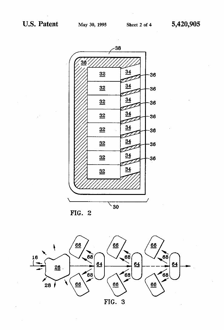
gage or other container, for example) is placed in the path of the bremsstrahlung beam 16 (by a conveyor belt, for example). The incident beam 16 resonantly excites the atoms of the target, and photons 28 are scattered from the target 26. The energies of the scattered pho tons 28 are characteristic of the spacings between the quantized energy states of the nuclei of the target 26. Detecting apparatus 30, including an array of detectors 32, captures, measures, counts, and records the energies of the photons 28 scattered in a given direction or direc tions. The detecting apparatus 30 preferably further includes a ?lter 34 over the face of each detector to absorb low energy photons, and shielding 36 and 38. As scattering and diffraction from the collimating apera ture 22 could lead to a signi?cant amount of photons
5
10
15
20
4 directed toward the detecting apparatus 30, a shadow shield 40 between the aperature 22 and the detecting apparatus 30 is suggested. A beam dump 42 is provided to absorb the energy of the beam 16 not absorbed in the target 26. Shielding 62 encloses the entire device while allowing convenient means for the entry and exit of targets. Data from the detecting apparatus 30 is sent to a computing apparatus 44 which analyzes the data and determines the abundances of particular elements. The computing apparatus is preferably adapted to compare the data for each target to pro?les of “normal” targets to determine if a target should be considered “suspi cious”. The electron source 10 is any accelerator capable of
producing a beam of electrons 12 at the required ener gies and with the required intensity and duty ratio. Among the suitable accelerators are linear accelerators, electrostatic accelerators, microtrons, and betatrons, all of which are commercially available. The electron ener gies required in the present invention are on the order of 10 MeV. It is preferable that energies not exceed about 10 MeV so that substantial radioactivity and neutron
- production does not occur. The beam intensity required
25
30
35
40
50
55
65
is on the order of at least 10 uA; however, an accelera tor capable of producing a beam of intensity in excess of 100 HA is preferred. Duty ratio is important and more than one percent is very desireable. Cost and size are also important considerations. The bremsstrahlung target 14 is a suitable thickness of
any material with a high atomic number or nuclear charge (high Z) and a high melting and boiling point, such as tungsten, tantalum, thorium, or uranium. As an electron in the beam passes through the bremsstrahlung target, it produces the electromagnetic radiation called bremsstrahlung radiation which consists of quanta of photons. The spectrum of energies of these photons is continuous, spanning from very low energies to a maxi mum energy equal to the kinetic energy of the electrons in the beam. This radiation goes by various terms de pending on the target and ?ltering process employed. The terms include thin target bremmstrahlung, thick target bremmstrahlung, ?ltered bremsstrahlung, and others. Here, the term bremsstrahlung is used to encom pass all varieties of bremsstrahlung radiation. A bremmstrahlung target comprising a thickness of
tungsten about the order of l gram/cm2 is appropriate for most embodiments of the present invention. A sub stantially thinner target would not provide a suf?cient intensity of high energy photons. A substantially thicker target would be self-absorptive of the high en ergy photons. The half-angle 0,, of the natural angular spread of the
bremsstrahlung beam for target thickness approaching zero is 0,,zmocZ/Eo, where 0 is in radians and is mea sured relative to the direction of the incident electron beam, mo is the mass of the electrons, c is the speed of light, and E0 is the energy of the electrons. For 10 MeV electrons, 0,,z2.9". This natural collimation is not com pletely useful in the present application, however, since the intensity of photons produced with a target thin enough to provide this natural collimation does not meet the present requirements without the use of much higher electron beam currents. An increased current would require a considerably more expensive accelera tor and shielding apparatus. Energy is also absorbed from the electrons in the
beam by atomic ionization and by the excitation of [the atomic electrons in the bremsstrahlung target. In the

5,420,905 5
process of interacting with the atoms of the target, the electrons often suffer a de?ection. The cumulative ef fect of these de?ections is to increase the angular spread of the bremsstrahlung beam. The rms half-angle 0 of the angular spread can be estimated roughly as 0 z20/PBVT, where 0 is in radians, P is the momentum of the electrons in MeV/0,13 is the velocity of the elec trons in units of the speed of light (and can be taken to be about 1 for this example), and T is the thickness of the bremsstrahlung target in radiation lengths that the electrons have traversed. As an example, 1.0 gram/cm2 of tungsten is about 0.15 radiation lengths and 0 is there fore about 0.77 radians or 45 degrees for a 10 MeV electron beam. Small angles of natural angular spread are therefore only achieved when a very thin brems strahlung target is used and overall ef?ciency for con verting the electron energy into photons is compro mised. Collimating the radiation with an aperature al lows almost any beam shape and angular size approach ing that produced by the scattering. The beam stopper 18 follows the bremsstrahlung
target. The beam stopper is preferably a low Z material, such as boron, beryllium, or silicon, that is not a species to be detected in the target, of a thickness suf?cient to absorb the remaining energy of the electrons 12 from the electron source 10. This low Z material will not generate bremsstrahlung radiation with the same ef? ciency as the bremsstrahlung target 14, but will be heav ily ionizing and therefore will stop the electrons. The thickness of the low Z material required to stop substan tially all the electrons in a 10 MeV beam is about 5.5 gram/cmz.
It is not advised to simply extend the thickness of the bremsstrahlung target 14 to act as the stopper, because a substantial increase in its thickness would be required to perform this function and, because it has a high Z, it would attenuate more of the useful high energy photons than the stopper 18 of the lighter material. For example, a thickness of 8 gram/cm2 of tungsten would be re quired to stop substantially all electrons of a 10 MeV beam.
It is possible to de?ect the electron beam with the use of magnets so that the stopper 18 need not be placed in the line of the bremsstrahlung beam 16. However, since a stopper of low Z material will not substantially affect the bremsstrahlung radiation at the energy levels of interest, this scheme introduces unnecessary complica tion and expense. On the other hand, the use of a magnet to de?ect the electron beam by modest amounts before its strikes the bremsstrahlung target, in order to sweep the resultant direction of the bremsstrahlung beam pro duced by the bremsstrahlung target, is a possible and useful embodiment for the purposes of imaging. A ?lter 20 preferably follows the beam stopper 18.
The ?lter is preferably a low Z material that is not a species to be detected in the target of a thickness suf? cient to absorb the low energy end of the bremsstrah lung spectrum preferentially over the high energy end, where the nuclear states of interest lie. The low energy photons in most embodiments of the present invention are those in the region of about 2 MeV or less. A suit able material for this ?lter is a material with atomic number in the range of carbon to iron. The ?lter may also be made using a combination of suitable materials. The ?lter can be tuned to optimize its ?ltering perfor mance over a desired energy range by selecting the atomic number of the ?lter material or materials and the thickness of the ?lter.
20
25
30
35
40
45
60
65
6 An indicated above, it is not practical to rely only on
the natural collimation of the bremsstrahlung radiation, since beam intensity is sacri?ced by the use of a very thin bremsstrahlung target. Therefore, the aperature 22 is required to produce a collimated beam. The aperature is preferably graded to result in a well-de?ned beam with little halo. It is advantageous to make the aperature rapidly adjustable to permit quick changes in the beam collimation. Preferred aperature geometries are dis cussed below in conjunction with imaging techniques. The shielding 24 includes a ?rst layer 46 of a high Z
material, such as bismuth, lead, or iron, of suf?cient ' thickness to absorb substantially all of the energy of the electrons and of the photons. The ?rst layer of shielding 46 and the collimating
aperature 22 could be a proli?c source of neutrons from the so-called Giant Electric Dipole resonance in heavier nuclei. These neutrons must be shielded from the detectors so as not to produce too much background from neutron induced reactions. A second layer of shielding 48 is therefore required to substantially absorb the neutrons produced by the bremsstrahlung radiation. This second layer is preferably a hydrogenous material loaded with boron or lithium to preferentially capture the neutrons so that no high energy photons are emit ted. Even at 10 MeV, substantial neutron shielding is required. At higher energies, this problem will be much more serious. This loaded hydrogenous material might also be required to cover the exit port of the collimating aperture. A ?nal layer of shielding 50 of a high Z material such
as bismuth, lead, or iron, is required to capture photons generated by neutron capture in the second layer 48 and the outer regions of the ?rst layer 46. The cross section for the absorption of the photons of
the bremsstrahlung beam 16 by the nuclei of the target 26 with ground state spin equal to zero, assuming only photon decay to the ground state (and not proton and other kinds of decay) is involved, is given by
do- _ xzrzc; de _ 2
ate-ewe) 1 where )t is the photon wavelength, 6 is the photon en ergy, so is the energy of the state, I" is the full width of the state, and G is a statistical factor equal to 21+ 1. In this last expression, 1 is the multipolarity of the transi tion. For nuclei with ground states of spin equal to zero, 1 is also the spin of the excited state. This cross section has the usual resonance behavior. The peak value of this cross section occurs when 5:60 and has the value A2(2l+l)/7r. The integrated cross section which is of interest for counting rate estimates is given by 0'=7t2I‘(2l+l)7r; that is, the peak value of the cross section multiplied by the width 1". If other modes of decay are possible, the ground state strength is attenu ated. If the ground state spin is different from zero, the formulas, although not precise, are still useful for esti mating the approximate cross sections. The nuclear states of interest in the present invention
are states that have a width I‘ that is mostly due to photon decay. Cascades via intermediate states that produce photon energies that are suf?ciently high so that they can bedetected above background are accept able. The widths of these states of interest are very small. For the decay of the 6.92 MeV state in oxygen to

5,420,905 7
the ground state, F is on the order of 1/10 eV. How ever, the cross sections are large at the peak because A is large. A value of about about 510 barns is expected for the 6.92 MeV state in oxygen. This means that most of the photons in the bremsstrahlung spectrum that lie within the resonance peak would be absorbed by a thickness of material that has about 1/ 10 of a gram of oxygen per cmz. The same would be true for nitrogen and carbon, or any other material of interest.
It is important to note that the line widths are made effectively very much wider by the Doppler shift due to the thermal motion of the molecules and atoms. As a consequence, the technique will not suffer from a short penetration depth because of the large peak cross sec tions. Unlike the situation where one is scattering a narrow line of photons from a resonance, the thermal broadening of the peak does not cause a loss of sensitiv ity. In the bremsstrahlung spectrum there are always photons to scatter at whatever energy the resonance is shifted to by thermal motion, molecular or atomic.
Nitrogen states of particular relevance to the pre ferred embodiment of the present invention include: the 4.92 MeV 0- state which decays to the 1+ ground
state with a width of 0.084 eV. the 7.03 MeV 2+ state which decays to the 1+ ground
state with a width of 0.078 eV. Oxygen states of particular relevance to the preferred
embodiment of the present invention include: the 6.92 MeV 2+ state which decays to the 0+ ground
state with a width of 0.097 eV. the 7.12 MeV 1- state which decays to the 0+ ground
state with a width of 0.055 eV. A carbon state of particular relevance to the pre
ferred embodiment of the present invention is: the 4.44 MeV 2+ state which decays to the 0+ ground
state with a width of 0.011 eV. Another oxygen state of interest is the 12.53 MeV 2
state which: decays to a 6.13 MeV 3- state producing a 6.40 MeV photon and a 6.13 MeV photon with a width of 2.1 eV; decays to a 7.12 MeV 1- state producing a 5.41 MeV photon and a 7.12 MeV photon with a width of 0.5 eV; and decays to a 8.87 MeV 2" state producing a 3.66 MeV photon, along with a 1.74 MeV photon and a 6.13 MeV photon in cascade most of the time, with a width of 0.9 eV. Another carbon state of interest is the 12.71 MeV 1+ state which: decays to the 0+ ground state with a width of 0.35 eV; and decays to the 4.44 MeV 2+ state producing a 8.27 MeV photon and a 4.44 MeV photon with a width of 0.053 eV. The total widths of the 12.53 MeV state in oxygen and the 12.71 MeV state in carbon are 97 eV and 18 Ev respectively. These states decay preferentially by proton and/ or alpha emis sion and the photon intensities will be considerably reduced. Thus, these states are not a part of a preferred embodiment because of their low intensity and high energy. The 15.11 MeV 1+ carbon state, which decays to the
0+ ground state with a width of 38 eV, is of great inter est. Its total width is about 43.6 eV and most of the width is ground state decay. However, exciting this state requires an electron beam with energies signi? cantly above the approximate limit of 10 MeV speci?ed above to limit the production of radioactivity and neu trons. Thus, the detection of carbon via this state, while attractive because of the very strong scattering, would involve additional complication and expense. There are numerous states of the isotopes chlorine 35
and chlorine 37 that are of interest, resulting in photons
20
25
30
35
40
45
50
55
60
65
8 With energies up to about 6 MeV with radiative widths in the range of the previous considerations. Thus, chlo rine abundances can be determined as easily as those of oxygen, nitrogen, and carbon. Many other elements have states of the appropriate
energy and radiative widths. Therefore, the present invention is general in its usefulness. The method of the present invention can be used to detect almost any clement of interest in a target. In addition to the explo sives detection embodiment described here, apparatus according to the present invention could be' used in many industrial and commercial applications. An important consideration for the estimation of
count rates is the number of photons that are found within the width of the resonance in the bremsstrahlun g spectrum. For a 10 MeV electron accelerator producing 10 11A of electrons, the bremsstrahlung spectrum can have about 106 photons per eV at 7 MeV. Thus, for a state that is about 1/ 10 eV wide, about 105 photons will lie within the range of the resonance and will be ab sorbed from the beam and re-emitted over all angles with very roughly an isotropic distribution. If 1/ 10 of the sphere is subtended with detectors that are 10% ef?cient, the counting rate will be about 1000 per sec ond.
In this example, it was assumed that all the resonant photons are absorbed by the target 26. This is roughly correct for a small amount of material because of the large cross sections when thermal motion is neglected. With thermal motion there are always more photons available to scatter and thus the counting rates could be larger. However, the exact result depends on the geom etry and thickness of the explosive material.
It is important to compare the signal with the ex pected background. In the preferred embodiments, the signal is a set of photon lines with energies of about 3-8 MeV. The background comes from the scattering of photons out of the beam by atomic processes. The major process is Compton scattering. Fortunately, if detectors are placed at scattering angles larger than 90 degrees to the bremsstrahlung beam, these Compton scattered photons will have energies below 0.5 MeV. The greater the angle past 90 degrees, the lower the energies of the Compton scattered photons, to a limit of 0.25 MeV for backscatter at 180 degrees. Pair produc tion will also provide secondary bremsstrahlung that is predominantly forward peaked, as well as 0.5 MeV photons from e+e- annihilation. There are coherent processes like Rayleigh scattering, but these should be very small at back angles, in particular for energies of a few MeV and above. Thus, there is no background in the energy region above 0.5 MeV except from mutiple scattering processes and pile-up in the counters.
In order to minimize the effects of Compton scatter ing and maximize the signal-to-noise ratio, the detecting apparatus 30 should be placed at an angle 11> with respect to the bremsstrahlung beam 16 of considerably more than 90 degrees. An angle approaching 150 degrees is strongly preferred to make the signal stand out suf? ciently from the noise of multiple processes. There are many ways to arrange individual detectors
32 within a detecting apparatus 30. A preferred geome try is an annular array of detectors positioned to collect photons scattered back from the target at an angle (b of about 150 degrees. This is the embodiment illustrated in FIG. 1 where the detecting apparatus 30 is shown in cross-section. Of course, the array need not form a complete annulus around the path of the bremsstrah
5,420,905 lung beam. For some embodiments, a vertical bank of detectors, one on each side of the bremsstrahlung beam 16, is preferred. There are a variety of suitable detectors 32 that can
be used in the detecting apparatus 30. In one embodi ment, the detectors are intrinsic germanium ionization chambers where charge detectors pick up the electrical signal. In another embodiment, the detectors are scintil lators such as NaI and BGO. The light emitted by these scintillators in response to incident photons can be con verted to an electrical signal using a photomultiplier tube. This can be almost 100% ef?cient, leading to higher counting rates in the above example. It is impor tant to recognize that the circuitry of the detecting apparatus must accomodate high counting rates and reduce pile—up. The ?lter 34 is preferably placed in front of the detec
tors 32 to ?lter out low energy photons while passing the high energy photons of interest. A suitable material for this ?lter is a material with a low Z somewhere between that of carbon and iron. A high Z shield 36 is preferably placed around all but the face of the detector apparatus to absorb photons incident on the back and sides of the apparatus. Lead, bismuth, and iron are ap propriate materials for this purpose. It may be necessary to include a shield 38 of hydrogenous material loaded with boron or lithium around the detectors to shield against neutrons generated in the luggage and beam dump. The high Z shadow shield 40 is preferably placed in the path between the aperature and the detectors to absorb all high-energy photons scattered from the aper ature in the direction of the detectors. Lead is a suitable material for this purpose. The function of the shadow shield 40 could be performed by a heavy section of the shield 36. The beam 16 passing through the target 26 may be
attenuated by a relatively small amount. This beam must be absorbed in a beam dump 42 designed to absorb substantially all of the energy. A suitable beam dump for 10 MeV may be a layer 52 of a hydrogenous material containing boron or lithium, a layer 54 of carbon, and a layer 56 of iron in a very deep cavity formed in a shield 58 of lead or iron, for example, to shield the sides and the detectors from back-streaming low energy photons. A layer 60 of a hydrogenous material containing boron or lithium preferably surrounds the outside of this shield. The depth of this cavity, the beam dimensions, the directive collimation of the detectors, and the exact location of the detectors are related parameters that must be made compatible so as to not allow back-scat tered photons from the beam dump to enter the detec tors. Additional shadow shields may be set up to help meet this goal. Imaging can be achieved in a variety of ways with the
technique of the present invention. The luggage can be scanned with the beam by moving the bremsstrahlung generating apparatus 5, the target 26, or simply the aperature 22. The electron beam can also be de?ected by a magnet to sweep the bremsstrahlung beam direc tion. Preferred beam geometries are spots and stripes. For example, if the beam 16 is collimated using a
small square aperature to an average angle of Qz l/2O radians (about 3 degrees), the spot 1 meter from the aperature will be about 10 cmX 10 cm, an excellent size for imaging the contents of a piece of luggage. If each measurement takes about 5 second, an entire suitcase could be scanned in several seconds.
30
35
45
55
60
65
10 If the beam 16 is collimated using a vertical slit apera~
ture to produce a thin stripe of 10 cm width at the point of incidence with a piece of luggage, a 60 cm long suit case could be scanned in a few seconds as the suitcase moves on a conveyor belt. Alternatively, the beam 16 could be collimated into a spot swept vertically by an adjustable collimator or by magnetic de?ection of the beam. Even if the collimation is in the form of a vertical stripe, the central intensity remains the highest, re?ect ing the natural collimation, and magnetic de?ection of the beam will be useful for imaging.
In another imaging technique, a large portion of the suitcase is ?ooded with bremsstrahlung radiation by using a large aperature, and the detectors 32 are adapted to be direction-speci?c, as illustrated in FIG. 2. In this embodiment, part of the ?lter 34 covering the faces of the individual detectors 32 is replaced with high Z shielding 36. A column of low Z ?lter 34 remains for each individual detector. In this way, each detector can be designed to only detect photons scattered from a small speci?c region of the suitcase in a particular direc tion. An array of such detectors can easily be designed to image the entire suitcase to a desired degree of reso lution. A combination of the above imaging techniques re
sults in a further embodiment of the present invention. For example, a thin slit aperature could be used to radi ate thin vertical stripes of the suitcase as the suitcase moves on a conveyor belt. The width of the stripe will determine the horizontal resolution of the imaging. The vertical resolution could be increased by using direc tional detectors. Such a method would result in fast measurements at a high resolution. Use of a rapidly adjustable collimating aperature 22
results in further embodiments with important advan tages. For example, a piece of luggage could ?rst be ?ooded with bremsstrahlung radiation in an effort to detect explosives in the form of thin sheets and/or to obtain an initial estimate of the abundances of various elements in its contents. The aperature could then be stopped down to image the suitcase in an effort to detect more localized explosive materials. Another mode of operation is possible with the appa
ratus of the present invention. With a second bank of detectors located behind the target 26 to detect photons transmitted through the target, the intensity of photons absorbed in the target can be monitored. In this way, a very precise image of the transmission density of the target can be constructed. Such an image will identify speci?c areas of high material density in the target which would be a further aid in detecting explosive materials. Similar density imaging could be achieved by detecting the low energy back-scatter from the target.
Shielding 62 is required to protect the public from photons and neutrons generated by the explosives de tection device. This shielding should enclose the device, while allowing a target to be quickly and conveniently moved into and out of the device. For example, if the targets aresuitcases on a conveyor belt, two sets of double doors, one set on each side of the device, would permit the entry and exit of the suitcases. This method is used in other explosives detection devices. Since the electron accelerator can be rapidly turned off and on, additional safety is provided by switching the accelera tor off while targets are entering and/or exiting the device. The fact that the electron accelerator can be rapidly
switched on and off can be used to other advantages.

5,420,905 11
For example, when the target is a large container, rang ing could be achieved by making pulses of bremsstrah lung radiation incident on the target and measuring the delay of the photons scattered to the detectors. In this way, the depth of an explosive material into the con tainer could be approximated. Pulses of less than a nano~ second in width are possible. The beam from an accelerator is often bunched, or
can be made to be bunched, in subnanosecond intervals. For example, electrons are typically injected into linear accelerators in discrete bunches at a rate on the order of 3 GHz. Each bunch in the stream typically lasts on the order of 10 picoseconds as it passes by some point in space. By adapting the source of electrons to skip por tions of the stream of bunches, a beam time structure can be achieved which includes bunches of electrons of duration on the order of 10 picoseconds, at any desired rate of repetition. This time structure can also be achieved with microtrons, betatrons, and electrostatic machines by properly preparing the electron gun and apparatus which serves to inject electrons into these accelerators. These electron bunches are converted into bunches of photons with approximately the same time structure using the bremsstrahlung generating appara tus described above. By using time bunches of photons, it is possible to determine the distance between the scattering element or compound in a target and the detectors when the detectors are suf?ciently greater than 0° from the line of the photon beam. In the pre ferred embodiment, the resonance ?uorescence detec tors are preferably at least 90°, and even more preferra-' bly, at least 150° away from this axis, providing excel lent ranging capability. The bunches of radiation may, for examples be made incident on the target at a suf? ciently slow rate so that the resonance ?uoresence from one bunch is detected before another hunch is sent, providing unambiguous timing data. For example, for a target three feet in dimension along the beam path, the bunches might be spaced 3 nanoseconds apart. The average intensity of a photon source can be maintained when bunches are removed by increasing the number of electrons in each bunch to compensate. Since most detectors, for example germanium, NaI, and BGO, inte grate over times of many nanoseconds, the effective duty ratio from the point of view of the in?uence of pile-up and event rate remains effectively the same for the bunched beam described above as compared to continuous beams. In view of the timing resolution easily provided with currently available accelerator and detector apparatus, a spatial resolution of target compo sition of less than two incites may be achieved. The computing apparatus 44 is adapted to analyze the
data obtained by the detecting apparatus 30. As with other explosives detecting devices, profiles of elements, such as nitrogen and oxygen, as they appear in “nor mal” suitcases are preferably either modelled or experi mentally determined. A suitcase which deviates signi? cantly from these pro?les would be considered “suspi cious”. The computing apparatus can be easily adapted to compare data to stored pro?les. If the pro?les are rigorously determined, a high probablity of explosives detection accompanied by a low rate of false alarms will be achieved. An alternate detecting scheme according to the pres
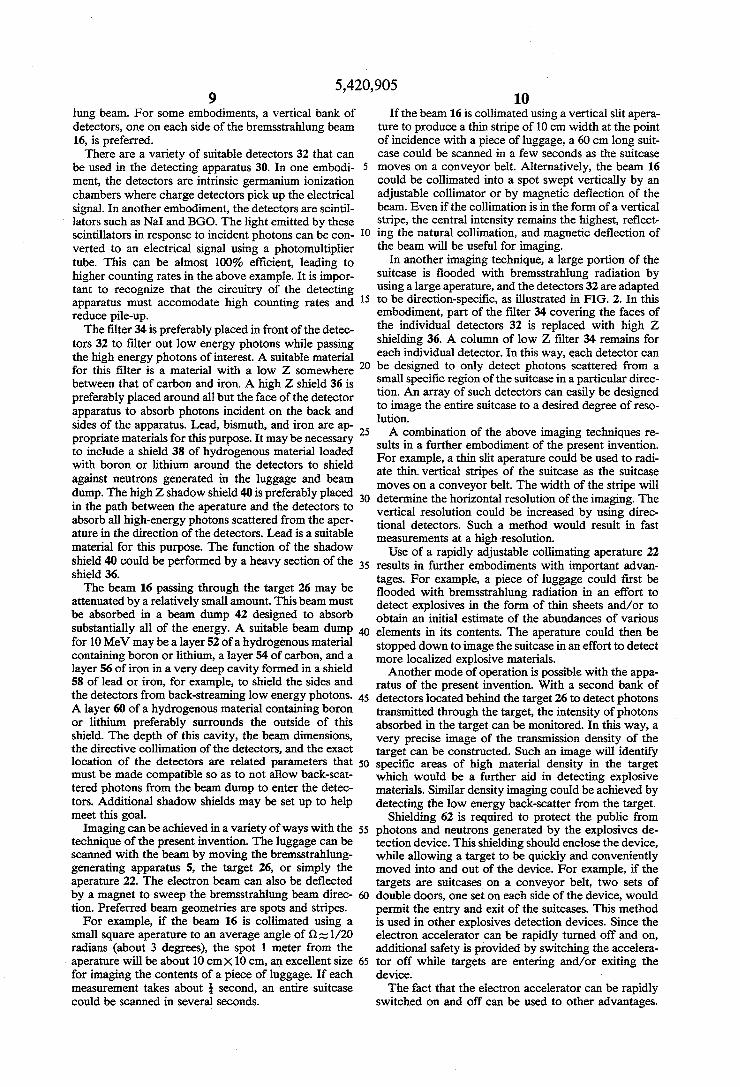
ent invention, which was disclosed in U.S. application Ser. No. 620,266, is illustrated in one embodiment in FIG. 3. For simplicity, only those elements relevant to the detecting scheme are shown. As shown, the brems
15
20
30
35
40
45
55
60
12 strahlung beam 16 is made incident on the target 26. As the beam 16 passes through the target 26, photons will be resonantly absorbed by the nuclei of the target. The energies of the absorbed photons correspond to the spacings between the quantized energy states of each nuclear species in the target. For these speci?c energies, the transmitted beam will be depleted of photons. For example, if the target contains nitrogen, photons of energies corresponding to nitrogen will be selectively absorbed. The amount of photons absorbed depends on the quantity of nitrogen in the target. Thus, the intensi ties of the photons of speci?c energies transmitted through the target contains information about the nu clear composition of the target. This information is exploited in the embodiment shown in FIG. 3. As shown, a series of reference resonance scatterers 64 are arranged behind the target 26. Each reference scatterer is composed of one or more of the elements that the explosives detecting device is to detect. A series of detecting apparatuses 66 is adapted to capture, measure, count, and record the photons 68 resonantly scattered from each of the reference scatterers 64. For example, in a simple embodiment, two reference scatterers are provided, one of nitrogen, the other of oxygen. A de tecting apparatus is adapted to detect photons reso nantly scattered from the nuclei in the nitrogen scat terer and another detecting apparatus is adapted to detect photons resonantly scattered from the nuclei in the oxygen scatterer.
This detecting scheme operates as follows. If no tar get 26 is placed in the path of the beam 16, the beam will directly strike the ?rst of the reference resonance scat terers 64. The detecting apparatus associated with the ?rst reference scatterer will detect a large number of photons, the energies of which correspond to the nu clear species of the ?rst reference scatterer. If a target 26 with a relatively small amount of the nuclear species of the ?rst reference scatterer is placed in the path of the beam, this strong signal at the ?rst detecting apparatus will be diminished by a relatively small amount due to the non-resonant processes. If however, a target 26 with a relatively large amount of the nuclear species of the ?rst reference scatterer is placed in the path of the beam, this signal will be further diminished considera bly, due to the resonant absorption of the photons of energies corresponding to the nuclear species of interest in the target 26. The non-resonant attenuation can be monitored by standard procedures and detectors and accounted for. Thus, an abundance of a nuclear species of interest in a target 26 will be detected as a decrease in the signal from the detecting apparatus associated with a reference scatterer composed substantially of that nuclear species due to the resonant absorption in the target in addition to standard non-resonant processes. Photons of energies not corresponding to the nuclear species of which a reference scatterer is substantially composed will be attenuated by only a relatively small amount by non-resonant processes. Thus, the method of detecting the nuclear species of the ?rst reference scat terer extends to each subsequent reference scatterer. An advantage of this detecting scheme is that if the energies corresponding to two or more nuclear species of inter est are very close, the detecting apparatus of the em bodiment of FIG. 1 may have dif?culty distinguishing the contributions from the two or more nuclear species. However, in the embodiment of FIG. 3, the energies corresponding to each nuclear species are detected separately, this ambiguity is diminished considerably,
5,420,905 13
and the ability of the detecting apparatus to resolve closely spaced photon energies is no longer very impor tant. When the energies corresponding to two or more nuclear species do not interfere within the detector resolution, a single reference scatterer can be composed of a combination of the species.
In this second detecting scheme, a transmission ge ometry is used. It is impractical to measure the nuclear resonance absorption of the target using bremsstrahlung and conventional detectors in transmission geometry. The absorption lines are very narrow, on the order of ten electron volts, due to the Doppler broadening of thermal motion, and they will not be evidenced easily if conventional non-resonance detectors are used that commonly have many kilovolts of resolution. The reso nant signal can be overwhelmed by all the non-resonant photons in the detector resolution interval. In our em bodiment, the resonance scattering from the reference scatterers has the same very narrow resolution as the absorption lines. The photon detectors are presented, via the scattering process, with only the resonant pho tons. Thus, the assembly is a resonance detector and thus the signal is only that of the resonant photons. We point out that this form of resonance detector has appli cations in other resonance absorption transmission tech niques wherein the photons being studied, although generally resonant, are further distributed in energy by the geometric aspects of the kinematics of nuclear reac tions. In such applications, use of our embodiment as a resonance detector will greatly improve the signal of the absorption lines relative to the signal obtained by the use of a non-resonance photon detector, such as NaI, BGO, intrinsic germanium, etc. The requirements for each detecting apparatus 66 in
the series are similar to those described for the detecting apparatus 30 of the embodiment of FIG. 1. Thus, each detecting apparatus preferably includes an array of detectors, and shielding and ?ltering means as indicated by elements 32, 34, 36, and 38 of FIG. 1. The various detecting apparatus con?gurations discussed above are applicable to the embodiment of FIG. 3. In order to minimize the effects of Compton scattering and maxi mize the signal-to-noise ratio, the detecting apparatuses 66 should be placed at an angle qb with respect to the bremsstrahlung beam 16 of considerably more than 90 degrees. An angle approaching 150 degrees is strongly preferred to help make the signal stand out suf?ciently from the noise of non-resonant multiple processes. It must be noted that backscatter from any of the refer ence scatterers 64 and the detecting apparatuses 66 must be taken into account when positioning the reference scatterers and detecting apparatuses. Appropriate shielding may be required to isolate the detectors from backscatter from reference resonance scatterers with which they are not associated. The adaptations for di rection detection pictured in FIG. 2 are also approriate for this alternate detecting scheme. Similarly, the imag ing methods described above can be employed in this scheme. A further imaging scheme appropriate to this embodiment employs reference resonance scatterers which are small with respect to the dimensions of the target 26. Imaging can then be achieved while the target 26 is ?ooded with bremsstrahlung radiation by moving the scatterers to scan the extent of the target.
All elements not shown in FIG. 3, such as the brems strahlung source, the beam dump, the shielding, and the computing apparatus are similar to that shown in FIG. 1. In this alternate detecting scheme, the computing
10
20
25
35
40
45
50
55
60
65
14 apparatus is adapted to compare the intensities of pho ton energy levels detected by the detecting apparatuses 66 to reference values. Clearly an appropriate set of reference values are the intensities measured when the target 26 is not placed in the path of the bremsstrahlung beam. In that way, the decreases in intensity measured when the target is placed in the path of the beam can be directly related to the composition of the target. Alter natively the reference values may be determined from pro?les of “normal” targets, as discussed above in rela tion to the first detecting scheme. A further detecting scheme involves a combination of
‘ the detecting schemes illustrated in FIG. land FIG. 3. It may be advantageous to detect both the photons resonantly scattered directly from the target 26 and those resonantly scattered from the reference scatterers 64. The two schemes are completely compatible and provide complementary information concerning the location of a resonant scatterer via the different paths experienced by the detected resonant photon and the different absorption along these paths. The detecting schemes discussed thus far are based on
the resonant absorption and resonant scattering of pho tons by nuclei. The nuclear species are detected via the characteristic energy lines that are present in the spec trum of photons scattered via the resonance process and detected by photon spectrometers. The nuclear species can also be detected by the preferential absorption via the resonance process of photons in the incident spec trum at these characteristic energies. In this latter tech nique, the presence of a nuclear species is detected as a diminution of the scattering of the photons with charac teristic energies from a special detector that views the scattering from a speci?c nuclear target that is placed in the beam after it passes through the target under investi gation; that is, the target produces absorption lines in the transmitted spectrum. The measurements are very speci?c as to the nuclear species.
Further detecting schemes according to the invention can be used to add to the certainty of the identi?cation of species in a target, especially with respect to size, density, and elemental composition. These schemes are based on other processes that occur naturally when a photon beam passes through the target, in particular, Compton scattering and pair production. These pro cesses are the ones that produce most of the non-reso nant absorption of the photons passing through the target. Compton scattering is a process that takes place when
photons are scattered by the atomic electrons of the target. The recoil of the electrons shifts the energy of the photons by an amount that depends on the incident photon energy and the scattering angle. At angles greater than 90 degrees, the photons are shifted to ener gies below 0.51 MeV. At smaller angles the energies are shifted by lesser amounts. For example, a photon of 5 MeV is shifted to an energy of about 1.3 MeV at 45 degrees. Thus, by looking a speci?c angles, one can identify from this transformed spectrum the photons of the original continuous spectrum that have Compton scattered from the atomic electrons. Neglecting the small effect of atomic binding, the Compton process depends only on the number of electrons. It thus pro vides information on the quantity
Z D7,

5,420,905 15
where D is the density, Z is the nuclear charge (number of protons), and A is the total number of nucleons (num ber of protons plus neutrons) of the species. Because Z is usually almost equal to A/ 2, the information describes the density alone. Since detectors can be made to be directional by the use of collimation, as described above, the Compton process can be detected and ana lyzed as a function of position along the beam of pho tons, thus allowing determination of the spatial distribu tion of the density of the target. Furthermore, the atten uation of the scattered photons along their path towards the detectors will depend upon the species along the path. For each position in the target, many paths can be used to view each location in the target by using differ ent detectors that view the same position. Similarly, different banks of detectors can view other positions. Or alternatively and in combination, the different banks of detectors could move together with the photon beam as it scans a target to image a species of interest in the target. This ensemble of information can be used to construct three-dimensional images of the target both as to the density, and as to the attenuation in the target. One embodiment of a scheme to detect Compton
scattering is illustrated by way of example in FIG. 4. For simplicity, only those elements relevant to the de tecting scheme are shown. All elements not shown in FIG. 4, such as the photon source, the beam dump, the shielding, and the computing apparatus are similar to
' that shown in FIG. 1. As shown, the photon beam 16 is made incident on the target 26. As the beam 16 passes through the target 26, the photons scatter from atomic electrons in the target. The recoil of the electrons signif icantly shifts the energies of the photons. The photons 70 in the shifted spectrum are detected by, for example, the two detecting apparatus 76 and 78 shown with nor mals 45° from the path of the photon beam. Of course other detector positions could be used as well, as dis cussed above. The requirements for the detecting appa ratus 76 and 78 are similar to those described for the detecting apparatus 30 of the embodiment of FIG. 1. Thus, each detecting apparatus preferably includes an array of detectors, and shielding and ?ltering means as indicated by elements 32, 34, 36, and 38 of FIG. 1. The geometry of the detectors and the shielding and ?ltering means must of course be modi?ed to optimize the de tecting apparatus to detect Compton scattered photons from the target. The various detecting apparatus con?g urations discussed above, including the con?gurations used for directional detection illustrated in FIG. 2 are applicable to the embodiment of FIG. 4. The coordi nates (x,y,t) of the detected photons in space and time, along with the directionality of the detectors and the photon spectrum, provide information required for analyzing the spatial distribution of the density of the target. The process of pair production takes place in the
electric ?eld of a nucleus when an incident photon is more energetic than about 1.02 MeV. A positive and negative electron pair is produced. Both are stopped in the surrounding material within a range of, for example, approximately one centimeter for 5 MeV photons and material densities of 1 g/cm3. The positive electron is captured by an atomic electron to form the positronium system that decays rapidly via the emission of two pho tons, each with an energy of 0.51 MeV. These photons are emitted back to back. Thus, two detectors whose joining line passes through the photon beam and the
15
20
25
30
35
45
50
55
60
65
16 target will see coincident events that signal the forma tion of a pair at a very speci?c location in the target. The resolution in space is limited by the size of the
detectors and the range of the positive electron. These detectors can be placed in any position that is conve nient and need not interfere with the detectors used in the Compton scattering scheme described above, or with the detectors used in the resonance methods de scribed earlier. The detectors are preferably positioned along a line that intersects the region of interest on opposite sides of the region. A detector location of 90° with respect to the direction of the photon beam might be favorable since the Compton scattered photons are then at or below 0.51 MeV in both detectors. On the other hand, it may be desireable to have one of the detectors at an angle of greater than 90° so that at least one detector has the 0.51 MeV photons from pair pro duction well-isolated from the Compton process. The timing of the events in two detectors whose
joining line passes through the photon beam and the target will also help remove the photons from the other processes as interfering background. The relative tim ing can also be used to identify the location of the pro cess along the line joining the two detectors since the photons travel at the speed of light and are emitted at the same time. The cross section for the pair production process is proportional to
22 D7 .
Thus this process samples a different function of the elemental composition than the Compton process and provides new information about the composition of a target. A three-dimensional image of the spatial distri bution of
Z2 DA
in a target can be formed using this pair production process. The 0.51 MeV photons will be attenuated along their paths to the detectors. By providing differ ent detector combinations to view the same position in the target, different paths are sampled for the same position. Similarly, different detector banks can view different positions simultaneously. Or, alternatively and in combination, these detector banks can move with the beam to follow it in the target as it moves to scan the target and form an image. This information can be used with the ensemble of data to provide a three-dimen sional image of the target for the attenuation process, along with the image formed for the resonance pro cesses, for the Compton processes and for the pair pro duction processes. The attenuation process for the Compton scattered photons can be tuned to be at a different energy than the photons for the pair produc tion process by selection of the angle of the Compton scattering that is used. This adds more element speci?c information to the data that is gathered. One embodiment of a scheme to detect pair produc
tion is illustrated by way of example in FIG. 5. For simplicity, only those elements relevant to the detecting scheme are shown. All elements not shown in FIG. 5, such as the photon source, the beam dump, the shield ing, and the computing apparatus are similar to that shown in FIG. 1. As shown, the photon beam 16 is


