Design of Temperature-Responsive Cell Culture Surfaces for ...
15
Review Article Design of Temperature-Responsive Cell Culture Surfaces for Cell Sheet Engineering Y. Akiyama Institute of Advanced Biomedical Engineering and Science, Tokyo Women’s Medical University, TWIns, Tokyo, Japan Correspondence should be addressed to Y. Akiyama; [email protected] Received 29 October 2020; Accepted 4 January 2021; Published 3 February 2021 Copyright © 2021 Y. Akiyama. Exclusive Licensee Beijing Institute of Technology Press. Distributed under a Creative Commons Attribution License (CC BY 4.0). Temperature-responsive cell culture surfaces, which modulate cell attachment/detachment characteristics with temperature, have been used to fabricate cell sheets. Extensive study on fabrication of cell sheet with the temperature-responsive cell culture surface, manipulation, and transplantation of the cell sheet has established the interdisciplinary field of cell sheet engineering, in which engineering, biological, and medical fields closely collaborate. Such collaboration has pioneered cell sheet engineering, making it a promising and attractive technology in tissue engineering and regenerative medicine. This review introduces concepts of cell sheet engineering, followed by designs for the fabrication of various types of temperature-responsive cell culture surfaces and technologies for cell sheet manipulation. The development of various methods for the fabrication of temperature-responsive cell culture surfaces was also summarized. The availability of cell sheet engineering for the treatment and regeneration of damaged human tissue has also been described, providing examples of the clinical application of cell sheet transplantation in humans. 1. Introduction Cell-based medicine has been expected as a promising approach to treat damaged tissues and organs. Langer and Vacanti proposed tissue engineering as a new research field by synergistically combining engineering and life sciences [1]. Their concept initiated the development of functional substitutes for damaged tissues and organs. Tissue engineer- ing involves cell culturing on synthetic and/or natural poly- meric porous scaffolds in the presence of growth factors. Some of the engineered tissues were successfully applied to the treatment of damaged tissue and/or tissue fabrication [2–5]. The engineered tissues and organs were constructed by seeding and culturing cells on a biodegradable scaffold. The immigrated and/or proliferated cells and their secreted extracellular matrix (ECM) filled spaces where the scaffold was degraded during the cell culture. Through these compli- cated processes, cells assemble and form three-dimensional (3D) tissue-like structures themselves. Adjustment of speed between scaffold degradation and space filling with cells was required for subsequent assembly and formation of the engineered 3D tissue. However, the adjustment remained difficult. In addition, in the deep part of the porous scaffold, diffusion of O 2 and nutrients, required for the survival of cells, was limited, possibly preventing cell migration toward the deep part. Therefore, the resulting engineered cultures have a lower cell density, different from native tissue with high cell density, such as the muscles and heart. In addition, acidic components degraded from the synthetic scaffold caused a nonspecific inflammation response after implanta- tion, damaging the cells [6]. In contrast with conventional tissue engineering, Okano’s group has shown a novel approach: cell sheet engineering, which deals with fabricating, manipulating, and transplanting cell sheets [7, 8]. A cell sheet was fabri- cated with an intelligent nano-bio-interface, temperature- responsive cell culture surface [9, 10]. Cell sheet engineering enables the assembly of 3D and cell-dense tissue-like struc- tures without scaffolds by layering the cell sheet. The cell sheet and/or layered cell sheets facilitate transplantation to biological tissues and organs without the need for sutures. Such transplantable cell sheets have been clinically applicable for the regeneration and treatment of damaged human tis- sues and organs such as the heart [11], corneal epithelium [12], esophagus epithelium [13, 14], periodontal ligaments [15, 16], cavity of the middle ear [17], knee cartilage [18], and lungs [19]. Cell sheet engineering is potentially promis- ing in both tissue engineering and regenerative medicine. AAAS Cyborg and Bionic Systems Volume 2021, Article ID 5738457, 15 pages https://doi.org/10.34133/2021/5738457
Transcript of Design of Temperature-Responsive Cell Culture Surfaces for ...
Review ArticleDesign of Temperature-Responsive Cell Culture Surfaces for CellSheet Engineering
Y. Akiyama
Institute of Advanced Biomedical Engineering and Science, Tokyo Women’s Medical University, TWIns, Tokyo, Japan
Correspondence should be addressed to Y. Akiyama; [email protected]
Received 29 October 2020; Accepted 4 January 2021; Published 3 February 2021
Copyright © 2021 Y. Akiyama. Exclusive Licensee Beijing Institute of Technology Press. Distributed under a Creative CommonsAttribution License (CC BY 4.0).
Temperature-responsive cell culture surfaces, which modulate cell attachment/detachment characteristics with temperature, havebeen used to fabricate cell sheets. Extensive study on fabrication of cell sheet with the temperature-responsive cell culture surface,manipulation, and transplantation of the cell sheet has established the interdisciplinary field of cell sheet engineering, in whichengineering, biological, and medical fields closely collaborate. Such collaboration has pioneered cell sheet engineering, making ita promising and attractive technology in tissue engineering and regenerative medicine. This review introduces concepts of cellsheet engineering, followed by designs for the fabrication of various types of temperature-responsive cell culture surfaces andtechnologies for cell sheet manipulation. The development of various methods for the fabrication of temperature-responsive cellculture surfaces was also summarized. The availability of cell sheet engineering for the treatment and regeneration of damagedhuman tissue has also been described, providing examples of the clinical application of cell sheet transplantation in humans.
1. Introduction
Cell-based medicine has been expected as a promisingapproach to treat damaged tissues and organs. Langer andVacanti proposed tissue engineering as a new research fieldby synergistically combining engineering and life sciences[1]. Their concept initiated the development of functionalsubstitutes for damaged tissues and organs. Tissue engineer-ing involves cell culturing on synthetic and/or natural poly-meric porous scaffolds in the presence of growth factors.Some of the engineered tissues were successfully applied tothe treatment of damaged tissue and/or tissue fabrication[2–5]. The engineered tissues and organs were constructedby seeding and culturing cells on a biodegradable scaffold.The immigrated and/or proliferated cells and their secretedextracellular matrix (ECM) filled spaces where the scaffoldwas degraded during the cell culture. Through these compli-cated processes, cells assemble and form three-dimensional(3D) tissue-like structures themselves. Adjustment of speedbetween scaffold degradation and space filling with cellswas required for subsequent assembly and formation of theengineered 3D tissue. However, the adjustment remaineddifficult. In addition, in the deep part of the porous scaffold,diffusion of O2 and nutrients, required for the survival of
cells, was limited, possibly preventing cell migration towardthe deep part. Therefore, the resulting engineered cultureshave a lower cell density, different from native tissue withhigh cell density, such as the muscles and heart. In addition,acidic components degraded from the synthetic scaffoldcaused a nonspecific inflammation response after implanta-tion, damaging the cells [6].
In contrast with conventional tissue engineering,Okano’s group has shown a novel approach: cell sheetengineering, which deals with fabricating, manipulating,and transplanting cell sheets [7, 8]. A cell sheet was fabri-cated with an intelligent nano-bio-interface, temperature-responsive cell culture surface [9, 10]. Cell sheet engineeringenables the assembly of 3D and cell-dense tissue-like struc-tures without scaffolds by layering the cell sheet. The cellsheet and/or layered cell sheets facilitate transplantation tobiological tissues and organs without the need for sutures.Such transplantable cell sheets have been clinically applicablefor the regeneration and treatment of damaged human tis-sues and organs such as the heart [11], corneal epithelium[12], esophagus epithelium [13, 14], periodontal ligaments[15, 16], cavity of the middle ear [17], knee cartilage [18],and lungs [19]. Cell sheet engineering is potentially promis-ing in both tissue engineering and regenerative medicine.
AAASCyborg and Bionic SystemsVolume 2021, Article ID 5738457, 15 pageshttps://doi.org/10.34133/2021/5738457

2. Temperature-Responsive Cell CultureSurface and Cell Sheet
2.1. Features of Temperature-Responsive Cell Culture Surfacesand Cell Sheet Recovered from the Surfaces. A temperature-responsive polymer, poly (N-isopropylacrylamide) (PIPAAm), was synthesized in the latter half of the 1960s. The phase
transition of PIPAAm in an aqueous solution was reported forthe first time (Figure 1(a)) [20]. When temperatures decreaseacross 32°C (lower critical solution temperature (LCST) ofPIPAAm), PIPAAm is dissolved and expands its chain confor-mation due to hydration of PIPAAm chains. In contrast, PIPAAm chains are quickly aggregated and insoluble because of thedehydration of PIPAAm chains when the temperature reaches
(a)
(b)
(c)
(i) Hydrophilic surface(ii) Cell detachment
CellCell sheet
ECM
PIPAAm layer
H2O
PIPAAm chain
Soluble Insoluble
Poly(N-isopropylacrylamide)(PIPAAm)
H3C
CH2 CH n
CH3
CHNHC O
(i)(ii)
Hydrophobic surfaceCell adhesion
Figure 1: Illustration of characteristics of (a) poly(N-isopropylacrylamide) (PIPAAm), (b) temperature-responsive cell culture surface, PIPAAm-TCPS, and (c) cell sheet recovery from PIPAAm-TCPS. (a) PIPAAm chains show reversible temperature-dependent hydration anddehydration across LCST. (b) Above LCST, the grafted PIPAAm chains dehydrated to exhibit enough hydrophobic and cell adhesionproperties (right). Below LCST, cells were detached spontaneously from the surface due to hydration of grafted PIPAAm chains (left).These images of (a) and (b) from reference [36] (Z. Tang and T. Okano., Recent development of temperature-responsive surfaces andtheir application for cell sheet engineering, Regen Biomater, 1, pp. 91-102) are under a Creative Commons Attribution 4.0 InternationalLicense (CC-BY-4.0). (c) After adhered cells were confluent at 37°C (left), lowering the temperature to 20°C (below LCST) enabled thedetachment of the cell sheet from PIPAAm-TCPS, preserving the deposited ECM and cell-cell junctions (right). Reprinted permissionfrom reference [8]. Copyright © 2021, Oxford University Press.
2 Cyborg and Bionic Systems

above the LCST. The temperature-dependent hydration anddehydration are reversible.
Approximately 30 years later, by grafting PIPAAm on atissue culture polystyrene surface (PIPAAm-TCPS), Okanoet al. successfully invented temperature-responsive surfacesfor the first time, in which hydrophilic and hydrophobicproperties are modulated with temperature (Figure 1(b))[9]. They grafted PIPAAm gel on a tissue culture polystyrene(TCPS) surface with electron beam (EB) irradiation andexploited PIPAAm-TCPS as a cell culture surface [9]. Theyshowed that bovine hepatocytes adhere to PIPAAm-TCPSat 37°C, where PIPAAm-TCPS surface became hydrophobicdue to dehydration of PIPAAm chains. The adhered cellsspontaneously detached at 20°C, where PIPAAm-TCPSare hydrophilic because of hydration of grafted PIPAAmchains (Figure 1(b)). PIPAAm-TCPS enables the modulationof cell adhesion/detachment characteristics through mildtemperature changes.
Okano’s group intensively investigated cell adhesion anddetachment mechanisms in terms of the cellular metabolicsystem [21, 22]. They found that cell adhesion to the PIPAAm-TCPS surface was involved in cellular metabolic pro-cesses as well as cytoskeletal reorganization, referred to asactive adhesion. Adhered cells proliferate to confluency onPIPAAm-TCPS surface. During cell culture, adhered cellssecrete ECM such as fibronectin [23]. After confluency, thecells were detached from PIPAAm-TCPS surface as a mono-layer sheet by lowering the temperature below LCST(Figure 1(c)). Conventionally, trypsin and/or EDTA treatmentis commonly employed for recovering adhered cells from cellculture surfaces, destroying cell-ECM interactions and cell-cell junctions. In contrast, the low-temperature treatmentavoids destruction, such as the cell-ECM interaction and cell-cell junction, enabling recovery of cell sheet, thereby preservingthe secreted ECM and cell-cell junctions (Figure 1(c)). As thedeposited ECM functions as a biological glue, recovered cellsheets are readily layered onto other cell sheets for the fabrica-tion of 3D tissues that are subsequently transplanted [24, 25].
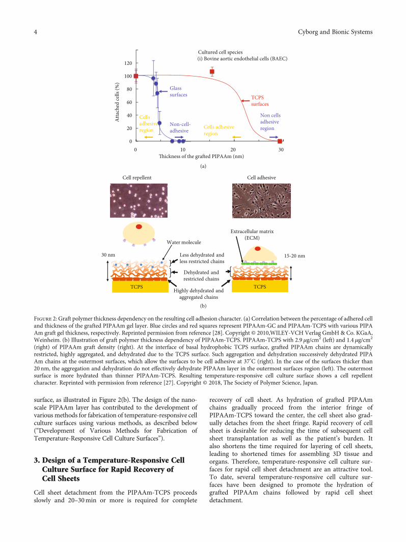
2.2. Design of Grafted PIPAAm Layer for Recovering CellSheet as a Temperature-Responsive Cell Culture Surface. Itwas observed that the grafted PIPAAm layer of PIPAAm-TCPS likely affected subsequent cell adhesion behavior,whereas the surface of the macroscopic PIPAAm gel, pre-pared using IPAAm and a cross-linking agent, did notexhibit cell adhesive character [10, 26]. To investigate theeffects of the grafted PIPAAm layer on the resultant celladhesive character, two types of PIPAAm-TCPS with dif-ferent thicknesses of the graft PIPAAm gel were preparedand characterized [10, 27]. The thicknesses of the graftedPIPAAm layers were directly measured by use of a UVexcimer laser and an atomic force microscope (AFM) andwere found to be 20 nm (20PIAAm-TCPS) and 30nm(30PIPAAm-TCPS). Bovine carotid artery endothelial cellsdid not attach to 30PIPAAm-TCPS at 37°C, where thePIPAAm chains are dehydrated. By contrast, cells adheredto the 20PIPAAm-TCPS as well as TCPS at 37°C. At a lowertemperature of 20°C, the adhered cells were completelydetached from 20PIPAAm-TCPS. The fact that adsorbed
fibronectin was not detected on 30PIPAAm-TCPS supportedthat 30PIPAAm-TCPS was a non-cell-adhesive surface. Thegrafted polymer thickness affects the subsequent cell attach-ment behavior. The graft polymer-thickness dependency oncell adhesion property was also observed in PIPAAm-graftedglass surface (PIPAAm-GS). The correlation between thegraft polymer thickness and cell adhesion ratio was graphi-cally plotted (Figure 2(a)) [28]. In the case of PIPAAm-GS,a 3.3 nm-thick grafted PIPAAm layer (3.3PIPAAm-GS) wasoptimal for expressing the temperature-dependent cellattachment/detachment. When the thickness was greaterthan the optimal polymer thickness (e.g., 8.8 nm-thickgrafted PIPAAm layer (8.8PIPAAm-GS)), cells were notadhered to PIPAAm-GS even at 37°C, similar to thatobserved in 30PIPAAm-TCPS [10]. The dependency of graftpolymer thickness on cell adhesion is explained in termsof the degree of molecular mobility and dehydration ofgraft PIPAAm chains [10, 28]. This consideration is illus-trated in Figure 2(b) [27]. The configuration and molecu-lar mobility of the grafted PIPAAm chains influence thesurface wettability with temperature changes [29–31]. Thisconsideration was applied to the nanoscale PIPAAm-graftedlayer of PIPAAm-TCPS and PIPAAm-GS surfaces. Hydro-phobic and immobile basal TCPS surface restrict graftedPIPAAm chains close to the TCPS surface, promoting aggre-gation and dehydration of grafted PIPAAm chains. Suchrestriction and dehydration of the grafted PIPAAm chainssuccessively promote dehydration at the outermost surfaceof the 20PIPAAm-TCPS chains, consequently providing20PIPAAm-TCPS with cell adhesive character. In contrast,in the case of 30PIPAAm-TCPS with the thicker polymericlayer, the successive restriction and dehydration of PIPAAm chains at the basal TCPS did not substantially affectthe dehydration of grafted PIPAAm chains at the outer-most regions. Polymer chains in the outermost regions of30PIPAAm-TCPS are more hydrated than those on20PIPAAm-TCPS. Whereby, cells do not attach to thethicker 30PIPAAm-TCPS at 37°C.
AFM observation of the increase and decrease in swellingof the graft PIPAAm layer of 3.3PIPAAm-GS and8.8PIPAAm-GS at 25°C and 37°C, respectively, under aque-ous conditions strongly supported the characteristics of thenanoscale PIPAAm graft layer as described above [28].Immersing 3.3PIPAAm-GS in an aqueous solution increasethe thickness from 3:3 nm ± 0:3 nm (dry state) to 8:0 nm ±1:4 nm (25°C) and 6:0 ± 1:4 nm (37°C). The thickness of thegraft PIPAAm layer was not substantially altered betweenabove and below the LCST. The graft PIPAAm layer of3.3PIPAAm-GS possibly maintained an unswollen stateunder aqueous conditions irrespective of temperature. Bycontrast, 8.8PIPAAm-GS shows a substantial increase ofthe graft PIPAAm layer, 25:4 nm ± 4:1 nm (25°C) and12:9 ± 2:2 nm (37°C) in aqueous conditions, dependingon temperature change. The PIPAAm graft layer of8.8PIPAAm-GS exhibited swelling and deswelling behaviorby temperature change. These results indicate that graftedPIPAAm chains in the thinner PIPAAm layers are readily sub-jected to successive dehydration and molecular restrictionfrom the grafted polymer chains, which are close to the basal
3Cyborg and Bionic Systems
surface, as illustrated in Figure 2(b). The design of the nano-scale PIPAAm layer has contributed to the development ofvarious methods for fabrication of temperature-responsive cellculture surfaces using various methods, as described below(“Development of Various Methods for Fabrication ofTemperature-Responsive Cell Culture Surfaces”).
3. Design of a Temperature-Responsive CellCulture Surface for Rapid Recovery ofCell Sheets
Cell sheet detachment from the PIPAAm-TCPS proceedsslowly and 20–30min or more is required for complete
recovery of cell sheet. As hydration of grafted PIPAAmchains gradually proceed from the interior fringe ofPIPAAm-TCPS toward the center, the cell sheet also grad-ually detaches from the sheet fringe. Rapid recovery of cellsheet is desirable for reducing the time of subsequent cellsheet transplantation as well as the patient’s burden. Italso shortens the time required for layering of cell sheets,leading to shortened times for assembling 3D tissue andorgans. Therefore, temperature-responsive cell culture sur-faces for rapid cell sheet detachment are an attractive tool.To date, several temperature-responsive cell culture sur-faces have been designed to promote the hydration ofgrafted PIPAAm chains followed by rapid cell sheetdetachment.
0
20
40
60
80
100
120
Thickness of the grafted PIPAAm (nm)
TCPS surfaces
Glass surfaces
Cultured cell species(i) Bovine aortic endothelial cells (BAEC)
Atta
ched
cells
(%)
Cells adhesiveregion
Non cellsadhesiveregion
0 10 20
(a)
(b)
30
Non-cell-adhesive
Cellsadhesiveregion
15-20 nm
Water molecule
TCPS
Extracellular matrix(ECM)
Cell repellent Cell adhesive
30 nm
Highly dehydrated and aggregated chains
Dehydrated and restricted chains
Less dehydrated and less restricted chains
TCPS
Figure 2: Graft polymer thickness dependency on the resulting cell adhesion character. (a) Correlation between the percentage of adhered celland thickness of the grafted PIPAAm gel layer. Blue circles and red squares represent PIPAAm-GC and PIPAAm-TCPS with various PIPAAm graft gel thickness, respectively. Reprinted permission from reference [28]. Copyright © 2010,WILEY-VCH Verlag GmbH & Co. KGaA,Weinheim. (b) Illustration of graft polymer thickness dependency of PIPAAm-TCPS. PIPAAm-TCPS with 2.9 μg/cm2 (left) and 1.4μg/cm2
(right) of PIPAAm graft density (right). At the interface of basal hydrophobic TCPS surface, grafted PIPAAm chains are dynamicallyrestricted, highly aggregated, and dehydrated due to the TCPS surface. Such aggregation and dehydration successively dehydrated PIPAAm chains at the outermost surfaces, which allow the surfaces to be cell adhesive at 37°C (right). In the case of the surfaces thicker than20 nm, the aggregation and dehydration do not effectively dehydrate PIPAAm layer in the outermost surfaces region (left). The outermostsurface is more hydrated than thinner PIPAAm-TCPS. Resulting temperature-responsive cell culture surface shows a cell repellentcharacter. Reprinted with permission from reference [27]. Copyright © 2018, The Society of Polymer Science, Japan.
4 Cyborg and Bionic Systems

3.1. Temperature-Responsive Cell Culture Surface UsingMicroporous Membrane and/or Incorporating HydrophilicPolymeric or Monomeric Components. Based on the conceptthat water molecules were efficiently supplied to the interfacebetween the cell sheets and the PIPAAm-grafted surface toaccelerate the hydration of PIPAAm chains below the cellsheet, PIPAAm gel was chemically deposited onto a porousmembrane (PM) such as a cell culture insert (PIPAAm-PM) (Figures 3(a) and 3(b)) [32]. The original microporestructure was preserved in PIPAAm-PM after grafting thePIPAAm gel, enabling the ready supply of water mole-cules through the pores to the basal part of the cell sheet.Indeed, PIPAAm-PM showed quicker cell sheet detachmentthan PIPAAm-TCPS at temperatures lower than 20°C. Inactual, PIPAAm-PM required ~30min for detachment of cellsheet, while PIPAAm-TCPS did ~75min (each cell culturearea was 4.2 cm2).
Polyethylene glycol (PEG) chains and PIPAAm cograftedPM (P(PIPAAm-co-PEG)-PM) further accelerated cell sheetdetachment (Figure 3(c)) [33]. The incorporated PEGmoietyallowed rapid water molecule diffusion from the peripherytoward the center of the cell sheet as well as beneath one,accelerating cell sheet detachment in comparison withPIPAAm-TCPS and PIPAAm-PM.
The monomer newly synthesized by Aoyagi et al. [34], 2-carboxyisopropylacrylamide (CIPAAm), is structurally simi-lar to N-isopropylacrylamide, besides the carboxyl group.IPAAm copolymerized with CIPAAm (P(IPAAm-co-CIPAAm)) showed almost the same LCST and temperatureresponse character as PIPAAm in aqueous solution. TheP(IPAAm-co-CIPAAm) gel-grafted TCPS (P(IPAAm-co-CIPAAm)-TCPS) exhibited cell adhesive characteristics sim-ilar to those of PIPAAm-TCPS and TCPS, whereas the cellsheet was more rapidly detached from P(IPAAm-co-CIPAAm) gel-grafted TCPS as the hydrophilic carboxylgroup of CIPAAm accelerated the hydration of the graftedP(IPAAm-co-CIPAAm) gel [34].
3.2. Temperature-Responsive Cell Culture Surface Incorporatedwith Free Mobile PIPAAm Chains. The comb-type graftedPIPAAm gel, which contains free mobile PIPAAm chains,showed rapid deswelling by temperature change in compari-son to a conventional PIPAAm cross-linked hydrogel(Figure 3(d), (1)) [35–37]. This is because the rapid dehydra-tion of the free mobile PIPAAm chains forms hydrophobicclusters inside the comb-type PIPAAm hydrogel above theLCST. The hydrophobic cluster promotes dehydrating thePIPAAm chains in the gel, enabling quick shrinkage of thePIPAAm gel. Based on the concept that free mobile PIPAAm chains also enabled rapid swelling of PIPAAm hydrogels,comb-type PIPAAm gels were chemically deposited on theTCPS (ctPIPAAm-TCPS) using IPAAm and PIPAAmmacromonomers (Figure 3(d), (2, 3)) [38]. At 37°C, cellsadhere and spread on ctPIPAAm-TCPS as well as PIPAAm-TCPS. After confluency, the cell sheets were more rap-idly detached from the ctPIPAAm-TCPS in comparison withPIPAAm-TCPS upon lowering temperature (20°C). Asexpected, the ctPIPAAm-TCPS containing free mobilePIPAAm chains attained more rapid cell sheet harvesting.
The free mobile PIPAAm chains promoted hydration of thedeposited comb-type PIPAAm gels.
3.3. PIPAAm Gel-Grafted Hydrophilic Polymeric Layer. Adouble polymeric gel layer consisting of nanoscale PIPAAmand polyacrylamide (PAAm) gels was grafted onto TCPS(PIPAAm-PAAm-TCPS) by successively grafting PAAmand PIPAAm (Figure 3(e)) [39]. The basal part of the doublepolymeric layer possibly formed an interpenetrating polymernetwork (IPN), which enabled the rapid swelling of thedouble polymeric gel layers, as the hydrophilic PAAmcomponent not only provides water molecules but alsofacilitates hydration of PIPAAm chains close to the basalhydrophobic TCPS surface [40, 41]. The PIPAAm-PAAm-TCPS has double layers, where the hydrophilicPAAm gel layer is sandwiched between the basal TCPSand PIPAAm gel layer. Optimization of the amount ofgrafted PIPAAm and PAAm showed cell adhesion charac-ter similar to PIPAA-TCPS at 37°C and rapid cell sheetdetachment by lowering temperature (20°C). However, cellswere not attached to PIPAAm-PAAm-TCPS with graftPAAm more than 3.3μg/cm2, because the PAAm layerlikely hydrates PIPAAm chains at its basal part, whereIPN was formed.
3.4. Stretchable Temperature-Responsive Cell Culture Surface.For a conventional PIPAAm-TCPS as introduced above, thethickness of the grafted PIPAAm gel was dominated by theamount of applied IPAAm monomer. The thickness ofgrafted PIPAAm layer could not be modulated after the poly-meric layer was grafted on hard base materials such as TCPSand glass. In contrast, as described above, cell attachment anddetachment properties depend on the thickness of the graftpolymeric layer. To readily modulate the thickness of thepolymeric graft layer, as well as subsequent cell attachmentand detachment events independent of the amount ofapplied monomer, PIPAAm gel grafted on an elastic polydi-methylsiloxane (PDMS) surface (PIPAAm-PDMS) was pre-pared as a stretchable temperature-responsive cell culturesurface via EB irradiation [42]. The grafted PIPAAm gel layerof PIPAAm-PDMS dynamically changed its thicknessthrough the application of mechanical stress such as stretch-ing and shrinking to the PIPAAm-PDMS. The PIPAAmlayer was more hydrophobic due to thinning the graft poly-meric layer when stretched, while the surface was morehydrophilic because of increasing the thickness of the layerwhen shrunken (Figure 3(f)). In reality, the uniaxiallystretched PIPAAm-PDMS was more hydrophobic and morecell adhesive than unstretched PIPAAm-PDMS at 37°C.PIPAAm-PDMS showed a chemically stable surface due tothe long-term temperature-dependent surface wettabilitychange. Moreover, shrink of the stretched PIPAAm-PDMSfollowed by lowering the temperature (20°C) (dual stimula-tion) promoted cell detachment compared to only tempera-ture change. Such a tendency was also observed in the cellsheet detachment process. Dual stimulation promoted cellsheet detachment from the stretched PIPAAm-PDMSsurface.
5Cyborg and Bionic Systems

Stretched PIPAAm-PDMS
Unstretched PIPAAm-PDMS
Temperature change
Rapid cell detachment
TCPS
PIPAAm
PAAm
: water molecule
Dual stimuliShrinking stressLowering temp.
PDMS PIPAAm gel layer
(1)
(2)
(3)
Low temp.
Deswelling of comb-type grafted PIPAAm gel
: Hydrophobic cluster
Swelling of comb-type grafted PIPAAm gel
Swelling of comb-type grafted PIPAAm-TCPSCell sheet
TCPS TCPS TCPS
Low temp.Cell sheet Water penetration from periphery
Slow detachmentPIPAAm layer (~20 nm) TCPSPIPAAm-TCPS
(a)
(b)
(c)
(d)
(e) (f)
Medium
Water penetration from periphery & bottom
Rapid detachmentMicroporous membrane(Pore size: 0.45 𝜇m, pore density: 1.6 106 pore/cm2)
PIPAAm-PM
Low temp.
Water penetration from periphery & bottom
Rapid detachmentPEG channelsP (IPAAm-co-PEG)-PM
PEGPIPAAm Cell
Low temp.
Figure 3: Schematic of temperature-responsive cell culture surface for acceleration of cell detachment. (a) PIPAAm-TCPS, (b) PIPAAm-PM,(c) P(IPAAm-co-PEG)-PM, (d) (1, 2) comb-type grafted PIPAAm gel and (3) ctPIPAAm-TCPS. These images of (a), (b), and (c) fromreference [37] (Z. Tang et al., Temperature-Responsive Polymer Modified Surface for Cell Sheet Engineering, Polymers, 4, pp. 1478) areunder a Creative Commons Attribution 3.0 International License (CC-BY-3.0). Illustration of (d) was reprinted with permission fromreference [38]. Copyright © 2010, Elsevier Ltd. (e) PIPAAm-PAAm-TCPS with a double polymer layer and (f) stretchable temperature-responsive cell culture surface (PIPAAm-PAAm-TCPS). Illustration of (e) was reprinted with permission from reference [39]. Copyright© 2014, Acta Materialia Inc. Published by Elsevier Ltd.
6 Cyborg and Bionic Systems

4. Development of Various Methods forFabrication of Temperature-Responsive CellCulture Surfaces
Information regarding the thickness dependency of thegrafted PIPAAm layer on cell adhesion behavior as describedabove has given valuable insights into the design and fab-rication of temperature-responsive cell culture surfaces,possibly motivating some researchers to develop new fabri-cation methods [43]. Methods such as polymer (or poly-meric gel particles) coating [44–53], photo-irradiation[54–57], and surface-initiated living radical polymerizationmethods such as atom transfer radical polymerization(ATRP) [58–63] and reversible addition-fragmentationchain transfer polymerization (RAFT) have been reported[64–66].
Surface-initiated ATRP and RAFT were attained toprecisely control the length of PIPAAm (molecular weight)on the polystyrene and glass surface with dense PIPAAm chains. Intensive research on the PIPAAm brush sur-face demonstrated that the PIPAAm chain length anddensity of brushes substantially influence cell attachmentand protein adsorption behaviors. When the molecularweight (Mn) and density of the graft polymeric chainswere optimized to be 23,000–58,000 and 0.03–0.04(chains/nm2), the PIPAAm brush surface enabled toexpress temperature-dependent cell attachment/detachmentproperty. At the optimal conditions, more importantly, thetemperature-dependent cell attachment/detachment prop-erty was due to the adsorbed fibronectin between the PIPAAm brushes, illustrated as ternary adsorption in previousreports [61, 63, 67].
Commercially available PIPAAm-TCPS (UpCell®) andconventional PIPAAm-TCPS were prepared via the EB irra-diation method. However, most researchers cannot accessspecial and expensive equipment as mentioned above forproducing temperature-responsive cell culture surfaces.Some researchers also pointed out that the newmethod couldpotentially reduce the cost of commercially available PIPAAm-TCPS. Based on this, facile and cost-effective spin coat-ing and UV or visible light irradiation methods have beendeveloped for the production of temperature-responsive cellculture surfaces.
A block copolymer (poly(butyl methacrylate)-b-PIPAAm) (PBMA) (PBMA-b-PIPAAm) was physically coatedon a polystyrene surface using a spin-coating method. Phys-ically coated temperature-responsive polymers tended tobe dissolved and eluted from the surface below the LCST[45, 52, 68], whereas, in the case of PBMA-b-PIPAAm-coated polystyrene surfaces, the hydrophobic PBMA unitswere strongly adsorbed on the surface through hydrophobicinteractions, preventing coated PBMA-b-PIPAAm from elu-tion from the surface below the LCST [44]. Optimizing thethickness of the coated PBMA-b-PIPAAm, PBMA-b-PIPAAm-coated surfaces enabled the expression of temperature-dependent cell attachment/detachment properties. PBMA-b-PIPAAm was also used for the preparation of a temperature-responsive cell culture insert [47].
The visible light irradiationmethod was also exploited forthe easy fabrication of temperature-responsive cell culturesurfaces [57]. The fabrication consists of two steps. Thiox-anthone photo-initiator groups are immobilized on polysty-rene (Th-PSt) dishes by incubating thiosalicylic aciddissolved in concentrated sulfuric acid. As a second step, anaqueous IPAAm solution containing N-methyldiethanola-mine was added to the Th-PSt surface followed by irradiationof visible light. The resultant PIPAAm-grafted PSt surfaceexhibited temperature-dependent cell sheet recovery. Thesenew methods would allow researchers to conveniently pre-pare temperature-responsive cell culture surfaces withoutusing special and expensive equipment such as EB irradiation.
5. Temperature-Responsive Cell CultureSurfaces Equipped with Bioactive Ligands
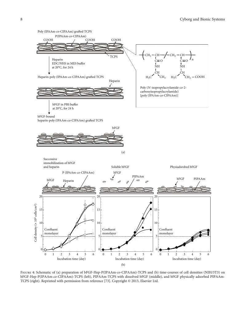
Bioactive peptides and proteinaceous growth factors werechemically immobilized on P(IPAAm-co-CIPAAm)-TCPSvia chemical reaction of the carboxyl groups of the surfacewith primary amine groups of the peptides [69, 70]. Ebaraet al. immobilized cell adhesive molecules, RGDS peptides,on the P(IPAAm-co-CIPAAm)-TCPS, and demonstratedthat human umbilical vein endothelial cells (HUVECs) werecultured at 37°C in the absence of serum [69]. After HUVECsreached confluency, they were recovered by reducing thetemperature (20°C) [70]. These results demonstrated con-trol of specific interactions between cultured cells and pep-tides. The P(IPAAm-co-CIPAAm) gel-grafted TCPS werealso available for immobilizing heparin molecules, whichhave a specific affinity for various heparin-binding pro-teins (e.g., basic fibroblast growth factor (bFGF or FGF-2) (Figure 4(a)), heparin-binding epithelial cell growth fac-tor (EGF)-like growth factor (HB-EGF), and VEGF [71–73]. Similar to heparan sulfate chains on proteoglycans,the immobilized heparin offers appropriate sites, whichmake growth factors form stable complexes. The growthfactors can maintain their activities and prevent diffusion.
Successively immobilized heparin and bFGF moleculeson P(IPAAm-co-CIPAAm)-TCPS (bFGF-Hep-P(PIPAAm-co-CIPAAm)-TCPS) showed faster proliferation of fibro-blasts than physically adsorbed bFGF surface and dissolvedbFGF in cell culture medium (Figure 4(b)) [73]. These resultssuggested that physically adsorbed bFGF molecules reducethe activity of the molecules due to their denaturation and/orunstable complex formation. Similarly, the heparin HB-EGFmolecules were also successively immobilized on heparin-immobilized temperature-responsive cell culture surfaces[71, 72]. Arisaka and Kobayashi et al. demonstrated thatHB-EGF-immobilized temperature-responsive cell culturesurfaces were available for the fabrication of hepatocytesheets. The hepatocyte sheet expressed genes specific forhepatocytes (albumin (Alb), hepatocyte nuclear factor 4alpha (Hnf4a), coagulation factor IX (F9), and coagulationfactor VII (F7)) during culture [72]. The specific gene expres-sion was higher than in hepatocyte sheets cultured in thepresence of dissolved HB-EGF. The hepatocyte sheet wasdetached from the surface at 20°C. Hepatocyte sheets could
7Cyborg and Bionic Systems
Poly (IPAAm-co-CIPAAm) grafted TCPSP(IPAAm-co-CIPAAm)
Heparin-poly (IPAAm-co-CIPAAm) grafted TCPS
Poly (N-isopropylacrylamide-co-2-carboxyisopropylacrylamide)[poly (IPAAm-co-CIPAAm)]
CH2 CH2CH
CHCH3 CH2H3CH3C COOH
CH
C
NH NH
O C O nCH
bFGF-boundheparin-poly (IPAAm-co-CIPAAm) grafted TCPS
bFGF in PBS buffer
HeparinEDC/NHS in MES buffer
TCPS
Heparin
bFGF
(a)
(b)
20
15
10
5
00 1 2
Confluentmonolayer
bFGF
bFGF
bFGFHeparin
P (IPAAm-co-CIPAAm)
Successiveimmobilization of bFGFand heparin Soluble bFGF Physiadsrobed bFGF
PIPAAmPIPAAm
Confluentmonolayer
Incubation time (day) Incubation time (day) Incubation time (day)
Confluentmonolayer
3 4 5 6 0 1 2 3 4 5 6 0 1 2 3 4 5 6
20
15
10
5
0
20
15
10
5
0
COOH COOH COOH
Figure 4: Schematic of (a) preparation of bFGF-Hep-P(IPAAm-co-CIPAAm)-TCPS and (b) time-courses of cell densities (NIH/3T3) onbFGF-Hep-P(IPAAm-co-CIPAAm)-TCPS (left), PIPAAm-TCPS with dissolved bFGF (middle), and bFGF physically adsorbed PIPAAm-TCPS (right). Reprinted with permission from reference [73]. Copyright © 2013, Elsevier Ltd.
8 Cyborg and Bionic Systems

be useful as new therapeutic approach for treatment of liverdisease, hemophilia [74].
6. Cell Sheet Manipulation Technology
Cell sheets generally shrink, wrinkle, and fold during detach-ing themselves from PIPAAm-TCPS. Such deformed cellsheets are troublesome for the manipulation and fabricationof cell-dense and thick tissue through layering and transplan-tation to native tissues and organs. To attain ideal manipula-tion and transplantation without deformation, commerciallyavailable products such as chitin membrane, porous PET,and hydrophilic modified poly(vinylidene difluoride) mem-branes were used as cell sheet carrier materials [75, 76]. Thesemembranes were physically overlaid on cultured cells at 37°Cafter aspirating almost the entire culture medium (retaininga small amount). By reducing the temperature below theLCST, the cell sheet was detached and attached to the over-laid cell sheet carrier membrane. By gently peeling off themembrane from the PIPAAm-TCPS with tweezers, thedetached cell sheet could be lifted up and manipulated withthe membrane without shrinking and wrinkling. Aftertransferring the cell sheet onto another cell sheet or biolog-ical tissue or organ, the cell sheet was released from themembrane with the addition of some medium. Addition-ally, commercially available membranes, synthetic polyioncomplex, poly(N,N-dimethylacrylamide-co-2-acrylamido-2-
methylpropane sulfonic acid) (P(DMAAm-co-AMPS)) andpoly(N,N-dimethylacrylamide-co-2-acryloxyethyltrimethy-lammonium chloride) (P(DMAAm-co-AETA-Cl)), werealso exploitable as cell sheet carriers [77].
In addition to the artificial polymeric membrane, gelatingel was also used as a cell sheet carrier. Gelatin was coated ona plunger-like device that aimed at being a cell sheet manip-ulator (Figure 5) [78, 79]. When the gelatin-coated devicewas deposited on confluent cells cultured on PIPAAm-TCPSat 20°C (Figure 5(b)), the cell sheet was detached from the sur-face and preferentially adhered to the gelatin-coated device. Itwas lifted from the device and placed on another cell sheet cul-tured on PIPAAm-TCPS (Figure 5(c)). Afterward, reducingthe temperature below LCST produced a double-layered cellsheet structure (Figure 5(d)). Repeating the process of layer-ing the cell sheet with the device enabled the fabrication ofdense and thick 3D tissue (Figure 5(e)). Additionally, a com-bination of the manipulation technique for cell sheet androbot technology successfully developed the automatic cellsheet stacking system, which enabled the automatic fabrica-tion of five-layered human skeletal muscle myoblast sheets(70–80μm thickness) in 100min [80]. This automation tech-nology contributed to the development of a flexible and auto-mated manufacturing facility: a tissue factory, which consistsof nine moduli and a material preparation isolator [81]. Thetissue factory automatically manufactures a five-layeredhuman myoblast sheet as a cell-based health care product.
(a)
(b) (c) (d)
(e)
ManipulatorGentian
Cell sheetCover on a cell sheet Recover by manipulator Transfer to new cell
culture surface
Manipulator
Gelatin
Silicon mold
Transfer on another cell sheet
Figure 5: Schematic of recovery, harvesting, transferring, and layering cell sheet using the gelatin-coated plunger device. (a) Macroscopicview of gelatin-coated plunger-like device as a cell sheet manipulator. (b) Cell sheet was covered by the gelatin-coated device andincubated at 20°C. (c) Cell sheet was harvested by the device. (d) The harvested cell sheet was transferred to a new cell culture surface. (e)The harvested cell sheet was layered on another cell sheet. The images from reference [36] (Z. Tang and T. Okano., Recent developmentof temperature-responsive surfaces and their application for cell sheet engineering, Regen Biomater, 2014, 1, pp. 91-102) are under aCreative Commons Attribution 4.0 International License (CC-BY-4.0).
9Cyborg and Bionic Systems

7. Cell Sheet-Based Regenerative Medicine
Cell sheet engineering has drawn attention, becoming apromising treatment for damaged tissues and organs. Cellsheet transplantation was superior to cell injectionapproaches in terms of the resultant survival rate of cellsand the subsequent recovery of damaged tissues and organs[82]. Transplanted cell sheets can adhere to biological tissuesvia their deposited ECM and survive thereon. However,injected cells did not stay on the tissues because they werescattered. Clinical application of autologous cell sheets hasbeen performed, demonstrating successful treatment of vari-ous damaged human tissues and organs (Figure 6(a)).
Nishida et al. pioneered corneal epithelial regenerationtreatment using a monolayer cell sheet. From tissues includ-ing epithelial stem cells derived from patients who lost eyevision due to alkali burn or drug side effects, corneal epithe-lial cells were expanded and recovered as a monolayer cellsheet from PIPAAm-TCPS [12, 83]. The corneal epithelialcell sheet was transplanted to treat the injured eye. For binoc-ular diseases, oral mucosal cell sheets were alternativelyavailable for treatment of patient eyes [12, 83]. Their abilityto see was improved after the cell sheet transplantation.Researchers in France also reported the efficacy of oral muco-sal cell sheet transplantation for regeneration of the ocularsurface in patients with binocular eye diseases [84].
Endoscopic submucosal dissection (ESD) is conducted asa less-invasive approach for resection of esophageal cancerthan a surgical one. However, after ESD, esophageal stenosisfrequently occurs due to artificial ulcer scarring, lowering thequality of life. Ohki et al. developed an approach for theregeneration of cell sheet-based esophagus tissue after EDStreatment [13]. They endoscopically transplanted oral muco-sal epithelial cell sheets onto the ulcer surface (Figure 6(b)).The transplanted cell sheet promotes reepithelialization ofthe esophagus and suppresses the stenosis event [14]. The cellsheet transplantation is available for treatment of patientswith Barrett’s esophagus [85].
After surgical treatment of adhesive otitis media and cho-lesteatoma middle ear, early postoperative mucosal regenera-tion of the middle ear and the mastoid cavity is desired forstructural and functional reconstruction of the cavity asmucosal regeneration prevents readhesion of the tympanicmembrane and recurrence of adhesive media and cholestea-toma. Yamamoto et al. have isolated nasal mucosa epithelialcells from the mucosal tissue of patients undergoing endos-copy and fabricated autologous nasal mucosa epithelial cellsheets using temperature-responsive cell culture inserts(Figure 6(c)) [17]. By combing tympanoplasty and autolo-gous nasal mucosal epithelial cell sheet transplantation, theydeveloped a new treatment method for postoperative muco-sal regeneration of the middle ear. The epithelial cell sheetswere transplanted to the damaged middle ear cavity, wheremucosa was lost. Transplantation successfully prevents thereappearance of cholesteatoma, tympanic membrane adhe-sion, and tympanic membrane retraction.
Air leaks after pulmonary resections can produce severecomplications without proper treatment. Kanzaki et al.applied autologous dermal fibroblasts cells, which were iso-
lated from patient skin tissue, followed by an expansion ontemperature-responsive cell culture surface, to close apatient’s pleural defect. The air leaks were completely sealedwith the cell sheet (Figure 6(d)) [19].
Cells from periodontal ligaments on wisdom teeth werecultured and harvested as periodontal ligament cell sheets.Once periodontal tissues are damaged, it is difficult to healspontaneously. Periodontal disease causes the loss of peri-odontal tissue, causing the instability or loss of teeth. Afterexpanding and culturing cells isolated from periodontal liga-ment (PDL) tissue of patients, three-layered autologousPDL-derived cell sheets were transplanted to root surface ofpatients with bone prosthetic materials (Figure 6(e)) by Iwataand Ishikawa et al. [15, 16]. As a result, transplantation notonly stabilizes teeth but also recovers periodontal ligamenttissue, reduces the periodontal probing depth, and improvesthe bone around the transplanted cell sheets.
Damaged articular cartilage cannot heal spontaneouslydue to a lack of blood supply and a low density of cells. Satoet al. developed a new treatment for damaged articular carti-lage [18]. Chondrocytes and synovial cells collected frompatients were cocultured on temperature-responsive cell cul-ture inserts and cell culture surfaces, respectively. Layeredchondrocyte sheets were transplanted to cartilage defects ofpatients that were treated with conventional surgical treat-ment. This demonstrates that the transplantation promoteshyaline cartilage repair.
Sawa et al. have transplanted autologous skeletal myo-blast sheet for the regeneration of ischemic myocardium[11]. Skeletal myoblast cells were isolated frommuscle tissuesof patient leg and cultured for the fabrication of autologousskeletal sheets. In a first clinical trial, they transplantedthree-layered autologous skeletal myoblast sheets onto theheart surfaces of a patient with serious cardiac insufficiencydue to dilated cardiomyopathy. Cell sheet transplantationimproved cardiac function in the patient. Later, it alsoenabled the removal of a left ventricular assist device fromthe patient. The improvement of cardiac function after cellsheet transplantation is attributable to cytokine secretion(such as VEGF, FGF, and HGF) from the transplanted cellsheets and paracrine effects inducing the recruitment of stemcells. Sawa et al. treated more patients with myoblast cellsheet transplantation. They demonstrated the feasibility andsafety of transplantation of autologous skeletal myoblastsheets in patients with severe chronic disease [86]. Based onthese insights, “Heart Sheet” was released as a medical prod-uct for patients with ischemic heart disease from TerumoCorporation, Japan in 2016.
8. Conclusions
Temperature-responsive cell culture surface established cellsheet engineering, which has offered fundamental technologyfor the fabrication of 3D tissues and organs as well as cellsheet-based regenerative medicine. Precise control of thenanoscale graft PIPAAm layer is essential for expression oftemperature-dependent cell attachment/detachment charac-ter and subsequent cell sheet recovery. Considerable progressin cell sheet engineering for years has created new and
10 Cyborg and Bionic Systems

Oral mucosalepithelialcellsheet
(i) Corneal tissue(ii) Esophagus tissue (b)
Nasal mucosalepithelial cell sheet
(iii) Middleear (c)
Dermal fibro blast sheet(iv) Lung (d)
Periodontal ligamentderivedcellsheets
(i) Periodontal ligament (e)
Chondrocyte sheets(ii) Articular cartilage
Myoblast cell sheets(iii) Heart
PIPAAm-TCPSMonolayer Multilayer
Manipulation
Layering cell sheets
Harvesting autologous cell sheet
Transplantation
(a)
(b) (c)
(d) (e)
(1) Biopsy of oral mucosal tissue
(2) Isolate and expand cells𝜃 6 mm
(3) Harvest cell sheets
(4) Endoscopic transplantation of cell sheets
Autologous oral mucosal epithelial cell sheets
(2) Isolate and expand cells
H&E staining
Autologous nasal mucosal epithelialcell sheets
(3) Harvest cell sheets
(1) Endoscopic biopsy of nasal mucosal tissue
(4) Transplantation ofcell sheets to middleear cavity
(1) Harvested dermal fibroblast sheet
(2) Transplanted dermal fibroblast sheetsealed the air leaks (rectangle area)
1. Scrape PDL tissue fromthe mid-third of tooth
2. Expand and createtriple-layered PDL cell sheets
𝛽-tricalcium phosphategranules were filled inbony defects
3. Transplanton the root
4.
(B)
(A)
Figure 6: Clinical applications of autologous cell sheet-based regenerative medicine. (a) Schematic of harvesting, manipulating, andtransplanting mono- and multilayered autologous cell sheets for regeneration of various types of human tissues and organs. (b)Transplantation of autologous oral mucosal epithelial cell sheet for treatments of damaged artificial ulceration after esophageal ESD. Thisillustration of (b) was adopted from reference [13] with permission form AGA Institute. Published by Elsevier Inc. (c) Transplantation ofautologous nasal mucosal epithelial cell sheet for regeneration of postoperative mucosal regeneration of the middle ear. This image fromreference [17] (K. Yamamoto et al., Middle ear mucosal regeneration by tissue-engineered cell sheet transplantation, 2017, npjRegenerative Medicine, 2, pp. 6) is licensed under a Creative Commons Attribution 4.0 International License (CC-BY-4.0). (d)Transplantation of autologous dermal fibroblast sheet for sealing the air leaks [19]. This image from reference [19] (M. Kanzaki et al.,Bio-artificial pleura using an autologous dermal fibroblast sheet, 2017, npj Regenerative Medicine, 2, pp. 26) is licensed under aCreative Commons Attribution 4.0 International License (CC-BY-4.0). (e) Transplantation of three-layered autologous PDL tissue derivedcell sheets on the root of the damaged tooth. This image was adopted from reference [15] with permission from The Japanese Society forRegenerative Medicine. Production and hosting by Elsevier B.V.
11Cyborg and Bionic Systems

various types of temperature-responsive cell culture surfaces,automated fabrication and manipulation systems, and newmedical treatments for damaged human tissues and organs.Currently, iPS and mesenchymal stem cells (MSCs) havebecome attractive cell sources for cell-based therapy and havebeen intensively researched in basic and clinical research[87–90]. Transplantation of allogenic cell sheets using MSCshas been conducted as a basic study for the development ofnew medical treatments. Various types of cells differentiatedfrom iPSCs and MSCs would extend the range of applicationof cell sheet-based therapies for damaged tissues and organs:salivary glands [91], diabetic nephropathy (chronic kidneydisease) [92, 93], cerebral ischemia [94], and so on, [95–97]with more complicated structures and functions.
Conflicts of Interest
The author declares no conflicts of interest associated withthis manuscript.
Acknowledgments
This work was partially supported by A Grant-in-aid forScientific Research (JSPS KAKENHI Grants 18K12084) anda Grant-in-aid for Scientific Research on Innovative Areas(MEXT KAKENHI Grants 23106009).
References
[1] R. Langer and J. P. Vacanti, “Tissue engineering,” Science,vol. 260, no. 5110, pp. 920–926, 1993.
[2] A. Atala, S. B. Bauer, S. Soker, J. J. Yoo, and A. B. Retik, “Tis-sue-engineered autologous bladders for patients needingcystoplasty,” The Lancet, vol. 367, no. 9518, pp. 1241–1246,2006.
[3] G. Zhou, H. Jiang, Z. Yin et al., “In vitro regeneration ofpatient-specific ear-shaped cartilage and its first clinical appli-cation for auricular reconstruction,” eBioMedicine, vol. 28,pp. 287–302, 2018.
[4] T. Shin'oka, Y. Imai, and Y. Ikada, “Transplantation of atissue-engineered pulmonary artery,” New England Journal ofMedicine, vol. 344, no. 7, pp. 532-533, 2001.
[5] M. Poh, M. Boyer, A. Solan et al., “Blood vessels engineeredfrom human cells,” The Lancet, vol. 365, no. 9477, pp. 2122–2124, 2005.
[6] J. Yang, M. Yamato, C. Kohno et al., “Cell sheet engineering:recreating tissues without biodegradable scaffolds,” Biomate-rials, vol. 26, no. 33, pp. 6415–6422, 2005.
[7] J. Kobayashi, A. Kikuchi, T. Aoyagi, and T. Okano, “Cell sheettissue engineering: cell sheet preparation, harvesting/manipu-lation, and transplantation,” Journal of Biomedical MaterialsResearch Part A, vol. 107, no. 5, pp. 955–967, 2019.
[8] Z. L. Tang, Y. Akiyama, and T. Okano, “Recent developmentof temperature-responsive cell culture surface using poly(N-isopropylacrylamide),” Journal of Polymer Science PartB-Polymer Physics, vol. 52, no. 14, pp. 917–926, 2014.
[9] N. Yamada, T. Okano, H. Sakai, F. Karikusa, Y. Sawasaki, andY. Sakurai, “Thermo-responsive polymeric surfaces; control ofattachment and detachment of cultured cells,” Die Makromo-lekulare Chemie, Rapid Communications, vol. 11, no. 11,pp. 571–576, 1990.
[10] Y. Akiyama, A. Kikuchi, M. Yamato, and T. Okano, “Ultrathinpoly(N-isopropylacrylamide) grafted layer on polystyrene sur-faces for cell adhesion/detachment control,” Langmuir, vol. 20,no. 13, pp. 5506–5511, 2004.
[11] Y. Sawa, S. Miyagawa, T. Sakaguchi et al., “Tissue engineeredmyoblast sheets improved cardiac function sufficiently todiscontinue LVAS in a patient with DCM: report of a case,”Surgery Today, vol. 42, no. 2, pp. 181–184, 2012.
[12] K. Nishida, M. Yamato, Y. Hayashida et al., “Corneal recon-struction with tissue-engineered cell sheets composed of autol-ogous oral mucosal epithelium,” The New England Journal ofMedicine, vol. 351, no. 12, pp. 1187–1196, 2004.
[13] T. Ohki, M. Yamato, M. Ota et al., “Prevention of esophagealstricture after endoscopic submucosal dissection using tissue-engineered cell sheets,” Gastroenterology, vol. 143, pp. 582–588, 2012.
[14] N. Yamaguchi, H. Isomoto, S. Kobayashi et al., “Oral epithelialcell sheets engraftment for esophageal strictures after endo-scopic submucosal dissection of squamous cell carcinomaand airplane transportation,” Scientific Reports, vol. 7, no. 1,p. 17460, 2017.
[15] T. Iwata, M. Yamato, K. Washio et al., “Periodontal regen-eration with autologous periodontal ligament-derived cellsheets - a safety and efficacy study in ten patients,” Regener-ative Therapy, vol. 9, pp. 38–44, 2018.
[16] T. Iwata, M. Yamato, K. Washio, T. Ando, T. Okano, andI. Ishikawa, “Cell sheets for periodontal tissue engineering,”Current Oral Health Reports, vol. 2, no. 4, pp. 252–256,2015.
[17] K. Yamamoto, M. Yamato, T. Morino et al., “Middle earmucosal regeneration by tissue-engineered cell sheet trans-plantation,” npj Regenerative Medicine, vol. 2, p. 6, 2017.
[18] M. Sato, M. Yamato, G. Mitani et al., “Combined surgery andchondrocyte cell-sheet transplantation improves clinical andstructural outcomes in knee osteoarthritis,” npj RegenerativeMedicine, vol. 4, p. 4, 2019.
[19] M. Kanzaki, R. Takagi, K. Washio, M. Kokubo, andM. Yamato, “Bio-artificial pleura using an autologous dermalfibroblast sheet,” npj Regenerative Medicine, vol. 2, p. 26, 2017.
[20] M. Heskins and J. E. Guillet, “Solution properties of poly(N-isopropylacrylamide),” Journal of Macromolecular Science:Part A - Chemistry, vol. 2, no. 8, pp. 1441–1455, 1968.
[21] T. Okano, N. Yamada, M. Okuhara, H. Sakai, and Y. Sakurai,“Mechanism of cell detachment from temperature-modulated,hydrophilic- hydrophobic polymer surfaces,” Biomaterials,vol. 16, no. 4, pp. 297–303, 1995.
[22] M. Yamato, M. Okuhara, F. Karikusa, A. Kikuchi, Y. Sakurai,and T. Okano, “Signal transduction and cytoskeletal reorgani-zation are required for cell detachment from cell culture sur-faces grafted with a temperature-responsive polymer,”Journal of Biomedical Materials Research, vol. 44, no. 1,pp. 44–52, 1999.
[23] A. Kushida, M. Yamato, C. Konno, A. Kikuchi, Y. Sakurai, andT. Okano, “Decrease in culture temperature releases mono-layer endothelial cell sheets together with deposited fibronec-tin matrix from temperature-responsive culture surfaces,”Journal of Biomedical Materials Research, vol. 45, no. 4,pp. 355–362, 1999.
[24] M. Harimoto, M. Yamato, M. Hirose et al., “Novel approachfor achieving double-layered cell sheets co-culture: overlayingendothelial cell sheets onto monolayer hepatocytes utilizing
12 Cyborg and Bionic Systems

temperature-responsive culture dishes,” Journal of BiomedicalMaterials Research, vol. 62, no. 3, pp. 464–470, 2002.
[25] T. Shimizu, M. Yamato, A. Kikuchi, and T. Okano, “Two-dimensional manipulation of cardiac myocyte sheets utilizingtemperature-responsive culture dishes augments the pulsatileamplitude,” Tissue Engineering, vol. 7, no. 2, pp. 141–151,2001.
[26] H. Sakai, T. Okano, N. Yamada, and Y. Sakurai, “Thermore-sponsive polymer surface for cell culture: analysis of thesurface and control of the cell attachment/detachment,” inAdvanced Biomaterials in Biomedical Engineering and DrugDelivery Systems, N. Ogata, S. Kim, J. Feijen, and T. Okano,Eds., pp. 231-232, Springer, Tokyo, 1996.
[27] Y. Akiyama and T. Okano, “Temperature-responsive cell cul-ture surface for cell-sheet tissue engineering and its design toexpress temperature-dependent cell attachment/detachmentcharacter,” Kobunshi Ronbunshu, vol. 75, no. 2, pp. 174–186,2018.
[28] K. Fukumori, Y. Akiyama, Y. Kumashiro et al., “Characteriza-tion of ultra-thin temperature-responsive polymer layer andits polymer thickness dependency on cell attachment/detach-ment properties,” Macromolecular Bioscience, vol. 10, no. 10,pp. 1117–1129, 2010.
[29] T. Yakushiji, K. Sakai, A. Kikuchi, T. Aoyagi, Y. Sakurai, andT. Okano, “Graft architectural effects on thermoresponsivewettability changes of poly(N-isopropylacrylamide)-modifiedsurfaces,” Langmuir, vol. 14, no. 16, pp. 4657–4662, 1998.
[30] T. Yakushiji, K. Sakai, A. Kikuchi, T. Aoyagi, Y. Sakurai, andT. Okano, “Effects of cross-linked structure on temperature-responsive hydrophobic interaction of poly(N-isopropylacry-lamide) hydrogel-modified surfaces with steroids,” AnalyticalChemistry, vol. 71, no. 6, pp. 1125–1130, 1999.
[31] Y. G. Takei, T. Aoki, K. Sanui, N. Ogata, Y. Sakurai, andT. Okano, “Dynamic contact angle measurement oftemperature-responsive surface properties for poly(N-isopro-pylacrylamide) grafted surfaces,” Macromolecules, vol. 27,no. 21, pp. 6163–6166, 1994.
[32] O. H. Kwon, A. Kikuchi, M. Yamato, Y. Sakurai, and T. Okano,“Rapid cell sheet detachment from poly(N-isopropylacryla-mide)-grafted porous cell culture membranes,” Journal of Bio-medical Materials Research, vol. 50, no. 1, pp. 82–89, 2000.
[33] O. Hyeong Kwon, A. Kikuchi, M. Yamato, and T. Okano,“Accelerated cell sheet recovery by co-grafting of PEG withPIPAAm onto porous cell culture membranes,” Biomaterials,vol. 24, no. 7, pp. 1223–1232, 2003.
[34] M. Ebara, M. Yamato, M. Hirose et al., “Copolymerization of2-carboxyisopropylacrylamide with N-isopropylacrylamideaccelerates cell detachment from grafted surfaces by reducingtemperature,” Biomacromolecules, vol. 4, no. 2, pp. 344–349,2003.
[35] R. Yoshida, K. Uchida, Y. Kaneko et al., “Comb-type graftedhydrogels with rapid deswelling response to temperaturechanges,” Nature, vol. 374, no. 6519, pp. 240–242, 1995.
[36] Z. Tang and T. Okano, “Recent development of temperature-responsive surfaces and their application for cell sheet engi-neering,” Regen Biomater, vol. 1, no. 1, pp. 91–102, 2014.
[37] Z. Tang, Y. Akiyama, and T. Okano, “Temperature-responsivepolymer modified surface for cell sheet engineering,” Polymers,vol. 4, no. 3, pp. 1478–1498, 2012.
[38] Z. Tang, Y. Akiyama, M. Yamato, and T. Okano, “Comb-typegrafted poly(N-isopropylacrylamide) gel modified surfaces for
rapid detachment of cell sheet,” Biomaterials, vol. 31, no. 29,pp. 7435–7443, 2010.
[39] Y. Akiyama, A. Kikuchi, M. Yamato, and T. Okano, “Acceler-ated cell-sheet recovery from a surface successively graftedwith polyacrylamide and poly(N-isopropylacrylamide),” ActaBiomaterialia, vol. 10, no. 8, pp. 3398–3408, 2014.
[40] Y. H. Bae, T. Okano, and S. W. Kim, ““On–off” thermocontrolof solute transport. I. Temperature dependence of swelling ofN-isopropylacrylamide networks modified with hydrophobiccomponents in water,” Pharmaceutical Research, vol. 8, no. 4,pp. 531–537, 1991.
[41] M. R. Guilherme, G. M. Campese, E. Radovanovic, A. F.Rubira, E. B. Tambourgi, and E. C. Muniz, “Thermo-respon-sive sandwiched-like membranes of IPN-PNIPAAm/PAAmhydrogels,” Journal of Membrane Science, vol. 275, no. 1-2,pp. 187–194, 2006.
[42] Y. Akiyama, M. Matsuyama, M. Yamato, N. Takeda, andT. Okano, “Poly(N-isopropylacrylamide)-grafted polydimeth-ylsiloxane substrate for controlling cell adhesion and detach-ment by dual stimulation of temperature and mechanicalstress,” Biomacromolecules, vol. 19, no. 10, pp. 4014–4022,2018.
[43] Y. V. Pan, R. A. Wesley, R. Luginbuhl, D. D. Denton, and B. D.Ratner, “Plasma polymerized N-isopropylacrylamide: synthe-sis and characterization of a smart thermally responsive coat-ing,” Biomacromolecules, vol. 2, no. 1, pp. 32–36, 2001.
[44] M. Nakayama, N. Yamada, Y. Kumashiro, H. Kanazawa,M. Yamato, and T. Okano, “Thermoresponsive poly(N-iso-propylacrylamide)-based block copolymer coating for opti-mizing cell sheet fabrication,” Macromolecular Bioscience,vol. 12, no. 6, pp. 751–760, 2012.
[45] M. E. Nash, W. M. Carroll, N. Nikoloskya et al., “Straightfor-ward, one-step fabrication of ultrathin thermoresponsive filmsfrom commercially available pNIPAm for cell culture andrecovery,” ACS Applied Materials & Interfaces, vol. 3, no. 6,pp. 1980–1990, 2011.
[46] D. Healy, M. Nash, A. Gorleov, K. Thompson, P. Dockery, andY. Rochev, “An investigation of cell growth and detachmentfrom thermoresponsive physically crosslinked networks,”Colloids and Surfaces B: Biointerfaces, vol. 159, pp. 159–165,2017.
[47] M. Nakayama, Y. Toyoshima, H. Chinen, A. Kikuchi,M. Yamato, and T. Okano, “Water stable nanocoatings ofpoly(N-isopropylacrylamide)-based block copolymers onculture insert membranes for temperature-controlled celladhesion,” Journal of Materials Chemistry B, vol. 8, no. 34,pp. 7812–7821, 2020.
[48] N. A. Dzhoyashvili, K. Thompson, A. V. Gorelov, and Y. A.Rochev, “Film thickness determines cell growth and cell sheetdetachment from spin-coated poly(N-isopropylacrylamide)substrates,” ACS Applied Materials & Interfaces, vol. 8,no. 41, pp. 27564–27572, 2016.
[49] V. M. Varghese, V. Raj, K. Sreenivasan, and T. V. Kumary, “Invitro cytocompatibility evaluation of a thermoresponsiveNIPAAm-MMA copolymeric surface using L929 cells,” Jour-nal of Materials Science: Materials in Medicine, vol. 21, no. 5,pp. 1631–1639, 2010.
[50] S. Schmidt, M. Zeiser, T. Hellweg, C. Duschl, A. Fery, andH. Möhwald, “Adhesion and mechanical properties of PNI-PAM microgel films and their potential use as switchable cellculture substrates,” Advanced Functional Materials, vol. 20,no. 19, pp. 3235–3243, 2010.
13Cyborg and Bionic Systems

[51] L. M. Mukundan, R. Nirmal, L. V. Thomas, U. S. Sajeev, andP. D. Nair, “Retrieval of rat aortic smooth muscle cells as intactcell sheet for regenerative medicine: a cost effective approachusing photo polymerization,” Biotechnology Letters, vol. 33,no. 10, pp. 2083–2089, 2011.
[52] M. T. Moran, W. M. Carroll, A. Gorelov, and Y. Rochev,“Intact endothelial cell sheet harvesting from thermorespon-sive surfaces coated with cell adhesion promoters,” J R SocInterface, vol. 4, no. 17, pp. 1151–1157, 2007.
[53] X. J. Loh,W. C. D. Cheong, J. Li, and Y. Ito, “Novel poly(N-iso-propylacrylamide)-poly[(R)-3-hydroxybutyrate]-poly(N-iso-propylacrylamide) triblock copolymer surface as a culturesubstrate for human mesenchymal stem cells,” Soft Matter,vol. 5, no. 15, pp. 2937–2946, 2009.
[54] M. E. Nash, W. M. Carroll, P. J. Foley et al., “Ultra-thin spincoated crosslinkable hydrogels for use in cell sheet recovery-synthesis, characterisation to application,” Soft Matter, vol. 8,no. 14, pp. 3889–3899, 2012.
[55] H. A. von Recum, S. W. Kim, A. Kikuchi, M. Okuhara,Y. Sakurai, and T. Okano, “Novel thermally reversible hydro-gel as detachable cell culture substrate,” Journal of BiomedicalMaterials Research, vol. 40, no. 4, pp. 631–639, 1998.
[56] Y. Ito, G. Chen, Y. Guan, and Y. Imanishi, “Patterned immo-bilization of thermoresponsive polymer,” Langmuir, vol. 13,no. 10, pp. 2756–2759, 1997.
[57] K. Fukumori, Y. Akiyama, M. Yamato, and T. Okano, “A facilemethod for preparing temperature-responsive cell culture sur-faces by using a thioxanthone photoinitiator immobilized on apolystyrene surface,” ChemNanoMat, vol. 2, no. 5, pp. 454–460, 2016.
[58] A. Mizutani, A. Kikuchi, M. Yamato, H. Kanazawa, andT. Okano, “Preparation of thermoresponsive polymer brushsurfaces and their interaction with cells,” Biomaterials,vol. 29, no. 13, pp. 2073–2081, 2008.
[59] L. Li, Y. Zhu, B. Li, and C. Gao, “Fabrication of Thermorespon-sive polymer gradients for study of cell adhesion and detach-ment,” Langmuir, vol. 24, no. 23, pp. 13632–13639, 2008.
[60] K. Nagase, M. Watanabe, A. Kikuchi, M. Yamato, andT. Okano, “Thermo-responsive polymer brushes as intelligentbiointerfaces: preparation via ATRP and characterization,”Macromolecular Bioscience, vol. 11, no. 3, pp. 400–409, 2011.
[61] C. Y. Xue, B. C. Choi, S. Choi, P. V. Braun, and D. E. Leckband,“Protein adsorption modes determine reversible cell attach-ment on poly(N-isopropyl acrylamide) brushes,” AdvancedFunctional Materials, vol. 22, no. 11, pp. 2394–2401, 2012.
[62] X. Sui, A. Di Luca, M. K. Gunnewiek et al., “Stability and celladhesion properties of poly(N-isopropylacrylamide) brusheswith variable grafting densities,” Australian Journal of Chemis-try, vol. 64, no. 9, pp. 1261–1268, 2011.
[63] S. Choi, B. C. Choi, C. Xue, and D. E. Leckband, “Proteinadsorption mechanisms determine the efficiency of thermallycontrolled cell adhesion on poly(N-isopropyl acrylamide)brushes,” Biomacromolecules, vol. 14, no. 1, pp. 92–100,2012.
[64] H. Takahashi, M. Nakayama, M. Yamato, and T. Okano,“Controlled chain length and graft density of thermorespon-sive polymer brushes for optimizing cell sheet harvest,” Bio-macromolecules, vol. 11, no. 8, pp. 1991–1999, 2010.
[65] N. Matsuzaka, H. Takahashi, M. Nakayama, A. Kikuchi,and T. Okano, “Effect of the hydrophobic basal layer ofthermoresponsive block co-polymer brushes on thermally-
induced cell sheet harvest,” Journal of Biomaterials Science.Polymer Edition, vol. 23, no. 10, pp. 1301–1314, 2011.
[66] N. Matsuzaka, M. Nakayama, H. Takahashi, M. Yamato,A. Kikuchi, and T. Okano, “Terminal-functionality effect ofpoly(N-isopropylacrylamide) brush surfaces on temperature-controlled cell adhesion/detachment,” Biomacromolecules,vol. 14, no. 9, pp. 3164–3171, 2013.
[67] A. Halperin and M. Kroger, “Theoretical considerations onmechanisms of harvesting cells cultured on thermoresponsivepolymer brushes,” Biomaterials, vol. 33, no. 20, pp. 4975–4987,2012.
[68] G. Rollason, J. E. Davies, andM. V. Sefton, “Preliminary reporton cell culture on a thermally reversible copolymer,” Biomate-rials, vol. 14, no. 2, pp. 153–155, 1993.
[69] M. Ebara, M. Yamato, T. Aoyagi, A. Kikuchi, K. Sakai, andT. Okano, “Immobilization of cell-adhesive peptides totemperature-responsive surfaces facilitates both serum-freecell adhesion and noninvasive cell harvest,” Tissue Engineer-ing, vol. 10, no. 7-8, pp. 1125–1135, 2004.
[70] M. Ebara, M. Yamato, T. Aoyagi, A. Kikuchi, K. Sakai, andT. Okano, “Temperature-responsive cell culture surfacesenable "on-off" affinity control between cell integrins andRGDS ligands,” Biomacromolecules, vol. 5, no. 2, pp. 505–510, 2004.
[71] J. Kobayashi, M. Hayashi, T. Ohno et al., “Surface design ofantibody-immobilized thermoresponsive cell culture dishesfor recovering intact cells by low-temperature treatment,”Journal of Biomedical Materials Research. Part A, vol. 102,no. 11, pp. 3883–3893, 2014.
[72] Y. Arisaka, J. Kobayashi, K. Ohashi et al., “A heparin-modifiedthermoresponsive surface with heparin-binding epidermalgrowth factor-like growth factor for maintaining hepatic func-tions in vitro and harvesting hepatocyte sheets,” RegenerativeTherapy, vol. 3, pp. 97–106, 2016.
[73] Y. Arisaka, J. Kobayashi, M. Yamato, Y. Akiyama, andT. Okano, “Switching of cell growth/detachment on heparin-functionalized thermoresponsive surface for rapid cell sheetfabrication and manipulation,” Biomaterials, vol. 34, no. 17,pp. 4214–4222, 2013.
[74] K. Tatsumi and T. Okano, “Hepatocyte transplantation: cellsheet technology for liver cell transplantation,” Current Trans-plantation Reports, vol. 4, no. 3, pp. 184–192, 2017.
[75] A. Kikuchi, M. Okuhara, F. Karikusa, Y. Sakurai, andT. Okano, “Two-dimensional manipulation of confluently cul-tured vascular endothelial cells using temperature-responsivepoly(N-isopropylacrylamide)-grafted surfaces,” Journal of Bio-materials Science. Polymer Edition, vol. 9, pp. 1331–1348,2012.
[76] M. Hirose, O. H. Kwon, M. Yamato, A. Kikuchi, and T. Okano,“Creation of designed shape cell sheets that are noninvasivelyharvested and moved onto another surface,” Biomacromole-cules, vol. 1, no. 3, pp. 377–381, 2000.
[77] Z. Tang, A. Kikuchi, Y. Akiyama, and T. Okano, “Novel cellsheet carriers using polyion complex gel modified membranesfor tissue engineering technology for cell sheet manipulationand transplantation,” Reactive & Functional Polymers,vol. 67, no. 11, pp. 1388–1397, 2007.
[78] T. Sasagawa, T. Shimizu, S. Sekiya et al., “Design of prevascu-larized three-dimensional cell-dense tissues using a cell sheetstacking manipulation technology,” Biomaterials, vol. 31,no. 7, pp. 1646–1654, 2010.
14 Cyborg and Bionic Systems
[79] Y. Haraguchi, T. Shimizu, T. Sasagawa et al., “Fabrication offunctional three-dimensional tissues by stacking cell sheetsin vitro,” Nature Protocols, vol. 7, no. 5, pp. 850–858, 2012.
[80] T. Kikuchi, T. Shimizu, M. Wada, M. Yamato, and T. Okano,“Automatic fabrication of 3-dimensional tissues using cellsheet manipulator technique,” Biomaterials, vol. 35, no. 8,pp. 2428–2435, 2014.
[81] T. Kikuchi, M. Kino-oka, M.Wada et al., “A novel, flexible andautomated manufacturing facility for cell-based health careproducts: tissue factory,” Regenerative Therapy, vol. 9,pp. 89–99, 2018.
[82] H. Sekine, T. Shimizu, I. Dobashi et al., “Cardiac cell sheettransplantation improves damaged heart function via superiorcell survival in comparison with dissociated cell injection,” Tis-sue Engineering. Part A, vol. 17, no. 23-24, pp. 2973–2980,2011.
[83] K. Nishida, M. Yamato, Y. Hayashida et al., “Functional bioen-gineered corneal epithelial sheet grafts from corneal stem cellsexpanded ex vivo on a temperature-responsive cell culture sur-face,” Transplantation, vol. 77, no. 3, pp. 379–385, 2004.
[84] C. Burillon, L. Huot, V. Justin et al., “Cultured autologous oralmucosal epithelial cell sheet (CAOMECS) transplantation forthe treatment of corneal limbal epithelial stem cell deficiency,”Investigative Ophthalmology & Visual Science, vol. 53, no. 3,pp. 1325–1331, 2012.
[85] E. Jonas, S. Sjöqvist, P. Elbe et al., “Transplantation of tissue-engineered cell sheets for stricture prevention after endoscopicsubmucosal dissection of the oesophagus,” United EuropeanGastroenterology Journal, vol. 4, no. 6, pp. 741–753, 2016.
[86] Y. Sawa, Y. Yoshikawa, K. Toda et al., “Safety and efficacy ofautologous skeletal myoblast sheets (TCD-51073) for the treat-ment of severe chronic heart failure due to ischemic heart dis-ease,” Circulation Journal, vol. 79, no. 5, pp. 991–999, 2015.
[87] M. Nakao, D. Inanaga, K. Nagase, and H. Kanazawa, “Charac-teristic differences of cell sheets composed of mesenchymalstem cells with different tissue origins,” Regenerative Therapy,vol. 11, pp. 34–40, 2019.
[88] R. Hayashi, Y. Ishikawa, R. Katori et al., “Coordinated genera-tion of multiple ocular-like cell lineages and fabrication offunctional corneal epithelial cell sheets from human iPS cells,”Nature Protocols, vol. 12, no. 4, pp. 683–696, 2017.
[89] N. Kaibuchi, T. Iwata, S. Onizuka et al., “Allogeneic multipo-tent mesenchymal stromal cell sheet transplantation promoteshealthy healing of wounds caused by zoledronate and dexa-methasone in canine mandibular bones,” Regenerative Ther-apy, vol. 10, pp. 77–83, 2019.
[90] K. Kim, S. Bou-Ghannam, H. Thorp, D. W. Grainger, andT. Okano, “Human mesenchymal stem cell sheets in xeno-free media for possible allogenic applications,” ScientificReports, vol. 9, no. 1, p. 14415, 2019.
[91] K. Nam, K. Kim, S. M. Dean et al., “Using cell sheets to regen-erate mouse submandibular glands,” npj Regenerative Medi-cine, vol. 4, p. 16, 2019.
[92] S. Takemura, T. Shimizu, M. Oka, S. Sekiya, and T. Babazono,“Transplantation of adipose-derived mesenchymal stem cellsheets directly into the kidney suppresses the progression ofrenal injury in a diabetic nephropathy rat model,” Journal ofDiabetes Investigation, vol. 11, no. 3, pp. 545–553, 2020.
[93] A. Imafuku, M. Oka, Y. Miyabe, S. Sekiya, K. Nitta, andT. Shimizu, “Rat mesenchymal stromal cell sheets suppress
renal fibrosis via microvascular protection,” Stem Cells Trans-lational Medicine, vol. 8, no. 12, pp. 1330–1341, 2019.
[94] B. Ryu, H. Sekine, J. Homma et al., “Allogeneic adipose-derived mesenchymal stem cell sheet that produces neurolog-ical improvement with angiogenesis and neurogenesis in a ratstroke model,” Journal of Neurosurgery JNS, vol. 132, no. 2,pp. 442–455, 2020.
[95] G. Kuramoto, S. Takagi, K. Ishitani, T. Shimizu, T. Okano, andH. Matsui, “Preventive effect of oral mucosal epithelial cellsheets on intrauterine adhesions,” Human Reproduction,vol. 30, pp. 406–416, 2014.
[96] G. Kuramoto, T. Shimizu, S. Takagi, K. Ishitani, H. Matsui,and T. Okano, “Endometrial regeneration using cell sheettransplantation techniques in rats facilitates successfulfertilization and pregnancy,” Fertility and sterility, vol. 110,pp. 172–181, 2018.
[97] A. Arauchi, T. Shimizu, M. Yamato, T. Obara, and T. Okano,“Tissue-Engineered Thyroid Cell Sheet Rescued Hypothyroid-ism in Rat Models After Receiving Total Thyroidectomy Com-paring with Nontransplantation Models,” Tissue EngineeringPart A, vol. 15, pp. 3943–3949, 2009.
15Cyborg and Bionic Systems