DESCRIPTIVE STUDY ON LACRIMAL GLAND LESIONS IN A …
118
DESCRIPTIVE STUDY ON LACRIMAL GLAND LESIONS IN A TERTIARY EYE CARE CENTRE DISSERTATION SUBMITTED FOR M.S Degree (Branch III) Ophthalmology THE TAMILNADU DR. M.G.R. MEDICAL UNIVERSITY CHENNAI APRIL - 2015
Transcript of DESCRIPTIVE STUDY ON LACRIMAL GLAND LESIONS IN A …
DESCRIPTIVE STUDY ON LACRIMAL
GLAND LESIONS IN A TERTIARY EYE
CARE CENTRE
DISSERTATION SUBMITTED FOR
M.S Degree (Branch III) Ophthalmology
THE TAMILNADU DR. M.G.R. MEDICAL UNIVERSITY CHENNAI
APRIL - 2015

CERTIFICATE
Certified that this dissertation entitled “DESCRIPTIVE STUDY ON
LACRIMAL GLAND LESIONS IN A TERTIARY EYE CARE
CENTRE” submitted for Master of Surgery (Branch III) Ophthalmology to the
Tamil Nadu Dr.M.G.R. Medical University, Chennai is the bonafide work done
by Dr. R.VIJAY, under our supervision and guidance in the Department of
Orbit and Oculoplasty at Aravind Eye Hospital and Postgraduate Institute of
Ophthalmology, Madurai from May 2012 to April 2015.
Dr. USHA KIM Dr. S.ARAVIND Guide and Chief Head of the Department, Orbit and Oculoplasty Aravind Eye Hospital, Aravind Eye Hospital, Madurai Madurai
Dr. M.SRINIVASAN Director,
Aravind Eye Hospital, Madurai.

DECLARATION
I, Dr. R.VIJAY solemnly declare that the dissertation titled
“DESCRIPTIVE STUDY ON LACRIMAL GLAND LESIONS IN A
TERTIARY EYE CARE CENTRE” has been prepared by me. I also declare
that this bonafide work or a part of this work was not submitted by me or any
other for any award, degree, diploma to any other university board either in
India or abroad.
This dissertation is submitted to the Tamil Nadu Dr.M.G.R Medical
University, Chennai in partial fulfillment of the rules and regulation for the
award of M.S. Ophthalmology ( Branch III) to be held in April 2015.
Place : Madurai Date : Dr. R.VIJAY
ACKNOWLEDGEMENT
I take this opportunity to pay my respect to our Honourable founder
Dr.G.Venkataswamy whose vision brought about the origin of this great
Institution.
I convey my heartful gratitude and sincere thanks to my guide
Dr.Ushakim, Head of Orbit and Oculoplasty, Department for giving me the
opportunity to do this study and for her guidance throughout my study.
I sincerely thank Dr.N.Venkatesh Prajna, Director of Academics,
Aravind Eye Care System, who offered his excellent guidance throughout my
residency programme.
I pay my gratitude to Dr.P.Namperumalsamy, Chairman Emeritus,
Dr.G.Natchiar, Director Emeritus, Dr.M.Srinivasan, Director Emeritus and
Dr.R.D.Ravindran, Chairman whose untiring efforts have contributed in a
major way to reduce blindness in our country. My sincere thanks to all
paramedical staff for their excellent co operation.
Last but not the least, I thank my patients who made this study possible.

CONTENTS Part I
S.No Page No
1. Introduction 1
2. Incidence of Lacrimal gland lesions 2
3. Anatomy of Lacrimal gland lesions 4
4. Classification of Lacrimal gland lesions 9
5. Clinical approach of Lacrimal gland lesions 23
6. Investigation 25
7. Management 29
8. Review of Literature 32
Part - II
9. Aims and Objective 36
10. Methodology 37
11. Case Reports 42
12. Analysis 87
13. Results 94
14. Discussion 97
15. Conclusion 100
Bibliography
Clinical Proforma
Master Chart

INTRODUCTION
There has been an increasing interest among ophthalmologists to study
the management algorithms of lacrimal gland lesions.
Unlike other ophthalmic conditions, lacrimal gland lesions have
difficulties in clinical examination, diagnosis and surgical approach, as final
diagnosis cannot be accurately predicted. Occurence of these lesions can have
relationship with age , gender, demographic and other socioeconomic factors.
Large number of various lesions in lacrimal gland have lead to varieties in the
series published by individual authors and different institutions.
The present study has been aimed to study the demographic profile ,
clinical features, treatment options and outcome of lacrimal gland lesions that
have been reported to Aravind Eye hospital , Madurai from the year 2012 –
2014.

INCIDENCE
Mayo case series (1948 – 1997)
Histological study of 83 cases of lacrimal gland lesions. It includes 29
cases (35 percentage) of Adenoid cystic carcinoma , 26 cases (31 percentage)
of benign mixed tumour, 16 cases (19 percentage) of malignant mixed tumour,
7 cases (8 percentage) of adenocarcinoma, 2 cases (2.5 percentage) of
mucoepidermoid carcinoma, 2 cases (2.5 percentage) of squamous cell
carcinoma and 1 case (1 percentage ) of apocrine carcinoma.
Reese’s study: Clinicopathological study of 112 cases of lacrimal gland
lesions. It included 50% Epithelial and 50% Non Epithelial lesions. Out of
Epithelial lesions 50% were pleomorphic adenoma , 25% were adenoid cystic
carcinoma and 25% were remaining other types of carcinoma. Among Non
epithelial lesions 50% were lymphoid lesions and 50% were pseudotumours.
Pseudotumours and lymphoproliferative disorders are being more
common. It is important to differentiate between these groups, as some of these
conditions are life threatening.

Lacrimal gland lesions are experienced in various age groups. Primary
tumours with rapid progression is mostly seen in children and adolescent.
Lymphomas are predominantly seen in adult life. Primary and secondary
tumours has equal occurence in middle age. Secondaries are more common in
old age.

ANATOMY OF LACRIMAL GLAND
Lacrimal gland is located in the lacrimal fossa, which is formed by the
orbital plate of frontal bone in the anterolateral part of orbital roof. Levator
aponeurosis divides the lacrimal gland into superior (orbital) and inferior
(palpebral portions). Both the portions are continuous with each other
posteriorly.
ORBITAL PART:
Larger than palpebral part. Two surfaces are present in this part
(Superior and inferior). There are two borders (anterior and posterior). There
are two aspects (Medial and Lateral).
Superior surface
It is convex in shape and related to the periorbita, which lines part
of frontal bone, forming lacrimal gland fossa. Fine trabeculae attaches
periorbita and superior surface of orbital part.

Inferior Surface
It is concave in shape. It is related with levator palpebrae superioris
muscle and lateral horn of levator aponeurosis.
Anterior Border
It is sharp and lies parallel to orbital margin. It is related with septum
orbitale.
Posterior Border
It is round in shape. It is continuous with palpebral lobe of lacrimal
gland.
Lateral aspect
It lies on lateral rectus muscle.
Medial aspect
It is related with levator palpebrae superioris muscle
PALPEBRAL PART:
Size of orbital part is 3 times more than palpebral part. It lies over the
orbital part of lacrimal ducts.
On everting upper lid, this part of the gland can be seen through
conjunctiva. Palpebral part of gland is continuous with orbital part posteriorly.
From the main lacrimal gland, 10-12 ducts pass down and open in lateral
aspect of superior fornix. Some ducts open in lateral aspect of inferior fornix.
Excision of palpebral lobe of lacrimal gland is similar to excision of
entire gland which aborts the whole secretory function of gland, since most of
the ducts pass through palpebral lobe of gland only.
HISTOPATHOLOGY:
Lacrimal gland is a serous acinous gland, which is tubuloalveolar and
the structure of lacrimal gland is similar to salivary gland.
It divided into three major parts – glandular tissue, Stroma, and Septa.
Outer limit of gland is lined by capsule.

Glandular tissue is contributed by acini and ducts which are arranged in
lobes. Fibrovascular septa separates this lobules from each other.
Pyrimidal cells lining the acini, are surrounded by layer of flattened
myoepitheliel cells. These cells have nucleus which is round in shape and
situated towards base. These cells are serous in type and have secretory
granules which are eosinophilic in nature. These cells are responsible for tear
secretion which is extracted by myofibril contraction.
Secretion of acinar units is first drained by intralobular part of the gland,
followed by extralobular part and finally by lacrimal ducts.
Lacrimal gland stroma contributed by mesodermal tissue is formed by
lymphoid tissue, blood vessels and elastic tissue.
Blood supply
Lacrimal Artery, a branch of ophthalmic Artery, supplies lacrimal gland
mainly. Transverse facial Artery also supplies the gland additionally.

Venous Drainage
Lacrimal gland is drained by lacrimal veins which is further drained by
ophthalmic veins.
Lymphatic Drainage:
Lacrimal gland is drained into preauricular lymph nodes.
NERVE SUPPLY:
v Sensory nerve supply from lacrimal nerve , a branch of ophthalmic
division of trigeminal nerve.
v Sympathetic nerve supply from carotid plexus of the cervical
sympathetic.
v Secretomotor fibres derived from superior salivary nucleus.
Accessory Lacrimal Glands:
1. Glands of wolfring
2. Glands of Krause
3. Infraorbital glands

Glands of Krause:
These glands lie in fornices (sub conjunctiva). In upper fornix , about
40-42 glands are present. In lower fornix about 6-8 glands are present.
Glands of Wolfring
These glands are located in superior tarsus (upper border) and also in
inferior tarsus (lower border).
CLASSIFICATION OF LACRIMAL GLAND LESIONS:
1. Neoplasm:
Benign: Pleomorphic adenoma
Warthin’s tumour
Oncocytoma
Myoepithelioma
Malignant: Adenoid cystic carcinoma
Carcinoma in Ex pleomorphic adenoma
Mucoepidermoid carcinoma
Adenocarcinoma
Undifferentiated carcinoma

2. Inflammatory:
Acute dacryoadenitis : Mumps, Influenza, Diphtheria,
Infectious mononucleosis.
Chronic dacryoadenitis : Tuberculosis, Sarcoidosis,
Lympho proliferative disorders,
Sjogrens syndrome, Wegeners
granulomatosis
3. Structural : Dacryops
CLINICAL PRESENTATION :
Lacrimal gland lesions clinically presented as 4 categories.
1. Benign tumours
2. Malignant tumours
3. Lymphoproliferative disorders
4. Inflammatory lesions

BENIGN TUMOURS:
Presented as chronic, painless, slowly progressive, non axial
proptosis. Lacrimal fossa remodelling is seen radiologically.
PLEOMORPHIC ADENOMA:
It is the most common benign epithelial tumour. Typically presents in
the second and fifth decade. This tumours are distributed equally between
males and females .Clinically as chronic slowly progressive swelling in the
upper outer quadrant causing non axial proptosis.
Histology reveals evidence of both epithelial and mesenchymal
differentiation. Proliferation of benign epithelial cells are arranged in double
layer to form lumen. Stromal differentiation can be seen in the formation of
bone and cartilage.
Immunohistochemistry reveals epithelial element and chondroid
elements staining separately.

CT scan reveals a well circumscribed, pseudoencapsulated lesion with
expansion and remodeling in the lacrimal fossa without bony erosion. This
tumour can undergo malignant transformation.
Treatment of choice is Excision biopsy through Lateral orbitotomy.
WARTHIN’S TUMOUR: (CYSTADENOLYMPHOMA)
It is a epithelial tumour of lacrimal gland which is very rare. Histology
reveals epithelial columnar cells arranged in solid nests or lining the cystic
spaces. The deep layer of epithelium is cuboidal and superficial layer is
columnar, and cytoplasm has finer granules. It resembles oncocytic cells.
ONCOCYTOMA:
Rare tumour occurring secondary to metaplasia of ductal cells.
Histology reveals large eosinophilic cells rich in mitochondria.
This cells are polygonal and cytoplasm has fine granules which are
eosinophilic.

MYOEPITHELIOMA:
Rare tumour with biological behavior similar to that of a pleomorphic
adenoma. It consists of 5 subtypes as spindle , plasmacytoid, epithelial, clear
and mixed.
It is contributed mainly by myoepithelial cells. Spindle cells and
plasmacytoid types are some variants of this tumor.
MALIGNANT TUMOURS:
Subacute presentation of short duration (4-6 months).
Radiologically presented as infiltration of adjacent structures, calcification
and irregular bony erosion.
ADENOID CYSTIC CARCINOMA: (CYLINDROMA)
Most common malignant epithelial tumour of lacrimal gland comprising
50 percent of malignant tumours of lacrimal gland and 25 percent of all
lacrimal gland tumours. Presented with bimodal distribution which peaks in
second and fourth decades. Clinically presents as proptosis of shorter duration
with pain and paraesthesia.

Histology revealed ductal cells forming spaces into which basement
membrane like material is deposited. This confirm a cribriform or Swiss-
cheese appearance to tissues
. Five histological patterns are seen in this lesions as follows.
1. Cribriform type ( most common subtype)
2. Sclerosing type
3. Basaloid type( worst prognosis)
4. Comedocarcinoma
5. Ductal / tubular type
Imaging reveals irregular mass with bony erosion (70%) , and occasional
calcification (20%).Features on CT imaging are larger sized tumours, with
calcification and bony erosions and with extension along lateral orbital wall.
MRI identifies perineural infiltration and cavernous sinus involvement.
Immunohistochemistry reveals cribriform and tubular areas which stains
positive for Alcian blue, S100 and CEA.

PRIMARY ADENOCARCINOMA:
Rare tumour with clinical features similar to Adenoid cystic carcinoma.
Histology reveals pleomorphic and mitotically active cells arranged in
sheets and cords.
Pathologically they are classified into Polymorphic, low grade
adenocarcinoma, Ductal adenocarcinoma, Acinic cell adenocarcinoma and
Basal cell adenocarcinoma.
CARCINOMA IN Ex. PLEOMORPHIC ADENOMA
May arise denova, as consequence of malignant transformation
following incomplete excision of benign adenoma or as malignant
transformation of a presumed benign adenoma. Presented as well
circumscribed pseudo capsulated lesion.
Clinically lacrimal gland mass is indolent. Rapid growth indicates
malignant transformation or with recurrence of previously excised lacrimal
mass.

Treated with mass excision followed by radiotherapy. Systemic
evolution for metastasis especially to lung is noted.
Indication of orbitectomy is orbital infiltration with bony involvement.
MUCOEPIDERMOID CARCINOMA:
Common neoplasm of major salivary glands which rarely affects the
lacrimal gland. Histology reveals epidermoid and mucus secreting cells
arranged in a pattern of cords and islands.
Clinically presented with slow or rapid growing mass in lacrimal fossa.
Bony invasion can also occur rarely.
ACINAR CELL CARCINOMA:
Occasionally involves the lacrimal gland. Grossly represented as
neoplasm of multipotential ductal cells. Histology reveals solid acinic
growth pattern with microcystic spaces containing mucoid material.

SQUAMOUS CELL CARCINOMA:
It is a very rare tumour. Histology reveals pure proliferation of
keratinized moderate to well differentiated squamous cells.
Orbital exenteration is recommended.
MALIGNANT MYOEPITHELIAL CARCINOMA:
Very rare low grade neoplasm with shorter duration of presentation. CT
scan imaging shows well circumscribed mass with foci of calcification.
Histology reveals encapsulated mass composed predominantely of spindle cells
and large clear myoepithelial differentiated cells. Immunohistochemistry
reveals focal positivity for smooth muscle actin, vimentin and glial fibrillary
acidic protein. Clear cell myoepithelial carcinoma is managed by Orbital
Exenteration.
CARCINOSARCOMA:
Very rare tumour that may arise from a pleomorphic adenoma.
SECONDARY TUMOURS:
Sinus tumours rarely invade the lacrimal gland. However basal cell
carcinoma from lateral canthus and conjunctiva can invade lacrimal gland.

Sebaceous cell carcinoma from the lid can also presents with mass in the
lacrimal fossa.
LYMPHOPROLIFERATIVE DISORDERS:
50% of orbital lymphomas arise in the lacrimal fossa.
Lymphoproliferative disorders represent a group with dense cellular infiltrate
that is composed predominantly of monoclonal B cells and of Non Hodgkins
lymphoma type.
T cell lymphoma is rare in orbit. It includes reactive and lymphomatous
lesions of Non Hodgkins lymphoma.
Ocular adnexal lymphoma constitute 6 -8 % of all lymphoma.
Occurs in 6th – 7th decade of life with a slight female predominance.
Usually occurs in the anterior orbit as fleshy pink subconjunctival
tumefaction (salmon patch), tends to mould the shape of the globe.
On palpation they are nodular and rubbery in consistency with well
defined margins. 35% of the patients progress to systemic lymphoma.

Histology reveals lymphoid infiltrates consisting of monomorphous ,
atypical population of cells. Subdivided according to R.E.A.L (Revised
European American classification of Lymphoma) which includes
• Extranodal marginal lymphoma
• Diffuse large B-cell lymphoma
• Follicular centre lymphoma
• Mantle cell lymphoma
• Lympho plasmacytic lymphoma
Immunohistochemical analysis with various cytogenic markers help in
precise classification and predicting prognosis.
CT or MRI reveals fairly well defined homogenous mass isodense to
extramuscles. Usually involves anterior, superior or lateral orbit. They almost
have an extraconal component. They tend to mould around pre- existing
structures without eroding bone or expanding the orbit. Lacrimal gland lesions
are well defined commonly with a lobulated or nodular edge.

INFLAMMATION:
Dacryoadenitis:
Typically presented as acute and subacute dacryoadenitis with pain and
tenderness in lacrimal gland. There may be minimal evidence of proptosis with
downward and inward displacement of globe. Common differential diagnosis
are sarcoidosis, sjogren’s syndrome, wegener’s granulomatosis and
idiopathic sclerosing inflammation.
On imaging, lacrimal gland is enlarged, with contrast enhancement and
irregular margins. Ultrasound features of mass are internal reflectivity echoes
and adjacent muscle thickening.
Histology revealed polymorphous cellular infiltration with edema and
vascular dilatation and with no evidence of lacrimal gland destruction.
If there is destruction of lacrimal gland , possibility of organ specific
immune disorder should be considered.

Idiopathic Sclerosing Inflammation:
Cicatricial infiltration with mild inflammation and mass effect
present. Corticosteroids , Cyclophosphomide and azothioprine are also tried in
this cases.
Sarcoidosis:
Seven percent of lacrimal gland mass was associated with
sarcoidosis. On imaging, circumscribed mass with infiltrative margins seen. It
is usually associated with other ocular findings . It is treated with oral
corticosteroids and other immunosuppressants.
Sjogrens Syndrome:
Lacrimal gland involvement in sjogrens syndrome is presented with
lymphocytic infiltration with mild dacryoadenitis and decreased secretion of
tears.
This damage due to T cell infiltration and auto antibody involvement.
Pathologicaly glands with periductal lymphocytic infiltration, acinar
atrophy and fibrosis are seen.
DACRYOPS:
Secondary cystic lesion which arise from the palpebral lobe of lacrimal
gland. It is transparent and do not need biopsy for diagnosis.
These swellings are mobile, tense in nature and have fluctuation. On
everting upper lid these cysts are transilluminant.
They enlarge very slowly. As cysts are discharging intermittently
variability in size is commonly present. All cases presented with unilateral or
bilateral involvement.
Histology reveals double layer of epithelium lining the cyst.
CLINICAL APPROACH
Important factors in clinical evaluation are
1. Clinical examination of lacrimal gland mass
2. Evaluation of proptosis
3. Histopathological Examination
4. Imaging
CLINICAL EXAMINATION OF LACRIMAL GLAND MASS:
1. INSPECTION:
Change in skin colour and texture is seen. Lid contour may present as S
shaped in lacrimal gland involvement which is surrounded by edema due to
pressure signs and lymphatic drainage interference. Width of palpebral fissure
measured and compared with other eye.
2. PALPATION:
On palpation warmth, consistency, and tenderness is noted. Resistance to
retropulsion is also seen. Mobility is restricted in secondary tumours rather
than in malignant tumours.

EVALUATION OF PROPTOSIS:
With Hertle’s Exophthalmometer forward distance of frontal
surface of the eye in relation to lateral rim of the orbit is measured and
compared with other eye in unilateral proptosis. Normally it measures 16 – 17
mm in adults and slightly less in children. Usually globe deviation is opposite
to the position of growing tumour. Lateral displacement is determined by
measuring distance between the pupils of the eye and midline of the bridge of
the nose. Anterior tumours mostly produce displacement and posterior
tumours produce proptosis.
Fundus examination with direct ophthalmoscopy may reveal fine
continuous radial lines of choroidal folds due to pressure effect.
HISTOPATHOLOGICAL EXAMINATION:
Aim of biopsy is to remove portion of orbital mass and better inspection
of histopathological study to establish the diagnosis. Excision biopsy requires
almost total removal of the tumour, primarily a method for benign tumour and
secondarily as a diagnostic procedure. Incision biopsy is done for lesions not
amenable to complete excision. FNAC and needle biopsy for lymphomas is
rarely done.

INVESTIGATIONS
ULTRASOUND:
Two echoes are used most commonly. A scan to detect particular
tissue component. B scan is used to localize the tumour.
CT SCAN:
Tomography is a type of body section Roentgenography permitting
study of tissue shadows in one plane of focus of X ray machine and depend
on difference in tissue electron density.
Principle of CT:
X- ray tube of machine emits a thin, collimated beam of X –rays. During
passing through tissues, this beams are attenuated and detected by array of
special detectors.
X-ray photons within detectors generate electric signals. Standard
radiation dose is 3-5 rads and for high resolution CT is 10 rads.
Spatial resolution of a CT scan depends on slice thickness. Thinner slice
effects higher resolution.

2mm cuts are optimal for eye and orbit. 1mm cuts are useful in orbital
apex and evaluation of orbital mass.
Intraocular tumor with proptosis are common indication for CT Scan.
Hounsfield Units:
Represent a scale of radiation attenuation values of tissues. The number
assigned is called Hounsfield number.
This number can range from -1000 to +1000 HU and above. Higher the
number, greater the attenuation of X-rays and higher the tissue density.
Contrast enhancement:
Contrast enhancing lesion is bright or more intense after contrast
medium infusion. An increase is Hounsfield value is more reliable indicator of
contrast enhancement.
Views
Commonly used are axial , coronal and sagittal sections. Lateral view is
important in lacrimal gland tumours to know the density, homogeneity of

lesion , lobular involvement within the gland, extension beyond the gland and
bony changes like moulding of lacrimal fossa, erosion, invasion and
destruction of adjacent bone.
Benign growth present as pseudoencapsulated moulding of lacrimal
gland fossa with remodeling.
Bony destruction is seen in Adenocarcinoma , Squamous cell carcinoma
and in secondary neoplasm. Small flecks of calcification can also be seen in CT
scan.
MRI:
Principle
Depends on rearrangement of Hydrogen Nuclei, where a tissue is
exposed to a strong electromagnetic pulse.
When the pulse subsides nuclei returns to normal position, re- radiating
some of energy they have absorbed.

Sensitive receivers pickup this electromagnetic echo. These signals are
analyzed, computed and displayed as a cross sectional image.
Contraindications of MRI
1. Presence of metal (pacemaker, aneurysmal clips)
2. Cardiac bypass surgery patients.
3. Claustrophobic patients.
Features to be seen in MRI:
1. Size, Shape and Site of tumor.
2. Circumscription of tumor.
3. Margin of tumor – smooth (benign) or irregular (malignant)
4. Effect on surrounding structures – fossa formation (benign) or
Hyperostosis
5. Consistency – Homogenous (benign) or Heterogenous (malignant)
Non invasive, costly but not associated with radiation. Images are
obtained in varieties of planes without repositioning patients.

Structure of the globe is better delineated in MRI than CT and more
useful to view the intracanalicular portion of the optic nerve.
MANAGEMENT
CHEMOTHERAPY:
Investigative tools like CT scan and Ultrasound distinguish lymphomas
which are radiosensitive and pseudotumours which are steroid responsive.
However tissue examination is needed for confirmation. For pseudotumours
60 – 80 mg of oral prednisolone per day is the treatment of choice. Intraarterial
cytoreductive chemotherapy may improve survival in adenocarcinoma.
Surgical excision followed by chemotherapy is used for patients with
systemic diseases.
RADIOTHERAPY:
Radiotherapy (30-40Gy) followed by surgical excision is given for
localized lymphoma. Radiotherapy of 6400-6800 Gy followed by surgical
excision is given in Adenoid cystic carcinoma.

SURGICAL APPROACH FOR INCISIONAL BIOPSY:
Skin incision is made at the beginning of lateral canthal tendon and
extended posteriorly 20 – 25 mm. Upper half of the lateral canthal tendon is
detached from the bone . The upper lid and attached soft tissue is reflected
upwards so that gland is identified for biopsy.
SURGICAL APPROACH FOR EXCISION BIOPSY: (LATERAL
ORBITOTOMY)
Extended upper lid crease incision is made on the skin with scalpel
blade and dissected through orbicularis muscle and deep fascia upto periosteum
of the orbital rim. Periosteum along the lateral orbital rim is cut and elevated.
Greater wing of sphenoid is removed to provide adequate exposure of the deep
orbit. The periorbita is opened and the lesion is located, excised and sent for
biopsy.
Complete excision without violating the pseudocapsule is the treatment
of choice in benign lesions especially pleomorphic adenoma.

SUMMARY OF TREATMENT ALGORITHM FOR LACRIMAL
GLAND LESIONS:
Tumour mass with predominant inflammatory features and without bony
changes, steroids is the treatment of choice.
Tumour mass without inflammatory features, but with bony changes
treated with biopsy , followed by specific therapy.
Tumour mass with benign features and no bony changes with
compression or expansion of the lacrimal fossa is treated with complete
excision and biopsy.
Tumour mass with malignant features and with bony changes including
erosion and destruction of the bone is treated with excisional biopsy followed
by radiotherapy with or without chemotherapy

REVIEW OF LITERATURE
1. In a retrospective analysis of lacrimal gland lesions by J.E. WRIGHT
(1992) with 50 cases observed that cranioorbital resection is done for 11
patients with malignant tumors. Combined tumor resection with
radiotherapy improved patient survival in adenoid cystic carcinoma.
2. JUNG YOUNG reported a rare clinical case of 50 year female with
pleomorphic adenocarcinoma who presented with spinal and intracranial
metastasis.
3. In a clinical study of patients with Adenoid cystic carcinoma involving
lacrimal gland (ASHTON series) ,Bony invasion is very common in
adenoid cystic carcinoma. Biological behavior of more aggressiveness
was seen in basaloid variety.
4. SHIELD’s reported that 6% of lacrimal gland lesions were dacryops
(8 of 142 patients). Mean age at time of presentation of dacryops was 38
years.

5. In case series conducted by WRIGHT, 50 % of patients with adenoid
cystic carcinoma presented with recurrences within two years. Mean age
of presentation of adenoid cystic carcinoma is 41 years.
6. ICE and colleagues studied survival of adenoid cystic carcinoma in 26
patients and observed that only 50 % of adenoid cystic carcinoma
patients survived after 2.5 years. Intracranial extension is the major
cause of death in adenoid cystic carcinoma.
7. In a largest series of tumors in lacrimal gland by EVIATOR, found that
Orbital Exenteration and radiotherapy is beneficial for Mucoepidermoid
carcinoma(High grade). Excision with or without radiotherapy is useful
in lesions with low grade potential.
8. Study of 262 consecutive orbital tumors by PALANISAMY
SUNDARRAJ revealed that Incidence of pleomorphic adenocarcinoma
is almost equal to adenoid cystic carcinoma.

9. A Comprehensive review of lacrimal gland lesions (26 patients) by
HIND. M. ALKATAN found that incidence of malignant tumors is
slightly higher than benign tumors.
10. EDWIN CHAN (HONG KONG STUDY) from 23 patients identified
that lymphoproliferative disorders are more common above 60 years.
11. AUSRALIAN COHORT of 263 patients with lacrimal gland biopsies
revealed that non specific etiology is more common in two thirds
patients with dacryoadenitis.
12. WASEE TULVATANA (THAILAND) studied 68 patients with lacrimal
gland lesions and recognized, that lymphoma is the most common
malignant lacrimal gland tumor.
13. RODRIGO SANTUS -10 year follow up of 180 cases with lacrimal
gland lesions revealed that malignant tumor is more common than
benign tumour in lacrimal gland.

14. Retrospective analysis of lacrimal gland tumors for a period of 15 years
(63 cases) by M. KOHLI observed that epithelial tumors are uncommon
below 10 years.
15. Dr. USHA KIM (ARAVIND EYE HOSPITAL) case series of 78
patients between 2005 and 2008. Two patients had Rosai-Dorman
disease and one had neurofibroma in benign category. Among 29
patients with pleomorphic adenoma one patient had recurrence.Among
12 patients with adenoid cystic carcinoma two patients underwent
Exenteration for recurrence. Among 2 patients with Mucoepidermoid
carcinoma one patient underwent excision and another underwent
Exenteration followed by radiotherapy.

AIMS AND OBJECTIVE
Aim:
To study the demographic profile, clinical features, treatment options
and outcome of Lacrimal gland Lesions.
Objective:
1. To determine demographic profile of patients presenting with Lacrimal
gland lesions including age and gender distribution.
2. To describe various clinical presentations of Lacrimal gland lesions.
3. To study the management algorithms, and histological features of
different lacrimal gland lesions.
4. To determine the treatment outcomes in patients with various Lacrimal
gland lesions.
Methodology
1. Cases presenting with Lacrimal gland lesions from the period of June
2012 to July 2014 are included for this study.
2. Retrospective, descriptive, case series conducted from June 2012 to July
2014.

History
1. Chief complaints:
Ø Protrusion of eyeball (Unilateral/Bilateral)
Ø Associated with lid swelling and drooping of Lids
Ø May be associated with pain
2. History of Present illness
Ø Onset
Ø Course
Ø Duration
Ø Progression of symptoms
3. History of co existing Systemic illness.
Past H/o
History of previous treatment/ surgery
History of similar episodes in past
General examination
Ø Facial contour
Ø Asymmetry
Ø Pallor

Ø Cyanosis
Ø Clubbing
Ø Jaundice
Ø Lymphadenopathy
Ocular Examination
Ø Visual Acuity
Ø Head Posture
Ø Ocular Alignment
1. Inspection
v Eyebrows
v Eyelids
Mass
v Unilateral/ Bilateral
v Pulsatile/ Not pulsatile
v Variation with posture
v Variation with valsalva
2. Palpation
v Warmth
v Tenderness
v Reducibility
v Pulsation
v Thrill
v Compressibility
v Finger insinuation
v Resistance to Retropulsion
v Variation with posture
Palpation of Mass
v Size
v Shape
v Number
v Margin
v Position
v Relation to Eyeball
3. Auscultation
v Bruit
S/L Evaluation
v Conjunctiva - Congestion / edema
chemosis / dilated episcleral vessles.
v Cornea
v Pupil – Size/Shape/RAPD
Systemic
v Para Nasal sinus.
v Thyroid / CNS/RS/CVS
v Lymph Node enlargement
Radiology
v X –Ray
v USG – B Scan
v CT
v MRI

Histopathologic Examination
v Macroscopic
v Microscopic
v Immunoreactivity

CASE REPORTS
1. Name : Priyanka Mondal Age: 17 years Sex:Female
Came to orbit clinic for Right eye Protrusion for 2 year
Swelling with insidious onset , not associated with pain
Not warmth, Firm in consistency
Non tender mass in superotemporal quadrant.
Finger insinuation not possible
Visual acuity: Right Eye – 6/6 and Left Eye – 6/6.
Fundus : Normal
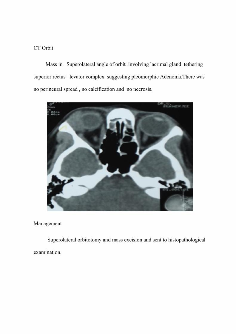
CT Orbit:
Mass in Superolateral angle of orbit involving lacrimal gland tethering
superior rectus –levator complex suggesting pleomorphic Adenoma.There was
no perineural spread , no calcification and no necrosis.
Management
Superolateral orbitotomy and mass excision and sent to histopathological
examination.

POST OPERATIVE (Lateral Approach)
HPE Report:
Evidence of both epithelial and mesenchymal differentiation.
Proliferation of benign epithelial cells are arranged in double layer to
form lumens. Stromal differentiation can be seen in the formation of bone and
cartilage suggestive of Pleomorphic Adenoma
Follow up:
In two year follow up there was no recurrence seen in this
patient.
2. Name : Parvathi Age: 45 years Sex: Female
Came to orbit clinic for drooping of left upper lid and swelling in
supero lateral aspect of orbit for 2 months.
Non tender mass with firm to hard in consistency
EOM: Restricted in Abduction
Visual Acuity in Both eyes - 6/6
Fundus: Normal
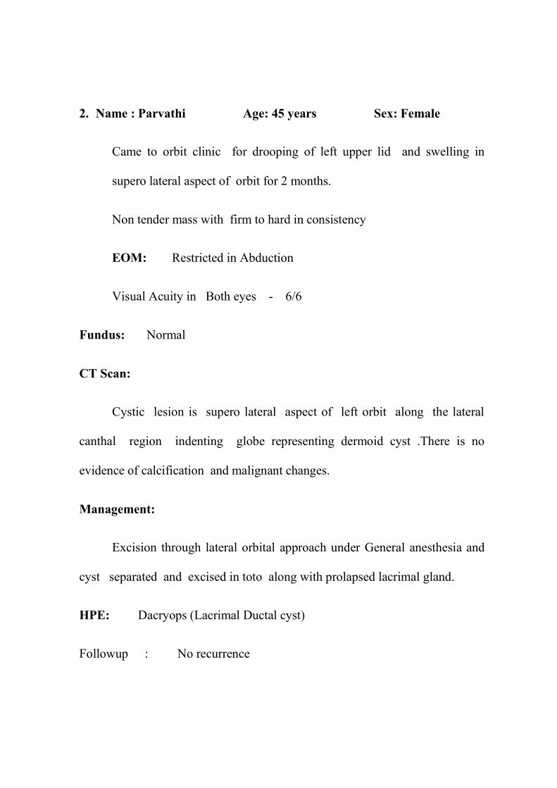
CT Scan:
Cystic lesion is supero lateral aspect of left orbit along the lateral
canthal region indenting globe representing dermoid cyst .There is no
evidence of calcification and malignant changes.
Management:
Excision through lateral orbital approach under General anesthesia and
cyst separated and excised in toto along with prolapsed lacrimal gland.
HPE: Dacryops (Lacrimal Ductal cyst)
Followup : No recurrence

3. Name : Rani Age: 41 years Sex: Female
Came to orbit clinic with complaints of left protrusion of Eyeball – 20 days
O/E: S- Shaped ptosis present not obscuring visual axis.
Firm mass in lacrimal gland , not freely mobile, no retropulsion
EOM: Full
Visual Acuity: Right Eye – 6/6
Left Eye – 6/6 Partial
Anterior Segment: Normal
Pupil: Reacting
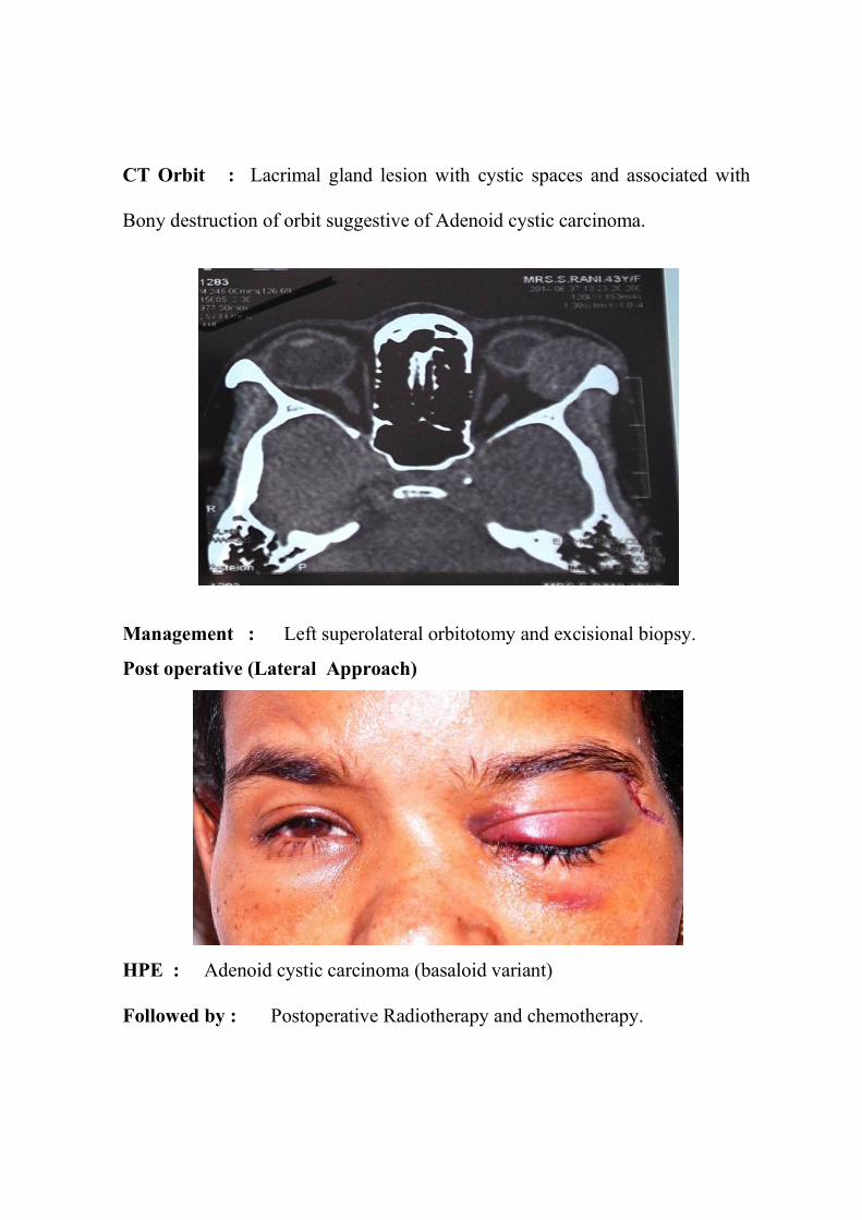
CT Orbit : Lacrimal gland lesion with cystic spaces and associated with
Bony destruction of orbit suggestive of Adenoid cystic carcinoma.
Management : Left superolateral orbitotomy and excisional biopsy.
Post operative (Lateral Approach)
HPE : Adenoid cystic carcinoma (basaloid variant)
Followed by : Postoperative Radiotherapy and chemotherapy.

4. Name : Haneef S.Iyad Age: 14 years Sex: Male
Came to orbit clinic with drooping of Left upper lid - 3 months which
was painless/ progressive.
Swelling is firm in consistency, not fixed to underlying structures.
Swelling - not warmth, not tender,
No associated systemic illness
No previous episodes.
Visual acuity: Right Eye - 6/6
Left Eye - 6/6.
Management:
Left eye cyst explored and dissected removed in toto and sent for
histopathological examination.
HPE:
Dacryops with squamous metaplasia.

5. Name : Janani Age : 12 years Sex : Female
Came to orbit clinic for Left eye swelling in superotemporal aspect of
orbit for 2 months.
Swelling is firm in consistency,
Not warmth, not tender, Not mobile.
Not fixed to underlying structures
Visual acuity : Both eyes - 6/6
Anterior Segment : Normal
Pupil : Reacting
Extra ocular movements : Normal
Management:
Left – Dacryops excision done.
HPE Report:
Lacrimal ductal cyst (dacryops)

6. Name : Ali Rizween Age: 26 years Sex: Male
Came to Eye O.P. for Right Eye protrusion- 6 months
Insidious onset , gradual and progressive
O/E: Right Eye – Eccentric proptosis with firm, tender mass in
superolateral quadrant.
Visual Acuity : Right Eye -6/18 and left eye-6/6
Anterior Segment : Normal
Fundus : BE- Normal
CT Orbit:
lacrimal gland lesion without bony remodeling and without calcification
seen
Management:
Superolateral orbitotomy with excision biopsy without violating capsule.
HPE:
Pleomorphic Adenoma with squamous metaplasia and Keratin production.

7. Name : Sasikumar Age: 27 years Sex: Male
Came to orbit clinic for swelling in the superotemporal aspect of left
eye for 6 months
O/E: Left Eye Inferior dystopia with resistance to retropulsion.
Orbital margins intact.
Mass firm in consistency.
Finger insinuation not possible.
EOM : Normal
Anterior segment : Normal
Visual Acuity: Right Eye - 6/6 and Left Eye - 6/6

CT scan :
Heterogenously enhancing soft tissue lesion displacing globe
downwards and cause bony indentation due to mass effect.
Management: Superior orbitotomy and mass excision biopsy
Post operative (Lateral Approach)
HPE: Lacrimal gland – Pleomorphic Adenoma

8. Name: Mahesh Age: 39 sex: Male
Came to orbit clinic with complaints of Bilateral Gradual
progressive swelling of superolateral orbit for 2 years.
Finger insinuation : Not Possible
EOM: Full
Visual Acuity: in Right Eye – 6/6 and in Left Eye – 6/6
Pupil: 2mm, Reacting
Fundus : Normal
CT report: B/L lacrimal gland enlargement – Suggestive of
lymphoproliferative disorder.
Management : Patient started on systemic steroids. After stopping
steroids, mass recurred.
Management : Left Eye -Incisional Biopsy of Lacrimal gland mass.
HPE Report : Non Hodgkin’s lymphoma
Patients was advised chemotherapy and regular follow up
9. Name: Maragatham Age: 37 yr Sex: Female
Came to orbit clinic with complaints of left Eye Protrusion – 3 years.
O/E: Upper lid and Lower lid congestion with inferior dystopia.
Eccentric proptosis and Restricted Retropulsion was present.
Extra ocular Movement : Full
Pupil : 3mm, reacting to light.
Visual acuity : Right Eye -6/6, Left Eye – 6/6- Partial
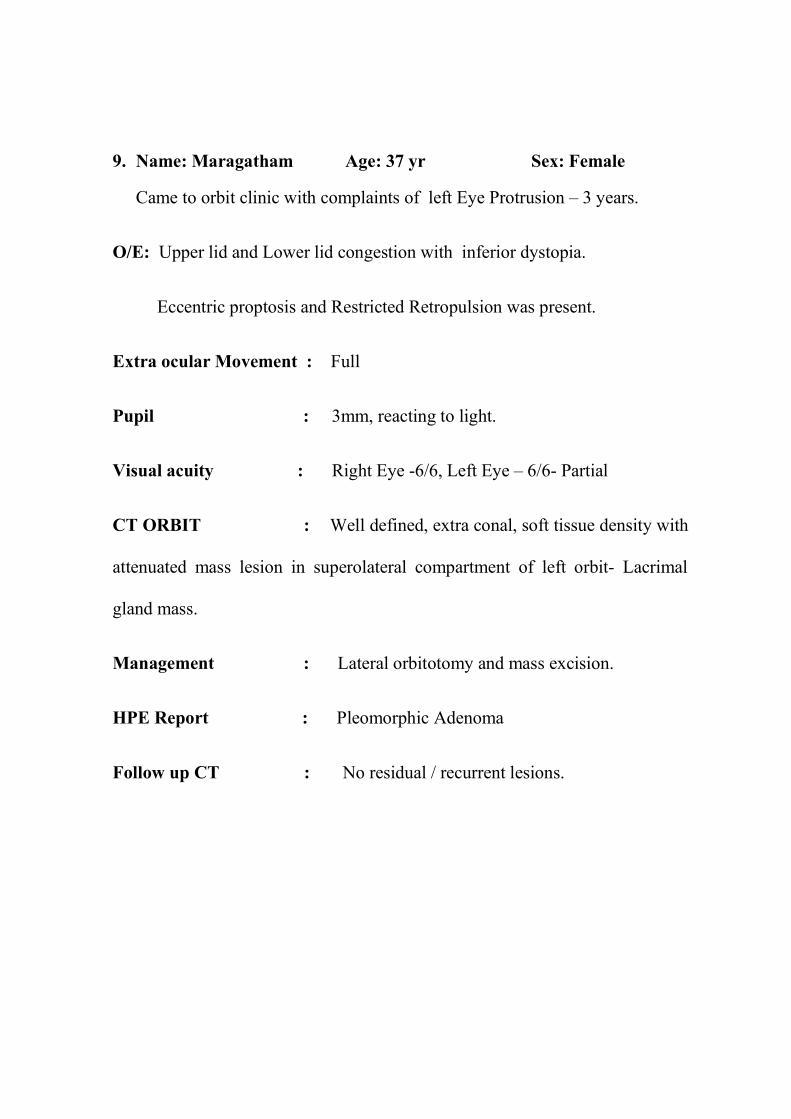
CT ORBIT : Well defined, extra conal, soft tissue density with
attenuated mass lesion in superolateral compartment of left orbit- Lacrimal
gland mass.
Management : Lateral orbitotomy and mass excision.
HPE Report : Pleomorphic Adenoma
Follow up CT : No residual / recurrent lesions.

10. Name : Abdul Rasheed Age : 7 years Sex : Male
Came to for orbit clinic with complaints of
Swelling in Right Eye – Upper lid for past -3 months
O/E: Right Eye – Swelling in the supero lateral aspect of Right Eye
Orbit
BE :
Visual Acuity: 6/6

CT Orbit:
Hyper dense lesion and enlargement of Right Eye - lacrimal gland
blends with lateral rectus.
Management:
Superolateral mass excision under incisional biopsy

POST OPERATIVE
Post operative (Lateral Approach)
HPE:
Structure of fibrofatty tissue with dense hyalinised collagen and
vascular proliferation. Perivascular lymphocytic infiltration with
interspersed eosinophils are seen suggestive of Idiopathic sclerosing orbital
inflammation.
Immunohistochemistry:
LCA: Diffuse positive reactivity
Post Operative Period:
Started on tab . prednisolone in tapering doses.
11. Name : Sundarajan Age: 43 years Sex: Male
Came to orbit clinic for pain with complaints of Right Eye – protrusion for
2 months.
Not warmth, firm in consistency
Swelling not associated with retropulsion
Not fixed to underlying structures
Finger insinuation not possible .
O/E: Right Eye – Eccentric proptosis with firm tender mass in
superotemporal aspect.
Visual Acuity: BE – 6/6
CT Scan: Heterogenous mass in superolateral aspect of Right orbit without
Bony erosion tethering lateral rectus.
Management: Right Eye – Lateral orbitotomy excision Biopsy followed by
chemotherapy and Radiotherapy.
Follow up: In one year, recurrence of Adenoid cystic carcinoma and
advised chemotherapy with Adriamycin and cisplatin.

12. Name : Alphonsa Age: 45 years Sex : Female
Came to orbit clinic with complaints of swelling in Right
superotemporal aspect for 2 years
Swelling is Painless and Progressive
Rubbery consistency
Finger insinuation not possible
O/E:
Right Eye ‘S’ shaped ptosis
Firm mass palpable
CT Orbit:
Mass replacing entire lacrimal gland and blends with globe and inferior
rectus muscle.
Management:
Right Lateral orbitotomy and incisional Biopsy.
HPE :
Non Hodgkins lymphoma – B Cell type and started chemotherapy.

13. Name: Fathima Age: 54years Sex: Female
Came to orbit clinic for Right Eye – Protrusion for one month.
O/E: Right Eye - Proptosis
Firm mass palpable in Right Lacrimal gland area
Finger insinuation not possible
Abduction restricted.
On palpation - mass not warmth ,
rubbery in consistency
not pulsatile, no bruit present
orbital margins intact.
No change in size of mass in various postures
Systemic examination : Generalised lymphadenopathy
Hepatosplenomegaly
Anterior segment examination: Normal
Pupil: reacting to light.

Visual Acuity: BE - 6/24
Complete hemogram - Normal
Chest X Ray - Normal
Ultrasound abdomen - Fatty liver
BMA - Leucopoiesis with small to medium sized atypical cleaved
nucleated lymphoid cells upto 50 percent. - lymphomatous infiltration of
bonemarrow with leukemic manifestation.

CT Orbit:
Lacrimal gland mass involving Right globe and lateral rectus muscle
with Bony erosion.
DIFFERENTIAL DIAGNOSIS ;
1. pleomorphic adenoma
2. Lymphoproliferative disorders
Management:
Lateral orbitotomy and excision biopsy

HPE Report:
B Cell type – Follicular lymphoma/ blastic differentiation. Lymphoid cells
arranged in follicles.
Followed by:
Chemotherapy ( chlorambucil and prednisolone) – advised for follow
up.

14. Name : Thangamma David Age:65 years Sex:
Female
Came to orbit clinic with complaints of
Swelling in Right Superolateral aspect of orbit for 3 months
Not warmth, not mobile
Finger insinuation not possible.
O/E: Right Eye -Mechanical ptosis
Swelling in superolateral quadrant
Salmon patch like appearance in tarsal conjunctiva.
Visual Acuity: Right Eye - 6/18
Left Eye - 6/12
CT Orbit: Soft tissue density mass in the superolateral aspect of right orbit.
Management: Mass excision biopsy.
HPE Report:
Necrobiotic Granulomatous inflammation (nonspecific). Patient was
started on steroids.

15. Name: Hamza Age : 24 years Sex: Male
Came to orbit clinic with complaints of
Left Protrusion of Eyeball for 10 days
No history of trauma
No history of previous episodes
No history of fever with seizures
O/E: Eccentric proptosis seen
mass felt in supero lateral region.
Globular in shape
Smooth surface
Mass firm in consistency
Mass not warmth , not tender
Not fixed with underlying structures
Finger insinuation not possible.

Visual Acuity: Right Eye - 6/6 and Left Eye - 6/9
Investigation :
Complete hemogram - Normal
Chest x ray - Normal
CT Orbit:
Extraconal soft tissue density lesion in left supero lateral aspect of orbit
without Bony erosion. Suggestive of pleomorphic adenoma.
Management:
Orbitotomy and excision biopsy.
HPE: Adenoid cystic carcinoma
Post operative Management:
Chemoradiotherapy and follow up

16. Name : Chinnaiah Age:41 years Sex: Male
Came to orbit clinic with complaints of Painless, gradual, Swelling in
Left Orbit for 20 days
Associated with Inferior dystopia and eccentric proptosis. Swelling is
firm in consistency. Not freely mobile, not associated with retropulsion.
Visual Acuity: Right Eye - 6/6 Partial and Left Eye - 6/6
Anterior Segment: Normal
EOM: Full
Pupil: Reacting
Fundus: Normal
CT Report: Left lacrimal gland Neoplasia with bony erosion with mild
infiltration of Lateral rectus muscle
. Management: Lateral orbitotomy with mass excision Biopsy.
HPE report: Hemangiopericytoma (intermediate grade) solitary fibrous
tumor complex. Diffuse CD34 and vimentin positive.
Follow up: No recurrence.

17. Name : Gnanaoli Age : 33 years sex: Male
Came to orbit clinic with complaints of Bilateral mass in superolateral
quadrant.
No H/o of trauma ,
No H/o of previous episodes in past
O/E:
Soft tender mass in superolateral quadrant of Both Eyes.
Not warmth, not tender
Finger insinuation not possible
Orbital margins intact
General Examination
B/L Parotid gland involvement
B/L submandibular gland involvement with
multiple enlarged lymph nodes and hepatosplenomegaly.
Visual Acuity: Right eye - 6/9

Left eye - 6/9
Both eyes - S shaped ptosis
- palpebral lobe of lacrimal gland enlargement
- subconjunctival hemorrhage
- orbital margins intact. EOM -Normal

CT Orbit ( axial view)
Bilateral Lacrimal gland enlargement.
CT Abdomen:
Diffuse bone marrow changes replacing axial and appendicular
skeleton with multiple lymphadenopathy and Hepatosplenomegaly.
Complete hemogram:
TC - 1,20, 000
DC - Atypical cells 80 percent.
ESR - 30 mm/hr

Peripheral Smear - Presence of large atypical cells with irregular
nuclei and prominent nucleoli.
Bone marraw aspiration - Lymphomatous infiltration of bone marrow
with leukemic manifestations
CT Brain – No evidence of subarachnoid hemorrhage and intraventricular
hemorrhage
MRI Spine / Abdomen
Diffuse marrow changes replacing axial and appendicular skeleton
Multiple enlarged lymph nodes and hepatosplenomegaly.
Peripheral Smear:
Presence of large atypical cells with irregular nuclei and prominent
nucleoli.
HPE Report:
Monomorphous atypical lymphoid cells with prominent nucleoli
Suggestive of B cell type Non Hodgkin’s lymphoma.

HISTO PATHOLOGY
IMMUNOHISTOCHEMISTRY
(Diffuse strong positive for CD 20)
Post Operatively:
Treated with chemotherapy- chlorambucil and prednisolone.
Follow Up : No recurrence was seen

18. Name : Pandi Age : 50 years Sex :Male
Came to for orbit clinic with Right Eye defective vision for 6 months
O/E:
Right sided proptosis with inferior dystopia
Mass in superotemporal quadrant of orbit with firm consistency
Mass not fixed to underlying structures
Not associated with retropulsion
Size of mass not varies with posture.
Anterior segment : Normal
EOM: Restricted in abduction and elevation in abduction.
Visual Acuity: Right Eye -6/18 Left Eye - 6/6
Fundus : Choroidal folds present
CT Orbit: Right side lacrimal gland neoplasm with mild bony erosion.
Management: Right – Lateral Orbitotomy with mass excision biopsy
HPE: Pleomorphic Adenoma.

19. Name : Maria Nevis Age: 43 years Sex: Female
Came to orbit clinic with Left swelling in Superolateral orbit – 5 years
Sudden onset with rapid growth
O/E: Mass in superolateral quadrant
Warmth, firm in consistency
Finger insinuation not possible
No pulsatile , no change in size after varying posture.
No similar treatment episodes in past.
Extra ocular movements: Normal
Visual Acuity: Both Eyes – 6/6.
IOP Both Eye - 15 mm Hg
Management:
Left lacrimal gland incisional biopsy done.
HPE report:
Pseudotumor.

20. Name : Ramamma Age:60 years Sex : Female
Came to orbit clinic with complaints of
Right Eye protrusion of Eyeball –for 5 months
Swelling sudden onset rapid growth
O/E: Right side – Lacrimal gland swelling chemosis and congestion at rectus
insertion site
Visual acuity: BE – 6/18.
CT Scan: Heterogenous lesion in superolateral compartment of Right lacrimal
gland with lacteral rectus and optic nerve involvement.
Thyroid: Cystic lesion with calcification with multiple lymph nodes.
Management: Superolateral orbitotomy with mass excision Biopsy.
HPE report: Adenocarcinoma of Lacrimal gland ( Basoloid type).

21. Name : Kannan Age : 40 years Sex: Female
Came to Orbit clinic for Right protrusion of eyeball for 6 months.
O/E: Eccentric Proptosis with firm swelling in superotemporal quadrant.
Not warmth, firm in consistency
Not fixed to underlying structures
Finger insinuation not possible
Not pulsatile, No bruit felt.
Visual Acuity: Both Eye - 6/6
Extra ocular movements - Normal
CT Orbit: Right Eye – Lacrimal gland mass with bone remodeling with no
perineural invasion, with no calcification.
Management:
Right Eye – lateral orbitotomy with mass excision Biopsy.
HPE Report:
Pleomorphic Adenoma.

22. Name : Johnson Age: 18 years Sex: Male
Came to orbit clinic with complaints of swelling in left lacrimal gland
area – One month sudden onset , rapid growth
O/E: Firm mass felt in lacrimal gland area
Resistance to retropulsion.
Not warmth, Not pulsatile
CT Orbit: Well defined lobulated, soft tissue density lesion in Left
lacrimal gland with Bony erosion infiltrating lateral rectus and
Infratemporal fossa and frontal lobe.
Management: Lacrimal gland mass incision biopsy.
HPE Report:
Lacrimal gland acini with infiltration of round to oval cells having
contorted and indented nuclei with pale eosinophilic cytoplasm. Focal
collections of eosinophils with prominent nucleoli and necrosis seen. Special
stain for AFB- Negative- suggestive of Histiocytosis more in favour of
Langerhan cell histiocytosis.
Follow Up : No recurrence was seen

23. Name: Ashokan Age: 58 years Sex: Male
Came to orbit clinic for Swelling Right lacrimal gland area – 3 months.
Insidious onset, painful
Progressive in nature
Not associated with fever
Not associated with other systemic illness
PAST H/o
No similar episodes in past
No H/o of trauma
O/E:
Firm mass in superotemporal aspect of lacrimal gland
Firm in consistency
Smooth surface
Warmth. Mild tenderness present.
Not fixed to underlying structures

Visual Acuity
Right Eye - 6/6
Left eye - 6/6
EOM- Normal
Investigations:
Complete hemogram- Normal
ESR- 45 mm/hr
CT Brain:
Hyper intense signal along superolateral aspect of Right lacrimal gland.
There was no evidence of calcification and perineural invasion. No evidence of
malignant changes.
Management:
Incisional biopsy for lacrimal gland mass.
HPE:
Right Dacryo adenitis (Non specific)
.Patient was Treated with steroids and advised follow up.

24. Name : Vijayan Age : 64 years Sex : Female
Came to orbit clinic with complaints of
Mass in Superolateral quadrant
Associated with pain, swelling – one month.
Not associated with lid swelling
Not associated with drooping of eye lids
O/E: Mass is firm in consistency
Finger insinuation not possible
Not warmth, not tender
Irregular surface.
No H/o of DM and Hypertension
No H/o of trauma
No H/o of contact with tuberculosis patients
No H/o mouth ulcers, fever, joint pain
No H/o of genital ulcers , urethral discharge

Previous recurrent history of dacryoadenitis which was treated with steroids,
Visual Acuity:
Both Eye - 6/6
Anterior segment examination :
Facial contour –normal
Eye lids – normal
Eccentric proptosis
RE- Temporal congestion
No evidence of scleritis or sclera abscess.
MRI (Brain and Orbit):
Swelling in superolateral aspect of lacrimal sac and gland.
Mantoux : Reactive (15mm)
ESR : Elevated (>150mm/Hg)
X-ray chest : Hilar lymphadenopathy
Nested PCR : Myco bacterium Tuberculosis positive.
Serum ANA : Negative
IgM for TB : Negative

IgG for Toxoplasmosis : Negative
IgM for Toxoplasmosis : Negative
Mantoux : 15mm. Reactive
Urine Bence Jones protein : Negative
Urine routine : 2to 3 pus cells
ESR : More than 150 mm/hr
CT Orbit:
Enlargement of right lacrimal gland, with edema, no evidence of
calcification, bony changes.
Management:
Right lacrimal gland – mass Incisional biopsy through skin crease
incision, gland identified, biopsy taken and material sent for culture and
sensitivity, biopsy
HPE Report:
Structure of lacrimal gland acini in lobules with destruction by infiltration
of lymphocytes and plasma cells suggestive of TB Dacryoadenitis Patient was
started Anti tuberculous Therapy. He responded well with no recurrence.

25. Name : Jolsna Benny Age: 36 years Sex : Female
Came to orbit clinic for swelling in Right superotemporal orbit - one year.
O/E: S- Shaped ptosis. Mass is firm to hard in consistency.
Anterior segment: Normal
EOM: Restricted in abduction and elevation in Abduction.
Visual Acuity : Right Eye -6/6 Left Eye - 6/6.
CT Orbit: Right – Lacrimal gland Neoplasia with mild Bony erosion.
Management: Right Eye – Lateral orbitotomy with mass excision Biopsy.
HPE: Pleomorphic Adenoma (with mixed epithelial cells and
mesenchymal cells)
Follow up : No recurrence.
26. Name : Geetha Age: 33 years Sex: Female
Came to orbit clinic with complaints of swelling in superotemporal
aspect of Left orbit for 3 months.
Swelling was insidious in onset.
Not associated with pain
Not warmth, not tender
Finger insinuation not possible.
O/E: Swelling – cystic in consistency
Transillumination was positive
No bruit felt on auscultation
Was not associated with pulsation
Visual Acuity: BE – 6/6.
Management:
Left Excision of mass in toto done and sent for histopatology.
HPE: Inflamed ductal cyst (Dacryops)

27. Name: Muthu Vadivoo Age : 64 years Sex: Female
Came to orbit clinic for protrusion of Left Eyeball – 2 months
O/E: Left Eye – eccentric Proptosis
Resistance to retropulsion
Firm in consistency
Finger insinuation not possible superiorly and laterally.
Visual Acuity: Right Eye -6/6 –partial and Left Eye -6/36.
Investigations:
Serum RA factor - Negative , ANA screening - Negative
CT orbit: Soft tissue density mass in superolateral aspect of left orbit
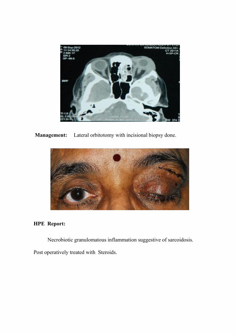
Management: Lateral orbitotomy with incisional biopsy done.
HPE Report:
Necrobiotic granulomatous inflammation suggestive of sarcoidosis.
Post operatively treated with Steroids.
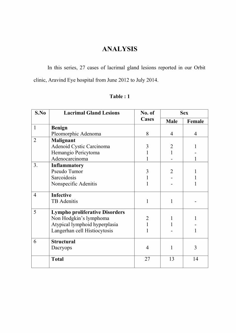
ANALYSIS
In this series, 27 cases of lacrimal gland lesions reported in our Orbit
clinic, Aravind Eye hospital from June 2012 to July 2014.
Table : 1
S.No Lacrimal Gland Lesions No. of Cases
Sex Male Female
1 Benign Pleomorphic Adenoma
8
4
4
2 Malignant Adenoid Cystic Carcinoma Hemangio Pericytoma Adenocarcinoma
3 1 1
2 1 -
1 - 1
3. Inflammatory Pseudo Tumor Sarcoidosis Nonspecific Adenitis
3 1 1
2 - -
1 1 1
4 Infective TB Adenitis
1
1
-
5 Lympho proliferative Disorders Non Hodgkin’s lymphoma Atypical lymphoid hyperplasia Langerhan cell Histiocytosis
2 1 1
1 1 -
1 - 1
6 Structural Dacryops
4
1
3
Total 27 13 14

18%4%
15%
15%
30%
18%
Benign
Malignant
Inflammatory
Infective
Lympho proliferative Disorders
Structural
Inflammatory
Lympho proliferative

Male
Female52%
Table : 2
Sex Ratio
Male Female
13 14
Male48%

Table : 3 Age wise Distribution
S.No Type of Tumor Total No.
of Case
0-10 yr 10-20 yr 20-30 yr 30-40 yr 40-50 yr 50-60 yr 60-70 yr
1 Pleomorphic Adenoma 8 - 1 3 2 1 1 -
2 Dacryops 4 - 2 - 1 1 - -
3 Pseudo Tumor 3 1 - - - 1 1 -
4 Non Hodgkins lymphoma 2 - - - 1 1 - -
5 Adenoid cystic Carcinoma 3 - - - - 2 - 1
6 Sarcoidosis 1 - - - - - - 1
7 Atypical lymphoid
Hyperplasia
1 - - - 1 - - -
8 Langerhan cell Histiocytosis 1 - 1 - - - - -
9 TB Adenitis 1 - - - - - - 1
10 Adenocarcinoma 1 - - - - - - 1
11 Hemangiopericytoma 1 - - - - 1 - -
12 Nonspecific Adenitis 1 - - - - - - 1

11%7%
11%
3%
4%4%
4%4%
4% 4%
29%
15%
Pleomorphic Adenoma
Dacryops
Psecudo Tumor
Non Hodgkins lymphoma
Adenoid cystic Carcinoma
Sarcoidosis
Atypical lymphoid Hyperplasia
Langerhan cell Histiocytosis
TB Adenitis
Adenocarcinoma
Henangiopericytoma
Nonspecific Adenitis
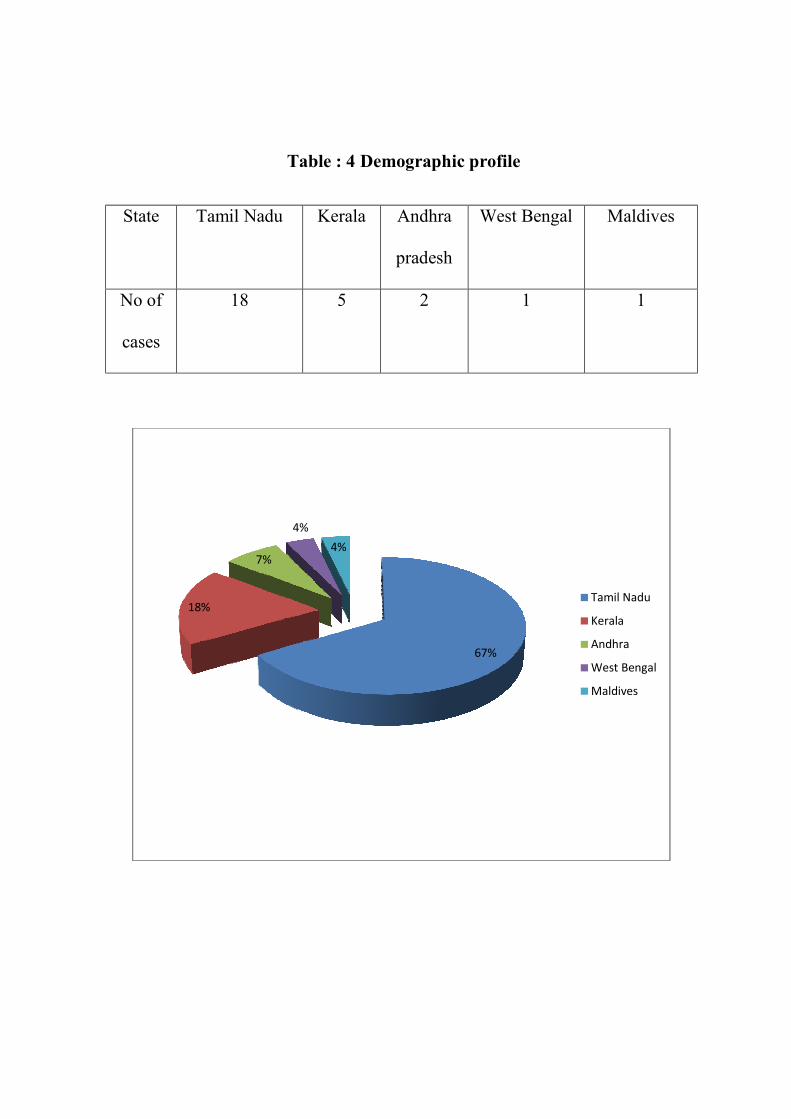
Table : 4
State Tamil Nadu
No of
cases
18
18%
7%
4%
Table : 4 Demographic profile
Kerala Andhra
pradesh
West Bengal
5 2 1
67%
4%
Tamil Nadu
Kerala
Andhra
West Bengal
Maldives
Maldives
1
Tamil Nadu
Kerala
Andhra
West Bengal
Maldives
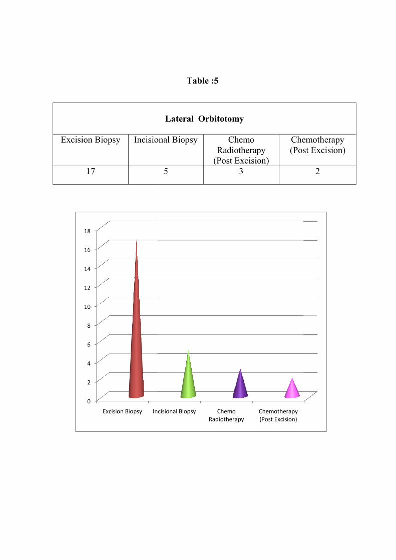
Excision Biopsy Incisional Biopsy
17
0
2
4
6
8
10
12
14
16
18
Excision Biopsy Incisional Biopsy
Table :5
Lateral Orbitotomy
Incisional Biopsy Chemo
Radiotherapy (Post Excision)
Chemotherapy (Post Excision)
5 3
Incisional Biopsy Chemo Radiotherapy
Chemotherapy (Post Excision)
Chemotherapy (Post Excision)
2
RESULTS
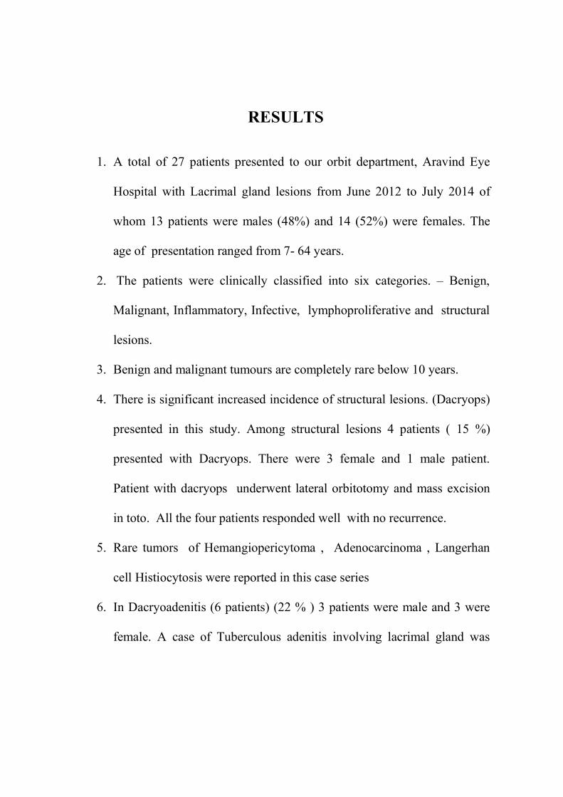
1. A total of 27 patients presented to our orbit department, Aravind Eye
Hospital with Lacrimal gland lesions from June 2012 to July 2014 of
whom 13 patients were males (48%) and 14 (52%) were females. The
age of presentation ranged from 7- 64 years.
2. The patients were clinically classified into six categories. – Benign,
Malignant, Inflammatory, Infective, lymphoproliferative and structural
lesions.
3. Benign and malignant tumours are completely rare below 10 years.
4. There is significant increased incidence of structural lesions. (Dacryops)
presented in this study. Among structural lesions 4 patients ( 15 %)
presented with Dacryops. There were 3 female and 1 male patient.
Patient with dacryops underwent lateral orbitotomy and mass excision
in toto. All the four patients responded well with no recurrence.
5. Rare tumors of Hemangiopericytoma , Adenocarcinoma , Langerhan
cell Histiocytosis were reported in this case series
6. In Dacryoadenitis (6 patients) (22 % ) 3 patients were male and 3 were
female. A case of Tuberculous adenitis involving lacrimal gland was

identified which was confirmed with nested PCR . He was treated with
excision Biopsy followed by Anti Tuberculous drugs with no recurrence.
7. In this category of Dacryoadenitis, two patients presented with
Necrobiotic granulamatous inflammation for which both of them
underwent excision biopsy and one was diagnosed as sarcoidosis and
another was diagnosed as Nonspecific adenitis. Both of them treated
with steroids with no recurrence.
8. Remaining 3 patients with dacryoadenitis were diagnosed as pscudo
tumor. They responded well with steroids.
9. Among 8 benign tumors (29 %) reported in this series, all patients
presented with pleomorphic adenoma. 4 patients were male and 4 were
female with equal sex preponderance. The age of presentation ranged
from 10 – 60 years. Duration of symptoms ranges from 4 months to 6
years.
10. The commonest symptom in these patients with pleomorphic Adenoma
is painless, insidious onset, orbital mass. The treatment included
complete excision without violating the capsule. There was no
recurrence during follow up.

11. Among 4 patients ( 14 %) presented with lymphoproliferative disorders,
two patients presented with bilateral lacrimal gland enlargement. One
patient was diagnosed as Langerhan cell Histiocytosis for which, he
underwent incisional biopsy followed by steroids with no recurrence.
12. Two patients were diagnosed as Non Hodgkins Lymphoma ( B cell
type). They underwent excision Biopsy followed by chemotherapy
alone. These 2 patients responded well with treatment with no
recurrence. Remaining one patient with Atypical lymphoid Hyperplasia,
was treated with steroids.
13. Five patients (18 %) in this study were diagnosed as malignant lesions, 3
had Adenoid cystic carcinoma, one with Adenocarcinoma and one with
Hemangiopericytoma.
14. Among 3 patients( 11%) with Adenoid cystic carcinoma 2 were male
and one was female. These patients treated with lateral orbitotomy and
excision biopsy followed by Chemo Radiotherapy and regular follow up.
15. Hemangiopericytoma (intermediate grade) was identified in one male
patient. Adenocarcinoma (basaloid types) was reported in one female
patient. Both of them underwent mass excision biopsy through lateral
orbitotomy, with no recurrence during followup.

DISCUSSION
A total of 27 patients presented to our orbit department, from Aravind Eye
Hospital with Lacrimal gland lesions from June 2012 to July 2014 . This study
is comparable to Henderson Series (1994), Rootman Series (1999), Reese
(1956), Ashton (1975), Stewart (1979), Shields (1989), British Columbia orbit
clinic series (1976 -1999) Dr.Usha kim , Aravind Eye Hospital series (2005-
2008) et al.
This study has its own unique features as Benign and malignant tumours
are completely rare below 10 years and increased incidence of structural
lesions (Dacryops). For Dacryops complete excision biopsy in toto was done.
This study also features rare tumors of Hemangiopericytoma,
Adenocarcinoma, Langerhan cell Histiocytosis in this case series.
Hemangiopericytoma is not included in other comparable case series like
British Columbia orbital clinic series.
In all nonspecific dacryoadenitis cases, whom not responding to medical
treatment, infective etiology especially tuberculosis should be ruled out.
Though tuberculosis is common in our country only one case reported with
dacryoadenitis due to tuberculosis which was confirmed with nested PCR.

Lacrimal gland lesions has wide range of clinical presentation and
Histopathological diagnosis. Lacrimal gland lesions can be divided into two
groups. First group with lesions conforming to lacrimal gland (Benign
lesions) which presents with painless lacrimal gland mass with insidious
onset. Pleomorphic adenoma is the common benign tumor in this study. All
patients need excision biopsy without violating capsule and followup is
needed.
The second group is with lesions not conforming to clinical picture of
lacrimal gland. This group contains variety of lesions like structural lesions
(Dacryops), lymphoma, malignant tumors, dacryoadenitis.
Adenoid cystic carcinoma is the common malignant tumor arising from
lacrimal gland. This tumor has high recurrence rate of of 40 to 100 percent in
various studies.In our study we found recurrence of 75 percent after a follow
up for 2 years. 6 months and 1 year followup is needed in these cases to detect
metastasis especially to lung.
Lymphoproliferative disorders involving lacrimal gland presented with
equal gender preponderance and bimodal presentation which is more in third to
fifth decade.

Cases with Bilateral lacrimal gland involvement and with suspicious
malignancy immediate complete metastatic workup with
immunohistochemistry confirmation are needed to prevent recurrence and to
improve life expectancy of patients.
All patients with lacrimal gland lesions need lateral orbitotomy and
excision biopsy.
In some cases with suspected infective and nonspecific inflammatory
etiology and cases with orbital invasion ( where complete excision is not
possible) are subjected to incisional biopsy.
Early and accurate diagnosis with proper followup and couselling is needed
in all lacrimal gland lesions, so that satisfactory outcome can be expected.

CONCLUSION
1. A total of 27 patients presented to our orbit department, Aravind Eye
Hospital with Lacrimal gland lesions from June 2012 to July 2014 .
2. Benign and malignant tumours are completely rare below 10 years.
3. There is significant increased incidence of structural lesions (Dacryops)
presented in this study
4. Rare tumors of Hemangiopericytoma , Adenocarcinoma , Langerhan
cell Histiocytosis were reported in this case series
5. Acute dacryoadenitis can occur at any age. Complete ophthalmic
examination with appropriate investigations is needed to identify
underlying etiology so that treatment plan can be tailored to each patient
individualy.
6. Benign tumours commonly seen in age group of 20 - 40 years and
malignant tumours are common in 60 – 70 years.
7. Most common benign tumour is pleomorphic adenoma. Since they
underwent complete excision without violating capsule, there was no
recurrence seen in this case series.

8. Adenoid cystic carcinoma is the common lacrimal gland malignant
tumour with recurrence rate of 75% in 2 years follow up in this series.
9. Incisional biopsy has also important role in this study in some cases
where complete excision is not possible.
10. Post surgical Chemoradiotherapy is beneficial in Adenoid cystic
carcinoma.
11. Post Surgical Chemotherapy is beneficial in Non Hodgkin’s lymphoma.

BIBLIOGRAPHY
1. Abboud I., Hanna L. Intermittent exophthalmos, Br.J. Ophthalmol. 55;
628, (1971).
2. Ashton N. Epithelial tumors of the lacrimal gland. Modern problems in
Opthamology 1975;14:306-23.
3. Blodi F.C., Gass J.M. Inflammatory pseudo tumor of the orbit, 1 Br. J.
Ophthalmol, 52; 79; (1968).
4. Coop M. pseudo tumor of the Orbit, Br.J. Ophthalmol. 45; 513 (1961).
5. Duke Elder. Vol. XIII, part -2, PP.1017 – 1226.
6. Ellis GL, Auclair PL. Tumors of the salivary gland. Washington, DC:
Armed Forces Institute of Pathology, 1996.
7. Forrest FW, Jr, Frazell EL. Tumors of the major salivary glands. Cancer
1953;6:1065-133.
8. Godtfredsen E. Pathology of mucous and salivary gland tumors in the
prognosis. Trans Am Acad Ophthlmol 1954;58:848-65.
9. Henderson J.W. Orbital tumors: Philadelphia W.E. Saunders co., (1973).
10. Henderson J.W. Farrow G.M. Orbital tumors, J.W. Henderson, G.M.
Farrow, eds. Philadelphia W.E. Saunders Co., (1973), P.555.
11. Henderson J.W. Orbital tumors. Text book.

12. Jackson H. pseudo tumor of the Orbit Br.J. Ophthalmol. 42: 212, (1958).
13. Jakobee orbital Diseases. Text book.
14. Mafee MF, Edward DP, Koeller KK, Dorodi S. Lacrimal gland tumors
and simulating lesions: clinicopathologic and MR imaging features.
Radiol Clin North AM 1999;37:219-39.
15. Moss H.M. Expanding lesions of the Orbit a clinical study of 230
consecutive cases, Am.J. ophthalmol.,54.
16. Ni C, Cheng SC, Dryja TP, Cheng TY. Lacrimal gland tumors: a
clinicopathological analysis of 160 cases. Int Ophthalmol Clin
1981;22:99-120.
17. Ni C, Kuo P-K. Histopathological classification of 272 primary
epithelial tumors of the lacrimal gland. Chin Med J 1992;105:481-5.
18. Reese AB. The treatment of expanding lesions of the orbit with
particulate regard to those arising in the lacrimal gland. Am J
Ophthalmol 1956;41:3-11.
19. Riley F.C. Henderson J.W. Report of a case of malignant transformation
in benign mixed tumor of the Lacrimal gland. Am.J. ophthalmol.,
70:767, (1970).
20. Shields CL, Shields JA. Lacrimal gland tumors. Int Ophthalmol Clin
1993;33:181-8.
21. Stewart WB, Krohel GB, Wright JE. Lacrimal gland and fossa lesions:
an approach to diagnosis and management. Opthalmology 1979:86:886-
95.
22. Wright JE, Stewart WB, Krohel GB. Clinical presentation and
management of lacrimal gland tumors. Br J Ophthalmol 1979;63:600-6.
23. Zimmerman LE, Sanders TE, Ackerman LV. Epithelial tumors of the
lacrimal gland: prognostic and therapeutic significance of histologic
types. Int Ophthalmol Clin 1962;2:337-67.
CLINICAL PROFORMA Name :
Age :
Sex :
M.R.No. :
Diagnosis :
Complaints :
Clinical Evaluation
BCVA
Pre op Evaluation
Visual Acuity
General Examination
Ø Facial contour
Ø Asymmetry
Ø Pallor
Ø Cyanosis
Ø Clubbing
Ø Jaundice

Ocular Examination
Ø Visual Acuity
Ø Head Posture
Ø Extraocular movements
Y-1 N-2
4. Inspection
v Eyebrows
v Eyelids
Mass
v Eccentric proptosis
P-1 A-2
v Unilateral/ Bilateral
1 / 2
v Inferior dystopia
P-1 A-2
v Pulsatile/ Not pulsatile
v Variation with posture
v Variation with valsalva

5. Palpation
v Warmth
v Tenderness
v Reducibility
v Pulsation
v Thrill
v Compressibility
v Finger insinuation
v Resistance to Retropulsion
v Variation with posture
v Transillumination
P-1 A-2
Palpation of Mass
v Size
v Shape
v Number
v Margin
v Position
v Relation to Eyeball
v Consistency
Firm -1 Cystic-2 Rubbery -3
6. Auscultation
v Bruit
S/L Evaluation
v Conjunctiva - Congestion / edema
chemosis / dilated episcleral vessles.
v Cornea
v Pupil – Size/Shape/RAPD
Systemic Involvement
Y-1 N-2
v Para Nasal sinus.
v Thyroid / CNS/RS/CVS
v Lymph Node enlargement
Radiology
v X –Ray

v USG – B Scan
v CT
v MRI
Biopsy
v Excisional / Incisional
1 / 2
Histopathologic Examination
v Macroscopic
v Microscopic
v Immunoreactivity
P-1 A-2
Nature of the lesion
Benign -1
Malignant -2
Inflammatory -3
Infective -4
Structural -5
Lymoproliferative -6

Management
v Orals steroids
Y-1 N-2
v Postop chemoradiotherapy -1
v Postop chemotherapy -2
v Postop radiotherapy-3
v Nil -4
Follow up
Recurrence
Y-1 N-2

ABBREVIATION
CT - Computed Tomography
MRI - Magnetic Resonance Imaging
RAPD - Relative afferent pupillary defect
BMA - Bone marrow aspiration
HPE - Histopathological examination
TC - Total Count
DC - Differential Count