DESCRIPTIONS OF TWO EXTINCT MAMMALS XENARTHRA …
27
DESCRIPTIONS OF TWO EXTINCT MAMMALS OF THE ORDER XENARTHRA FROM THE PLEISTOCENE OF TEXAS. By Oli\t3r p. Hat, Research Associate of the Carnegie Institution of Washington. Few of the many remarkable animals of the Pleistocene epoch in North America are more interesting than are those which have been known as Edentata, but which now are more properly called Xenarthra. Our interest in them is due in part to their usually large size and their strange forms and habits; in part to the fact that their presence furnishes evidence that about the beginning of the Pleistocene or earlier, there was a sufficiently free communication* between the two American continents, that many South American genera of animals migrated into North America and other genera passed from the latter continent into the more southern. On the plains bordering on the Gulf of Mexico and those stretching north- ward from Texas, the overgrown and unwieldy South American Xenarthra met more highly organized forms, many themselves immi- grants from Asia, and in the contest with them suffered extinction. GLYPTODON PETALIFERUS Cope. Plates 3-5. In the United States National Museum there are considerable parts of a glyptodon which the writer is permitted to describe. It has the catalogue number 6071. This specimen was found in 1908 by Mr. O. S. Shelton near Wolfe City, Hunt County, Tex. This place is in the northeast corner of the State and its position is approxi- mately latitude 33° IC/ and longitude 96° 3'. In a letter written November 18, 1908, Mr. Shelton stated that the remains had been found along the banks of Middle Sulphur Creek, at a depth of about 9 feet from the surface. The bones lay on a bed of gravel and were overlain with clay. Where the enveloping matrix is present it con- sists of fine clay. Proceedings U. S. National Museum, Vol. 51— No. 2147. 107
Transcript of DESCRIPTIONS OF TWO EXTINCT MAMMALS XENARTHRA …
DESCRIPTIONS OF TWO EXTINCT MAMMALS OF THEORDER XENARTHRA FROM THE PLEISTOCENE OFTEXAS.
By Oli\t3r p. Hat,
Research Associate of the Carnegie Institution of Washington.
Few of the many remarkable animals of the Pleistocene epoch in
North America are more interesting than are those which have been
known as Edentata, but which now are more properly called
Xenarthra. Our interest in them is due in part to their usually
large size and their strange forms and habits; in part to the fact that
their presence furnishes evidence that about the beginning of the
Pleistocene or earlier, there was a sufficiently free communication*
between the two American continents, that many South Americangenera of animals migrated into North America and other genera
passed from the latter continent into the more southern. On the
plains bordering on the Gulf of Mexico and those stretching north-
ward from Texas, the overgrown and unwieldy South AmericanXenarthra met more highly organized forms, many themselves immi-grants from Asia, and in the contest with them suffered extinction.
GLYPTODON PETALIFERUS Cope.
Plates 3-5.
In the United States National Museum there are considerable parts
of a glyptodon which the writer is permitted to describe. It has
the catalogue number 6071. This specimen was found in 1908 byMr. O. S. Shelton near Wolfe City, Hunt County, Tex. This place
is in the northeast corner of the State and its position is approxi-
mately latitude 33° IC/ and longitude 96° 3'. In a letter written
November 18, 1908, Mr. Shelton stated that the remains had been
found along the banks of Middle Sulphur Creek, at a depth of about
9 feet from the surface. The bones lay on a bed of gravel and wereoverlain with clay. Where the enveloping matrix is present it con-
sists of fine clay.
Proceedings U. S. National Museum, Vol. 51—No. 2147.
107
108 PROCEEDINQS OF THE NATIONAL MUSEUM. vol. 51.
The specimen presents considerable parts of the skull, most of
the anterior half of the vertebral column, six caudal vertebrae, con-
siderable parts of the limbs, a fragment of the scapula, a few frag-
ments of the pelvis, and a considerable number of the osseous plates
which made up the carapace and sheath of the tail. Undoubtedly
much more of the skeleton was present and might have been saved
had it been exhumed by a practiced hand.
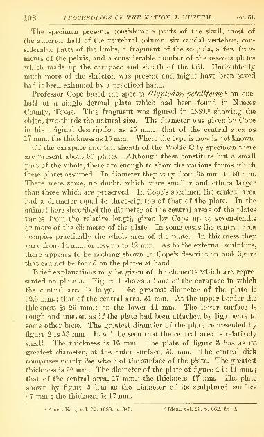
Professor Cope based the species Glyptodon petaliferus'^ on one-
half of a single dermal plate which had been found in Nueces
County, Texas. This fragment was figured in 1889,^ showing the
object two-thirds the natural size. The diameter was given by Cope
in his original description as 45 mm. ; that of the central area as
17 mm., the thickness as 15 mm. Where the type is now is not known.
Of the carapace and tail sheath of the Wolfe City specimen there
are present about 80 plates. Although these constitute but a small
part of the whole, there are enough to show the various forms which
these plates assumed. In diameter they vary from 35 mm. to 50 mm.There were some, no doubt, which were smaller and others larger
than those which are preserved. In Cope's specimen the central area
had a diameter equal to three-eighths of that of the plate. In the
animal here described the diameter of the central areas of the plates
varies from the relative length given by Cope up to seven-tenths
or more of the diameter of the plate. In some cases the central area
occupies practicall}^ the whole area of the plate. In thickness they
vary from 14 mm. or less up to 42 mm. As to the external sculpture,
there appears to be nothing shown in Cope's description and figure
that can not be found on the plates at hand.
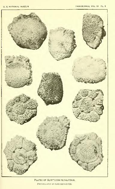
Brief explanations may be given of the elements which are repre-
sented on plate 5. Figure 1 shows a bone of the cai'apace in which
the central area is large. The greatest diameter of the plate is
52.5 mm. ; that of the central area, 31 mm. At the upper border the
thickness is 29 mm.; on the lower 44 mm. The lower surface is
rough and uneven as if the plate had been attached by ligaments to
some other bone. The greatest diameter of the plate represented by
figure 2 is 53 mm. It will be seen that the central area is relatively
small. The thickness is 16 mm. The pla.te of figure 3 has as its
greatest diameter, at the outer surface, 50 mm. The central disk
comprises nearly the whole of the surface of the plate. The greatest
thickness is 22 mm. The diameter of the plate of figure 4 is 44 mm.
;
that of the central area, 17 mm. ; the thickness, 17 mm. The plate
shown by figure 5 has as the diameter of its sculptured surface
47 mm. ; the thickness is 17 mm.
1 Amer. Nat., vol. 22, 1S8S, p. 345. = Idem, vol. 2S, p. 662, fig. 2.
NO. 2147. TWO EXTINCT MAMilALS FROM TEXAS—HAT. 109
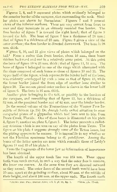
Figures 7, 8, and 9 represent plates which evidently belonged to
the anterior border of the carapace, that surrounding the neck. Simi-
lar iDlates are shown by Burmeister.^ Figures 7 and 9 present
views of the inferior surfaces. These are very convex from front to
rear and they terminate in an obtusely rounded free border. Thefree border of figure 7 is toward the right hand; that of figure 9
toward the left. The bone of figure 7 has a thickness of 26 mm.;that of figure 9 a thickness of 23 mm. Figure 8 gives a view of the
outer surface ; the free border is directed downward. The bone is 28
mm. thick.
Figures G, 10, and 11 give views of plates which belonged on the
tail.^ From a rather thin front border, about 10 mm., these bones
thicken backward and end in a relatively acute point. At this point
the bone of figure 10 is 23 mm. thick; that of fi.gure 11, 31 mm. Thebone of figure G belonged to one of the rings which alternated with
the rings composed of such bones as those of figures 10 and 11. Theupper half of the figure, which represents the hinder half of the bone,
was evidently overlapped by such a bone as that of figure 10, while
the lower border joined the front edge of another bone similar to
figure 10. The convex pitted outer surface is shown in the lower half
of figure G. The bone is 16 mm. thick.
Another plate belonging to the tail, or possibly to the borders of
the carapace, and resembling that of figure 11, has a thickness of
32 mm. at the proximal border and of 42 mm. near the hinder border.
In the second volume of the Transactions of the Wagner Free In-
stitute of Science (p. 25) Dr. Joseph Leidy called attention to some
carapacial plates of a glyptodon which had been sent to him from
Peace Creek, Florida. One of these bones is illustrated on his plate
4, figure 9 ; another on plate 6, figure 1. The latter presents a radiat-
ing striation which is not seen on any of the plates from Texas. Thefigure on his plate 4 suggests strongly some of the Texan bones, but
the pitting appears to be coarser. It is impossible to say whether or
not the Floridan specimens belong to G. petaUfey'us. Among the
bones of the latter species are none which resemble those of Leidy's
figures 11 and 12 of his plate 5.
From the fragments of the lower jaw no information of importance
has been obtained.
The length of the upper tooth line was 165 mm. These upperteeth were much curved, in such a way that the outer face is concave,
the inner one convex. At the same time they are directed outwardas they ascend. The outer faces of the second teeth would have been
65 mm. apart at the grinding surface, about 90 mm. at the middle of
their height, and about 100 mm. at the upper ends. The fourth tooth
1 Anales Mus. Pub., Buenos Aires, vol. 2, pi. 41, flg. 4. s Idem, pis. 37-40.
110 PROCEEDINGS OF THE NATIONAL MUSEUM. vol.51.
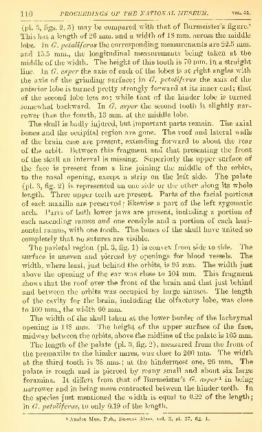
(pi. 3, figs. 2, 3) may be compared with that of Burmeister's figure.^
This has a length of 26 mm. and a width of 18 mm. across the middle
lobe. In G. j}etaUferus the corresponding measurements are 22.5 mm.
and 15.5 mm., the longitudinal measurements being taken at the
middle of the width. The height of this tooth is TO mm. in a straight
line. In G. asper the axis of each of the lobes is at right angles with
the axis of the grinding surface; in G. petaliferus the axis of the
anterior lobe is turned pretty strongly forward at its inner end ; that
of the second lobe less so; while that of the hinder lobe is turned
somewhat backward. In G. asper the second tooth is slightly nar-
rower than the fourth, 13 mm. at the middle lobe.
The skull is badly injured, but important parts remain. The axial
bones and the occipital region are gone. The roof and lateral walls
of the brain case are present, extending forward to about the rear
of the orbit. Between this fragment and that presenting the front
of the skull an interval is missing. Superiorly the upper surface of
the face is present from a line joining the middle of the orbits,
to the nasal opening, except a strip on the left side. The palate
(pi. 3, fig. 2) is represented on one side or the other along its whole
length. Three upper teeth are present. Parts of the facial portions
of each maxilla are preserved ; likewise a part of the left zygomatic
arch. Parts of both lower jaws are present, including a portion of
each ascending ramus and one condyle and a portion of each hori-
zontal ramus, with one tooth. The bones of the skull have united so
completely that no sutures are visible.
The parietal region (pi. 3, fig. 1) is convex from side to side. The
surface is uneven and pierced by openings for blood vessels. The
width, where least, just behind the orbits, is 95 mm. The width just
above the opening of the ear was close to 101 mm. This fragment
shows that the roof over the front of the brain and that just behind
and between the orbits was occupied by large sinuses. The length
of the cavity for the brain, including the olfactory lobe, was close
to 100 mm., the width 60 mm.The width of the skull taken at the lower border of the lachrymal
opening is 118 mm. The height of the upper surface of the face,
midway between the orbits, above the midline of the palate is 105 mm.The length of the palate (pi. 3, fig. 2), measured from the front of
the premaxilse to the hinder nares, was close to 200 mm. The width
at the third tooth is 38 mm. ; at the hindermost one, 26 mm. The
palate is rough and is pierced by many small and about six large
foramina. It differs from that of Burmeister's G. asper ^ in being
narrower and in being more contracted between the hinder teeth. In
the species just mentioned the width is equal to 0.22 of the length;
in G. pefalifenis, to only 0.19 of the length.
1 Anales Mus. Pub., Buenos Aires, vol. 2, pi. 27, flg. 1.
NO. 2147. Tiro EXTINCT 2IAJ,BIALS FR02I TEXAS—HAY. HI
In G. petaUferus the second tooth (pi. 3, figs. 2, -1) is different
from the fourth. Its length is 20 mm., its width only 9 mm. The outer
ends of all the lobes are much reduced. It resembles considerably
the first tooth of G. asper. The first tooth is missing in the Texas
species, but the inner wall of the socket is present. From this it is
evident that the lobes were much reduced on the inner side also. It
is pretty certain that this tooth was thin and simple in construction,
but the length of its grinding surface nearly equaled that of the
second tooth.
The lower teeth were nearly straight, as shown by the one present
and by the sockets in the fragments of the lower jaw. The one
present (pi. 3, fig. 5) had a height of 75 mm., a length of 21 mm.on the grinding surface, and a width of 12.5 mm. on the middle lobe.
The tooth present, belonging on the right side, is placed opposite
the front border of the ascending ramus and is probably the sixth
in the series. There were at least two others behind it. In G. asper^
as figured by Burm.eister, the grinding surface of this tooth has a
length of 21 mm. and a width of IG mm. across the middle lobe.
In G. petalifei-us the length is 21 mm., the width 13 mm. In the
tooth of this species the axes of the lobes are little turned from a
perpendicular to the longitudinal axis; in G. asper they are muchmore strongly deflected.
In all the teeth, upper and lower, the central core of vasodentine
which sends lateral branches into the lobes undergoes secondary
divisions there, as in G. asper.
The atlas is missing. In the giyptodonts the axis and the suc-
ceeding three or four cervicals are consolidated into one mass.
Usually in the genus Glyptodon the mass includes the sixth cer-
vical, but from Burmeister's description ^ it seems that in two species
it is sometimes free and sometimes confluent. In the specimen at
hand the sixth was evidently free and is missing from the collection.
The consolidated second to fifth vertebrae (pi. 3, fig. 6) are injured
somewhat; especially, the transverse processes are gone. The massresembles much that of the forms figured by Burmeister. From the
outside of one lateral articular surface for the atlas to that of the
other is 80 mm. Hence the bone is smaller than any of those figured
on the plate just cited. The distance from the outer side of one
postzygapophysis to that of the other of the fifth vertebra is like-
wise 80 mm. The height of the neural spine above the floor of the
neural canal is 63 mm.Judging from the character of the surfaces by which the sixth
cervical was united with the seventh, there was not much motion
between them.
1 Anales Mus. Pub., Buenos Aires, vol. 2, p. 296, pi. 29.
112 PROCEEDINGS OF THE NATIONAL MUSEUM. vol.51.
As usual in the glyptodonte, the seventh cervical is united solidly
with the first and second dorsals. The width of the mass (pi. 3,
fig. 7) near the rear is 150 mm. That of G. asper^ appears to have
been about 180 mm. wide behind and wider still in front. This
mass, as figured and described by Burmeister, had along each border
two rather deep notches and three processes. These are not seen in
the specimen before us. On each side is an irregular surface, with
several small facets for union with the head of the first rib. Themotion here was evidently unimportant. The surface on each side
for the second rib indicates more liberal movement. On each side
below are two large openings for nerves. These divide each into two
canals, one opening out on the upper surface of the mass, the other
on the lower. The superior openings are much larger than those of
Burmeister's figures. At the rear of the mass the postzygapophysial
surfaces of the two sides coalesce under the spine. On each lateral
process is a surface for union with corresponding surface on the
front of the third dorsal.
The third dorsal and the succeeding ones, up to and including the
twelfth are, in the glyptodonts, consolidated into a single mass in
which the individual vertebrae can be distinguished only by the
foramina for nerves and the facets for the ribs. In G. jyetallferus
the floor of the spinal canal is in places less than a millimeter in
thickness : in the last dorsal, however, 5 mm. thick. The dorsal spines
are greatly reduced and coalesced into a median ridge of small and
irregular height. In the series, as preserved, on the assumption that
there were twelve, there is missing most of the sixth and of the
seventh dorsals and a part of the eleventh. The front of the third
dorsal presents, superiorly (pi. 3, fig. 8) a crescentic zygapophysial
surface for the second dorsal; also on each side a semicylindrical
surface on the lateral process, for union with a corresponding surface
on the second dorsal, already noted above. Above the rear of the
articulatory surfaces for the fourth pair of ribs the bone correspond-
ing to the fourth vertebra is 109 mm. wide. According to Bur-
meister's figure of G. asfer"^ the same bone had a width of about
132 mm. Burmeister's figur^ indicates that the front end of this
vertebral tube, in the region of the articulations of the third, fourth,
and fifth pairs of ribs, was bounded on each side by a ridge ; but in
the specimen here described there are here no such ridges. However,
further backward these ridges become very prominent. Again, the
median ridge, composed of the coalesced spinous processes, which
in Burmeister's figure is still prominent opposite the tenth and
eleventh pairs of ribs, is obsolete in G. petallferus. The rear of the
twelfth vertebra is rough and was joined to the first lumbar probably
1 Burmeister, Anales Mus. Pub., Buenos Aires, vol. 2, pi. 30. ^ idem, pi. 30, flg. 1.
TWO EXTINCT 2IA2niALS FROM TEXAS—HAY. 113
by fibro-cartilage ; hence there was some movement at this point of
the vertebral column.
The whole congeries of vertebrae which compose the lumbosacral
tube is missing, except a fragment which appears to represent tlie
fourth and fifth sacrals, and another fragment which furnishes the
hinder part of the centrum of the seventh sacral and the whole of
the eight. To the latter is attached a large part of each lateral
process. On the front edge of each of these processes is a stump of
the lateral process of the seventh sacral. The hinder end of the
centrum of the eighth sacral, smooth for movable union with the
first caudal, has a width of 73 mm. and a height of 55 mm.There are present six caudal vertebrae. The average length of
these is 77 mm. These belong at the base of the tail and all bear
facets for chevrons. According to Burmeister's figure ^ and that of
Lydekker- the tail of Glyptodon has 11 vertebrae. Of the whole length
of the tail the basal six vertebrae occupy a little more than one-half.
It seems probable, therefore, that the tail of G. petaliferus had a
length of about 810 mm. An estimate indicates that our Texas
species had a length of head, body, and tail of about 7 feet.
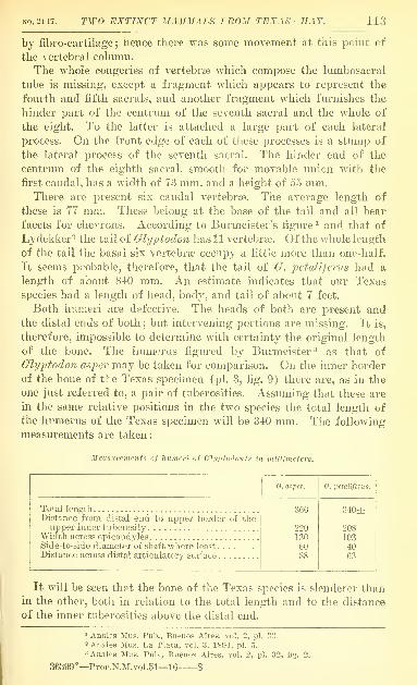
Both humeri are defective. The heads of both are present andthe distal ends of both ; but intervening portions are missing. It is,
therefore, impossible to determine with certainty the original length
of the bone. The humerus fig-ured by Burmeister^ as that of
Glyptodon asper may be taken for comparison. On the inner border
of the bone of the Texas specimen (pi. 3, fig. 9) there are, as in the
one just referred to, a pair of tuberosities. Assuming that these are
in the same relative positions in the tw^o species the total length of
the humerus of the Texas specimen will be 310 mm. The following
measurements are taken
:
Measurements of humeri of Glijpiodonts in miUimeters.
114 PROCEEDINGS OF THE NATIONAL MUSEUM.
Both ulnae are preserved and the left one is wholly uninjured.
This may be compared with the corresponding bone of Burmeister's
Glyptodon asyer.^ The one of the right side is figured (pi. 4, fig. 1)
because with it may be shown the corresponding radius.
Measurements of ulnae of Ghjptodonts in millimeters.
KO. 2147. Tiro EXTINCT MAMMALS FROM TEXAS—HAY. 115
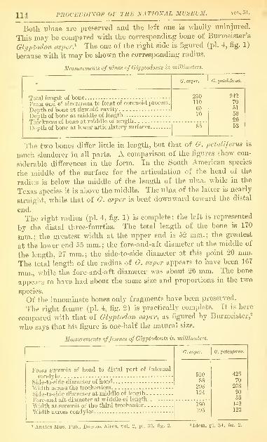
The femur measured by Burmeister is, as seen, considerably longer
than that of the Texas species here measured. In the upper part the
proportions are nearly the same, the length of the bone being madethe standard of comparison. However, the width at the middle is
somewhat less in G. petaliferus. "Wliile the width across the third
trochanter of G. asper is 0.425 of the length of the bone, in G.
petaliferus this width is only 0.331 of the length. Likewise, the
width across the condyles of G. asper is 0.382 of the length, in G.
petaliferus only 0.29.
The patella of the left leg is present. Its general form is quadrate.
Its length is 81 mm. ; its width near the upper end is 75 mm. ; near
the lower end 60 mm. The two lines of measurement are not, how-ever, in the same plane, the outer end of the lower one being car-
ried somewhat forward.
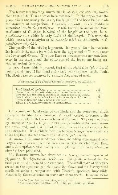
A part of each tibia is present, that of the right side (pi. 1, fig. 3)
lacking that part of the distal end which was ankylosed to the fibula.
The fibulas are represented by a single fragment of each.
Measurements of the tibiae of Glyptodon petaliferus in millimeters.
Total length of the boneDistance acro33 the articulatory surfaces for the femurFore-and-aft diameter of surface for inner condyle of femiu* . .
.
Side-to-side diameter of surface for inner condyle of femur . . .
Greatest diameter where bone is smallest ,
Width of articulatory surface for astra^galus
On account of the absence of the fibula and the consequent slight
injury to the tibia here described, it is not possible to compare the
latter accurately with the same bone of G. asper. The one measuredby Burmeister^ had a length of 210 mm., a width of 111 mm. across
the upper end, and a width of 96 mm. across the articulation for
the astragalus. It is evident that this bone in G. asper was, relatively
to its length, a stouter bone than that of G. petaliferus.
A considerable number of foot bones, including nine ungTial pha-langes, are preserved, but no foot can be reconstructed from themand a description would hardly add anything of value to what hasalready been published.
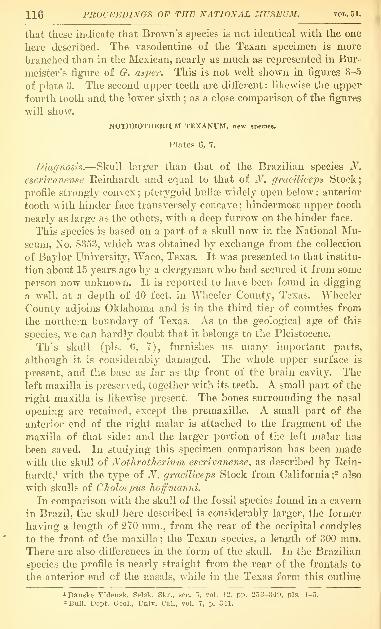
]Mr. Barnum Brown has described^ a new genus and species of
glyptodon, Brachyostraeon mexicanus. The genus is based for the
most part on the form of the carapace. The small part of this pre-
served in the specimen which I describe above and its disorganized
condition make a comparison with Brown's specimen impossible.
Practically the only common parts are three teeth. It seems to me
1 Anales Mus. Pub., Bnenos Aires, vol. 2, p. 348." Buil. Amer. Mus. Nat. Hist., vol. 31, 1912, pp. 167-177, pis. 13-18.
116 PROCEEDINGS OF THE NATIONAL MUSEUM. vol.51.
that these indicate that Brown's species is not identical with the one
here described. The vasodentine of the Texan specimen is more
branched than in the Mexican, nearly as much as represented in Bur-
meister's figure of G. asper. This is not well shown in figures 3-5
of plate 3. The second upper teeth are different ; likewise the upper
fourth tooth and the lower sixth ; as a close comparison of the figures
will show.
NOTHROTHERIUM TEXANUM, new species.
Flutes 6, 7.
Diagnosis.—Skull larger than that of the Brazilian species N.
escrivanense Reinhardt and equal to that of N. graciliceps Stock;
profile strongly convex;pterygoid bullse widely open below : anterior
tooth with hinder face transversely concave ; hindermost upper tooth
nearly as large as the others, with a deep furrow on the hinder face.
This species is based on a part of a skull now in the National Mu-seum, No. 8353, which was obtained by exchange from the collection
of Baylor University, Waco, Texas. It w^as presented to that institu-
tion about 15 years ago by a clergyman who had secured it from some
person now unknown. It is reported to have been found in digging
a well, at a depth of 40 feet, in Wheeler Count}^, Texas. AMieeler
County adjoins Oklahoma and is in the third tier of counties from
the northern boundary of Texas. As to the geological age of this
species, we can hardly doubt that it belongs to the Pleistocene.
This skull (pis. 6, 7), furnishes us many im.portant parts,
although it is considerably damaged. The whole upper surface is
present, and the base as far as thp front of the brain cavity. Theleft maxilla is preserved, together with its teeth. A small part of the
right maxilla is likewise present. The bones surrounding the nasal
opening are retained, except the premaxillse. A small part of the
anterior end of the right malar is attached to the fragment of tlie
maxilla of that side; and the larger portion of the left malar has
been saved. In studying this specimen comparison has been madewith the skull of Nothrotherium escrivanense^ as described by Rein-
hardt,^ with the type of N. graciliceps Stock from California;- also
with skulls of Choloepus liojfmanm.
In comparison with the skull of the fossil species found in a cavern
in Brazil, the skull here described is considerably larger, the former
having a length of 270 mm., from the rear of the occipital condyles
to the front of the maxilla ; the Texan species, a length of 300 mm.There are also differences in the form of the skull. In the Brazilian
species the profile is nearly straight from the rear of the frontals to
the anterior end of the nasals, while in the Texas form this outline
iDanske Vidensk, Selsk. Skr., ser. 5, vol. 12, pp. 253-349, pis. 1-5.
2 Bull. Dept. Geol., Univ. Cal., vol. 7, p. 341.
NO. 2147. T^YO EXTINCT MAMMALS FROM TEXAS—HAY 117
is convex (pi. G), but undulating. Also, while the parietal part
of the profile in the Brazilian species is pretty strongly convex,
in the Texan species it is undulating and little convex.
On the other hand, N, texanum is very closely related to N. grcDcili-
ceps Stock. The differences which are believed to exist are con-
sidered below.
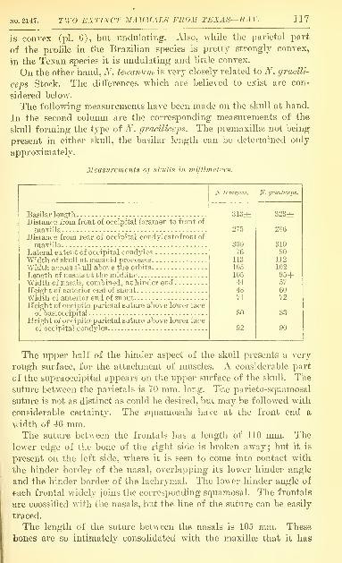
The following measurements have been made on the skull at hand.
In the second column are the corresponding measurements of the
skull forming the type of N. grddlheps. The premaxillaj not being
present in either skull, the basilar length can be determined only
approximately.
Measurements of skulls in millimeters.
N. graciliceps.
Basilar lengthDistance from front of occipital foramen to front of
maxilla.Distance from rear of occipital condyles to front of
maxillaLateral extent of occipital condylesWidth of skull at mastoid processes
Width across ekidl above the orbits
Length of nasals at the midlineWidth of nasals, combined, at hinder endHeight of anterior end of snoutWidth of anterior end of snoutHeight of occipito-parietal suture above lower face
of basioccipital
Height of occipito-parietal suture above lower face
of occipital condyles
313z 323rh
286
500
118 ntOCEEDINGS OF THE NATIONAL MUSEUM.
been somewhat difficult to determine the line of union. On the left
side there is, at a distance of 32 mm. from the midline and at the
anterior border of the frontal bone, a small foramen from which an
indistinct, irregular line may be traced for a few millimeters for-
ward. This line is shown on plate 7, figure 1. At the corresponding
position on the left side of the figure is seen a white line. The bone
on the right of this line had been separated and later cemented on
again. On close examination it is found that there are here well-
defined sutural surfaces, the maxilla joining the outer border of the
nasal. At this point the distance from the outer edge of one nasal
to that of the other is 57 mm. To what extent the naso-maxillary
sutures determined the lines of fracture seen on the upper surface
of the snout is uncertain.
The lachrymal is articulated principally with the maxilla, but its
upper hinder border joins the frontal; while below it is united with
the anterior end of the malar. It shows a large lachrymal foramen
well in front of the orbit. This foramen is the outer opening of a
canal which followed inward soon turns and is directed forward,
opening into the nasal chamber just in front of the upper end of the
first tooth.
In viewing the skull from below (pi. 7, fig. 2) there are observed
behind the ear opening the small condyloid foramen and the large
foramen lacerum posterius. The ear opening has a diameter of 10
mm. On the right side the tympanic bone is in its place, forming a
ring which is incomplete above. Below it is inflated into a bulla of
moderate size whose external surface is rough. On the left side the
tympanic is missing, a fact which shows that it had not become
ankylosed to the contiguous bones. The absence of the bone permits
a view of a part of the petrosal. In front of the petrosal is seen
the foramen lacerum medius. This, as it appears, is divided into
two parts, the more anterior and outer being well in front of the
external auditory meatus.
In front of the great pterygoid bulla is seen the foramen ovale.
On the right side there is, in front of the ovale, an opening, the
sphenoidal fissure. On the right side there are here two foramina,
the hinder of which is probably the foramen rotundum. Farther
in front and somewhat higher up and nearer the midline are the
canals for the optic nerves. It is evident that these opened out at
points in advance of the middle of the length of the skull.
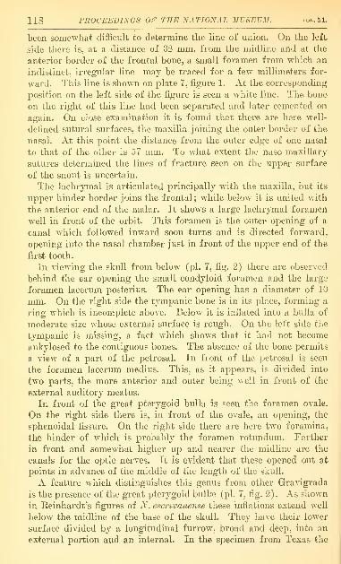
A feature which distinguishes this genus from other Gravigrada
is the presence of the great pterygoid bulla? (pi. 7, fig. 2). As shown
in Reinhardt's figures of N. es'cnvanense these inflations extend well
below the midline of the base of the skull. They have their lower
surface divided by a longitudinal furrow, broad and deep, into an
external portion and an internal. In the specimen from Texas the
NO. 2147. TWO EXTINCT MAMMALS FROM TEXAS—HAY. 119
lower floor of the bullae is missing, so that the form of this part, if
ever present, can not be observed. According to the description of
these bulloe in the Brazilian species there is along the median line
a space only about 5 mm. wide between them. They are evidently
marked off along their inner boundary much more sharply than in
the Texan species. In this animal there is between them a broad,
longitudinal groove whose sides slope downward and outward grad-
ually into the walls of the bullae. The length of each bulla is 50
mm.; the width may be taken as 35 mm. The distance from the
outer wall of one to that of the other is 90 mm. The median side of
each cavity extends inward and upward into the base of the skull
until the two are only 15 mm. apart.
The pterygoid bullae of N. graciUceps have been described by
Stock. They are called by him tympanic bullae, but they are not
such. Mr. Gerrit Miller has directed my attention to similarly placed
and apparently homologous cavities at the base of the skull in vari-
ous bats. As shown by Stock the roof of these bullae is formed by
the alisphenoids. The side walls and floor in the Brazilian and the
Californian species are certainly formed by the pterygoids. In N.
graciUceps Stock there is along the inner face of the bulla a slit
about 30 mm. long which puts the cavity of the bulla in communi-
cation with the pharynx. The bulla of N. texanum appears not to
have had a floor. The pterygoids seem to form a wall which sur-
rounds the cavity on both sides. On the median side the edge of
the wall is partly intact, partly injured. On the outer side the
w^all comes down to a sharp thin edge which appears to be little if
at all injured. In places the edge is certainly wholly natural. Such
being the case the bulla is incomplete and is a cavity opening below
by a mouth 30 mm. wide. In N. graciUceps the outer wall has
grown downwards and inwards until it has nearly met the inner
wall; in N. eserivanense the space between the two walls was ap-
parently abolished. In Choloepus hoffmanni there are homologous
bullae which open at the anterior end into the mesopterygoid fossa.
Similarly placed bullae are found in the great anteater {Myrmeco-
phaga juhafa), but their structure is somewhat doubtful.^
In the Texas specimen there is a rough and sharp ridge which
begins on the midline between the front ends of the pterygoid bul-
lae and runs forward as far as the bone is uninjured. A similar struc-
1 From an examination of skulls of the great anteater in the United States National
Museum the writer concludes that the pterygoids and the alisphenoids of each side are so
completely coossifled that the line of union can not be determined unless it be in youngerindividuals than are at hand. The bullte in adult individuals are completely closed. In
a not fully grown specimen the impression given is that the bullte remained open longest
on the outer side, near the border of the temporal bone. It is believed that the area
called alisphenoid in Weber's figure 332 taken from Pouchet (Saugetiere, p. 434) is not
such. Certainly the foramen ovale pierces the alisphenoid ; and it is this bone, not the
pterygoid, which joins the basisphenoid.
120 PROCEEDINGS OF THE NATIONAL MUSEUM. VOL. 51.
ture is shown in one of Keinhardt's figures. This ridge appears to
be on the vomer.
In our specimen the greater part of the palate, the front of the
vomer, and the ethmoid bones have been broken away. A part of
the hard palate is seen in front, and the underside of this is rough.
In the rear of this injured region the cribriform plate has been
broken through so as to leave a small opening to the brain-cavity
on the left side and a much larger one on the right. In front of
this, on the right side (left side of the illustration, pi. T, fig. 2),
are seen openings into sinuses in the frontal bone. The larger of
these on each side extend backward to the hinder end of the frontal.
Some of the plates of bone nearer the midline evidently belong to
the olfactory apparatus. On the right side there remains about
30 mm. of the malar bone. On the left side the front part of the
malar is missing, but the hinder part is present. The malar was a
triradiate bone. The anterior process joined the lachrymal. The
hinder process was directed upward and backward and had a notch
in the hinder part of the lower border to receive the anterior end of
the zygomatic process of the temporal bone. The lower process is
pointed, and it descended about 60 mm. below the level of the palate.
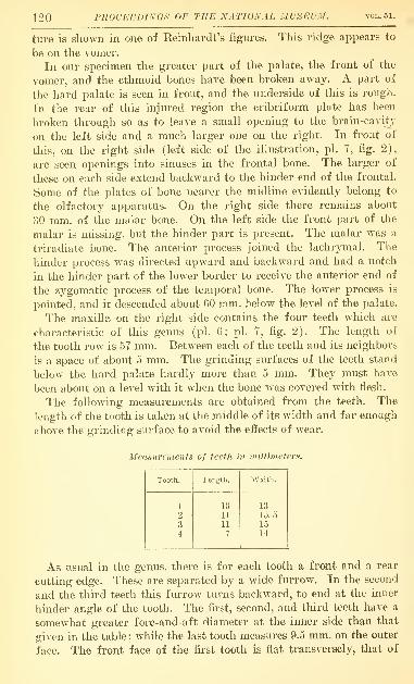
The maxilla on the right side contains the four teeth which are
characteristic of this genus (pi. 6; pi. 7, fig. 2). The length of
the tooth row is 57 mm. Between each of the teeth and its neighbors
is a space of about 5 mm. The grinding surfaces of the teeth stand
below the hard palate hardly more than 5 mm. They must have
been about on a level with it Avhen the bone was covered with flesh.
The following measurements are obtained from the teeth. The
length of the tooth is taken at the middle of its width and far enough
above the grinding surface to avoid the effects of wear.
Measurements of teeth in millimeters.
Tooth.
NO. 2147. TWO EXTINCT MAMMALfi FROM TEXAS—HAY. 121
the others convex. The hinder faces of all are concave—that of the
fourth tooth most so of all. The inner faces are flat or slightly con-
vex—that of the fourth rather strongly so. The outer faces are some-
what concave, showing a shallow groove along their whole height.
All of these teeth have a height of about 50 mm. They are hollow
down to within about 10 mm. of the grinding surface.
On the front end of each maxillar}^ there is a surface for the articu-
lation of the corresponding premaxilla. The two surfaces are sepa-
rated by a space of 20 mm., and each has a length of 30 mm. On the
lower side of the maxilla is another surface for a backwardly di-
rected process of the premaxilla. In case the premaxillae corre-
sponded in size to those of the Brazilian species mentioned above,
each had a length of about 30 mm.This is not the first discovery of the genus Nothrot/ierhimm'^orth.
America. In 1905 ^ Sinclair reported it, with some doubt, from Potter
Creek cave, Shasta Count}'', California, He had for description a
part of a lower jaw without teeth and fourteen loose molars. Thename N. shastense was given to the species.
In order to determine the relationship of the Texan specimen to
that found in northern California, it is necessary to compare with
the teeth of the former those which Sinclair has represented byfigures 3, 5, and 8 of his plate 23. figures 3 and 5 must be second
and third teeth. Of the tooth- represented by his figure 3 both the
front and the rear faces are convex in section, whereas both the second
and the third teeth of N. texanum have the front face convex and the
rear face concave. Sinclair's figure 5 resembles somewhat the sec-
tion of the third tooth of the Texan species ; but here, as in the tooth
of his figure 3, the inner face of the tooth is more or less concave;
whereas, in the Texan animal, the inner face is flat. However, it is
in the hindermost tooth that the greatest difference is found. In
the California species the front of the tooth is convex, the rear flat.
In the Texan species the rear of the tooth is deeply concave. It
appears to be evident that two distinct species are indicated.
In 1913,^ Stock described a skull, lacking the lower jaw and some
other parts, which he called N. graciUceps. The type is now in the
Los Angeles Museum of History, Science, and Art, where the writer
has had the privilege of examining it. This skull resembles closely
that from Texas in size and proportions, as may be seen from the
measurements given on page 117. There are, however, in the Texan
slmll, certain deviations from that of N. gradliceips which appear to
make it advisable to give to it a distinctive specific name. One can
not rely wholly on the differences which are seen in the two skulls
for additional specimens may be intermediate.
iBull. Dept. Geol., Univ. Cal., vol. 4, p. 153, pi. 23.
= Idem, vol. 7, pp. 341-352, figs. 1-8.
122 PROCEEDINGS OF THE NATIONAL MUSEUM. vol.51.
It seems to the writer that N. grcaciZiceps had the skull more de-
pressed at the anterior half of the frontals. As a result of this, as
Stock says, the nasals have their upper surface transversely convex
in front, but becoming flattened posteriorly. In N. texanum these
bones are rather more convex just in front of the hinder end than
in front. In N. texanum the end of the snout is apparently more
depressed than in N. graciUceps. The width is nearly the same in
the two skulls, but in the latter the height is 60 mm., while in N.
texanum it is only 48 mm. Unless a serious error is committed as
to the structure of the pterygoid bullae in N. texanum, these are
sufficient to differentiate the two species. In N. graeilheps the nasals
have a combined width of only U mm. ; in .V. texanum the width
is 57 mm.There are apparently differences in the two species as regards the
teeth. The type of N. graicili)ceps had not retained the teeth ; but the
size and forms of these may be determined from the sockets. Stock
had one tooth, apparently the second molar, which had been found
in the Rancho La Brea deposits. The sockets of the type skull and
the tooth mentioned show that the teeth of N. graciUceps were larger
than those of N. texanum. The anteroposterior diameter of the
second molar is 13 mm., and thus 2 mm. greater than in the same
tooth of N. texanum. In A'', gramlkeps the hinder face of the first
tooth was evidently convex from side to side; in N. texanum it is
slightly concave.
NO. 2147. TWO EXTINCT MAMMALS FROM TEXAS—HAY. 123
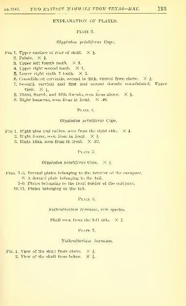
EXPLANATION OF PLATES.
Plate 3.
Glyptodon petaUferns Cope.
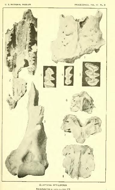
Fig. 1. Upper surface of rear of skull. X i.
2. Palate. X h3. Upper left fourth tooth. X 1.
4. Upper right second tooth. X 1.
5. Lower right sixth ? tooth. X 1.
6. Consolidated cervicals, second to fifth, viewed from above. X J.
7. Seventh cervical and first and second dorsals consolidated. Upper
view. X ^.
8. Third, fourth, and fifth dorsals, seen from above. X J.
9. Right humerus, seen from in front. X .46.
Plate 4.
Glyptodon petaliferus Cope.
Fig. 1. Right ulna and radius, seen from the right side. X J.
2. Right femur, seen from in front. X J.
3. Right tibia, seen from in front. X .32.
I'LATE 5.
Glyptodon petaliferus Cope. X ^.
Figs. 1-5. Dermal plates belonging to the interior of the carapace.
6. A dermal plate belonging to the tail.
7-9. Plates belonging to the front border of the carapace,
10, 11. Plates belonging to the tail.
Plate 6.
Notlirothermm texanutn, new species.
Skull seen from the left side. X i.
Plate 7.
Nothrotheriuni texanum.
Fig. 1. View of the skull from above. X ^.
2. View of the skull from below. X h

U. S. NATIONAL MUSEUM PROCEEDINGS, VOL. 51 PL. 3
Glyptodon PETALIFERUS.
For explanation of plate see page 123.
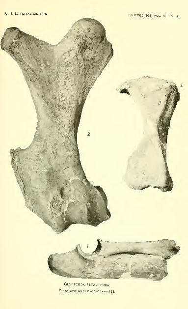
U. S. NATIONAL MUSEUM PROCEEDINGS, VOL. 51 PL. 4
GLYPTODON PETALIFERUS.
For explanation of plate see page 123.

U. S. NATIONAL MUSEUM PROCEEDINGS, VOL. 51 PL. 5
-i^'^^^r:^'k
Plates of Glyptodon petaliferus.
For explanation of plate see page 123.

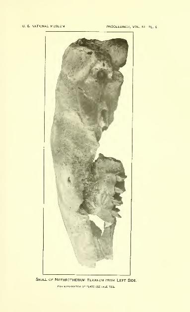
U. S. NATIONAL MUSEUM PROCEEDINGS, VOL. 51 PL. 6
Skull of Nothrotherium texanum from Left Side.
For Explanation of plate see page 123.
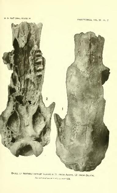
U. S. NATIONAL MUSEUMPROCEEDINGS, VOL. 51 PL. 7
Skull of Nothrotherium texanum (1) from Above, (2) from Below.For explanation of plate see page 123.