
Dermatology Potpourri: Interesting Cases - CME Conferenes · Dermatology Potpourri: Interesting...
90
©Pvernon 2016 Dermatology Potpourri: Interesting Cases Peggy Vernon, RN, MA, C-PNP, DCNP, FAANP
Transcript of Dermatology Potpourri: Interesting Cases - CME Conferenes · Dermatology Potpourri: Interesting...

©Pvernon 2016
Dermatology Potpourri: Interesting Cases
Peggy Vernon, RN, MA, C-PNP, DCNP, FAANP
©Pvernon 2016
Disclosures
• There are no financial interests or
relationships to disclose
• Any unlabeled/unapproved uses of drugs or
products referenced will be discussed
©Pvernon 2016
Restrictions
• Permission granted to the Skin, Bones, Hearts, and Private
Parts 2017 and its attendees
• All rights reserved. No part of this presentation may be
reproduced, stored, or transmitted in any form or by any
means without written permission of the author
• For permission contact Peggy Vernon at

©Pvernon 2016
Session Objectives
• Name common contacts in
phytophotodermatitis
• List two common causes of Majocchi
Granuloma
• List two treatment options for Granuloma
Annularae

©Pvernon 2016
31 YEAR OLD MALE
• Two day history pruritic, tender papules and
vesicles
• Recent camping trip
• Recent fever, headache, lethargy, “flu
symptoms”
• Developed rash on trunk day two
• Day three increased lesions spreading to
entire body with increased lethargy and
fever
©Pvernon 2013
©Pvernon 2016
DIFFERENTIAL DIAGNOSIS
• Rocky Mountain
Spotted Fever
• Lyme Disease
• Varicella
©Pvernon 2016
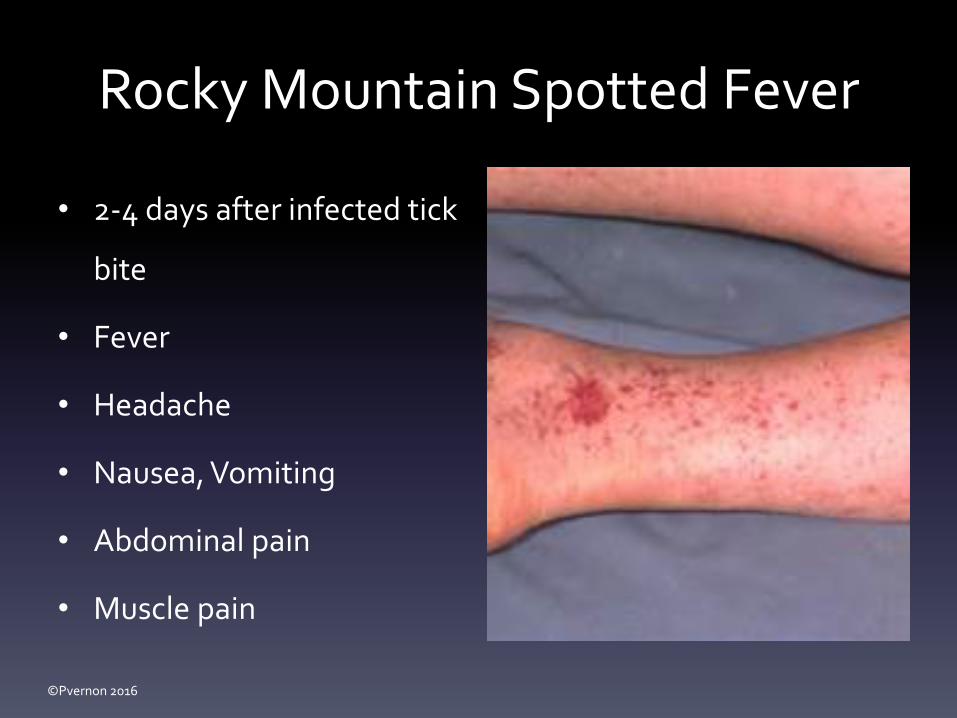
Rocky Mountain Spotted Fever
• 2-4 days after infected tick
bite
• Fever
• Headache
• Nausea, Vomiting
• Abdominal pain
• Muscle pain
©Pvernon 2016
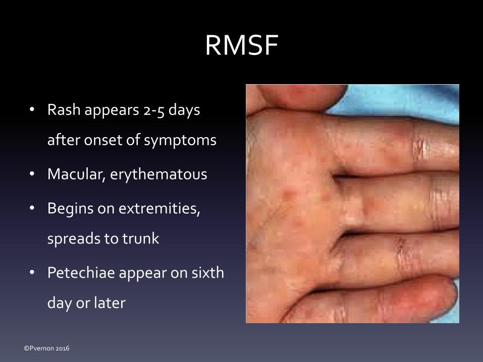
RMSF
• Rash appears 2-5 days
after onset of symptoms
• Macular, erythematous
• Begins on extremities,
spreads to trunk
• Petechiae appear on sixth
day or later

©Pvernon 2016
Lyme Disease• Fever
• Headache
• Fatigue
• Rash at site of tick bite
– circular outwardly expanding rash
(erythema migrans)
– innermost portion dark red,
indurated (bull’s eye)
©Pvernon 2016
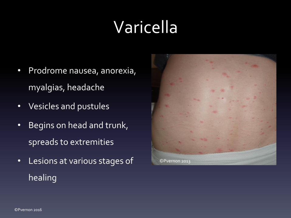
Varicella
• Prodrome nausea, anorexia,
myalgias, headache
• Vesicles and pustules
• Begins on head and trunk,
spreads to extremities
• Lesions at various stages of
healing
©Pvernon 2013
©Pvernon 2016
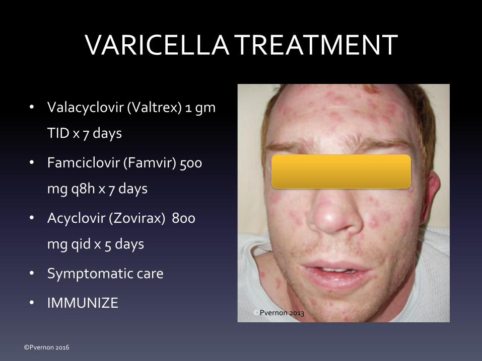
VARICELLA TREATMENT
• Valacyclovir (Valtrex) 1 gm
TID x 7 days
• Famciclovir (Famvir) 500
mg q8h x 7 days
• Acyclovir (Zovirax) 800
mg qid x 5 days
• Symptomatic care
• IMMUNIZE©Pvernon 2013
©Pvernon 2016
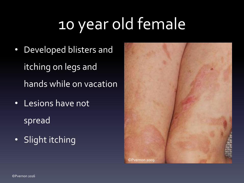
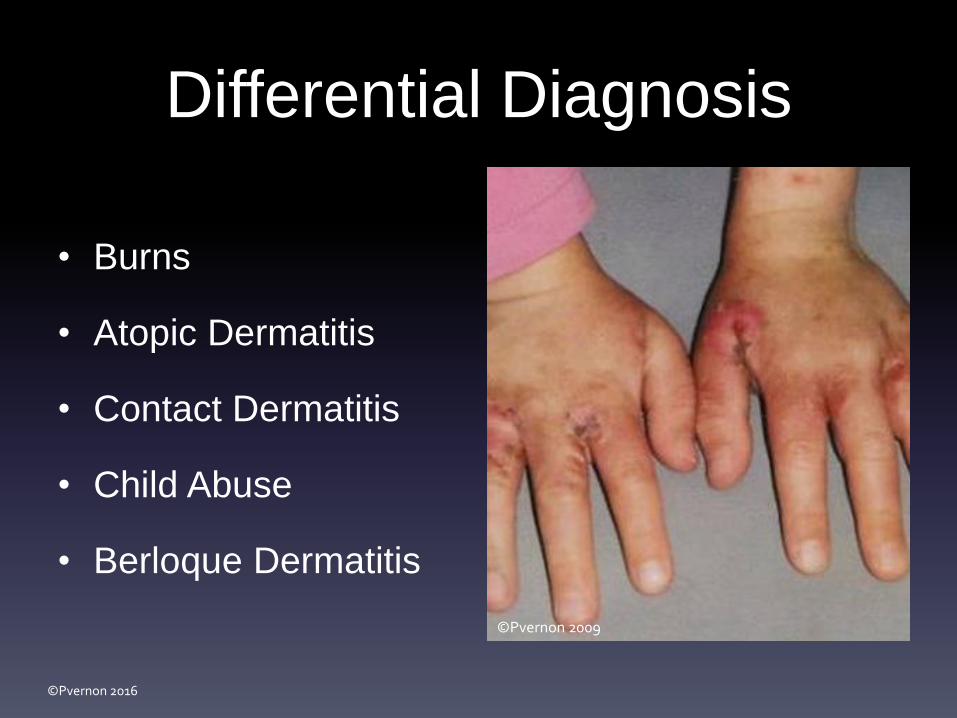
10 year old female
• Developed blisters and
itching on legs and
hands while on vacation
• Lesions have not
spread
• Slight itching
©Pvernon 2009
©Pvernon 2016
Differential Diagnosis
• Burns
• Atopic Dermatitis
• Contact Dermatitis
• Child Abuse
• Berloque Dermatitis
©Pvernon 2009
©Pvernon 2016
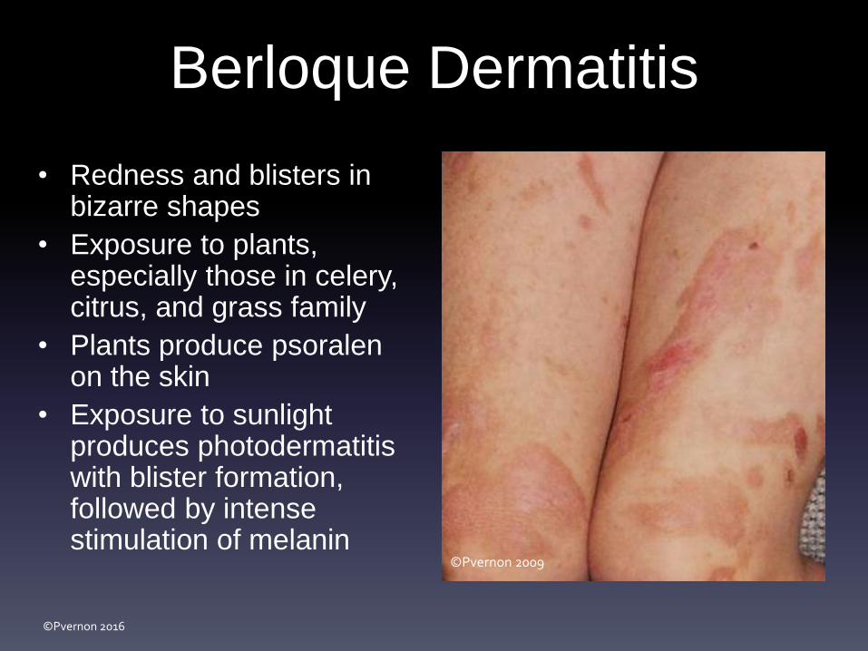
Berloque Dermatitis
• Redness and blisters in bizarre shapes
• Exposure to plants, especially those in celery, citrus, and grass family
• Plants produce psoralenon the skin
• Exposure to sunlight produces photodermatitiswith blister formation, followed by intense stimulation of melanin
©Pvernon 2009

©Pvernon 2016
Berloque Dermatitis Causes
• Citrus and Lime found in drinks and food
– Figs
– Celery
– Lemon and Lime oil
– Queen Anne’s lace
– Giant Russian hogweed
• Bergapten
– Component of bergamot oil
– Found in cosmetics, perfumes, lotions, sunscreens and household
products
©Pvernon 2016
Workup
• Clinical suspicion
• Photopatch test if photoallergy suspected:
– Occlusive application of test chemical(s)
– Irradiation with UV light at several intervals
– Phototoxicity: controls positive
– Photoallergy: controls negative
©Pvernon 2016
Treatment
• Remove offending substance
• No treatment necessary if asymptomatic
• Topical corticosteroids if pruritic
• Analgesics
• Sunscreen
• Treat resulting PIH

©Pvernon 2016
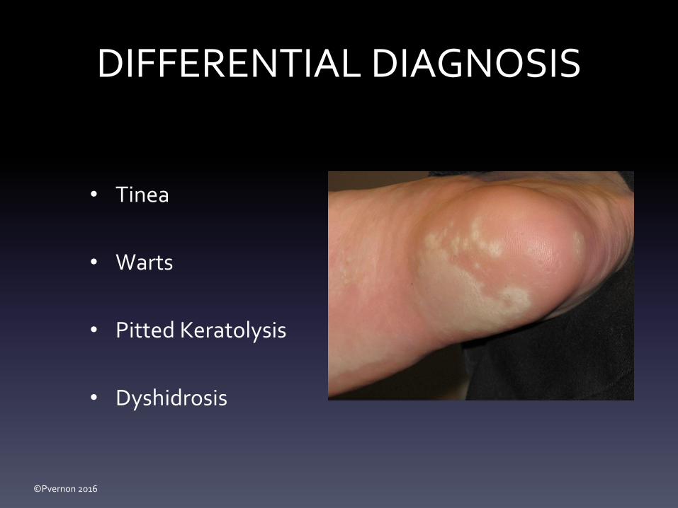
23 YEAR OLD MALE
– Multiple pits on
soles of feet
– Feet, socks, and
shoes damp
– Malodorous
– Asymptomatic

©Pvernon 2016
©Pvernon 2016
DIFFERENTIAL DIAGNOSIS
• Tinea
• Warts
• Pitted Keratolysis
• Dyshidrosis

©Pvernon 2016
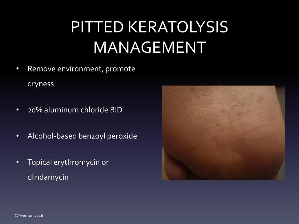
PITTED KERATOLYSIS• Superficial bacterial infection of the soles
of the foot, lateral toes, occasionally
palms
• Asymptomatic erythematous plaques and
shallow pits; occasionally painful
• Often misdiagnosed as tinea
• Hyperhidrosis, moist socks, humid
environment, occlusive shoes and
prolonged immersion in water are
predisposing factors
©Pvernon 2016
PITTED KERATOLYSIS MANAGEMENT
• Remove environment, promote
dryness
• 20% aluminum chloride BID
• Alcohol-based benzoyl peroxide
• Topical erythromycin or
clindamycin

©Pvernon 2016
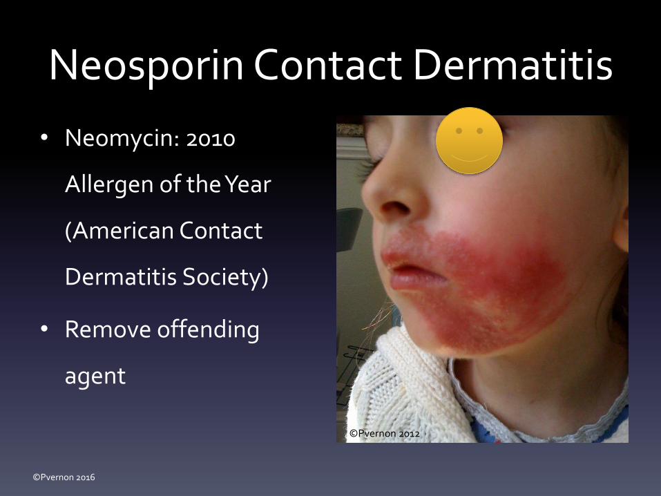
8 year old female
• Developed red, pruritic
rash
• Began as small cut at
oral commissure
• Spreading to chin and
cheeks
©Pvernon 2012
©Pvernon 2016
Neosporin Contact Dermatitis
• Neomycin: 2010
Allergen of the Year
(American Contact
Dermatitis Society)
• Remove offending
agent
©Pvernon 2012

©Pvernon 2016
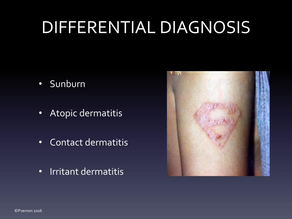
5 YEAR OLD MALE
• Erythematous plaque in
bizarre shape on upper
right arm
• Pruritic
• Recent travel to Mexico
©Pvernon 2016
DIFFERENTIAL DIAGNOSIS
• Sunburn
• Atopic dermatitis
• Contact dermatitis
• Irritant dermatitis

©Pvernon 2016
CONTACT DERMATITIS TREATMENT
• Topical
corticosteroid BID x
2 weeks
• Moisturizer
• Sunscreen
©Pvernon 2016
©Pvernon 2016
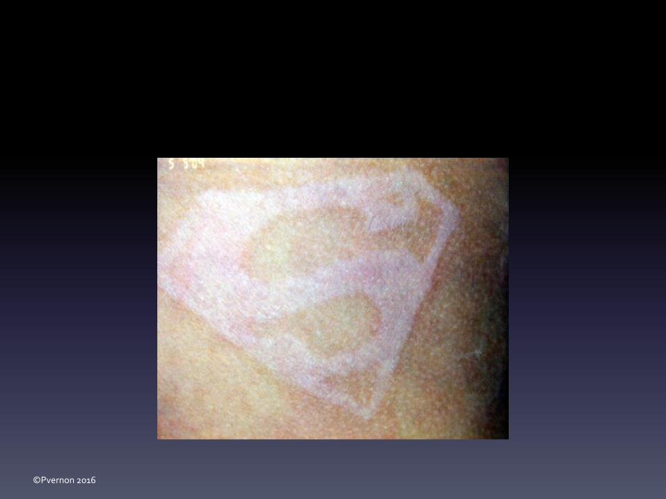
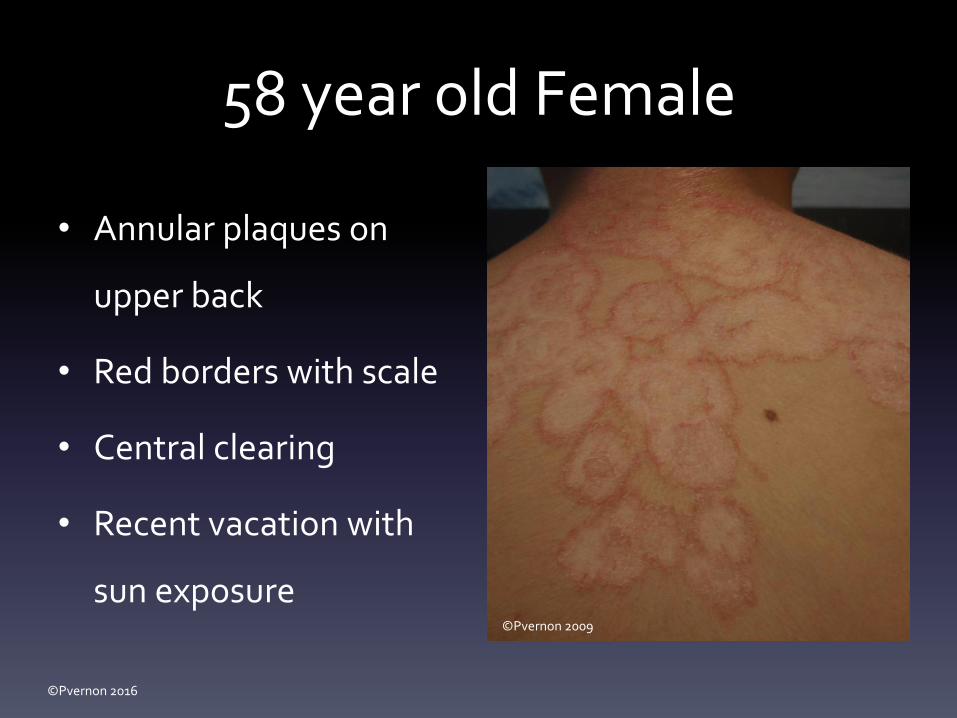
58 year old Female
• Annular plaques on
upper back
• Red borders with scale
• Central clearing
• Recent vacation with
sun exposure©Pvernon 2009
©Pvernon 2016
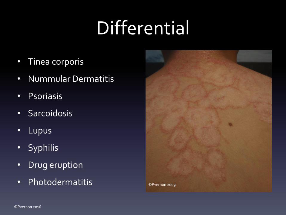
Differential
• Tinea corporis
• Nummular Dermatitis
• Psoriasis
• Sarcoidosis
• Lupus
• Syphilis
• Drug eruption
• Photodermatitis ©Pvernon 2009
©Pvernon 2016
Diagnostics
• ANA, CBC with Differential, ESR (sedimentation rate)
• UA
• Biopsy
– Hematoxylin and eosin staining (H & E)
– Direct immunofluorescnce (DIF) on lesional and peri-
lesional skin

©Pvernon 2016
Lupus Management
• Refer to rheumatology and dermatology for co-
management
• Sunscreen
• Topical and intralesional steroids
• Oral steroids
• Azathioprine
• Cyclophosphamide
• Cyclosporine
• Plaquenil
• Mycophenolate
• Methotrexate
• Benlysta
©Pvernon 2009
©Pvernon 2016
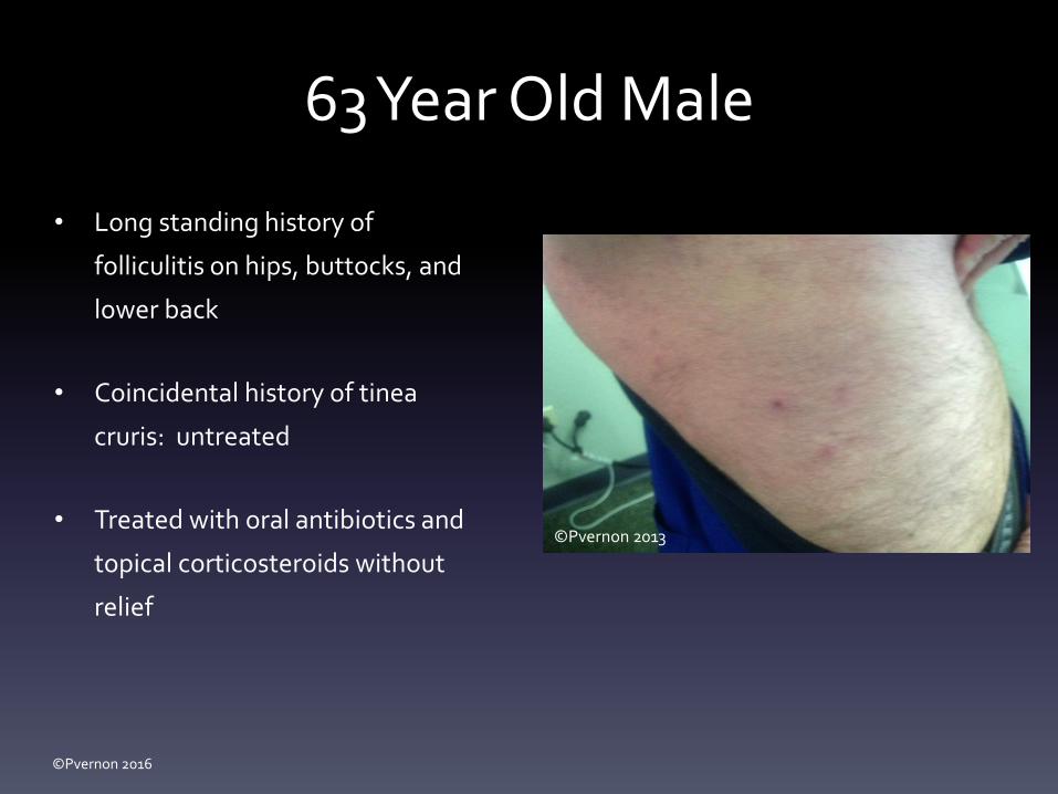
63 Year Old Male
• Long standing history of
folliculitis on hips, buttocks, and
lower back
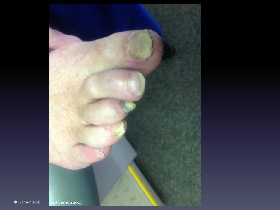
• Coincidental history of tinea
cruris: untreated
• Treated with oral antibiotics and
topical corticosteroids without
relief
©Pvernon 2013
©Pvernon 2016 ©Pvernon 2013
©Pvernon 2016
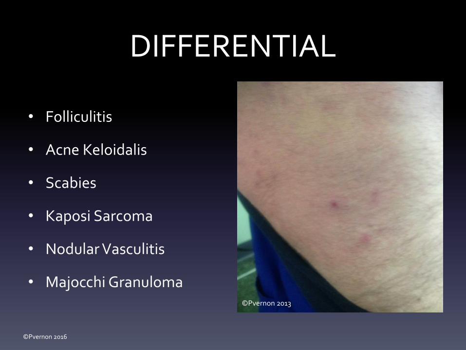
DIFFERENTIAL
• Folliculitis
• Acne Keloidalis
• Scabies
• Kaposi Sarcoma
• Nodular Vasculitis
• Majocchi Granuloma©Pvernon 2013
©Pvernon 2016
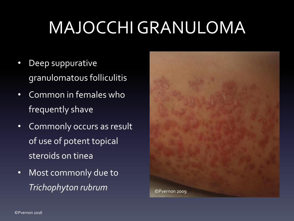
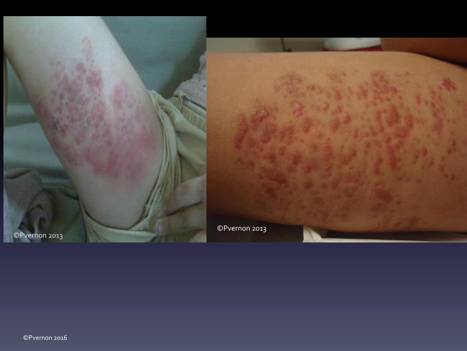
MAJOCCHI GRANULOMA
• Deep suppurative
granulomatous folliculitis
• Common in females who
frequently shave
• Commonly occurs as result
of use of potent topical
steroids on tinea
• Most commonly due to
Trichophyton rubrum ©Pvernon 2009
©Pvernon 2016
©Pvernon 2013©Pvernon 2013
©Pvernon 2016
DIAGNOSIS
• KOH usually negative
• Tissue biopsy
• Gram stains
• Periodic acid-Schiff (PAS) stains reveal fungal hyphae
in tissue, surrounded by granulomatous reaction

©Pvernon 2016
TREATMENT
• Systemic antifungals:
terbinafine x 6 weeks
• Remove exacerbating
factors: topical steroids
• Antibiotics for secondary
bacterial infections
©Pvernon 2013
©Pvernon 2013
©Pvernon 2013
©Pvernon 2016
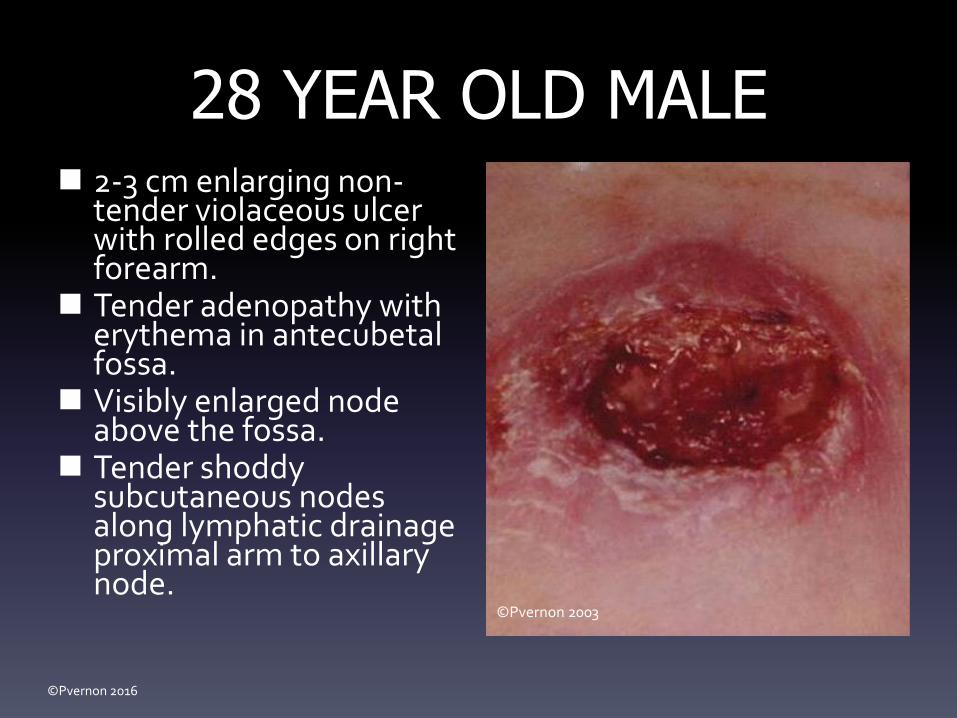
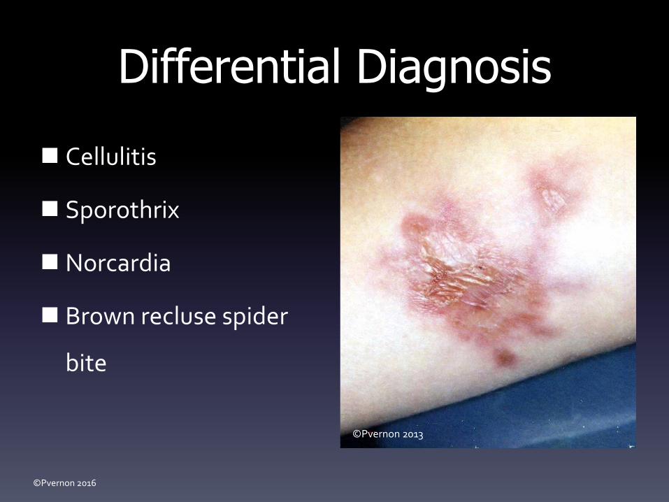
28 YEAR OLD MALE 2-3 cm enlarging non-
tender violaceous ulcer with rolled edges on right forearm.
Tender adenopathy with erythema in antecubetal fossa.
Visibly enlarged node above the fossa.
Tender shoddy subcutaneous nodes along lymphatic drainage proximal arm to axillary node.
©Pvernon 2003
©Pvernon 2016
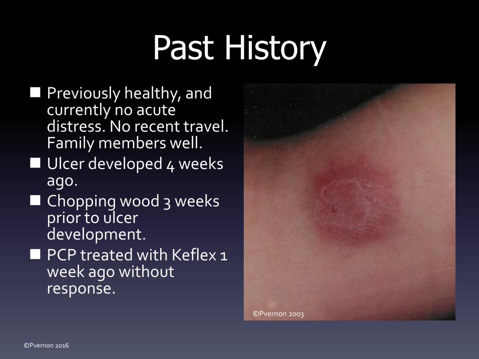
Past History Previously healthy, and
currently no acute distress. No recent travel. Family members well.
Ulcer developed 4 weeks ago.
Chopping wood 3 weeks prior to ulcer development.
PCP treated with Keflex 1 week ago without response.
©Pvernon 2003
©Pvernon 2016
Differential Diagnosis
Cellulitis
Sporothrix
Norcardia
Brown recluse spider
bite
©Pvernon 2013

©Pvernon 2016
SPOROTRICHOSIS
Granulomatous fungal infection
Occurs in all ages in patients
exposed to contaminated soil or
vegetation
Usually follows a wound inflicted
by a contaminated object (splinter,
thorn, straw, grain, rock, glass, cat
bite, or cat scratch)
©Pvernon 2013
©Pvernon 2016
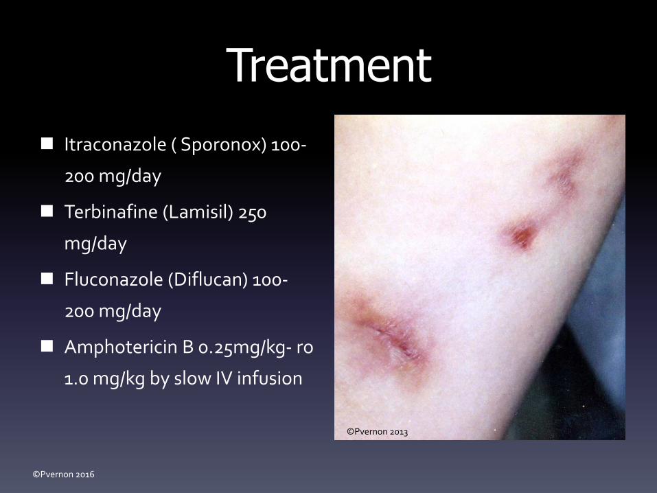
Treatment
Itraconazole ( Sporonox) 100-
200 mg/day
Terbinafine (Lamisil) 250
mg/day
Fluconazole (Diflucan) 100-
200 mg/day
Amphotericin B 0.25mg/kg- ro
1.0 mg/kg by slow IV infusion
©Pvernon 2013
©Pvernon 2016
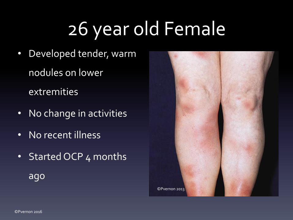
26 year old Female• Developed tender, warm
nodules on lower
extremities
• No change in activities
• No recent illness
• Started OCP 4 months
ago©Pvernon 2013
©Pvernon 2016
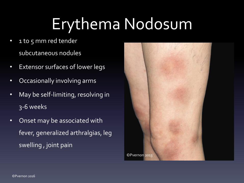
Erythema Nodosum• 1 to 5 mm red tender
subcutaneous nodules
• Extensor surfaces of lower legs
• Occasionally involving arms
• May be self-limiting, resolving in
3-6 weeks
• Onset may be associated with
fever, generalized arthralgias, leg
swelling , joint pain
©Pvernon 2013
©Pvernon 2016
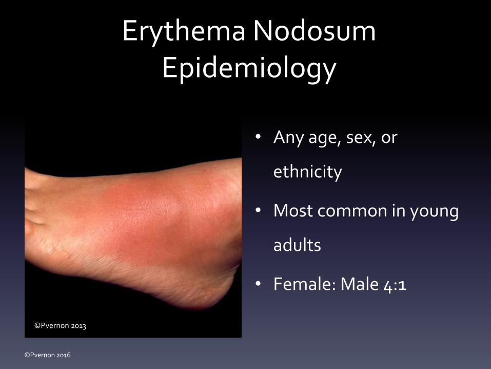
Erythema NodosumEpidemiology
• Any age, sex, or
ethnicity
• Most common in young
adults
• Female: Male 4:1
©Pvernon 2013

©Pvernon 2016
Erythema Nodosum:Infections
• Strep infections, esp. upper
respiratory
• Ulcerative colitis
• Histoplasmosis
• Syphilis, Leprosy
• Sarcoidosis
• Fungal infections:
coccisiodomycosis,
histoplasmosis
©Pvernon 2013
©Pvernon 2016
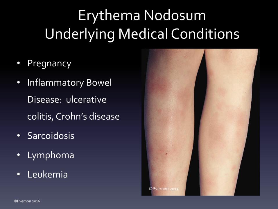
Erythema NodosumUnderlying Medical Conditions
• Pregnancy
• Inflammatory Bowel
Disease: ulcerative
colitis, Crohn’s disease
• Sarcoidosis
• Lymphoma
• Leukemia
©Pvernon 2013
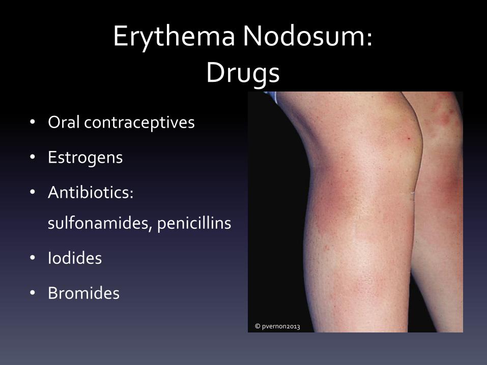
Erythema Nodosum:Drugs
• Oral contraceptives
• Estrogens
• Antibiotics:
sulfonamides, penicillins
• Iodides
• Bromides
© pvernon2013
©Pvernon 2016
Erythema Nodosum Treatment
• Supportive: can be self-healing
• Rest, elevation
• NSAIDS
• Oral or intralesional steroids
• Remove/treat underlying cause
©Pvernon 2016
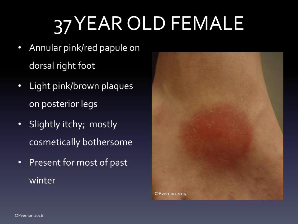
37 YEAR OLD FEMALE• Annular pink/red papule on
dorsal right foot
• Light pink/brown plaques
on posterior legs
• Slightly itchy; mostly
cosmetically bothersome
• Present for most of past
winter©Pvernon 2015
©Pvernon 2016
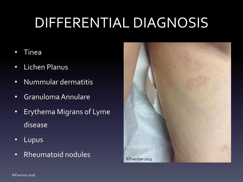
DIFFERENTIAL DIAGNOSIS
• Tinea
• Lichen Planus
• Nummular dermatitis
• Granuloma Annulare
• Erythema Migrans of Lyme
disease
• Lupus
• Rheumatoid nodules©Pvernon 2013
©Pvernon 2016
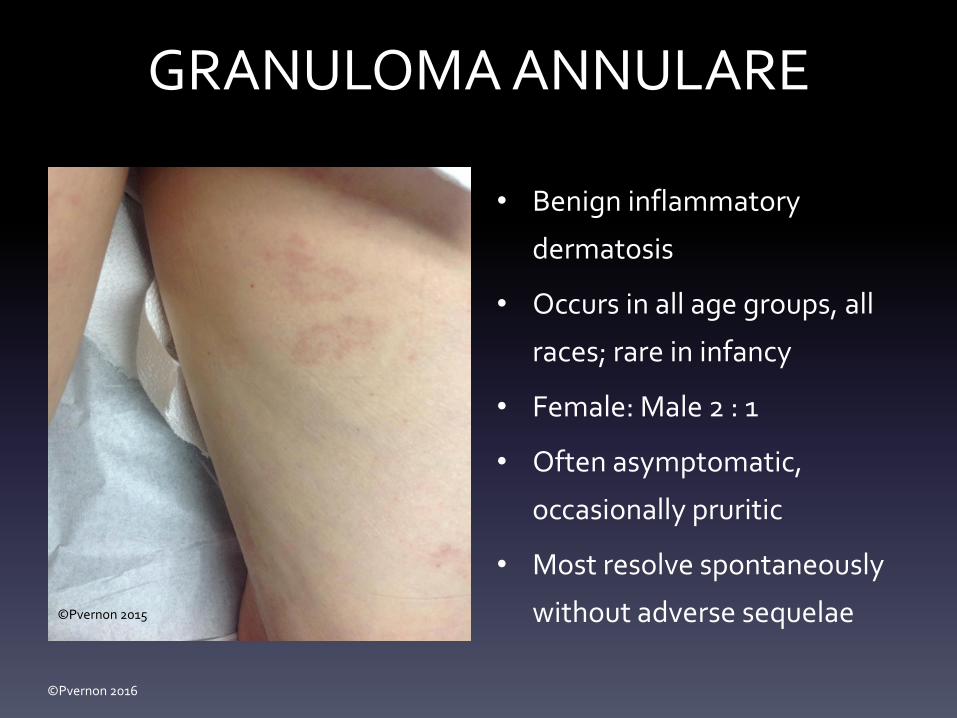
GRANULOMA ANNULARE
• Benign inflammatory
dermatosis
• Occurs in all age groups, all
races; rare in infancy
• Female: Male 2 : 1
• Often asymptomatic,
occasionally pruritic
• Most resolve spontaneously
without adverse sequelae©Pvernon 2015

©Pvernon 2016
TREATMENT
• Intralesional corticosteroid
injections
• Topical corticosteroid
• Cryotherapy
• UVB
©Pvernon 2015
©Pvernon 2016
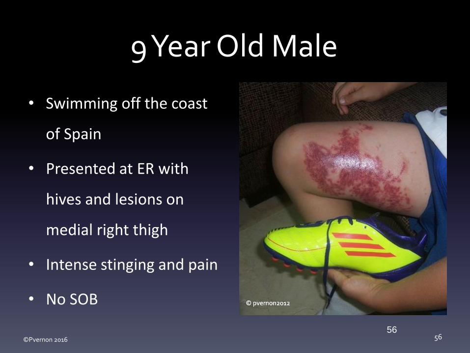
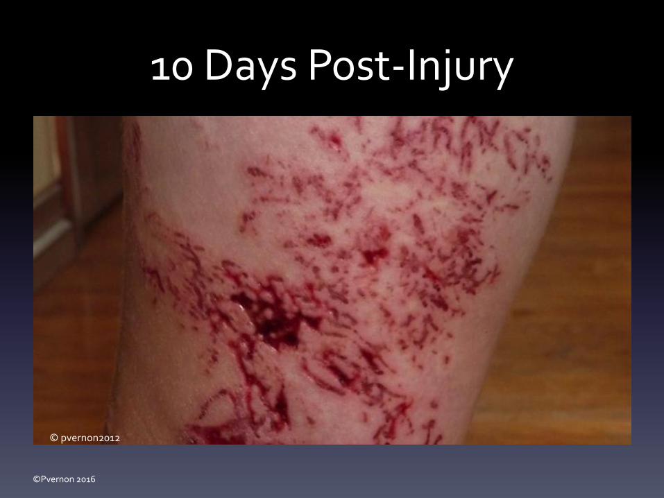
9 Year Old Male
• Swimming off the coast
of Spain
• Presented at ER with
hives and lesions on
medial right thigh
• Intense stinging and pain
• No SOB
5656
© pvernon2012© pvernon2012
©Pvernon 2016
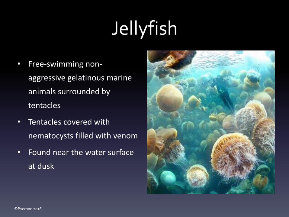
Jellyfish
• Free-swimming non-
aggressive gelatinous marine
animals surrounded by
tentacles
• Tentacles covered with
nematocysts filled with venom
• Found near the water surface
at dusk

©Pvernon 2016
Jellyfish Symptoms
• Intense stinging, pain, rash
• Progressive symptoms: nausea, vomiting,
diarrhea, adenopathy, muscle spasms
• Severe reactions cause difficulty breathing,
coma, death
©Pvernon 2016
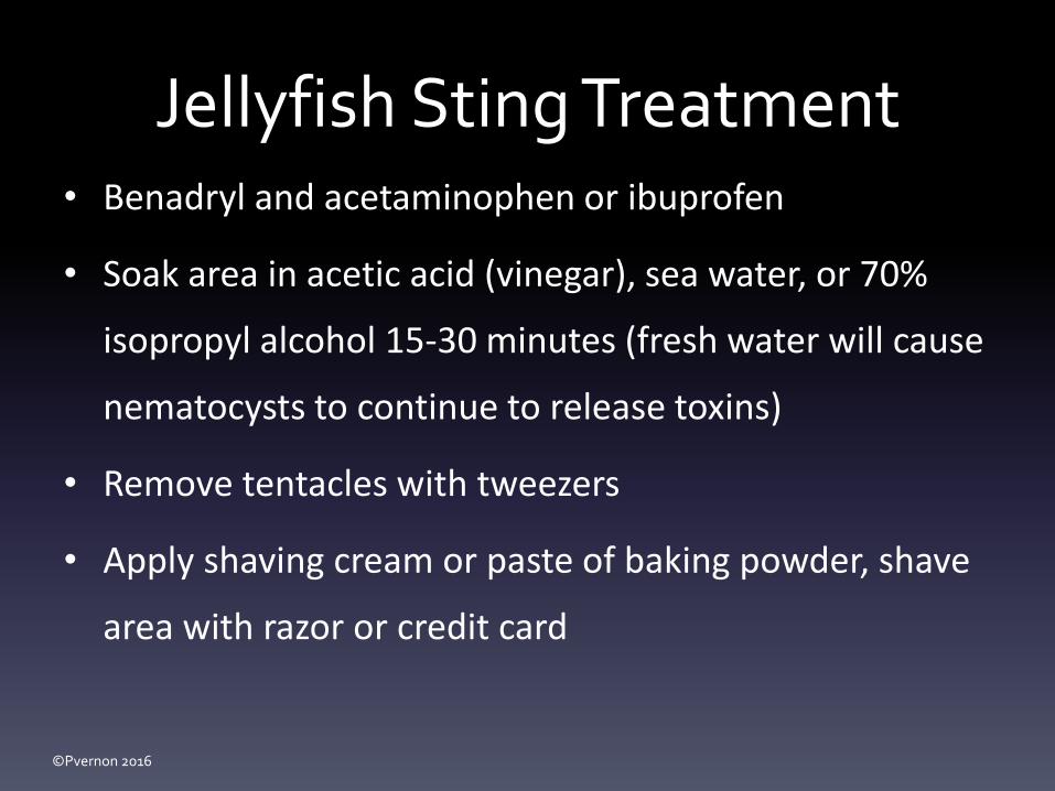
Jellyfish Sting Treatment• Benadryl and acetaminophen or ibuprofen
• Soak area in acetic acid (vinegar), sea water, or 70%
isopropyl alcohol 15-30 minutes (fresh water will cause
nematocysts to continue to release toxins)
• Remove tentacles with tweezers
• Apply shaving cream or paste of baking powder, shave
area with razor or credit card
©Pvernon 2016
10 Days Post-Injury
© pvernon2012

©Pvernon 2016
2 Weeks Post-Injury
© pvernon2012
©Pvernon 2016
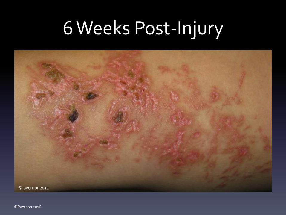
6 Weeks Post-Injury
© pvernon2012

©Pvernon 2016
© pvernon2012

©Pvernon 2016
36 Year Old Female• Developed rash on 4th
day of vacation in Costa
Rica
• Developed papular,
pruritic rash after
swimming in ocean
• Now spreading on trunk© pvernon2016

©Pvernon 2016
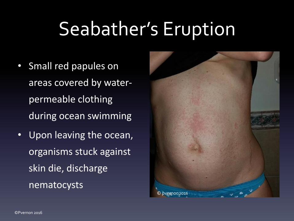
Seabather’s Eruption
• Pruritic dermatitis
• Hypersensitivity
reaction to nematocysts
of larval-stage thimble
jellyfish
• Sometimes called “sea
lice”© pvernon2016
©Pvernon 2016
Seabather’s Eruption
• Small red papules on
areas covered by water-
permeable clothing
during ocean swimming
• Upon leaving the ocean,
organisms stuck against
skin die, discharge
nematocysts © pvernon2016
©Pvernon 2016
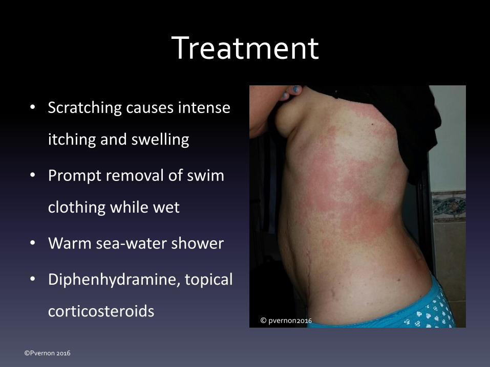
Treatment
• Scratching causes intense
itching and swelling
• Prompt removal of swim
clothing while wet
• Warm sea-water shower
• Diphenhydramine, topical
corticosteroids© pvernon2016
©Pvernon 2016
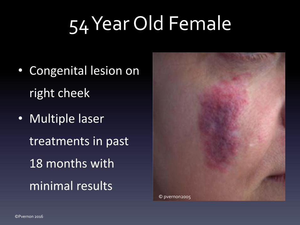
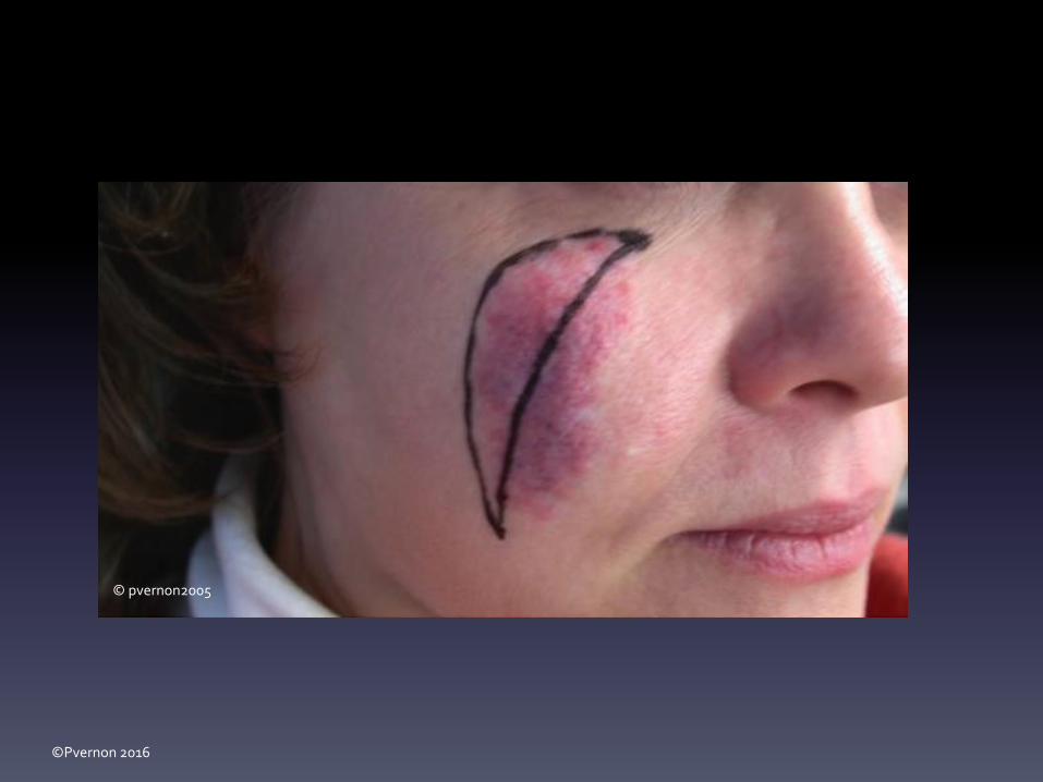
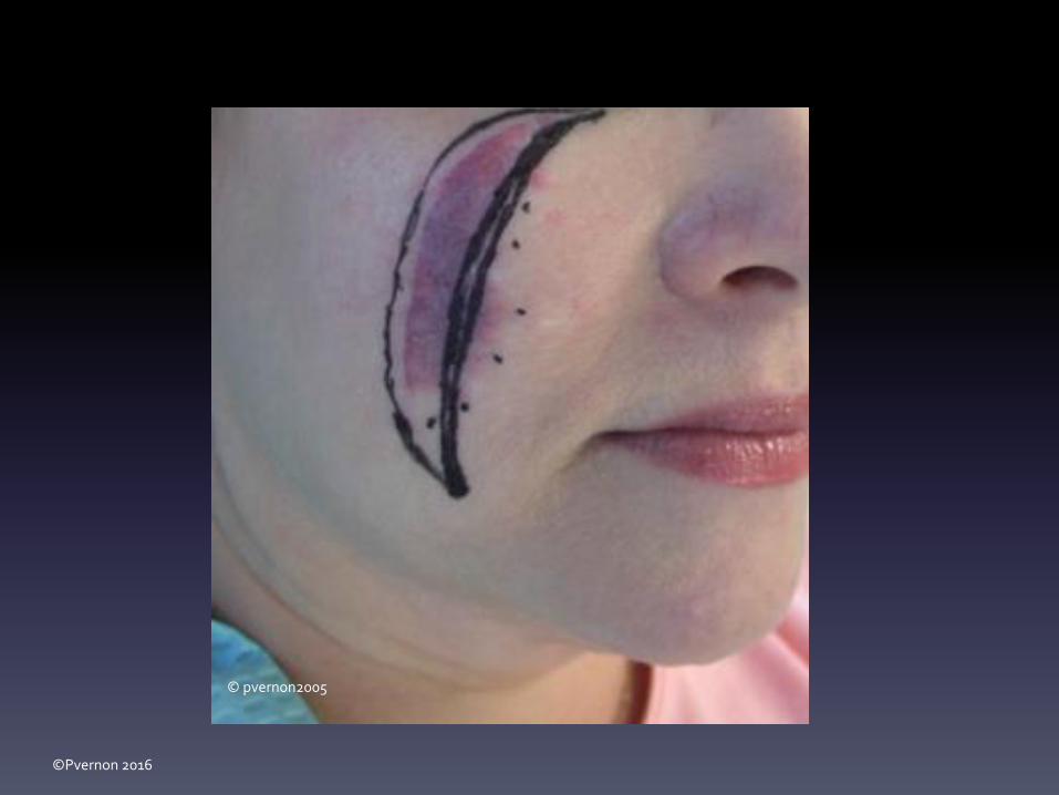
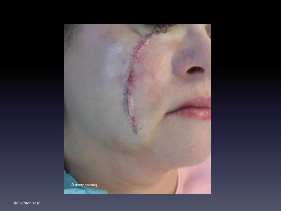
54 Year Old Female
• Congenital lesion on
right cheek
• Multiple laser
treatments in past
18 months with
minimal results© pvernon2005
©Pvernon 2016
005© pvernon2005
©Pvernon 2016
© pvernon2005

©Pvernon 2016
© pvernon2005
©Pvernon 2016
©Pvernon 2005© pvernon2005
©Pvernon 2016
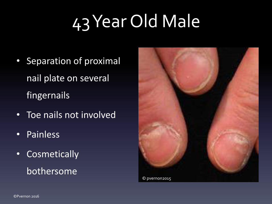
43 Year Old Male
• Separation of proximal
nail plate on several
fingernails
• Toe nails not involved
• Painless
• Cosmetically
bothersome© pvernon2015

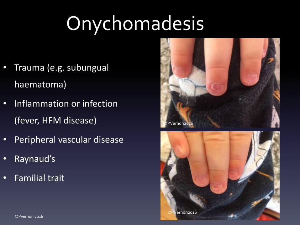
Onychomadesis
• Painless
spontaneous
separation of
proximal nail plate
©Pvernon 2015
© pvernon2015
©Pvernon 2016
Onychomadesis
• Trauma (e.g. subungual
haematoma)
• Inflammation or infection
(fever, HFM disease)
• Peripheral vascular disease
• Raynaud’s
• Familial trait
©PVernon2016
©PVernon2016
©Pvernon 2016
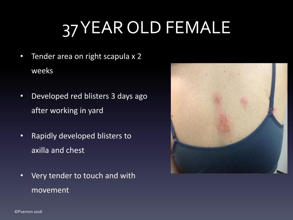
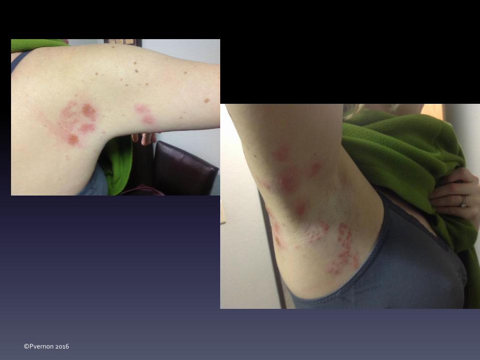
37 YEAR OLD FEMALE
• Tender area on right scapula x 2
weeks
• Developed red blisters 3 days ago
after working in yard
• Rapidly developed blisters to
axilla and chest
• Very tender to touch and with
movement
©Pvernon 2016
©Pvernon 2016
HERPES ZOSTER– Pre-eruptive phase
• Sensory phenomena along dermatome: itching, tingling, burning, pain
• 1-10 days
– Acute eruptive phase
• Grouped vesicles on erythematous base along a dermatome
• Pain, often severe; itching
• 10-15 days
– Chronic phase
• Persistent or recurring pain lasting 30 days or more, weeks to years
©Pvernon 2016
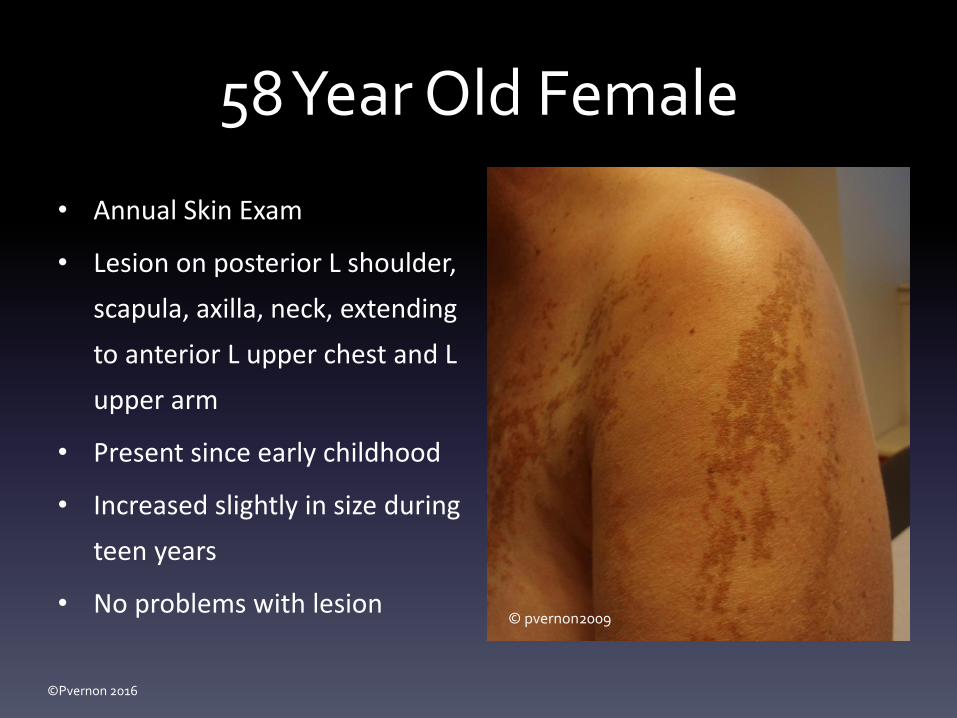
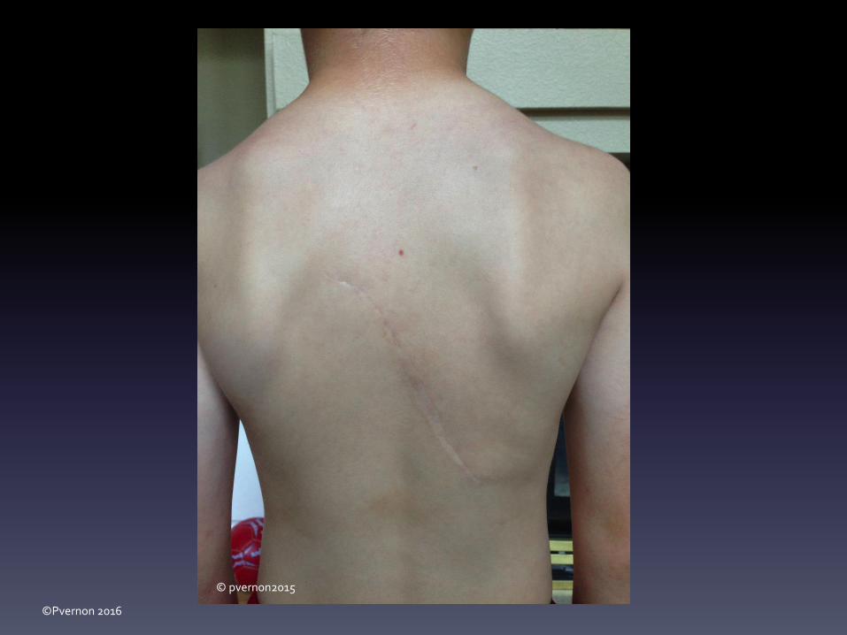
58 Year Old Female
• Annual Skin Exam
• Lesion on posterior L shoulder,
scapula, axilla, neck, extending
to anterior L upper chest and L
upper arm
• Present since early childhood
• Increased slightly in size during
teen years
• No problems with lesion© pvernon2009
©Pvernon 2016
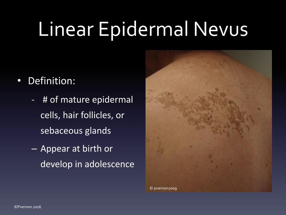
Linear Epidermal Nevus
• Definition:
- # of mature epidermal
cells, hair follicles, or
sebaceous glands
– Appear at birth or
develop in adolescence
© pvernon2009

©Pvernon 2016
Linear Epidermal Nevus
Epidermal Nevi
• Appear anywhere on the body
• Often linear or oval• Warty surface• Majority lesions present
at birth• DDx: Warts, ichthyosis,
dermatitis, lichen striatus• Treatment: Excision,
keratolytics, patient education
© pvernon2002
© pvernon2009

©Pvernon 2016
6 month old male
• Large congenital lesion
• Increasing in size with
growth
• Lesion crosses mid-line
© pvernon2008
©Pvernon 2016
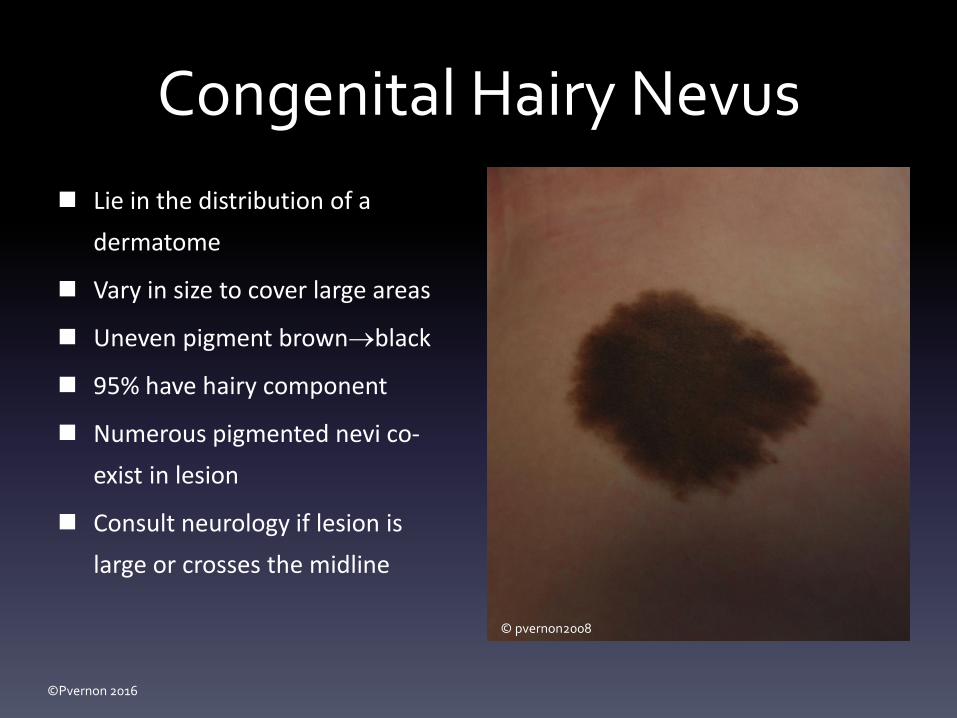
Congenital Hairy Nevus
Lie in the distribution of a
dermatome
Vary in size to cover large areas
Uneven pigment brownblack
95% have hairy component
Numerous pigmented nevi co-
exist in lesion
Consult neurology if lesion is
large or crosses the midline
© pvernon2008
©Pvernon 2016
© pvernon2015
©Pvernon 2016
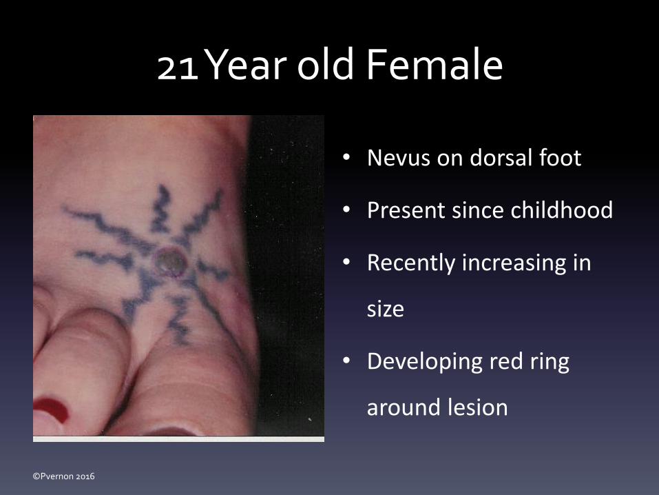
21 Year old Female
• Nevus on dorsal foot
• Present since childhood
• Recently increasing in
size
• Developing red ring
around lesion

©Pvernon 2016
• Tattoo in and around
lesion 5 years ago
• Lesion has always been
cosmetically
bothersome; patient
thought tattoo would
help

©Pvernon 2016
Dysplastic Nevi 2-5% Of Caucasian population
Type A: no family member with dysplastic nevi or melanoma: lifetime risk
of developing melanoma ~6%
Type B: Dysplastic Nevus Syndrome:
100/more moles
1/more moles 8 mm
1/more atypical moles
FAMMM (familial atypical multiple mole melanoma syndrome):
1/more first or second degree relatives with melanoma
lifetime risk of melanoma 500 x general population
©Pvernon 2016
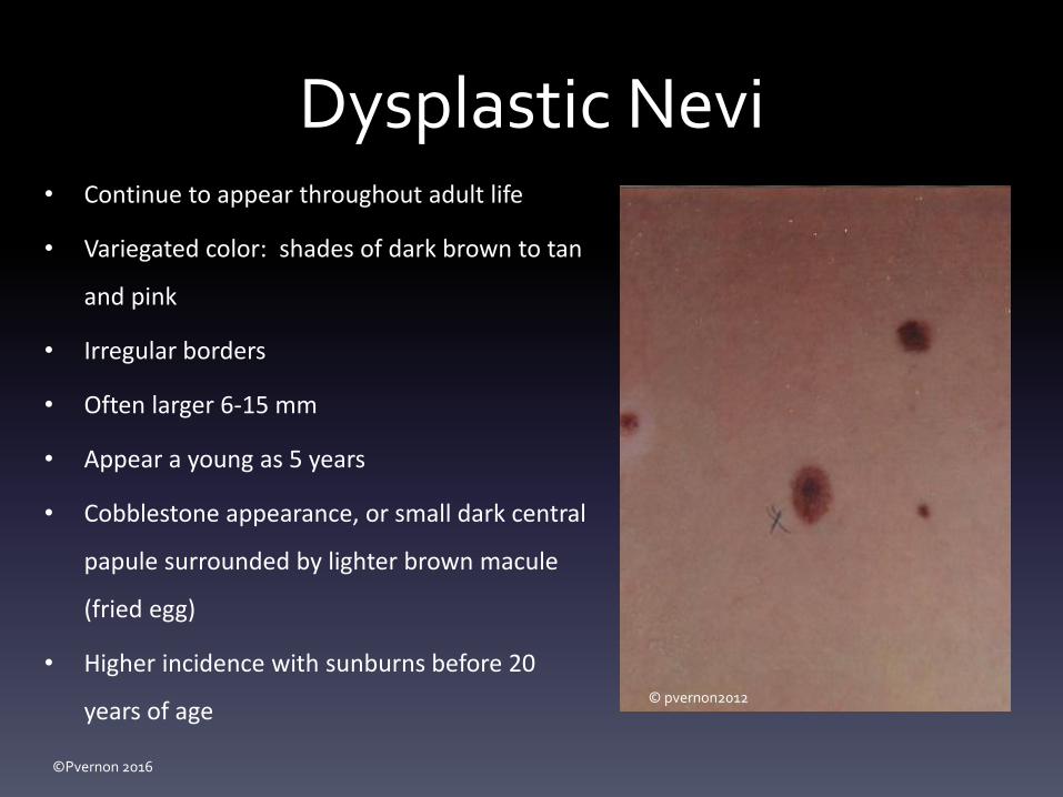
Dysplastic Nevi• Continue to appear throughout adult life
• Variegated color: shades of dark brown to tan
and pink
• Irregular borders
• Often larger 6-15 mm
• Appear a young as 5 years
• Cobblestone appearance, or small dark central
papule surrounded by lighter brown macule
(fried egg)
• Higher incidence with sunburns before 20
years of age© pvernon2012
©Pvernon 2016
Dysplastic Nevi Treatment• Grading:
– Mild: Observe, annual exam
– Moderate: Conservative excision, annual exam
– Severe: Excision 5 mm margins, annual exam
• Biopsy of changing nevi
• Annual Skin Exam
• Self Exams
• Sunscreen
• Protective clothing

©Pvernon 2016
References• Bobonich, M, Nolen, M. Dermatology for Advanced Practice Clinicians. Wolters Kluwer, 2015. First Edition.
• Foe, Donna Poma, Cutaneous Drug Eruption: A Case Study and Review; Journal of the Dermatology Nurses’ Association, Nov/Dec
2009, Vol 1 Issue 6, p 345-409
• Furaro, T, Bernaix L, Schmidt, C., and Clement, J. Nurse Practitioner Knowledge and Practice Regarding Malignant Melanoma
Assessment and Counseling. Journal of the American Academy of Nurse Practitioner. 2008, 20 (7), p. 367-75.
• Geller, A.C., Switter, S.M., Brooks, K., et al. Screening, Early Detection, and Trends for Melanoma: Current Status and Future
Directions. Journal of the American Academy of Dermatology. May 2003, p. 780-78ms & Wilkins 2009
• Goodhearts, Herbert P. Goodheart’s Photoguide to Common Skin Disorders, Third Edition, Lippincott Williams & Wilkins 2009
• Habif, Thomas. Clinical Dermatology. Fourth Edition, Mosby, 2004
• Sulzberger and Zaidems: “Psychogenic factors in Dermatological Disorders”. Medical Clinics of North America, 1948, Vol. 32, P. 669.
• Wollf, Klaus et al. Fitzpatrick’s Color Atlas & Synopsis of Clinical Dermatology, Sixth Edition, McGraw-Hill, 200
• Skin Cancer Foundation
• National Conference of State Legislatures


![Action-Items XCI [Potpourri]](https://static.fdocuments.us/doc/165x107/577ccf061a28ab9e788eb048/action-items-xci-potpourri.jpg)
