Assessment of Myocardial Viability in Patients with Heart Failure*
Upload
michael-beckerCategory
view
213download
0
J A C C : C A R D I O V A S C U L A R I M A G I N G V O L . 4 , N O . 4 , 2 0 1 1
© 2 0 1 1 B Y T H E A M E R I C A N C O L L E G E O F C A R D I O L O G Y F O U N D A T I O N I S S N 1 9 3 6 - 8 7 8 X / $ 3 6 . 0 0
P U B L I S H E D B Y E L S E V I E R I N C . D O I : 1 0 . 1 0 1 6 / j . j c m g . 2 0 1 1 . 0 1 . 0 1 0
Dependency of Cardiac ResynchronizationTherapy on Myocardial Viability at the LVLead Position
Michael Becker, MD,* Christian Zwicker, MD,* Markus Kaminski, MD,*Andreas Napp, MD,* Ertunc Altiok, MD,* Christina Ocklenburg, MSC,†Zvi Friedman, PHD,‡ Dan Adam, PHD,‡ Patrick Schauerte, MD,* Nikolaus Marx, MD,*Rainer Hoffmann, MD*
Aachen, Germany; and Haifa, Israel
O B J E C T I V E S This study sought to analyze the effectiveness of cardiac resynchronization therapy
(CRT) related to the viability in the segment of left ventricular (LV) lead position defined by myocardial
deformation imaging.
B A C K G R O U N D Echocardiographic myocardial deformation analysis allows determination of LV
lead position as well as extent of myocardial viability.
M E T H O D S Myocardial deformation imaging based on tracking of acoustic markers within
2-dimensional echo images (GE Ultrasound, GE Healthcare, Horton, Norway) was performed in 65
heart failure patients (54 � 6 years of age, 41 men) before and 12 months after CRT implantation.
In a 16-segment model, the LV lead position was defined based on the segmental strain curve with
earliest peak strain, whereas the CRT system was programmed to pure LV pacing. Nonviability of a
segment (transmural scar formation) was assumed if the peak systolic circumferential strain was
�–11.1%.
R E S U L T S In 47 patients, the LV lead was placed in a viable segment, and in 18 patients, it was
placed in a nonviable segment. At 12-month follow-up there was greater decrease of LV end-diastolic
volumes (58 � 13 ml vs. 44 � 12 ml, p � 0.0388) and greater increase of LV ejection fraction (11 � 4%
vs. 5 � 4%, p � 0.0343) and peak oxygen consumption (2.5 � 0.9 ml/kg/min vs. 1.7 � 1.1 ml/kg/min,
p � 0.0465) in the viable compared with the nonviable group. The change in LV ejection fraction and
the reduction in LV end-diastolic volumes at follow-up correlated to an increasing peak systolic
circumferential strain in the segment of the LV pacing lead (r � 0.61, p � 0.0274 and r � 0.64, p
� 0.0412, respectively). Considering only patients with ischemic heart disease, differences between
viable and nonviable LV lead position group were even greater.
C O N C L U S I O N S Preserved viability in the segment of the CRT LV lead position results in greater
LV reverse remodeling and functional benefit at 12-month follow-up. Deformation imaging allows
analysis of viability in the LV lead segment. (J Am Coll Cardiol Img 2011;4:366–74) © 2011 by the
American College of Cardiology Foundation
From the *Department of Cardiology, RWTH Aachen University, Aachen, Germany; †Department of Medical Statistics,RWTH Aachen University, Aachen, Germany; and the ‡Department of Biomedical Engineering, Technion, Haifa, Israel. Thisstudy was supported by a research grant from the German-Israeli Foundation for Scientific Research and Development (GIF,I-873-77.10/2005). The authors have reported that they have no other relationships to disclose.
Manuscript received October 11, 2010; revised manuscript received January 3, 2011, accepted January 7, 2011.
Cosicec
pet
nPforEaawws
ltfipbmiaolAcwaSM
tea6(vpL
A1odeL
f3uwaw
mption
J A C C : C A R D I O V A S C U L A R I M A G I N G , V O L . 4 , N O . 4 , 2 0 1 1
A P R I L 2 0 1 1 : 3 6 6 – 7 4
Becker et al.
Dependency of CRT on Viability
367
ardiac resynchronization therapy (CRT) isused for the treatment of advanced drug-refractory heart failure of ischemic andnonischemic origin (1–5). However, up to
ne-third of patients do not respond to CRT usingtandard clinical selection criteria (5,6). Factorsnfluencing the patient’s response to CRT are notompletely understood. Echocardiographic param-ters suggested to evaluate mechanical dyssyn-hrony and predict CRT success have not been
See page 375
confirmed in a large multicenter study (7–9). Tech-nical and procedural factors such as optimal leftventricular (LV) lead placement seem to have animportant impact. Butter et al. (10) demonstratedin an experimental analysis that the LV lead shouldbe placed in the area of greatest delay in mechanicalcontraction and electrical activation to achieve theoptimal resynchronization effect. Clinical studiesconfirmed that concurrence of the LV lead positionand the LV segment with latest contraction beforeCRT results in significantly better effectiveness ofCRT on LV function and clinical outcome (11,12).Ischemic etiology of heart failure has been identi-fied as a predictor of impaired responsiveness (13).The extent of scar tissue has been shown to definethe response to CRT (14–17). The response toCRT may thus be directly related to the extent ofmyocardial viability in the area of the LV lead andnonresponse in ischemic heart failure may be theconsequence of the LV lead being positioned in ascarred segment without functional capacity.
Myocardial deformation imaging can be used todefine CRT LV lead position and determine myo-cardial viability. Temporal analysis of segmentalmyocardial deformation curves has been shown toallow definition of LV lead position (11,18). Themagnitude of peak segmental myocardial strainclosely relates to segmental viability (19,20).
This study sought to determine CRT effective-ness related to the viability of the segment with theLV lead position as well as the area surrounding theLV lead segment. Viability was defined by analysisof myocardial deformation.
M E T H O D S
Patients. We included in this study 65 consecutiveatients (mean age 55 � 4 years, 39 men) withnd-stage heart failure severe LV systolic dysfunc-
ion (ejection fraction [EF] �35%), scheduled for mew implantation of a biventricular pacemaker.atients had to be in New York Heart Association
unctional class III (n � 48) or IV (n � 17) despiteptimal pharmacologic therapy and show sinushythm with a QRS interval duration �120 ms.tiology of heart failure was ischemic in 46 patients
nd nonischemic in 19 patients based on coronaryngiography. No studies to assess myocardial viabilityere performed before CRT implantation. This studyas approved by the local ethical committee and all
ubjects gave written informed consent.Biventricular device implantation. The LV pacingead was inserted by a transvenous approachhrough the coronary sinus into a cardiac vein of theree wall. An average of 2.1 veins were triedntraoperatively to achieve an optimal LV leadosition. Optimal LV lead position was considered toe when the width of the QRS complex was mini-ized and the arterial systolic pressure increased. No
nformation about presence of myocardial viability orrea of latest activation was provided intra-peratively. The right atrial and ventricu-ar leads were positioned conventionally.
ll patients received a biventricularardioverter-defibrillator (Attain-Systemith InSync Marquis, Medtronic, Minne-
polis, Minnesota [n � 40] or Aesula-ystem with Epic HF V-339, St. Judeedical, St. Paul, Minnesota [n � 25]).Post-operatively the optimal atrioven-
ricular time was determined by Dopplerchocardiography and set between 100nd 150 ms (mean time 122 � 10 ms) in1 patients and between 70 and 85 msmean time 75 � 8 ms) in 4 patients. Theentriculo-ventricular time was set to 0 in allatients. Thresholds for sensing and pacing of theV lead at the final position were documented.To exclude LV lead dislocation and change of
V time, the device was controlled at 6- and2-month follow-up. Seven days after implantationf the CRT system, transient programming of theevice to pure LV pacing was performed during anchocardiographic examination to determine theV lead position.
Echocardiography. All studies were performed be-ore CRT, one day after implantation, and at 12 (�)-month follow-up using a Vivid Seven digitalltrasound scanner (General Electric, Horton, Nor-ay). Using apical 4- and 2-chamber views, LVEF
nd left ventricular end-diastolic volume (LVEDV)ere determined employing biplane Simpson
A B B
A N D
CRT �
therap
EF �
LV �
LVED
volum
ROC �
chara
VO2m
consu
ethod. The physician performing the echoc
R E V I A T I O N S
A C R O N YM S
cardiac resynchronization
y
ejection fraction
left ventricle/ventricular
V � LV end-diastolic
e
receiver-operator
cteristic
ax � peak oxygen
ardio-
LwsmTfoUfaBass(miVppwaQTco
dafpssp
oswiemw�((seosa
LolLap
ciC
tcrrdctcttCc
J A C C : C A R D I O V A S C U L A R I M A G I N G , V O L . 4 , N O . 4 , 2 0 1 1
A P R I L 2 0 1 1 : 3 6 6 – 7 4
Becker et al.
Dependency of CRT on Viability
368
graphic analyses was blinded to the physician per-forming the LV lead placement.Analysis of myocardial deformation. For analysis of
V myocardial deformation, the 16-segment modelas applied on 3 parasternal short-axis views: 6
egments each on the basal and on the papillaryuscle levels and 4 segments on the apical level.he frame rate was between 56 and 92 frames/s, the
ocus was adjusted to the center of the LV cavity forptimized characterization of myocardial tissue.sing 2 consecutive cardiac cycles, myocardial de-
ormation analysis was performed offline with theid of a customized software package (EchoPACT 05.2, General Electric). This software followscoustic markers within the myocardium duringeveral consecutive frames (21) and calculates meantrain values for whole pre-defined LV segments22). It is assumed that these natural acousticarkers change their position from frame to frame
n accordance with the surrounding tissue motion.isual control of width between endocardial andericardial trace as well as tracking quality waserformed to ensure accurate analysis. End-systoleas determined in the apical long-axis view as
ortic valve closure. The time difference from theRS complex was transferred to the other views.he focus was adjusted to the center of the LV
avity to optimize myocardial speckle characteristicsf all segments.Circumferential strain relates to circumferential
eformation along the LV curvature. It is calculateds mean over the whole segment. Myocardial de-ormation analysis was used to define the LV leadosition. The LV lead position was defined as theegment with the earliest peak on the segmentaltrain curve analysis, whereas the CRT system wasrogrammed to pure LV pacing (18).Viability of LV segments was determined based
n the segmental peak systolic circumferentialtrain before CRT implantation. This informationas not provided to the physician performing the
mplantation procedure. A peak systolic circumfer-ntial strain �–11.1% was considered to indicateyocardial viability (no scar or nontransmural scar),hereas a peak systolic circumferential strain–11.1% was considered to indicate nonviability
transmural scar formation) as shown before (19)Fig. 1). In addition, viability of the adjacentegments of the LV lead position segment wasvaluated. This assessment was based on applicationf a 16-segment LV model. There were 4 adjacentegments in case of midventricular LV lead position
nd 3 adjacent segments in case of basal or apical sV lead position. Viability of the surrounding areaf the LV lead segment was defined as viability in ateast 3 adjacent segments in case of midventricularV lead position, and as viability in at least 2djacent segments in case of basal or apical LV leadosition.
Peak oxygen consumption. Patients underwent bicy-le cardiopulmonary exercise testing (10 W per minncrements) at baseline and after 12 (�3) months ofRT. The peak oxygen consumption (VO2max) at
peak exercise was defined as the highest oxygenconsumption measured during the symptom-limited exercise test and expressed as ml/kg/min.Statistics. Continuous data are expressed as mean� SD and have been compared using Student test or analysis of variance as appropriate. Pearsonorrelation coefficient was determined and linearegression analysis was performed to define theelationship between parameters with continuousata. Categorical data are presented as frequen-ies and were compared with Pearson chi-squareest. The receiver-operator characteristics (ROC)urve for peak circumferential systolic strain inhe segment of the LV pacing lead was examinedo define the optimal cutoff for prediction ofRT response. The area under ROC curve was
alculated. A p value of �0.05 was considered
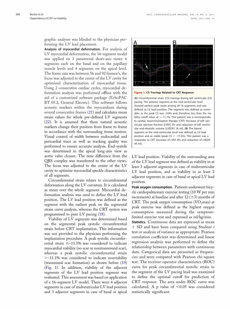
Figure 1. CS Tracings Related to CRT Response
(A) Circumferential strain (CS) tracings during left ventricular (LV)pacing. The anterior segment at the mid-ventricular levelshowed earliest peak strain among all 16 segments and wasdefined as LV lead position. The segment was defined as nonvi-able, as the peak CS was –9.8% and, therefore, less than the via-bility cutoff value of �–11.1%. This patient was a nonresponderto cardiac resynchronization therapy (CRT) (increase of left ven-tricular ejection fraction [LVEF] 2% and reduction of left ventric-ular end-diastolic volume [LVEDV] 18 ml). (B) The lateralsegment at the mid-ventricular level was defined as LV leadposition and as viable (peak CS � –17.2%). This patient was aresponder to CRT (increase of LVEF 8% and reduction of LVEDV44 ml).
tatistically significant.
sihc2mv00a
5omps2
Lis0Pwd1w
sso
ttsir
witaacLwtoLvbL
iwaapotC
J A C C : C A R D I O V A S C U L A R I M A G I N G , V O L . 4 , N O . 4 , 2 0 1 1
A P R I L 2 0 1 1 : 3 6 6 – 7 4
Becker et al.
Dependency of CRT on Viability
369
R E S U L T S
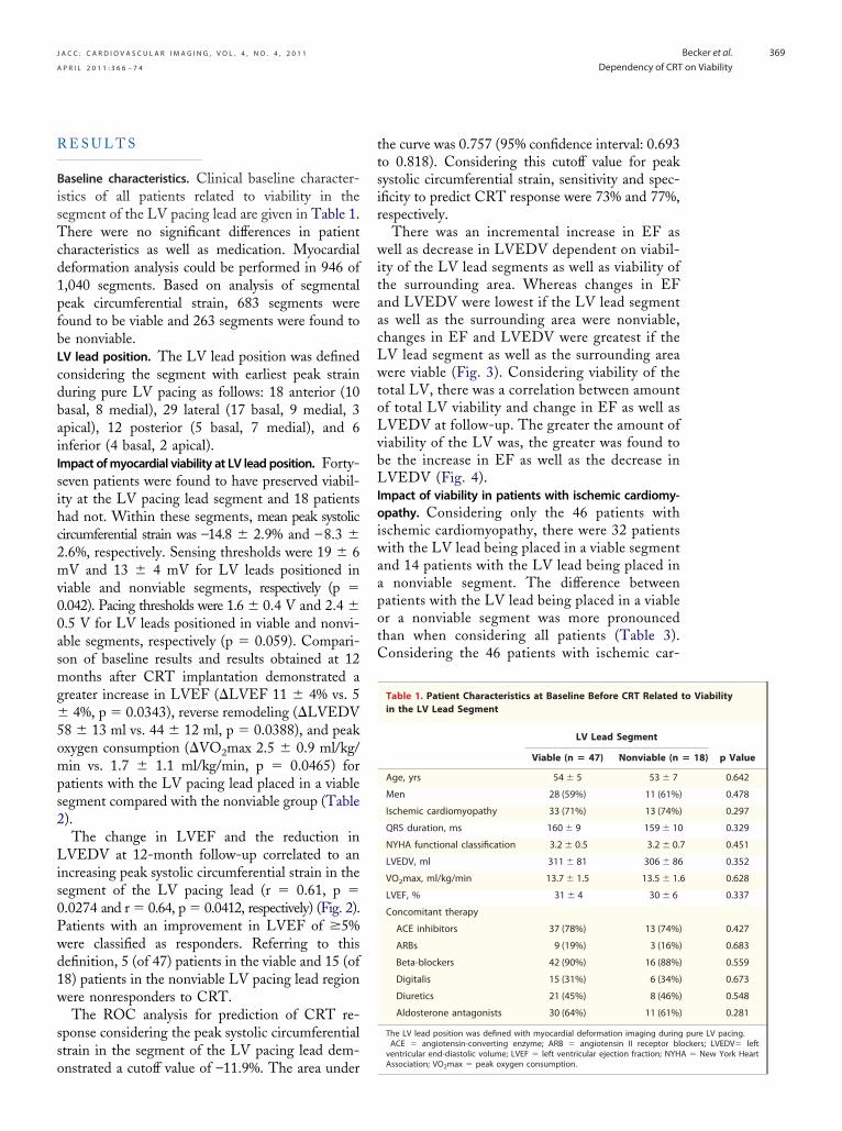
Baseline characteristics. Clinical baseline character-istics of all patients related to viability in thesegment of the LV pacing lead are given in Table 1.There were no significant differences in patientcharacteristics as well as medication. Myocardialdeformation analysis could be performed in 946 of1,040 segments. Based on analysis of segmentalpeak circumferential strain, 683 segments werefound to be viable and 263 segments were found tobe nonviable.LV lead position. The LV lead position was definedconsidering the segment with earliest peak strainduring pure LV pacing as follows: 18 anterior (10basal, 8 medial), 29 lateral (17 basal, 9 medial, 3apical), 12 posterior (5 basal, 7 medial), and 6inferior (4 basal, 2 apical).Impact ofmyocardial viability at LV lead position. Forty-even patients were found to have preserved viabil-ty at the LV pacing lead segment and 18 patientsad not. Within these segments, mean peak systolicircumferential strain was –14.8 � 2.9% and –8.3 �.6%, respectively. Sensing thresholds were 19 � 6V and 13 � 4 mV for LV leads positioned in
iable and nonviable segments, respectively (p �.042). Pacing thresholds were 1.6 � 0.4 V and 2.4 �.5 V for LV leads positioned in viable and nonvi-ble segments, respectively (p � 0.059). Compari-
son of baseline results and results obtained at 12months after CRT implantation demonstrated agreater increase in LVEF (�LVEF 11 � 4% vs. 5� 4%, p � 0.0343), reverse remodeling (�LVEDV8 � 13 ml vs. 44 � 12 ml, p � 0.0388), and peakxygen consumption (�VO2max 2.5 � 0.9 ml/kg/in vs. 1.7 � 1.1 ml/kg/min, p � 0.0465) for
atients with the LV pacing lead placed in a viableegment compared with the nonviable group (Table).The change in LVEF and the reduction in
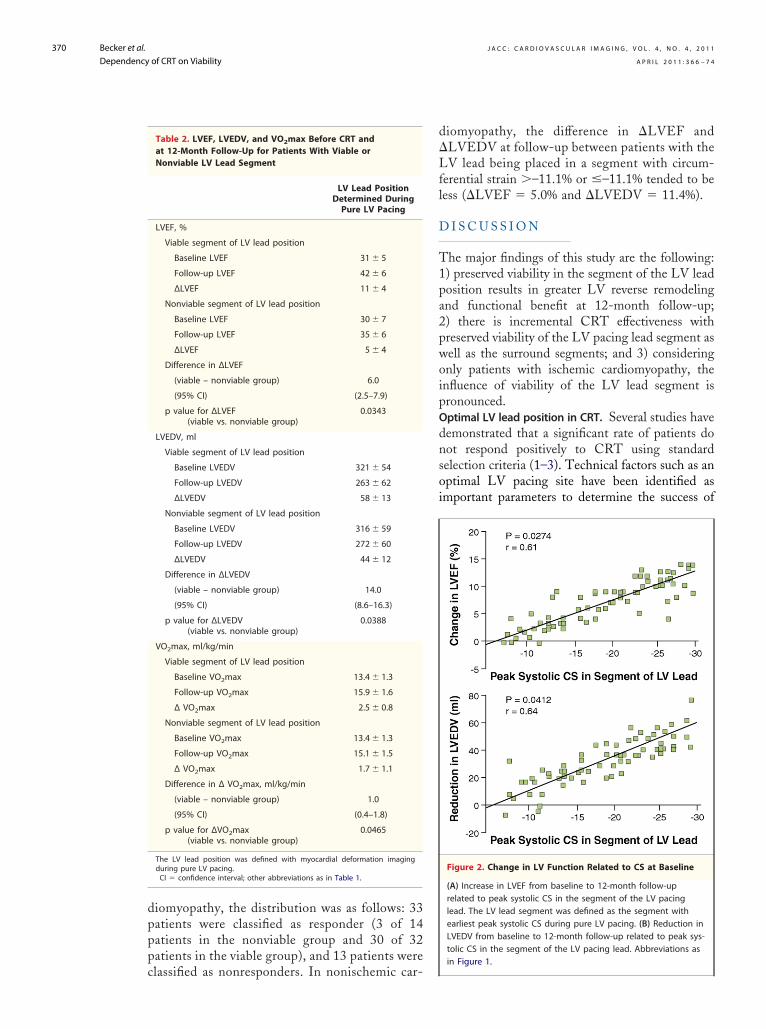
VEDV at 12-month follow-up correlated to anncreasing peak systolic circumferential strain in theegment of the LV pacing lead (r � 0.61, p �.0274 and r � 0.64, p � 0.0412, respectively) (Fig. 2).atients with an improvement in LVEF of �5%ere classified as responders. Referring to thisefinition, 5 (of 47) patients in the viable and 15 (of8) patients in the nonviable LV pacing lead regionere nonresponders to CRT.The ROC analysis for prediction of CRT re-
ponse considering the peak systolic circumferentialtrain in the segment of the LV pacing lead dem-
nstrated a cutoff value of –11.9%. The area underhe curve was 0.757 (95% confidence interval: 0.693o 0.818). Considering this cutoff value for peakystolic circumferential strain, sensitivity and spec-ficity to predict CRT response were 73% and 77%,espectively.
There was an incremental increase in EF asell as decrease in LVEDV dependent on viabil-
ty of the LV lead segments as well as viability ofhe surrounding area. Whereas changes in EFnd LVEDV were lowest if the LV lead segments well as the surrounding area were nonviable,hanges in EF and LVEDV were greatest if theV lead segment as well as the surrounding areaere viable (Fig. 3). Considering viability of the
otal LV, there was a correlation between amountf total LV viability and change in EF as well asVEDV at follow-up. The greater the amount ofiability of the LV was, the greater was found toe the increase in EF as well as the decrease inVEDV (Fig. 4).
Impact of viability in patients with ischemic cardiomy-opathy. Considering only the 46 patients withschemic cardiomyopathy, there were 32 patientsith the LV lead being placed in a viable segment
nd 14 patients with the LV lead being placed innonviable segment. The difference between
atients with the LV lead being placed in a viabler a nonviable segment was more pronouncedhan when considering all patients (Table 3).onsidering the 46 patients with ischemic car-
Table 1. Patient Characteristics at Baseline Before CRT Relatedin the LV Lead Segment
LV Lead Segment
Viable (n � 47) Nonviable (n
Age, yrs 54 � 5 53 � 7
Men 28 (59%) 11 (61%)
Ischemic cardiomyopathy 33 (71%) 13 (74%)
QRS duration, ms 160 � 9 159 � 10
NYHA functional classification 3.2 � 0.5 3.2 � 0.
LVEDV, ml 311 � 81 306 � 86
VO2max, ml/kg/min 13.7 � 1.5 13.5 � 1.
LVEF, % 31 � 4 30 � 6
Concomitant therapy
ACE inhibitors 37 (78%) 13 (74%)
ARBs 9 (19%) 3 (16%)
Beta-blockers 42 (90%) 16 (88%)
Digitalis 15 (31%) 6 (34%)
Diuretics 21 (45%) 8 (46%)
Aldosterone antagonists 30 (64%) 11 (61%)
The LV lead position was defined with myocardial deformation imaging duringACE � angiotensin-converting enzyme; ARB � angiotensin II receptor bl
ventricular end-diastolic volume; LVEF � left ventricular ejection fraction; NYH
to Viability
� 18) p Value
0.642
0.478
0.297
0.329
7 0.451
0.352
6 0.628
0.337
0.427
0.683
0.559
0.673
0.548
0.281
pure LV pacing.ockers; LVEDV� leftA � New York Heart
Association; VO2max � peak oxygen consumption.
�Lfl
dnsoi
J A C C : C A R D I O V A S C U L A R I M A G I N G , V O L . 4 , N O . 4 , 2 0 1 1
A P R I L 2 0 1 1 : 3 6 6 – 7 4
Becker et al.
Dependency of CRT on Viability
370
diomyopathy, the distribution was as follows: 33patients were classified as responder (3 of 14patients in the nonviable group and 30 of 32patients in the viable group), and 13 patients were
Table 2. LVEF, LVEDV, and VO2max Before CRT andat 12-Month Follow-Up for Patients With Viable orNonviable LV Lead Segment
LV Lead PositionDetermined DuringPure LV Pacing
LVEF, %
Viable segment of LV lead position
Baseline LVEF 31 � 5
Follow-up LVEF 42 � 6
∆LVEF 11 � 4
Nonviable segment of LV lead position
Baseline LVEF 30 � 7
Follow-up LVEF 35 � 6
∆LVEF 5 � 4
Difference in ∆LVEF
(viable – nonviable group) 6.0
(95% CI) (2.5–7.9)
p value for ∆LVEF(viable vs. nonviable group)
0.0343
LVEDV, ml
Viable segment of LV lead position
Baseline LVEDV 321 � 54
Follow-up LVEDV 263 � 62
∆LVEDV 58 � 13
Nonviable segment of LV lead position
Baseline LVEDV 316 � 59
Follow-up LVEDV 272 � 60
∆LVEDV 44 � 12
Difference in ∆LVEDV
(viable – nonviable group) 14.0
(95% CI) (8.6–16.3)
p value for ∆LVEDV(viable vs. nonviable group)
0.0388
VO2max, ml/kg/min
Viable segment of LV lead position
Baseline VO2max 13.4 � 1.3
Follow-up VO2max 15.9 � 1.6
∆ VO2max 2.5 � 0.8
Nonviable segment of LV lead position
Baseline VO2max 13.4 � 1.3
Follow-up VO2max 15.1 � 1.5
∆ VO2max 1.7 � 1.1
Difference in ∆ VO2max, ml/kg/min
(viable – nonviable group) 1.0
(95% CI) (0.4–1.8)
p value for ∆VO2max(viable vs. nonviable group)
0.0465
The LV lead position was defined with myocardial deformation imagingduring pure LV pacing.CI � confidence interval; other abbreviations as in Table 1.
classified as nonresponders. In nonischemic car-
diomyopathy, the difference in �LVEF andLVEDV at follow-up between patients with theV lead being placed in a segment with circum-
erential strain �–11.1% or �–11.1% tended to beess (�LVEF � 5.0% and �LVEDV � 11.4%).
D I S C U S S I O N
The major findings of this study are the following:1) preserved viability in the segment of the LV leadposition results in greater LV reverse remodelingand functional benefit at 12-month follow-up;2) there is incremental CRT effectiveness withpreserved viability of the LV pacing lead segment aswell as the surround segments; and 3) consideringonly patients with ischemic cardiomyopathy, theinfluence of viability of the LV lead segment ispronounced.Optimal LV lead position in CRT. Several studies have
emonstrated that a significant rate of patients doot respond positively to CRT using standardelection criteria (1–3). Technical factors such as anptimal LV pacing site have been identified asmportant parameters to determine the success of
Figure 2. Change in LV Function Related to CS at Baseline
(A) Increase in LVEF from baseline to 12-month follow-uprelated to peak systolic CS in the segment of the LV pacinglead. The LV lead segment was defined as the segment withearliest peak systolic CS during pure LV pacing. (B) Reduction inLVEDV from baseline to 12-month follow-up related to peak sys-tolic CS in the segment of the LV pacing lead. Abbreviations as
in Figure 1.
.05.
J A C C : C A R D I O V A S C U L A R I M A G I N G , V O L . 4 , N O . 4 , 2 0 1 1
A P R I L 2 0 1 1 : 3 6 6 – 7 4
Becker et al.
Dependency of CRT on Viability
371
CRT. Experimental data suggested placing the LVlead in the area of the latest contraction beforeCRT (10). Clinical studies confirmed the impor-
Figure 3. Change in LV Function and Volumes Related to Viabil
Increase in LVEF and reduction in LVEDV related to viability (�: via(LS) and viability of surrounding segments (SS). #p � 0.001; *p � 0
Figure 4. LV Viability and Follow-Up Change in LV Functionand Volumes
Correlation between percentage of viable segments determined bymyocardial deformation analysis (peak systolic circumferential strain�–11.1%) and effectiveness of CRT defined as increase in LVEF (A)
and reduction in LVEDV (B). Abbreviations as in Figure 1.tance of optimal LV lead placement site, demon-strating improved exercise tolerance, greater im-provement in LV function, and better outcome forpatients with concordance of LV lead position tosite of latest contraction before CRT (11,12). Dif-ferent imaging techniques have been used to definethe LV lead site. Fluoroscopy has been used in alarge study demonstrating differences in patientoutcome depending on the LV lead site defined byfluoroscopy (12). However, because of the limitedfluoroscopic views applied in clinical studies, thesegmental definition of the lead position is con-
,�: no viability) of LV segment with assumed LV lead positionAbbreviations as in Figure 1.
Table 3. Comparison of Baseline to 12-Month Follow-UpResults (LVEF, LVEDV, and VO2max) Considering OnlyPatients With Ischemic Cardiomyopathy (n � 46)
LV Lead PositionDetermined DuringPure LV Pacing
Difference in ∆LVEF, %
(viable – nonviable group) 7.0
(95% CI) (3.7–9.8)
p value for ∆LVEF (viable vs. nonviable) �0.001
Difference in ∆LVEDV, ml
(viable – nonviable group) 16.0
(95% CI) (11.3–19.7)
p value for ∆LVEDV (viable vs. nonviable) �0.001
Difference in ∆VO2max, ml/kg/min
(viable – nonviable group) 1.4
(95% CI) (0.6–2.6)
p value for ∆VO2max (viable vs. nonviable) �0.001
ity
bility
Abbreviations as in Tables 1 and 2.

tdslcbt
bwi(tihitsdgrfelttic
wdicattwraedt
emmaciv1b
LsvpiLcllomCfia
fMwets(f�iswesna2smEsCol
J A C C : C A R D I O V A S C U L A R I M A G I N G , V O L . 4 , N O . 4 , 2 0 1 1
A P R I L 2 0 1 1 : 3 6 6 – 7 4
Becker et al.
Dependency of CRT on Viability
372
fined. Techniques based on myocardial deforma-ion imaging have been shown to allow improvedefinition of LV lead position (11,18). In thistudy, we used a technique that defines the LVead position as the segment with earliest me-hanical activity during pure LV pacing. It haseen demonstrated to provide facilitated access tohe LV pacing site (18).Impact of viability on response to CRT. It has also
een suggested that the extent of scar tissue asell as the extent of viable myocardium are
mportant factors defining the response to CRT14 –17). Bleeker et al. (23) suggested that pacinghe LV in nonviable or scared myocardium resultsn a less optimal response to CRT. Some studiesave found global extent of LV scar to be
mportant (15,16), whereas other studies foundhe size of septal or lateral scar significant (17). Atudy by Ypenburg et al. (17) demonstrated airect association between viability determined byated single-photon emission computed tomog-aphy and response to CRT. The increase in LVunction and the decrease of LV end-systolic andnd-diastolic volumes at 6-month follow-up wereinearly related to the extent of viability. Addi-ionally, the extent of scar tissue was also impor-ant for the response to CRT as reflected by annverse relation between the scar tissue and thehange in LV function and LV volumes.
In an echocardiographic study, transmural scaras diagnosed based on measurement of an end-iastolic wall thickness �0.5 cm associated with
ncreased tissue acoustic reflectance (24). The echo-ardiographic extent of scar tissue was significantlyssociated with LV reverse remodeling. The greaterhe pre-implant myocardial scar tissue, the lowerhe reverse remodeling effect of CRT. This is in lineith results by Mangiavacchi et al. (25), who
eported that the number of scar segments evalu-ted by echocardiography was less in CRT respond-rs than in nonresponders. However, these studiesid not specifically focus on myocardial viability ofhe LV pacing segment.
In the present study, a peak systolic circumfer-ntial strain of �–11.1% was used to define seg-ental myocardial viability. This value was deter-ined in a recent study and had a sensitivity of 70%
nd a specificity of 71% to detect transmural myo-ardial infarction defined by magnetic resonancemaging (19). In this study, we evaluated LVolumes and functional capacity at baseline and at2 months after CRT implantation related to via-
ility of the LV lead segment. A greater increase inVEF, reverse remodeling, and peak oxygen con-umption could be demonstrated for patients withiability at the LV pacing lead position versusatients in the nonviable group. Furthermore, themprovement in LVEF and the reduction inVEDV correlated to an increasing peak systolicircumferential strain in the region of LV pacingead as a marker of increasing viability. The simi-arity of the cutoff value found in the ROC analysisn peak systolic circumferential strain in the seg-ent with the LV pacing lead for prediction ofRT response to a previously reported cutoff value
or circumferential strain to predict myocardial viabil-ty (19) stresses the importance of myocardial viabilityt the LV lead position for CRT effectiveness.
The results on importance of myocardial viabilityor CRT effectiveness are consistent to results by
angiavacchi et al. (25) as well as Chalil et al. (26),ho reported an association between increasing
xtent and transmurality of scar and poor responseo CRT. Patients with scar transmurality of �52%howed only one-half symptomatic responder rateimprovement was New York Heart Associationunctional class �1 or 6-min walking distance
25%) observed in patients with a scar transmural-ty of �51%. Considering the results of thesetudies, there may be a limit of scar size beyondhich resynchronization becomes ineffective. Sev-
ral factors might contribute to this finding: 1) aubstantial amount of viable myocardium iseeded for improvement in systolic LV functionfter CRT, whereas scar tissue does not contract;) pacing of scar tissue is likely to result inuboptimal resynchronization of the neighboringyocardium as electrical conduction is delayed.valuation of the extent and location of scar tissue
hould be considered in the selection process forRT to avoid nonresponse, especially for the areaf latest contraction as optimal segment for the LVead placement.Study limitations. Because there was an insufficientnumber of echocardiographic images, myocardialdeformation parameters for analysis of myocar-dial viability and optimal location for LV leadcould not be determined in 9% of segments.Thus, the segment with assumed LV lead posi-tion may have been missed in some patients.However, this should not have affected the prin-ciple findings of this study.
The location of the LV lead was determinedbased on the assumption that the electric current ofthe LV lead will affect the nearest segment stron-
gest and first. This assumption was affirmed before
J A C C : C A R D I O V A S C U L A R I M A G I N G , V O L . 4 , N O . 4 , 2 0 1 1
A P R I L 2 0 1 1 : 3 6 6 – 7 4
Becker et al.
Dependency of CRT on Viability
373
by a high match in the LV lead position determinedby fluoroscopy (11).
There are potential difficulties of speckle trackingimaging. Circumferential strain values may be im-paired owing to very tight coronary stenosis(�90%) inducing acute myocardial ischemia (27).This study focused on analysis of circumferentialstrain. This focus considered former analysis dem-onstrating superiority of circumferential strain tolongitudinal motion analysis in terms of adequatedemonstration of CRT benefit (28).
The analysis approach used in this study maysimplify the complexity of many factors having animpact on CRT effectiveness. This relates in par-ticular to technical and procedural factors that arenot all considered. However, myocardial viability,optimal location of LV lead position, and actual LVlead position are 3 important factors that influenceCRT effectiveness, and the applied analysis givesaccess to all 3 parameters using 1 modality. Thestudy was not intended to analyze differences be-tween ischemic and nonischemic cardiomyopathies.This study found a more pronounced impact of
2140–50.
1
1al. Impact of left v
further studies should address this issue withinproportionate subgroups. The viability cutoff valuedefined for ischemic cardiomyopathy was appliedalso to patients with nonischemic cardiomyopathy.However, as the definition of viability based ondeformation analysis relates to functional capacity,application of the same cutoff value for nonischemiccardiomyopathy to predict functional response toCRT should be a valid approach.
C O N C L U S I O N S
Myocardial deformation imaging provides informa-tion on LV lead position and myocardial viability.Preserved viability in the segment of the LV leadresults in greater LV reverse remodeling and func-tional benefit at 12-month follow-up. The impactof viability in the LV lead position is particularlypronounced in patients with ischemic heart disease.
Reprint requests and correspondence: Dr. Rainer Hoff-mann, Medical Clinic I, RWTH Aachen University,Pauwelsstrasse 30, 52057 Aachen, Germany. E-mail:
viability in patients with ischemic heart disease but [email protected].
R E F E R E N C E S
1. Cleland JG, Daubert JC, Erdmann E,et al., for CARE-HF Study Investi-gators. The effect of cardiac resyn-chronization on morbidity and mor-tality in heart failure. N Engl J Med2005;352:1539–49.
2. Abraham WT, Fisher WG, Smith AL,et al, for MIRACLE Study Group.Multicenter InSync Randomized Clin-ical Evaluation. Cardiac resynchroniza-tion in chronic heart failure. N EnglJ Med 2002;346:1845–53.
3. Linde C, Leclercq C, Rex S, et al.Long-term benefits of biventricularpacing in congestive heart failure: re-sults from the multisite stimulation incardiomyopathy (MUSTIC) study.J Am Coll Cardiol 2002;40:111–8.
4. Auricchio A, Stellbrink C, Sack S, etal., for PATH-CHF Study Group.Long-term clinical effect of hemo-dynamically optimized cardiac resyn-chronization therapy in patients withheart failure and ventricular conduc-tion delay. J Am Coll Cardiol 2002;39:2026–33.
5. Bristow MR, Saxon LA, Boehmer J,et al., for COMPANION Investiga-tors. Cardiac-resynchronization ther-apy with or without an implantabledefibrillator in advanced chronic heartfailure. N Engl J Med 2004;350:
6. Young JB, Abraham WT, Smith AL,et al., for MIRACLE ICD Trial In-vestigators. Combined cardiac resyn-chronization and implantable cardio-version defibrillation in advancedchronic heart failure: the MIRACLEICD trial. JAMA 2003;289:2685–94.
7. Penicka M, Bartunek J, De Bruyne B,et al. Improvement of left ventricularfunction after cardiac resynchroniza-tion therapy is predicted by tissueDoppler imaging echocardiography.Circulation 2004;109:978–83.
8. Yu CM, Chau E, Sanderson JE, et al.Tissue Doppler echocardiographic ev-idence of reverse remodeling and im-proved synchronicity by simultane-ously delaying regional contractionafter biventricular pacing therapy inheart failure. Circulation 2002;105:438–45.
9. Chung ES, Leon AR, Tavazzi L, etal. Results of the Predictors of Re-sponse to CRT (PROSPECT) trial.Circulation 2008;117:2608–16.
0. Butter C, Auricchio A, Stellbrink C,et al., for Pacing Therapy for ChronicHeart Failure II Study Group. Effectof resynchronization therapy stimula-tion site on the systolic function ofheart failure patients. Circulation2001;104:3026–9.
1. Becker M, Kramann R, Franke A, et
entricular lead po-sition in cardiac resychronizationtherapy on left ventricular remodeling.A circumferential strain analysis basedon 2D echocardiography. Eur Heart J2007;28:1211–20.
12. Ypenburg C, Van Bommel RJ, Del-gado V, et al. Optimal left ventricularlead position predicts reverse remod-eling and survival after cardiac resyn-chronization therapy. J Am Coll Car-diol 2008;52:1402–9.
13. Dı́az-Infante E, Mont L, Leal J, et al.,for SCARS Investigators. Predictorsof lack of response to resynchroniza-tion therapy. Am J Cardiol 2005;95:1436–40.
14. Hummel JP, Lindner JR, Belcik JT, etal. Extent of myocardial viability pre-dicts response to biventricular pacingin ischemic cardiomyopathy. HeartRhythm 2005;2:1211–7.
15. Adelstein EC, Saba S. Scar burden bymyocardial perfusion imaging predictsechocardiographic response to cardiacresynchronization therapy in ischemiccardiomyopathy. Am Heart J 2007;153:105–12.
16. Buch E, Lellouche N, De Diego C, etal. Left ventricular apical wall motionabnormality is associated with lack ofresponse to cardiac resynchronizationtherapy in patients with ischemic car-diomyopathy. Heart Rhythm 2007;4:
1300–5.
1
1
2
2
2
2
2
2
J A C C : C A R D I O V A S C U L A R I M A G I N G , V O L . 4 , N O . 4 , 2 0 1 1
A P R I L 2 0 1 1 : 3 6 6 – 7 4
Becker et al.
Dependency of CRT on Viability
374
17. Ypenburg C, Schalij MJ, Bleeker GB,et al. Impact of viability and scar tissueon response to cardiac resynchroniza-tion therapy in ischaemic heart failurepatients. Eur Heart J 2007;28:33–41.
8. Becker M, Altiok E, Ocklenburg C, etal. Analysis of left ventricular leadposition in cardiac resynchronizationtherapy using different imaging mo-dalities. J Am Coll Cardiol Img 2010;3:472–81.
9. Becker M, Hoffmann R, Kuehl H, etal. Analysis of myocardial deformationbased on ultrasonic pixel tracking todetermine transmurality in chronicmyocardial infarction. Eur Heart J2006;27:2560–6.
0. Hanekom L, Jenkins C, Jeffries L, etal. Incremental value of strain rateanalysis as an adjunct to wall-motionscoring for assessment of myocardialviability by dobutamine echocardiog-raphy: a follow-up study after revascu-larization. Circulation 2005;112:3892–3900.
1. Reisner S, Lysyansky P, Agmon Y,Mutlak D, Lessick J, Friedman Z.
Global longitudinal strain: a novel in-dex of left ventricular systolic func-tion. J Am Soc Echocardiogr 2004;17:630–3.
2. Leitman M, Lysyansky P, Sidenko S,et al. Two-dimensional strain-a novelsoftware for real-time quantitativeechocardiographic assessment of myo-cardial function. J Am Soc Echocar-diogr 2004;17:1021–9.
3. Bleeker GB, Kaandrop TAM, LambHJ, et al. Effect of posterolateral scartissue on clinical and echocardio-graphic improvement after cardiac re-synchronization therapy. Circulation2006;113:969–76.
4. Mele D, Agricola E, Galderisi M, etal. Echocardiographic myocardial scarburden predicts response to cardiacresynchronization therapy in ischemicheart failure. J Am Soc Echocardiogr2009;22:702–8.
5. Mangiavacchi M, Gaspaarini M,Faletra F, Klersy C, Morenghi E,Galimberti P. Clinical predictors ofmarked improvement in left ventricu-lar performance after cardiac resyn-chronization therapy in patients with
chronic heart failure. Am Heart J2006;151:477,e1–6.26. Chalil S, Stegemann B, MuhyaldeenSA, et al. Effect of posterolateral leftventricular scar on mortality and mor-bidity following cardiac resynchroni-zation therapy. Pacing Clin Electro-physiol 2007;30:1201–9.
27. Reant P, Labrousse L, Lafitte S, et al.Experimental validation of circumfer-ential, longitudinal, and radial2-dimensional strain during dobut-amine stress echocardiography in isch-emic conditions. J Am Coll Cardiol2008;51:149–57.
28. Helm RH, Leclercq C, Faris OP, etal. Cardiac dyssynchrony analysis us-ing circumferential versus longitudinalstrain: implications for assessing car-diac resynchronization. Circulation2005;111:2760–7.
Key Words: cardiacresynchronization therapy yechocardiography y heart failurey left ventricular function ymyocardial deformation
imaging.