

Dental X-Rays Are Safer Than They Used To Be
35
Dental X-Rays Are Safer Than They Used To Be
-
Upload
kay-frenzer-zeeh -
Category
Health & Medicine
-
view
174 -
download
0
Transcript of Dental X-Rays Are Safer Than They Used To Be
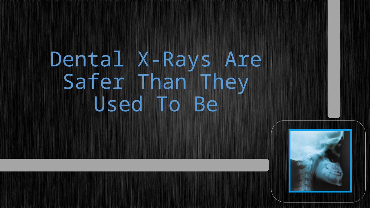
Dental X-Rays Are Safer Than They Used
To Be

One of the most important tools family dentists use is the dental x-ray.

X-rays allow dentists to determine conditions that may not be evident during visual examination.

Hidden conditions, such as root disease, impacted teeth and problems of the jaw can be found.

Problems that may affect successful rejuvenation of teeth can be detected by cosmetic dentists.

Today, both x-ray equipment and techniques have been refined to a high level of precision that makes them safer than ever.

History of X-Rays in Dentistry

The history of dental x-rays begins with the discovery of radium by Wilhelm Conrad Roentgen in 1895.
1895

The ability to see the boney structures inside the body opened up new opportunities to understand and treat numerous diseasesand injuries.

Very soon after this discovery, Dr. Otto Walkhoff in Braunschweig, Germany, took the first x-ray of the oral cavity.

These early x-rays took significantly more time to produce an image, often as long as 25 minutes.

The new technology quickly spread throughout Europe and to the United States, where it was used to help patients with hidden dental conditions.

Recommendations onDental X-rays

Because of the dangers of excessive amounts of radiation during diagnostic x-rays, strict controls have been put on the use of x-ray equipment.

Agencies such as the U.S. Food & Drug Administration, as well as the American Dental Association, provides a number of guidelines in regard to who should get dental x-rays, how many x-rays should be administered and the safest way to administer x-rays.

These recommendations are implemented to ensure that minimum amounts of radiation are used to diagnose dental problems.
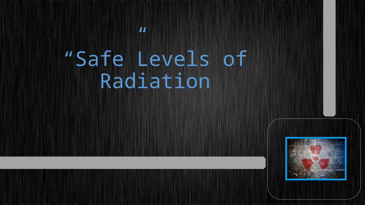
“Safe”Levels of Radiation

Radiation occurs naturally in the environment, and people are exposed to it as they go about their normal lives.

Some activities, such as taking an airplane flight, expose people to slightly higher levels of radiation.

The amount of radiation patients receive as a result of dental x-rays is thousands of times less than natural radiation.

Unfortunately, there is no known “safelevel of radiation.
Because of this uncertainty, medical and dental equipment designers continuously try to develop equipment that uses less radiation than previous equipment, yet still effectively creates the images that help to diagnose and treat medical and dental conditions.

Dental X-Rays Have Improved
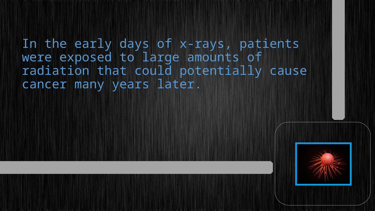
In the early days of x-rays, patients were exposed to large amounts of radiation that could potentially cause cancer many years later.

Today, new technology has allowed x-ray equipment to use minute amounts of radiation to produce high-quality images of the structures inside of the teeth and jaw, with minimal risk.

Digital radiography uses up to 90 percent less radiation than traditional film x-rays.
90%

The digital equipment also eliminates the need for chemicals to process the images, making them more environmentally friendly.

Computer tomography, or CT scans, that allow the even greater examination and diagnosis of internal structures possible, using very small amounts of radiation.

Advances in Dental X-Ray Technology

The hand-held x-ray machine and other developments are helping to bring the powerof radiologic diagnosis to areas of the worldthat previously could not support thiscomplex technology.

No doubt greater improvements will bring even more safety and convenience to x-ray equipment in the future.

If you are seeking professional dental advice, it's time to call Shumway Dental Care.

Their Chandler dentists will ask you questions, examine you, and determine exactly what needs to be done to get you back to normal.

SHUMWAY DENTAL CARE3150 S Gilbert Rd Suite 1Chandler, AZ 85286(480) 420-7551


![DENTAL REGISTRATION AND HISTORY C] NO PATIENT … · DENTAL HISTORY Reason for today's visit Former Dentist City/State Date of last dental visit Date of last dental X-rays Relationship](https://static.fdocuments.us/doc/165x107/5f0b66ac7e708231d4305626/dental-registration-and-history-c-no-patient-dental-history-reason-for-todays.jpg)


