Degenerative Disorders - كلية الطب · Degenerative Disorders. Inherited Metabolic Disorders...
43
Degenerative Disorders
Transcript of Degenerative Disorders - كلية الطب · Degenerative Disorders. Inherited Metabolic Disorders...

Degenerative Disorders
Inherited Metabolic Disorders
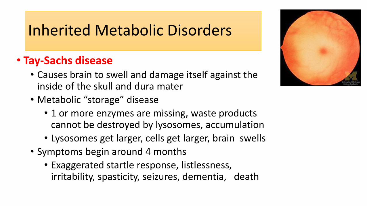
• Tay-Sachs disease • Causes brain to swell and damage itself against the
inside of the skull and dura mater
• Metabolic “storage” disease
• 1 or more enzymes are missing, waste products cannot be destroyed by lysosomes, accumulation
• Lysosomes get larger, cells get larger, brain swells
• Symptoms begin around 4 months
• Exaggerated startle response, listlessness, irritability, spasticity, seizures, dementia, death
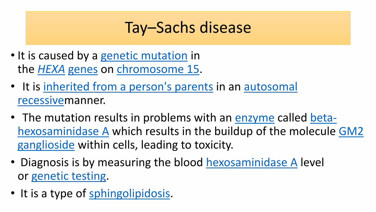
Tay–Sachs disease
• It is caused by a genetic mutation in the HEXA genes on chromosome 15.
• It is inherited from a person's parents in an autosomal recessivemanner.
• The mutation results in problems with an enzyme called beta-hexosaminidase A which results in the buildup of the molecule GM2 ganglioside within cells, leading to toxicity.
• Diagnosis is by measuring the blood hexosaminidase A level or genetic testing.
• It is a type of sphingolipidosis.
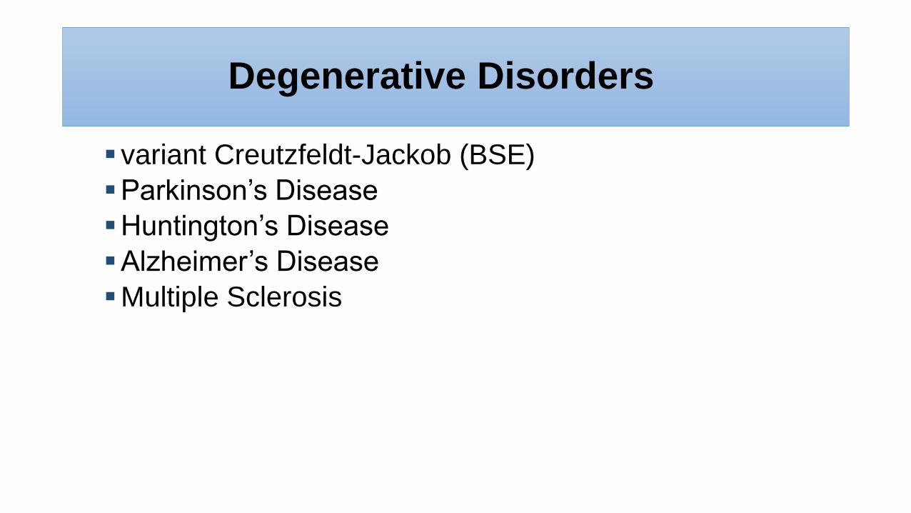
Degenerative Disorders
variant Creutzfeldt-Jackob (BSE)
Parkinson’s Disease
Huntington’s Disease
Alzheimer’s Disease
Multiple Sclerosis
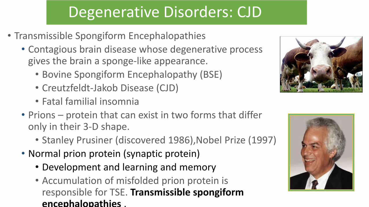
Degenerative Disorders: CJD
• Transmissible Spongiform Encephalopathies
• Contagious brain disease whose degenerative process gives the brain a sponge-like appearance.
• Bovine Spongiform Encephalopathy (BSE)
• Creutzfeldt-Jakob Disease (CJD)
• Fatal familial insomnia
• Prions – protein that can exist in two forms that differ only in their 3-D shape.
• Stanley Prusiner (discovered 1986),Nobel Prize (1997)
• Normal prion protein (synaptic protein)
• Development and learning and memory
• Accumulation of misfolded prion protein is responsible for TSE. Transmissible spongiform encephalopathies .
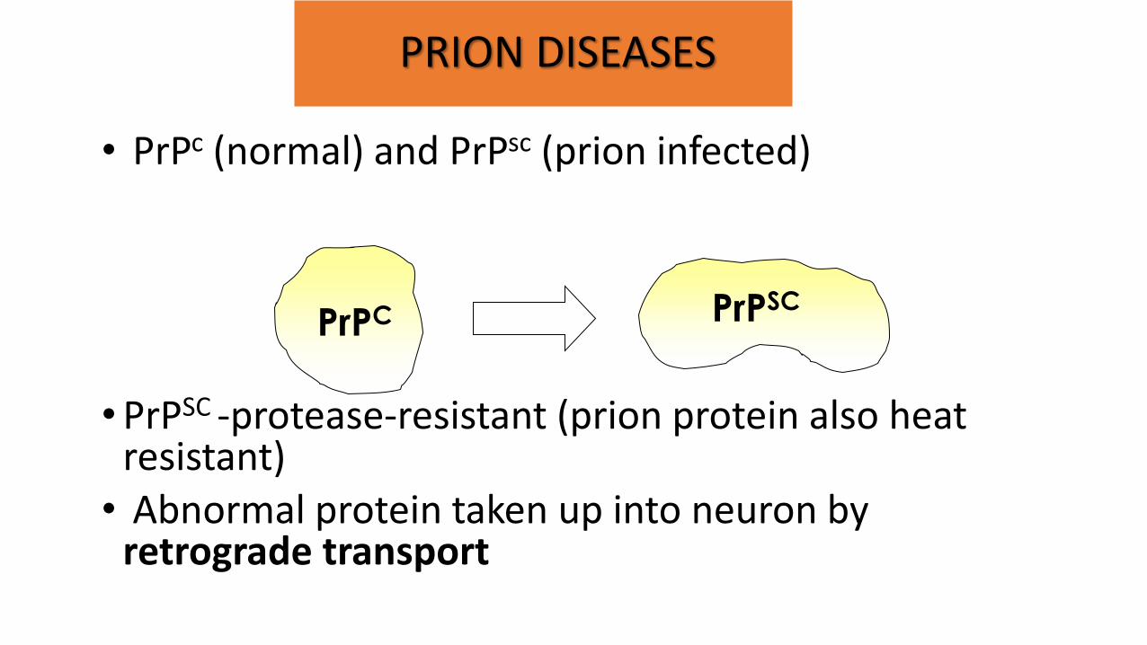
PRION DISEASES
• PrPc (normal) and PrPsc (prion infected)
• PrPSC -protease-resistant (prion protein also heat
resistant) • Abnormal protein taken up into neuron by
retrograde transport
PrPC PrPSC
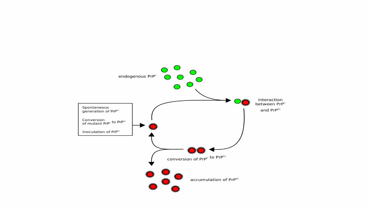
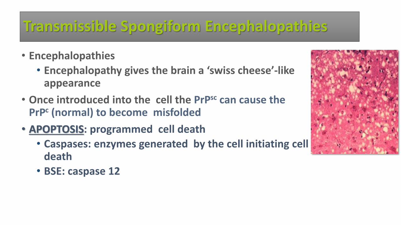
Transmissible Spongiform Encephalopathies
• Encephalopathies
• Encephalopathy gives the brain a ‘swiss cheese’-like appearance
• Once introduced into the cell the PrPsc can cause the PrPc (normal) to become misfolded
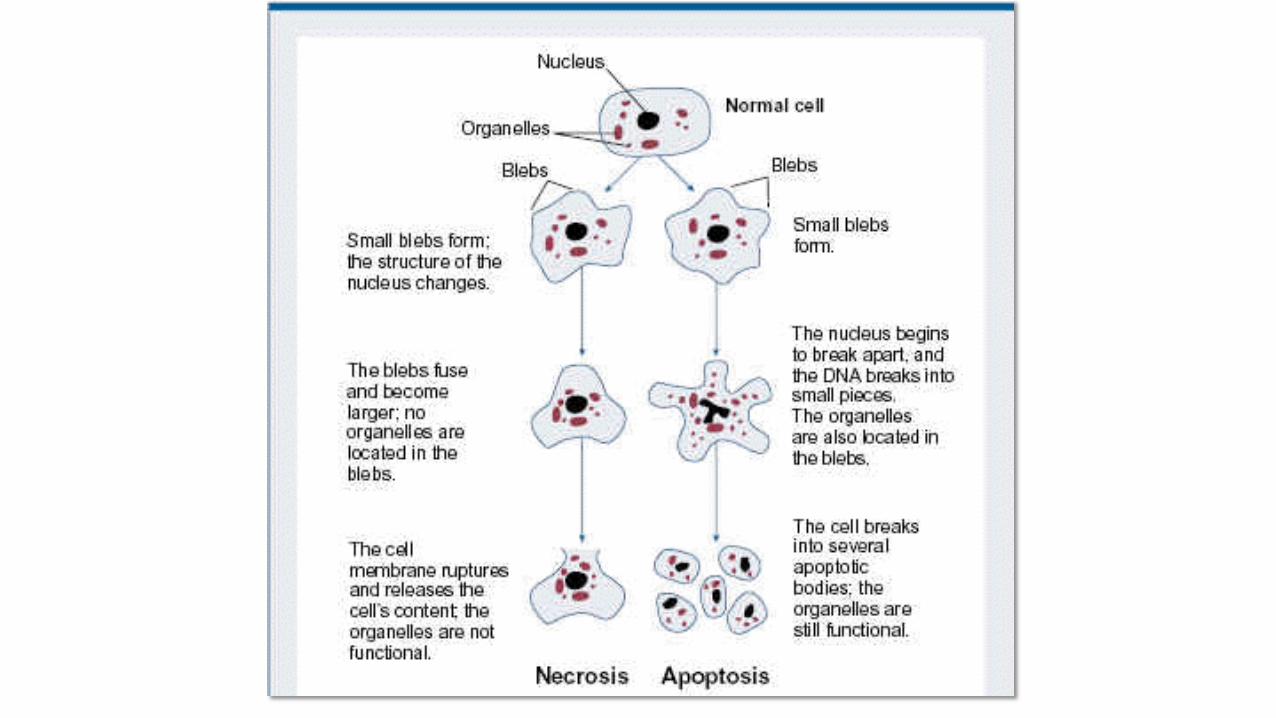
• APOPTOSIS: programmed cell death
• Caspases: enzymes generated by the cell initiating cell death
• BSE: caspase 12
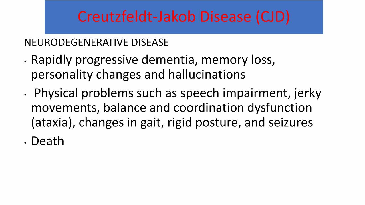
Creutzfeldt-Jakob Disease (CJD)
NEURODEGENERATIVE DISEASE
• Rapidly progressive dementia, memory loss, personality changes and hallucinations
• Physical problems such as speech impairment, jerky movements, balance and coordination dysfunction (ataxia), changes in gait, rigid posture, and seizures
• Death
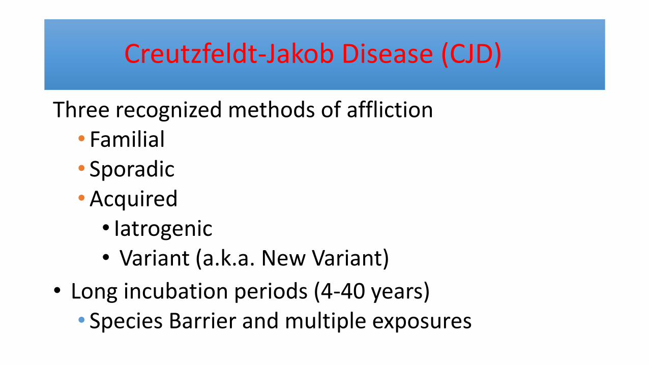
Creutzfeldt-Jakob Disease (CJD)
Three recognized methods of affliction • Familial • Sporadic • Acquired
• Iatrogenic • Variant (a.k.a. New Variant)
• Long incubation periods (4-40 years) • Species Barrier and multiple exposures

FOOD FOR THOUGHT
50,000 BSE-infected cattle are estimated to have entered the human food chain before its recognition in 1986
“You’re sick, Jessy!…Sick, sick, sick!”
Degenerative Disorders
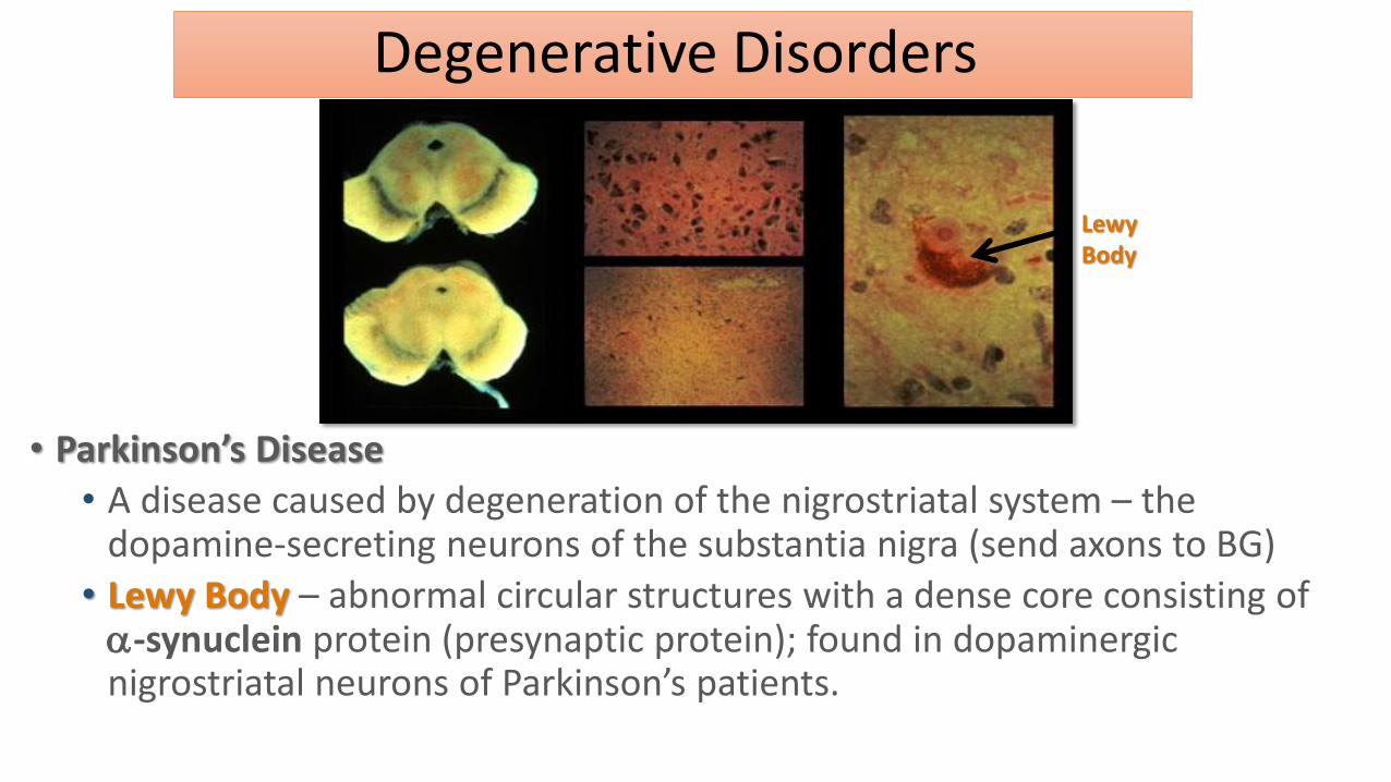
• Parkinson’s Disease
• A disease caused by degeneration of the nigrostriatal system – the dopamine-secreting neurons of the substantia nigra (send axons to BG)
• Lewy Body – abnormal circular structures with a dense core consisting of -synuclein protein (presynaptic protein); found in dopaminergic nigrostriatal neurons of Parkinson’s patients.
Lewy Body
Parkinson’s Disease
• 1% of people over 65
• Symptoms: • Muscular rigidity • Slowness of movement • Resting tremor • Postural instability • Difficulties with handwriting or making facial
expressions
Genetic causes of PD
• Gene mutations • Mutation on chromosome 4
• Gene that codes for alpha-synuclein (SNCA) – located in presynaptic terminal of DA cells
• Toxic gain of function
• Dominant
• Abnormal SNCA becomes misfolded, forms aggregations - make up lewy bodies
Toxic gain of function – genetic disorder caused by a dominant mutation
that involves a faulty gene that produces a protein with toxic effects
Genetic causes of PD
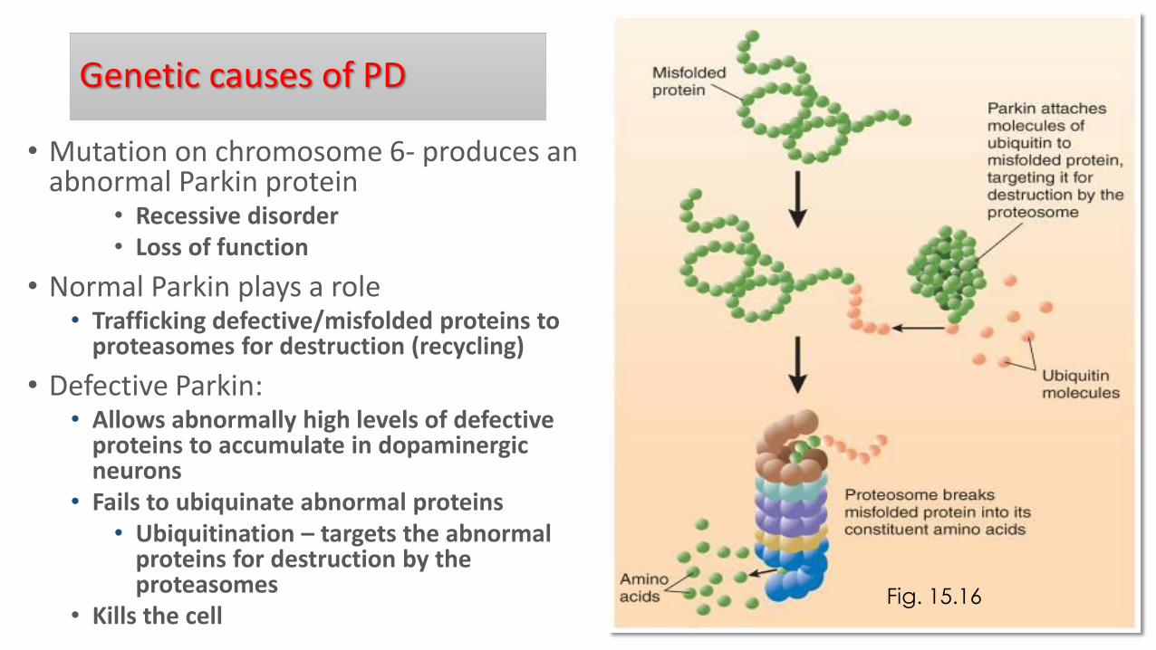
• Mutation on chromosome 6- produces an abnormal Parkin protein
• Recessive disorder • Loss of function
• Normal Parkin plays a role • Trafficking defective/misfolded proteins to
proteasomes for destruction (recycling)
• Defective Parkin: • Allows abnormally high levels of defective
proteins to accumulate in dopaminergic neurons
• Fails to ubiquinate abnormal proteins • Ubiquitination – targets the abnormal
proteins for destruction by the proteasomes
• Kills the cell
Fig. 15.16
Sporadic PD
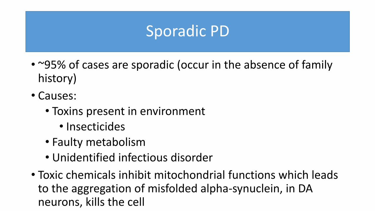
• ~95% of cases are sporadic (occur in the absence of family history)
• Causes: • Toxins present in environment
• Insecticides • Faulty metabolism • Unidentified infectious disorder
• Toxic chemicals inhibit mitochondrial functions which leads to the aggregation of misfolded alpha-synuclein, in DA neurons, kills the cell
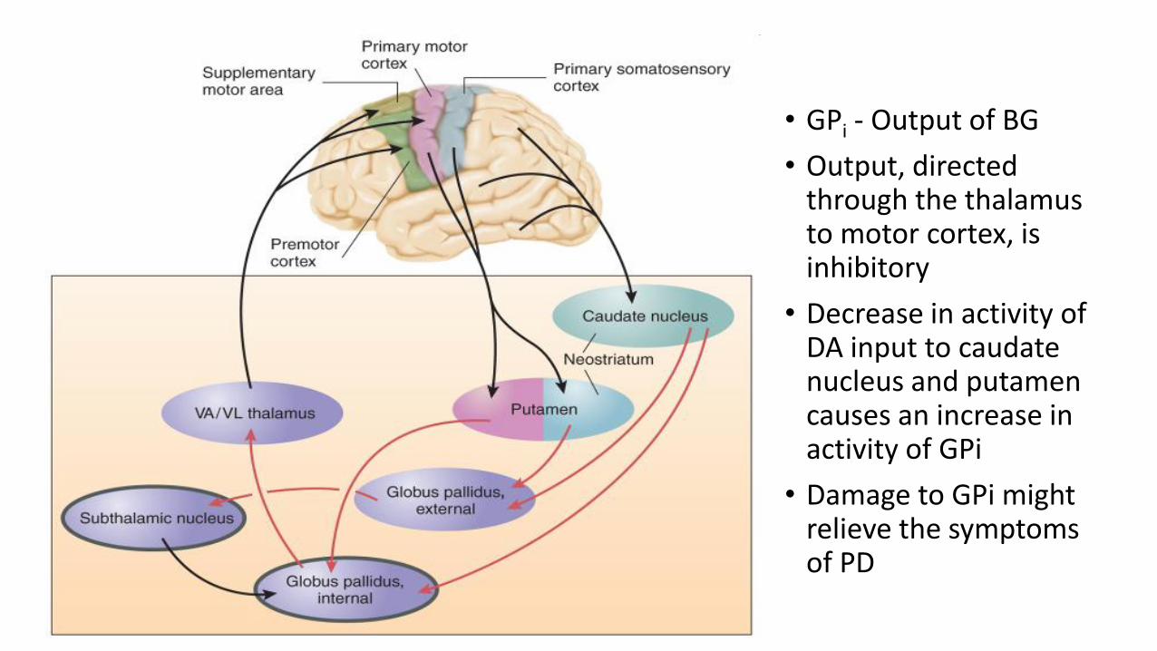
• GPi - Output of BG
• Output, directed through the thalamus to motor cortex, is inhibitory
• Decrease in activity of DA input to caudate nucleus and putamen causes an increase in activity of GPi
• Damage to GPi might relieve the symptoms of PD
Treatment of PD
• Stimulation of subthalamus (deep brain stimulation)
• Implant electrodes in subthalamic nucleus and attach a device that permits PD patient to electrically stimulate the brain
• Fewer side effects (compared to surgery)
• How can stimulation and lesions of the same area produce the same effect???
Gene Therapy as a Treatment of PD
• Genetically modified virus into the subthalamic nucleus of PD patients
• Delivered a gene for GAD (enzyme that makes GABA)
• Production of GAD turned some of the glutamate neurons into inhibitory, GABA neurons
• Activity of GPi decreased, activity of supplementary motor area increased, symptoms improved
Huntington’s Disease (also known as Huntington's chorea)
•Degeneration of caudate nucleus and putamen
•Uncontrollable movements, jerky limb movements
•Progressive, cognitive and emotional changes
•Death (10-15 years)
HD
• The disease can affect both men and women.
• HD is caused by an autosomal dominant mutation in either of an individual's two copies of a gene called Huntingtin, which means any child of an affected person typically has a 50% chance of inheriting the disease.
• Physical symptoms of Huntington's disease can begin at any age from infancy to old age, but usually begin between 35 and 44 years of age.
• About 6% of cases start before the age of 21 years with an akinetic-rigid syndrome; they progress faster and vary slightly.
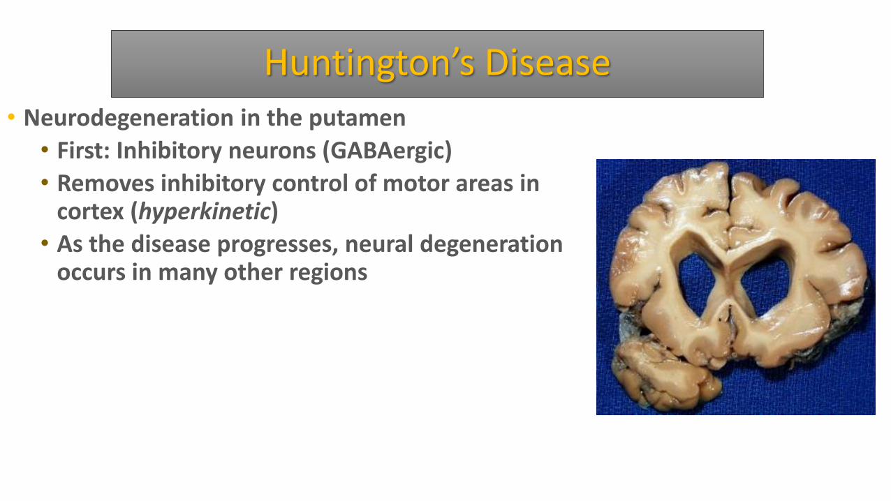
Huntington’s Disease • Neurodegeneration in the putamen
• First: Inhibitory neurons (GABAergic)
• Removes inhibitory control of motor areas in cortex (hyperkinetic)
• As the disease progresses, neural degeneration occurs in many other regions
Huntington’s Disease
• GENETICS • Dominant gene on chromosome 4 • Gene that codes the huntingtin protein (htt) • Repeated sequence of bases that code for
the amino acid glutamine • Abnormal htt becomes misfolded and
forms aggregates in nucleus • Cell death: apoptosis
Huntington’s Disease
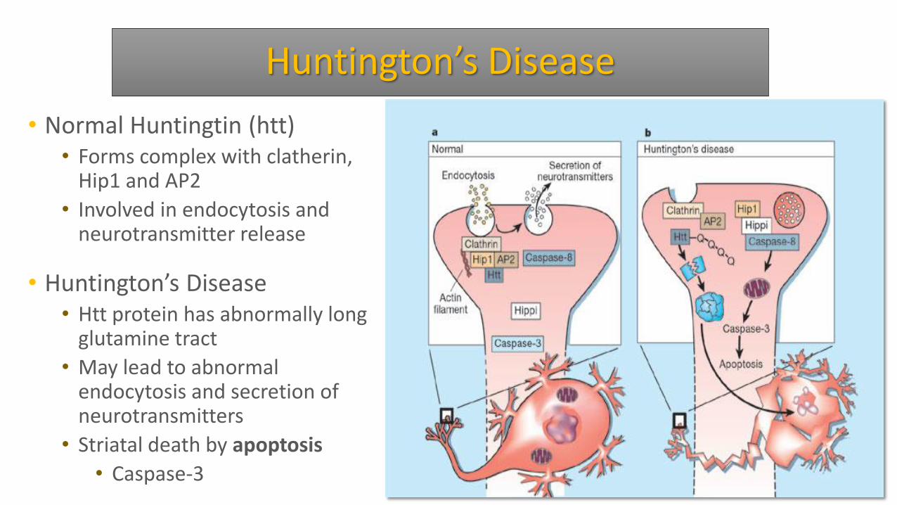
• Normal Huntingtin (htt) • Forms complex with clatherin,
Hip1 and AP2
• Involved in endocytosis and neurotransmitter release
• Huntington’s Disease • Htt protein has abnormally long
glutamine tract
• May lead to abnormal endocytosis and secretion of neurotransmitters
• Striatal death by apoptosis
• Caspase-3
Huntington’s Disease
• Normal htt facilitates the production and transport of brain derived neurotropic factor (BDNF) • BDNF: neurotropic factor critical for the survival of
neurons • BDNF produced in cortex and transported to basal
ganglia
• Abnormal htt interferes with BDNF in 2 ways: • Inhibits the expression of the BDNF gene • Interferes with the transport of BDNF from the
cerebral cortex to the BG
Huntington’s Disease
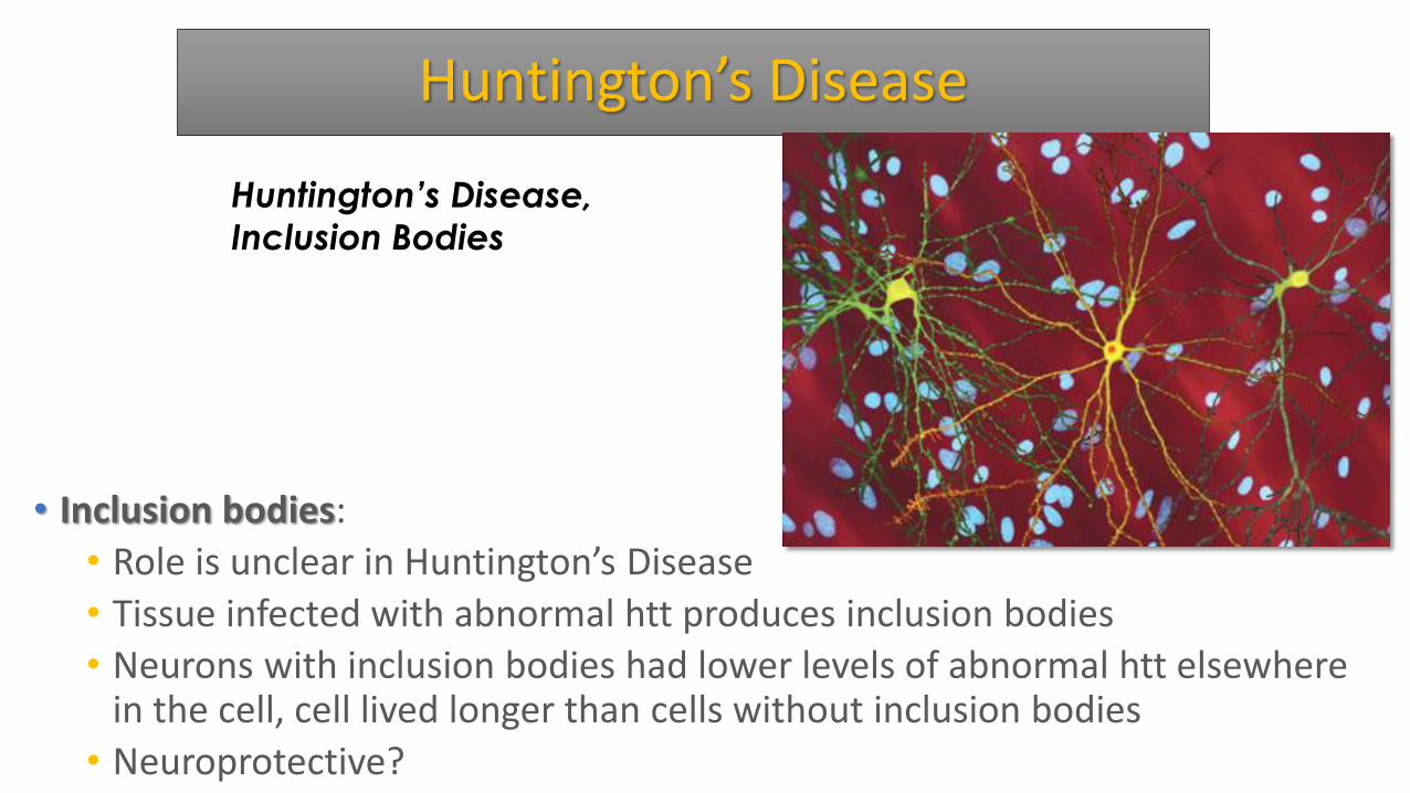
• Inclusion bodies:
• Role is unclear in Huntington’s Disease
• Tissue infected with abnormal htt produces inclusion bodies
• Neurons with inclusion bodies had lower levels of abnormal htt elsewhere in the cell, cell lived longer than cells without inclusion bodies
• Neuroprotective?
Huntington’s Disease,
Inclusion Bodies
Alzheimer’s Disease
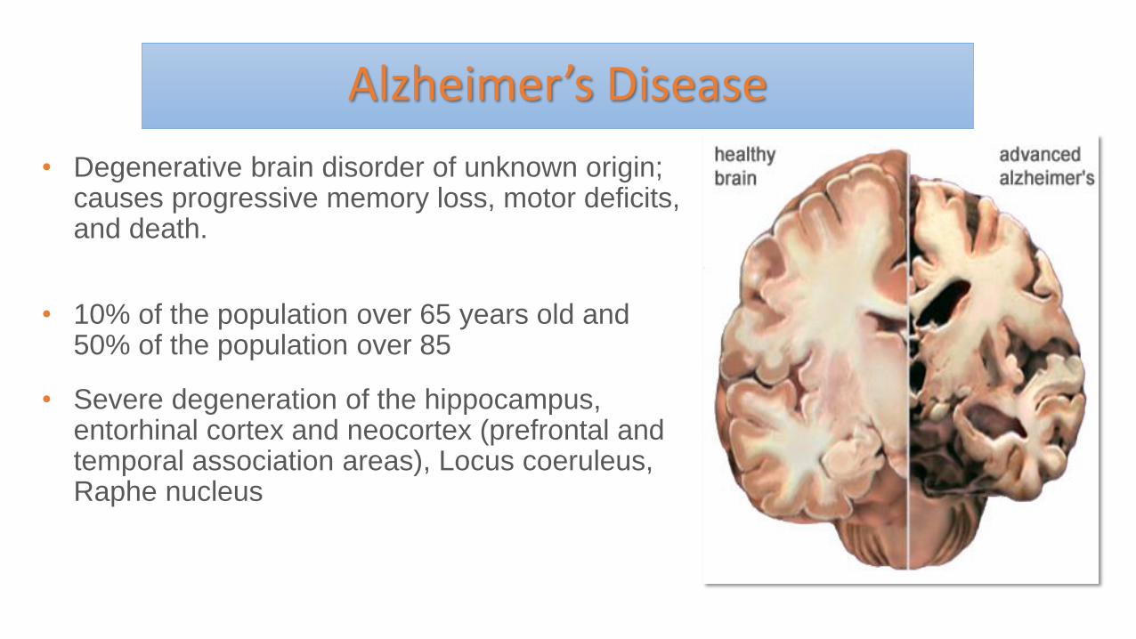
• Degenerative brain disorder of unknown origin; causes progressive memory loss, motor deficits, and death.
• 10% of the population over 65 years old and 50% of the population over 85
• Severe degeneration of the hippocampus, entorhinal cortex and neocortex (prefrontal and temporal association areas), Locus coeruleus, Raphe nucleus
Signs of Alzheimer’s Disease
1. Memory loss that disrupts daily life
2. Challenges in planning or solving problems
3. Difficulty completing familiar tasks at home, at work or at leisure
4. Confusion with time or place
5. Trouble understanding visual images and spatial relationships
6. New problems with words in speaking or writing
7. Misplacing things and losing the ability to retrace steps
8. Decreased or poor judgment
9. Withdrawal from work or social activities
10.Changes in mood and personality
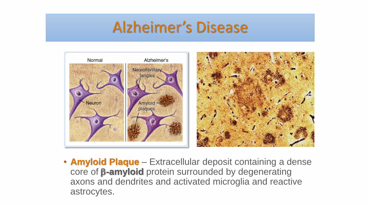
Alzheimer’s Disease
• Amyloid Plaque – Extracellular deposit containing a dense core of -amyloid protein surrounded by degenerating axons and dendrites and activated microglia and reactive astrocytes.
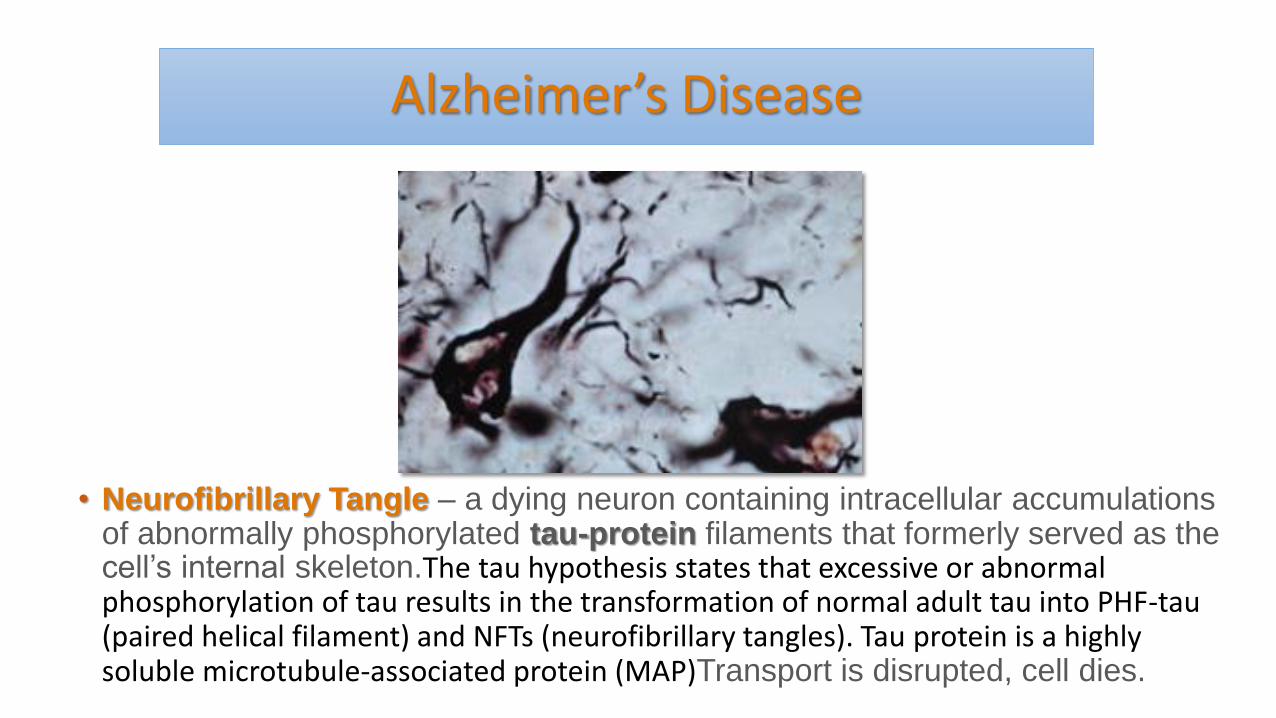
Alzheimer’s Disease
• Neurofibrillary Tangle – a dying neuron containing intracellular accumulations of abnormally phosphorylated tau-protein filaments that formerly served as the cell’s internal skeleton.The tau hypothesis states that excessive or abnormal phosphorylation of tau results in the transformation of normal adult tau into PHF-tau (paired helical filament) and NFTs (neurofibrillary tangles). Tau protein is a highly soluble microtubule-associated protein (MAP)Transport is disrupted, cell dies.
Tau proteins • (or τ proteins, after the Greek letter with that name) are proteins
that stabilize microtubules. They are abundant in neurons of the central nervous system and are less common elsewhere, but are also expressed at very low levels in CNS astrocytes and oligodendrocytes. Pathologies and dementias of the nervous system such as Alzheimer's disease and Parkinson's disease are associated with tau proteins that have become defective and no longer stabilize microtubules properly.
• The tau proteins are the product of alternative splicing from a single gene that in humans is designated MAPT (microtubule-associated protein tau) and is located on chromosome 17q21
Alzheimer’s Disease
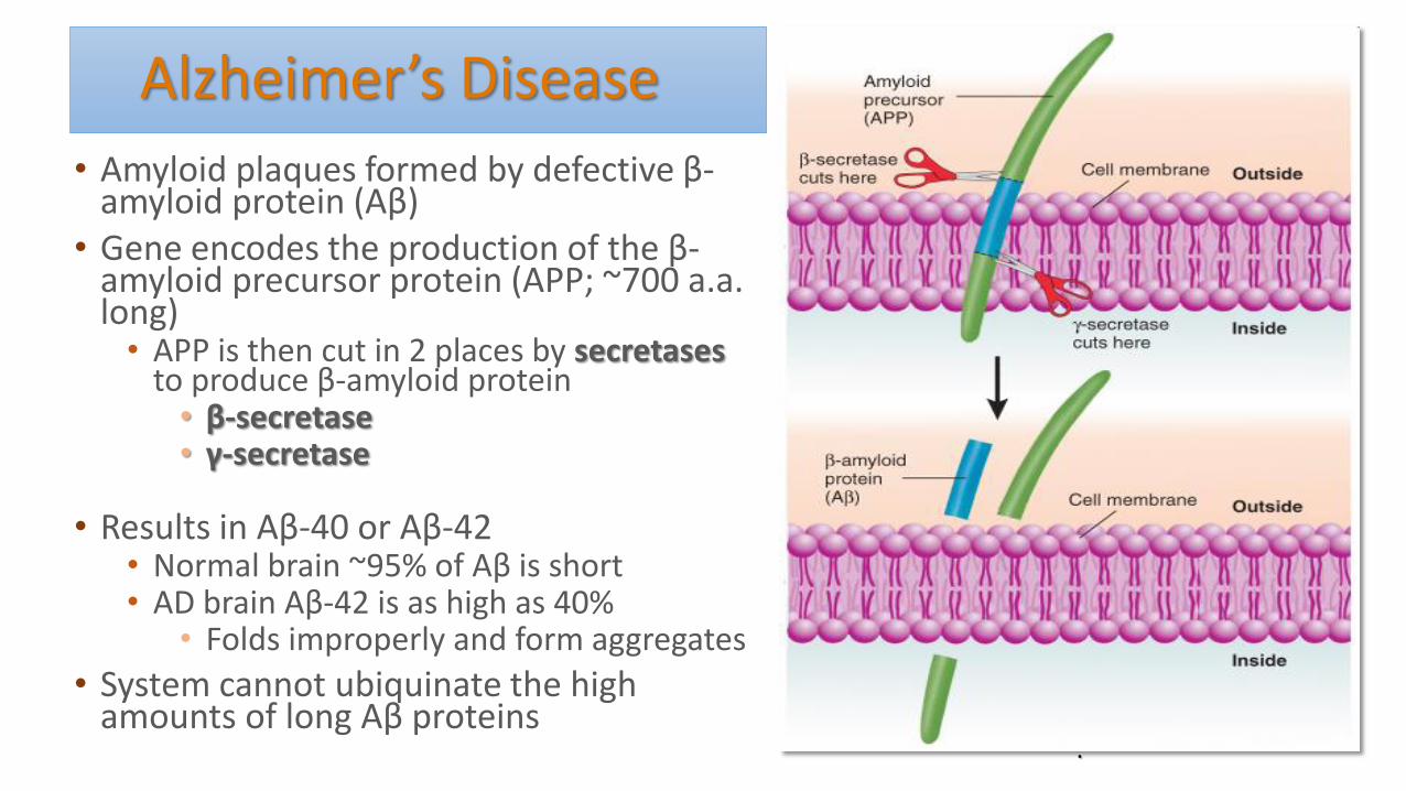
• Amyloid plaques formed by defective β-amyloid protein (Aβ)
• Gene encodes the production of the β-amyloid precursor protein (APP; ~700 a.a. long)
• APP is then cut in 2 places by secretases to produce β-amyloid protein
• β-secretase • γ-secretase
• Results in Aβ-40 or Aβ-42 • Normal brain ~95% of Aβ is short • AD brain Aβ-42 is as high as 40%
• Folds improperly and form aggregates
• System cannot ubiquinate the high amounts of long Aβ proteins
.
Alzheimer’s Disease • Some forms of AD are familial
• APP gene – chromosome 21 • Gene for the amyloid beta precursor protein (APP) is located on
chromosome 21, and people with trisomy 21 (Down Syndrome) who thus have an extra gene copy almost universally exhibit AD by 40 years of age.
• Two presenilin genes found on chromosomes 1 and 14 • Subunits of γ-secretase
• Apolipoprotein E (ApoE) – glycoprotein that transports cholesterol in the blood and also plays a role in cellular repair
• ApoE4 – interfers with removal of long form of Aβ
• Other causes: • Traumatic brain injury
Alzheimer’s Disease
• Aβ inside cell (not plaques) is the cause of neural degeneration
• Aggregated forms of amyloid (Aβ oligomers) • interact with microglia, causing an inflammatory response that
triggers the release of toxic cytokines (chemicals produced by the immune system that destroy infected cells)
• trigger XS release of glutamate by glial cells, causes excitotoxicity (increased inflow of Ca2+ through neural NMDA receptors
• Cause synaptic dysfunction and suppress the formation of LTP • Alzheimer's is the sixth leading cause of death in the United
States.
Treatment
• Decline in Ach levels
• Cholingeric agonists (acetylcholinesterase inhibitors)
• NMDA receptor antagonist (memantine)
• Immunotherapeutic approach • Amyloid vaccine to reduce plaque deposits and improve
performance on memory tasks in a transgenic mouse model
• Mixed results • Dangerous side effects
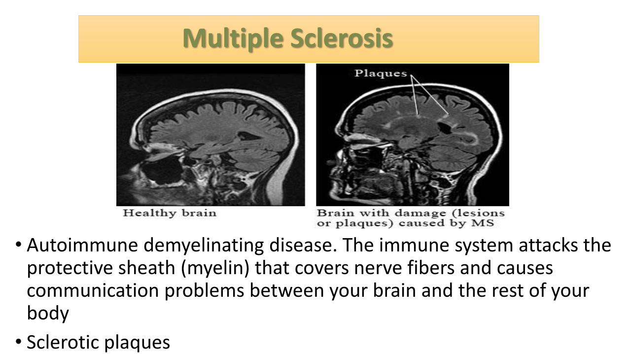
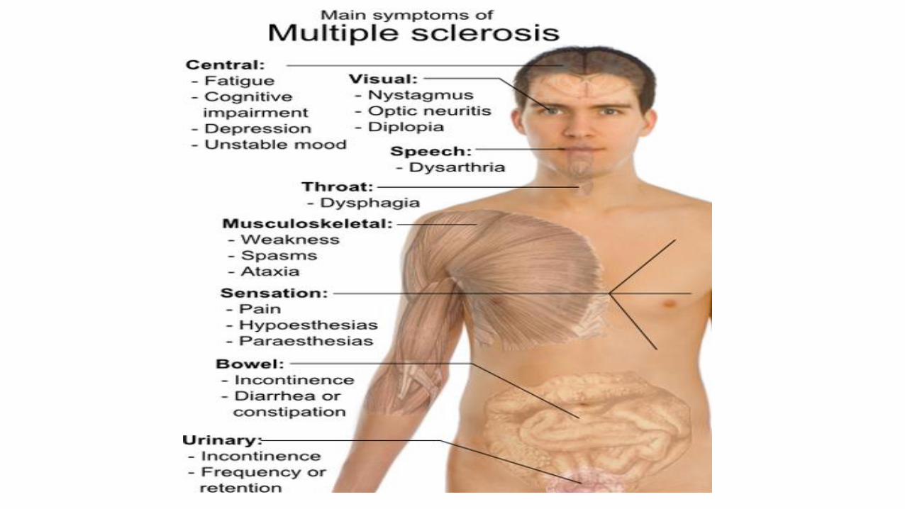
Multiple Sclerosis
• Autoimmune demyelinating disease. The immune system attacks the protective sheath (myelin) that covers nerve fibers and causes communication problems between your brain and the rest of your body
• Sclerotic plaques
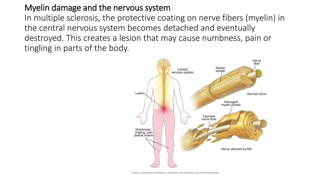
Myelin damage and the nervous system In multiple sclerosis, the protective coating on nerve fibers (myelin) in the central nervous system becomes detached and eventually destroyed. This creates a lesion that may cause numbness, pain or tingling in parts of the body.
Multiple Sclerosis
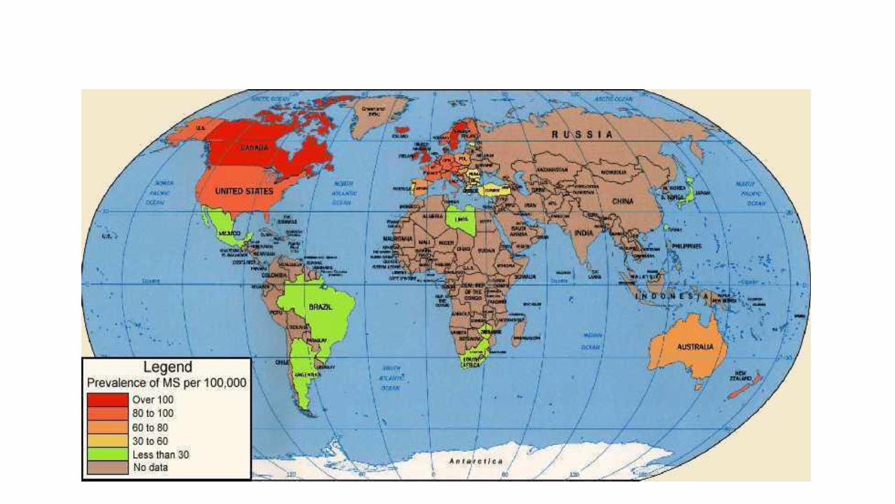
• Epidemiology • More women then men • Late twenties-thirties • Childhood in colder climates • Canada has amongst the highest MS incidence estimates in the
world • 55,000 – 75,000

Multiple Sclerosis
• TREATMENTS: • Interferon β
• Modulates the responsiveness of the immune system
• Treatment slows the progression and severity of the attacks
• Glaterimer acetate (copaxone) • Peptides composed of random sequences of
glutamate, alanine and lysine • May stimulate anti-inflammatory responses