Definitions, Pathophysiology and Clinical...
19
Canine Urolithiasis: Definitions, Pathophysiology and Clinical Manifestations Carl A. Osborne Jody P. Lulich Lisa K. Ulrich CLINICAL IMPORTANCE Urolithiasis is a common disorder of the urinary tract in dogs. It was diagnosed in 3,628 of 676,668 dogs (0.53%) admitted to veterinary teaching hospitals in North America between 1980 and 1993. The proportion of dogs with urolithiasis admitted to veterinary hospitals in Germany was similar (Lulich et al, 1995). In 2011, 69.3 million dogs resided in the USA and urolith analysis was performed for 61 thousand resulting in an incidence of 0.1% of the canine population. These values are likely an underestimate because not all uroliths are submitted for analysis and not all dogs with disease are presented for care. Clinical signs of urolithiasis may be the first indication of underlying systemic disorders, or defects in the structure or function of the urinary tract (Table 38-1). Uroliths may pass through various parts of the excretory pathway of the urinary tract, they may dissolve, they may become inactive or they may continue to form and grow. If uroliths associated with clinical signs are allowed to remain untreated, they may result in serious sequelae. Despite urolith removal by dissolution, surgery, or minimally invasive procedures, uroliths frequently recur if risk factors associated with their formation are not suppressed or corrected. Urolithiasis should not be viewed as a single disease, but rather as a sequela of one or more underlying abnormalities. The fact that urolith formation is often erratic and unpredictable indicates that several interrelated complex physiologic and pathologic factors are involved. Therefore, detection of uroliths is only the beginning of the diagnostic process. Determination of urolith composition narrows etiologic possibilities. Knowledge of the patient’s food and how it is fed and serum and urine concentrations of lithogenic minerals, crystallization promoters, crystallization inhibitors and their interactions aids in the diagnosis, treatment and prevention of urolithiasis (Box 38-1). FORMATION OF UROLITHS Initiation and Growth Urolith formation is associated with two complementary but separate phases: initiation and growth. It appears that initiating events are not the same for all types of uroliths. In addition, factors that initiate urolith formation may be different from those that allow urolith growth. The initial step in urolith formation is formation of a crystal nidus (or crystal embryo). This initiation phase of urolith for- “If the patient you treat is harmed more than helped, then best leave the stones alone. But by taking a look at the thoughts in this book, ways to treat stones by how patients eat you’ll be shown.” Carl A. Osborne, 1999 38
Transcript of Definitions, Pathophysiology and Clinical...

Canine Urolithiasis: Definitions, Pathophysiology
and Clinical Manifestations Carl A. Osborne
Jody P. Lulich
Lisa K. Ulrich
CLINICAL IMPORTANCE Urolithiasis is a common disorder of the urinary tract in dogs. It was diagnosed in 3,628 of 676,668 dogs (0.53%) admitted to veterinary teaching hospitals in North America between 1980 and 1993. The proportion of dogs with urolithiasis admitted to veterinary hospitals in Germany was similar (Lulich et al, 1995). In 2011, 69.3 million dogs resided in the USA and urolith analysis was performed for 61 thousand resulting in an incidence of 0.1% of the canine population. These values are likely an underestimate because not all uroliths are submitted for analysis and not all dogs with disease are presented for care.
Clinical signs of urolithiasis may be the first indication of underlying systemic disorders, or defects in the structure or function of the urinary tract (Table 38-1). Uroliths may pass through various parts of the excretory pathway of the urinary tract, they may dissolve, they may become inactive or they may continue to form and grow. If uroliths associated with clinical signs are allowed to remain untreated, they may result in serious sequelae. Despite urolith removal by dissolution, surgery, or minimally invasive procedures, uroliths frequently recur if risk factors associated with their formation are not suppressed or corrected. Urolithiasis should not be viewed as a single disease, but rather as a sequela of one or more underlying abnormalities.
The fact that urolith formation is often erratic and unpredictable indicates that several interrelated complex physiologic and pathologic factors are involved. Therefore, detection of uroliths is only the beginning of the diagnostic process. Determination of urolith composition narrows etiologic possibilities. Knowledge of the patient’s food and how it is fed and serum and urine concentrations of lithogenic minerals, crystallization promoters, crystallization inhibitors and their interactions aids in the diagnosis, treatment and prevention of urolithiasis (Box 38-1).
FORMATION OF UROLITHS Initiation and Growth Urolith formation is associated with two complementary but separate phases: initiation and growth. It appears that initiating events are not the same for all types of uroliths. In addition, factors that initiate urolith formation may be different from those that allow urolith growth.
The initial step in urolith formation is formation of a crystal nidus (or crystal embryo). This initiation phase of urolith for-
“If the patient you treat is harmed more than helped, then best leave the stones alone. But by taking a look at the thoughts in this book,
ways to treat stones by how patients eat you’ll be shown.” Carl A. Osborne, 1999
38

814 Small Animal Clinical Nutrition
mation, called nucleation, is dependent on supersaturation of urine with lithogenic crystalloids. The inciting factor and the precise sequence of events that lead to the formation of most types of stones are still unknown. The degree of urine supersaturation may be influenced by the magnitude of renal excretion of crystalloids, urinary pH and/or crystallization inhibitors or promoters in urine. Noncrystalline proteinaceous matrix substances may also play a role in nucleation in some instances.
Three theories have been proposed to explain initiation of lithogenesis: 1) supersaturation-crystallization theory, 2) matrix-nucleation theory and 3) crystallization-inhibition theory (Osborne and Kruger, 1984). Each theory emphasizes a single factor. The supersaturation-crystallization theory incriminates excessive supersaturation of urine with urolith-forming crystalloids as the primary event. In this hypothesis, crystal nucleation is considered to be a physiochemical process involving precipitation of crystalloids from a supersaturated solution. Urolith formation is thought to occur independently of preformed matrix or crystallization inhibitors.
The matrix-nucleation theory incriminates preformed organic matrix (thought to be a mucoprotein with mineral-
binding properties) as the primary determinant in lithogenesis. This theory is based on the assumption that preformed organic matrix forms an initial nucleus that subsequently permits urolith formation by precipitation of crystalloids. The role of organic matrix in lithogenesis has not been defined with certainty; however, the similarity of the overall composition of matrix from human uroliths of various mineral composition supports this hypothesis.
The crystallization-inhibition theory proposes that reduction or absence of organic and inorganic inhibitors of crystallization is the primary determinant of calcium oxalate and calcium phosphate lithogenesis. This theory is based on the fact that several lithogenic substances in urine are maintained in solution at concentrations significantly higher than is possible in water (i.e., driving forces for crystal precipitation of normally saturated urine are minimized by crystallization inhibitors). Similarly, inhibitors are important in minimizing crystal growth and aggregation. These three theories are not mutually exclusive. In fact, supersaturation of urine with the crystal’s components is a prerequisite for each theory of nucleation.
Further growth of the crystal nidus depends on: 1) whether or not it remains in the lumen of the excretory pathway of the urinary system, 2) the degree and duration of supersaturation of urine with crystalloids identical or different from those in the nidus and 3) physical characteristics of the crystal nidus. Crystals that are compatible with other crystalloids may align themselves and grow on the surface of other crystals. This is called epitaxial growth. Epitaxy may represent a heterogeneous form of nucleation, and may account for some mixed and compound uroliths. For example, in people, the structural similarities of uric acid and calcium oxalate permit urolith growth by epitaxy (Coe, 1977).
Nucleation Nucleation refers to the initial event in the formation (or precipitation) of uroliths and is characterized by the appearance of submicroscopic molecular aggregates of crystalloids. Initially, the aggregates are approximately 100 molecules in size and represent potential crystal embryos (or a nidus). Crystals represent an orderly arrangement of atoms in a periodic pattern or lattice. To become a urolith, crystal embryos must have a lattice arrangement that allows continued growth. They must also be large enough to prevent dispersion back into the dissolved phase (Pak, 1976).
Nucleation has been classified as homogeneous (also called self-nucleation or generalized nucleation) or heterogeneous (also called localized nucleation) (Lyon and Vermeulen, 1965). Homogeneous nucleation occurs spontaneously in highly supersaturated urine in the absence of foreign substances (Figure 38-1). Therefore, the nidus is composed of identical crystalloids. Heterogeneous nucleation is catalyzed by foreign material such as suture material, indwelling catheters, tissue debris, crystal embryos of different composition, etc. (Figure 38-2). Urine contains many impurities that might promote heterogeneous nucleation and initiate crystal formation at a concentration of crystalloids below the formation concentration.
Table 38-1. Clinical importance of urolithiasis.
First evidence of an underlying systemic disorder Hypercalcemia
Calcium oxalate uroliths Calcium phosphate uroliths
Cushing’s syndrome Calcium oxalate uroliths Calcium phosphate uroliths Struvite uroliths
Defects in purine metabolism Portal vascular anomalies
Ammonium urate uroliths Enzyme defects
Ammonium urate uroliths Xanthine uroliths 2,8-dihydroadenine uroliths
First evidence of an underlying urinary tract disorder Renal tubular transport defect
Cystinuria Cystine uroliths
Renal tubular acidosis Calcium oxalate uroliths Calcium phosphate uroliths
Defects in local host defenses against urease-producing microbes
Struvite uroliths Calcium phosphate carbonate uroliths
Foreign bodies in urinary tract Suture material
Usually struvite uroliths Catheters
Usually struvite and sometimes calcium oxalate uroliths Sequelae to urolithiasis Dysuria, pollakiuria, urge incontinence, hematuria Secondary microbial urinary tract infection Partial or total obstruction to urine outflow
Bacterial urinary tract infection that may progress Impaired renal function and postrenal azotemia Rupture of the outflow tract
Uroperitoneum Inflammation of tissues adjacent to various portions of
the urinary tract Formation of inflammatory bladder polyps

Introduction to Canine Urolithiasis 815
Box 38-1. Urolithiasis Terms and Concepts.
UROLITHIASIS The urinary system is designed to dispose of waste products in soluble form. However, some waste products are sparingly soluble and occasionally precipitate out of solution to form crystals. Growth or aggregation of microscopic crystals may lead to formation of macroscopic uroliths. Urolithiasis may be conceptually defined as the formation of uroliths anywhere in the urinary tract from less soluble crystalloids of urine as a result of multiple congenital and/or acquired physiologic and pathologic processes. If such crystalloids become trapped in the urinary system, they may grow to sufficient size to cause clinical signs.
Urolithiasis should not be thought of as a single disease, but rather as a sequela of one or more underlying abnormalities. The fact that urolith formation is often erratic and unpredictable indicates that several interrelated complex physiologic and pathologic factors are involved. Therefore, detection of uroliths is only the beginning of the diagnostic process. Determination of urolith composition narrows etiologic possibilities. Knowledge of the patient’s food, and serum and urine concentrations of lithogenic minerals, crystallization promoters, crystallization inhibitors and their interactions aids in the diagnosis, treatment and prevention of urolithiasis.
UROLITHS Uroliths are polycrystalline concretions that typically contain more than 95% organic or inorganic crystalloids, and less than 5% organic matrices (weight vs. weight ratio). (The exception to this generality is infection-induced uroliths which contain as much as 50% matrix). Uroliths may also contain a number of minor constituents. A variety of different types of uroliths may occur in dogs (Figure 1). Uroliths are typically composed of organized crystal aggregates with a complex internal structure. Cross sections of uroliths frequently reveal nuclei and laminations, and less frequently radial striations. Urine that bathes uroliths varies in composition (and probably in degree of saturation with lithogenic crystalloids) from day to day and perhaps from hour to hour. This phenomenon is of conceptual importance in understanding the physical characteristics of uroliths.
The incidence and composition of uroliths may be influenced by a variety of factors including: 1) species, 2) breed, 3) gender, 4) age, 5) geography, 6) food, 7) anatomic abnormalities, 8) physiologic abnormalities, 9) urinary tract infection and 10) urinary pH. Uroliths may be named according to mineral composition, location (i.e., nephroliths, ureteroliths, cystoliths, vesical calculi, urethroliths) or shape (i.e., smooth, faceted, pyramidal, laminated, mulberry, jackstone, staghorn or branched). Characteristic shapes of crystals and uroliths are influenced primarily by the internal structure of crystals and the environment in which they form. Crystals of calcium oxalate monohydrate tend to fuse, producing smoothly rounded or mammillated uroliths. Local factors that influence the size and shape of uroliths include: 1) number of uroliths present, 2) mobility or fixation of uroliths, 3) flow characteristics of urine and 4) anatomic configuration of the structure in which uroliths grow.
MINERAL A mineral is a naturally occurring, inorganically formed substance that has a characteristic chemical composition and usually has an
ordered atomic arrangement that may influence its external geometric form. Minerals commonly found in uroliths often have a chemical name and a crystal (or mineral) name. Even though a particular mineral usually predominates, the mineral composition of many uroliths may be mixed. Occasionally, the center of a urolith may be composed of one type of crystalloid (e.g., silica), whereas outer layers are composed of a different crystalloid (especially struvite). Detection, treatment and prevention of the underlying causes of urolithiasis depend on knowledge of the composition and structure of all portions of uroliths.
MATRIX The nondialyzable portion of uroliths that remains after crystalline components have been dissolved with mild solvents is organic matrix. Uroliths consistently contain variable quantities of organic matrix substances in addition to crystalloids. Organic matrix substances identified in human uroliths and experimentally produced in animals include matrix substances A, Tamm-Horsfall glycoprotein, uromucoid, serum albumin and alpha and gamma globulins. Of these, matrix substance A, Tamm-Horsfall glycoprotein and uromucoid appear to be quantitatively more significant than alpha and gamma globulins.
The complex of diverse mucoprotein compounds composing matrix substances may represent the skeleton of uroliths. Although the physical characteristics of uroliths suggest organized relationships between the matrix skeleton and crystalline building blocks, the role of each of these components in formation, retention and growth of uroliths is still poorly understood.
Organic matrix may affect urolith formation by one or more of several mechanisms including: 1) sites of heterogeneous nucleation, 2) templates for organizing and modifying growth of crystals 3) binding agents that cement urolith particles together andpromote retention of crystals and 4) protective colloids thatprevent further growth of uroliths. Organic matrix may also becomposed of passive substances that have no effect on urolithformation or growth.
NUCLEI AND LAMINATIONS Examination of cross sections of uroliths often reveals a nucleus and adjacent peripheral laminations. Laminated uroliths may be detected by radiography. Nuclei are focal points (or cores) that differ in appearance from more peripheral portions of the urolith. Nuclei are usually but not invariably located in the center of uroliths. Nuclei may be of crystalline composition or they may be composed of foreign material, tissue debris, blood clots, bacteria, etc. The mineral composition of crystalline nuclei may be identical or different from the remainder of the urolith. Nuclei surrounded by well-defined layers (or lamellae) of solid material suggest an early phase of urolith evolution. However, crystalline nuclei large enough to be detected visually are too large to represent an initial crystalline nidus for crystal nucleation in the physiochemical sense. Centrally located nuclei imply that the urolith was freely accessible to urine from all sides and that growth proceeded at a similar rate on all sides.
Laminated uroliths are common and may represent: 1) alternating bands of different mineral types, 2) periods during which urolith

816 Small Animal Clinical Nutrition
Box 38-1 continued
growth occurred without interruption or 3) alternating periods of precipitation of minerals and gel. Although a difference in appearance between two consecutive layers should prompt suspicion of differences in composition, this is not always the case.
MATRIX CONCRETIONS By definition, a urolith must contain some minerals. However, concretions composed primarily (more than 65%) of matrix may occur. These concretions, commonly called matrix stones, often occur in the urethra of male cats and sheep, and sometimes occur in dogs and people. They may form a cast of that portion of the excretory pathway in which they are formed (e.g., urethral plugs), implying a rapid rate of formation. In dogs, matrix concretions usually occur secondary to bacterial infections.
COMPOUND UROLITHS Compound uroliths have one or more layers of mineral composition (e.g., struvite) different from minerals identified in the nucleus (e.g., calcium oxalate).
MIXED UROLITHS Mixed uroliths contain more than one mineral, neither of which composes at least 70% of the urolith, but without a nucleus or well-defined laminations.
Figure 1. Different mineral types of canine uroliths illustrating common sizes, shapes and surface characteristics. 1) Calcium oxalate dihydrate; 2) Calcium oxalate dihydrate; 3) Calcium oxalate monohydrate; 4) Calcium oxalate monohydrate; 5) Calcium oxalate monohydrate; 6) Calcium oxalate dihydrate; 7) Cystine; 8) Cystine; 9) Ammonium urate (left urolith has been bisected to illustrate laminations); 10) Ammonium urate; 11) Ammonium urate; 12) Struvite; 13) Struvite; 14) Compound urolith with a nidus of calcium oxalate monohydrate surrounded by a shell of struvite and calcium carbonate apatite; 15) Compound urolith with a nidus of silica surrounded by shells containing a mixture of calcium oxalate, silica and ammonium urate; 16) Silica; 17) Silica; 18) Silica; 19) Struvite that has the shape of the urinary bladder and proximal urethra; 20) Struvite that has the shape of the renal pelvis and proximal ureter.
The Bibliography for Box 38-1 can be found at www.markmorris.org.
These substances may be thought of as facilitators or potentiators of crystallization. Any crystal type may be a potential nidus for nucleation of another crystal type. A greater degree of supersaturation (i.e., a higher formation product) is required for homogeneous nucleation than for heterogeneous nucleation. Once nucleation has occurred, however, crystal growth can occur at any degree of supersaturation (even at metastability).
Undersaturated Solutions An undersaturated solution contains a sufficiently low concentration of a crystalloid to permit dissolution of additional quantities of the crystalloid. Urine is undersaturated when the solute concentration (or activity product) is less than the solubility of the solute in question. Formation of urine that is undersaturated with lithogenic crystalloids may permit varying degrees of urolith dissolution.
Saturated Solutions Saturated solutions are in equilibrium with undissolved solute
at a given temperature. Saturated solutions contain so much dissolved substances that no more can be dissolved at a given temperature. With respect to urine, the saturation concentration is that concentration of a crystalloid that remains unchanged when the urine is mixed with uroliths (or the solid phase) containing that crystalloid. The saturation of salts in urine is influenced by several variables including pH, ionic strength and temperature.
Supersaturated Solutions A supersaturated solution is more saturated with a substance at a given temperature than would be normally expected (i.e., it is any concentration greater than the saturation concentration). Supersaturated urine contains a greater concentration of a crystalloid (cystine, phosphate, calcium, ammonium, etc.) than the associated solvent (water) would be predicted to be able to normally hold in solution. Supersaturation can vary in degree. Urine is metastable at lower levels of supersaturation. At higher levels of supersaturation, however, urine becomes unstable
14
20
1 2 13
3 12 19
5
4 6 15
11
7 8 9 16 17
18 10

Introduction to Canine Urolithiasis 817
with regard to its capacity to keep lithogenic substances in solution (Figure 38-3). Factors that increase the saturation of crystalloids in urine predispose patients to precipitation of crystals and thus urolith formation. Spontaneous precipitation will occur if the concentration of the crystalloid is greater than its formation product.
Metastable Region The metastable region refers to the degree of supersaturation of a crystalloid that lies between the solubility product and the formation product. Metastability applies to those liquids (e.g., urine) that have the capacity to retain more of a compound in solution than would be predicted by knowledge of its true solubility in water. The term “metastable” is appropriate because it implies a condition subject to change. A metastable solution is thermodynamically unstable, but does not contain enough energy to initiate crystal formation. However, crystals already present may grow. The region of metastability varies with the type of lithogenic crystalloid. For example, in people, it has been estimated that the difference between the solubility product and the formation product of calcium oxalate in urine is a multiple of about 8.5 to 10.0 (Coe, 1978).
Oversaturated Solutions An oversaturated solution is one in which the degree of supersaturation of a crystalloid is greater than the formation product (Figure 38-3). Recall that supersaturated urine exceeds the solubility product, but does not exceed the formation product. Oversaturated urine is no longer metastable. Nucleation will take place in the absence of heterogeneous factors. Oversaturation of urine is thought to cause crystals observed by microscopic examination of urine sediment.
Inhibitors and Promoters of Crystal Formation
Urine is a complex solution containing a variety of substances that can inhibit or promote crystal formation and growth. Inhibitors include molecules that reduce calcium oxalate and calcium phosphate supersaturation. Some inhibitors (e.g., citrate, magnesium, pyrophosphate) form soluble salts with calcium, oxalic acid or phosphoric acid, thereby reducing the quantity of these metabolites available for precipitation. Other inhibitors (e.g., nephrocalcin, uropontin, glycosaminoglycans, Tamm-Horsfall glycoprotein, other inert ions) interfere with the ability of calcium and oxalic acid to combine, thereby minimizing crystal formation and growth. Also, glycosaminoglycans act as protectors by preventing crystals from adhering to the urinary tract mucosa.
Clinical Concepts of Urine Supersaturation Salts (crystals) are neutral compounds derived from the reversible interaction of a cation (e.g., calcium) and an anion (e.g., oxalic acid). The ability of a salt to dissolve in solution depends on the concentration of its ions in solution, and its interaction with other ions and neutral molecules in the same solution. For example, the state of urine saturation for any specific crystal system is the product of urine solute concentration, pH, ionic strength, tem-
Figure 38-1. Layered urocystolith composed of 100% calcium oxalate dihydrate removed from an adult male miniature schnauzer. The difference in color of the center of the urolith vs. the outer layer is due to the large quantity of blood in the matrix of the outer layer.
Figure 38-2. Struvite uroliths that have formed on a hair shaft.
Figure 38-3. Probable events in formation of crystals in urine. A variety of factors influence the solubility of minerals in urine including concentration of lithogenic and non-lithogenic minerals, the concentration of crystallization inhibitors and crystallization promoters, urine temperature, urinary pH and urine ionic strength.

818 Small Animal Clinical Nutrition
perature and preformed chemical complexes. To illustrate these principles, consider pure water as a
solution and calcium oxalate as a salt. Small amounts of calcium oxalate added to water dissolve completely because water is undersaturated with calcium and oxalic acid ions. As more calcium oxalate is added, the water’s capacity to dissolve additional calcium oxalate is decreased until the solution becomes saturated. In this context, saturation of the solution with calcium and oxalic acid ions occurs when no additional calcium oxalate can be dissolved at a given pH and temperature of the solution. If additional calcium oxalate is added, it will appear as a solid.
As in water, calcium oxalate can also be dissolved in undersaturated urine. However, unlike water, urine is a complex solution containing a unique combination of ionic and nonionic molecules that may increase the solubility of calcium oxalate. Therefore, calcium oxalate added beyond the point of saturation will remain in solution. Thus, the solution becomes supersaturated with calcium and oxalic acid ions. Supersaturation is conceptually significant because the solution contains enough energy to form solids from dissolved ions (i.e., it is thermodynamically unstable). When supersaturated, the solution must “struggle” to maintain the homogeneous nature between the ions it contains. One method by which the solution returns to thermodynamic stability is by concentrating excess calcium and oxalic acid ions as solids or crystals on pre-existing surfaces or templates (e.g., other crystals or foreign material). This phenomenon is called heterogeneous nucleation. However, if the solution becomes oversaturated by addition of more calcium and oxalic acid ions, calcium oxalate crystals will form without an existing template (so-called homogeneous nucleation). After crystals have formed, available thermodynamic energy favors crystal growth whereby free ions become incorporated into the crystals. Crystal growth continues until ions in solution become depleted, allowing the solution to return to thermodynamic stability (or saturation). Crystals retained in the urinary tract may grow (the second phase of urolith formation).
Urine is a complex solution containing “inert” ions (i.e., sulfate, sodium, potassium, magnesium) unlikely to chemically bond with calcium and oxalic acid. In this way, they increase calcium oxalate solubility. The negative ions (e.g., sulfate) surround positive calcium ions, and the positive ions (e.g., sodium, potassium, magnesium) surround negative oxalic acid ions. The net effect is a decrease in attraction between calcium and oxalic acid ions. Because calcium and oxalic acid ion interaction is required for crystal formation, the solubility of calcium oxalate increases as the concentration of “inert” ions increases.
Supersaturation of urine with certain lithogenic ions also depends on another group of substances called “crystallization inhibitors.” These include citric acid and pyrophosphates that chelate calcium but remain dissolved in solution because they are more soluble than calcium oxalate. Likewise, certain mucoproteins, glycosaminoglycans, glycoproteins (e.g., nephrocalcin) and other poorly identified substances may interact with calcium. The result is a decrease in the amount of calcium available to bind with oxalic acid (and phosphoric acid). It is of significance that these inhibitors have been found to be deficient or abnormal in some calcium oxalate urolith-forming patients.
Activity Product The product of the chemical activities of two ionic materials is called the activity product. It is a mathematical expression used to estimate the degrees of saturation (i.e., undersaturation, supersaturation or oversaturation) of a dog’s urine with lithogenic minerals (Figure 38-3). In addition to concentration of minerals, it encompasses other variables including urinary pH and ionic strength of the solution (Pak et al, 1977). Activity product encompasses solubility product and formation product. Activity products are calculated by measuring total concentrations of major ionizable solutes in urine. For efficiency, computer programs are commonly used to aid calculation of ion concentrations and activity products (Brown et al, 1994).
Solubility Product The solubility product is a type of activity product reflecting the urine’s ability to dissolve a known concentration of lithogenic ions at variable but known pH and temperature. It is constant for each mineral component at a given temperature and pH. Urine is saturated when the solubility product value is reached. Below this value, urine is undersaturated with lithogenic ions; above this value urine is supersaturated. When devising dietary and medical protocols to dissolve or prevent urolith formation, the goal is to achieve an activity product less than the solubility product (or a state of undersaturation) (Figure 38-3).
Formation Product The formation product is a type of activity product reflecting the concentration of ions at which precipitation of solute (homogeneous nucleation and eventually crystal formation) occurs at a given pH and temperature. It is the upper limit of metastability. Urinary pH may affect the ionization of some urine constituents and thus their solubility. If urinary pH varies during the day, urine may be intermittently supersaturated or oversaturated. Ion activities above the formation product are associated with an unstable state of oversaturation resulting in spontaneous crystal formation and rapid crystal growth. Because this condition may be influenced by the product of several factors (including the time of incubation, a crystallizable matrix and inhibitors of nucleation) in addition to the concentration of lithogenic crystalloids, it is commonly called the formation product. In people, as mentioned above, the formation product for calcium oxalate is approximately 8.5 to 10 times greater than its solubility product (Coe, 1978). This indicates that urine, because of the addition of a variety of crystallization inhibitors, must be saturated at least eight times above the solubility product before crystals will form. In general, urine of urolith formers is more supersaturated with respect to the constituents of their uroliths than is the urine of normal subjects.
PATIENT ASSESSMENT History and Physical Examination The history of dogs with urolithiasis depends on: 1) anatomic location(s) of uroliths, 2) duration of uroliths in specific loca-

Introduction to Canine Urolithiasis 819
tion(s), 3) physical characteristics of uroliths (size, shape, number), 4) secondary urinary tract infection (UTI) and virulence of infecting organism(s) and 5) presence of concomitant diseases in the urinary tract and other body systems. After a diagnosis of urolithiasis has been confirmed, the history and physical examination should focus on detection of underlying illnesses that predispose to urolith formation.
A dietary history should also be obtained for all patients with urolithiasis, with the objective of identifying risk factors that predispose the patient to specific mineral types. Likewise, owners should be questioned about vitamin-mineral supplements, previous illnesses and medications that may predispose the patient to various types of uroliths.
Signs typical of lower urinary tract disease include dysuria, pollakiuria, hematuria, urge incontinence, paradoxical incontinence and voiding small uroliths during micturition. Signs of uremia may occur if urine flow has been obstructed for a sufficient period, or if there is extravasation of urine into the peritoneal cavity due to rupture of the excretory pathways.
Signs of upper tract disease include painless hematuria and polyuria if sufficient nephrons have impaired function. Abdominal pain may occur if there is overdistention of the renal pelvis with urine due to outflow obstruction (Table 38-2). Many patients with uroliths have no clinical signs. Absence of signs is especially common in patients with nephroliths.
If gross hematuria is present, determining when during the process of micturition it is most severe may be of value in localizing its source. If hematuria occurs throughout micturition, lesions (including uroliths) are likely present in the kidneys, ureters, and/or urinary bladder. If hematuria occurs primarily at the end of micturition, lesions of the ventral bladder wall or intermittent renal hematuria should be suspected. If hematuria occurs at the beginning or is independent of micturition, lesions in the urethra or genital tract should be suspected.
Digital palpation of the entire urethra, including evaluation by rectal examination, may reveal urethroliths or uroliths lodged in the bladder neck. A firm, non-yielding mass may be palpated in the urinary bladder if a solitary urolith is present; a grating sensation confined to the bladder may be detected if multiple uroliths are present. It may be impossible to palpate small or solitary urocystoliths if the bladder wall is contracted and/or thickened due to inflammation. Likewise, it may be impossible to palpate uroliths in a distended or overdistended bladder. In this situation, the bladder should be repalpated after urine has been eliminated by voiding, manual compression of the bladder, cystocentesis or catheterization. One should suspect urethroliths when urethral catheters cannot be advanced into the bladder. However, inability to advance a catheter through the urethra may also be associated with urethral strictures or space occupying lesions that partially or totally occlude the urethral lumen.
In the absence of infection or outflow obstruction, abnormalities are usually not associated with nephroliths unless bilateral nephroliths are associated with sufficient renal damage to cause uremia. If infection or obstruction is present, there may be pain
in the area of the kidneys and/or palpable enlargement of the affected kidney(s). Concomitant bacterial pyelonephritis may be associated with polysystemic signs due to sepsis.
Diagnostic Studies Urinalysis Results of urinalysis are usually characterized by abnormalities typical of inflammation (pyuria, proteinuria, hematuria and increased numbers of epithelial cells), which may or may not be associated with infection. Whereas urease-producing microbes (staphylococci, Proteus spp., ureaplasmas) may cause infectioninduced struvite (magnesium ammonium phosphate) uroliths to form, opportunistic bacteria that are not lithogenic (e.g., Escherichia coli and streptococci) may colonize the urinary tract as a result of urolith-induced alterations in local host defenses. Quantitative urine culture of all patients with uroliths is recom-
Table 38-2. Clinical signs of uroliths that may be associated with urinary system dysfunction.
Urethroliths Asymptomatic Dysuria, pollakiuria, urge incontinence and/or periuria Visible hematuria Palpable urethral uroliths Spontaneous voiding of small uroliths Partial or complete urine outflow obstruction
Overflow incontinence Anuria Palpation of an overdistended and painful urinary bladder Urinary bladder rupture, abdominal distention and
abdominal pain Signs of postrenal azotemia (anorexia, depression, vomiting
and diarrhea) Signs associated with concurrent urocystoliths, ureteroliths
and/or renoliths Urocystoliths Asymptomatic Dysuria, pollakiuria and urge incontinence Visible hematuria Palpable bladder uroliths Palpable thickened urinary bladder wall Partial or complete urine outflow obstruction of bladder neck
(See Urethroliths.) Other signs associated with concurrent urethroliths, ureteroliths
and/or renoliths Ureteroliths Asymptomatic Visiblehematuria Constant abdominal pain Unilateral or bilateral urine outflow obstruction
Palpably enlarged kidney(s) Signs of postrenal azotemia (See Urethroliths.)
May have other signs associated with concurrent urethroliths, urocystoliths and/or nephroliths
Nephroliths Asymptomatic Visible hematuria Constant abdominal pain Signs of systemic illness if generalized renal infection is present
(anorexia, depression, fever and polyuria) Palpably enlarged kidney(s) Signs of postrenal azotemia (See Urethroliths.) Other signs associated with concurrent urethroliths,
urocystoliths and/or ureteroliths

820 Small Animal Clinical Nutrition
mended because knowledge of bacterial type is important in predicting the mineral composition of uroliths, and in selecting an appropriate antimicrobial agent for treatment.
The pH of urine obtained from patients with uroliths is variable; however, it may become persistently alkaline if secondary infection with urease-producing bacteria occurs. The significance of a single urinary pH measurement should be interpreted cautiously because there are significant fluctuations throughout the day, especially with respect to the time, amount and types of food consumption. In general, magnesium ammonium phosphate and calcium phosphate uroliths are associated with alkaline urine, whereas ammonium urate, sodium urate, uric acid, calcium oxalate, cystine and silica uroliths tend to be associated with acidic urine.
The advent of effective dietary and medical protocols to dissolve and prevent uroliths in dogs and cats has resulted in renewed interest in detection and interpretation of crystalluria. Evaluation of urine crystals may aid in: 1) detection of disorders predisposing animals to urolith formation, 2) estimation of the mineral composition of uroliths and 3) evaluation of the effectiveness of dietary and medical protocols initiated to dissolve or prevent uroliths.
Crystals form only in urine that is or recently has been supersaturated with lithogenic substances. Therefore, crystalluria represents a risk factor for urolithiasis. However, detection of urine crystals is not synonymous with urolithiasis and clinical signs associated with uroliths. Nor are urine crystals irrefutable evidence of a urolith-forming tendency. For example, crystalluria that occurs in individuals with anatomically and functionally normal urinary tracts is usually harmless because the crys-
tals are eliminated before they aggregate or grow to sufficient size to interfere with normal urinary function. In addition, crystals that form after elimination or removal of urine from the patient often are of no clinical importance. Identification of crystals that have formed in vitro (i.e. during urine storage or shipment) does not justify therapy.
Detection of some types of crystals (e.g., cystine and ammonium urate) in clinically asymptomatic patients, frequent detection of large aggregates of crystals (e.g., calcium oxalate or magnesium ammonium phosphate) in apparently normal individuals, or detection of any form of crystals in fresh urine collected from patients with confirmed urolithiasis may be of diagnostic, prognostic and therapeutic importance. Large crystals and aggregates of crystals are more likely to be retained in the urinary tract, and therefore may be of greater clinical significance than small or single crystals.
Although there is not a direct relationship between crystalluria and urolithiasis, detection of crystals in urine is proof that the urine sample is oversaturated with lithogenic substances. However, oversaturation may occur as a result of in vitro events in addition to or instead of in vivo events. Therefore, care must be used not to overinterpret the significance of crystalluria. In vivo variables that influence crystalluria include: 1) the concentration of lithogenic substances in urine (which in turn is influenced by their rate of excretion and the volume of water in which they are excreted), 2) urinary pH (Table 38-3), 3) the solubility of lithogenicsubstances and 4) excretion of diagnostic agents (e.g.,radiopaque contrast media) and medications (e.g.,sulfonamides).
In vitro variables that influence crystalluria include: 1) temperature, 2) evaporation, 3) urinary pH and 4) the technique of
Table 38-3. Common characteristics of selected urine crystals.
Urinary pH at which crystals commonly form
Crystal types Appearances Acidic Neutral Alkaline Ammonium urate Yellow-brown spherulites, thorn apples + + + Amorphous urates Amorphous or spheroidal yellow-brown structures + ± - Bilirubin Reddish-brown needles or granules + - - Calcium carbonate Large yellow-brown spheroids with radial striations, or - ± +
small crystals with spheroidal or dumbbell shapes Calcium oxalate dihydrate Small colorless envelopes (octahedral form) + + ± Calcium oxalate monohydrate Small spindles “hempseed” or dumbbells + + ± Calcium phosphate Amorphous or long thin prisms ± + + Cholesterol Flat colorless plates with corner notch + + - Cystine Flat colorless hexagonal plates + + ± Hippuric acid Four- to six-sided colorless elongated plates or prisms + + ±
with rounded corners Leucine Yellow-brown spheroids with radial and concentric laminations + + - Magnesium ammonium phosphate Three- to six-sided colorless prisms ± + + Sodium urate Colorless or yellow-brown needles or slender prisms, + ± -
sometimes in clusters or sheaves Sulfa metabolites Sheaves of needles with central or eccentric binding, + ± -
sometimes fan-shaped clusters Tyrosine Fine colorless or yellow needles arranged in sheaves or rosettes + - - Uric acid Diamond or rhombic rosettes, or oval plates, structures + - -
with pointed ends, occasionally six-sided plates Xanthine Yellow-brown amorphous, spheroidal or ovoid structures + ± -
Key: + = crystals commonly occur at this pH, ± = crystals may occur at this pH, but are more common at the other pH, - = crystals are uncommon at this pH.

Introduction to Canine Urolithiasis 821
Figure 38-4. Photomicrographs of common crystals found in urine sediment. Calcium oxalate monohydrate (dumbbell form, large arrow) and calcium oxalate dihydrate (octahedral form, small arrows) (Top, Left). Calcium oxalate dihydrate; octahedral form (Top, Right). Magnesium ammonium phosphate (struvite); prisms (Middle, Left). Cystine; flat, colorless hexagonal plates (Middle, Right). Ammonium urate; thorn apple form (Bottom, Left). Amorphous xanthine; spheroids (Bottom, Right).
specimen preparation (e.g., centrifugation vs. noncentrifugation and volume of urine examined) and preservation. As mentioned above, in vitro changes that occur after urine collection may enhance formation or dissolution of crystals. Although in vitro changes may be used to enhance detection of certain types of crystals (e.g., acidification to cause precipitation of cystine), in vitro crystal formation may have no clinical relevance to in vivo formation of crystals in urine. When knowledge of in vivo urine crystal type is especially important, fresh, warm specimens should be serially examined. The number, size and struc-
ture of crystals should be evaluated, as well as their tendency to aggregate.
Urinary pH influences the formation and persistence of several types of crystals. Therefore, it is often useful to consider pH when interpreting crystalluria (Table 38-3). Different crystals tend to form and persist in certain urinary pH ranges, although there are exceptions. Exceptions may be related to large concentrations of lithogenic substances in urine or recent in vivo or in vitro changes in urinary pH.
Refrigeration is an excellent method to preserve many phys-
822 Small Animal Clinical Nutrition
ical, chemical and morphologic properties of urine sediment. However, refrigeration must be used with caution when evaluating crystalluria from qualitative and quantitative standpoints. Although refrigeration of urine samples is likely to enhance formation of various types of crystals, this phenomenon may have no relationship to events occurring in the patient’s body.
Crystalluria may also be influenced by food, including water intake. Dietary influence on crystalluria is of diagnostic importance because urine crystal formation that occurs while patients are consuming hospital foods may be dissimilar to urine crystal formation that occurs when patients are consuming foods fed at home.
Microscopic evaluation of urine crystals should not be used as the sole criterion to predict the mineral composition of macroliths in patients with confirmed urolithiasis (Table 38-3 and Figure 38-4). Only quantitative analysis can provide definitive information about the mineral composition of the entire urolith. However, interpretation of crystalluria in light of other
clinical findings often allows the clinician to tentatively identify the mineral composition of uroliths, especially their outermost layers. Subsequent reduction or elimination of crystals by therapy provides a useful index of the efficacy of medical and dietary protocols designed to dissolve or prevent uroliths.
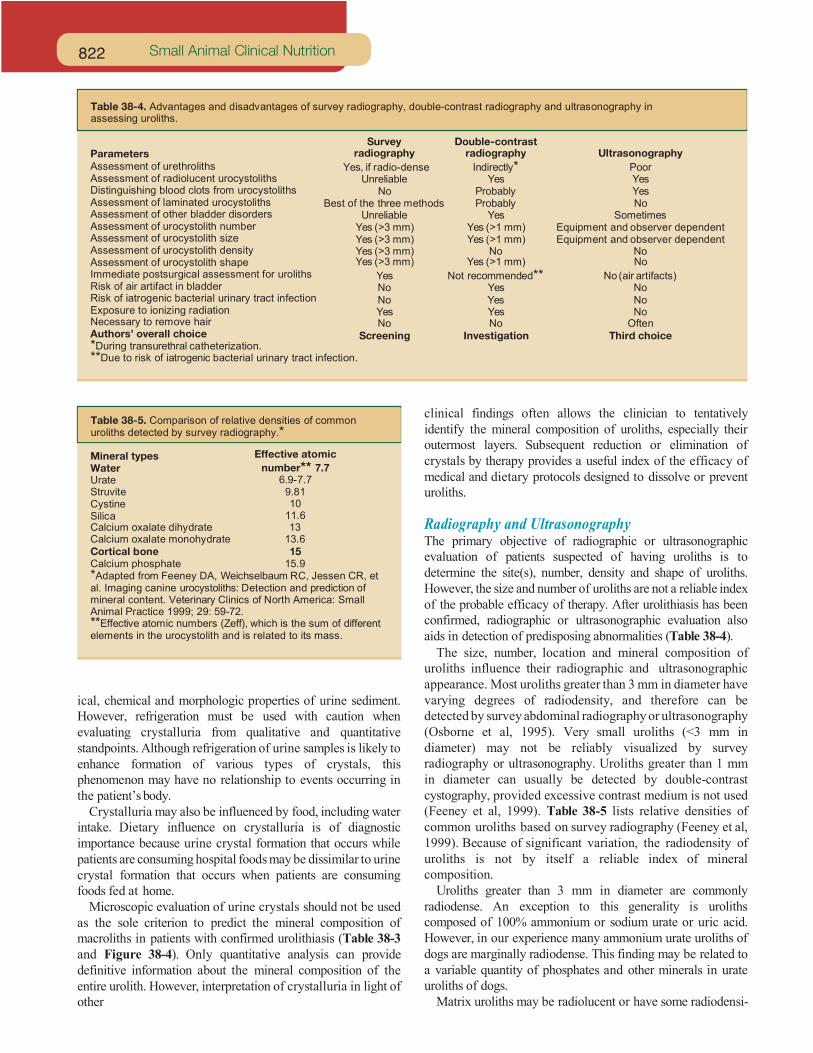
Radiography and Ultrasonography The primary objective of radiographic or ultrasonographic evaluation of patients suspected of having uroliths is to determine the site(s), number, density and shape of uroliths. However, the size and number of uroliths are not a reliable index of the probable efficacy of therapy. After urolithiasis has been confirmed, radiographic or ultrasonographic evaluation also aids in detection of predisposing abnormalities (Table 38-4).
The size, number, location and mineral composition of uroliths influence their radiographic and ultrasonographic appearance. Most uroliths greater than 3 mm in diameter have varying degrees of radiodensity, and therefore can be detected by survey abdominal radiography or ultrasonography (Osborne et al, 1995). Very small uroliths (<3 mm in diameter) may not be reliably visualized by survey radiography or ultrasonography. Uroliths greater than 1 mm in diameter can usually be detected by double-contrast cystography, provided excessive contrast medium is not used (Feeney et al, 1999). Table 38-5 lists relative densities of common uroliths based on survey radiography (Feeney et al, 1999). Because of significant variation, the radiodensity of uroliths is not by itself a reliable index of mineral composition.
Uroliths greater than 3 mm in diameter are commonly radiodense. An exception to this generality is uroliths composed of 100% ammonium or sodium urate or uric acid. However, in our experience many ammonium urate uroliths of dogs are marginally radiodense. This finding may be related to a variable quantity of phosphates and other minerals in urate uroliths of dogs.
Matrix uroliths may be radiolucent or have some radiodensi-
Table 38-4. Advantages and disadvantages of survey radiography, double-contrast radiography and ultrasonography in assessing uroliths.
Parameters Assessment of urethroliths Assessment of radiolucent urocystoliths Distinguishing blood clots from urocystoliths Assessment of laminated urocystoliths Assessment of other bladder disorders Assessment of urocystolith number Assessment of urocystolith size Assessment of urocystolith density Assessment of urocystolith shape Immediate postsurgical assessment for uroliths Risk of air artifact in bladder Risk of iatrogenic bacterial urinary tract infection Exposure to ionizing radiation Necessary to remove hair Authors’ overall choice *During transurethral catheterization.
Survey Double-contrast radiography radiography Ultrasonography
Yes, if radio-dense Indirectly* Poor Unreliable Yes Yes
No Probably Yes Best of the three methods Probably No
Unreliable Yes Sometimes Yes (>3 mm) Yes (>1 mm) Equipment and observer dependentYes (>3 mm) Yes (>1 mm) Equipment and observer dependentYes (>3 mm) No No Yes (>3 mm) Yes (>1 mm) No
Yes Not recommended** No (air artifacts) No Yes No No Yes No Yes Yes No No No Often
Screening Investigation Third choice
**Due to risk of iatrogenic bacterial urinary tract infection.
Table 38-5. Comparison of relative densities of common uroliths detected by survey radiography.*
Mineral types Water Urate Struvite Cystine Silica
Effective atomic number** 7.7
6.9-7.7 9.81 10
11.6 Calcium oxalate dihydrate 13 Calcium oxalate monohydrate 13.6 Cortical bone 15
*Adapted from Feeney DA, Weichselbaum RC, Jessen CR, etal. Imaging canine urocystoliths: Detection and prediction ofmineral content. Veterinary Clinics of North America: SmallAnimal Practice 1999; 29: 59-72.**Effective atomic numbers (Zeff), which is the sum of differentelements in the urocystolith and is related to its mass.
Calcium phosphate 15.9

Introduction to Canine Urolithiasis 823
ty. Blood clots are radiolucent and may be mistaken for radiolucent uroliths. Radiolucent uroliths may be readily distinguished from blood clots when evaluated by two-dimensional, grayscale ultrasonography. Uroliths are usually in the dependent portion of the bladder lumen, produce sharply marginated shadows containing few echoes and are associated with acoustic shadowing. Blood clots may be located anywhere in the bladder lumen, typically have an irregular outline and indistinct margins and are not associated with acoustic shadowing.
Uroliths that are radiodense on survey radiographs may appear to be radiolucent when evaluated by positive-contrast radiography. This finding is related to the fact that many uroliths are more radiodense than body tissue, but less radiodense than the contrast material. A diagnosis of radiolucent uroliths should be based on their radiodensity compared with soft tissues, and not their radiodensity compared with positive-contrast medium.
A urolith may be larger than that depicted by its radiodensity if only a portion of it contains radiodense minerals. This phenomenon is most likely to occur with rapidly growing struvite uroliths that contain large quantities of matrix.
Hematology and Serum Chemistry Hemograms of dogs with uroliths are usually normal unless there is concomitant generalized infection of the kidneys or prostate gland associated with leukocytosis. Microcytosis, anemia, target cells and leukocytosis have occasionally been associated with portal vascular anomalies in dogs with and without urate uroliths (Cornelius et al, 1975; Ewing et al, 1974; Griffiths et al, 1981; Rothuizen and van den Ingh, 1980).
Serum chemistry values are usually normal in patients with infection-induced magnesium ammonium phosphate, cystine and silica uroliths unless obstruction of urine outflow or generalized renal infection leads to changes characteristic of renal failure. Although most patients with calcium oxalate and calcium phosphate uroliths are normocalcemic, some are hypercalcemic.
Calcium phosphate and sterile struvite uroliths may be associated with distal renal tubular acidosis characterized by hyperchloremic (normal anion gap) metabolic acidosis, urinary pH values consistently greater than approximately 6 and hypokalemia.
A variety of biochemical alterations may exist in patients with urate urolithiasis. The following changes may be observed in patients with urate uroliths due to congenital or acquired hepatic disorders (Rothuizen and van den Ingh, 1980; Barrett et al, 1976; Marretta et al, 1981): 1) decreased urea nitrogen concentrations, 2) decreased total protein and albumin concentrations, 3) increased bile acid concentrations, 4) increased concentrations of total bilirubin and fasting blood ammonia and 5) increased serum alanine aminotransferase and serum alkaline phosphatase enzyme activities. Dogs with portal vascular anomalies typically have reduced hepatic functional mass and altered portal blood flow evidenced by abnormally elevated bile acid concentrations, prolonged sulfobromophthalein retention times and abnormal ammonia tolerance tests (Griffiths et al,
1981; Rothuizen and van den Ingh, 1980; Barrett et al, 1976; Marretta et al, 1981; Center et al, 1985).
Urine Chemistry Detection of the underlying causes of specific types of urolithiasis is often linked to evaluation of the biochemical composition of urine. For best results, at least one and preferably two consecutive 24-hour urine samples should be collected because determination of fractional excretion of many metabolites in “spot” urine samples may not accurately reflect 24-hour metabolite excretion (Table 38-6).
Water consumption and hydration status must be considered when interpreting laboratory results. Decreased water consumption and dehydration are associated with several alterations, including decreased renal clearance of metabolites and increased urine specific gravity and urine solute concentrations (Taburu et al, 1993). Caution must be used in interpreting 24-hour excretion of solutes in the diagnosis and therapy of urolithiasis if hospitalized animals consume less water than in the home environment.
Urine concentrations of potentially lithogenic metabolites are also influenced by the amount and composition of food consumed, and whether urine was collected during conditions of fasting or food consumption (Lulich et al, 1991, 1991a). Aldosterone secretion increases following food deprivation. Increased aldosterone secretion promotes renal tubular sodium reabsorption and potassium excretion. As a consequence, plasma potassium concentration decreases, urinary potassium excretion increases and urinary sodium and chloride excretion decrease (Lulich et al, 1991a). Urinary calcium, magnesium and uric acid excretions are reduced during fasting. However, urinary excretion of phosphorus, oxalate and citrate are apparently unaffected by fasting (Lulich et al, 1991a). In dogs, urinary ammonia, titratable acid and hydrogen ion excretion decrease and urinary pH values increase when food is withheld (Lulich et al, 1991a; Lemieux and Plante, 1968). Therefore, values for 24-hour urinary solute excretion may differ when measured following food consumption vs. values obtained when food is withheld.
Consumption of food stimulates gastric secretion of hydrochloric acid. As a result, concentrations of chloride decrease and bicarbonate increase in venous blood draining the stomach. Total serum concentration of carbon dioxide increases. The resulting metabolic alkalosis is commonly called the postprandial alkaline tide. Urinary pH will increase unless acidifying substances are contained in the food. In a study of healthy beagles, eating was associated with increased urinary excretion of hydrogen ions, ammonia, sodium, potassium, calcium, magnesium and uric acid (Lulich et al, 1991a).
Laboratory results may be markedly affected by changes in foods fed in a home environment vs. different foods fed in a hospital environment. For example, urinary excretion of potentially lithogenic metabolites while animals consume foods fed in the hospital may be different from those excreted by animals eating at home. To determine the influence of home-fed foods on laboratory test results, consider asking clients to bring

824 Small Animal Clinical Nutrition
Table 38-6. Protocol for measuring 24-hour urinary excretion of various substances associated with urolithiasis.
Technique 1. To allow for food acclimation, feed the patient either the food it was consuming just before urolith formation or a standard food at
home for 10 to 14 days. We commonly use Prescription Diet k/d Canine* as the standard food.2. If possible, house and feed the dog in the urine collection cage for at least one day before urine collection. As dogs become
acclimated to their new environment, they are more likely to consume quantities of food and water similar to that consumed in theirhome environment.
3. Begin each 24-hour urine collection period by removing urine from the urinary bladder by transurethral catheterization. This urine isdiscarded. Record the actual time that urine collection is initiated.
4. Weigh the dog.5. Then feed the dog its food as if at home. Water should be continuously available for consumption. 6. Begin administering a broad-spectrum antibiotic that achieves high concentrations in urine to prevent catheter-induced urinary tract
infection. The dosage, dosing interval and route of administration should be based on manufacturer recommendations.7. Keep the patient in the collection cage during urine collection. When using metabolism cages designed for urine collection,
catheterization of the urinary tract is unnecessary except at the end of the 24 hours. House-trained dogs may not voluntarily void intheir cage. Bladder catheterization may be necessary to obtain urine from these dogs. Dogs may be catheterized as often asnecessary to keep them comfortable (usually every six to eight hours).
8. Catheterize the urinary bladder at the end of 24 hours to remove all urine. Save this urine.9. Record the exact time of collection termination.
10. Pool all urine collected during the 24-hour period in a single container and measure its volume.11. Thoroughly mix the pooled urine before removing aliquots for analysis.
Preservation 1. Preservatives have different roles, but are often used to minimize bacterial growth, reduce chemical decomposition, solubilize
constituents that might otherwise precipitate out of solution, or decrease atmospheric oxidation of unstable compounds.2. The method of preservation may vary depending on the substances being measured and the tests used to measure them. Consult
the laboratory to determine the recommended method of preservation.3. Preservatives should not be added to some specimens because of possible interference with analytical methods.4. Refrigeration is a common method for preserving urine collected for analysis. Urine removed by intermittent catheterization can be
stored in a refrigerator in clean containers with screw top lids. Containers used for continuous collection beneath metabolism cagescan be surrounded by ice packs and then insulated. Refrigeration causes some minerals to precipitate out of solution.
5. Specimens can be acidified (add 10 ml of 1 N hydrochloric acid per liter to achieve a pH of 3 or less) to preserve oxalate and calciumfor analysis. However, acidified urine is unsuitable for measuring uric acid because it precipitates in acidic solutions.
Storage of selected analytes in urine 1. No single preservative is ideal if multiple substances in urine are to be analyzed. To minimize degradation, we routinely collect urine
under conditions of refrigeration. Immediately following urine collection, preservatives are added to appropriate aliquots of urine forstorage until analysis.
2. Uric acid and xanthine: Aliquots of urine should be diluted (1 ml of urine with 19 ml of distilled water) to preserve uric acid andxanthine. This mixture can then be frozen.
3. Ammonia: Aliquots of urine (3 to 5 ml) may be frozen for up to 30 days.4. Oxalate: Aliquots of urine (2 ml) are diluted with 1 N hydrochloric acid (1.66 ml) and then frozen.
Calculations 1. Calculating 24-hour urine volume
a. Although 24-hour urine specimens are recommended to minimize the effects of short-term biologic variations in mineral excretion, collecting perfectly timed 24-hour samples may be difficult. The following formula can be used to adjust actual urine volume to a24-hour period: 1,440 ÷ actual time interval (minutes) x urine volume (1,440 = number of minutes in 24 hours).
b. Example: A 24-hour urine collection was started at 9:30 a.m. and ended the following day at 8:30 a.m. A total of 350 ml of urinewere collected during this period. What is the 24-hour urine volume? 1,440 ÷ 1,380 x 350 = 356.2 ml.
2. Converting mmol/l to mg/dla. Scientists are striving to adopt a uniform system of measurement termed the System International d ‘Unites to standardize
measurements. In this system, concentration is often expressed as moles, millimoles or micromoles of a substance per liter offluid. Most normal values in the United States are expressed as mg/dl. The following formula can be used to convert mmol/l tomg/dl: mmol/l x atomic weight of substance ÷ 10. The atomic weights of elements can be found in the periodic tables of generalchemistry books.
b. Example: The concentration of calcium from a 24-hour urine sample was 1.35 mmol/l. Convert this value to mg/dl for comparisonwith normal values. The atomic weight of calcium is 40.08. 1.35 mmol/l x 40.08 ÷ 10 = 5.4 mg/dl.
3. Calculating mg/kg/24-hour or mEq/kg/24-hour excretiona. Excretion of metabolites is often expressed on a per kg basis to standardize excretion for dogs of different weights. The following
formula can be used to standardize excretion rates: Concentration of substance x 24-hour urine volume ÷ body weight in kg. Theunits used to express the volume of urine and the concentration of the substance evaluated must be the same.
b. Example: The concentration of calcium in a 24-hour urine sample was 5.4 mg/dl. A total of 356.2 ml of urine was collected. Thedog weighed 10 kg. What is the daily calcium excretion on a per kg basis? First, express the volume of urine collected in the sameunits as the concentration of the substance measured. The 356.2 ml = 3.562 dl; therefore, 5.4 mg/dl x 3.562 dl ÷ 10 kg = 1.92mg/kg/24 hours.
Additional considerations 1. Midpoint blood samples. Evaluation of blood during the midpoint of a 24-hour urine collection may help determine if changes in urine
concentration reflect changes in serum or plasma concentration of analytes. This information can help detect underlying causes andmechanisms of abnormal mineral excretion. Likewise, evaluation of blood concentrations of some hormones (i.e., parathyroidhormone, calcitriol, etc.) may be helpful in determining the role of hormones in the regulation of mineral excretion.

Introduction to Canine Urolithiasis 825
Table 36-6 continued
2. Antimicrobialsa. Antimicrobials are administered to prevent iatrogenic urinary tract infection during catheterization of the urinary bladder. The dosage,
dosing interval and route should be based on the recommendations of the manufacturer. We recommend that antimicrobialadministration be continued for three to five days after urine collection. This represents the time required for normal urothelial repair and replacement.
b. Select antimicrobials that are primarily excreted in the urine and that minimally affect urine concentrations of minerals, promoters and inhibitors associated with urolith formation. Because some antimicrobials are formulated as salts of sodium or potassium, highconcentrations of sodium or potassium may be excreted in urine. We routinely use cephalosporins when collecting urine for aminoacid evaluation (i.e., cystine urolithiasis), and ampicillin when collecting urine from dogs with calcium oxalate or urate uroliths.
3. Fasting urine collectiona. Fasting urine collections have been evaluated to characterize the pathophysiologic mechanism of hypercalciuria in dogs with
calcium oxalate uroliths. Dogs that absorb excessive amounts of calcium from their food and subsequently excrete large quantitiesof calcium in their urine have intestinal hypercalciuria. Hypercalciuria primarily occurs during food consumption; normal or lowerquantities of urine calcium are excreted when food is withheld. In addition, dogs with intestinal hypercalciuria have normal serumconcentrations of calcium and normal or low serum concentrations of parathyroid hormone. In contrast, urinary calcium excretion during fed and nonfed conditions is similar in dogs with primary hyperparathyroidism (resorptive hypercalciuria) or impaired renal tubular absorption of calcium (renal-leak hypercalciuria).
b. Fasting urine collections are initiated immediately following collection of urine during standard feeding.4. Urinary pH. The solubility of mineral salts is influenced by urinary pH. Determination of urinary pH from 24-hour urine samples may be
helpful in understanding crystal formation, and can also be used to calculate activity products for several mineral salts commonlyfound in uroliths. We use an ion selective electrode and pH meter to accurately measure urinary pH.
5. Activity productsa. The activity product of urine is a mathematical expression used to estimate the degree of saturation of urine with mineral salts.
Activity products are calculated by measuring concentrations of major ionizable solutes in urine. For efficiency, computer programsare commonly used to aid in calculating activity products.
b. Urine in which the activity product exceeds the solubility product is saturated for that particular mineral salt. Although crystals maynot form at this degree of saturation, uroliths already present are likely to grow.
c. Urine in which the activity product exceeds the formation product for a particular mineral salt is associated with an unstable state ofoversaturation. Crystal nucleation and rapid crystal growth are likely at this urine concentration.
*Hill’s Pet Nutrition, Inc., Topeka, KS, USA.
home-fed foods for use during periods of diagnostic hospitalization (Osborne et al, 1990).
Urolith Analysis Small uroliths in the urinary bladder or urethra are commonly voided during micturition by female dogs and occasionally by male dogs. Uroliths with a smooth surface (e.g., those composed of ammonium urate or calcium oxalate monohydrate) are more likely to pass through the urethra than uroliths with a rough surface (e.g., those composed of calcium oxalate dihydrate or silica). Commercially manufactured tropical fish nets designed for household aquariums facilitate retrieval of uroliths during voiding (Osborne et al, 1992). They are much less expensive than collection cups with wire mesh bottoms designed for people and available from medical supply houses. Urocystoliths may also be obtained by voiding urohydropropulsion (Figure 38-5 and Table 38-7). If the unaided eye can detect a urolith, it will usually be sufficient size for quantitative analysis.
CATHETER-ASSISTED RETRIEVAL OF UROCYSTOLITHS
Small urocystoliths may be retrieved for analysis by aspirating them through a urethral catheter into a syringe (Osborne et al, 1992; Lulich and Osborne, 1992). Urocystoliths detected by survey radiography may be too large to be removed with the aid of a urethral catheter. However, large urocystoliths are often associated with small ones that may be detected by double-contrast cystography. The diameter of uroliths retrieved is limited by the size of openings or “eyes” in the proximal portion of the
Figure 38-5. To remove urocystoliths by voiding urohydropropulsion, position the patient so that its vertebral column is approximately vertical (Left). The urinary bladder is then gently agitated to promote gravitational movement of urocystoliths into the bladder neck. To expel urocystoliths (Right), voiding is induced by applying steady digital pressure to the urinary bladder. (Adapted from Lulich JP, Osborne CA, Carlson M, et al. Nonsurgical removal of uroliths from dogs and cats by voiding urohydropropulsion. Journal of the American Veterinary Medical Association 1993; 203: 660-663.)
catheter and by the diameter of the catheter lumen. It is best to select the largest-diameter catheter that can be advanced into the bladder lumen without traumatizing the urethral mucosa.
826 Small Animal Clinical Nutrition
Well-lubricated, soft, flexible catheters are preferable to less flexible ones. The size of openings in the proximal portion of the catheter may be enlarged with a scalpel, razor blade or scissors to facilitate retrieval of urocystoliths. However, care must be used not to weaken the catheter to the point where it could break while being inserted into or removed from the urethra and urinary bladder.
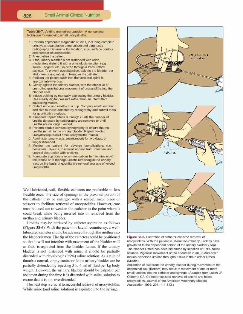
Uroliths may be retrieved by catheter aspiration as follows (Figure 38-6). With the patient in lateral recumbency, a well-lubricated catheter should be advanced through the urethra into the bladder lumen. The tip of the catheter should be positioned so that it will not interfere with movement of the bladder wall as fluid is aspirated from the bladder lumen. If the urinary bladder is not distended with urine, it should be partially distended with physiologic (0.9%) saline solution. As a rule of thumb, a normal, empty canine or feline urinary bladder can be partially distended by injecting 3 to 4 ml of fluid per kg body weight. However, the urinary bladder should be palpated per abdomen during the time it is distended with saline solution to ensure that it is not overdistended.
The next step is crucial to successful retrieval of urocystoliths. While urine (and saline solution) is aspirated into the syringe,
Figure 38-6. Illustration of catheter-assisted retrieval of urocystoliths. With the patient in lateral recumbency, uroliths have gravitated to the dependent portion of the urinary bladder (Top). The bladder lumen has been distended by injection of 0.9% saline solution. Vigorous movement of the abdomen in an up-and-down motion disperses uroliths throughout fluid in the bladder lumen (Middle). Aspiration of fluid from the urinary bladder during movement of the abdominal wall (Bottom) may result in movement of one or more small uroliths into the catheter and syringe. (Adapted from Lulich JP, Osborne CA. Catheter assisted retrieval of canine and feline urocystoliths. Journal of the American Veterinary Medical Association 1992; 201: 111-113.)
Table 38-7. Voiding urohydropropulsion: A nonsurgical technique for removing small urocystoliths.
1. Perform appropriate diagnostic studies, including completeurinalysis, quantitative urine culture and diagnosticradiography. Determine the location, size, surface contourand number of urocystoliths.
2. Anesthetize the patient.3. If the urinary bladder is not distended with urine,
moderately distend it with a physiologic solution (e.g.,saline, Ringer’s, etc.) injected through a transurethral catheter. To prevent overdistention, palpate the bladder perabdomen during infusion. Remove the catheter.
4. Position the patient such that the vertebral spine isapproximately vertical.
5. Gently agitate the urinary bladder, with the objective ofpromoting gravitational movement of urocystoliths into thebladder neck.
6. Induce voiding by manually expressing the urinary bladder.Use steady digital pressure rather than an intermittentsqueezing motion.
7. Collect urine and uroliths in a cup. Compare urolith numberand size to those detected by radiography and submit themfor quantitative analysis.
8. If needed, repeat Steps 3 through 7 until the number ofuroliths detected by radiography are removed or untiluroliths are no longer voided.
9. Perform double-contrast cystography to ensure that nouroliths remain in the urinary bladder. Repeat voidingurohydropropulsion if small urocystoliths remain.
10. Administer prophylactic antimicrobials for two days, orlonger if needed.
11. Monitor the patient for adverse complications (i.e.,hematuria, dysuria, bacterial urinary tract infection andurethral obstruction with uroliths).
12. Formulate appropriate recommendations to minimize urolith recurrence or to manage uroliths remaining in the urinarytract on the basis of quantitative mineral analysis of voidedurocystoliths.
Introduction to Canine Urolithiasis 827
an assistant should vigorously and repeatedly move the patient’s abdomen in an up-and-down motion. This maneuver disperses uroliths located in the dependent portion of the bladder throughout fluid in the bladder lumen. Small uroliths in the vicinity of the catheter tip may then be aspirated into the catheter along with the urine-saline mixture. It may be necessary to repeat this sequence of steps several times before a sufficient number of uroliths are retrieved. The bladder lumen should be redistended with saline solution each time. Difficulty in aspirating urine and saline solution into the syringe may be caused by poor positioning of the catheter tip or by partial occlusion of the catheter lumen with one or more uroliths. Flushing saline solution through the catheter after it has been removed from the patient often results in the retrieval of uroliths that occlude the catheter lumen.
Care must be used not to overdistend the urinary bladder with saline solution because this will increase the space in which the uroliths are suspended. Because patients with uroliths are predisposed to catheter-induced bacterial UTIs, antimicrobial therapy should be considered immediately before this procedure and for an appropriate period afterward. Proper selection, insertion and positioning of urethral catheters minimize iatrogenic trauma to the lower urinary tract.
COLLECTION AND QUANTITATIVE ANALYSIS OF URINE CRYSTALS If available data do not indicate the probable mineral
composition of uroliths and if uroliths cannot be retrieved with the aid of a urethral catheter, consider preparing a large pellet of urine crystals by centrifugation of urine in a conical-tip centrifuge tube (Osborne et al, 1992, 1995). The quantity of crystalline sediment available for analysis may be increased by repeatedly removing the supernatant after centrifugation, adding additional noncentrifuged urine to the tube containing sediment and again centrifuging the preparation. If the conditions that caused urolith formation are still present, evaluation of the pellet formed from crystalline sediment by quantitative methods designed for urolith analysis may provide meaningful information about the mineral composition of a patient’s uroliths. However, crystals identified by this method may only reflect the outer portions of compound uroliths. Therefore, results of quantitative urine crystal analysis should be interpreted in conjunction with other pertinent clinical data.
QUANTITATIVE ANALYSIS OF UROLITHS The location, number, size, shape, color and consistency of
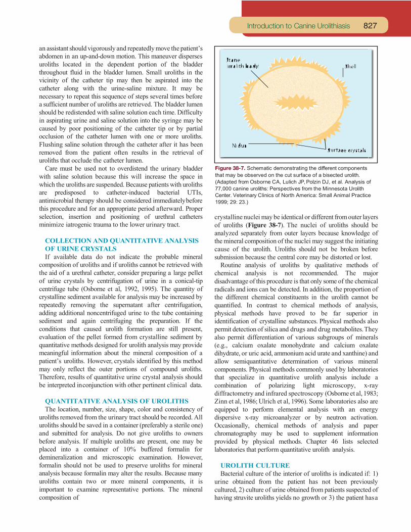
uroliths removed from the urinary tract should be recorded. All uroliths should be saved in a container (preferably a sterile one) and submitted for analysis. Do not give uroliths to owners before analysis. If multiple uroliths are present, one may be placed into a container of 10% buffered formalin for demineralization and microscopic examination. However, formalin should not be used to preserve uroliths for mineral analysis because formalin may alter the results. Because many uroliths contain two or more mineral components, it is important to examine representative portions. The mineral composition of
Figure 38-7. Schematic demonstrating the different components that may be observed on the cut surface of a bisected urolith. (Adapted from Osborne CA, Lulich JP, Polzin DJ, et al. Analysis of 77,000 canine uroliths: Perspectives from the Minnesota Urolith Center. Veterinary Clinics of North America: Small Animal Practice 1999; 29: 23.)
crystalline nuclei may be identical or different from outer layers of uroliths (Figure 38-7). The nuclei of uroliths should be analyzed separately from outer layers because knowledge of the mineral composition of the nuclei may suggest the initiating cause of the urolith. Uroliths should not be broken before submission because the central core may be distorted or lost.
Routine analysis of uroliths by qualitative methods of chemical analysis is not recommended. The major disadvantage of this procedure is that only some of the chemical radicals and ions can be detected. In addition, the proportion of the different chemical constituents in the urolith cannot be quantified. In contrast to chemical methods of analysis, physical methods have proved to be far superior in identification of crystalline substances. Physical methods also permit detection of silica and drugs and drug metabolites. They also permit differentiation of various subgroups of minerals (e.g., calcium oxalate monohydrate and calcium oxalate dihydrate, or uric acid, ammonium acid urate and xanthine) and allow semiquantitative determination of various mineral components. Physical methods commonly used by laboratories that specialize in quantitative urolith analysis include a combination of polarizing light microscopy, x-ray diffractometry and infrared spectroscopy (Osborne et al, 1983; Zinn et al, 1986; Ulrich et al, 1996). Some laboratories also are equipped to perform elemental analysis with an energy dispersive x-ray microanalyzer or by neutron activation. Occasionally, chemical methods of analysis and paper chromatography may be used to supplement information provided by physical methods. Chapter 46 lists selected laboratories that perform quantitative urolith analysis.
UROLITH CULTURE Bacterial culture of the interior of uroliths is indicated if: 1)
urine obtained from the patient has not been previously cultured, 2) culture of urine obtained from patients suspected of having struvite uroliths yields no growth or 3) the patient has a

828 Small Animal Clinical Nutrition
Table 38-8. Mineral composition of 339886 canine uroliths evaluated at the Minnesota Urolith Center by quantitative methods:2012 to 2016.
Proportion of Predominant mineral type predominant mineral (%) Number Percent Magnesium ammonium
phosphate•6H2O 70-100* 127,063 37 Calcium oxalate 70-100 141618 42
Calcium phosphate 70-100 2213 1
Brushite 70-100 881 <1
Purines - 13774 4
Cystine 70-100 7357 2 Silica 70-100 1,354 <1
Other - 203 <1
Mixed** - 11231 3 Compound*** - 34192 10 *Urolith composed of 70 to 100% of mineral type listed; no nucleus and shell detected.**Uroliths did not contain at least 70% of mineral type listed; no nucleus or shell detected.***Uroliths contained an identifiable nucleus and one or more surrounding layers of a different mineral type.
UTI with bacteria that do not produce urease. Bacteria harbored inside uroliths are not always the same as those present in urine. Bacteria detected within uroliths probably represent those present at the time they were formed, and may serve as a source of recurrent UTI. Bacteria may remain viable within uroliths for long periods. In a pilot study, we cultured viable staphylococci from struvite uroliths removed from a miniature schnauzer up to three months following surgery. If all the uroliths have not been removed from the patient, knowledge of the type and antimicrobic susceptibility of bacteria inside uroliths that have been voided or removed may be of therapeutic significance. Procedures for culture of microbes from the inner portions of uroliths have been developed and can be performed by most veterinary diagnostic laboratories (Osborne et al, 1983; Ruby and Ling, 1986).
GUESSTIMATION OF UROLITH COMPOSITION Formulation of effective dietary and medical protocols for urolith dissolution depends on knowledge of the mineral composition of uroliths. Because a variety of different types of uroliths and nephroliths occur in dogs (Tables 38-8 and 38-9), veterinarians should use a protocol that facilitates determination of the mineral composition of uroliths based on probability (Tables 38-10 and 38-11). Formulation of dietary and medical therapy based on the mineral composition of uroliths determined by this protocol is usually associated with a high degree of success in dissolving uroliths or arresting their growth.
Attempts to induce dissolution of uroliths may be hampered if the uroliths are heterogeneous in composition. This has not been a significant problem in dogs with uroliths composed primarily of magnesium ammonium phosphate with small quantities of calcium phosphate carbonate and calcium apatite because the solubility characteristics of these minerals are similar. Veterinarians have encountered difficulty in dissolving uroliths composed primarily of struvite with an outer shell composed primarily of calcium apatite. Difficulty will also be encountered in attempting to induce complete dissolution of a urolith with a nucleus of calcium oxalate, calcium phosphate, ammonium urate or silica and a shell of struvite because the solubility characteristics of this combination of minerals are dissimilar. These phenomena should be considered if dietary and medical therapy seems to be ineffective after initially reducing the size of uroliths (Box 38-2).
REFERENCES
The references for Chapter 38 can be found at www.markmorris.org.

Introduction to Canine Urolithiasis 829
Table 38-9. Mineral composition of 5,050 canine nephroliths and ureteroliths evaluated at the Minnesota Urolith Center by quantitative methods: 1991 to 2006.
Proportion of Predominate mineral type predominant mineral (%) Number Percent Calcium oxalate - 2,174 43.1 Calcium oxalate monohydrate 100 (1,168) -
70-99 (511) - Calcium oxalate dihydrate 70-100* (234) - Calcium oxalate monohydrate and dihydrate 70-100 (161) - Calcium phosphate - 81 1.6 Calcium apatite 70-100 (67) - Calcium hydrogen phosphate•2H2O 70-100 (14) - Magnesium ammonium phosphate•6H2O 70-100 1,299 25.7 Purines - 671 13.3 Ammonium acid urate 70-100 (476) - Sodium acid urate 70-100 (47) - Sodium calcium urate 70-100 (17) - Uric acid 70-100 104 - Xanthine 70-100 (24) - Cystine 70-100 77 - Silica 70-100 18 0.4 Mixed** - 132 2.6 Compound*** - 575 11.4 Matrix - 13 0.3 Drug metabolite na 1 0.02 Other na 7 0.1 Key: na = not applicable. *Uroliths composed of 70 to 100% of mineral type listed; no nucleus and shell detected.**Uroliths contained less than 70% of predominant mineral; no nucleus or shell detected.***Uroliths contained an identifiable nucleus and one or more surrounding layers of a different mineral type.
Table 38-10. Problem-specific and therapeutic-specific database for diagnosis and management of urolithiasis.
1. Obtain appropriate history and perform physical examination, including a rectal examination of the urethra.2. Perform complete urinalysis; save aliquots of urine for possible determination of mineral concentration.3. Obtain quantitative urine culture and determine urease activity; test for antimicrobial susceptibility if bacterial pathogens are identified.
Consider attempts to isolate ureaplasmas if urease-positive urine is bacteriologically sterile.4. Perform a complete blood cell count.5. Freeze an aliquot of serum collected at the time the sample was obtained for the complete blood cell count for possible determination
of urea nitrogen, creatinine, calcium and/or uric acid concentrations.6. Obtain radiographs.
a. Take survey radiographs of the entire urinary system.b. Consider intravenous urography for patients with renal or ureteral uroliths.c. Consider intravenous urography or contrast cystography for patients with bladder uroliths.d. Consider contrast urethrography for patients with urethral uroliths.e. Ultrasonography or CT is recommended in patients with radiolucent uroliths or ureteral obstruction.
7. Determine mineral composition of uroliths.a. Submit uroliths passed during micturition or retrieved during diagnostic procedures for quantitative analysis.b. Use results obtained from the history, physical examination, laboratory examination and radiography to determine probable mineral
composition of uroliths.8. Initiate therapy to eradicate urinary tract infection, if present.9. Initiate therapy for urolithiasis.
a. Initiate therapy to promote dissolution of uroliths if amenable to dietary and medical therapy.Formulate followup protocol to monitor dissolution of uroliths. Formulate alternative treatment options if uroliths do not dissolve or if problems such as recurrent outflow obstruction occur. b. Remove uroliths by voiding urohydropropulsion.c. Remove uroliths by nephrotomy or cystotomy.Obtain bladder or kidney biopsy specimens for microscopic examination during surgical procedure.Correct any anatomic defects, if present.Compare number of uroliths removed during surgery with the number identified by radiography.Obtain postprocedural radiographs to evaluate completeness of urolith removal.Submit uroliths for quantitative analysis.
10. After uroliths are removed or medically dissolved, initiate therapy to prevent recurrence.11. Formulate followup protocol with clients.

830 Small Animal Clinical Nutrition
Box 38-2. Principles of Urolith Treatment and Prevention.
Almost every urinary stone can be managed by surgery. However, the role of the clinician is to determine if surgery is the best option for care after considering the nature of the problem, the feasibility of the procedure (e.g. laser lithotripsy in a small male dog may not be feasible because the urethral is too narrow to accommodate a cystoscope), and the wishes of the client. Although each case is individual in its needs and approach, there are several considerations that can be applied universally.
1. Therapy should not be initiated before appropriate samples have been collected for diagnosis (e.g. urinalysis, urine culture, medical imaging, genetic testing, others) of the disease and its underlying causes.
2. Stones with a high success of dietary/medical dissolution (i.e. struvite) should be dissolved medically. Unless the patient is obstructed, dissolution is the first choice. Results of experimental and clinical studies have revealed that the size and number of uroliths do not dictate the likelihood of response to therapy. We have successfully dissolved uroliths that are small and large, single and multiple. However, the rate of dissolution is related to size and surface area of the urolith exposed to urine. Difficulty in inducing complete dissolution by creating urine that is undersaturated with the suspected lithogenic crystalloid should prompt consideration that: 1) the wrong mineral component was identified, 2) the nucleus of the urolith is of different mineral composition than outer portions and/or 3) the owner or the patient is not complying with therapy. In asymptomatic dogs with urate and cystine uroliths, medical dissolution should be considered first, although dissolution of these mineral types is less predictable, the cost and complications of removal are often higher.
3. Bladder stones of sufficiently small size (i.e. those that would easily pass through the urethra) should be removed nonsurgically by spontaneous voiding, voiding urohydropropulsion or basket retrieval.
However, therapy to minimize urolith growth, may delay intervention. This is especially true for highly recurrent uroliths whose underlying cause and risks are difficult to ameliorate.
5. Complete obstruction is an emergency. Restoring urine flow preserves renal function, eliminates pain, and minimizes spread of infection, when present. In most cases, stones will need to be removed (surgery, lithotripsy, laparoscopy, others)
6. Avoid procedures that permanently alter and impair urinary tract function without considering their absolute necessity. Instead of Urethral urethrostomy or urethrotomy) consider retrograde urohydropropulsion to reposition stones in the bladder for removal or laser lithotripsy to fragment urethroliths.
7. Minimize injury to healthy tissues. Instead of routine cystotomy consider dissolution, transurethral laser lithotripsy or percutaneous cystolithotomy. Avoiding or reducing surgery is associated with reduced infection, reduced pain, shorter hospitalization, faster return to function, fewer complications and less scarring.
UROLITH REMOVALDetection of uroliths is not in itself an indication for removal. However, along with dietary and medical management, nonsurgical and surgixcal intervention play important roles in therapy of urolithiasis.
PREVENTION OF UROLITH RECURRENCEUroliths tend to recur. Prevention of recurrent uroliths, which reduces the need for dietary and medical therapy and surgery, is therefore cost effective. In general, preventive strategies are designed to eliminate or control the underlying causes of various types of uroliths. When causes cannot be identified, preventive strategies encompass efforts to minimize risk factors associated with lithogenesis. These strategies commonly include dietary modification.
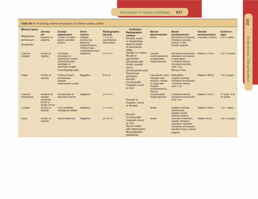
Introduction to Canine Urolithiasis 831
Table 38-11. Predicting mineral composition of common canine uroliths.
Mineral types Urinary Crystal Urine Radiographic
Predictors Radiographic contour Smooth, round or faceted May assume shape of renal pelvis, ureter, bladder or urethra Rough or speculated (dihydrate salt) Small, smooth, round (monohydrate salt) Sometimes jackstone Smooth (occasionally irregular) round or oval
Smooth or irregular, round or faceted
Smooth (occasionally irregular) round or oval Round center with radial spoke-like projections (jackstone)
Serum Breed Gender Common pH appearance culture density abnormalities predisposition predisposition agesNeutral to Three- to eight- Urease- 1+ to 4+ None Miniature schnauzer, Females (>80%) 2 to 9 years
ammonium alkaline sided colorless producing (sometimes miniature poodle,prisms bacteria laminated) Bichon Frise,
(staphylococci, cocker spanielProteus spp.,Ureaplasma spp.)
Calcium Acidic to Colorless Negative 2+ to 4+ Usually Miniature schnauzer, Males (>70%) 5 to 12 yearsoxalate neutral envelope or normocalcemic, standard schnauzer,
octahedral shape occasionally Lhasa apso,(dihydrate salt) hypercalcemic Yorkshire terrier,Spindles or miniature poodle,dumbbell shape Shih Tzu,(monohydrate salt) Bichon Frise
Urate Acidic to Yellow-brown Negative 0 to 2+ Low serum urea Dalmatian, Males (>90%) 1 to 5 years neutral amorphous nitrogen and English bulldog,
shapes albumin values miniature schnauzer,(ammonium urate) In dogs with Yorkshire terrier,
hepatic Shih Tzuportosystemicshunts
Calcium Alkaline to Amorphous or Negative 2+ to 4+ Occasionally Yorkshire terrier, Males (>55%) <1 year, 6 tophosphate neutral long thin prisms hypercalcemic miniature schnauzer, 10 years
(brushite Shih Tzuforms inacidic urine)
Cystine Acidic to Flat colorless Negative 1+ to 2+ None English bulldog, Males (>98%) 1 to 7 yearsneutral hexagonal plates dachshund,
basset hound,Newfoundland
Silica Acidic to None observed Negative 2+ to 3+ None German shepherd, Males (95%) 3 to 10 yearsneutral golden retriever,
Labrador retriever,miniature schnauzer,cavalier King Charlesspaniel
832 S
mall A
nim
al C
linic
al Nutritio
n
Magnesium
phosphate