Definition of acute kidney injury (acute renal failure) file · Web viewAcute Kidney Injury and...
26
Acute Kidney Injury and Chronic Kidney Disease I. Contributor : dr. Atma Gunawan SpPD.KGH II. Learning objective At the end of this module, student should be able to know : AKI 1. definition of AKI 2. criteria of AKI 3. the causes of AKI 4. pathogenesis of AKI 5. clinical manifestations of AKI 6. diagnosing AKI 7. prevention and management of AKI CKD 1. definition of CKD 2. criteria of CKD 3. stage of CKD 4. the causes of CKD 5. natural history of CKD 6. management to delay declining renal function 7. treatment complications of CKD 8. indications for renal replacement therapy 1
Transcript of Definition of acute kidney injury (acute renal failure) file · Web viewAcute Kidney Injury and...
Acute Kidney Injury and Chronic Kidney Disease
I. Contributor : dr. Atma Gunawan SpPD.KGH
II. Learning objective
At the end of this module, student should be able to know :
AKI
1. definition of AKI2. criteria of AKI
3. the causes of AKI
4. pathogenesis of AKI
5. clinical manifestations of AKI
6. diagnosing AKI
7. prevention and management of AKI
CKD
1. definition of CKD2. criteria of CKD
3. stage of CKD
4. the causes of CKD
5. natural history of CKD
6. management to delay declining renal function
7. treatment complications of CKD
8. indications for renal replacement therapy
9. indications for referral to nephrologist
III. Method
This module is designed for medical students at six semester, and will be processed by combines lectures and small group discussion.
1

IV. Overview
Acute Kidney Injury (AKI/ARF)
INTRODUCTION — Acute renal failure (ARF) has traditionally been defined as the abrupt loss of kidney function that results in the retention of urea and other nitrogenous waste products and in the dysregulation of extracellular volume and electrolytes. The loss of kidney function is most easily detected by measurement of the serum creatinine which is used to estimate the glomerular filtration rate (GFR).
The lack of consensus in the quantitative definition of ARF, in particular, has hindered clinical research since it confounds comparisons between studies. The Acute Dialysis Quality Initiative (ADQI) was created by a group of expert intensivists and nephrologists to develop consensus and evidence based guidelines for the treatment and prevention of acute renal failure. Recognizing the need for a uniform definition for ARF, the ADQI group proposed a consensus graded definition, called the RIFLE criteria. A modification of the RIFLE criteria was subsequently proposed by the Acute Kidney Injury Network (AKIN), which included the ADQI group as well as representatives from other nephrology and intensive care societies .
Because of these initiatives, the term acute kidney injury (AKI) was proposed to represent the entire spectrum of acute renal failure.
AKIN CRITERIA — The AKIN (Acute Kidney Injury Network), which representatives from nephrology and intensive care societies, proposed diagnostic criteria for ARF and a staging system. The proposed diagnostic criteria for ARF are an abrupt (within 48 hours) absolute increase in the serum creatinine concentration of ≥ 0.3 mg/dL (26.4 micromol/L) from baseline, a percentage increase in the serum creatinine concentration of ≥ 50 percent, or oliguria of less than 0.5 mL/kg per hour for more than six hours. The addition of an absolute change in serum creatinine of ≥ 0.3 mg/dL is based on epidemiologic data that have demonstrated an 80 percent increase in mortality risk associated with changes in serum creatinine concentration of as little as 0.3 to 0.5 mg/dL. Including a time constraint of 48 hours is based upon data that showed that poorer outcomes were associated with small changes in the creatinine when the rise in creatinine was observed within 24 to 48 hours Two additional caveats were proposed by the AKIN group: The diagnostic criteria should be applied only after volume status had been optimized Urinary tract obstruction needed to be excluded if oliguria was used as the sole diagnostic criterion.
2

A flaw with the last caveat is that, according to the current definition, AKI would still be used to describe the patient with acute urinary tract obstruction and an acute increase in serum creatinine.
Classification/staging system for acute kidney injury (AKIN criteria)*
1 . Increase in serum creatinine of more than or equal to 0.3 mg/dL (26.4 mircomol/L) or increase to more than or equal to 150 to 200 percent (1.5- to 2-fold) from baseline or urine output less than 0.5 mL/kg per hour for more than 6 hours.
2 . Increase in serum creatinine to more than 200 to 300 percent (>2- to 3-fold) from baseline or urine output less than 0.5 mL/kg per hour for more than 12 hours.
3 . Increase in serum creatinine to more than 300 percent (>3-fold) from baseline (or serum creatinine of more than or equal to 4.0 mg/dL [354 micromol/L] with an acute increase of at least 0.5 mg/dL [44 micromol/L]) or urine output less than 0.3 mL/kg per hour for 24 hours or anuria for 12 hours.
* The diagnostic criteria should only be applied after volume status has been optimized. Only one criterion (creatinine or urine output) has to be fulfilled to qualify for a stage.
200 to 300 percent increase = 2- to 3-fold increase.
PATHOGENESIS AND ETIOLOGY — The causes of acute renal disease can be related to the renal anatomy most affected by the disorder as follows: Vascular, Glomeruli ,Renal tubule, Urinary tract .Any process that interferes with any of these structures and/or functions can cause renal disease.The causes of AKI can therefore be categorized as prerenal, renal, or postrenal.
Prerenal — Prerenal azotemia results from either: Volume depletion due to bleeding (surgery, trauma, gastrointestinal bleeding), gastrointestinal (vomiting, diarrhea), urinary (diuretics, diabetes insipidus), or cutaneous losses (burns). Decreased effective arterial pressure and/or effective circulating volume seen in heart failure, shock, or cirrhosis
Intrinsic renal disorders — Intrinsic renal disease includes disorders that involve the renal vascular, glomerular, and/or tubular/interstitial pathology.
Vascular — Vascular causes of AKI include thrombosis (arterial and venous), hemolytic-uremic syndrome, malignant hypertension, and vasculitis.
3

Glomerular — The principal glomerular cause of AKI is acute glomerulonephritis, which is commonly postinfectious. AKI can be observed with most of the glomerulonephritides that can occur in childhood. Tubular and interstitial disease — Acute tubular necrosis (ATN) results from ischemia due to decreased renal perfusion or injury from tubular nephrotoxins. All causes of prerenal azotemia can progress to ATN if renal perfusion is not restored and/or nephrotoxic insults are not withdrawn
The administration of nephrotoxic agents, including aminoglycosides,methicillin, beta-lactam, amphotericin B, and contrast agents, is a common cause of tubular disease. Currently, the most common drug causes of AIN include: NSAIDs, including selective COX-2 inhibitors. Penicillins and cephalosporins Rifampin Sulfonamides, including trimethoprim-sulfamethoxazole and, much less often, furosemide, bumetanide, thiazide-type diuretics, Ciprofloxacin and, Cimetidine, Allopurinol, Proton pump inhibitors.
Postrenal — Postrenal AKI is due to bilateral urinary tract obstruction unless there is a solitary kidney. In neonates, urinary tract obstruction, due to posterior urethral valves is the most common cause of postrenal failure.
Causes of AKI — Seven studies primarily from Asia (India [3 reports], New Zealand [1 trial], Singapore [1 trial]) that reported the following were the most common causes of ARF ranked in order of incidence: Acute tubular necrosis (ATN), (23 percent) Hemolytic uremic syndrome (HUS), (21 percent) Glomerulonephritis (13 percent) Intrinsic renal disease (9 percent), causes not specified Postoperative (7 percent) Sepsis (6 percent) Ischemia/prerenal (4.5 percent) Urinary tract obstruction (3 percent) Miscellaneous causes (13.8 percent) including metabolic disorders, renal venous thrombosis, hepatorenal syndrome, complication of organ transplantation.
CLINICAL PRESENTATION — A careful history and physical examination can frequently identify events and/or disease processes that underlie AKI and suggest an underlying diagnosis:
A history of vomiting, diarrhea, hemorrhage, sepsis and/or decreased oral intake resulting in hypovolemia, associated with decreased urine output suggests AKI due to prerenal disease or ATN. Physical examination findings that include tachycardia, dry mucous membranes, sunken eyes, orthostatic blood pressure changes, and decreased skin turgor suggest hypovolemia, resulting in AKI due to prerenal disease or ATN.
Bloody diarrhea with oliguria (defined as less than 500 mL/1.73 m2 per day in children and less than 0.5 mL/kg per hour in infants) or anuria (absent urine) is consistent with the hemolytic-uremic syndrome.
4

A history of pharyngitis or impetigo, a few weeks prior to the onset of gross hematuria suggests post-infectious glomerulonephritis. Nephrotic syndrome, heart failure, and liver failure may result in edema and other signs of specific organ dysfunction.
Hemoptysis in the presence of renal impairment suggests a diagnosis of pulmonary-renal syndrome, which includes Goodpasture's syndrome or Wegener's granulomatosis.
Skin findings, such as purpura, malar rash, or petechiae, and/or joint pain favor a diagnosis of systemic vasculitis, such as systemic lupus erythematosus or Henoch Schönlein
In the hospital, ATN resulting from hypotension (due to sepsis or intraoperative events) or from the administration of nephrotoxic medications (such as aminoglycosides or amphotericin-B) is the common cause of AKI .
EVALUATION AND DIAGNOSIS — In addition to a careful history and physical examination, the initial evaluation includes an estimation of the glomerular filtration rate, examination of the urine, and the use of other modalities.
Several formulas that utilize easily obtained values have been developed that help estimate the GFR in patients with chronic renal failure. The most common methods utilized to estimate the GFR are the serum creatinine concentration, the creatinine clearance, or estimation equations based upon the serum creatinine: such as the Cockcroft-Gault equation and Modification of Diet in Renal Disease (MDRD) Study equations. In children, the most commonly used formula to estimate creatinine clearance is the Schwartz formula .
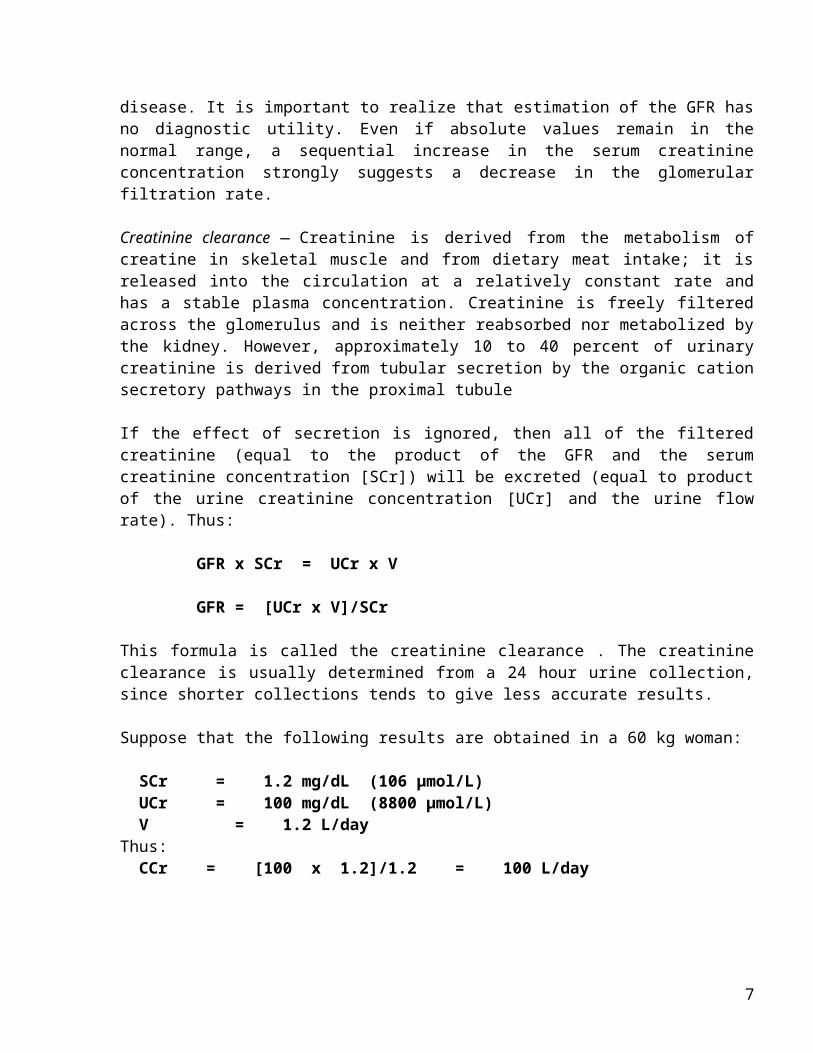
Estimation of the glomerular filtration rate (GFR), usually by the serum creatinine concentration, is used clinically to assess the degree of renal impairment and to follow the course of the disease. It is important to realize that estimation of the GFR has no diagnostic utility. Even if absolute values remain in the normal range, a sequential increase in the serum creatinine concentration strongly suggests a decrease in the glomerular filtration rate.
Creatinine clearance — Creatinine is derived from the metabolism of creatine in skeletal muscle and from dietary meat intake; it is released into the circulation at a relatively constant rate and has a stable plasma concentration. Creatinine is freely filtered across the glomerulus and is neither reabsorbed nor metabolized by the kidney. However, approximately 10 to 40 percent of urinary creatinine is derived from tubular secretion by the organic cation secretory pathways in the proximal tubule
If the effect of secretion is ignored, then all of the filtered creatinine (equal to the product of the GFR and the serum creatinine concentration [SCr]) will be
5

excreted (equal to product of the urine creatinine concentration [UCr] and the urine flow rate). Thus:
GFR x SCr = UCr x V
GFR = [UCr x V]/SCr
This formula is called the creatinine clearance . The creatinine clearance is usually determined from a 24 hour urine collection, since shorter collections tends to give less accurate results.
Suppose that the following results are obtained in a 60 kg woman:
SCr = 1.2 mg/dL (106 µmol/L) UCr = 100 mg/dL (8800 µmol/L) V = 1.2 L/day Thus: CCr = [100 x 1.2]/1.2 = 100 L/day
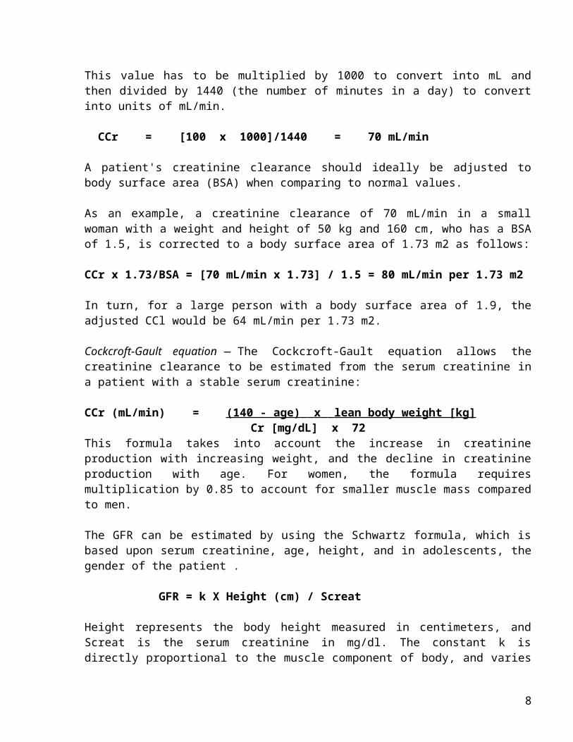
This value has to be multiplied by 1000 to convert into mL and then divided by 1440 (the number of minutes in a day) to convert into units of mL/min.
CCr = [100 x 1000]/1440 = 70 mL/min
A patient's creatinine clearance should ideally be adjusted to body surface area (BSA) when comparing to normal values. As an example, a creatinine clearance of 70 mL/min in a small woman with a weight and height of 50 kg and 160 cm, who has a BSA of 1.5, is corrected to a body surface area of 1.73 m2 as follows:
CCr x 1.73/BSA = [70 mL/min x 1.73] / 1.5 = 80 mL/min per 1.73 m2
In turn, for a large person with a body surface area of 1.9, the adjusted CCl would be 64 mL/min per 1.73 m2.
Cockcroft-Gault equation — The Cockcroft-Gault equation allows the creatinine clearance to be estimated from the serum creatinine in a patient with a stable serum creatinine:
CCr (mL/min) = (140 - age) x lean body weight [kg] Cr [mg/dL] x 72
This formula takes into account the increase in creatinine production with increasing weight, and the decline in creatinine production with age. For women, the formula requires multiplication by 0.85 to account for smaller muscle mass compared to men.
6
The GFR can be estimated by using the Schwartz formula, which is based upon serum creatinine, age, height, and in adolescents, the gender of the patient .
GFR = k X Height (cm) / Screat
Height represents the body height measured in centimeters, and Screat is the serum creatinine in mg/dl. The constant k is directly proportional to the muscle component of body, and varies with age. The value for k is 0.33 in premature infants through the first year of life, 0.45 for term infants through the first year of life, 0.55 in children and adolescent girls, and 0.7 in adolescent boys.
Serum BUN/creatinine ratio — In adults and older children, the serum BUN/creatinine ratio is normal at 10 to 15:1 in ATN, and may be greater than 20 : 1 in prerenal disease due to the increase in the passive reabsorption of urea that follows the enhanced proximal transport of sodium and water. Thus, a high ratio is highly suggestive of prerenal disease. This ratio is not useful in infants and smaller children as their serum creatinine levels are much lower.
Urinalysis — The urinalysis is the most important noninvasive test in the diagnostic evaluation, since characteristic findings on microscopic examination of the urine sediment strongly suggest certain diagnoses .
As examples:A normal or near-normal urinalysis, characterized by few cells with little or no casts or proteinuria, suggests prerenal disease, urinary tract obstruction, and some cases of acute tubular necrosis (ATN).Muddy brown granular casts and epithelial cell casts are highly suggestive of ATN.The finding of a red cell cast is diagnostic of glomerulonephritis, while the presence of proteinuria is generally indicative of some form of glomerular disease.The concurrent presence of hematuria with red cell casts, dysmorphic red cells, heavy proteinuria, or lipiduria can also help subclassify patients into those with an active "nephritic" sediment. This is commonly associated with AKI due to glomerulonephritis. Pyuria with white cell and granular or waxy casts and varying levels of proteinuria is suggestive of tubular or interstitial disease or urinary tract infection.White cells and white cell casts can also be seen in acute glomerulonephritis, particularly postinfectious glomerulonephritis. In this setting, however, there are also other signs of glomerular disease, such as hematuria, red cell casts, and proteinuria. Hematuria and pyuria with no or variable casts (excluding red cell casts) may be seen in acute interstitial nephritis, glomerular disease, vasculitis, obstruction, and renal infarction.
7
Urine sodium excretion — With AKI, measurement of the urine sodium concentration is helpful in distinguishing ATN from prerenal AKI due to effective volume depletion. The urine sodium concentration is usually above 30 to 40 mEq/L and below 10 mEq/L in the former and latter conditions, respectively.
Fractional excretion of sodium (FENa) — The effect of variations in urine volume can be eliminated by calculating the FENa :
FENa (percent) = UNa x PCr x 100 PNa x UCr
where UCr and PCr are the urine and serum creatinine concentrations, respectively, and UNa and PNa are the urine and serum sodium concentrations, respectively.
The FENa is a screening test that differentiates between prerenal AKI and ATN in children. A value below 1 percent suggests prerenal disease, where the reabsorption of almost all of the filtered sodium represents an appropriate response to decreased renal perfusion. A value between 1 and 2 percent may be seen with either disorder. A value above 2 percent usually indicates ATN. In newborns, prerenal disease and ATN are associated with FENa values of less than 2.5 percent and greater than 2.5 to 3.5 percent, respectively, because of their decreased ability to reabsorb sodium.
Urine osmolality — Loss of concentrating ability is an early and almost universal finding in ATN with the urine osmolality usually being below 350 mosmol/kg. In contrast, a urine osmolality above 500 mosmol/kg is highly suggestive of prerenal disease.
Response to volume repletion — Unless contraindicated, a child with a clinical history consistent with fluid loss (such as vomiting and diarrhea), a physical examination consistent with hypovolemia (hypotension and tachycardia), and/or oliguria should be administered intravenous fluid therapy. This fluid challenge attempts to identify prerenal failure that can progress to ATN if not treated promptly. However, such fluid infusion is contraindicated in those with obvious volume overload or heart failure.
Commonly used fluids are crystalloid solutions, such as normal saline (20 mL/kg) administered over 20 to 30 minutes, which may be repeated. Restoration of adequate urine flow and improvement in renal function with fluid resuscitation is consistent with prerenal disease. However, if urine output does not increase and renal function fails to improve with the restoration of
8

intravascular volume, invasive monitoring may be required to adequately assess the fluid status and help guide further therapy. Additional laboratory measurements
Complete blood count — Severe microangiopathic hemolytic anemia associated with thrombocytopenia in the setting of AKI confirms the diagnosis of HUS . Severe hemolysis, whether drug-induced or secondary to hemoglobinopathies, may also result in ATN due to massive hemoglobinuria.
Renal imaging — Renal ultrasonography should be performed in all children with AKI of unclear etiology. It can document the presence of one or two kidneys, delineate renal size, and help survey renal parenchyma . It is particularly useful in diagnosing urinary tract obstruction or occlusion of the major renal vessels.
Renal biopsy — A renal biopsy is most commonly obtained when noninvasive evaluation has been unable to establish the correct diagnosis .
Biomarkers — Biomarkers have been identified that may identify in the beginning stages of AKI. Urinary levels of neutrophil gelatinase-associated lipocalin (NGAL) were found to be elevated 2 hours before subsequently developed AKI . In addition to NGAL, interlukin-18 has been reported as an early marker of AKI .
PREVENTION OF AKI — General measures to help prevent AKI include close monitoring of serum levels of nephrotoxic drugs, adequate fluid repletion in those with hypovolemia, fluid challenge if suspected prerenal hypovolumia, and aggressive hydration and alkalinization of the urine prior to chemotherapy.
MANAGEMENT OF AKI — The basic principles of the general management of acute kidney injury (AKI) include: - Maintenance of electrolyte and fluid balance - Adequate nutritional support Avoidance of life-threatening complications- Treatment of the underlying cause
Hyperkalemia — Hyperkalemia can be asymptomatic or severe enough to constitute a medical emergency. As the extracellular potassium levels increase in AKI, the gradient and membrane potential are affected, resulting in the clinical signs of muscle weakness and cardiac arrhythmias. Electrocardiographic findings associated with hyperkalemia consist of peaked T waves, flattened P waves, increased PR interval, and widening of the QRS. Bradycardia, supraventricular or ventricular tachycardia, and ventricular fibrillation may occur
9

The following modalities are for treatment of severe hyperkalemia (above 7 meq/L). - Stabilization of the cardiac membrane with the intravenous calcium
(calcium gluconate 10 percent solution in a dose 0.5 to 1.0 mL per kilogram intravenously over 5 to 15 minutes).
- Promotion of potassium movement from the extracellular fluid (ECF) into the cells via three different therapies: 1. Administration of intravenous glucose and insulin (0.5 to 1.0 g of
glucose per kilogram over 30 minutes and 0.1 unit of insulin per kilogram intravenously or subcutaneously);
2. Administration of intravenous sodium bicarbonate (in a dose of 1 to 2 milliequivalent per kilogram over 30 to 60 minutes)
3. Administration of beta agonists, such as albuterol 5 mg, via nebulization
The above modalities only transiently lower the plasma potassium concentration; as a result, additional therapy is required to remove potassium from the body. Thus, kayexalate, an ion exchange resin, can be used to effect a net elimination of potassium, at a dose of 1 gram per kilogram orally or rectally. For patients who have mild to moderate hyperkalemia (serum potassium levels greater than 6.0 mEq/L and less than 7.0 mEq/L, and who are asymptomatic without electrocardiographic changes), kayexalate therapy may be useful to prevent further increases in serum potassium levels.
Acidosis — In AKI, not only is acid excretion impaired, acid production frequently is increased on account of underlying comorbid conditions such as shock and sepsis. Administration of sodium bicarbonate should be done only in life-threatening situations in which maximal respiratory compensation is inadequate, and/or the acidosis is contributing to hyperkalemia. In cases of severe or progressive acidosis following shock, serious infections or other hypercatabolic states, supplemental bicarbonate may be required to correct and maintain arterial pH above 7.2 until the underlying disease is controlled. Patients with serum bicarbonate levels that are above 14 mEq/L or with arterial pH greater than 7.2 do not require intervention.Administer 1 mEq of intravenous sodium bicarbonate/kg of body weight over 30-60 minutes, and monitor arterial pH and bicarbonate levels to determine further therapy.
Intravascular volume — Based upon the underlying cause, comorbid conditions, and possible previous therapy, may be hypovolemic, euvolemic, or hypervolemic (including pulmonary edema and heart failure).
Fluid If clinical history and physical exam consistent with fluid loss, and/or oliguria requires immediate intravenous fluid therapy in an attempt to restore
10
renal function and perhaps prevent ischemic renal injury. Commonly used fluids are crystalloid solutions, such as normal saline (20 mL/kg) administered over 20 to 30 minutes, which may be repeated. Patient with oliguria, overload volume, or heart failure, require immediate fluid removal and/or fluid restriction. In such cases a trial of furosemide (2 to 5 mg/kg per dose) may be attempted to induce a diuresis and convert oliguric to non-oliguric renal failure . However, diuretics should not be continued in an unresponsive paients. If a diuresis does not ensue and/or the patient has evidence of fluid overload with pulmonary edema, renal replacement therapy should be initiated.
Nutrition — AKI is associated with marked catabolism, and inadequate nutrition can delay recovery of the patient's renal function. Infants should receive at least maintenance calories (120 Kcal/kg per day). Adult should receive at least calories 40 Kcal/kg per day, protein 1,2 – 1,5 gram/kg per day.
Renal replacement therapy — Renal replacement therapy in AKI should be initiated for the following : - Signs and symptoms of uremia symptoms that include pericarditis,
neuropathy or an otherwise unexplained decline in mental status regardless of the serum BUN or creatinine concentration.
- Azotemia (BUN greater than 80 to 100 mg/dL [29 to 36 mmol/L]).- Severe fluid overload as manifested by hypertension, pulmonary edema
or heart failure that is refractory to supportive medical therapy. - Severe electrolyte abnormalities including hyperkalemia, hypernatremia,
hyponatremia, and acidosis that are refractory to supportive medical therapy.
- Need for intensive nutritional support in a child with oliguria or anuria.
Available renal replacement modalities for the management of acute kidney injury include the following: Hemodialysis, Peritoneal dialysis, Continuous renal replacement therapy (CRRT) — The use of renal replacement therapy to remove fluid and solute of toxins until renal recovery
Chronic Kidney Disease NATURAL HISTORY OF RENAL DISEASE — The initial injury to the kidney may result in a variety of clinical manifestations, ranging from asymptomatic hematuria to renal failure requiring dialysis. Many individuals fully recover and subsequently suffer from little or no sequelae. Poststreptococcal glomerulonephritis in children, for example, most frequently has a long-term benign prognosis. By comparison, some patients, such as those with lupus nephritis, experience repeated and chronic insults to the renal parenchyma, thereby resulting in lasting damage. Furthermore, others in whom the initial
11
disease is either inactive or cured may still develop progressive renal disease due to hemodynamic and other mechanisms.
In addition to variations in the activity of the individual diseases, these different manifestations are partly due to how the kidney responds to injury. The kidney is able to adapt to damage by increasing the filtration rate in the remaining normal nephrons, a process called adaptive hyperfiltration. As a result, the patient with mild renal insufficiency often has a normal or near-normal serum creatinine concentration. Additional homeostatic mechanisms (most frequently occurring within the renal tubules) permit the serum concentrations of sodium, potassium, calcium, and phosphorous and the total body water to also remain within the normal range, particularly among those with mild to moderate renal failure. (See "Assessment of kidney function: Serum creatinine; BUN; and GFR").
Adaptive hyperfiltration, although initially beneficial, appears to result in long-term damage to the glomeruli of the remaining nephrons, which is manifest by proteinuria and progressive renal insufficiency. This process appears to be responsible for the development of renal failure among those in whom the original illness is either inactive or cured. The institution of measures to help prevent this process, such as antihypertensive therapy with an angiotensin converting enzyme inhibitor or an angiotensin II receptor blocker, may slow progressive disease and even preserve renal function. If these modalities are effective, the benefit is likely to be greatest if begun before a great deal of irreversible scarring has occurred. (See "Secondary factors and progression of chronic kidney disease").
The gradual decline in function in patients with chronic kidney disease (CKD) is initially asymptomatic. However, as previously mentioned, different signs and symptoms may be observed with advanced renal dysfunction, including volume overload, hyperkalemia, metabolic acidosis, hypertension, anemia, and bone disease. The onset of end-stage renal disease results in a constellation of signs and symptoms referred to as uremia.
Manifestations of the uremic state include anorexia, nausea, vomiting, pericarditis, peripheral neuropathy, and central nervous system abnormalities (ranging from loss of concentration and lethargy to seizures, coma, and death). No direct correlation exists between the absolute serum levels of blood urea nitrogen (BUN) or creatinine, and the development of these symptoms. Some patients have relatively low levels (eg, a BUN of 60 mg/dL [21.4 mmol/L] in an older patient) but are markedly symptomatic, while others have marked elevations (eg, a BUN of 140 mg/dL [50 mmol/L]) but remain asymptomatic. To continue life, uremic patients require the institution of renal replacement therapy with hemodialysis, peritoneal dialysis, or renal transplantation.
12
DEFINITIONS AND CLASSIFICATION — The definition, classification and guideline of chronic kidney were proposed from the National Kidney Foundation of the United States through its Kidney Disease Outcomes Quality Initiative (K/DOQI) program. These guidelines have been reviewed and accepted internationally.
The K/DOQI working group defined chronic kidney disease as:
1. Evidence of structural or functional kidney abnormalities (abnormal urinalysis, imaging studies, or histology) that persist for at least three months, with or without a decreased GFR (as defined by a GFR of less than 60 mL/min per 1.73 m2). The most common manifestation of kidney damage is persistent albuminuria, including microalbuminuria.
2. Or decreased GFR, with or without evidence of kidney damage.
Based upon these definitions, the following is the recommended classification of chronic kidney disease by stage and the estimated prevalence within the United States of each stage, as determined by a National Health and Nutrition Examination Survey (NHANES) performed in 1999 to 2004 :
- Stage 1 disease is defined by a normal GFR (greater than 90 mL/min per 1.73 m2) and persistent albuminuria.
- Stage 2 disease is a GFR between 60 to 89 mL/min per 1.73 m2 and persistent albuminuria.
- Stage 3 disease is a GFR between 30 and 59 mL/min per 1.73 m2. - Stage 4 disease is a GFR between 15 and 29 mL/min per 1.73 m2. - Stage 5 disease is a GFR of less than 15 mL/min per 1.73 m2 or end-
stage renal disease. The K/DOQI guidelines have also suggested that microalbuminuria alone that persists for more than three months falls within the definition of chronic kidney disease in patients without diabetes. GENERAL MANAGEMENT OF CHRONIC KIDNEY DISEASE — The general management of the patient with chronic kidney disease involves the following issues :
- Treatment of reversible causes of renal dysfunction- Preventing or slowing the progression of renal disease - Treatment of the complications of renal dysfunction - Identification and adequate preparation of the patient in whom renal
replacement therapy will be required
Reversible causes of renal dysfunction — patients with chronic renal disease with a recent decrease in renal function may be suffering from an underlying reversible process, which if identified and corrected may result in the recovery of function.
13

Decreased renal perfusion — Hypovolemia (such as vomiting, diarrhea, diuretic use, bleeding), hypotension (due to myocardial dysfunction or pericardial disease), infection (such as sepsis), and the administration of drugs which lower the GFR (such as nonsteroidal antiinflammatory drugs [NSAIDs] and ACE inhibitors) are common causes of potentially reversible declines in renal function.
Urinary tract obstruction — Urinary tract obstruction should always be considered in the patient with unexplained worsening renal function. Renal ultrasonography is often performed to exclude urinary tract obstruction in patients with an unexplained elevation in the serum creatinine.
Slowing the rate of progression — Studies in experimental animals and humans suggest that progression in chronic renal disease may be due at least in part to secondary factors that are unrelated to the activity of the initial disease. The major factors are thought to be intraglomerular hypertension and glomerular hypertrophy (which are primarily responsible for the adaptive hyperfiltration described above), leading to glomerular scarring (glomerulosclerosis).The major histologic manifestation of hemodynamically-mediated renal injury is secondary focal segmental glomerulosclerosis . Thus, proteinuria typically occurs in patients with progressive chronic kidney disease.
Aggressive goals are recommended for both proteinuria and blood pressure. Antihypertensive therapy is given for both renal protection and cardiovascular protection, since chronic kidney disease is associated with a marked increased in cardiovascular risk.
- A reduction in protein excretion to less than 500 to 1000 mg/day - A reduction in blood pressure to less than 130/80 mmHg. These
aggressive goals will, in most patients, require therapy with multiple drugs. ACE inhibitors and ARBs has benefit to reduce proteinuria, reduce blood pressure, slowing decline in renal function.
- Low protein diet. Restrict protein intake to 0.8 to 1.0 g/kg per day of high biologic value protein, with the lower value (0,6 to 0,75 g/kg perday) used in patients with progressive chronic kidney disease. Calories should be provided approximately 30 to 35 kcal/kg per day.
- Both hyperlipidemia and metabolic acidosis should be treated, in part because there is some evidence that they may enhance the rate of progression of the renal.
- Smoking cessation should be encouraged, with smoking stoppage being associated with a reduced rate of progression of chronic kidney disease.
14
Treatment of the complications of renal dysfunction — A wide range of disorders may develop as a consequence of the loss of renal function. These include disorders of fluid and electrolyte balance, such as volume overload, hyperkalemia, metabolic acidosis, and hyperphosphatemia, as well as abnormalities related to hormonal or systemic dysfunction, such as anorexia, nausea, vomiting, fatigue, hypertension, anemia, malnutrition, hyperlipidemia, and bone disease.
Volume overload — Sodium and intravascular volume balance are usually maintained via homeostatic mechanisms until the GFR falls below 10 to 15 mL/min. However, the patient with mild to moderate chronic kidney disease, despite being in relative volume balance, is less able to respond to rapid infusions of sodium and is therefore prone to fluid overload.
Patients with chronic kidney disease and volume overload generally respond to the combination of dietary sodium restriction and diuretic therapy, usually with a loop diuretic given daily. In case of severe volume overload , furosemide (2 to 5 mg/kg per dose) may be attempted to induce a diuresis and convert oliguric to non-oliguric renal failure
Hyperkalemia — hyperkalemia generally develops in the patient who is oliguric or who has an additional problem such as a high potassium diet, increased tissue breakdown, or hypoaldosteronism (due in some cases to the administration of an ACE inhibitor or ARB).Hyperkalemia due to ACE inhibitor or ARB therapy is most likely to occur in patients in whom the serum potassium concentration is elevated or in the high normal range prior to therapy. In this setting, institution of a low-potassium diet or concurrent use of a loop diuretic (to increase urinary potassium losses) often ameliorates the degree of hyperkalemia. In selected patients, low dose Kayexalate (5 grams with each meal) can be used to lower the serum potassium concentration without the side effects associated with larger doses. (see treatment hyperkalemia in subject of AKI).
Metabolic acidosis — There is an increasing tendency to retain hydrogen ions among patients with chronic renal disease . This can lead to a progressive metabolic acidosis with the serum bicarbonate concentration tending to stabilize between 12 and 20 meq/L, and rarely falling below 10 meq/L We recommend alkali therapy to maintain the serum bicarbonate concentration above 23 meq/L. If alkali is given, sodium bicarbonate (in a daily dose of 0.5 to 1 meq/kg per day) is the agent of choice. (see also treatment of metabolic acidosis in subject of AKI).
Hyperphosphatemia — A tendency toward phosphate retention begins early in renal disease, due to the reduction in the filtered phosphate load. Although this problem is initially mild with hyperphosphatemia being a relatively late
15

event, phosphate retention is intimately related to the common development of secondary hyperparathyroidism. From the viewpoint of calcium and phosphate balance, the hypersecretion of parathyroid hormone (PTH) is initially appropriate, since PTH can correct both hyperphosphatemia and hypocalcemia. As a result, phosphate balance and a normal serum phosphate concentration are generally maintained in patients with a GFR of greater than 30 mL/min. The price paid is secondary hyperparathyroidism and the development of renal osteodystrophy. Dietary phosphate restriction may limit the development of secondary hyperparathyroidism in patients with chronic kidney disease. Once the GFR falls below 25 to 30 mL/min, the addition of oral phosphate binders are usually required to prevent hyperphosphatemia.One of the preferred agents to bind intestinal phosphate is calcium salts, of which one of the most widely used is calcium carbonate. In patients with stage 3 to 5 CKD, the K/DOQI guidelines suggest that total elemental calcium intake (including both dietary calcium intake and calcium-based phosphate binders) should not exceed 2,000 mg/day. Other phosphate binder are sevelamer and lanthanum
Hypertension — Hypertension is present in approximately 80 to 85 percent of patients with chronic kidney disease . Treating hypertension can both slow the progression of proteinuric CKD and reduce the rate of cardiovascular complications. The desired degree of blood pressure control can usually be safely achieved with combined therapy which usually begins with an ACE inhibitor or angiotensin II receptor blocker and a diuretic. Volume expansion, often in the absence of overt edema, contributes to the elevation in blood pressure in most forms of chronic renal disease. As a result, before other medications are added, the dose of diuretics should be increased until the blood pressure is normalized or the patient has attained "dry weight" which, in the presence of persistent hypertension, is defined as the weight at which further fluid loss will lead either to symptoms (fatigue, orthostatic hypotension) or to decreased tissue perfusion as evidenced by an otherwise unexplained elevation in the BUN and plasma creatinine concentration.
A loop diuretic is recommended for the treatment of hypertension and edema in patients with chronic kidney disease. The thiazide diuretics in conventional dosage become less effective as monotherapy when the GFR falls below 20 mL/min. They do, however, produce an additive effect when administered with a loop diuretic for refractory edema.
We recommend a blood pressure goal of less than 130/80 mmHg, which is consistent with JNC 7 and the K/DOQI Clinical Practice Guidelines on hypertension and antihypertensive agents in chronic kidney disease . However, evidence from the Modification of Diet in Renal Disease study, the AASK trial, and a meta-analysis from the ACE inhibition and Progressive Renal
16

Disease (AIPRD) study group suggest that an even lower systolic pressure may be more effective in slowing progressive renal disease in patients with a spot urine total protein-to-creatinine ratio ≥ 1000 mg/g (which represents protein excretion of greater than 1000 mg/day) . Caution is advised about lowering the systolic blood pressure below 110 mmHg.
Anemia — The anemia of chronic kidney disease is, in most patients, normocytic and normochromic, and is due primarily to reduced production of erythropoietin by the kidney (a presumed reflection of the reduction in functioning renal mass), and to shortened red cell survival. Anemia is a common feature in many patients with chronic kidney disease who do not yet require dialysis, with anemia becoming increasingly common as glomerular filtration rates (GFRs) decline below 60 mL/min per 1.73 m2 .
As stated in the 2006 K/DOQI guidelines, the evaluation of anemia in those with CKD should begin when the Hgb level is less than 12 g/dL in females, and Hgb levels of less than 13.5 g/dL in adult males. The evaluation of patients should therefore include red blood cell indices, absolute reticulocyte count, serum iron, total iron binding capacity, percent transferrin saturation, serum ferritin, white blood cell count and differential, platelet count, and testing for blood in stool. An erythropoietic agent should be given to the predialysis patient with CKD and anemia. Target Hgb levels in the range of 11 to 12 g/dL in predialysis patients with CKD. Above such levels have been associated with adverse cardiovascular outcomes. The erythropoietin dose should be approximately 50 to 100 U/kg given two to three doses per week.
K/DOQI guidelines suggest administering iron to maintain the percent transferrin saturation ≥ 20 percent, and the serum ferritin level to be greater than 100 ng/mL
PREPARATION FOR AND INITIATION OF RENAL REPLACEMENT THERAPY — It is important to identify patients who may eventually require renal replacement therapy since adequate preparation can decrease morbidity and perhaps mortality. Early identification enables dialysis to be initiated at the optimal time with a functioning chronic access and may also permit the recruitment and evaluation of family members for the placement of a renal allograft prior to the need for dialysis. In addition, the ability of the individual to psychologically accept the requirement of life-long renal replacement therapy is often diminished if inadequate time has elapsed between the time of recognition of end-stage renal disease and the initiation of dialysis.
Referral to nephrologists — Patients with CKD should be referred to a nephrologist early in the course of their disease, preferably before the plasma creatinine concentration exceeds 1.2 (106 micromol/L) and 1.5 mg/dL (133
17
micromol/L) in women and men, respectively, or the eGFR is less than 60 mL/min per 1.73 m2. These subspecialists are trained to help counsel the patient in choosing the optimal renal replacement therapy and to manage the many issues associated with chronic kidney disease. Lower costs and/or decreased morbidity and mortality may be associated with early referral and care by subspecialists.
Choice of renal replacement therapy — Once it is determined that renal replacement therapy will eventually be required, the patient should be counseled to consider the advantages and disadvantages of hemodialysis (in-center or at home), peritoneal dialysis (continuous or intermittent modalities), and renal transplantation (living or deceased donor). The 2006 K/DOQI guidelines recommend that patients with a GFR less than 30 mL/min per 1.73 m2 should be educated concerning these issues .
Kidney transplantation is the treatment of choice for end-stage renal disease. A successful kidney transplant improves the quality of life and reduces the mortality risk for most patients, when compared with maintenance dialysis.
Indications for renal replacement therapy — The decision to initiate dialysis in a patient with chronic kidney disease involves the consideration of subjective and objective parameters by the physician and the patient. There are a number of clinical indications to initiate dialysis in patients with CKD. These include : - Pericarditis or pleuritis (urgent indication). - Progressive uremic encephalopathy or neuropathy, with signs such as
confusion, asterixis, myoclonus, wrist or foot drop, or, in severe, cases, seizures (urgent indication).
- A clinically significant bleeding diathesis attributable to uremia (urgent indication).
- Fluid overload refractory to diuretics. - Hypertension poorly responsive to antihypertensive medications. - Persistent metabolic disturbances that are refractory to medical therapy.
These include hyperkalemia, metabolic acidosis, hypercalcemia, hypocalcemia, and hyperphosphatemia.
- Persistent nausea and vomiting, weight loss or signs of malnutrition.
However, these indications are potentially life-threatening. They occur when the patient has very advanced chronic kidney disease, such as may be observed in those who present with severe uremia and have not had prior medical contact. For patients under medical care, delaying initiation of dialysis until one or more of these complications is present may put the patient at unnecessary jeopardy; dialysis should therefore be initiated well before these indications have developed. Patients with chronic kidney disease should therefore be closely followed and the GFR estimated.
18

A number of characteristics have therefore been proposed as possible indications for early institution of maintenance dialysis: The 2006 National Kidney Foundation Dialysis Outcomes Quality Initiative (K/DOQI) for peritoneal dialysis and hemodialysis adequacy published guidelines concerning the initiation of dialysis among patients with advanced chronic kidney disease. The work group suggested that the benefits and risks of initiating renal replacement therapy should be considered in patients with a GFR of less than 15 mL/min per 1.73 m2 (stage 5 CKD). Initiation of dialysis prior to stage 5 chronic kidney disease may also be required in patients with certain characteristics and/or complications, such as declining health due to the loss of kidney function.
V. Module Tasks
1. a woman, 23 yo, got diarrhea for three days, frequency more than ten times a week, and vomitting. She admitted hospital with decrease of consciousnes. Blood pressure was 90/40 mmHg, pulse rate 120 x/min, cold acral. Urine collection for 6 hours was 50 ml. Creatinine serum 2,5 mg/dl.
a. what is problem list of the patient ?b. is patient has renal failure ? acute or chronic?c. How is management of the patient ?
19
2. a male, 55 yo, 50 kg bw, has long standing of hypertension. He also complaint of pain in the right flank area. He look pale. Blood pressure is 180/100 mgHg. Hemoglobin serum level 9 mg/dl. Creatinin serum 23 mg/dl, kalium 7 meq/L. Urinalysis reveal albuminuria +3, many erythrocyte sediment. USG show hydronephrosis ren extra, small stones in lower pole. Ren dextra contracted.
a. what is problem list of the patient ?b. calculate GFR of the patient ?c. does patient has acute or chronic renal failure ?d. what is probable the cause of renal failure?e. Does the patient will be recovery if the causes is corrected?f. Do planning to treat patients problem
20