Defining Developmental Dyslexia: An Overview of ...
33
Defining Developmental Dyslexia: An Overview of Etiological Theories and the Neurobiological Basis of the Disorder Dana Elaameir A dissertation submitted in partial fulfillment of the requirements for the degree of Masters in Education University of Washington 2019 Reading Committee: Gail Joseph, Chair Deborah McCutchen Program Authorized to Offer Degree: Education
Transcript of Defining Developmental Dyslexia: An Overview of ...
Defining Developmental Dyslexia: An Overview of Etiological Theories and the Neurobiological Basis of the Disorder
Dana Elaameir
A dissertation
submitted in partial fulfillment of the
requirements for the degree of
Masters in Education
University of Washington
2019
Reading Committee:
Gail Joseph, Chair
Deborah McCutchen
Program Authorized to Offer Degree:
Education

2 Defining Developmental Dyslexia
© Copyright 2019
Dana Elaameir
3 Defining Developmental Dyslexia
University of Washington
Abstract
Defining Developmental Dyslexia: An Overview of Etiological Theories and the Neurobiological Basis of the Disorder
Dana Elaameir
Chair of the Supervisory Committee:
Dr. Gail Joseph College of Education
Researchers have long been debating the most accurate definition for developmental
dyslexia, likely due to its complicated and unknown etiology, and its variability in expression.
The phonological deficit theory, the cerebellar theory, and the magnocellular theory each argue
the primary characteristics of dyslexia that inform their varying definitions. In this review, I
explore what is known about developmental dyslexia, the research that supports its etiological
theories as well as the interventions that address them. Though the evidence is correlational, new
research indicates potential answers in the genetic association of dyslexia. Perhaps the causality
of dyslexia is not as imperative as the proper intervention of its consequential reading
difficulties.

4 Defining Developmental Dyslexia
Introduction
Historically, developmental dyslexia was perceived as a hereditary defect, one that
affects some aspects of the visual processing of words without influencing intellect, or verbal
skills (Stein, 2018). In 1878, renowned German physician Adolf Kussmaul, observing his stroke
patients, noted selective deficiencies in reading but otherwise unaffected non-verbal reasoning
and oral skills, thus introducing the term “word blindness” (Stein, 2008). Rudolph Berlin later
renamed this condition to dyslexia in 1884, though the term only gained popularity almost 50
years later. Physician Pringle Morgan published the first academic paper on dyslexia, noting the
earliest case of what he called “congenital word blindness,” a visual processing issue with
written words that had no bearing on intelligence (Snowling, 1995). By the mid to late 20th
century, dyslexia was established as a hereditary defect that maintained oral and verbal
intelligence but affected visual processing of reading. However, this was later rejected, and
dyslexia was redefined as a language disorder—an inability to develop the necessary
phonological skills involved in reading (Stein, 2008). The general public has maintained this
view of dyslexia and most existing research supports this interpretation, though it is certainly not
the only viable explanation. Recent developments in technology have revealed the neurological
component of the disorder and corresponding theories, like the visual processing deficit, have
been reconsidered, piquing the curiosities of many researchers in the field. Experts continue to
deliberate as to what characteristics are universal to dyslexics and thus essential to its definition.
Despite the significant progress in this endeavor, the understanding of dyslexia remains in
constant flux, and causality remains a mystery.
5 Defining Developmental Dyslexia
Methods
The following literature review aims to present a summary of what is known and agreed
upon by researchers regarding the nature of dyslexia. The goal is to highlight the major extant
theories explaining the etiology of dyslexia as revealed by recent neurological research. Several
procedures were taken to ensure that a high quality of literature was compiled and reviewed.
Databases such as EBSCOHOST, ERIC, Google Scholar, Jstor, ScienceDirect and PubMed were
utilized during a widespread search for peer reviewed journals and medical publications. The
reference section of each article was thoroughly explored and served as another valuable source
of literature. During the collection process, a general search of studies on dyslexia and other
literature reviews that comprehensively describe the diagnosis were accumulated for research.
After realizing that the controversy was rooted in the definition, search criteria was refined to
scholarly educational and medical journals, specifically on the theories that frame dyslexia and
the numerous studies that have attempted to prove them. For the sake of focusing recent
developments in the field, any source not published in well-known, peer-reviewed journals after
1990 were excluded from the search, though a few exceptions have been made in an effort to
holistically approach particular aspects of the theories and definitions. The studies chosen
focused predominantly on children with dyslexia, though this scope severely limited the research
available, and thus adults with dyslexia were also taken into account. Keywords for the search
included dyslexia, reading disabilities, neurological, phonological deficits, the brain, vision
pathways, magnocellular, brain abnormalities, genetics and interventions. Of the aggregated
search results, I found 70 references to be most relevant.
6 Defining Developmental Dyslexia
Overview: What do we know?
Under current definitions, individuals with dyslexia account for 7% of the population,
(Peterson and Pennington, 2012), roughly 3% -17.5% of all school-age children (dependent on
definition and diagnostic criteria) and 80% of all reading disabilities (Saviour and Ramachandra,
2006). Children with dyslexia are often diagnosed early in their elementary school experience, as
difficulties in reading begin to surface and hinder their academic performance. Boys are often
affected more drastically than girls, possibly due to fetal testosterone levels in late pregnancies
(Saviour and Ramachandra, 2006) or referral bias in the classroom (Shaywitz, 1990). This can
also be explained by higher prevalence of comorbid externalizing disorders such as attention
deficit hyperactivity disorder (ADHD) in boys (Peterson and Pennington, 2012). Other
comorbidities of dyslexia include language impairment (difficulties in syntax and vocabulary
development), motor skills, executive functioning and speech sound disorder (difficulties in
intelligible and accurate reproduction of sounds in speech) (Gooch, 2014; Peterson and
Pennington, 2012). These conditions are typically detected far earlier than dyslexia and could
therefore be important indicators for later reading complications (Peterson and Pennington,
2012).
In the past, only a handful of research has focused on cross-cultural manifestations of
dyslexia. Cross-cultural research hoped to discover whether dyslexia existed only in languages
with complex orthographies like English. Ziegler’s 2003 study determined that children from
both Australia and Germany exhibited non-word reading deficits, indicating the universality of
non-word decoding speed deficiency in dyslexics, regardless of the language’s orthography.
Studies like that of Lindgren (1985) found the frequency of dyslexia to be higher in the United
States than in Italy; while both were similar in their association with verbal processing issues,
7 Defining Developmental Dyslexia
English dyslexics were unique in their visual-motor deficits. Overall, the research tends to agree
on a common neurocognitive basis for dyslexia, though the severity and variation of reading
behaviors seen among different countries is dependent on the complexity of different
orthographies (Paulesu et al., 2001).
The Defining Controversy
The definition of dyslexia has proven to be the most controversial aspect of the disorder.
For decades, researchers in every related field have attempted to set definitional parameters, yet
there is still no universal agreement for its essential characteristics. The DSM V no longer
provides a definition for dyslexia, instead grouping it with other disorders placed under the
category of Specific Learning Disabilities (American Psychiatric Association, 2013). Some argue
that dyslexia is neurodevelopmental in nature, characterized by slow and inaccurate word
recognition (Peterson and Pennington, 2012). Others argue that it is “unexpected, specific and
persistent” in its inability to facilitate proficient reading skills (Démonet, 2004), independent of
factors such as learning opportunity, instruction, and motivation (Critchley, 1970; World Health
Organization, 1993). Yet another definition with more historical acceptance emphasizes the
impairment in vision processing that produces the visual confusion of written words (Stein,
2000). To many, dyslexia is more complicated than a specific reading disability (Catts, 1989),
though the general public tends to use dyslexia and reading disability interchangeably, partly due
to the vague and conflicting definitions used to describe both.
The more common classification of dyslexia is that it is a language disorder involving
deficits in phonological processing (Castles and Coltheart, 2004). Still, the vision pathway deficit
theory has made a recent comeback, attempting to explain the apparent movement of words and
letters that some individuals experience. The trouble with having a conglomerate of competing
8 Defining Developmental Dyslexia
definitions is that they are often exclusionary, addressing only some of the experienced
symptoms. These parameters are therefore discernibly limited, as many people who have the
diagnosis report deficiencies that are not always highlighted. Arguably, the biggest downfall of
the current diagnosis process is a heavy reliance on the presence of reading difficulties, meaning
that affected children must fail before being identified (Catts, 1989). The complicated and varied
nature of the disorder and the way it affects each individual, makes it difficult to characterize a
universal definition, and research that attempts to do so will prove futile (Stein 2018).
Despite the lack of consensus, there are a few fundamentals of dyslexia that researchers
tend to agree on. For example, dyslexia is widely assumed to be genetic in origin, with some
elements of neurological abnormalities at play (Smith et al., 1998), though the extent of those
abnormalities is debated. A large majority of people with dyslexia demonstrate cognitive issues
in phonological processing such as encoding speech sound information into long term memory
(Kamhi, 1988). In other words, people with dyslexia need multiple encounters and depictions of
words before they can reliably reproduce them (Catts, 1989). Individuals with dyslexia also have
difficulty retaining verbal information in short term memory, causing poor performance on
activities depending on the short term recall of lists, whether they be letters, words, numbers or
phrases (Cohen and Netley, 1981). Similarly, there is a general complication with retrieving any
phonological information from long term memory (Wagner and Torgesen, 1987). Lack of
sensitivity to the speech sound structure of language also characterizes one of the neurological
abnormalities, though this may stem from issues with limited exposure to reading, as reading
promotes improved experience with speech sound and phoneme awareness (Catts, 1989).
One of the most widely accepted and relatively inclusive definitions is that of Catts
(1989), who outlines dyslexia as a developmental language disorder present at birth and
9 Defining Developmental Dyslexia
persisting into adulthood; the specific reading disability is characterized by difficulties in
phonological processing such as coding, retrieving and code use of phonological information in
memory, extending to lack of awareness of speech sound structures (Catts,1989). However, even
this definition leaves many questions unanswered. A major controversy surrounding dyslexia lies
in the perceived importance of limited phonological processing, namely that it may not be the
only defining characteristic and many experts in the field argue that this should not be a primary
component of the definition. In contrast to Catts, Saviour and Ramachandra (2006) base their
definition of dyslexia more broadly in the difficulties in processing new words and single word
decoding, while struggling with the reversal and rearranging of words and letters. They also note
an association between dyslexia and delayed milestones in development and motor skills
(Saviour and Ramachandra, 2006). The neurological basis of dyslexia is undeniably present in
the diagnosed population and definitions modeled around Saviour and Ramachandra’s (2006)
framework maintain the reality that cognitive abnormalities should be leading contenders in the
primary characterization of dyslexia. This leads us to the heart of the definition controversy:
what is universally experienced by individuals with dyslexia? Researchers have generated
several theories that seek to explain the indicators of dyslexia by turning to observable
abnormalities in the structure and function of brain.
Structural and Functional Brain Abnormalities
Data gathered from MRI scans of individuals with dyslexia reveals the absence of typical
asymmetry of the planum temporale, in the center of the Wernicke’s area—an important area for
language processing, analyzing sounds, naming objects and recalling words (Duara, 1991;

10 Defining Developmental Dyslexia
Galaburda,1979). In the average
brain, the larger side of the
Wernicke’s area, specifically the
planum temporale on the left, is
possibly related to the language
dominance being localized in the left
brain hemisphere (Duara, 1991). Due
to enlargement of the right
hemisphere, this area appears to be symmetrical in dyslexic brains (Velluntino, 1996), yielding
poor performance on non-word reading tasks (Saviour and Ramachandra, 2006). The parietal
lobe area in dyslexic brains also shows more symmetry, correlating with poor phonological
performance (Saviour and Ramachandra, 2006). Additionally, the posterior part of the corpus
callosum, referred to as the splenium, is larger in dyslexic brains, but little is understood about
what that may mean for cognitive and functional abilities (Duara, 1991). Also larger in dyslexic
brains is the angular gyrus and posterior pole in the posterior region of the parietal lobe, which is
involved in a multitude of processes related to language processing (Duara, 1991).
The connectivity between the middle and temporal gyrus, superior temporal sulcus and
the supramarginal and angular gyri show reduced activity in dyslexics and is generally correlated
with the underdevelopment of reading skills in children (Darki, 2012). The occipitotemporal
cortex, which holds the area for visual word form, along with the temporoparietal and inferior
frontal cortices, which play a role in phonological and semantic processing of words, and the
inferior frontal cortex, involved in the formation of speech sounds, are all areas of the brain
associated with reading that are altered in people with dyslexia (Richlan et al, 2011; IDA 2015).
Figure 1. Areas of the left hemisphere involved in reading that are altered in dyslexic brains. Reprinted from “Developmental Dyslexia: dysfunction of a left hemisphere reading network” by F. Richlan, 2012, Frontiers in human neuroscience, 6, 120.

11 Defining Developmental Dyslexia
Microscopic cortical malformations have also been discovered in language areas of
dyslexic brains, such as the interior frontal gyrus and the temporoparietal area (Galaburda, 1985).
These malformations appear as ectopias, misplaced collections of neurons caused by
disturbances in neural migration that results in changes in both the white and gray matter
structure. They can also appear as microgyrias and dysplasia: lost structural organization among
cortical neurons (Saviour and Ramachandra, 2006; Darki 2012). This could be an indication of
abnormal cortical development in dyslexic brains (Saviour and Ramachandra, 2006), possibly
dictated by an undercurrent of genetic risk factors.
Additionally, PET scans show white matter density and connectivity between posterior
and frontal regions to be significantly altered in dyslexic brains (Saviour and Ramachandra,
2006). Variability in white matter structure is correlated with variability in reading ability since
connectivity between language regions in the brain is critical for literacy (Darki, 2012). In
children and adults with dyslexia, white matter structure is altered in the left temporoparietal area
and in the left interior frontal gyrus of the Broca’s area, which is responsible for language
processing and speech production (Peterson and Pennington, 2012; Stein, 2018). According to
Hoeft (2006), the fMRI data analysis revealed that children with dyslexia exhibited less
activation in their left parietotemporal lobe than age-matched children in the control group with
the same reading ability. Other regions that exhibit reduced activation include the right
occipitotemporal cortex and the bilateral frontal cortices (Hoeft, 2006). The results of the study
indicate that the reduced activation in these brain regions is characteristic of dyslexia, rather than
a consequence of low reading proficiency (Hoeft, 2006).
Silani (2005) noted that decrease of gray matter density among dyslexics corresponds
with under-activation of the left medial temporal gyrus, affecting the performance in tasks
12 Defining Developmental Dyslexia
requiring phonological retrieval such as
reading. While reduced grey matter density is
observed in the left middle temporal region, a
higher density of grey matter surrounds the
area down to the inferior temporal cortex; this
phenomenon was associated with greater
impairment in reading tasks performed by the
subjects (Silani, 2005). Significant reduction of
white matter density in dyslexic brains was
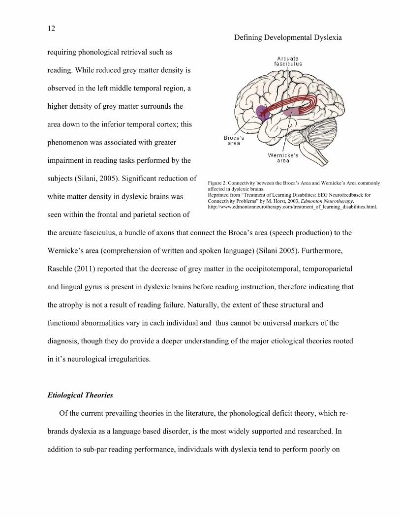
seen within the frontal and parietal section of
the arcuate fasciculus, a bundle of axons that connect the Broca’s area (speech production) to the
Wernicke’s area (comprehension of written and spoken language) (Silani 2005). Furthermore,
Raschle (2011) reported that the decrease of grey matter in the occipitotemporal, temporoparietal
and lingual gyrus is present in dyslexic brains before reading instruction, therefore indicating that
the atrophy is not a result of reading failure. Naturally, the extent of these structural and
functional abnormalities vary in each individual and thus cannot be universal markers of the
diagnosis, though they do provide a deeper understanding of the major etiological theories rooted
in it’s neurological irregularities.
Etiological Theories
Of the current prevailing theories in the literature, the phonological deficit theory, which re-
brands dyslexia as a language based disorder, is the most widely supported and researched. In
addition to sub-par reading performance, individuals with dyslexia tend to perform poorly on
Figure 2. Connectivity between the Broca’s Area and Wernicke’s Area commonly affected in dyslexic brains. Reprinted from “Treatment of Learning Disabilites: EEG Neurofeedbasck for Connectivity Problems” by M. Horst, 2003, Edmonton Neurotherapy. http://www.edmontonneurotherapy.com/treatment_of_learning_disabilities.html.

13 Defining Developmental Dyslexia
tasks that require phonological awareness such as manipulating speech sounds (Ramus, 2003).
According to Démonet (2004), people with dyslexia have difficulties in the representation,
retrieval and storage of phonological information from long term memory. In other words, there
is an inability to separate sounds that match with the letter itself, a skill known as phonemic
awareness (Stein, 2018). It exists as part of a larger process of learning known as phonological
awareness, which is the ability to identify and manipulate units of sound in language such as
words and syllables, vital to the foundation of reading and fluency. Without this skill, children
are unable to efficiently read and understand text. The phonological processing deficit interferes
with their phonemic awareness and subsequent phoneme-grapheme mapping, the process of
matching sounds to the corresponding written letter (Peterson and Pennington, 2012).
Essentially, the lack of phonological awareness experienced by children with dyslexia explains
the root of their reading incompetence. Any impairments in processing are associated with
abnormalities in the neural connectivity and structure of the left hemisphere language network,
including the temporal, occipital and parietal lobe (Stein, 2018).
This theory, though highly probable, remains undeniably incomplete. Critics note that it is
impossible to differentiate dyslexia from other factors that yield reading failure, such as lack of
resources, low general ability, poor teaching, stress, etc. (Stein, 2018). No singular deficit in
phonological processing is enough to entirely explain the causation of dyslexia, and in chicken-
or-egg fashion, it remains unclear whether phonological awareness problems are a cause or effect
of dyslexia. This is especially muddled because the relationship between phonological skills and
reading is bidirectional; over time, poor reading can cause poor phonological awareness
(Peterson and Pennington, 2012). There are also studies of individuals who have difficulties in
non-word reading yet demonstrate adequate phonological awareness (Castles and Coltheart,

14 Defining Developmental Dyslexia
2004). Interventions that aim to remediate the deficits of dyslexia via phonological training often
neglect the possible visual impairments experienced by many dyslexics. Though this theory
dominates current understanding of the diagnosis, the recent renewal in interest regarding the
visual explanation for dyslexia seems to fill in ignored gaps, but produces many more questions
of its own.
On the other hand, the magnocellular theory takes into account the notable visual
impairments associated with dyslexia. According to this theory, sensory stimuli of both visual
and auditory varieties cannot be processed efficiently and are therefore distorted in time due to
abnormalities in the neurons of the magnocellular pathway (Petkov, 2005). Anatomically, the
magnocellular pathway consists of neurons that connect the lateral geniculate nucleus of the
thalamus to the primary visual cortex where information received from the eye is processed
(Demb, 1998). These large neurons are sensitive to motion perception and are important for the
control of eye movements and rapid transmission of signals throughout the magnocellular layer.
Impairments in this pathway can lead to visual confusion and letters may “appear to move
around,” a phenomenon experienced by some individuals with dyslexia. As a result, the
consequential unsteadiness of the eyes can explain visual reading errors, qualifying it as another
marker of the disorder (Saviour and Ramachandra, 2006).
Both the slow recognition of letters and a general inability to sequence the order of those
letters and sounds are also outcomes of impairments in temporal processing—the timing of
visual information during reading and of sounds in the word while hearing it (Breznitz, 2003).
Typically, magnocellular neurons mediate temporal processing when the visual system first
senses a letter and directs the eyes’ focus in order to identify it (Stein, 2018). In dyslexic brains,
the magnocells in this pathway are abnormal in shape; size is important in the rapid processing

15 Defining Developmental Dyslexia
and transmission of signals concerning timing, in which the larger the magnocells, the better. As
a result, dyslexics with impaired magnocells cannot maintain the speed of reading, as their eyes
simply cannot appropriately fixate on the letters due to slow motion processing.
Due to this impairment, their eyes compensate with saccades—quick and simultaneous
involuntary movement of both eyes between two or more points of fixation in the same direction
(Adler, 1978; Stein, 2018). Saccades serve as a mechanism for fixation by signaling any slip in
focus to bring the eyes back to target (Fischer, 2000). In non-dyslexic brains, this feedback is
relayed to the retina, but in dyslexic brains, this process is slowed, and fixation on the letters
when reading is unstable. Consequently, people with dyslexia struggle with fixation stability, as
they find difficulty keeping their eyes focused on the same points, since they jump around rather
than glide through letters and words smoothly. This lack of stability renders inaccurate saccades,
causing letters to move or appear distorted, thus leaving no choice but to fixate on words longer
in an attempt to make sense of them (Tiadi, 2016). Abnormal magnocellular layers are also a
consequence of impaired function of the dorsal stream, which plays a critical role in the control
of focus of visual attention and eye movements in reading (Stein, 2018). Individuals with
dyslexia traditionally have low levels of Docosahexaenoic acid—an Omega-3 fatty chain
imperative to the flexibility of the membrane in magnocellular neurons which accounts for their
sensitivity and efficient functionality in processing and sending information— in their blood
(Haag, 2003; Muskiet, 2004). Research reveals that supplementing Docosahexaenoic acid in the
diet of children with dyslexia can improve reading behavior, reaffirming the significant role of
magnocellular functioning in reading (Cyhlarova, 2007; Richardson, 2005).
While the most famously reported side effects of dyslexia are distorted and moving letters,
most individuals with dyslexia experience auditory impairments as well. Some have impairments
16 Defining Developmental Dyslexia
in auditory temporal processing which manifest as difficulties with auditory sequencing of
written words—an important cause of reading failure (Stein, 2018). Accompanied by visual
sequencing of written letters, reading also requires auditory sequencing of phonemes in spoken
word (Stein, 2018). Differentiation of phonemes relies on the ability to sense the frequency and
amplitude changes that characterize them. A specialized set of neurons, often referred to as the
auditory magnocellular neurons, detect these subtle changes in frequency and amplitude
modulations. Therefore, sensitivity to these modulations is indicative of one’s phonological
awareness and reading ability (Stein ,1995). Lab studies show that individuals with dyslexia are
less sensitive to frequency and amplitude modulations (McAnally,1996) because the specialized
cells found in the medial geniculate nucleus of the thalamus are smaller in size (Galaburda,
1994). To address this, studies like that of Thomson (2012) demonstrate that musical and
rhythmic training interventions are strong options to improve phonological skills by increasing a
dyslexics person’s sensitivity to frequency and amplitude modulations. According to Witton
(1998), auditory sensitivity modulations correlate quite strongly with visual magnocellular
sensitivity, possibly indicating similar genetic and environmental influences.
Most research in support of this theory argues that the magnocellular pathway manages to
account for all known manifestations of dyslexia, whether it be visual, auditory, tactile, motor
and phonological (Ramus, 2003). However, this theory is not as recognized as the phonological
deficit theory. In actuality, only a small percentage of people with dyslexia experience visual
problems in reading tasks, which means it cannot be a universal indicator, nor generalizable
extension. Children with impairments in the magnocellular pathway still learn to read despite the
neurological abnormality and thus cannot provide a sufficient baseline condition for the
understanding of dyslexia (Skoyles, 2004). Additionally, the dorsal stream and visual motor

17 Defining Developmental Dyslexia
pathway are not entirely magnocellular as much of its function includes the parvocellular
pathway and thus cannot be solely attributed to difficulties in reading (Skottun, 2015; Stein,
2018). Lastly, most studies regarding this theory are correlational in nature, and magnocellular
deficits can in fact be a consequence of reading impairment rather than the cause (Gori, 2016).
With regards to the auditory deficits in the magnocellular pathway, it is debatable whether they
are capable of adequately predicting phonological deficits, and the failure in the replication of
study findings on auditory disorders in dyslexia renders the data unreliable (Ramus, 2003).
Another alternative, the cerebellar theory, seeks to explain the motor function deficit
associated with dyslexia. Fundamentally, there is an experienced impairment in the cerebellum,
affecting the ability to perform skills automatically (Nicholson, 2001). Though it is not widely
regarded or studied, there is evidence indicating poor performance in motor tasks, (Fawcett et al.,
1996), impaired balance automatization (Nicolson and Fawcett, 1990) and time estimation
among people with dyslexia (Nicolson et al., 1995). Of its many functions, the cerebellum is
responsible for the automatization of learned tasks such as texting, writing, reading, etc. A weak
capacity to automatize in reading would affect the learning of grapheme and phoneme
correspondences (Ramus 2003). This theory also provides an explanation for the poor quality of
handwriting, as it is essentially a motor skill contingent on timing and coordination of muscle
movement, a challenging feat for individuals with dyslexia with cerebellar dysfunction
(Nicholson, 2001).
From a structural level, the functional deficit of the right side of the cerebellum is evident
in the decreased blood flow (proxy of activation) in response to learned and new motor skills
(Nicholson, 1999). The ratio of left grey matter to right grey matter is greater in the cerebella of
dyslexic individuals (Saviour and Ramachandra, 2006). Cerebral laterality, or the dominance of
18 Defining Developmental Dyslexia
one side of the brain in controlling organs and activities, is ipsilateral to handedness (Saviour and
Ramachandra, 2006) in that there is a larger portion of cerebral grey matter in the dominant
hemisphere. This balance tends to be irregular in dyslexic brains; the ratio of left grey matter to
right grey matter is greater in the dyslexic cerebella (Saviour and Ramachandra, 2006). Higher
density of gray matter in regions of the brain are associated with higher ability in the
corresponding functions. Other abnormalities include the altered ratios of choline and creatine in
the right cerebellum, indicating altered development of this region and subsequent motor skills,
including reading (Rae, 1998). Studies have indicated that choline is associated with white
matter density and abnormal levels affect connectivity and myelination in dyslexic cerebella
(Pugh, 2014). Some children with dyslexia also struggle with postural balance and motor
coordination in attention-demanding tasks (Fawcett, 1999). Despite the soundness of these
findings, cerebellar abnormalities are not commonly reported in dyslexia diagnoses and are less
prevalent than visual and phonological deficit impairments.
Genetics
Developing research in genetics has succeeded in revealing both the genetic influences on
dyslexia and the underlying deficits in the language areas associated with the disorder in the
brain (Saviour and Ramachandra, 2006). According to Velluntino (2004), the risk of dyslexia is
eight times higher in children with parental history of reading difficulties. Similarly, Hallgren’s
1950 genetic study on the inheritance of dyslexia concluded that this disorder is familial and
autosomal dominant, though data also noted the possibility of sex-related autosomal
transmission. Identifying the gene for dyslexia is complicated because it affects multiple and
varying aspects of reading, with no sole source. Any changes in the structure or function of

19 Defining Developmental Dyslexia
corresponding brain regions typically implies genetic mutations; in the case of dyslexia, these
changes tend to affect the development of areas of the brain involved in reading (Saviour and
Ramachandra, 2006). Still, the varying manifestations of dyslexia are more likely a combination
of many genetic and environmental factors, not just genetic mutations.
Comprehensive genetic research on dyslexia is scarce and not enough studies have been
conducted to associate chromosomes to the expression of dyslexia. Still, a locus on chromosome
6 has been discovered to possess a role in phonological awareness and some degree of single-
word reading, and a locus on chromosome 15 seems to affects single word reading (Fisher,
1999). A handful of genetic studies have identified corresponding chromosomal loci for different
dyslexia related phenotypes, suggesting that many genes contribute to the predisposition for the
disorder. Common genes associated with dyslexia are DYX1C1, DCDC2, and KIAA0319. Their
involvement in neural migration, the method by which neurons travel from their birthplace to
their final position in the brain, affects brain connectivity between the temporal and parietal
regions which in turn affect reading ability (Darki, 2012). Additionally, a protein expressed by
DCDC2 affecting cell polarity has been found to have an influence on neuronal migration,
thought the research remains unclear as to why these genes affect neuronal migration specifically
in the temporal and partial cortex (Massinen, 2011).
According to Darki (2012), the DYX1C1 gene may influence both early brain
development and additional pathways that affect later myelination of neurons, affecting white
matter density (Darki, 2012). Darki’s (2012) study on gene susceptibility of dyslexia concluded
that both the DCDC2 and KIAA0319 genes affect early brain development, specifically the
neuronal migration altering white matter density in the left temporoparietal region of the brain,
which has been shown to impact reading scores and reading accuracy. Another study also found

20 Defining Developmental Dyslexia
that single nucleotide polymorphisms affect the expression of the KIAA0319 gene, associated
with dyslexia (Pinel, 2012). Specifically, the natural asymmetry in activation of the superior
temporal sulcus, an area of the temporal lobe linked to phonological processing, is reduced in the
left hemisphere in participants with the KIAA0319 gene variant (Pinel, 2012; Cope, 2012).
Unfortunately, most of the existing research on genetics has been inadequately replicated,
if at all. A genome-wide study of dyslexia has yet to be conducted, and most of the known loci
fail to explain the heritability in dyslexia that has been described in twin studies (Peterson and
Pennington, 2012). It remains unclear as to what loci are shared or not shared between
comorbidities of dyslexia. Whether dyslexia shows any genetic mechanisms that other
neurodevelopmental disorders present, like epigenetic effects, is also unknown (Peterson and
Pennington, 2012). Lastly, the role of the environment in the development of dyslexia has yet to
be established.
The Importance of Informed Interventions
Phonological Interventions:
In the last 50 years, efforts
have been focused on interventions
that can address the learning
deficiencies that are characteristic
of the diagnosis. Research from
Yale university discovered different
activation patterns in the brains of individuals with dyslexia who learn to read compared to those
with poor reading ability (Marshall, 2003). While people with dyslexia typically show less
Non-dyslexic readers use left temporal area for sounding out words.
Dyslexic readers do not use a particular area for sounding out words.
Improved dyslexic readers depend on right hemisphere for sounding out words.
Figure 3. Brain imaging of activation during reading in both dyslexic and non-dyslexics. Adapted from “Brain Scans Show Dyslexics Read Better with Alternative Strategies,” by A. Marshall, 2015, Davis Dyslexia Association International.

21 Defining Developmental Dyslexia
activation in the left posterior and temporal areas of the brain during reading tasks, those who
develop reading skills via phonological trainings and interventions show increased activation in
the right temporal areas and left frontal areas, as noted in Figure 3 (Marshall, 2003).
Phonological interventions, like the Phono-Graphix technique, are based on the notion that poor
reading skills are due to a child’s inability to utilize the phonemic level of language and is most
effective for dyslexic children with some word-reading skills. Phono-Graphix aims to teach the
alphabetic principle via letters and letter combinations as pictures of sounds and its evaluation
study noted meaningful improvements in word recognition and nonsense word decoding tasks
(McGuiness, 1996; Simos, 2006). After eighty hours of one-on-one instruction, the Woodcock
Johnson’s “Word Attack” scores of students with dyslexia increased from the 20th percentile to
the average range of their corresponding age group (Papanicolaou, 2003). Figure 4 illustrates the
activation changes in the temporoparietal lobe following the Phono-Graphix intervention as
noted in Papanicolaou (2003). For
dyslexic children with more severe
phonological processing deficits, the
Lindamood program is unique in its
simpler focus on pictures and
modeling of lip and tongue
movements. This technique improves
sound awareness in children, and
helps them move towards more
structured understanding of letter
sound pairings (Simos, 2006).
Figure 4. The activation maps for a 9-year-old with dyslexia before and after the Phono-Graphix intervention program. Significant increases in activation of temporoparietal areas after 8 weeks of enrollment in the program. Reprinted from “Brain mechanisms for Reading in children with and without dyslexia: A Review of Studies of Normal Development and Plasticity,” by A. Papanicolaou, P.Simos, J. Breier, 2003, Developmental Neuropsychology, 24(2-3),

22 Defining Developmental Dyslexia
Alternatively, the Auditory Discrimination in Depth program is effective in developing
stronger analytic decoding skills and phonological awareness by consciously focusing on the
sensory—especially auditory—feedback from the mouth, ears and eyes in distinguishing and
labeling vowels, constants and common phoneme pairings (Alexander, 1991). Most, if not all,
researched phonological based interventions emphasize training in phonemic awareness, and all
are successful in remediating these deficits., However, these methods do not address other
reported impairments, such as visual and auditory confusion.
Magnocellular Interventions:
There has been progress in the last 20 years in interventions targeting the magnocellular
visual pathway, with the assumption that training the pathway to improve its detection of fast
movements can further reinforce its connection to reading ability and dyslexia. Chouake et al.
(2012) proved that continuous magnocellular based visual training improved reading ability by
strengthening neural connections critical for lexical decision and reading accuracy. This result
showed that training the magnocellular pathway to detect progressively faster movements was
followed by improved lexical decision and reading accuracy. Unique interventions like that of
Gori et al. (2016) demonstrate that video games encourage the continued use of the pathway to
process the speed of moving scenes and objects. This improved ability, coupled with
simultaneous motor engagement, improves the visual magnocellular function and subsequent
reading behavior in children with dyslexia. Eye movement training aimed at increasing the
accuracy of saccades has also been shown to drastically improve reading fluency and
comprehension (Leong et al., 2014).
Davis Learning Strategies:
23 Defining Developmental Dyslexia
Of all the aforementioned interventions, the Davis Learning Strategies, also referred to as
the Davis Dyslexia Correction, remains the most popular. Created by Ron Davis, who overcame
his own severe dyslexia by this visual, meaning-based method, this intervention does not follow
any of the previously outlined theories. In fact, Davis emphasizes that his methodology lies “not
[with] phonics or a phonetic theory; it is simply letter and word recognition” (Marshall, 2009).
According to Davis’ hypothesis and personal experience, individuals with dyslexia think
in pictures. Therefore, breaking down words into their phoneme-grapheme units is inherently
difficult for them. Using a visual and meaning-based approach to reading, he argues, is not only
easier to learn but also incredibly effective, as demonstrated by a quick progress in reading
development, which is almost always guaranteed. By using clay to model letters and the concepts
related to the word’s meaning, students can expect to learn 200 or so common sight words by
using clay to model the letters. The Davis Spell Reading and Sweep-Sweep-Spell exercises train
the eye and brain to develop instant word recognition, a naturally occurring process among non-
dyslexic readers. During training, students read a passage out loud in the presence of a teacher or
parent, and if an unfamiliar word is encountered, the student spells out the word letter by letter
until they recognize it and move on. If they cannot identify the word by the last letter, the parent
or teacher says the word, and the student continues reading after repeating said word (Marshall,
2009). It is important to note that students engaged in this intervention are discouraged from
using phonics and decoding sounds because this is what confuses and disorients the reader.
These strategies aim to strengthen the neural pathways in the Visual Word Form Area in the
occipital lobe which identifies words from memory instantaneously before one is aware of seeing
the word, improving reading speed, accuracy and fluency (Marshall, 2009).

24 Defining Developmental Dyslexia
Ron Davis himself, with the addition of a few colleagues, conducted a longitudinal study
on first graders with dyslexia in two San Francisco Bay area elementary schools. The
participants were divided equally: 48 participants in the control group and 48 participants
receiving instruction in the Davis Learning Strategies (Pfeiffer et al., 2001). The results noted
that those who received the Davis Learning Strategies intervention in class demonstrated
significant increases in accuracy on sight word recognition tests and by the third grade, none of
these children required special services (Pfeiffer et al., 2001). Most of the students in the
intervention group also benefited in their sight-word reading accuracy, though more research is
required in evaluating the benefits of the intervention in earlier age groups. The data was
inconclusive when the study was conducted on kindergartners in the same schools, requiring a
more discriminating assessment tool that highlights early word recognition and reading ability
(Pfeiffer et al., 2001).
Implications for Future Study
While leading experts disagree on
the principle characteristic of the disorder,
these theories may not be as mutually
exclusive as they are currently perceived
(Démonet, 2004). Figure 5 illustrates the
many potential pathways that yield
dyslexia and the intertwining nature of its
etiological theories. Perhaps it is not as
crucial to determine causation as it is to Figure 5. Possible Pathways of causation that lead to dyslexia. Descriptive phrase that serves as title and description. Reprinted from “Biological Basis of Dyslexia: A Maturing Perspective,” by P. Saviour and N.B. Ramachandra, 2006, Current Science, 90(2), 173. https://doi.org/10.2307/24088978

25 Defining Developmental Dyslexia
understand its various forms and create informed strategies that address them. Nonetheless,
continued study in the field can only widen our scope of knowledge. There is much to be
uncovered in the cerebellum’s role in dyslexia and to what extent its deficits co-exist with
magnocellular deficits (Nicholson, 2001). Additionally, dyslexia has primarily been studied in
individuals with affluent backgrounds, and little research has been done on lower socioeconomic
groups (Peterson and Pennington, 2012). Such investigation would prove important, as it could
possibly reveal the importance of environmental factors on the individual experience of dyslexia.
In this capacity, a more thorough investigation on the interplay of these factors could broaden
our understanding of genetics and environmental influence. In regards to genetics, future
advancement in technology may aid in uncovering specific loci on chromosomes for heightened
risk of dyslexia (Démonet, 2004). Replication of genetic studies on particular chromosomes can
make that data more reliable. As noted previously, magnocellular irregularities in temporal
processing can be diagnosed earlier than dyslexia. This will allow for earlier testing in temporal
processing in children, before they experience reading failure (Stein, 2018). As some questions
are answered and more surface, it is essential that the focus of research remains on the proper
interventions and trainings that can motivate children towards academic success despite the
struggle they experience with reading. We may never fully understand dyslexia, or narrow it
down to a definite origin. Perhaps it is a necessary reminder of the natural variance in learning
behavior and the individuality of the human experience.

26 Defining Developmental Dyslexia
References
Adler-Grinberg, D., & Stark, L. (1978). Eye movements, scanpaths, and dyslexia. American
Journal of Optometry and Physiological Optics, 55(8), 557-70.
Alexander, Ann W., & Others. (1991). Phonological Awareness Training and Remediation of
Analytic Decoding Deficits in a Group of Severe Dyslexics. Annals of Dyslexia, 41, 193-
206.
American Psychiatric Association. (2013). Diagnostic and statistical manual of mental
disorders (5th ed.). Arlington, VA: American Psychiatric Publishing.
Association, T. I. D. (2014). Dyslexia and the Brain. Just the Facts, 5.
Breznitz, Z.; Meyler, A. (2003). Speed of lower-level auditory and visual processing as a basic
factor in dyslexia: Electrophysiological evidence. Brain Lang, 85, 166–184.
Catts, H. W. (1989). Defining dyslexia as a developmental language disorder. Annals of
Dyslexia, 39(1), 50–64.
Castles, A., & Coltheart, M. (2004). Is there a causal link from phonological awareness to
success in learning to read? Cognition, 91(1), 77–111.
Cohen, R. L., & Netley, C. (1981). Short-Term Memory Deficits in Reading Disabled Children,
in the Absence of Opportunity for Rehearsal Strategies. Intelligence, 5(1), 69-76.
Cope, Eicher, Meng, Gibson, Hager, Lacadie, . . . Gruen. (2012). Variants in the DYX2 locus are
associated with altered brain activation in reading-related brain regions in subjects with
reading disability. NeuroImage, 63(1), 148-156.
Cyhlarova, E.; Bell, J.G.; Dick, J.R.; MacKinlay, E.; Stein, J.F.; Richardson, A.J. (2007).
Membrane fatty acids, reading and spelling in dyslexic and non-dyslexic
adults. European Neuropsychopharmacology,17(2), 116-121.

27 Defining Developmental Dyslexia
Darki, F., Peyrard-Janvid, M., Matsson, H., Kere, J., & Klingberg, T. (2012). Three dyslexia
susceptibility genes, DYX1C1, DCDC2, and KIAA0319, affect temporo-parietal white
matter structure. Biological Psychiatry, 72(8), 671–676.
Demb, J. B., Boynton, G. M., & Heeger, D. J. (1998). Functional magnetic resonance imaging of
early visual pathways in dyslexia. The Journal of Neuroscience : The Official Journal of
the Society for Neuroscience, 18(17), 6939–6951.
Démonet, J.-F., Taylor, M. J., & Chaix, Y. (2004). Developmental Dyslexia. The Lancet, 363,
1451–1460.
Duara, R., Kushch, A., Grossglenn, K., Barker, W. W., Jallad, B., Pascal, S., … Lubs, H. (1991).
Neuroanatomical differences between dyslexic and normal readers on magnetic-
resonance-imaging scans. Archives of Neurology, 48(4), 410–416.
Fawcett, A., Nicolson, R., & Dean, P. (1996). Impaired Performance of Children with Dyslexia
on a Range of Cerebellar Tasks. Annals of Dyslexia, 46, 259-283.
Fawcett, A., & Nicolson, R. (1999). Performance of Dyslexic Children on Cerebellar and
Cognitive Tests. Journal of Motor Behavior, 31(1), 68-78.
Fischer, B., & Hartnegg, K. (2000). Stability of gaze control in dyslexia. Strabismus, 8(2), 119-
122.
Fisher, S., Marlow, A., Lamb, J., et al. (1999). A Quantitative-Trait Locus on Chromosome 6p
Influences Different Aspects of Developmental Dyslexia. The American Journal of
Human Genetics, 64(1), 146-156.
Galaburda, A., & Kemper, T. (1979). Cytoarchitectonic abnormalities in developmental dyslexia:
A case study. Annals of Neurology, 6(2), 94-100.

28 Defining Developmental Dyslexia
Galaburda, A., Sherman, G., Rosen, G., Aboitiz, F., & Geschwind, N. (1985). Developmental
dyslexia: Four consecutive patients with cortical anomalies. Annals of Neurology,18(2),
222-233.
Galaburda, A., Menard, M., & Rosen, G. (1994). Evidence for aberrant auditory anatomy in
developmental dyslexia. Proceedings of the National Academy of Sciences of the United
States of America,91(17), 8010-3.
Gooch, D., Hulme, C., Nash, H. M., & Snowling, M. J. (2014). Comorbidities in preschool
children at family risk of dyslexia. Journal of Child Psychology and Psychiatry and
Allied Disciplines, 55(3), 237–246.
Gori, S., Seitz, A., Ronconi, L., Franceschini, S., & Facoetti, A. (2016). Multiple Causal Links
Between Magnocellular–Dorsal Pathway Deficit and Developmental Dyslexia. Cerebral
Cortex, 26(11), 4356-4369.
Haag, M. (2003). Essential Fatty Acids and the Brain. The Canadian Journal of
Psychiatry, 48(3), 195-203.
Hallgren, B. (1950). Specific dyslexia: a clinical and genetic study. Acta Psychiatr Neurolog
Scand, 65.
Hoeft, F., Hernandez, A., McMillon, G., Taylor-Hill, H., Martindale, J. L., Meyler, A., …
Gabrieli, J. D. E. (2006). Neural Basis of Dyslexia: A Comparison between Dyslexic and
Nondyslexic Children Equated for Reading Ability. Journal of Neuroscience, 26(42),
10700–10708.
Kamhi, A., Catts, H., Mauer, D., Apel, K., & Gentry, B. (1988). Phonological and spatial
processing abilities in language- and reading-impaired children. The Journal of Speech
and Hearing Disorders, 53(3), 316-27.

29 Defining Developmental Dyslexia
Leonard, C., Lombardino, L., Mercado, L., Browd, S., Breier, J., & Agee, O. (1996). Cerebral
Asymmetry and Cognitive Development in Children: A Magnetic Resonance Imaging
Study. Psychological Science,7(2), 89-95.
Lindgren, Scott D., et al. (1985). Cross-National Comparisons of Developmental Dyslexia In
Italy and the United States. Child Development,56(6), 1404-17.
Marshall, A. (2003). Brain Scans Show Dyslexics Read Better with Alternative Strategies.
Retrieved June 14, 2012 from Dyslexia, the Gift.
URL: http://www.dyslexia.com/science/different_pathways.htm
Marshall, A. (2009). Davis Reading Exercises: Spell Reading & Sweep-Sweep-Spell. Davis
Dyslexia Association International.
Massinen S, Hokkanen ME, Matsson H, Tammimies K, Tapia-Paéz I, Dahlström-Heuser V, et al.
(2011). Increased expression of the dyslexia candidate gene DCDC2 affects length and
signaling of primary cilia in neurons. PloS One, 6(6), E20580.
Mcanally, K., & Stein, J. (1996). Auditory Temporal Coding in Dyslexia. Proceedings:
Biological Sciences, 263(1373), 961-965.
Mcguinness, C. (2016). Phono-Graphix TM : A New Method for Remediating Reading
Difficulties Author ( s ): Diane McGuiness , Carmen McGuinness , Diane McGuinness
and Geoffrey Stable URL : http://www.jstor.org/stable/23769456 Phono-Graphix TM : A
New Method for Remediating Reading , 46(1996), 73–96.
Muskiet, F., Fokkema, M., Schaafsma, A., & Crawford, M. (2004). Is Docosahexaenoic Acid
(DHA) Essential? Lessons from DHA Status Regulation, Our Ancient Diet,
Epidemiology and Randomized Controlled Trials. The Journal of Nutrition, 134(1), 183-
6.
30 Defining Developmental Dyslexia
Nicolson, R.I., Fawcett, A.J. (1990). Automaticity: A new framework for dyslexia
research? Cognition,35(2), 159-182.
Nicolson, R., Fawcett, A., & Dean, P. (1995). Time Estimation Deficits in Developmental
Dyslexia: Evidence of Cerebellar Involvement. Proceedings of the Royal Society B:
Biological Sciences, 259(1354), 43-47.
Nicolson, R., Fawcett, A., & Berry, E. (1999). Association of abnormal cerebellar activation with
motor learning difficulties in dyslexic adults. The Lancet,353(9165), 1662-7.
Nicholson, R. I., Fawcett, A.J., & Dean, Paul. (2001). Developmental dyslexia: The cerebellar
deficit hypothesis. Trends in Neurosciences, 24(9), 508-11
Papanicolaou, A., Simos, P., Breier, J., Fletcher, J., Foorman, B., Francis, D., . . . Davis, R.
(2003). Brain Mechanisms for Reading in Children With and Without Dyslexia: A
Review of Studies of Normal Development and Plasticity. Developmental
Neuropsychology, 24(2-3), 593-612.
Paulesu, E., Demonet, J., Fazio, F., McCrory, E., Chanoine, V., Brunswick, N., et al. (2001).
Dyslexia: cultural diversity and biological unity. Science, 291, 2165–2167.
Peterson, R. L., & Pennington, B. F. (2012). Developmental dyslexia. The Lancet, 379(9830),
1997–2007.
Petkov, C., O'Connor, K., Benmoshe, G., Baynes, K., & Sutter, M. (2005). Auditory perceptual
grouping and attention in dyslexia. Brain Research. Cognitive Brain Research, 24(2),
343-54.
Pfeiffer, S., Davis, R., Kellogg, E., Hern, C., McLaughlin, T.F., Curry, G. (2001). The Effect of
the Davis Learning Strategies on First Grade Word Recognition and Subsequent Special
Education Referrals. Reading Improvement, 38(2).

31 Defining Developmental Dyslexia
Pinel, P., Fauchereau, F., Moreno, A., Barbot, A., Lathrop, M., Zelenika, D., . . . Dehaene, S.
(2012). Genetic variants of FOXP2 and KIAA0319/TTRAP/THEM2 locus are associated
with altered brain activation in distinct language-related regions. The Journal of
Neuroscience: The Official Journal of the Society for Neuroscience, 32(3), 817-25.
Pugh, K., Frost, S., Rothman, D., Hoeft, F., Del Tufo, S., Mason, G., . . . Fulbright, R. (2014).
Glutamate and choline levels predict individual differences in reading ability in emergent
readers. The Journal of Neuroscience: The Official Journal of the Society for
Neuroscience, 34(11), 4082-9.
Rae, C. et al. (1998). Metabolic abnormalities in developmental dyslexia detected by 1H
magnetic resonance spectroscopy. The Lancet, 351(9119), 1849-1852.
Raschle, Chang, & Gaab. (2011). Structural brain alterations associated with dyslexia predate
reading onset. NeuroImage,57(3), 742-749.
Ramus, F., Rosen, S., Dakin, S. C., Day, B. L., Castellote, J. M., White, S., & Frith, U. (2003).
Theories of developmental dyslexia: Insights from a multiple case study of dyslexic
adults. Brain, 126(4), 841–865.
Richardson, A., & Montgomery, P. (2005). The Oxford-Durham study: A randomized, controlled
trial of dietary supplementation with fatty acids in children with developmental
coordination disorder. Pediatrics, 115(5), 1360-6.
Richlan, F., Kronbichler, M., & Wimmer, H. (2011). Meta-analyzing brain dysfunctions in
dyslexic children and adults. Neuroimage, 56(3), 1735–1742.
Richlan F. (2012). Developmental dyslexia: dysfunction of a left hemisphere reading
network. Frontiers in human neuroscience, 6, 120. doi:10.3389/fnhum.2012.00120

32 Defining Developmental Dyslexia
Saviour, P., & Ramachandra, N. B. (2006). Biological basis of dyslexia: A maturing perspective.
Current Science, 90(2), 168–175.
Shaywitz, S.E., Shaywitz, B. A., Fletcher, J. M., & Escobar, M. D. (1990). Prevalence of
Reading Disability in Boys and Girls: Results of the Connecticut Longitudinal
Study. JAMA, 264(8), 998-1002.
Silani, G., Frith, U., Demonet, J.F., et al. (2005). Brain abnormalities underlying altered
activation in dyslexia: A voxel based morphometry study. Brain,128(10), 2453-2461.
Simos, P. G., Fletcher, J. M., Denton, C., Sarkari, S., Billingsley-Marshall, R., & Papanicolaou,
A. C. (2006). Magnetic source imaging studies of dyslexia interventions. Developmental
Neuropsychology, 30(1), 591–611.
Skottun, B. (2015). The need to differentiate the magnocellular system from the dorsal stream in
connection with dyslexia. Brain and Cognition, 95, 62-66.
Skoyles, J., & Skottun, B. C. (2004). On the Prevalence of Magnocellular Deficits in the Visual
System of Non-Dyslexic Individuals. Brain and Language, 88(1), 79-82.
Smith, S., Kelley, P., & Brower, A. (1998). Molecular Approaches to the Genetic Analysis of
Specific Reading Disability. Human Biology, 70(2), 239-256.
Snowling, M. (1995). Phonological processing and developmental dyslexia. Journal of Research
in Reading, 18(2), 132-138.
Stein, J., & Mcanally, K. (1995). Auditory Temporal Processing in Developmental
Dyslexics. The Irish Journal of Psychology,16(3), 220-228.
Stein, J., Talcott, J., & Walsh, V. (2000). Controversy about the visual magnocellular deficit in
developmental dyslexics. Trends in Cognitive Sciences, 4(6), 209-211.

33 Defining Developmental Dyslexia
Stein, J. (2018). What is developmental dyslexia? Brain Sciences, 8(2).
Thomson, J.M., Leong, V., Goswami, U. (2013). Auditory Processing Interventions and
Developmental Dyslexia: A Comparison of Phonemic and Rhythmic
Approaches. Reading and Writing: An Interdisciplinary Journal, 26(2), 139-161.
Tiadi, A., Gérard, C.L., Peyre, H., Bui-Quoc, E., Bucci, M.P. (2016). Immaturity of Visual
Fixations in Dyslexic Children. Front. Hum. Neuroscience, 10.
Vellutino, F. R., Fletcher, J. M., Snowling, M. J., & Scanlon, D. M. (2004). Specific reading
disability (dyslexia): what have we learned in the past four decades?, 1, 2–40.
Wagner, R., Torgesen, J., & Masters, John C. (1987). The Nature of Phonological Processing
and Its Causal Role in the Acquisition of Reading Skills. Psychological Bulletin,101(2),
192-212.
Witton, C., Talcott, J.B., Hansen, P.C., Richardson, A.J., Griffiths, T.D., Rees, A., Stein, J.F.,
Green, G.G.R. (1998). Sensitivity to dynamic auditory and visual stimuli predicts non-
word reading ability in both dyslexic and normal readers. Current Biology,8(14), 791-
797.
Ziegler, J. C., Perry, C., Ma-Wyatt, A., Ladner, D., & Schulte-Körne, G. (2003). Developmental
dyslexia in different languages: Language-specific or universal? Journal of Experimental
Child Psychology, 86(3), 169–193.