Deficiencies in tRNA synthetase editing activity cause ... · Deficiencies in tRNA synthetase...
6
Deficiencies in tRNA synthetase editing activity cause cardioproteinopathy Ye Liu a , Jakob S. Satz a , My-Nuong Vo b , Leslie A. Nangle b,1 , Paul Schimmel b,c,2 , and Susan L. Ackerman a,2 a Howard Hughes Medical Institute, The Jackson Laboratory, Bar Harbor, ME 04609; b The Skaggs Institute for Chemical Biology, The Scripps Research Institute, La Jolla, CA 92037; and c The Scripps Research Institute, Scripps Florida, Jupiter, FL 33458 Contributed by Paul Schimmel, October 25, 2014 (sent for review May 26, 2014) Misfolded proteins are an emerging hallmark of cardiac diseases. Although some misfolded proteins, such as desmin, are associated with mutations in the genes encoding these disease-associated proteins, little is known regarding more general mechanisms that contribute to the generation of misfolded proteins in the heart. Reduced translational fidelity, caused by a hypomorphic mutation in the editing domain of alanyl-tRNA synthetase (AlaRS), resulted in accumulation of misfolded proteins in specific mouse neurons. By further genetic modulation of the editing activity of AlaRS, we generated mouse models with broader phenotypes, the severity of which was directly related to the degree of compromised editing. Severe disruption of the editing activity of AlaRS caused embryonic lethality, whereas an intermediate reduction in AlaRS editing efficacy resulted in ubiquitinated protein aggregates and mitochon- drial defects in cardiomyocytes that were accompanied by pro- gressive cardiac fibrosis and dysfunction. In addition, autophagic vacuoles accumulated in mutant cardiomyocytes, suggesting that autophagy is insufficient to eliminate misfolded proteins. These findings demonstrate that the pathological consequences of di- minished tRNA synthetase editing activity, and thus translational infidelity, are dependent on the cell type and the extent of editing disruption, and provide a previously unidentified mechanism underlying cardiac proteinopathy. translational fidelity | mistranslation | protein misfolding | protein quality control | cardiomyopathy P roteins are the building blocks and major signaling molecules of cells, and as such, their synthesis and degradation are tightly regulated. Disruption of protein homeostasis (proteostasis) can result in the accumulation of abnormal proteins that lead to cellular pathogenesis. Like the neuroproteopathies, misfolding of specific proteins in cardiomyocytes can be caused by genetic mutations that alter the primary structure of aggregated proteins. For example, mutations in the desmin gene, which encodes a muscle-specific intermediate filament protein, or in the gene en- coding αB-crystallin, a molecular chaperone for desmin, result in aggregation of these proteins and are a primary cause of heredi- tary cardiomyopathy (1, 2). Systemic amyloidosis, the extracellular accumulation of abnormal proteinaceous fibrils, can also occur in the heart with detrimental effects on cardiac function (3, 4). In addition, misfolded proteins and changes in the ubiquitin pro- teasome system or autophagy have been widely reported in failing hearts (4, 5), demonstrating the importance of protein quality control systems for cardiomyocyte homeostasis. However, whether such proteostatic changes induce pathological changes, or are consequences of the diseased state, is not clear. In addition to genetic mutations, defective proteins can be generated by inaccurate translation. Translational fidelity is largely controlled by the precise aminoacylation of tRNAs with their cognate amino acids, a function carried out by the aminoacyl- tRNA synthetases (aaRSs). To improve substrate specificity, an editing site, which hydrolyses misactivated noncognate amino acids or mischarged tRNAs and is distinct from the aminoacylation domain, is found in approximately half of the aaRSs (6, 7). Failure of proofreading by these aaRSs may result in incorporation of the wrong amino acid into the nascent peptide, which could be detri- mental to protein folding and function. Although errors in aminoacylation are relatively low (8, 9), even small decreases in aaRS proofreading have dramatic effects on cell survival (10–12). Bacterial alanyl-tRNA synthetase (AlaRS) can misactivate glycine or serine, and Escherichia coli expressing a severe editing-deficient form (C666A) of AlaRS had increased death when grown in elevated concentrations of these amino acids (11). In mice, a point mutation (A734E) in the AlaRS editing domain in the mutant strain “sticky” caused a twofold increase in Ser-mischarged tRNA Ala that resulted in formation of ubiquiti- nated protein aggregates in cerebellar Purkinje cells and de- generation of these neurons (10). However, because cell loss in other regions of the brain or in other tissues was not observed in the sti mutant mouse, the importance of AlaRS editing activity in other mammalian cell types remains unknown. Here we hypothesized that further decreases in AlaRS editing function could lead to misfolded protein accumulation in additional cell types. Indeed mouse embryos homozygous for a point mutation at C723 (which corresponds to the C666 amino acid in the editing domain of bacterial AlaRS) died by midgestation. Furthermore, decreasing the amount of the sti-associated AlaRS A734E enzyme or placing the sti mutation and the more severe AlaRS (Aars C723A ) mutation in trans caused extensive aggregation of misfolded proteins in mouse cardiomyocytes, eventually leading to cardio- myopathy. Using this proteome-wide mistranslation system, we demonstrate that different cell types are distinctly sensitive to errors in protein synthesis and that mistranslation can induce Significance Misfolded proteins are a hallmark of diverse human cardiac disorders including desmin-related cardiomyopathy and sys- temic amyloidosis. Defects in translational fidelity can cause neurodegeneration, however the consequences of mistranslation in other tissues, including the heart, are unknown. The fidelity of protein synthesis is largely ensured by aminoacyl-tRNA synthe- tases, and many tRNA synthetases contain editing domains that hydrolyze mischarged tRNAs, preventing incorporation of in- correct amino acids into proteins. Here, we show that diminished editing efficacy of the alanyl-tRNA synthetase causes misfolded protein aggregation and cell death in the mammalian heart. These results illuminate the importance of translational fidelity in cardiac homeostasis and suggest that genetic factors that disrupt the accuracy of translation may contribute to proteinopathies of the heart and other tissues. Author contributions: Y.L., J.S.S., M.-N.V., L.A.N., P.S., and S.L.A. designed research; Y.L., J.S.S., M.-N.V., and L.A.N. performed research; Y.L., J.S.S., M.-N.V., L.A.N., P.S., and S.L.A. analyzed data; and Y.L., J.S.S., M.-N.V., L.A.N., P.S., and S.L.A. wrote the paper. The authors declare no conflict of interest. 1 Present address: aTyr Pharma Inc., San Diego, CA 92121. 2 To whom correspondence may be addressed. Email: [email protected] or susan. [email protected]. This article contains supporting information online at www.pnas.org/lookup/suppl/doi:10. 1073/pnas.1420196111/-/DCSupplemental. 17570–17575 | PNAS | December 9, 2014 | vol. 111 | no. 49 www.pnas.org/cgi/doi/10.1073/pnas.1420196111
Transcript of Deficiencies in tRNA synthetase editing activity cause ... · Deficiencies in tRNA synthetase...
Deficiencies in tRNA synthetase editing activitycause cardioproteinopathyYe Liua, Jakob S. Satza, My-Nuong Vob, Leslie A. Nangleb,1, Paul Schimmelb,c,2, and Susan L. Ackermana,2
aHoward Hughes Medical Institute, The Jackson Laboratory, Bar Harbor, ME 04609; bThe Skaggs Institute for Chemical Biology, The Scripps Research Institute,La Jolla, CA 92037; and cThe Scripps Research Institute, Scripps Florida, Jupiter, FL 33458
Contributed by Paul Schimmel, October 25, 2014 (sent for review May 26, 2014)
Misfolded proteins are an emerging hallmark of cardiac diseases.Although some misfolded proteins, such as desmin, are associatedwith mutations in the genes encoding these disease-associatedproteins, little is known regarding more general mechanisms thatcontribute to the generation of misfolded proteins in the heart.Reduced translational fidelity, caused by a hypomorphic mutationin the editing domain of alanyl-tRNA synthetase (AlaRS), resultedin accumulation of misfolded proteins in specific mouse neurons.By further genetic modulation of the editing activity of AlaRS, wegenerated mouse models with broader phenotypes, the severity ofwhich was directly related to the degree of compromised editing.Severe disruption of the editing activity of AlaRS caused embryoniclethality, whereas an intermediate reduction in AlaRS editingefficacy resulted in ubiquitinated protein aggregates and mitochon-drial defects in cardiomyocytes that were accompanied by pro-gressive cardiac fibrosis and dysfunction. In addition, autophagicvacuoles accumulated in mutant cardiomyocytes, suggesting thatautophagy is insufficient to eliminate misfolded proteins. Thesefindings demonstrate that the pathological consequences of di-minished tRNA synthetase editing activity, and thus translationalinfidelity, are dependent on the cell type and the extent of editingdisruption, and provide a previously unidentified mechanismunderlying cardiac proteinopathy.
translational fidelity | mistranslation | protein misfolding |protein quality control | cardiomyopathy
Proteins are the building blocks and major signaling moleculesof cells, and as such, their synthesis and degradation are
tightly regulated. Disruption of protein homeostasis (proteostasis)can result in the accumulation of abnormal proteins that lead tocellular pathogenesis. Like the neuroproteopathies, misfolding ofspecific proteins in cardiomyocytes can be caused by geneticmutations that alter the primary structure of aggregated proteins.For example, mutations in the desmin gene, which encodes amuscle-specific intermediate filament protein, or in the gene en-coding αB-crystallin, a molecular chaperone for desmin, result inaggregation of these proteins and are a primary cause of heredi-tary cardiomyopathy (1, 2). Systemic amyloidosis, the extracellularaccumulation of abnormal proteinaceous fibrils, can also occur inthe heart with detrimental effects on cardiac function (3, 4). Inaddition, misfolded proteins and changes in the ubiquitin pro-teasome system or autophagy have been widely reported in failinghearts (4, 5), demonstrating the importance of protein qualitycontrol systems for cardiomyocyte homeostasis. However, whethersuch proteostatic changes induce pathological changes, or areconsequences of the diseased state, is not clear.In addition to genetic mutations, defective proteins can be
generated by inaccurate translation. Translational fidelity is largelycontrolled by the precise aminoacylation of tRNAs with theircognate amino acids, a function carried out by the aminoacyl-tRNA synthetases (aaRSs). To improve substrate specificity, anediting site, which hydrolyses misactivated noncognate amino acidsor mischarged tRNAs and is distinct from the aminoacylationdomain, is found in approximately half of the aaRSs (6, 7). Failureof proofreading by these aaRSs may result in incorporation of the
wrong amino acid into the nascent peptide, which could be detri-mental to protein folding and function.Although errors in aminoacylation are relatively low (8, 9), even
small decreases in aaRS proofreading have dramatic effects oncell survival (10–12). Bacterial alanyl-tRNA synthetase (AlaRS)can misactivate glycine or serine, and Escherichia coli expressinga severe editing-deficient form (C666A) of AlaRS had increaseddeath when grown in elevated concentrations of these amino acids(11). In mice, a point mutation (A734E) in the AlaRS editingdomain in the mutant strain “sticky” caused a twofold increase inSer-mischarged tRNAAla that resulted in formation of ubiquiti-nated protein aggregates in cerebellar Purkinje cells and de-generation of these neurons (10). However, because cell loss inother regions of the brain or in other tissues was not observed inthe sti mutant mouse, the importance of AlaRS editing activityin other mammalian cell types remains unknown.Here we hypothesized that further decreases in AlaRS editing
function could lead to misfolded protein accumulation in additionalcell types. Indeed mouse embryos homozygous for a point mutationat C723 (which corresponds to the C666 amino acid in the editingdomain of bacterial AlaRS) died by midgestation. Furthermore,decreasing the amount of the sti-associated AlaRSA734E enzyme orplacing the sti mutation and the more severe AlaRS (AarsC723A)mutation in trans caused extensive aggregation of misfoldedproteins in mouse cardiomyocytes, eventually leading to cardio-myopathy. Using this proteome-wide mistranslation system, wedemonstrate that different cell types are distinctly sensitive toerrors in protein synthesis and that mistranslation can induce
Significance
Misfolded proteins are a hallmark of diverse human cardiacdisorders including desmin-related cardiomyopathy and sys-temic amyloidosis. Defects in translational fidelity can causeneurodegeneration, however the consequences of mistranslationin other tissues, including the heart, are unknown. The fidelity ofprotein synthesis is largely ensured by aminoacyl-tRNA synthe-tases, and many tRNA synthetases contain editing domains thathydrolyze mischarged tRNAs, preventing incorporation of in-correct amino acids into proteins. Here, we show that diminishedediting efficacy of the alanyl-tRNA synthetase causes misfoldedprotein aggregation and cell death in the mammalian heart.These results illuminate the importance of translational fidelity incardiac homeostasis and suggest that genetic factors that disruptthe accuracy of translation may contribute to proteinopathies ofthe heart and other tissues.
Author contributions: Y.L., J.S.S., M.-N.V., L.A.N., P.S., and S.L.A. designed research; Y.L.,J.S.S., M.-N.V., and L.A.N. performed research; Y.L., J.S.S., M.-N.V., L.A.N., P.S., and S.L.A.analyzed data; and Y.L., J.S.S., M.-N.V., L.A.N., P.S., and S.L.A. wrote the paper.
The authors declare no conflict of interest.1Present address: aTyr Pharma Inc., San Diego, CA 92121.2To whom correspondence may be addressed. Email: [email protected] or [email protected].
This article contains supporting information online at www.pnas.org/lookup/suppl/doi:10.1073/pnas.1420196111/-/DCSupplemental.
17570–17575 | PNAS | December 9, 2014 | vol. 111 | no. 49 www.pnas.org/cgi/doi/10.1073/pnas.1420196111
cardiac proteinopathy, which leads to cardiac fibrosis and dec-rements in heart function.
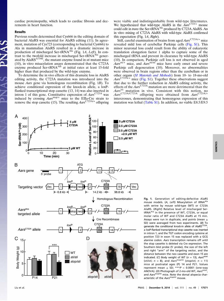
ResultsPrevious results determined that Cys666 in the editing domain ofbacterial AlaRS was essential for AlaRS editing (11). In agree-ment, mutation of Cys723 (corresponding to bacterial Cys666) toAla in mammalian AlaRS resulted in a dramatic increase inproduction of mischarged Ser-tRNAAla (Fig. 1A, Left). In con-trast to the twofold increase in mischarged Ser-tRNAAla gener-ated by AlaRSA734E, the mutant enzyme found in sti mutant mice(10), in vitro misacylation assays demonstrated that the C723Aenzyme produced Ser-tRNAAla at initial rates at least 15-foldhigher than that produced by the wild-type enzyme.To determine the in vivo effects of this dramatic loss in AlaRS
editing activity, the C723A mutation was introduced into themouse Aars gene via homologous recombination (Fig. 1B). Toachieve conditional expression of the knock-in allele, a loxP-flanked transcriptional stop cassette (13, 14) was also inserted inintron 1 of this gene. Constitutive expression of AarsC723A wasinduced by crossing Aarsstop/+ mice to the EIIa-Cre strain toremove the stop cassette (15). The resulting AarsC723A/+ offspring
were viable and indistinguishable from wild-type littermates.We hypothesized that wild-type AlaRS in the AarsC723/+ mousecould edit in trans the Ser-tRNAAla produced by C723A AlaRS. Anin vitro mixing of C723A AlaRS with wild-type AlaRS confirmedthis expectation (Fig. 1A, Right).Still, careful examination of brains from aged AarsC723A/+ mice
revealed mild loss of cerebellar Purkinje cells (Fig. S1). Thisminor neuronal loss could result from the ability of eukaryotictranslation elongation factor 1 alpha to capture some of themischarged tRNA and prevent its clearance by wild-type AlaRS(10). In comparison, Purkinje cell loss is not observed in agedAarssti/+ mice, and Aarssti/sti mice have early onset and severePurkinje cell degeneration (10). Moreover, no abnormalitieswere observed in brain regions other than the cerebellum or inother organs (SI Materials and Methods) from 10- to 18-mo-oldAarsC723A/+ mice (Fig. S1). Together these observations suggestthat due to the further reduction in AlaRS editing activity, theeffects of the AarsC723A mutation are more detrimental than theAarssti mutation in vivo. Consistent with this notion, noAarsC723A/C723A offspring were obtained from AarsC723A/+
intercrosses, demonstrating that homozygous expression of thismutation was lethal (Table S1). In addition, no viable E8.5/E9.5
Fig. 1. Generation of editing-defective AlaRSmouse models. (A, Left) Misacylation of tRNAAla
with serine by mouse wild-type (WT) or C723AAlaRS. (Right) Relative level of mischarged Ser-tRNAAla in the presence of WT, C723A, or equalmolar ratio of WT and C723A AlaRS at 15 min.Assays were run in duplicate, and points (mean ±SD) were averaged from two experiments. (B) Togenerate the conditional knock-in allele of AlaRSC723A,a loxP-flanked transcriptional stop cassette was insertedin intron 1, and the TGT codon encoding cysteine atposition 723 in exon 15 was replaced with a GCGalanine codon. Aars transcription remains off untilthe stop cassette is deleted via Cre expression. TheSouthern blot probe (5′ probe), the size of the leftand right “arm” of the targeting vector, and thedistance between the neo cassette and exon 15 areindicated. (C) Body weight of WT (n = 13), Aarssti/sti
(sti/sti; n = 8), and Aarsstop/sti (stop/sti; n = 11)mice at postnatal ages (P) 14 and P21. Valuesrepresent mean ± SD; ***P < 0.0001 (one-wayANOVA). (D) Photograph of 2-mo-old WT, Aarssti/sti,and Aarsstop/sti mice. Note the dorsal alopecia char-acteristic of the Aarsstop/sti mouse.
Liu et al. PNAS | December 9, 2014 | vol. 111 | no. 49 | 17571
GEN
ETICS
AarsC723A/C723A embryos were observed, and 26% (n = 9/34) ofdecidua at these time points contained embryos that were tooreabsorbed for genotyping, suggesting that the homozygous em-bryos died at an earlier stage of development (Table S1). Theseresults, compared with our previous findings of neuron-specificdefects in the sti mutant mouse (10), indicate that the AarsC723A
mutation likely causes a higher rate of translational errors in vivothan the sti mutation and that phenotypes caused by deficienciesin AlaRS editing are dependent on the extent of the residualediting activity of the mutant enzyme.To create a mouse model with an editing deficiency between
that of AarsC723A/C723A and Aarssti/sti mice, we generated Aarsstop/sti
compound heterozygotes. These mice were viable and present atweaning age in the expected Mendelian ratios. Although initiallyindistinguishable from wild-type littermates, at 3 wk of ageAarsstop/sti mice were smaller than wild-type or Aarssti/sti mice(Fig. 1C). In addition, larger regions of alopecia were consis-tently observed in Aarsstop/sti than in Aarssti/sti mice (Fig. 1D). Thegene dosage effect of the Aarssti mutation reinforces the notion thatcells differ in their sensitivity to disruptions in translational fidelity.Histological analysis of the Aarsstop/sti brain demonstrated that
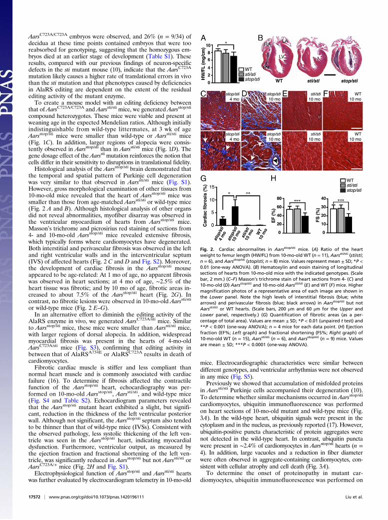
the temporal and spatial pattern of Purkinje cell degenerationwas very similar to that observed in Aarssti/sti mice (Fig. S1).However, gross morphological examination of other tissues from10-mo-old mice revealed that the heart of Aarsstop/sti mice wassmaller than those from age-matched Aarssti/sti or wild-type mice(Fig. 2 A and B). Although histological analysis of other organsdid not reveal abnormalities, myofiber disarray was observed inthe ventricular myocardium of hearts from Aarsstop/sti mice.Masson’s trichrome and picrosirius red staining of sections from4- and 10-mo-old Aarsstop/sti mice revealed extensive fibrosis,which typically forms where cardiomyocytes have degenerated.Both interstitial and perivascular fibrosis was observed in the leftand right ventricular walls and in the interventricular septum(IVS) of affected hearts (Fig. 2 C and D and Fig. S2). Moreover,the development of cardiac fibrosis in the Aarsstop/sti mouseappeared to be age-related: At 1 mo of age, no apparent fibrosiswas observed in heart sections; at 4 mo of age, ∼2.5% of theheart tissue was fibrotic; and by 10 mo of age, fibrotic areas in-creased to about 7.5% of the Aarsstop/sti heart (Fig. 2G). Incontrast, no fibrotic lesions were observed in 10-mo-old Aarssti/sti
or wild-type mice (Fig. 2 E–G).In an alternative effort to diminish the editing activity of the
AlaRS enzyme in vivo, we generated AarsC723A/sti mice. Similarto Aarsstop/sti mice, these mice were smaller than Aarssti/sti mice,with larger regions of dorsal alopecia. In addition, widespreadmyocardial fibrosis was present in the hearts of 4-mo-oldAarsC723A/sti mice (Fig. S3), confirming that editing activity inbetween that of AlaRSA734E or AlaRSC723A results in death ofcardiomyocytes.Fibrotic cardiac muscle is stiffer and less compliant than
normal heart muscle and is commonly associated with cardiacfailure (16). To determine if fibrosis affected the contractilefunction of the Aarsstop/sti heart, echocardiography was per-formed on 10-mo-old Aarsstop/sti, Aarssti/sti, and wild-type mice(Fig. S4 and Table S2). Echocardiogram parameters revealedthat the Aarsstop/sti mutant heart exhibited a slight, but signifi-cant, reduction in the thickness of the left ventricular posteriorwall. Although not significant, the Aarsstop/sti septum also tendedto be thinner than that of wild-type mice (IVSs). Consistent withthe observed pathology, less systolic thickening of the left ven-tricle was seen in the Aarsstop/sti heart, indicating myocardialdysfunction. Furthermore, ventricular output, as measured bythe ejection fraction and fractional shortening of the left ven-tricle, was significantly reduced in Aarsstop/sti but not Aarssti/sti orAarsC723A/+ mice (Fig. 2H and Fig. S1).Electrophysiological function of Aarsstop/sti and Aarssti/sti hearts
was further evaluated by electrocardiogram telemetry in 10-mo-old
mice. Electrocardiographic characteristics were similar betweendifferent genotypes, and ventricular arrhythmias were not observedin any mice (Fig. S5).Previously we showed that accumulation of misfolded proteins
in Aarssti/sti Purkinje cells accompanied their degeneration (10).To determine whether similar mechanisms occurred in Aarsstop/sti
cardiomyocytes, ubiquitin immunofluorescence was performedon heart sections of 10-mo-old mutant and wild-type mice (Fig.3A). In the wild-type heart, ubiquitin signals were present in thecytoplasm and in the nucleus, as previously reported (17). However,ubiquitin-positive puncta characteristic of protein aggregates werenot detected in the wild-type heart. In contrast, ubiquitin punctawere present in ∼2.4% of cardiomyocytes in Aarsstop/sti hearts (n =4). In addition, large vacuoles and a reduction in fiber diameterwere often observed in aggregate-containing cardiomyocytes, con-sistent with cellular atrophy and cell death (Fig. 3A).To determine the onset of proteinopathy in mutant car-
diomyocytes, ubiquitin immunofluorescence was performed on
Fig. 2. Cardiac abnormalites in Aarsstop/sti mice. (A) Ratio of the heartweight to femur length (HW/FL) from 10-mo-old WT (n = 11), Aarssti/sti (sti/sti;n = 6), and Aarsstop/sti (stop/sti; n = 8) mice. Values represent mean ± SD; *P <0.01 (one-way ANOVA). (B) Hematoxylin and eosin staining of longitudinalsections of hearts from 10-mo-old mice with the indicated genotypes. (Scalebar, 2 mm.) (C–F) Masson’s trichrome stain of heart sections from 4- (C) and10-mo-old (D) Aarsstop/sti and 10-mo-old Aarssti/sti (E) and WT (F) mice. Highermagnification photos of a representative area of each image are shown inthe Lower panel. Note the high levels of interstitial fibrosis (blue; whitearrows) and perivascular fibrosis (blue; black arrows) in Aarsstop/sti but notAarssti/sti or WT hearts. (Scale bars, 200 μm and 60 μm for the Upper andLower panel, respectively.) (G) Quantification of fibrotic areas (as a per-centage of total area). Values are mean ± SD; *P < 0.01 (unpaired t test) and**P < 0.001 (one-way ANOVA); n = 4 mice for each data point. (H) Ejectionfraction (EF%; Left graph) and fractional shortening (FS%; Right graph) of10-mo-old WT (n = 15), Aarssti/sti (n = 6), and Aarsstop/sti (n = 9) mice. Valuesare mean ± SD; ***P < 0.0001 (one-way ANOVA).
17572 | www.pnas.org/cgi/doi/10.1073/pnas.1420196111 Liu et al.
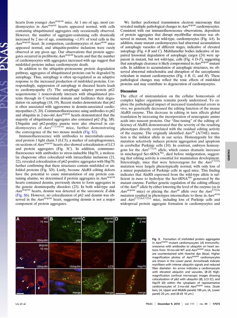
hearts from younger Aarsstop/sti mice. At 1 mo of age, most car-diomyocytes in Aarsstop/sti hearts appeared normal, with cellscontaining ubiquitinated aggregates only occasionally observed.However, the number of aggregate-containing cells drasticallyincreased by 2 mo of age, constituting ∼1.8% of total cells in theAarsstop/sti heart. In comparison, cardiomyocytes of Aarssti/sti miceappeared normal, and ubiquitin-positive inclusions were rarelyobserved at any given age. Our observations that protein aggre-gates occurred in prefibrotic Aarsstop/sti hearts and that the numberof cardiomyocytes with aggregates increased with age suggest thatmisfolded proteins induce cardiomyocyte death.In addition to the ubiquitin–proteasome protein degradation
pathway, aggregates of ubiquitinated proteins can be degraded byautophagy. Thus, autophagy is often up-regulated as an adaptiveresponse to the increased production of misfolded proteins. Cor-respondingly, suppression of autophagy in diseased hearts leadsto cardiomyopathy (5). The autophagic adapter protein p62/sequestosome 1 noncovalently interacts with ubiquitinated pro-teins through its C-terminal domain and facilitates their degra-dation via autophagy (18, 19). Recent studies demonstrate that p62is often associated with aggresomes in desmin-associated cardio-myopathies (5, 20). Coimmunofluorescence using antibodies to p62and ubiquitin in 2-mo-old Aarsstop/sti hearts demonstrated that themajority of ubiquitinated aggregates also contained p62 (Fig. 3B).Ubiquitin and p62-positive puncta were also observed in car-diomyocytes of AarsC723A/sti mice, further demonstratingthe convergence of the two mouse models (Fig. S3).Immunofluorescence with antibodies to microtubule-associ-
ated protein 1 light chain 3 (LC3), a marker of autophagosomes,on sections of Aarsstop/sti hearts also showed colocalization of LC3and protein aggregates (Fig. 3C). In addition, coimmuno-fluorescence with antibodies to stress-inducible Hsp70, a molecu-lar chaperone often colocalized with intracellular inclusions (21,22), revealed colocalization of p62-positive aggregates with Hsp70,further confirming that these structures contain misfolded or un-folded proteins (Fig. 3D). Lastly, because AlaRS editing defectshave the potential to cause mistranslation of any protein con-taining alanine, we determined if protein aggregates in Aarsstop/sti
hearts contained desmin, previously shown to form aggregates inthe genetic desminopathy disorders (23). In both wild-type andAarsstop/sti hearts, desmin was detected at the sarcomeric Z-disk(Fig. S6). However, no colocalization of p62 and desmin was ob-served in the Aarsstop/sti heart, suggesting desmin is not a majorcomponent of protein aggregates.
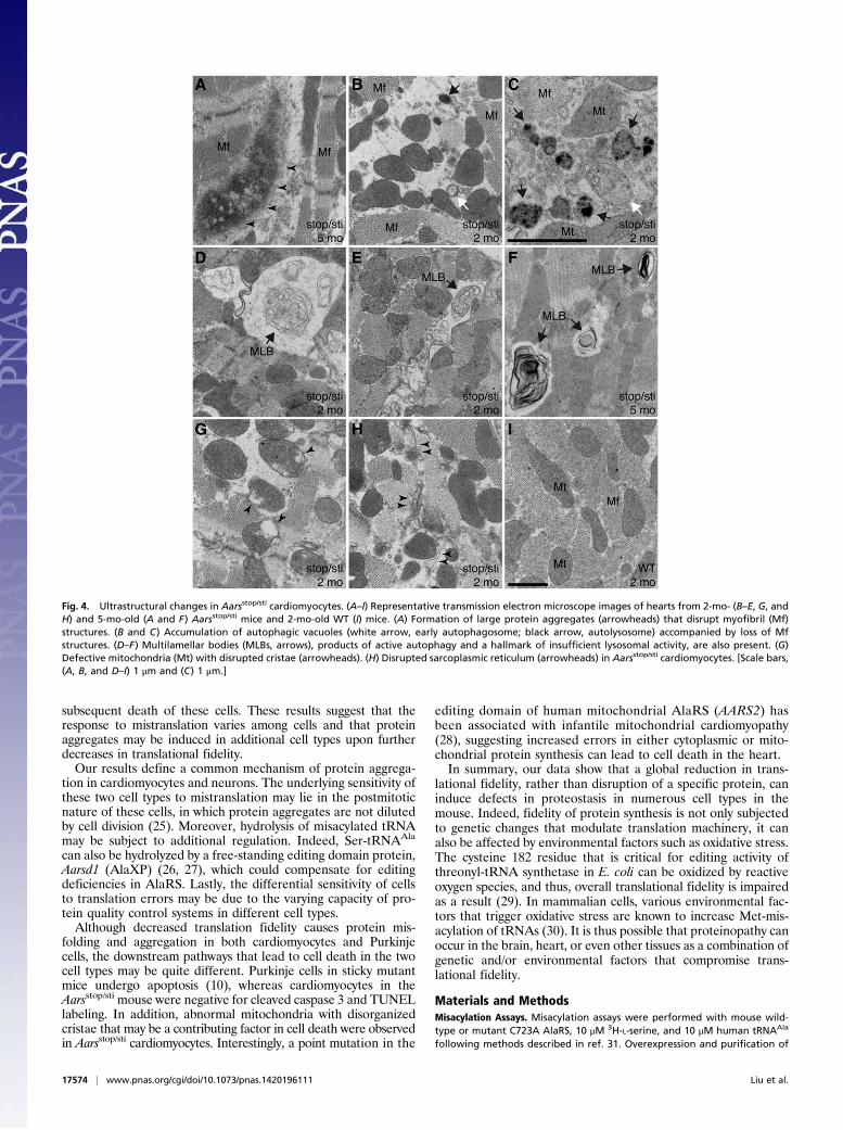
We further performed transmission electron microscopy thatrevealed multiple pathological changes in Aarsstop/sti cardiomyocytes.Consistent with our immunofluorescence observations, depositionof protein aggregates that disrupt myofibrillar structure was ob-served in mutant, but not wild-type, cardiomyocytes (Fig. 4A). Inaddition, many mutant cardiomyocytes had abnormal accumulationof autophagic vacuoles of different stages, indicative of elevatedautophagy (Fig. 4 B and C). Multilamellar bodies indicative of im-paired lysosomal degradation of autophagic cargos (24) were ap-parent in mutant, but not wild-type, cells (Fig. 4 D–F), suggestingthat autophagic clearance is likely compromised inAarsstop/sti mutantcells. In addition to accumulation of autophagic structures, we ob-served abnormal mitochondria and disruption of the sarcoplasmicreticulum in mutant cardiomyocytes (Fig. 4 B, G, and H). Thesepathological changes may reflect the toxic effects of misfoldedproteins and may contribute to degeneration of cardiomyocytes.
DiscussionThe effect of mistranslation on the cellular homeostasis ofcomplex higher organisms remains poorly understood. To ex-plore the pathological impact of increased translational errors invivo, we genetically decreased the editing function of the mouseAlaRS enzyme. This decrease in editing in turn affects globaltranslation by increasing the incorporation of noncognate aminoacids into nascent proteins. Our “fine-tuning” of the editing ef-ficiency of AlaRS demonstrated that the severity of the resultingphenotypes directly correlated with the residual editing activityof the enzyme. The originally identified Aarssti (A734E) muta-tion is the mildest allele in our series. Homozygosity for thismutation selectively induces protein aggregation and apoptosisin cerebellar Purkinje cells (10). In contrast, embryos homozy-gous for the AarsC723A allele, which causes dramatic increasesin mischarged Ser-tRNAAla, died before midgestation, suggest-ing that editing activity is essential for mammalian development.Interestingly, mice that were heterozygous for the AarsC723A
mutation were largely phenotypically normal, with only loss ofa minor population of Purkinje cells in aged mice. This findingindicates that AlaRS expressed from the wild-type allele is suf-ficient in trans to hydrolyze the Ser-tRNAAla generated by themutant enzyme. Further genetic regulation of the editing efficacyof the Aarssti allele by either lowering the level of the enzyme (as inAarsstop/sti mice) or placing the Aarssti allele over the AarsC723A
mutation resulted in phenotypes intermediate to those in Aarssti/sti
and AarsC723A/C723A mice, including loss of Purkinje cells andwidespread protein aggregate formation in cardiomyocytes and
Fig. 3. Formation of misfolded protein aggregatesin Aarsstop/sti mutant cardiomyocytes. (A) Immunoflu-orescence with antibodies to ubiquitin on heart sec-tions from 10-mo-old WT and Aarsstop/sti mice. Nucleiare counterstained with Hoechst dye (blue). Highermagnification photos of Aarsstop/sti cardiomyocytesare shown in the Lower panel. Arrowheads indicatemyofibers with intense ubiquitin signals and reducedfiber diameter. An arrow indicates a cardiomyocytewith elevated ubiquitin and vacuoles. (B–D) High-magnification confocal microscopic images showingcolocalization of p62 with ubiquitin (B), LC3 (C), andHsp70 (D) within the cytoplasm of representativecardiomyocytes of 2-mo-old Aarsstop/sti mice. [Scalebars, (A, Upper and Middle panels) 100 μm, (A, Lowerpanel) 20 μm, and (B–D) 10 μm.]
Liu et al. PNAS | December 9, 2014 | vol. 111 | no. 49 | 17573
GEN
ETICS
subsequent death of these cells. These results suggest that theresponse to mistranslation varies among cells and that proteinaggregates may be induced in additional cell types upon furtherdecreases in translational fidelity.Our results define a common mechanism of protein aggrega-
tion in cardiomyocytes and neurons. The underlying sensitivity ofthese two cell types to mistranslation may lie in the postmitoticnature of these cells, in which protein aggregates are not dilutedby cell division (25). Moreover, hydrolysis of misacylated tRNAmay be subject to additional regulation. Indeed, Ser-tRNAAla
can also be hydrolyzed by a free-standing editing domain protein,Aarsd1 (AlaXP) (26, 27), which could compensate for editingdeficiencies in AlaRS. Lastly, the differential sensitivity of cellsto translation errors may be due to the varying capacity of pro-tein quality control systems in different cell types.Although decreased translation fidelity causes protein mis-
folding and aggregation in both cardiomyocytes and Purkinjecells, the downstream pathways that lead to cell death in the twocell types may be quite different. Purkinje cells in sticky mutantmice undergo apoptosis (10), whereas cardiomyocytes in theAarsstop/sti mouse were negative for cleaved caspase 3 and TUNELlabeling. In addition, abnormal mitochondria with disorganizedcristae that may be a contributing factor in cell death were observedin Aarsstop/sti cardiomyocytes. Interestingly, a point mutation in the
editing domain of human mitochondrial AlaRS (AARS2) hasbeen associated with infantile mitochondrial cardiomyopathy(28), suggesting increased errors in either cytoplasmic or mito-chondrial protein synthesis can lead to cell death in the heart.In summary, our data show that a global reduction in trans-
lational fidelity, rather than disruption of a specific protein, caninduce defects in proteostasis in numerous cell types in themouse. Indeed, fidelity of protein synthesis is not only subjectedto genetic changes that modulate translation machinery, it canalso be affected by environmental factors such as oxidative stress.The cysteine 182 residue that is critical for editing activity ofthreonyl-tRNA synthetase in E. coli can be oxidized by reactiveoxygen species, and thus, overall translational fidelity is impairedas a result (29). In mammalian cells, various environmental fac-tors that trigger oxidative stress are known to increase Met-mis-acylation of tRNAs (30). It is thus possible that proteinopathy canoccur in the brain, heart, or even other tissues as a combination ofgenetic and/or environmental factors that compromise trans-lational fidelity.
Materials and MethodsMisacylation Assays. Misacylation assays were performed with mouse wild-type or mutant C723A AlaRS, 10 μM 3H-L-serine, and 10 μM human tRNAAla
following methods described in ref. 31. Overexpression and purification of
Fig. 4. Ultrastructural changes in Aarsstop/sti cardiomyocytes. (A–I) Representative transmission electron microscope images of hearts from 2-mo- (B–E, G, andH) and 5-mo-old (A and F) Aarsstop/sti mice and 2-mo-old WT (I) mice. (A) Formation of large protein aggregates (arrowheads) that disrupt myofibril (Mf)structures. (B and C) Accumulation of autophagic vacuoles (white arrow, early autophagosome; black arrow, autolysosome) accompanied by loss of Mfstructures. (D–F) Multilamellar bodies (MLBs, arrows), products of active autophagy and a hallmark of insufficient lysosomal activity, are also present. (G)Defective mitochondria (Mt) with disrupted cristae (arrowheads). (H) Disrupted sarcoplasmic reticulum (arrowheads) in Aarsstop/sti cardiomyocytes. [Scale bars,(A, B, and D–I) 1 μm and (C) 1 μm.]
17574 | www.pnas.org/cgi/doi/10.1073/pnas.1420196111 Liu et al.
human tRNAAla was performed as previously described (10). Details areprovided in SI Materials and Methods.
Mice. All animal protocols were approved by The Jackson Laboratory AnimalCare and Use Committee. Homologous recombination of the Aarsstop allelewas performed in C57BL/6J-Tyrc-2J ES cells. Aarssti/sti and Tg(EIIa-cre)C5379Lmgd/J mice were maintained on a C57BL/6 genetic background (10,15). Generation of the knock-in mouse and genotyping information aredescribed in SI Materials and Methods.
Echocardiography and Electrocardiography. Cardiac function, heart dimen-sions, and electrical activity of the heart were evaluated by echocardiographyand electrocardiography on anesthetized mice. Details are provided in SIMaterials and Methods.
Histology. Hematoxylin and eosin, Masson’s trichrome, and picrosirius redstaining was performed on 10% (vol/vol) neutral buffered formalin (NBF)-fixed tissue sections. Details are described in SI Materials and Methods.
Immunofluorescence. The 10% NBF-fixed, paraffin-embedded heart sectionswere deparaffinated, rehydrated, and microwaved in 0.01 M citrate buffer(pH 6.0) three times for 2 min each. Tissue sections were then blocked with4% (wt/vol) goat serum in phosphate-buffered saline with Tween-20 (PBST)for 30 min, incubated with primary antibodies overnight at 4 °C, washed in
PBST, and incubated with secondary antibodies for 1 h. Details about anti-bodies are described in SI Materials and Methods.
Electron Microscopy. Mice were transcardially perfused with 2% (wt/vol)paraformaldehyde and 2% (wt/vol) glutaraldehyde. Tissues were preparedfor analysis using standard procedures.
Statistics. Results are mean ± SD. Statistical significance was determined byunpaired t tests or one-way ANOVA followed by Dunnett’s multiple-com-parisons test. P values less than 0.05 were considered significant.
ACKNOWLEDGMENTS. We are grateful to Jennifer Cook and Krystal-LeighBaker for technical assistance, Litao Sun for tRNAAla, Jennifer Ryan for ECGand echocardiography recordings, Mark Lessard for help with imagingand quantification, Pete Finger for electron microscopy assistance, andDoug McMinimy for mouse photos. We also thank Dr. Karen Svensonfor helpful discussions, Dr. Greg Cox for comments on the manuscript,and The Jackson Laboratory sequencing, histology, and microinjectionservices for their contributions. These studies were supported by NationalInstitutes of Health Grant NS042613 (to S.L.A.), and services used in thisstudy were supported by Cancer Center Core Grant CA34196 (The JacksonLaboratory). Support was also provided by aTyr Pharma, Inc., The NationalFoundation for Cancer Research, and National Cancer Institute GrantCA92577. J.S.S. was supported by a fellowship from the American HealthAssistance Foundation. S.L.A. is an investigator of the Howard HughesMedical Institute.
1. Goldfarb LG, Dalakas MC (2009) Tragedy in a heartbeat: Malfunctioning desmincauses skeletal and cardiac muscle disease. J Clin Invest 119(7):1806–1813.
2. McLendon PM, Robbins J (2011) Desmin-related cardiomyopathy: An unfolding story.Am J Physiol Heart Circ Physiol 301(4):H1220–H1228.
3. Quarta CC, Kruger JL, Falk RH (2012) Cardiac amyloidosis. Circulation 126(12):e178–e182.
4. Wang X, Robbins J (2006) Heart failure and protein quality control. Circ Res 99(12):1315–1328.
5. Zheng Q, Su H, Ranek MJ, Wang X (2011) Autophagy and p62 in cardiac proteinopathy.Circ Res 109(3):296–308.
6. Ling J, Reynolds N, Ibba M (2009) Aminoacyl-tRNA synthesis and translational qualitycontrol. Annu Rev Microbiol 63:61–78.
7. Schimmel P (2008) Development of tRNA synthetases and connection to genetic codeand disease. Protein Sci 17(10):1643–1652.
8. Loftfield RB, Vanderjagt D (1972) The frequency of errors in protein biosynthesis.Biochem J 128(5):1353–1356.
9. Jakubowski H, Goldman E (1992) Editing of errors in selection of amino acids forprotein synthesis. Microbiol Rev 56(3):412–429.
10. Lee JW, et al. (2006) Editing-defective tRNA synthetase causes protein misfolding andneurodegeneration. Nature 443(7107):50–55.
11. Beebe K, Ribas De Pouplana L, Schimmel P (2003) Elucidation of tRNA-dependentediting by a class II tRNA synthetase and significance for cell viability. EMBO J 22(3):668–675.
12. Nangle LA, Motta CM, Schimmel P (2006) Global effects of mistranslation from anediting defect in mammalian cells. Chem Biol 13(10):1091–1100.
13. Soriano P (1999) Generalized lacZ expression with the ROSA26 Cre reporter strain. NatGenet 21(1):70–71.
14. Srinivas S, et al. (2001) Cre reporter strains produced by targeted insertion of EYFPand ECFP into the ROSA26 locus. BMC Dev Biol 1:4.
15. Lakso M, et al. (1996) Efficient in vivo manipulation of mouse genomic sequences atthe zygote stage. Proc Natl Acad Sci USA 93(12):5860–5865.
16. Weber KT, Brilla CG, Janicki JS (1993) Myocardial fibrosis: Functional significance andregulatory factors. Cardiovasc Res 27(3):341–348.
17. Hilenski LL, Terracio L, Haas AL, Borg TK (1992) Immunolocalization of ubiquitinconjugates at Z-bands and intercalated discs of rat cardiomyocytes in vitro and in vivo.J Histochem Cytochem 40(7):1037–1042.
18. Vadlamudi RK, Joung I, Strominger JL, Shin J (1996) p62, a phosphotyrosine-independent ligand of the SH2 domain of p56lck, belongs to a new class of ubiquitin-binding proteins. J Biol Chem 271(34):20235–20237.
19. Pankiv S, et al. (2007) p62/SQSTM1 binds directly to Atg8/LC3 to facilitate degradationof ubiquitinated protein aggregates by autophagy. J Biol Chem 282(33):24131–24145.
20. Olivé M, et al. (2008) Expression of mutant ubiquitin (UBB+1) and p62 in my-otilinopathies and desminopathies. Neuropathol Appl Neurobiol 34(1):76–87.
21. Warrick JM, et al. (1999) Suppression of polyglutamine-mediated neurodegenerationin Drosophila by the molecular chaperone HSP70. Nat Genet 23(4):425–428.
22. Waelter S, et al. (2001) Accumulation of mutant huntingtin fragments in aggresome-like inclusion bodies as a result of insufficient protein degradation.Mol Biol Cell 12(5):1393–1407.
23. Clemen CS, Herrmann H, Strelkov SV, Schröder R (2013) Desminopathies: Pathologyand mechanisms. Acta Neuropathol 125(1):47–75.
24. Hariri M, et al. (2000) Biogenesis of multilamellar bodies via autophagy. Mol Biol Cell11(1):255–268.
25. Soonpaa MH, Field LJ (1997) Assessment of cardiomyocyte DNA synthesis in normaland injured adult mouse hearts. Am J Physiol 272(1 Pt 2):H220–H226.
26. Nawaz MH, Merriman E, Yang XL, Schimmel P (2011) p23H implicated as cis/transregulator of AlaXp-directed editing for mammalian cell homeostasis. Proc Natl AcadSci USA 108(7):2723–2728.
27. Chong YE, Yang XL, Schimmel P (2008) Natural homolog of tRNA synthetase editingdomain rescues conditional lethality caused by mistranslation. J Biol Chem 283(44):30073–30078.
28. Götz A, et al. (2011) Exome sequencing identifies mitochondrial alanyl-tRNA syn-thetase mutations in infantile mitochondrial cardiomyopathy. Am J Hum Genet 88(5):635–642.
29. Ling J, Söll D (2010) Severe oxidative stress induces protein mistranslation throughimpairment of an aminoacyl-tRNA synthetase editing site. Proc Natl Acad Sci USA107(9):4028–4033.
30. Netzer N, et al. (2009) Innate immune and chemically triggered oxidative stressmodifies translational fidelity. Nature 462(7272):522–526.
31. Beebe K, Waas W, Druzina Z, Guo M, Schimmel P (2007) A universal plate format forincreased throughput of assays that monitor multiple aminoacyl transfer RNA syn-thetase activities. Anal Biochem 368(1):111–121.
Liu et al. PNAS | December 9, 2014 | vol. 111 | no. 49 | 17575
GEN
ETICS