
Defibrillation and Cardioversion - NSW Agency for … · Appendix 1. Definitions:2 ... Pay careful...
12
Liverpool Hospital ICU Guideline: Defibrillation and Cardioversion Intensive Care Unit Systems_Cardiovascular ICU2015_ICU_Guidelines_Systems_Cardiovascular_Defibrillation_and_Cardioversion Page 1 of 12 Guideline Title: Defibrillation and Cardioversion Summary: 1 Electrical cardioversion and defibrillation are procedures in the management of patients with cardiac arrhythmias. Cardioversion is the delivery of energy that is synchronised to the QRS complex, while defibrillation is the non-synchronised delivery of a shock randomly during the cardiac cycle. Approved by: ICU Medical Director Prof Michael Parr Publication (Issue) Date: July 2015 Next Review Date: July 2018 Replaces Existing Guideline: Defibrillation and Cardioversion_ November 2011 Contents: 1. Definitions 2. Background Information 3. Introduction 4. Policy Statement 5. Principles/guidelines 6. Clinical issues 7. Indications 8. Contraindications 9. Precautions 10. Troubleshooting 11. Performance Measures 12. References 13. Appendix 1. Definitions: 2 Energy: Energy in a defibrillator is expressed in joules. A joule is the unit of work associated with one amp of current passed through one ohm of resistance for one second. When we express it in a formula, it is generally stated as follows: Joules (Energy) = Voltage X Current X Time Joules have become a surrogate for current in modern defibrillator language. Current: Current is what actually defibrillates the heart. It is also expressed as Voltage/Impedance (resistance). Impedance: Resistance to Flow; there is resistance in the electrical circuit itself as well as in the patient. The amount of impedance in a patient is difficult to determine as it relates to body mass, temperature, diaphoresis, and quality of the contact with paddles or pads. Impedance is expressed in ohms (ohms).
Transcript of Defibrillation and Cardioversion - NSW Agency for … · Appendix 1. Definitions:2 ... Pay careful...
Liverpool Hospital ICU Guideline: Defibrillation and Cardioversion Intensive Care Unit Systems_Cardiovascular
ICU2015_ICU_Guidelines_Systems_Cardiovascular_Defibrillation_and_Cardioversion Page 1 of 12
Guideline Title: Defibrillation and Cardioversion Summary: 1
Electrical cardioversion and defibrillation are procedures in the management of patients with cardiac arrhythmias. Cardioversion is the delivery of energy that is synchronised to the QRS complex, while defibrillation is the non-synchronised delivery of a shock randomly during the cardiac cycle.
Approved by: ICU Medical Director Prof Michael Parr
Publication (Issue) Date: July 2015 Next Review Date: July 2018 Replaces Existing Guideline: Defibrillation and Cardioversion_ November 2011 Contents:
1. Definitions 2. Background Information 3. Introduction 4. Policy Statement 5. Principles/guidelines 6. Clinical issues 7. Indications 8. Contraindications 9. Precautions 10. Troubleshooting 11. Performance Measures 12. References 13. Appendix
1. Definitions: 2 Energy:
Energy in a defibrillator is expressed in joules. A joule is the unit of work associated with one amp of current passed through one ohm of resistance for one second.
When we express it in a formula, it is generally stated as follows: Joules (Energy) = Voltage X Current X Time Joules have become a surrogate for current in modern defibrillator language.
Current: Current is what actually defibrillates the heart. It is also expressed as
Voltage/Impedance (resistance). Impedance:
Resistance to Flow; there is resistance in the electrical circuit itself as well as in the patient. The amount of impedance in a patient is difficult to determine as it relates to body mass, temperature, diaphoresis, and quality of the contact with paddles or pads. Impedance is expressed in ohms (ohms).
Liverpool Hospital ICU Guideline: Defibrillation and Cardioversion Intensive Care Unit Systems_Cardiovascular
ICU2015_ICU_Guidelines_Systems_Cardiovascular_Defibrillation_and_Cardioversion Page 2 of 12
2. Background Information: 1
Defibrillation is treatment for life-threatening cardiac arrhythmias such as ventricular fibrillation and pulseless ventricular tachycardia. Electrical energy is delivered to the heart via a device called a defibrillator and pads which are placed on the chest. This depolarises a critical mass of the heart muscle, terminates the arrhythmia, and allows normal sinus rhythm to be re-established by the body's natural pacemaker, in the sino- atrial node of the heart. Defibrillators can be external, transvenous, or implanted, depending on the type of device used or needed. This guideline will only discuss external defibrillation.
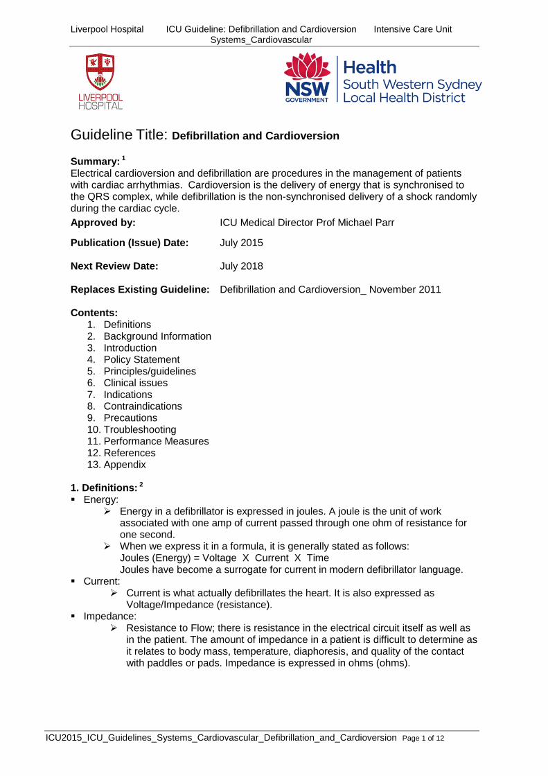
Most defibrillators are energy-based, meaning that the devices charge a capacitor to a selected level and then deliver a pre-specified amount of energy in joules. The amount of energy which arrives at the myocardium is dependent upon the selected energy level and the transthoracic impedance (which varies by patient)1 Defibrillators can also deliver energy in a variety of waveforms, characterised as monophasic, where the current flows in one direction, or biphasic, where there are two current pulses in opposite directions. Biphasic waveforms defibrillate at lower energies than monophasic waveforms. Biphasic defibrillators are now the only one used in ICU.
Biphasic waveforms. www.resuscitationcentral.com
There is good evidence to suggest that minimal time delay to defibrillation for Ventricular Fibrillation and Pulseless Ventricular Tachycardia improves patient survival.
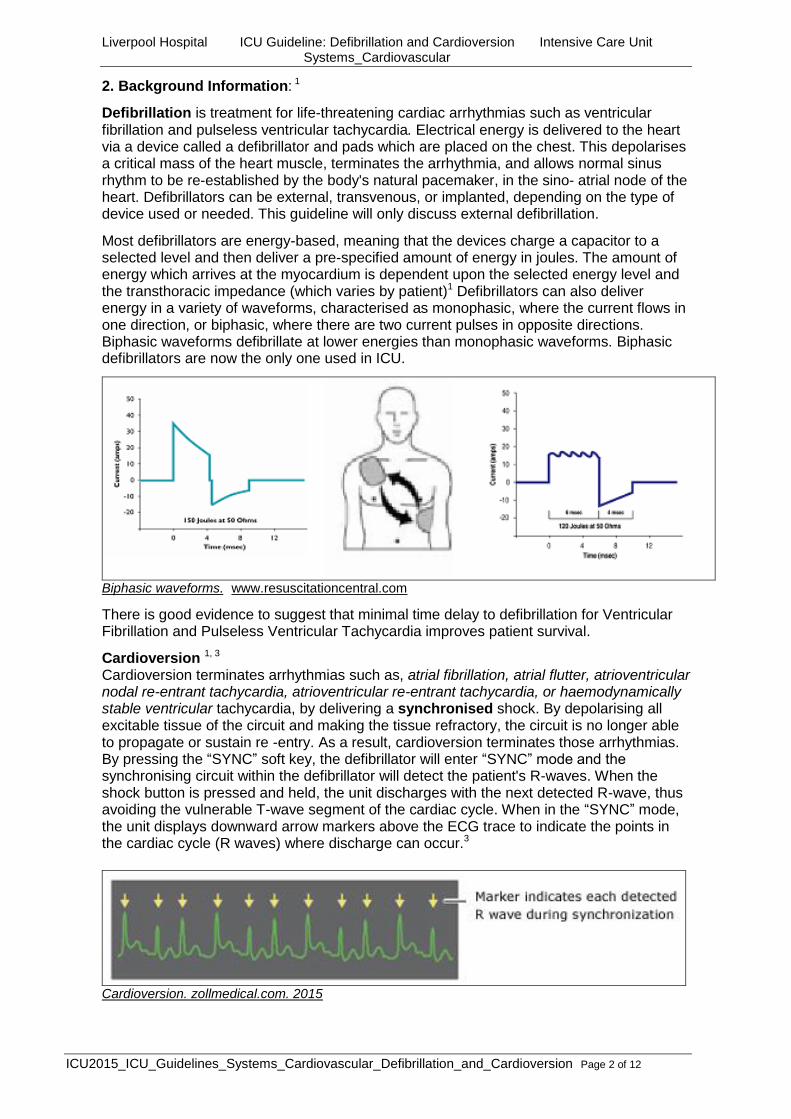
Cardioversion 1, 3 Cardioversion terminates arrhythmias such as, atrial fibrillation, atrial flutter, atrioventricular nodal re-entrant tachycardia, atrioventricular re-entrant tachycardia, or haemodynamically stable ventricular tachycardia, by delivering a synchronised shock. By depolarising all excitable tissue of the circuit and making the tissue refractory, the circuit is no longer able to propagate or sustain re -entry. As a result, cardioversion terminates those arrhythmias. By pressing the “SYNC” soft key, the defibrillator will enter “SYNC” mode and the synchronising circuit within the defibrillator will detect the patient's R-waves. When the shock button is pressed and held, the unit discharges with the next detected R-wave, thus avoiding the vulnerable T-wave segment of the cardiac cycle. When in the “SYNC” mode, the unit displays downward arrow markers above the ECG trace to indicate the points in the cardiac cycle (R waves) where discharge can occur.3
Cardioversion. zollmedical.com. 2015

Liverpool Hospital ICU Guideline: Defibrillation and Cardioversion Intensive Care Unit Systems_Cardiovascular
ICU2015_ICU_Guidelines_Systems_Cardiovascular_Defibrillation_and_Cardioversion Page 3 of 12
3. Introduction: The risk addressed by this policy:
Patient Safety
The Aims / Expected Outcome of this policy:
Staff managing a patient who requires defibrillation or cardioversion will have the skills and knowledge to perform this skill effectively and safely.
Related Standards or Legislation
NSQHS Standard 1 Governance
National Standard 4 Medication Safety Related Policies
LH_PD2013_C03.01 Drug Administration
LH_PD2013_C03.00 Drug Prescribing
LH_PD2013_C03.12 Administration of Intravenous (IV) Medications
LH_ICU_2011 Management of Arrhythmias in ICU
LH_ICU_2014 Cardiac Monitoring
LH_ICU_2011 Adrenaline
LH_ICU_2014 Amiodarone
LH_ICU_2011 Atropine
LH_ICU_2013 Suxamethonium
LH_ICU_2013 Vecuronium
LH_ICU_2012 Midazolam
LH_ICU_2012 Propofol
4. Policy Statement: All care provided within Liverpool Hospital will be in accordance with infection
prevention/control, manual handling and minimisation and management of aggression guidelines.
Defibrillation or cardioversion should only be done by accredited staff following ALS assessment
If there are no signs of life ( loss of consciousness, no pulse, abnormal (agonal)breathing) commence immediate CPR and call a MET: dial 666 and state ward and bed number, except if the ICU team are at the bedside
Emergency trolley must be checked each shift by an RN Infection Control guidelines are to be followed. All drugs administered during an emergency (under the direction of a medical officer)
are to be documented during the event, then prescribed and signed following the event. Defibrillation pads must be in good contact with chest wall Defibrillation pads must be checked for an expiry date One defibrillation pad must be positioned at the mid axillary line, left 6th intercostal
space and one to the right parasternal area 2nd intercostal space Before discharging the defibrillator “Stand clear” must be stated loudly and clearly and
a visual sweep of the bed area for any hazards

Liverpool Hospital ICU Guideline: Defibrillation and Cardioversion Intensive Care Unit Systems_Cardiovascular
ICU2015_ICU_Guidelines_Systems_Cardiovascular_Defibrillation_and_Cardioversion Page 4 of 12
Electrical hazards ( jewellery, water, ECG electrodes, GTN patches) must be removed before discharge of defibrillator
When Cardioversion procedure is going to be performed the “SYNC” mode must be activated
5. Principles / Guidelines Equipment:
Defibrillator Multi function adult pads Emergency trolley IV access Mask size 3 or 4 and resuscitation bag Suction equipment Sedative agent for cardioversion as appropriate
Zoll M series: ICU Emergency trolleys
Zoll R series defibrillator: ICU4
Monitoring functions
Pacing functions Rate Output
Defibrillation functions
Monitoring functions
Pacing functions Rate Output
Defibrillation functions

Liverpool Hospital ICU Guideline: Defibrillation and Cardioversion Intensive Care Unit Systems_Cardiovascular
ICU2015_ICU_Guidelines_Systems_Cardiovascular_Defibrillation_and_Cardioversion Page 5 of 12
Zoll X series: MET trolley
Multi function pads
Procedure for Defibrillation Defibrillation as soon as possible provides the best chance of survival in patients with
VF or pulseless VT ARC guidelines for shockable rhythms should be commenced (see Appendix 1) Clean and shave the area where the pads need to be applied (for cardioversion) Remove pads from package and separate lead wires Remove pad protective liner Connect the pads to the defibrillator
Connection from pads to Defibrillator
Pacing functions
Defibrillation functions
Monitoring functions

Liverpool Hospital ICU Guideline: Defibrillation and Cardioversion Intensive Care Unit Systems_Cardiovascular
ICU2015_ICU_Guidelines_Systems_Cardiovascular_Defibrillation_and_Cardioversion Page 6 of 12
Apply the defibrillator ECG electrodes to patients chest
Apply a pad to mid axillary line, left 6th intercostal space and one to the right
parasternal area 2nd intercostal space
Correct placement of defibrillation pads. ARC Guideline 7
If patient has implantable cardioverter defibrillator (ICD) or permanent pacemaker the pads should be placed on chest wall at least 8cms from the device
Ensure there are no IV lines or ECG electrodes under the pads Smooth the pads from the centre outward to the edges with finger tips to ensure there
are no air pockets under the pads Pads are not repositionable. Replace with new pads if they need to be repositioned Replace pads every 24 hours or 50 defibrillations ( Manufacturers recommendations) Turn dial onto defibrillation (Defib)
Steps for defibrillation of shockable rhythms
1. Turn dial to Defib
2. Ensure energy is 200joules
3. Press Charge
4. Press Shock

Liverpool Hospital ICU Guideline: Defibrillation and Cardioversion Intensive Care Unit Systems_Cardiovascular
ICU2015_ICU_Guidelines_Systems_Cardiovascular_Defibrillation_and_Cardioversion Page 7 of 12
The defibrillator will default to biphasic mode and energy 200 joules if not press energy
select button to change joules Charge the defibrillator In a loud clear voice say “STAND CLEAR” and ensure all staff have moved away from
the bed Deliver the shock and recommence compressions Observe patient and ECG monitor for results Continue with ARC algorithm for shockable rhythms (see Appendix 1) Procedure for Cardioversion Explain procedure to patient Sedation may be required if the patient is fully conscious Follow ARC Algorithm for Tachycardia’s (see Management of Arrhythmias
Guideline_2011) Place ECG electrodes from the defibrillator behind the shoulders and away from where
the defibrillation pads are placed Pay careful attention to skin preparation; make sure the surface is dry, free of hair and
lotions that can impact adhesion. Remove pads from the package and separate the lead wires Smooth the pads from the centre outwards to ensure there is no air between the pads
and patients skin If patient has implantable cardioverter defibrillator (ICD) or permanent pacemaker the
pads should be placed on chest wall at least 8cms from the device Ensure there are no IV lines or ECG electrodes under the pads Smooth the pads from the centre outward to the edges with finger tips to ensure there
are no air pockets under the pads Pads are not repositionable. Replace with new pads if they need to be repositioned Replace pads every 24 hours The defibrillation pads for Cardioversion can be placed either Anterior–Posterior (AP) or
Anterior-Anterior (AA), though AP placement is preferable for maximum current flow through the atria2
Posterior pad is placed left lateral of the spine and just under the scapula Anterior pad is placed mid clavicular,4th intercostal space, lateral to the sternum
Anterior placement of defibrillation pad3
Posterior placement of defibrillation pad3
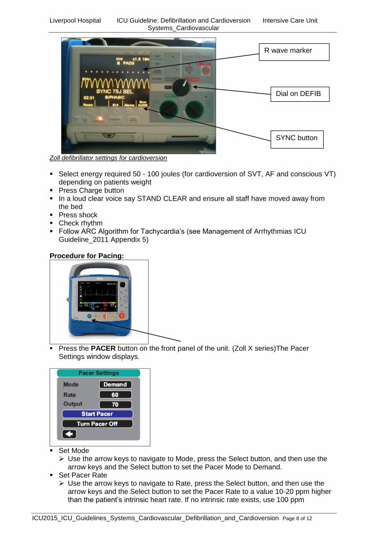
Turn defibrillator dial to Defib Ensure SYNC mode is activated by pressing SYNC button on defibrillator Ensure R wave marker is seen on ECG trace, if not increase amplitude of ECG trace
Liverpool Hospital ICU Guideline: Defibrillation and Cardioversion Intensive Care Unit Systems_Cardiovascular
ICU2015_ICU_Guidelines_Systems_Cardiovascular_Defibrillation_and_Cardioversion Page 8 of 12
Zoll defibrillator settings for cardioversion
Select energy required 50 - 100 joules (for cardioversion of SVT, AF and conscious VT)
depending on patients weight Press Charge button In a loud clear voice say STAND CLEAR and ensure all staff have moved away from
the bed Press shock Check rhythm Follow ARC Algorithm for Tachycardia’s (see Management of Arrhythmias ICU
Guideline_2011 Appendix 5) Procedure for Pacing:
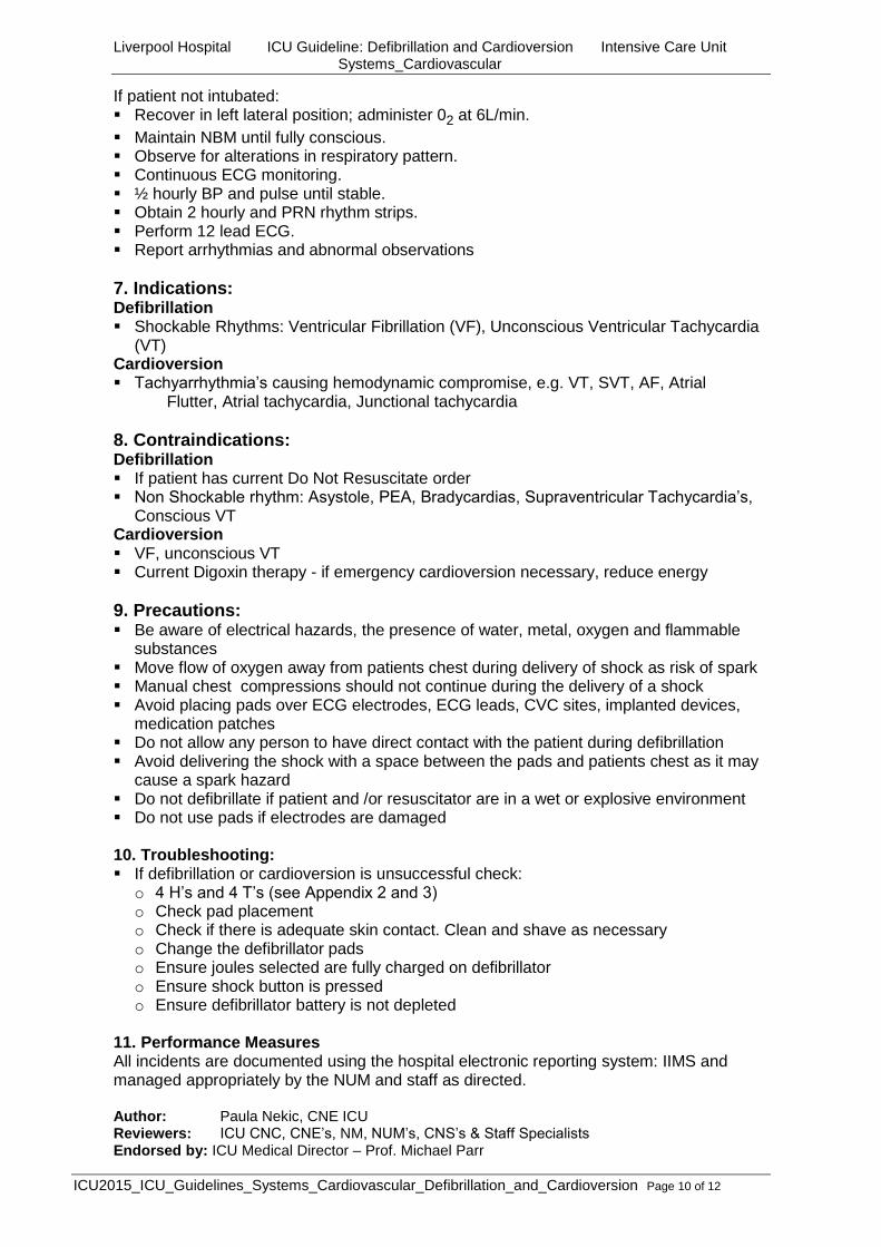
Press the PACER button on the front panel of the unit. (Zoll X series)The Pacer
Settings window displays.
Set Mode Use the arrow keys to navigate to Mode, press the Select button, and then use the
arrow keys and the Select button to set the Pacer Mode to Demand. Set Pacer Rate Use the arrow keys to navigate to Rate, press the Select button, and then use the
arrow keys and the Select button to set the Pacer Rate to a value 10-20 ppm higher than the patient’s intrinsic heart rate. If no intrinsic rate exists, use 100 ppm
SYNC button
Dial on DEFIB
R wave marker

Liverpool Hospital ICU Guideline: Defibrillation and Cardioversion Intensive Care Unit Systems_Cardiovascular
ICU2015_ICU_Guidelines_Systems_Cardiovascular_Defibrillation_and_Cardioversion Page 9 of 12
Turn On Pacer Use the arrow keys to navigate to Start Pacer, then press the Select button to select
it. The Pacing window displays behind the Pacer Settings window
Set Pacer Output In the Pacer Settings window, use the arrow keys and the Select button to adjust the
pacer output. The pacer output is adjustable in 10 mA increments when increasing the output, and
in 5 mA increments when decreasing the output. Observe the ECG for evidence of electrical capture. Select the lowest output current that achieves both electrical and mechanical
capture. Note: If the Pacer Settings window disappears before you have set the output current, press the PACER button again to display the settings window Determine Capture It is important to recognize when pacing stimulation has produced a ventricular
response (capture). Determination of capture must be assessed both electrically and mechanically in
order to ensure appropriate circulatory support of the patient. Electrical capture is determined by the presence of a widened QRS complex, the
loss of any underlying intrinsic rhythm, and the appearance of an extended, and sometimes enlarged, T-wave.
Ventricular response is normally characterized by suppression of the intrinsic QRS complex.
Determine Optimum Threshold
The ideal pacer current is the lowest value that maintains capture — it is usually about 10% above threshold.
Turn down output till capture is lost. Then slowly increase output till capture returns. Then set output 10mv above threshold
For Zoll M and R series Turn dial to pacer (Zoll M and R series) Set rate to 80-100bpm Increase output till capture occurs Find threshold and safety settings
6. Clinical Issues: Minimise interruptions to CPR when defibrillating Manual chest compressions should stop only when delivering a shock Avoid placing pads over ECG electrodes, ECG leads, CVC sites, implanted devices,
medication patches Move patients limbs away from metal fixtures e.g. bed rails Move flow of oxygen away from patients chest during delivery of shock as risk of spark Check that the patient has motor response to shock which indicates delivery of the
charge. If no response may be that defibrillator has flat battery or lead fracture Replace electrode pads every 24hours or 50 defibrillation shocks (Manufacturers
recommendations) Check patients skin for burns
Liverpool Hospital ICU Guideline: Defibrillation and Cardioversion Intensive Care Unit Systems_Cardiovascular
ICU2015_ICU_Guidelines_Systems_Cardiovascular_Defibrillation_and_Cardioversion Page 10 of 12
If patient not intubated: Recover in left lateral position; administer 02 at 6L/min.
Maintain NBM until fully conscious. Observe for alterations in respiratory pattern. Continuous ECG monitoring. ½ hourly BP and pulse until stable. Obtain 2 hourly and PRN rhythm strips. Perform 12 lead ECG. Report arrhythmias and abnormal observations
7. Indications: Defibrillation Shockable Rhythms: Ventricular Fibrillation (VF), Unconscious Ventricular Tachycardia
(VT) Cardioversion Tachyarrhythmia’s causing hemodynamic compromise, e.g. VT, SVT, AF, Atrial
Flutter, Atrial tachycardia, Junctional tachycardia
8. Contraindications: Defibrillation If patient has current Do Not Resuscitate order Non Shockable rhythm: Asystole, PEA, Bradycardias, Supraventricular Tachycardia’s,
Conscious VT Cardioversion VF, unconscious VT Current Digoxin therapy - if emergency cardioversion necessary, reduce energy
9. Precautions: Be aware of electrical hazards, the presence of water, metal, oxygen and flammable
substances Move flow of oxygen away from patients chest during delivery of shock as risk of spark Manual chest compressions should not continue during the delivery of a shock Avoid placing pads over ECG electrodes, ECG leads, CVC sites, implanted devices,
medication patches Do not allow any person to have direct contact with the patient during defibrillation Avoid delivering the shock with a space between the pads and patients chest as it may
cause a spark hazard Do not defibrillate if patient and /or resuscitator are in a wet or explosive environment Do not use pads if electrodes are damaged
10. Troubleshooting: If defibrillation or cardioversion is unsuccessful check:
o 4 H’s and 4 T’s (see Appendix 2 and 3) o Check pad placement o Check if there is adequate skin contact. Clean and shave as necessary o Change the defibrillator pads o Ensure joules selected are fully charged on defibrillator o Ensure shock button is pressed o Ensure defibrillator battery is not depleted
11. Performance Measures All incidents are documented using the hospital electronic reporting system: IIMS and managed appropriately by the NUM and staff as directed. Author: Paula Nekic, CNE ICU Reviewers: ICU CNC, CNE’s, NM, NUM’s, CNS’s & Staff Specialists Endorsed by: ICU Medical Director – Prof. Michael Parr

Liverpool Hospital ICU Guideline: Defibrillation and Cardioversion Intensive Care Unit Systems_Cardiovascular
ICU2015_ICU_Guidelines_Systems_Cardiovascular_Defibrillation_and_Cardioversion Page 11 of 12
12. References / Links 1. Basic principles and technique of cardioversion and defibrillation. www.uptodate.com 2015.
Bradley P Knight MD, FAC 2. Resuscitation Central. Defibrillation. www.resuscitationcentral.com 2015 3. Cardioversion. Zoll Medical Corporation. www.zoll.com 2015 4. Australian Resuscitation Guideline. 2010. Guideline 11.4 Electrical therapy for Adult Advanced
Life Support, accessed 2nd
July 2015 via http://www.resus.org.au/policy/guidelines
13. APPENDIX
Management of Reversible causes: 4 H’s

Liverpool Hospital ICU Guideline: Defibrillation and Cardioversion Intensive Care Unit Systems_Cardiovascular
ICU2015_ICU_Guidelines_Systems_Cardiovascular_Defibrillation_and_Cardioversion Page 12 of 12
4 H’s MANAGEMENT
Hypoxia Check and maintain airway Insert Guedel, ETT, LMA, surgical airway if required Check oxygenation and ventilation
Hypovolaemia Replace blood or fluid loss Replacement of blood with:
- Crystalloid/ Colloid - Blood Products Anaphylaxis:
Management of ABC - Adrenaline (IMI, S/C, or IV) - Hydrocortisone - Correct hypovolaemia
Hypo/Hyperkalaemia Hypokalaemia Potassium of less than 3.5mmol/L Replace Potassium
Hyperkalaemia IV calcium, 10 mLs 10% CaCl2, up to 3 ampoules, each
over 5 minutes hyperventilation: CO2 + H2O H2CO3 H+ + HCO3- 50mls 50 % glucose + 10 units Actrapid over 10-15
minutes. NaHCO3 to correct acidosis Nebulised salbutamol
Hypo/Hyperthermia Hypothermia Active core re-warming Warmed humidified oxygen Warmed intravenous fluids Peritoneal lavage Extracorporeal warming Pleural lavage
Hyperthermia Cooling Blankets Cooling packs or ice to head, axilla, chest, groin and legs Cooled IV fluids
Management of reversible causes: 4 T’s
4 T’s MANAGEMENT
Tamponade Pericardiocentesis open sternotomy wound if post cardiac surgery
Tension Pneumothorax
Thorococentesis -Chest tube insertion if there is time or a large bore needle through the 2nd intercostal space in the mid-clavicular line
Toxins/tablets Antidote Charcoal (within 1 hr of ingestion) Supportive measures ABCDEFG
Thrombus Thrombolysis, embolectomy or cardiopulmonary bypass to allow operative removal of the clot.


![Check List Cardioversion I.gallastegi[1]](https://static.fdocuments.us/doc/165x107/55cf8e57550346703b912349/check-list-cardioversion-igallastegi1.jpg)