Deep brain stimulation for dystonia and essential tremor
190
Deep brain stimulation for dystonia and essential tremor November 2008 MSAC Application 1109 Assessment report
Transcript of Deep brain stimulation for dystonia and essential tremor
Deep brain stimulation for
dystonia and essential tremor
November 2008
MSAC Application 1109
Assessment report
© Commonwealth of Australia 2008
ISBN (Print) 1-74186-717-7
ISBN (Online) 1-74186-718-5
ISSN (Print) 1443-7120
ISSN (Online) 1443-7139
First printed November 2008
Paper-based publications
© Commonwealth of Australia 2008 This work is copyright. Apart from any use as permitted under the Copyright Act 1968, no part may be reproduced by any process without prior written permission from the Commonwealth. Requests and inquiries concerning reproduction and rights should be addressed to the Commonwealth Copyright Administration, Attorney General’s Department, Robert Garran Offices, National Circuit, Barton ACT 2600 or posted at http://www.ag.gov.au/cca Internet sites © Commonwealth of Australia 2008 This work is copyright. You may download, display, print and reproduce this material in unaltered form only (retaining this notice) for your personal, non-commercial use or use within your organisation. Apart from any use as permitted under the Copyright Act 1968, all other rights are reserved. Requests and inquiries concerning reproduction and rights should be addressed to Commonwealth Copyright Administration, Attorney General’s Department, Robert Garran Offices, National Circuit, Barton ACT 2600 or posted at http://www.ag.gov.au/cca Electronic copies of the report can be obtained from the Medical Service Advisory Committee’s Internet site at http://www.msac.gov.au/
Printed copies of the report can be obtained from:
The Secretary Medical Services Advisory Committee Department of Health and Ageing Mail Drop 106 GPO Box 9848 Canberra ACT 2601
Enquiries about the content of the report should be directed to the above address.
The Medical Services Advisory Committee (MSAC) is an independent committee which has been established to provide advice to the Minister for Health and Ageing on the strength of evidence available on new and existing medical technologies and procedures in terms of their safety, effectiveness and cost-effectiveness. This advice will help to inform government decisions about which medical services should attract funding under Medicare.
MSAC recommendations do not necessarily reflect the views of all individuals who participated in the MSAC evaluation. This report was prepared by the Medical Services Advisory Committee with the assistance of Ms Eliana Della Flora, Dr Alun Cameron and Ms Caryn Perera from the Australian Safety and Efficacy Register of New Interventional Procedures – Surgery (ASERNIP-S) and Mr Richard Norman from the Centre for Health Economics Research Evaluation (CHERE). The report was edited by ASERNIP-S.
The Minister for Health and Ageing noted MSAC’s advice on 28 August 2008.
Publication approval number: P3-4469
Deep brain stimulation for dystonia and essential tremor iii
Contents
Executive summary ..........................................................................................................xiii Introduction..........................................................................................................................1 Background ......................................................................................................................... 2
Introduction ..................................................................................................................... 2 The procedure .................................................................................................................. 2 Intended purpose............................................................................................................. 4 Dystonia ............................................................................................................................ 5
Age of onset............................................................................................................... 5 Aetiology .................................................................................................................... 5
Primary dystonia................................................................................................. 5 Secondary dystonia ............................................................................................ 5 Post-anoxic dystonia.......................................................................................... 6 Post-traumatic dystonia..................................................................................... 6 Tardive dystonia ................................................................................................. 6 Paroxysmal dystonia .......................................................................................... 6
Distribution of affected body regions.................................................................... 6 Generalised dystonia.......................................................................................... 6 Focal dystonia ..................................................................................................... 6
Clinical need and burden of disease of dystonia .................................................. 7 Incidence and prevalence of dystonia.................................................................... 7 Existing procedures for dystonia............................................................................ 8 Therapy for dystonia in Australia ........................................................................... 9 Comparator.............................................................................................................. 10
Essential tremor ............................................................................................................. 12 Clinical need and burden of disease of essential tremor ................................... 12 Incidence and prevalence of essential tremor..................................................... 12 Existing procedures for essential tremor............................................................. 13 Comparator.............................................................................................................. 13
Rating scales for essential tremor and dystonia......................................................... 16 Clinical rating scales: dystonia ............................................................................... 16
Burke-Fahn-Marsden Dystonia Rating Scale (BFMDRS).......................... 16 Toronto Western Spasmodic Torticollis Rating Scale (TWSTRS) ........... 16 Unified Dystonia Rating Scale (UDRS) ........................................................ 16 Global Rating Scale (GRS), also called Global Dystonia Rating Scale (GDS)....................................................................................................... 16 Electromyography (EMG).............................................................................. 16
Clinical rating scales: essential tremor.................................................................. 16 Fahn-Tolosa-Marin Scale (FTM), also called Essential Tremor Rating Scale (ETRS) ........................................................................................ 16
iv Deep brain stimulation for dystonia and essential tremor
Accelerometry................................................................................................... 17 Quality of life measures ......................................................................................... 17
SF-36 .................................................................................................................. 17 EuroQoL........................................................................................................... 17 Activities of Daily Living Scale (ADL) ......................................................... 17
Marketing status of the technology............................................................................. 18 Current reimbursement arrangement ......................................................................... 18
Approach to assessment .................................................................................................... 20 Search strategy................................................................................................................ 20 Inclusion criteria ............................................................................................................ 20 Data analysis ................................................................................................................... 21
Meta-analysis............................................................................................................ 21 Handling of nonrandomised data......................................................................... 21
Included and excluded studies ..................................................................................... 21 Current ongoing trials ................................................................................................... 21 Expert advice.................................................................................................................. 21 Research questions ........................................................................................................ 21
Results of assessment ........................................................................................................ 23 Overall results of the literature search........................................................................ 23 Overall comment on variety of studies ...................................................................... 23 Discussion of results of the systematic reviews ........................................................ 23 Additional studies .......................................................................................................... 25
Results of assessment - dystonia ....................................................................................... 26 Descriptive characteristics of included studies.......................................................... 26
Studies reporting outcomes for dystonia patients before and after DBS........................................................................................................................... 26 Studies comparing dystonia patients with DBS switched on versus off......... 27
Quality of included studies........................................................................................... 27 Technical characteristics of dystonia studies ...................................................... 28
Is it safe for dystonia? ................................................................................................... 29 Adverse events during DBS testing ..................................................................... 29 Intra-operative and peri-operative complications .............................................. 29 Long-term safety outcomes................................................................................... 30 Hardware-related complications ........................................................................... 32 Patient-related complications ................................................................................ 32 Stimulation-related complications ........................................................................ 33 Safety concerns for specific patient groups ........................................................ 33
Is it effective for dystonia? ........................................................................................... 34 Comparative evidence for primary dystonia ....................................................... 34 Focal dystonia (primary or secondary dystonia unspecified)............................ 35 Primary generalised dystonia ................................................................................. 36
Quality of life .................................................................................................... 41
Deep brain stimulation for dystonia and essential tremor v
Primary focal dystonia............................................................................................ 43 Primary cervical dystonia ....................................................................................... 43
TWSTRS............................................................................................................ 44 Subgroups – phasic vs. tonic .......................................................................... 44 Global Function Outcome and Glasgow Outcome Score ........................ 45 Quality of life .................................................................................................... 45
Primary focal dystonia............................................................................................ 46 TWSTRS............................................................................................................ 46 BFMDRS........................................................................................................... 47 Quality of life .................................................................................................... 47 Meta-analyses of TWSTRS scores for total primary focal dystonia (including cervical dystonia) ........................................................................... 48
Secondary dystonia ................................................................................................. 49 Secondary focal dystonia................................................................................. 51 Mixed secondary dystonia............................................................................... 51 Basal ganglia calcifications .............................................................................. 51 Cervical dyskinesias and cervical myelopathy .............................................. 52 Dystonia secondary to basal ganglia haemorrhage...................................... 52 Dystonia secondary to cerebral palsy ............................................................ 52 Dystonia secondary to Huntington’s disease ............................................... 52 Chorea-neuroacanthocytosis .......................................................................... 53 Dystonia secondary to multiple sclerosis...................................................... 53 Dystonia secondary to Parkinson’s disease .................................................. 54 Dystonia secondary to striatal necrosis......................................................... 54 Encephalitic secondary dystonia .................................................................... 55 GM1 Type 3 gangliosidosis secondary dystonia.......................................... 55 Pantothenate Kinase-Associated Neurodegeneration ................................ 55 Post-anoxic secondary dystonia ..................................................................... 58 Rapid-onset dystonia-Parkinsonism.............................................................. 58 Tardive dyskinesia/dystonia ........................................................................... 58
Discussion................................................................................................................ 60 Medication ...................................................................................................................... 62
Medications for primary generalised dystonia ............................................. 62
Results of assessment – essential tremor .......................................................................... 65 Descriptive characteristics of included studies ............................................ 65 Studies assessing essential tremor with DBS switched on compared to off................................................................................................ 65 Quality of included studies ............................................................................. 65 Characteristics of included studies................................................................. 66
Is it safe for essential tremor? ............................................................................... 70 Other adverse events ....................................................................................... 70
Is it effective for essential tremor? ....................................................................... 73
vi Deep brain stimulation for dystonia and essential tremor
Studies assessing essential tremor before and after DBS......................................... 79 Quality of included studies ............................................................................. 79
Is it safe?................................................................................................................... 80 Is it effective?........................................................................................................... 80
Studies reporting tremors associated with a brain insult ......................................... 81 Post-traumatic secondary tremor................................................................... 82 Holmes tremor ................................................................................................. 83
Studies reporting effect of DBS settings to clinical outcomes in patients with essential tremor ..................................................................................................... 84 Summary of essential tremor studies .......................................................................... 87
What are the economic considerations?............................................................................ 90 Background..................................................................................................................... 90 Assumptions for DBS................................................................................................... 90 Search strategies and existing literature ...................................................................... 91 Costing DBS for essential tremor and dystonia........................................................ 92 Sensitivity analysis.......................................................................................................... 96
Quality of life for dystonia..................................................................................... 96 Quality of life for essential tremor ....................................................................... 98 Productivity gains for dystonia and essential tremor......................................... 98
Cost burden of DBS for essential tremor and dystonia........................................... 98 Treatment for DBS overseas ....................................................................................... 98
Discussion ......................................................................................................................... 99 Conclusions ...................................................................................................................... 101
Safety ............................................................................................................................. 101 Effectiveness ................................................................................................................ 101 Cost-effectiveness........................................................................................................ 102
Advice ............................................................................................................................... 104 Appendix A MSAC terms of reference and membership ....................................... 105 Appendix B Advisory Panel and Evaluators............................................................ 106
Advisory Panel - Deep brain stimulation for dystonia and essential tremor No. 1109 ....................................................................................................................... 106 Evaluators ..................................................................................................................... 106
Appendix C Studies included in the review............................................................ 107 Appendix D Studies excluded from the review....................................................... 112 Appendix E Searching............................................................................................. 117 Appendix F Inclusion criteria................................................................................. 121 Appendix G New studies ......................................................................................... 123 Appendix H Current ongoing trials.......................................................................... 124 Appendix I Further data for dystonia studies......................................................... 126 Appendix J Further data for essential tremor studies............................................. 139

Deep brain stimulation for dystonia and essential tremor vii
Abbreviations.................................................................................................................... 160 References......................................................................................................................... 163
viii Deep brain stimulation for dystonia and essential tremor
Tables
Table 1 Principal diagnosis of dystonia in ICD-10-AM, Australia, 2004-05................... 8 Table 2 Current MBS item numbers for comparator procedures .................................. 10 Table 3 Principal diagnosis of essential tremor in ICD-10-AM, Australia,
2004-05 ..................................................................................................................... 13 Table 4 TGA registrations and listing numbers of medical devices used for
DBS........................................................................................................................... 18 Table 5 Current MBS listing of deep brain stimulation ................................................... 19 Table 6 PICO (population, intervention, comparator, outcome) criteria ..................... 20 Table 7 Adverse events during DBS testing ...................................................................... 29 Table 8 Intra-operative and peri-operative safety outcomes........................................... 30 Table 9 Adverse events reported in the follow-up period............................................... 31 Table 10 Patient characteristics: mixed primary dystonia ................................................... 34 Table 11 Technical characteristics: mixed primary dystonia............................................... 34 Table 12 BFMDRS scores with DBS switched ON or OFF ............................................. 34 Table 13 Patient characteristics: focal dystonia (unstated type) ......................................... 35 Table 14 Technical characteristics of DBS for focal dystonia (unstated type) ................ 35 Table 15 Movement scores before and after DBS in patients with segmental
dystonia..................................................................................................................... 36 Table 16 BFMDRS: primary generalised dystonia only....................................................... 38 Table 17 BFMDRS scores with DBS switched ON or OFF ............................................. 40 Table 18 UDRS and GOS scores before and after DBS in primary generalised
dystonia patients...................................................................................................... 41 Table 19 SF-36 scores before and 12 months after DBS implantation in patients
with primary generalised dystonia ...................................................................... 41 Table 20 PDQ-39, EuroQoL1 and EuroQoL2 scores before and 12 months
after DBS implantation in patients with primary generalised dystonia..................................................................................................................... 42
Table 21 Patient characteristics: primary cervical dystonia ................................................. 43 Table 22 Technical characteristics: primary cervical dystonia ............................................ 44 Table 23 TWSTRS: primary cervical dystonia ...................................................................... 45 Table 24 Modified TWSTRS: primary cervical dystonia ..................................................... 45 Table 25 Patient characteristics: primary focal dystonia...................................................... 46 Table 26 Technical characteristics: primary focal dystonia................................................. 46 Table 27 TWSTRS: primary focal dystonia ........................................................................... 47 Table 28 BFMDRS: focal dystonia......................................................................................... 47
Deep brain stimulation for dystonia and essential tremor ix
Table 29 EuroQoL1, EuroQoL2, PDQ-39 and SF-36 scores before and after DBS implantation in PFD patient ........................................................................ 48
Table 30 BFMDRS scores in patients with secondary dystonia before and after DBS........................................................................................................................... 50
Table 31 TWSTRS scores in patients with secondary focal dystonia before and after DBS.................................................................................................................. 51
Table 32 Patient characteristics: PKAN ................................................................................ 56 Table 33 BFMDRS scores before and after DBS in patients with PKAN....................... 57 Table 34 BFMDRS before and after DBS for tardive dystonia ......................................... 59 Table 35 AIMS before and 18 months after DBS for tardive dystonia ............................ 60 Table 36 Patients on medications for primary generalised dystonia before and
after DBS.................................................................................................................. 63 Table 37 Patients on medications for secondary dystonia before and after DBS ........... 64 Table 38 Medications for secondary dystonia before and after DBS................................ 64 Table 39 Study characteristics: essential tremor – DBS ON/OFF ................................... 67 Table 40 Patient characteristics: essential tremor – DBS ON/OFF................................. 69 Table 41 Studies reporting resolved adverse events in patients with essential
tremor – DBS ON/OFF....................................................................................... 70 Table 42 Summary of adverse events for essential tremor – DBS ON/OFF
studies ....................................................................................................................... 72 Table 43 Effectiveness outcomes: FTM tremor rating scale .............................................. 74 Table 44 Essential tremor: DBS ON/OFF – ADL scores ................................................ 77 Table 45 Essential tremor: DBS ON/OFF – other scores ................................................ 78 Table 46 Study characteristics: essential tremor before and after DBS ............................ 79 Table 47 Adverse events reported in patients receiving DBS for essential tremor
studies ....................................................................................................................... 80 Table 48 Tremor scores before and after DBS in patients with essential tremor ........... 81 Table 49 Quality of Life scores in patients with essential tremor before and after
DBS........................................................................................................................... 81 Table 50 Neuropsychological scores in patients with essential tremor before and
after DBS.................................................................................................................. 81 Table 51 Study characteristics: effect of DBS settings in essential tremor....................... 84 Table 52 Technical characteristics: effect of DBS settings in essential tremor................ 85 Table 53 Patient characteristics: effect of DBS settings in essential tremor .................... 85 Table 54 Adverse events related to DBS settings for essential tremor studies................ 86 Table 55 Minimum DBS voltage required to elicit a side effect ........................................ 86 Table 56 Effect of lead location on outcome by distance of lead from optimal
target at AC-PC plane ............................................................................................ 87 Table 57 Effectiveness outcomes ........................................................................................... 87
x Deep brain stimulation for dystonia and essential tremor
Table 58 Hardware costs.......................................................................................................... 92 Table 59 MBS items relevant to DBS .................................................................................... 93 Table 60 Costing Medicare expenditure on DBS per patient over 10 years .................... 94 Table 61 Costs associated with surgery.................................................................................. 94 Table 62 Summary of costs ..................................................................................................... 95 Table 63 SF-36 Scores and Quality of Life in Primary Generalised Dystonia................. 96 Table 64 Generating Incremental QALY’s ........................................................................... 97 Table 65 Breakdown of dystonia and essential tremor separations (2004/5) by
age ............................................................................................................................. 98 Table 66 Search terms utilised............................................................................................... 117 Table 67 Bibliographic databases searched ......................................................................... 117 Table 68 Inclusion criteria for identification of relevant studies: essential tremor ....... 121 Table 69 Inclusion criteria for identification of relevant studies: primary
generalised dystonia .............................................................................................. 121 Table 70 Inclusion criteria for identification of relevant studies: primary focal
dystonia................................................................................................................... 121 Table 71 Inclusion criteria for identification of relevant studies: other forms of
dystonia................................................................................................................... 122 Table 72 Characteristics of studies reporting outcomes for dystonic patients
before and after DBS ........................................................................................... 126 Table 73 Characteristics of studies of dystonic patients comparing DBS
switched on versus off ......................................................................................... 129 Table 74 Technical characteristics: secondary dystonia..................................................... 130 Table 75 Study characteristics of additional case reports for secondary dystonia......... 132 Table 76 Technical characteristics of dystonia case series used to assess safety............ 133 Table 77 Details of adverse events reported in the follow-up period............................. 135 Table 78 Patient characteristics: primary generalised dystonia......................................... 136 Table 79 Technical characteristics of dystonia case series for primary generalised
dystonia patients.................................................................................................... 137 Table 80 Patient characteristics: tardive dyskinesia/dystonia........................................... 138 Table 81 Technical characteristics: essential tremor – DBS ON/OFF.......................... 139 Table 82 Adverse effects in studies assessing patients with essential tremor with
DBS switched on versus off: stimulation factors............................................. 141 Table 83 Effectiveness outcomes: FTM.............................................................................. 148 Table 84 Effectiveness outcomes: other assessment scales .............................................. 153 Table 85 Technical characteristics: essential tremor before and after DBS ................... 157 Table 86 Patient characteristics: essential tremor before and after DBS........................ 157 Table 87 Study characteristics: Tremors associated with a brain insult .......................... 158

Deep brain stimulation for dystonia and essential tremor xi
Table 88 Patient characteristics: Tremors associated with a brain insult ........................ 158 Table 89 Technical characteristics: Tremors associated with a brain insult ................... 159
xii Deep brain stimulation for dystonia and essential tremor
Figures
Figure 1 Clinical decision pathway of deep brain stimulation for dystonia ..................... 11 Figure 2 Clinical decision pathway of deep brain stimulation for essential
tremor ....................................................................................................................... 15 Figure 3 Meta-analysis of BFMDRS-clinical scores before and after DBS for
patients with primary generalised dystonia ......................................................... 39 Figure 4 Meta-analysis of BFMDRS-clinical scores before and after DBS for
patients with primary generalised dystonia (excluding two studies) ................ 40 Figure 5 Meta-analysis of BFMDRS-functional scores before and after DBS................ 40 Figure 6 Meta-analysis of TWSTRS (total) scores before and after DBS in
patients with primary focal dystonia .................................................................... 48 Figure 7 Meta-analysis of TWSTRS (severity) scores before and after DBS in
patients with primary focal dystonia .................................................................... 48 Figure 8 Meta-analysis of TWSTRS (disability) scores before and after DBS in
patients with primary focal dystonia .................................................................... 49 Figure 9 Meta-analysis of TWSTRS (pain) scores before and after DBS in
patients with primary focal dystonia .................................................................... 49 Figure 10 The relationship between natural decline in Quality of Life without
DBS and the cost per QALY of DBS relative to no DBS ............................... 97
Deep brain stimulation for dystonia and essential tremor xiii
Executive summary
The procedure
Deep brain stimulation (DBS) for the management of tremor conditions is a non-destructive surgical treatment and is thought to allow the irregularly firing neurones of the brain to function more correctly (Greene 2005). DBS is currently MBS-listed for the treatment of Parkinson’s disease. The procedure involves the placement of electrodes into one (unilateral) or both (bilateral) sides of the basal ganglia of the brain and is generally performed in two separate steps. First, the electrodes and leads are implanted, in a position determined by the patient’s response to stimulation (involving physical evaluation of the lower limbs and face muscles) and interpretation of the microelectrode recording data. Once the target eliciting the best response has been localised, the testing electrodes are removed and replaced with permanent leads. Secondly, the neurostimulator/implantable pulse generator (IPG), to which the leads are connected, is implanted below the clavicle while the patient is fully anaesthetised. The IPG delivers electrical pulses and contains a battery, which needs to be replaced at intervals of 2-5 years, depending on the condition. The IPG and leads are internalised by subcutaneous tunnelling and the neurologist or neurosurgeon uses an external programming unit to adjust the stimulation parameters (pulse width, stimulation amplitude and stimulation frequency) to the patient’s needs. Although the precise mechanism of DBS is still not understood, it is known that it appears to mimic the effects of ablative procedures (Benabid et al 2002).
Medical Services Advisory Committee – role and approach
The Medical Services Advisory Committee (MSAC) was established by the Australian Government to strengthen the role of evidence in health financing decisions in Australia. MSAC advises the Minister for Health and Ageing on the evidence relating to the safety, effectiveness and cost-effectiveness of new and existing medical technologies and procedures and under what circumstances public funding should be supported.
A rigorous assessment of evidence is thus the basis of decision making when funding is sought under Medicare. A team from the Australian Safety and Efficacy Register of New Interventional Procedures – Surgical (ASERNIP-S) was engaged to conduct a systematic review of literature on deep brain stimulation for dystonia and essential tremor. An Advisory Panel with expertise in this area then evaluated the evidence and provided advice to MSAC.
xiv Deep brain stimulation for dystonia and essential tremor
MSAC’s assessment of deep brain stimulation for essential tremor and dystonia
Clinical need
Dystonia
Dystonia is a movement disorder often resulting in painful repetitive twisting movements or abnormal postures caused by sustained muscle contractions (Albanese et al 2006). The symptoms may significantly impact a patient’s quality of life. There are many different types of dystonia, classified by aetiology or distribution of affected body region. Aetiologies comprise primary dystonia (not attributable to any exogenous cause or degenerative disorder) and secondary dystonia (caused by an exogenous source or due to other degenerative or inherited disorders). The range of secondary dystonia may include pantothenate kinase-associated neurodegeneration (PKAN), post-anoxic dystonia, post-traumatic dystonia, tardive dystonia and paroxysmal dystonia. Affected body regions may be generalised, focal, segmental, multifocal or hemidystonia.
Currently, there are no estimates of the prevalence of dystonia within the Australasian region (Lim 2007). There is a wide variance in the estimates of prevalence reported in international studies, most probably due to the absence of validated clinical criteria, diagnostic tests and biological markers for diagnosing dystonia (Logroscino et al 2003). Rates vary widely from 0.2 to 5 cases per 100,000 for early onset dystonia and between 3 and 732 cases per 100,000 for late onset dystonia (Defazio et al 2004). Categorised by distribution of affected body regions, the prevalence of primary generalised dystonia and focal dystonia have been reported at 3.4 and 29.5 per 100,000 (Nutt et al 1988; Warner 2000). Some secondary disorders are very rare, such as PKAN which has been estimated at approximately one case per 1 million (Castelnau et al 2005). The only study to date reporting the incidence of dystonia was conducted in the United States and reported an incidence of early-onset and late-onset primary dystonia of 0.2 and 2.4 per 100,000 people per year respectively (Nutt et al 1988).
Essential tremor
Essential tremor is the most common movement disorder (Leehey 2003; Louis 2005). A key feature of essential tremor is kinetic tremor of the arms during voluntary movement. In severe cases this can spread to other body parts or occur at rest and lead to an inability of the patient to independently feed or toilet (Louis 2005). The disorder is clinically progressive in nature and as many as 4 to 5 per cent of people over the age of 40 are affected (Dogu et al 2003; Louis 2005). The prevalence of essential tremor in populations in the 6th to 8th decade of life has been estimated at between 6 and 9 per cent (Dogu et al 2003; Louis et al 1998). Among the general population, the prevalence of essential tremor has been conservatively estimated at between 0.4 and 5 per cent, although it is expected that the true prevalence is much higher due to the existence of many undiagnosed patients (Louis 1999; Zesiewicz et al 2005). The wide range of these estimates is a result of an absence of uniform methodology by which to diagnose the disorder (Louis 2006).
Alternative treatments
To date, no curative treatment exists for essential tremor or dystonia and management of the disorders is primarily focused on controlling the symptoms. The first line of
Deep brain stimulation for dystonia and essential tremor xv
treatment is pharmacotherapy; however, treatment effects vary and it is estimated that a large proportion of patients will become refractory to medication. For some patients with focal dystonia, treatment with botulinum toxin injections may also be attempted (Albanese et al 2006), although there is a need for repeated injections and most patients develop a resistance to the treatment.
Surgical treatment options for dystonia and essential tremor include lesional surgery (pallidotomy or thalamotomy) or DBS. Pallidotomy involves the creation of lesions in the globus pallidus and thalamotomy involves the creation of lesions in the ventrolateral thalamus. These lesions can inhibit the neuronal pathways involved in the specific movement disorder; however, these procedures are rarely used now due to association with severe adverse events and their destructive and irreversible nature (Katayama 2005). DBS is a newer procedure which appears to have similar effectiveness to lesional surgery but with less adverse effects and it is more easily reversed. Consequently DBS may in effect be described as an ‘orphan procedure’ for which there is no directly relevant comparator.
Limitations of the evidence and Advisory Panel comments
The quality of available evidence was limited. One randomised controlled trial (RCT) was identified for primary dystonia. In the absence of high quality evidence, case series and case reports were used to assess the safety and effectiveness of DBS. In total, 44 studies were included to assess the safety and/or effectiveness of DBS in patients with dystonia and 17 studies were included to assess the safety and/or effectiveness of DBS in patients with essential tremor. There was a great variety in the manner in which studies reported the use of DBS for movement disorders. Many studies reported a combination of disorders together (such as Parkinson’s, dystonia and essential tremor). Some studies reported outcomes pre- and post-intervention, while others reported outcomes of stimulation compared to no stimulation (ie the IPG switched off). Where possible, clinically-relevant conditions were reported separately.
The members of the expert Advisory Panel estimate that DBS should be considered a low volume and invasive procedure, which will not be chosen lightly by patients. Most patients endure symptoms until they have significant impairment in quality of life (ie the patient is unable to independently feed or toilet). At this point the patient will have failed all alternative treatments, including multiple courses of medication and botulinum toxin in the case of focal dystonia.
Although there may not be conclusive evidence that DBS is effective for rarer disorders such as secondary tremor and secondary dystonia, the expert Advisory Panel noted that there is also no evidence that DBS is ineffective in these conditions. Given the low prevalence of these conditions, the Advisory Panel considered that the suitability of individual patients with secondary tremor and secondary dystonia for treatment with DBS should be assessed by a movement disorder surgeon and a neurologist.
Safety The safety of DBS was assessed from one RCT and 28 studies of level IV evidence for dystonia and from 19 studies of level IV evidence for essential tremor. There was large inter-study variation in the reporting of adverse events; some studies detailed adverse events including side effects experienced during stimulation testing, while others only reported serious adverse events or did not report them at all.
xvi Deep brain stimulation for dystonia and essential tremor
The great majority of adverse events were minor and were resolved simply by changing the stimulation parameters. The most serious adverse events reported in any of the DBS studies were two suicides of dystonia patients that occurred in the postoperative period in one study; however, the contribution of DBS treatment to these events is unclear. Importantly, there were no reported incidences of meningitis. There were two reported cases of haemorrhage, one of which resolved spontaneously (dystonia), whilst the second resulted in mild hemiparesis (essential tremor). There were also two cases of ischaemic stroke in patients with essential tremor. One of these resolved spontaneously, while the outcome of the second was not reported in the study. Reporting upon three dystonia patients who used DBS during pregnancy indicated that DBS is not a barrier to conception or delivery of a healthy baby. None of the women experienced an exacerbation of symptoms during pregnancy.
From the available evidence DBS is a relatively safe treatment for essential tremor and dystonia. Most adverse events are mild and can be resolved completely with or without minor intervention, such as changing the stimulation parameters. Most of the hardware-related complications were resolved by treatment of the local infection or replacement of the affected hardware. In two cases complications led to the removal of all hardware but did not result in any further patient complications. The more severe events are relatively rare and may not affect long-term outcomes; however, many of the studies poorly reported the overall long-term outcomes related to these events.
Effectiveness The effectiveness of DBS was assessed from one RCT and 28 studies of level IV evidence for dystonia and from 19 studies of level IV evidence for essential tremor. The assessment of the effectiveness of DBS for the treatment of dystonia and essential tremor was limited by the relatively small number of individuals who were analysed, the paucity of high level evidence and the variety of studies included. Evidence was best for primary generalised dystonia, primary focal dystonia and essential tremor.
Primary generalised dystonia
A total number of 200 patients with primary generalised dystonia had a weighted mean improvement of 60 per cent in the Burke-Fahn-Marsden Dystonia Rating Scale (BFMDRS) clinical score at the maximal follow-up after DBS of 12.6 months (P<0.0001).
Primary focal dystonia
Patients with primary focal dystonia also appeared to benefit from DBS. Seven studies reported mean Toronto Western Spasmodic Torticollis Rating Scale (TWSTRS) (total) scores before and after DBS treatment and a meta-analysis revealed that the weighted mean improvement in the total TWSTRS score after DBS (median follow-up: 15 months) was a reduction of 30 points in the 85 point scale (95% CI: 25-36, P<0.00001). All TWSTRS sub-scores (severity, disability and pain) showed a statistically significant improvement after DBS (P<0.00001 for all cases). Patients with primary cervical dystonia noted improvements in TWSTRS scores after DBS treatment compared to before DBS, with a mean percentage improvement in total TWSTRS scores of 62 per cent.

Deep brain stimulation for dystonia and essential tremor xvii
Secondary dystonia
The effectiveness of DBS treatment for secondary dystonia appeared to vary between the different types of dystonia. The evidence was very limited by the small patient numbers for these conditions. Although DBS appears to improve secondary dystonia in the majority of cases, there may be some bias in results due to the inclusion of a number of case reports of single patient outcomes. The limited evidence suggests that DBS may be effective for mixed secondary dystonia, as one group of 26 patients all reported improvements in total BFMDRS score. Although DBS may not be conclusively effective for some disorders, patients with these disorders should not be immediately excluded from potential treatment. The Advisory Panel considered that the final decision to treat a patient suffering from a type of secondary dystonia with DBS should be made on a case-by-case basis through discussion with a movement disorder surgeon and a neurologist.
Essential tremor
In total, two hundred and seventy patients were included for essential tremor. For all rating scales used (including the Fahn-Tolosa-Marin tremor rating scale and the activities of daily living) there was a statistically significant improvement in outcomes following DBS compared to baseline pre-surgical scores in all studies. In addition DBS was reported to be significantly better in testing when the stimulation was on, compared to off or baseline. Meta-analysis of the overall outcomes was not possible as in many cases studies did not clearly define the specific sub-scores which were used.
Certain tremors were also identified which are associated with brain insult (Holmes tremor, post-traumatic tremor and tremor secondary to multiple sclerosis). As with secondary dystonia, the evidence was limited to a small number of case reports; therefore, a conclusive statement on the effectiveness of DBS in the treatment of these conditions is not possible. It may be that the final decision to treat a patient suffering from tremor as a result of brain insult with DBS should be made on a case-by-case basis through discussion with a movement disorder surgeon and a neurologist.
In summary, DBS is an effective treatment for essential tremor and for primary generalised and primary focal dystonia; however, the absence of high quality comparative studies available for inclusion should be taken into account. The Advisory Panel considers that secondary forms of tremor or dystonia that are subsequently shown to benefit from DBS should also be considered for treatment, rather than only those that are currently known.
Cost-effectiveness Due to limited effectiveness data, the base case in this analysis considers only the resource use of deep brain stimulation (DBS) for essential tremor (ET) and dystonia patients. In the sensitivity analysis, the introduction of the limited existing generic quality of life data is investigated. Productivity benefits associated with return to work are likely to be substantial.
Using a 10-year time horizon, the DBS cost per patient is $91,250 for essential tremor and $136,278 for dystonia. The reason for divergence is because dystonia patients need more frequent battery replacement as the unit is turned on for a greater period of time per day. Using estimates of the total burden of disease in Australia (ie 60 patients per year for ET and 20 patients per year for dystonia), the total cost of DBS in this population is estimated to be $8.201 million.
xviii Deep brain stimulation for dystonia and essential tremor
Advice
MSAC has considered the safety, effectiveness and cost effectiveness of deep brain stimulation as end stage treatment for primary and secondary dystonia and essential tremor.
This treatment is indicated where other therapies are insufficient and the patient has severe disability including inability to feed or toilet independently.
DBS is relatively safe in the context of the clinical condition and the net benefit of the treatment.
MSAC considers the treatment is sufficiently effective in these conditions.
Robust information on cost effectiveness is unlikely to emerge but the total cost is acceptable.
MSAC recommends public funding of DBS for primary and secondary dystonia and essential tremor in patients where other therapies are insufficient and the patient has severe disability including inability to feed or toilet independently.
The Minister for Health and Ageing noted MSAC’s advice on 28 August 2008.
Deep brain stimulation for dystonia and essential tremor 1
Introduction
The Medical Services Advisory Committee (MSAC) has reviewed the use of deep brain stimulation, which is a device for the treatment of essential tremor and dystonia. MSAC evaluates new and existing health technologies and procedures for which funding is sought under the Medicare Benefits Scheme in terms of their safety, effectiveness and cost-effectiveness, while taking into account other issues such as access and equity. MSAC adopts an evidence-based approach to its assessments, based on reviews of the scientific literature and other information sources, including clinical expertise.
MSAC’s terms of reference and membership are at Appendix A. MSAC is a multidisciplinary expert body, comprising members drawn from such disciplines as diagnostic imaging, pathology, surgery, internal medicine and general practice, clinical epidemiology, health economics, consumer health and health administration.
This report summarises the assessment of current evidence for MSAC Application 1109, deep brain stimulation as a treatment for dystonia and essential tremor.
2 Deep brain stimulation for dystonia and essential tremor
Background
An MSAC review was published in May 2006, entitled ‘Deep brain stimulation for the symptoms of Parkinson’s disease’ (Application 1092). MSAC’s recommendation, endorsed by the Minister for Health and Ageing, was that ‘there is sufficient evidence of safety and effectiveness, and robust information on cost-effectiveness is unlikely to emerge but the total cost is acceptable for patients in whom other therapies are insufficient’. As a consequence, deep brain stimulation is currently listed for Medicare rebate for Parkinson’s disease (Table 5) and there are a number of items related to DBS listed on the Therapeutic Goods Administration (Table 4).
MSAC Application 1109 (Deep brain stimulation for essential tremor and dystonia) is concerned with two other common and debilitating movement disorders. Both conditions are occasionally referred to as benign as there is the perception that there is no reduction in life expectancy. However, essential tremor and dystonia are associated with significant physical and psychosocial disability.
Introduction Movement disorders can lead to significant functional and social impairment. This review aims to consider two specific movement disorders: essential tremor (ET) and dystonia. Essential tremor is kinetic tremor of the arms during voluntary movement, which in severe cases can spread to other body parts or occur at rest (Louis 2005). Dystonia is a movement disorder often resulting in repetitive twisting movements or abnormal postures caused by sustained muscle contractions (Albanese et al 2006). Patients with ET and dystonia may have significant physical impairment and a markedly decreased quality of life; in addition, patients may become unable to work or dependent upon welfare and the condition may place a burden on hospital resources and caregivers.
The procedure Deep brain stimulation is a relatively new procedure which may be an alternative to lesional surgery but with the potential benefits of fewer adverse effects and it is more easily reversed. It may be an effective treatment for a wide range of movement disorders, including Parkinson’s disease, essential tremor and dystonia. The subthalamic nucleus (STN) may play an important role in basal ganglia disorders, especially in Parkinson’s disease, where STN stimulation improves rest tremor, bradykinesia and rigidity (Chou et al 2005). STN DBS has been shown to markedly reduce action tremor in patients with Parkinson’s disease and improve dystonia after withdrawal of medication. This suggests that DBS of the STN might also suppress tremor and dystonia in disorders other than Parkinson’s disease (Chou et al 2005). DBS is a lifelong therapy, requiring lifelong maintenance and follow-up. Although non-destructive and minimally invasive, DBS may lead to many complications and side effects, some of which are neither reversible nor adaptable (Hariz 2002). Due to the nature of the treatment, appropriate patient selection is essential, that is, patients who are medically-refractory with a significant impairment in quality of life. (Hariz 2002).
The procedure involves the placement of electrodes into one (unilateral) or both (bilateral) sides of the basal ganglia of the brain and is generally performed in two separate surgical steps. First, the electrodes and leads are implanted, followed by
Deep brain stimulation for dystonia and essential tremor 3
implantation of the neurostimulator/implantable pulse generator (IPG), to which the leads are connected. Stage one is performed under local anaesthesia assisted with sedation and comprises frame fixation and microelectrode recording or macrostimulation. The placement of the electrode at a particular site is determined by the patient’s response to stimulation (involving physical evaluation of the lower limbs and face muscles), interpretation of the microelectrode recording data and ascertainment of any side effects. Once the target eliciting the best response has been localised, the testing electrodes are removed and replaced with permanent leads. Stage two, performed under general anaesthesia, comprises subcutaneous tunneling of the extension leads down the neck and placement of the IPG usually over the anterior chest wall. The IPG delivers electrical pulses and may be switched off, which is often referred to as the off state.
The targets for DBS are the thalamus, the sub-thalamic nucleus (STN) and the globus pallidus internus (GPi). The target site where DBS electrodes are placed is dependent on specific symptoms to be treated. For example:
Thalamic DBS is used predominantly for tremor (Starr et al 1998) (Nicholson & Milne 1999);
STN DBS is used for tremor, dyskinesia, rigidity, bradykinesia, akinesia, speech difficulties and freezing after withdrawal of medication (Nicholson & Milne 1999);
GPi DBS is used for dyskinesias, reduction in state after withdrawal of medication (to increase overall mobility), tremor rigidity, bradykinesia and akinesia (Nicholson & Milne 1999).
It is important to note, however, that the exact target location and indication for each of these procedures has not been standardised (Starr et al 1998).
From 12 hours (Merello et al 1999) to several days (Schuurman et al 2000) after surgery to position the electrodes, the neurostimulator is implanted below the clavicle while the patient is fully anaesthetised. The IPG contains a battery and once the IPG and leads are internalised by subcutaneous tunnelling, the neurologist uses an external programming unit to adjust the stimulation parameters (pulse width, stimulation amplitude and stimulation frequency) to the patient’s needs. These stimulation parameters typically have a pulse width of 60–120 µs, amplitude of 1–3 V and frequency of 135–185 Hz. In some cases, such as in patients with essential tremor, the patient may turn the IPG on or off, according to the physician’s instructions, with an external magnet; however, this is not recommended in patients with GPi stimulation (ie dystonia). Many patients with essential tremor turn the IPG off at night to conserve battery life.
Although the precise mechanism of DBS is still not understood, it is known that the high frequency electrical stimulation of these targets inhibits neuronal somatic structures and appears to mimic the effects of ablative procedures (Benabid et al 2002). DBS for the management of tremor conditions is a non-destructive surgical treatment and allows the irregularly firing neurones in this area of the brain to function more correctly (Greene 2005). Following the complications of surgery, patients may still face the prospects of longer-term device-related problems irrespective of clinical outcome (Joint et al 2002; Voges et al 2006; Yianni et al 2004).
Due to the nature of the treatment, only appropriate patients should be considered for DBS, that is, medically-refractory patients with a significant impairment in quality of life.

4 Deep brain stimulation for dystonia and essential tremor
Although these patients possibly consider any benefit of surgery to be advantageous compared to no therapy, the Advisory Panel considered that the potential for treatment with DBS should be assessed on a case-by-case basis by a movement disorder surgeon and a neurologist.
Intended purpose For the purpose of this assessment, the use of DBS has been considered for dystonia and essential tremor.
Deep brain stimulation for dystonia and essential tremor 5
Dystonia Dystonia (International Classification of Diseases (ICD)-10 block G24) is a movement disorder often resulting in painful repetitive twisting movements or abnormal postures caused by sustained muscle contractions (Albanese et al 2006). Dystonia is not a diagnosis in itself, instead it is a symptom or feature of various disorders of different aetiologies (Kartha 2006). The disorder presents in various forms and is classified according to age of onset, distribution of affected body regions and aetiology (Albanese et al 2006).
Age of onset Dystonia can be classified by the age of onset, divided into early onset or late onset. Early onset dystonia refers to presentation of the disorder before the age of 20 while late onset dystonia refers to presentation of the disorder after the age of 20 (Defazio et al 2004). Other studies have reported the dividing age between early onset and late onset dystonia at 26 years (Kartha 2006).
Aetiology
Primary dystonia
Primary (idiopathic) dystonia is not attributable to any exogenous cause or degenerative disorder, with dystonia (occasionally associated with tremor) being the only clinical symptom. Idiopathic dystonia is most commonly observed in young people while familial primary dystonia is thought to be linked to the DYT-1 gene (Albanese et al 2006; Holloway et al 2006).
Secondary dystonia
Secondary dystonia is caused by an exogenous source such as perinatal injury, stroke, trauma or drugs, or may be due to other degenerative or inherited disorders. Secondary dystonia also encompasses dystonia-plus syndromes which present in conjunction with other movement disorders (such as myoclonus, levodopa-responsive-dystonia or Parkinsonism) and heterodegenerative dystonia (ie Wilson’s disease, pantothenate kinase-associated neurodegeneration and X-linked dystonia Parkinsonism).
Pantothenate kinase–associated neurodegeneration
Pantothenate kinase-associated neurodegeneration (PKAN), also known as Hallervorden-Spatz syndrome, is a disorder characterised by neurodegeneration and accumulation of iron in the brain (Castelnau et al 2005). The disorder mainly develops during childhood and is categorised as ‘classic’ or ‘atypical’ (Balas et al 2006). The ‘classic’ form of PKAN, characterised by early onset, typically develops during the first decade of life and rapidly progresses leading to a loss of independent ambulation within 10 to 15 years of onset. The ‘atypical’ form of PKAN has a late onset and develops during the second or third decade of life. This form of the disorder progresses much more slowly than the classic form and leads to a loss of independent ambulation between 15 and 40 years of onset (Hayflick 2003). Sufferers of PKAN develop various motor symptoms including dystonia, Parkinsonism, choreoathetosis, corticospinal tract involvement, optic
6 Deep brain stimulation for dystonia and essential tremor
atrophy, pigmentary retinopathy and cognitive impairment (Kapoor et al 2005). The major clinical feature of PKAN is progressive generalised dystonia, with its associated aberrant postures (Castelnau et al 2005). Some patients with atypical PKAN may also suffer from speech disorders (Hayflick 2003). In severe cases the disorder may lead to life threatening complications (Balas et al 2006).
The presence of PKAN can be diagnosed radiologically using magnetic resonance imaging (MRI) or genetically by the identification of a mutant pantothenate kinase 2 (PANK2) gene (Zhou et al 2001). Radiologic diagnosis of PKAN involves the identification of iron deposits in the basal ganglia in an MRI image. This characteristic phenomenon, known as the ‘eye of the tiger’ sign, shows on the MRI image as bilateral areas of hyperintensity within a region of hypointensity in the medial globus pallidus. The genetic diagnosis of PKAN involves genetic testing for a mutation in the PANK2 gene to serve as confirmation of the disease (Castelnau et al 2005). Unfortunately, there is no cure for PKAN. Pharmacological management has shown to have limited efficacy and does not prevent the disorder from progressing to disability (Balas et al 2006).
Post-anoxic dystonia
Occurs following lack of oxygen to the brain, such as during birth.
Post-traumatic dystonia
Occurs after trauma to the head (usually), such as a car accident or fall.
Tardive dystonia
Occurs as the result of neuroleptic treatment such as medications for schizophrenia, depression, anxiety and mania and dystonia is often sustained after withdrawal from medications.
Paroxysmal dystonia
Brief episodes of dystonia, with normality in-between episodes.
Distribution of affected body regions Dystonia may also be classified by affected body region, which encompasses a wide variety of associated disorders (Defazio et al 2004).
Generalised dystonia
Dystonia spread throughout the body; for example, affecting the leg, trunk and one other body part.
Focal dystonia
Focal dystonia may occur at one specific region of the body or in multiple locations and is classified as follows:
Focal dystonia: affecting a single body region (eg blepharospasm, cervical dystonia/ spasmodic torticollis).
Deep brain stimulation for dystonia and essential tremor 7
Segmental dystonia: affecting continuous body regions (eg cranial and cervical dystonia).
Multifocal dystonia: affecting non-continuous regions of the body (eg cervical and foot dystonia).
Hemidystonia: affecting an ipsilateral arm and leg.
Clinical need and burden of disease of dystonia Sufferers of dystonia experience involuntary muscle contractions which force affected body parts into abnormal and sometimes painful positions or movements (Chen & Hallett 1998). In extreme cases, generalised dystonia can lead to total disability and need for continuous care by others.
Aside from the physical consequences of the disorder, dystonia sufferers often experience a substantial impact on their quality of life. Studies have demonstrated an adverse impact on the health-related quality of life of focal, segmental and generalised dystonia patients irrespective of age or gender (Page et al 2007). Not surprisingly, the domains of physical and social functioning were the greatest affected.
A recent study documented the effects of various types of dystonia in 55 New Zealand and 41 Australian patients (Lim 2007). When compared to the national norms of the respective countries, the dystonia patients for both countries were significantly lower in the eight dimensions of the Short Form-36 (SF-36) health-related quality of life survey, suggesting a negative impact of dystonia on the quality of life of dystonia patients.
The burden of disease of dystonia is not limited to dystonic patients. It may also resonate through economic (inability to work, use of welfare), institutional (burden on hospital resources) and caregiver spheres. A small study of caregivers of New Zealand and Australian patients revealed that these caregivers did not show statistically lower quality of life scores than their national norms. However, this single study assessed only 32 caregivers and the findings may not apply to all caregivers of dystonic patients (Lim 2007).
Incidence and prevalence of dystonia Currently, there are no estimates of the prevalence rates of dystonia within the Australasian region (Lim 2007). The most likely reason for this absence of information may be the fact that dystonia is a rare condition perceived to have low morbidity and generally non-fatal (Defazio et al 2004).
The prevalence of dystonia has been reported in a number of international studies. Unfortunately there is a wide variance in the estimates reported between studies. The most likely reason for such large variances may be the absence of validated clinical criteria, diagnostic tests and biological markers for diagnosing dystonia (Logroscino et al 2003). Therefore, dystonia is often diagnosed on clinical grounds (Defazio et al 2004), which may be open to bias and result in under- or mis-diagnosis (Albanese et al 2006; Defazio et al 2004).
The only study to date reporting the incidence of dystonia was conducted in the United States and reported an incidence of early-onset and late-onset primary dystonia of 0.2
8 Deep brain stimulation for dystonia and essential tremor
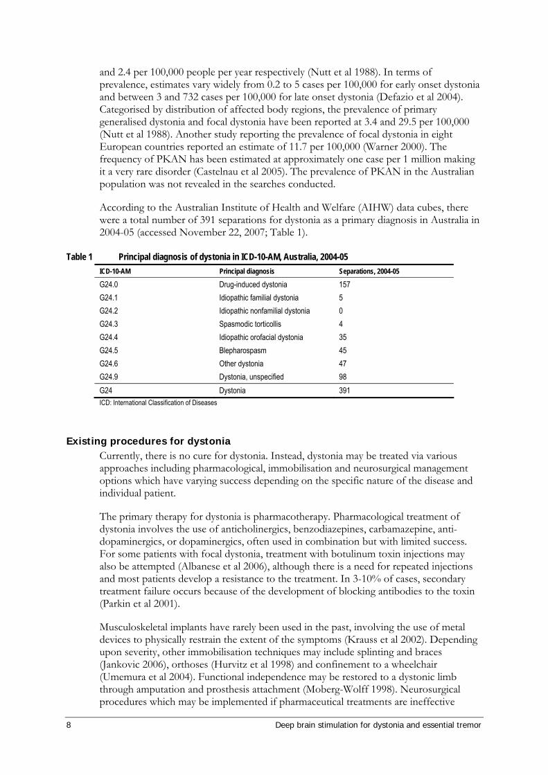
and 2.4 per 100,000 people per year respectively (Nutt et al 1988). In terms of prevalence, estimates vary widely from 0.2 to 5 cases per 100,000 for early onset dystonia and between 3 and 732 cases per 100,000 for late onset dystonia (Defazio et al 2004). Categorised by distribution of affected body regions, the prevalence of primary generalised dystonia and focal dystonia have been reported at 3.4 and 29.5 per 100,000 (Nutt et al 1988). Another study reporting the prevalence of focal dystonia in eight European countries reported an estimate of 11.7 per 100,000 (Warner 2000). The frequency of PKAN has been estimated at approximately one case per 1 million making it a very rare disorder (Castelnau et al 2005). The prevalence of PKAN in the Australian population was not revealed in the searches conducted.
According to the Australian Institute of Health and Welfare (AIHW) data cubes, there were a total number of 391 separations for dystonia as a primary diagnosis in Australia in 2004-05 (accessed November 22, 2007; Table 1).
Table 1 Principal diagnosis of dystonia in ICD-10-AM, Australia, 2004-05 ICD-10-AM Principal diagnosis Separations, 2004-05
G24.0 Drug-induced dystonia 157 G24.1 Idiopathic familial dystonia 5 G24.2 Idiopathic nonfamilial dystonia 0 G24.3 Spasmodic torticollis 4 G24.4 Idiopathic orofacial dystonia 35 G24.5 Blepharospasm 45 G24.6 Other dystonia 47 G24.9 Dystonia, unspecified 98 G24 Dystonia 391 ICD: International Classification of Diseases
Existing procedures for dystonia Currently, there is no cure for dystonia. Instead, dystonia may be treated via various approaches including pharmacological, immobilisation and neurosurgical management options which have varying success depending on the specific nature of the disease and individual patient.
The primary therapy for dystonia is pharmacotherapy. Pharmacological treatment of dystonia involves the use of anticholinergics, benzodiazepines, carbamazepine, anti-dopaminergics, or dopaminergics, often used in combination but with limited success. For some patients with focal dystonia, treatment with botulinum toxin injections may also be attempted (Albanese et al 2006), although there is a need for repeated injections and most patients develop a resistance to the treatment. In 3-10% of cases, secondary treatment failure occurs because of the development of blocking antibodies to the toxin (Parkin et al 2001).
Musculoskeletal implants have rarely been used in the past, involving the use of metal devices to physically restrain the extent of the symptoms (Krauss et al 2002). Depending upon severity, other immobilisation techniques may include splinting and braces (Jankovic 2006), orthoses (Hurvitz et al 1998) and confinement to a wheelchair (Umemura et al 2004). Functional independence may be restored to a dystonic limb through amputation and prosthesis attachment (Moberg-Wolff 1998). Neurosurgical procedures which may be implemented if pharmaceutical treatments are ineffective
Deep brain stimulation for dystonia and essential tremor 9
include deep brain stimulation, selective peripheral denervation/myectomy, intrathecal baclofen and radiofrequency lesions such as pallidotomy and thalamotomy (Albanese et al 2006; Defazio et al 2004).
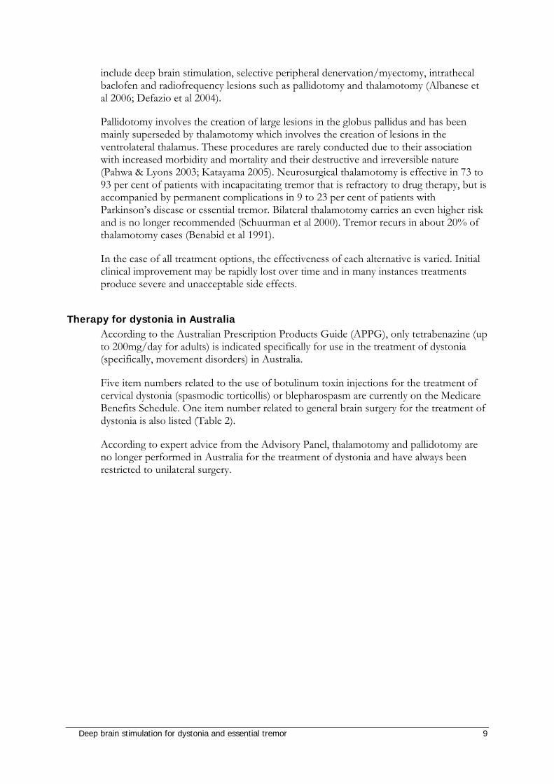
Pallidotomy involves the creation of large lesions in the globus pallidus and has been mainly superseded by thalamotomy which involves the creation of lesions in the ventrolateral thalamus. These procedures are rarely conducted due to their association with increased morbidity and mortality and their destructive and irreversible nature (Pahwa & Lyons 2003; Katayama 2005). Neurosurgical thalamotomy is effective in 73 to 93 per cent of patients with incapacitating tremor that is refractory to drug therapy, but is accompanied by permanent complications in 9 to 23 per cent of patients with Parkinson’s disease or essential tremor. Bilateral thalamotomy carries an even higher risk and is no longer recommended (Schuurman et al 2000). Tremor recurs in about 20% of thalamotomy cases (Benabid et al 1991).
In the case of all treatment options, the effectiveness of each alternative is varied. Initial clinical improvement may be rapidly lost over time and in many instances treatments produce severe and unacceptable side effects.
Therapy for dystonia in Australia According to the Australian Prescription Products Guide (APPG), only tetrabenazine (up to 200mg/day for adults) is indicated specifically for use in the treatment of dystonia (specifically, movement disorders) in Australia.
Five item numbers related to the use of botulinum toxin injections for the treatment of cervical dystonia (spasmodic torticollis) or blepharospasm are currently on the Medicare Benefits Schedule. One item number related to general brain surgery for the treatment of dystonia is also listed (Table 2).
According to expert advice from the Advisory Panel, thalamotomy and pallidotomy are no longer performed in Australia for the treatment of dystonia and have always been restricted to unilateral surgery.
10 Deep brain stimulation for dystonia and essential tremor
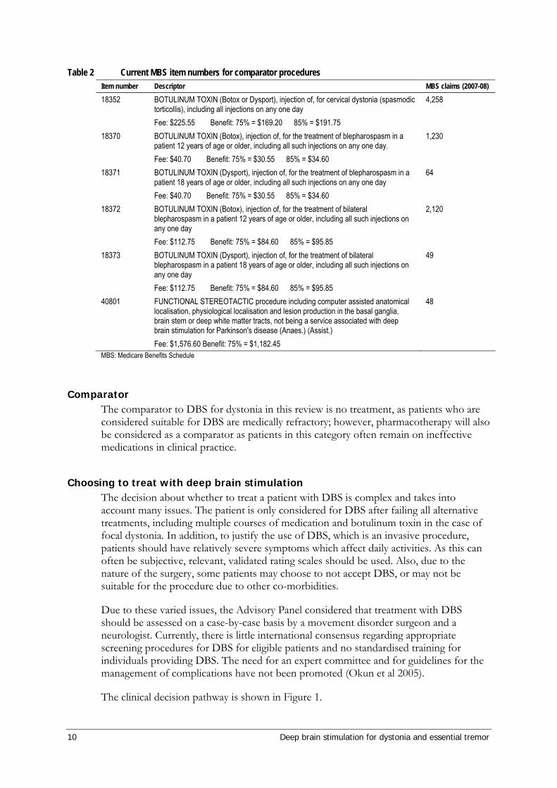
Table 2 Current MBS item numbers for comparator procedures Item number Descriptor MBS claims (2007-08)
18352 BOTULINUM TOXIN (Botox or Dysport), injection of, for cervical dystonia (spasmodic torticollis), including all injections on any one day Fee: $225.55 Benefit: 75% = $169.20 85% = $191.75
4,258
18370 BOTULINUM TOXIN (Botox), injection of, for the treatment of blepharospasm in a patient 12 years of age or older, including all such injections on any one day. Fee: $40.70 Benefit: 75% = $30.55 85% = $34.60
1,230
18371 BOTULINUM TOXIN (Dysport), injection of, for the treatment of blepharospasm in a patient 18 years of age or older, including all such injections on any one day Fee: $40.70 Benefit: 75% = $30.55 85% = $34.60
64
18372 BOTULINUM TOXIN (Botox), injection of, for the treatment of bilateral blepharospasm in a patient 12 years of age or older, including all such injections on any one day Fee: $112.75 Benefit: 75% = $84.60 85% = $95.85
2,120
18373 BOTULINUM TOXIN (Dysport), injection of, for the treatment of bilateral blepharospasm in a patient 18 years of age or older, including all such injections on any one day Fee: $112.75 Benefit: 75% = $84.60 85% = $95.85
49
40801 FUNCTIONAL STEREOTACTIC procedure including computer assisted anatomical localisation, physiological localisation and lesion production in the basal ganglia, brain stem or deep white matter tracts, not being a service associated with deep brain stimulation for Parkinson's disease (Anaes.) (Assist.) Fee: $1,576.60 Benefit: 75% = $1,182.45
48
MBS: Medicare Benefits Schedule
Comparator The comparator to DBS for dystonia in this review is no treatment, as patients who are considered suitable for DBS are medically refractory; however, pharmacotherapy will also be considered as a comparator as patients in this category often remain on ineffective medications in clinical practice.
Choosing to treat with deep brain stimulation The decision about whether to treat a patient with DBS is complex and takes into account many issues. The patient is only considered for DBS after failing all alternative treatments, including multiple courses of medication and botulinum toxin in the case of focal dystonia. In addition, to justify the use of DBS, which is an invasive procedure, patients should have relatively severe symptoms which affect daily activities. As this can often be subjective, relevant, validated rating scales should be used. Also, due to the nature of the surgery, some patients may choose to not accept DBS, or may not be suitable for the procedure due to other co-morbidities.
Due to these varied issues, the Advisory Panel considered that treatment with DBS should be assessed on a case-by-case basis by a movement disorder surgeon and a neurologist. Currently, there is little international consensus regarding appropriate screening procedures for DBS for eligible patients and no standardised training for individuals providing DBS. The need for an expert committee and for guidelines for the management of complications have not been promoted (Okun et al 2005).
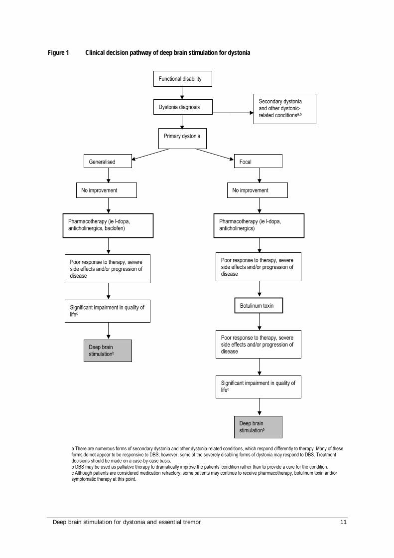
The clinical decision pathway is shown in Figure 1.
Deep brain stimulation for dystonia and essential tremor 11
Figure 1 Clinical decision pathway of deep brain stimulation for dystonia
a There are numerous forms of secondary dystonia and other dystonia-related conditions, which respond differently to therapy. Many of these forms do not appear to be responsive to DBS; however, some of the severely disabling forms of dystonia may respond to DBS. Treatment decisions should be made on a case-by-case basis. b DBS may be used as palliative therapy to dramatically improve the patients’ condition rather than to provide a cure for the condition. c Although patients are considered medication refractory, some patients may continue to receive pharmacotherapy, botulinum toxin and/or symptomatic therapy at this point.
Primary dystonia
No improvement
Pharmacotherapy (ie l-dopa, anticholinergics, baclofen)
Deep brain stimulationb
Botulinum toxin
Functional disability
Deep brain stimulationb
No improvement
Secondary dystonia and other dystonic-related conditionsa,b
Poor response to therapy, severe side effects and/or progression of disease
Significant impairment in quality of lifec
Pharmacotherapy (ie l-dopa, anticholinergics)
Generalised Focal
Poor response to therapy, severe side effects and/or progression of disease
Poor response to therapy, severe side effects and/or progression of disease
Significant impairment in quality of lifec
Dystonia diagnosis
12 Deep brain stimulation for dystonia and essential tremor
Essential tremor Essential tremor is a progressive neurological disorder that usually occurs later in life (Rincon & Louis 2005). The term essential tremor has been in use since the mid-twentieth century to describe a form of kinetic tremor for which a cause has not yet been established and which is often familial (Louis 2006). Traditionally, essential tremor has been viewed as a bland neurological tremor; however, this view is changing and the disorder is now considered to be a specific disease entity with a varied set of clinical characteristics including action tremor of the hands, voice and head (Louis 2006; Rincon & Louis 2005). It is thought that the onset of essential tremor may be influenced by both genetic and environmental factors despite the considerable clinical, genetic and pharmacological heterogeneity among essential tremor patients. Current research suggests that essential tremor may be mediated by central nervous system (CNS) gamma-aminobutyric acid (GABA)-ergic mechanisms and further modulated by peripheral (muscle) adrenoreceptors (Rincon & Louis 2005).
Clinical need and burden of disease of essential tremor A key feature of essential tremor is kinetic tremor of the arms during voluntary movement which in severe cases can spread to other body parts or occur at rest (Louis 2005). The regions of the body most often affected by essential tremor other than the arms include the head, face, voice, trunk and legs. Exacerbation of symptoms can occur during emotional or physiological stress (Pahwa & Lyons 2003). Essential tremor may be linked with increased risk of mortality; however, further research is needed to support this association (Louis et al 2007).
Incidence and prevalence of essential tremor Essential tremor (ICD-10 G25.0) is one of the most common neurological disorders and is the most common movement disorder (Leehey 2003; Louis 2005). Among the general population, the prevalance of essential tremor has been conservatively estimated at between 0.4 and 5 per cent, although it is expected that the true prevalance is much higher due to the existence of many undiagnosed patients (Louis 1999; Zesiewicz et al 2005). The wide range of these estimates is a result of an absence of uniform methodology by which to diagnose the disorder (Louis 2006). There is currently no diagnostic laboratory test for essential tremor. Diagnosis must be based on the history and physical examination of the individual patient (Louis 2001). Hospital separations relating to essential tremor by ICD-10 classification in 2004-05 are presented in Table 3.
Symptoms of essential tremor can develop at any age, from birth through to advanced age. The disorder, however, is clinically progressive in nature and as many as 4 to 5 per cent of people over the age of 40 are affected (Dogu et al 2003; Louis 2005). The prevalence of essential tremor in populations in the 6th to 8th decade of life has been estimated at between 6 and 9 per cent (Dogu et al 2003; Louis et al 1998). As the onset of essential tremor is usually later in life, prevalence is largely increased with age; according to the National Hospital Morbidity Database, 75 per cent of all patients with essential tremor are at least 70 years of age. Studies have not reported any differences in the prevalence of the disorder between men and women (Pahwa & Lyons 2003).
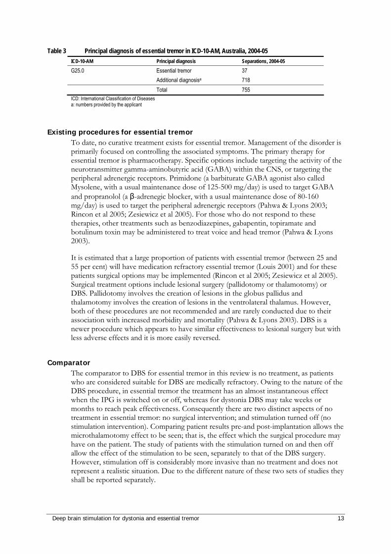
Deep brain stimulation for dystonia and essential tremor 13
Table 3 Principal diagnosis of essential tremor in ICD-10-AM, Australia, 2004-05 ICD-10-AM Principal diagnosis Separations, 2004-05
G25.0 Essential tremor 37 Additional diagnosisa 718 Total 755 ICD: International Classification of Diseases a: numbers provided by the applicant
Existing procedures for essential tremor To date, no curative treatment exists for essential tremor. Management of the disorder is primarily focused on controlling the associated symptoms. The primary therapy for essential tremor is pharmacotherapy. Specific options include targeting the activity of the neurotransmitter gamma-aminobutyric acid (GABA) within the CNS, or targeting the peripheral adrenergic receptors. Primidone (a barbiturate GABA agonist also called Mysolene, with a usual maintenance dose of 125-500 mg/day) is used to target GABA and propranolol (a β-adrenegic blocker, with a usual maintenance dose of 80-160 mg/day) is used to target the peripheral adrenergic receptors (Pahwa & Lyons 2003; Rincon et al 2005; Zesiewicz et al 2005). For those who do not respond to these therapies, other treatments such as benzodiazepines, gabapentin, topiramate and botulinum toxin may be administered to treat voice and head tremor (Pahwa & Lyons 2003).
It is estimated that a large proportion of patients with essential tremor (between 25 and 55 per cent) will have medication refractory essential tremor (Louis 2001) and for these patients surgical options may be implemented (Rincon et al 2005; Zesiewicz et al 2005). Surgical treatment options include lesional surgery (pallidotomy or thalamotomy) or DBS. Pallidotomy involves the creation of lesions in the globus pallidus and thalamotomy involves the creation of lesions in the ventrolateral thalamus. However, both of these procedures are not recommended and are rarely conducted due to their association with increased morbidity and mortality (Pahwa & Lyons 2003). DBS is a newer procedure which appears to have similar effectiveness to lesional surgery but with less adverse effects and it is more easily reversed.
Comparator The comparator to DBS for essential tremor in this review is no treatment, as patients who are considered suitable for DBS are medically refractory. Owing to the nature of the DBS procedure, in essential tremor the treatment has an almost instantaneous effect when the IPG is switched on or off, whereas for dystonia DBS may take weeks or months to reach peak effectiveness. Consequently there are two distinct aspects of no treatment in essential tremor: no surgical intervention; and stimulation turned off (no stimulation intervention). Comparing patient results pre-and post-implantation allows the microthalamotomy effect to be seen; that is, the effect which the surgical procedure may have on the patient. The study of patients with the stimulation turned on and then off allow the effect of the stimulation to be seen, separately to that of the DBS surgery. However, stimulation off is considerably more invasive than no treatment and does not represent a realistic situation. Due to the different nature of these two sets of studies they shall be reported separately.
14 Deep brain stimulation for dystonia and essential tremor
Choosing to treat with deep brain stimulation As with dystonia, the decision about whether to treat essential tremor with DBS is complex and takes into account many issues. The patient is only considered for DBS after failing all the alternative treatments, including multiple courses of medication. In addition, to justify the use of DBS, which is an invasive procedure, patients should have relatively severe symptoms which affect daily activities, such as the inability to independently feed or toilet. As this can often be subjective, relevant validated rating scales should be used. Due to the nature of the surgery, some patients may choose to not accept DBS, may delay DBS until the symptoms become so extreme that they are unable to look after themselves, or may not be suitable for the procedure due to other co-morbidities. The Advisory Panel considered that the potential for treatment with DBS should be assessed on a case-by-case basis by a movement disorder surgeon and a neurologist.
The clinical decision pathway for essential tremor is shown in Figure 2. There is little international consensus regarding appropriate screening procedures for DBS for eligible patients and no standardised training for individuals providing DBS. The need for an expert committee and for guidelines for the management of complications have not been promoted (Okun et al 2005).
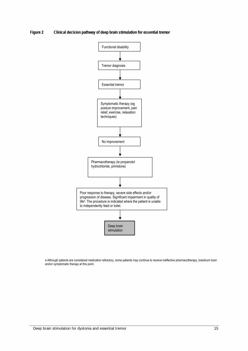
Deep brain stimulation for dystonia and essential tremor 15
Figure 2 Clinical decision pathway of deep brain stimulation for essential tremor
a Although patients are considered medication refractory, some patients may continue to receive ineffective pharmacotherapy, botulinum toxin and/or symptomatic therapy at this point.
Tremor diagnosis
Pharmacotherapy (ie propanolol hydrochloride, primidone)
Symptomatic therapy (eg posture improvement, pain relief, exercise, relaxation techniques)
Deep brain stimulation
Poor response to therapy, severe side effects and/or progression of disease. Significant impairment in quality of lifea. The procedure is indicated where the patient is unable to independently feed or toilet.
Functional disability
Essential tremor
No improvement
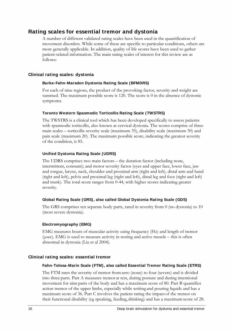
16 Deep brain stimulation for dystonia and essential tremor
Rating scales for essential tremor and dystonia A number of different validated rating scales have been used in the quantification of movement disorders. While some of these are specific to particular conditions, others are more generally applicable. In addition, quality of life scores have been used to gather patient-related information. The main rating scales of interest for this review are as follows:
Clinical rating scales: dystonia
Burke-Fahn-Marsden Dystonia Rating Scale (BFMDRS)
For each of nine regions, the product of the provoking factor, severity and weight are summed. The maximum possible score is 120. The score is 0 in the absence of dystonic symptoms.
Toronto Western Spasmodic Torticollis Rating Scale (TWSTRS)
The TWSTRS is a clinical tool which has been developed specifically to assess patients with spasmodic torticollis, also known as cervical dystonia. The scores comprise of three main scales – torticollis severity scale (maximum 35), disability scale (maximum 30) and pain scale (maximum 20). The maximum possible score, indicating the greatest severity of the condition, is 85.
Unified Dystonia Rating Scale (UDRS)
The UDRS comprises two main factors – the duration factor (including none, intermittent, constant); and motor severity factor (eyes and upper face, lower face, jaw and tongue, larynx, neck, shoulder and proximal arm (right and left), distal arm and hand (right and left), pelvis and proximal leg (right and left), distal leg and foot (right and left) and trunk). The total score ranges from 0-44, with higher scores indicating greater severity.
Global Rating Scale (GRS), also called Global Dystonia Rating Scale (GDS)
The GRS comprises ten separate body parts, rated in severity from 0 (no dystonia) to 10 (most severe dystonia).
Electromyography (EMG)
EMG measures bouts of muscular activity using frequency (Hz) and length of tremor (µsec). EMG is used to measure activity in resting and active muscle – this is often abnormal in dystonia (Liu et al 2004).
Clinical rating scales: essential tremor
Fahn-Tolosa-Marin Scale (FTM), also called Essential Tremor Rating Scale (ETRS)
The FTM rates the severity of tremor from zero (none) to four (severe) and is divided into three parts. Part A measures tremor at rest, during posture and during intentional movement for nine parts of the body and has a maximum score of 80. Part B quantifies
action tremor of the upper limbs, especially while writing and pouring liquids and has a maximum score of 36. Part C involves the patient rating the impact of the tremor on their functional disability (eg speaking, feeding, drinking) and has a maximum score of 28.
Deep brain stimulation for dystonia and essential tremor 17
The maximum FTM score is 144, obtained by summing the three parts. The tremor rating scale (TRS) also has a separate item relating to global assessment of tremor-related disability, rated by both the patient and the examiner on a 5 point scale (Hariz et al 2002).
Accelerometry
Accelerometry involves recording of the movements from a body segment to allow measurement of frequency, amplitude or intensity of a tremor. The intensity of a tremor is a measure of the overall magnitude of movement (the amplitude of tremor multiplied by its frequency) (Ferreria & Sampaio 2007).
Quality of life measures Over the years many quality of life (QoL) scales have been developed. Some are specific to particular indications and others are more general. While the specific manner in which this is done differs between the scales, they all aim to describe and measure health states and wellbeing. As such they are more patient-oriented than the clinical, physician-marked scales. Currently, two of the most commonly used standardised, non-disease specific scales include EuroQoL (EuroQoL Group 1990) and the SF-36 (www.sf-36.org/tools/sf36.shtml, accessed 22 November 2007).
SF-36
The Short Form-36 health-related quality of life survey (SF-36) is a widely-used generic short-form health survey which has been widely evaluated (Garratt et al 2002). It has only 36 questions covering physical functioning and role, bodily pain, general health, vitality, social functioning, emotion and mental health. The SF-36 score is often normalised to a 100-point algorithm, with a higher score reflective of best health. The SF-12 is a shortened version of the SF-36.
EuroQoL
EuroQoL is a utility measure more commonly used for economic evaluation and incorporates preferences for health states (Garratt et al 2002). As with the SF-36 it is marked on a 0-100 point scale, with 0 being the worst imaginable health state (EuroQoL Group 1990).
Activities of Daily Living Scale (ADL)
A relatively common score for a variety of movement disorders, including Parkinson’s disease and essential tremor, is the Activities of Daily Living (ADL) or a variant thereof. One example developed in the 1950s is the Schwab and England ADL scale (Putzke et al 2005). ADLs more specifically related to essential tremor are part of the Essential Tremor Rating Scale (ETRS) and Tremor Activities of Daily Living Scale (TADLS) (Lyons et al 1998; Sydow et al 2003). All these scales rate a number of activities (ranging in number from 6 to 30), including items such as eating, drinking, threading a needle, driving, shaving and tying shoes. These are each rated on a 0 (normal) to 4 (unable to do) scale to give a total score. ADLs may be rated by the patient and by the clinician. The ADLs therefore are an indirect measure of the quality of life for the patient.
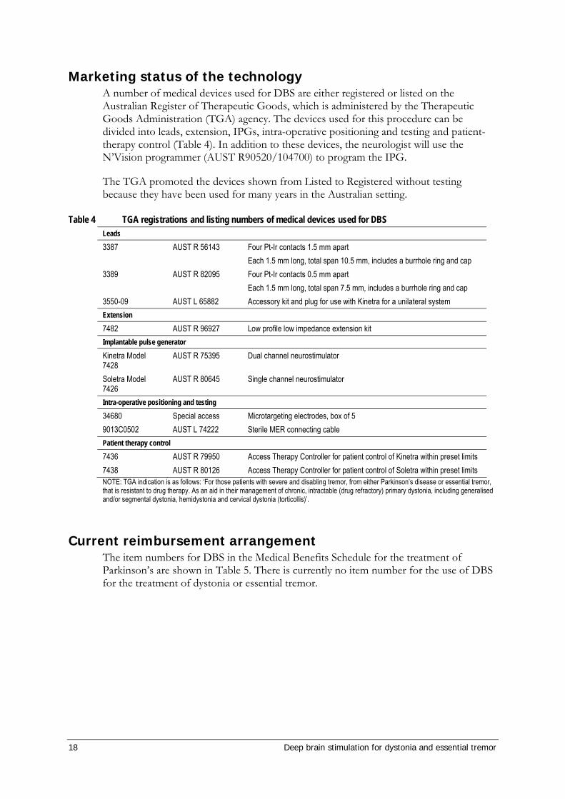
18 Deep brain stimulation for dystonia and essential tremor
Marketing status of the technology A number of medical devices used for DBS are either registered or listed on the Australian Register of Therapeutic Goods, which is administered by the Therapeutic Goods Administration (TGA) agency. The devices used for this procedure can be divided into leads, extension, IPGs, intra-operative positioning and testing and patient-therapy control (Table 4). In addition to these devices, the neurologist will use the N’Vision programmer (AUST R90520/104700) to program the IPG.
The TGA promoted the devices shown from Listed to Registered without testing because they have been used for many years in the Australian setting.
Table 4 TGA registrations and listing numbers of medical devices used for DBS Leads
3387 AUST R 56143
Four Pt-Ir contacts 1.5 mm apart Each 1.5 mm long, total span 10.5 mm, includes a burrhole ring and cap
3389
AUST R 82095
Four Pt-Ir contacts 0.5 mm apart Each 1.5 mm long, total span 7.5 mm, includes a burrhole ring and cap
3550-09 AUST L 65882 Accessory kit and plug for use with Kinetra for a unilateral system Extension
7482 AUST R 96927 Low profile low impedance extension kit Implantable pulse generator
Kinetra Model 7428
AUST R 75395 Dual channel neurostimulator
Soletra Model 7426
AUST R 80645 Single channel neurostimulator
Intra-operative positioning and testing
34680 Special access Microtargeting electrodes, box of 5 9013C0502 AUST L 74222 Sterile MER connecting cable Patient therapy control
7436 AUST R 79950 Access Therapy Controller for patient control of Kinetra within preset limits 7438 AUST R 80126 Access Therapy Controller for patient control of Soletra within preset limits NOTE: TGA indication is as follows: ‘For those patients with severe and disabling tremor, from either Parkinson’s disease or essential tremor, that is resistant to drug therapy. As an aid in their management of chronic, intractable (drug refractory) primary dystonia, including generalised and/or segmental dystonia, hemidystonia and cervical dystonia (torticollis)’.
Current reimbursement arrangement The item numbers for DBS in the Medical Benefits Schedule for the treatment of Parkinson’s are shown in Table 5. There is currently no item number for the use of DBS for the treatment of dystonia or essential tremor.
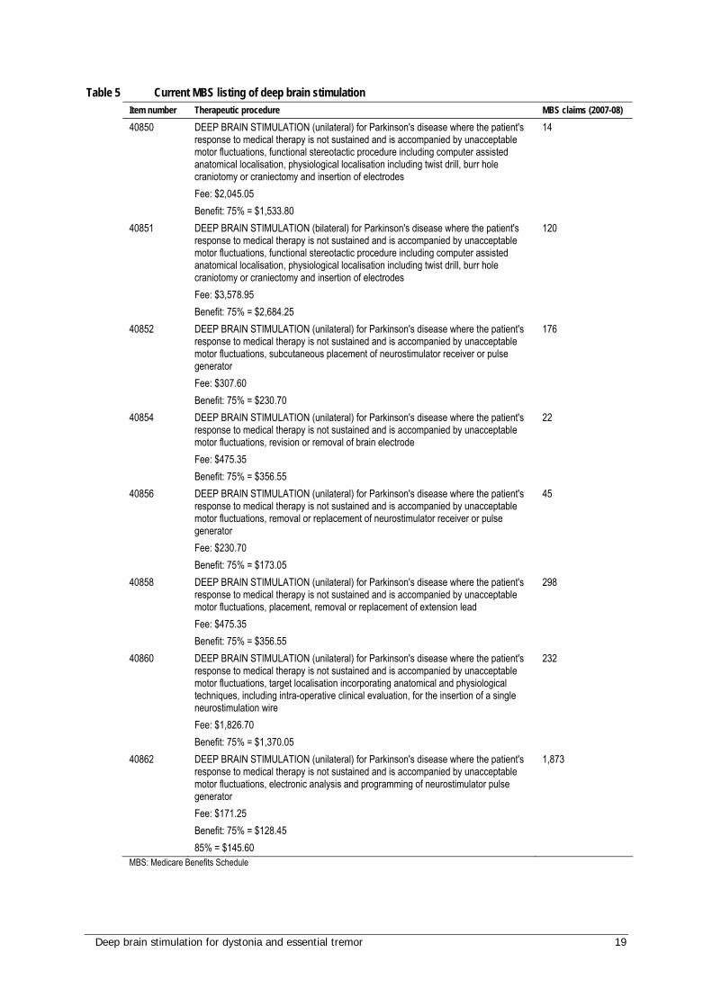
Deep brain stimulation for dystonia and essential tremor 19
Table 5 Current MBS listing of deep brain stimulation Item number Therapeutic procedure MBS claims (2007-08)
40850 DEEP BRAIN STIMULATION (unilateral) for Parkinson's disease where the patient's response to medical therapy is not sustained and is accompanied by unacceptable motor fluctuations, functional stereotactic procedure including computer assisted anatomical localisation, physiological localisation including twist drill, burr hole craniotomy or craniectomy and insertion of electrodes Fee: $2,045.05 Benefit: 75% = $1,533.80
14
40851 DEEP BRAIN STIMULATION (bilateral) for Parkinson's disease where the patient's response to medical therapy is not sustained and is accompanied by unacceptable motor fluctuations, functional stereotactic procedure including computer assisted anatomical localisation, physiological localisation including twist drill, burr hole craniotomy or craniectomy and insertion of electrodes Fee: $3,578.95 Benefit: 75% = $2,684.25
120
40852 DEEP BRAIN STIMULATION (unilateral) for Parkinson's disease where the patient's response to medical therapy is not sustained and is accompanied by unacceptable motor fluctuations, subcutaneous placement of neurostimulator receiver or pulse generator Fee: $307.60 Benefit: 75% = $230.70
176
40854 DEEP BRAIN STIMULATION (unilateral) for Parkinson's disease where the patient's response to medical therapy is not sustained and is accompanied by unacceptable motor fluctuations, revision or removal of brain electrode Fee: $475.35 Benefit: 75% = $356.55
22
40856 DEEP BRAIN STIMULATION (unilateral) for Parkinson's disease where the patient's response to medical therapy is not sustained and is accompanied by unacceptable motor fluctuations, removal or replacement of neurostimulator receiver or pulse generator Fee: $230.70 Benefit: 75% = $173.05
45
40858 DEEP BRAIN STIMULATION (unilateral) for Parkinson's disease where the patient's response to medical therapy is not sustained and is accompanied by unacceptable motor fluctuations, placement, removal or replacement of extension lead Fee: $475.35 Benefit: 75% = $356.55
298
40860 DEEP BRAIN STIMULATION (unilateral) for Parkinson's disease where the patient's response to medical therapy is not sustained and is accompanied by unacceptable motor fluctuations, target localisation incorporating anatomical and physiological techniques, including intra-operative clinical evaluation, for the insertion of a single neurostimulation wire Fee: $1,826.70 Benefit: 75% = $1,370.05
232
40862 DEEP BRAIN STIMULATION (unilateral) for Parkinson's disease where the patient's response to medical therapy is not sustained and is accompanied by unacceptable motor fluctuations, electronic analysis and programming of neurostimulator pulse generator Fee: $171.25 Benefit: 75% = $128.45 85% = $145.60
1,873
MBS: Medicare Benefits Schedule
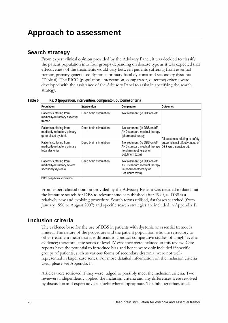
20 Deep brain stimulation for dystonia and essential tremor
Approach to assessment
Search strategy From expert clinical opinion provided by the Advisory Panel, it was decided to classify the patient population into four groups depending on disease type as it was expected that effectiveness of the treatments would vary between patients suffering from essential tremor, primary generalised dystonia, primary focal dystonia and secondary dystonia (Table 6). The PICO (population, intervention, comparator, outcome) criteria were developed with the assistance of the Advisory Panel to assist in specifying the search strategy.
Table 6 PICO (population, intervention, comparator, outcome) criteria Population Intervention Comparator Outcomes
Patients suffering from medically-refractory essential tremor
Deep brain stimulation ‘No treatment’ (ie DBS on/off)
Patients suffering from medically-refractory primary generalised dystonia
Deep brain stimulation ‘No treatment’ (ie DBS on/off) AND standard medical therapy (pharmacotherapy)
Patients suffering from medically-refractory primary focal dystonia
Deep brain stimulation ‘No treatment’ (ie DBS on/off) AND standard medical therapy (ie pharmacotherapy or Botulinum toxin)
Patients suffering from medically-refractory severe secondary dystonia
Deep brain stimulation ‘No treatment’ (ie DBS on/off) AND standard medical therapy (ie pharmacotherapy or Botulinum toxin)
All outcomes relating to safety and/or clinical effectiveness of DBS were considered.
DBS: deep brain stimulation
From expert clinical opinion provided by the Advisory Panel it was decided to date limit the literature search for DBS to relevant studies published after 1990, as DBS is a relatively new and evolving procedure. Search terms utilised, databases searched (from January 1990 to August 2007) and specific search strategies are included in Appendix E.
Inclusion criteria The evidence base for the use of DBS in patients with dystonia or essential tremor is limited. The nature of the procedure and the patient population who are refractory to other treatment mean that it is difficult to conduct comparative studies of a high level of evidence; therefore, case series of level IV evidence were included in this review. Case reports have the potential to introduce bias and hence were only included if specific groups of patients, such as various forms of secondary dystonia, were not well-represented in larger case series. For more detailed information on the inclusion criteria used, please see Appendix F.
Articles were retrieved if they were judged to possibly meet the inclusion criteria. Two reviewers independently applied the inclusion criteria and any differences were resolved by discussion and expert advice sought where appropriate. The bibliographies of all
Deep brain stimulation for dystonia and essential tremor 21
retrieved publications were handsearched for any relevant references missed in the database search (pearling).
Data analysis
Meta-analysis Where outcomes could be sensibly combined (outcomes measured in comparable ways and no apparent heterogeneity), relative risks or weighted mean differences with 95% confidence intervals (CI) were calculated using RevMan 4.2. Subgroup analyses were carried out for certain variables where possible.
Handling of nonrandomised data Where statistical pooling was not possible, medians of rates (for dichotomous outcomes) or medians of means (for continuous outcomes) for all studies reporting the outcome were calculated.
Included and excluded studies The studies identified as fulfilling the inclusion criteria for the review are listed in Appendix C. The studies which were excluded from the review are listed in Appendix D, together with the reason for exclusion.
Current ongoing trials A list of the current ongoing trials which would add to the currently available evidence base is provided in Appendix H.
Expert advice An Advisory Panel with expertise in neurology and surgery was established to evaluate the evidence and provide advice to MSAC from a clinical perspective. In selecting members for Advisory Panels, MSAC’s practice is to approach the appropriate medical colleges, specialist societies and associations and consumer bodies for nominees. Membership of the Advisory Panel is provided at Appendix B.
Research questions Is deep brain stimulation as safe as or safer than the comparator for the treatment of essential tremor?
Is deep brain stimulation as or more effective than the comparator for essential tremor?
Is deep brain stimulation as or more cost-effective than the comparator for the treatment of essential tremor?
Is deep brain stimulation as safe or safer than the comparator for the treatment of primary generalised dystonia?

22 Deep brain stimulation for dystonia and essential tremor
Is deep brain stimulation as or more effective than the comparator for the treatment of primary generalised dystonia?
Is deep brain stimulation as or more cost-effective than the comparator for the treatment of primary generalised dystonia?
Is deep brain stimulation as safe or safer than the comparator for the treatment of primary focal dystonia?
Is deep brain stimulation as or more effective than the comparator for the treatment of primary focal dystonia?
Is deep brain stimulation as or more cost-effective than the comparator for the treatment of primary focal dystonia?
Is deep brain stimulation as safe or safer than the comparator for the treatment of severe secondary dystonia?
Is deep brain stimulation as or more effective than the comparator for the treatment of severe secondary dystonia?
Is deep brain stimulation as or more cost-effective than the comparator for the treatment of severe secondary dystonia?
Deep brain stimulation for dystonia and essential tremor 23
Results of assessment
Overall results of the literature search The search strategy identified 1093 articles; however 961 were excluded because they were not primary studies or did not meet the inclusion criteria. A further 18 studies were excluded because outcomes were not considered to be clinically relevant and 14 were excluded because outcomes were reported for a mixed group of patients with a variety of disorders other than dystonia or essential tremor. As the quality of the evidence was limited, case series including three or more patients were used to assess safety and effectiveness of DBS in patients with essential tremor or dystonia. Single case reports were included to assess the effectiveness of DBS for dystonia and essential tremor if there was insufficient evidence for specific patient subgroups (ie different types of secondary dystonia and tremor associated with a brain insult). Thirty case reports of dystonic patients treated with DBS were excluded as they did not contribute further to the evidence base. In total, 44 studies were included to assess the safety and/or effectiveness of DBS in patients with dystonia and 17 studies were included to assess the safety and/or effectiveness of DBS in patients with essential tremor.
Overall comment on variety of studies The systematic searching revealed a great variety of studies, with several case series and one RCT. There were many studies concerning the use of DBS for essential tremor and dystonia; however, many of these were mixed studies which also considered other conditions such as Parkinson’s disease.
The dystonia studies were particularly difficult to analyse as there were few studies which reported on DBS for dystonia alone and these mostly comprised studies which reported on a mixture of dystonia conditions. These mixed studies were challenging to decipher and synthesise.
The essential tremor studies concerned patients both before/after DBS and with stimulation on/off. The stimulation on/off studies were considered by the Advisory Panel to provide the most information regarding the effectiveness of DBS for essential tremor; hence these studies have been reported separately. Several of the before/after DBS studies described a microthalamotomy effect; that is, the placement of the electrodes alone was sometimes sufficient to elicit an effect.
Discussion of results of the systematic reviews A number of systematic reviews were identified in the international literature which investigated DBS in the treatment of dystonia and essential tremor. Of these, three were unavailable for various reasons. Two reports were published by Hayes (USA) in 2004, entitled ‘Deep brain stimulation for treatment of dystonia’ and ‘Deep brain stimulation for Parkinson's disease and essential tremor’. Hayes’ reports require a fee to be paid for access. Also, IECS (Instituto de Efectividad Clínica y Sanitaria, Argentina) published a report in 2005 entitled ‘Deep brain stimulation for generalized dystonia treatment’. The full text of this report is freely available through their website; however, it is only available in Spanish. In the English language abstract the authors comment on the lack of
24 Deep brain stimulation for dystonia and essential tremor
available quality evidence. The overall conclusion is that bilateral DBS of the GPi may be beneficial for patients with generalised, segmental or cervical primary dystonia that is resistant to pharmacology. Given the nature of the condition and the fact that the majority of dystonias may be adequately controlled through medications, the authors suggest that the procedure will be infrequently performed.
The literature searches identified 5 full text systematic reviews and health technology assessments on DBS for essential tremor and dystonia (Diamond & Jankovic 2005; Holloway et al 2006; NICE overview 2006; Ontario MAS 2005; Yianni et al 2005). Of these, two included an assessment of Parkinson’s disease (Diamond & Jankovic 2005, Ontario MAS 2005).
Diamond and Jankovic (2005) systematically reported only on studies which reported health-related quality of life outcomes (including activities of daily living or ADLs). As a result, only two studies for essential tremor and three studies for dystonia were included in the assessment and reported narratively. The authors conclude that there is a paucity of quality of life data for these conditions, but state that there is growing evidence that DBS has a favourable impact on quality of life for movement disorders.
Holloway and colleagues (2006) conducted a meta-analysis of DBS for dystonia. Mean percentage changes in BFM scores pre- and post-procedure were calculated and corrected to normality for patients separated according to disease aetiology. Primary dystonia was separated into DYT1-positive and DYT1-negative as opposed to generalised or focal. Total numbers of included patients were small, ranging from 1 (specific secondary dystonia) to 40 (primary unspecified). Overall the authors suggest that DBS treatment brings about a statistically significant improvement for all conditions, excepting cerebral palsy, birth injury and encephalitis.
The study by Yianni et al (2005) is not a systematic review; rather, the authors have retrospectively collected patient information (n=26) using the Euroquol (EQ-5D) and Willingness-to-Pay (WTP) questionnaires in order to inform a cost-benefit analysis. Data were collected before and after the DBS procedure. The specific nature or aetiology of dystonia in the included patients was not provided. Overall costs per patient, based on a bilateral procedure to the GPi, were estimated at £31,866 (Great British sterling, 2003 prices), achieving a gain of 0.94 quality affected life years (QALY) units. Mean WTP value was £291,000, representing a large perceived value in the treatment.
Two health technology assessments were identified. The most recent was a NICE overview and its associated guidance from 2006 (UK), entitled ‘Interventional procedure overview of deep brain stimulation for tremor and dystonia (excluding Parkinson’s disease)’. It is worth noting that NICE overviews are not comprehensive systematic reviews. The evidence presented in this document is based on six studies including one systematic review, together with the opinion of five specialist advisors. The subsequent guidance supports the use of the procedure, provided that patient selection and management should be carried out in the context of a multidisciplinary team.
A more comprehensive systematic review has been published by Medical Advisory Secretariat, Ontario Ministry of Health (Canada) in March 2005, ‘Deep Brain Stimulation for Parkinson’s Disease and Other Movement Disorders’. Literature searches were from January 2001 and results were provided separately for Parkinson’s disease, essential tremor and primary dystonia. Three studies were included for essential tremor and one for primary dystonia. The study concludes that there is a shortfall in the numbers of
Deep brain stimulation for dystonia and essential tremor 25
patients currently having DBS for movement disorders in Ontario and that the DBS procedures should be limited to a small number of specialist centres in order to reduce complication rates. The suggestion is made that the cost-per-procedure and the shortage of expertise will be natural limiting factors for the further diffusion of DBS.
Despite the lack of comparative studies, all of the identified systematic reviews concluded that DBS is a safe and effective treatment for movement disorders, including essential tremor and primary dystonia. Where investigated, the use of DBS with secondary dystonia was not reported as being as beneficial. There was a general consensus that the use of DBS will not be a commonly performed procedure.
Additional studies An additional study concerning DBS for dystonia was published in September 2007 (Kiss et al 2007) and was identified through the Advisory Panel. This study was not included in the review as it was published after the official dates of the search strategy. For further information on this study, please see Appendix G.
26 Deep brain stimulation for dystonia and essential tremor
Results of assessment - dystonia
Descriptive characteristics of included studies Expert clinical opinion from the Advisory Panel suggested that different treatment effects result from DBS for essential tremor and dystonia. It is thought that stimulation elicits an immediate response in essential tremor patients, seen within minutes or hours. As a result, patients with essential tremor are often instucted to turn off their stimulator at night to conserve the battery and to prevent tissue habituation (Plaha et al 2004). A more gradual, cumulative effect of DBS is seen in patients with dystonia which may take weeks or months to take full effect. Many dystonia patients experience a residual effect after their stimulation is turned off.
The dystonia studies were often challenging to analyse. Many of the studies reported on dystonia in conjunction with other conditions such as Parkinson’s and there were few studies which reported on the use of DBS solely for dystonia. Further, those studies that did report solely on dystonia mostly comprised studies which reported on a mixture of types of dystonia. One RCT for DBS in patients with dystonia was identified, along with 25 case series. The results for the RCT shall be reported separately to reflect the higher level of evidence.
An additional study concerning DBS for dystonia was published in September 2007 (Kiss et al 2007), but was not included in the review as it was published outside the official dates of the search strategy. There were a total of ten participants in this study. Briefly, the results were a significant difference for TWSTRS and quality of life scores between baseline and 12-month follow-up after DBS for patients with medication-refractory cervical dystonia (P=0.003 for both). For further information on this study, please see Appendix G.
Studies reporting outcomes for dystonia patients before and after DBS The characteristics of the 28 case series reporting safety or effectiveness outcomes of DBS for the treatment of dystonia that were included for review are presented in Table 72, together with one RCT. All case series that reported outcomes relating to the safety of DBS in dystonic patients were used to assess the safety of DBS for the treatment of dystonia; however, studies were generally not included if they reported outcomes for dystonic patients together with patients with different movement disorders. An exception was the study by Paluzzi et al (2006b), which reported safety outcomes for 19 dystonic patients among a larger cohort of 96 patients treated with DBS for various conditions and assessed safety, but not effectiveness. One additional paper (Paluzzi et al 2006a) reporting outcomes for three dystonic patients undergoing DBS during pregnancy was included for the assessment of safety outcomes as it was the only identified study which reported outcomes for DBS during pregnancy. One paper which reported on outcomes for safety (Fonke et al 2006) was not used to assess effectiveness as the method used to assess improvements of dystonia was not reported. Additional case reports were also used to assess the effectiveness of DBS for various forms of secondary dystonia and dystonia-related disorders, as there were few patients with these disorders in the included case series due to the rarity of these conditions.
Deep brain stimulation for dystonia and essential tremor 27
Studies comparing dystonia patients with DBS switched on versus off In addition to reporting outcomes for patients before and after DBS, four of the included studies also reported outcomes for patients with the stimulators switched on compared to outcomes with the stimulators switched off (Kupsch et al 2006; Detante et al 2004; Grips et al 2007; Tisch et al 2007). Kupsch et al (2006) compared sham stimulation (DBS electrodes and IPG implanted but switched off) with DBS between two groups of randomised patients in a double-blinded manner. The other three studies (Detante et al 2004; Grips et al 2007; Tisch et al 2007) were internally comparative, comparing patient response while on and off stimulation. These studies can help to differentiate the effects of DBS from other factors that may affect patient condition such as the placebo effect, assessor bias or the effects associated with the implantation of DBS equipment; however, they are not ideal to assess the effectiveness of DBS compared to ‘no treatment’, as sham stimulation is considerably more invasive than no treatment and is therefore not realistic. The on/off studies can be useful for the optimisation of the procedure, determination of ideal stimulation settings and conditions, as well as to determine any residual benefits of DBS that may be sustained when the stimulator is switched off. Outcomes relating to the effects of DBS switched on versus off will be used to enrich the evidence regarding the effectiveness of the procedure; however, many important safety outcomes are related to the implantation of DBS equipment.
Quality of included studies One RCT (level II on the National Health and Medical Research Council (NHMRC) levels of evidence) was included (Kupsch et al 2006), whilst all other included studies were case series or case reports and therefore of a low methodological quality (level IV on the NHMRC levels of evidence). There are certain factors of interest that may highlight differences of study quality between these studies such as the use of consecutive patients and follow-up. Fifteen studies used consecutive patients (Table 72). Of these, three were retrospectively examined (Eltahawy et al 2004a; Paluzzi et al 2006a; Vercueil et al 2001). Two studies did not report the follow-up period (Loher et al 2000; Parkin et al 2001) and ten studies did not specifically indicate any patient losses during the follow-up period. Where losses to follow-up were reported, sixteen studies reported that no patients were lost and one study reported that one patient was lost (Krause et al 2004). This patient lived at a location which was distant to the centre at which the DBS was implanted. Following infection a surgeon at her local hospital removed the IPG approximately three weeks after the initial surgery.
Inclusion and exclusion criteria, where reported, varied in the detail provided. The majority of the studies included patients with severe generalised dystonia (Table 72). Five studies included only cervical dystonia (Eltahawy et al 2004b; Kiss et al 2004; Kleiner-Fisman et al 2007; Krauss et al 2002; Wang et al 2006) and one study each included only segmental (Grips et al 2007) or torsion dystonia (Tisch et al 2006). Exclusion criteria were not widely used. Where reported, one study excluded patients who had received previous brain surgery (Eltahawy et al 2004a) and one study excluded patients with psychiatric disturbance (Vidailhet et al 2005).
In addition to the case series listed above, nine case reports were included to provide additional information regarding secondary dystonias (Burbaud et al 2002; Deutschlander et al 2005; Foote & Okun 2005; Guehl et al 2007; Nikkah et al 2004; Paluzzi et al 2006a; Parkin et al 2001; Roze et al 2006; Trottenberg et al 2005) (Table 75). These were poorly
28 Deep brain stimulation for dystonia and essential tremor
recorded in the larger studies, possibly as a result of their rarity. Further information on the study characteristics is available in Appendix I.
Technical characteristics of dystonia studies There were many similarities in the technical characteristics of the included dystonia studies (Table 11, Table 14, Table 22, Table 26, Table 74, Table 76 and Table 79). Where reported, the electrode used was the Medtronic 3387 or 3389 and the implantable pulse generator was of the Itrel, Soletra or Kinetra type (all Medtronic). Bilateral implantation to the GPi was the most common type of procedure. Unilateral implantation was carried out in thirteen patients (Diamond et al 2006; Eltahawy et al 2004a; Krauss et al 2002; Kupsch et al 2003; Starr et al 2006; Vercueil et al 2001). Where reported, the stimulation between the studies was equally-proportioned between monopolar and bipolar stimulation, although the majority of the studies did not report the polarity of the electrodes. There appeared to be no major differences overall between the studies reporting primary generalised- (Table 79), primary focal- (Table 14) or secondary-dystonia (Table 74) with the dystonia case series as a whole (Table 76). The values of the final stimulation parameters were slightly higher for both pulse width and frequency in unipolar compared to bipolar stimulation.
Summary: Dystonia studies Twenty-nine studies which reported safety or effectiveness outcomes in patients with dystonia after DBS treatment compared to patient status before treatment were included for review. In addition, four of these studies compared patient status with DBS switched on compared to off, to research the effect of the stimulation alone. Many studies reported results for dystonia in conjunction with results for other conditions such as Parkinson’s. Of the studies that reported solely on dystonia, many reported simultaneously on various types of dystonia. There were few studies which clearly reported the effect of DBS on one type of dystonia. The evidence was limited by the lack of comparative data as only one of the included studies was level II evidence (Kupsch 2006) and the remaining studies were level IV evidence. The treatment criteria of medically-refractory, severely disabled patients, combined with the invasive nature of DBS and lack of a true alternative, make comparative studies logistically and ethically difficult to perform. Hence DBS for dystonia may be considered an orphan procedure.
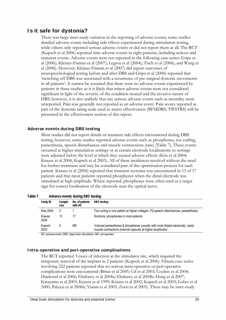
Deep brain stimulation for dystonia and essential tremor 29
Is it safe for dystonia? There was large inter-study variation in the reporting of adverse events; some studies detailed adverse events including side effects experienced during stimulation testing, while others only reported serious adverse events or did not report them at all. The RCT (Kupsch et al 2006) reported nine adverse events in eight patients, including serious and transient events. Adverse events were not reported in the following case series: Grips et al (2006); Kleiner-Fisman et al (2007); Legros et al (2004); Tisch et al (2006); and Wang et al (2006). However, Kleiner-Fisman et al (2007) did report outcomes of neuropsychological testing before and after DBS and Grips et al (2006) reported that ‘switching off DBS was associated with a recurrence of pre-surgical dystonic movements in all patients’. It cannot be assumed that there were no adverse events experienced by patients in these studies as it is likely that minor adverse events were not considered significant in light of the severity of the condition treated and the invasive nature of DBS; however, it is also unlikely that any serious adverse events such as mortality went unreported. Pain was generally not reported as an adverse event. Pain scores reported as part of the dystonia rating scale used to assess effectiveness (BFMDRS; TWSTRS) will be presented in the effectiveness section of this report.
Adverse events during DBS testing Most studies did not report details of transient side effects encountered during DBS testing; however, some studies reported adverse events such as phosphenes, toe curling, paraesthesia, speech disturbances and muscle contractions (rare) (Table 7). These events occurred at higher stimulation settings or at certain electrode localisations so settings were adjusted below the level at which they caused adverse effects (Kiss et al 2004; Krause et al 2004; Kupsch et al 2003). All of these incidences resolved without the need for further treatment and may be considered part of the optimisation process for each patient. Krause et al (2004) reported that transient scotoma was encountered in 13 of 17 patients and that most patients reported phosphenes when the distal electrode was stimulated at high amplitude. Where reported, phosphenes were often used as a target sign for correct localisation of the electrode near the optical nerve.
Table 7 Adverse events during DBS testing Study ID Sample
size No. of patients with AE
DBS testing
Kiss 2004 3 1 Toe curling in one patient at higher voltages. P2-speech disturbances, paraestheisa Krause 2004
13 17 Scotoma, phosphenes in most patients
Kupsch 2003
5 NR ‘Induced paraestheisa & phosphenes (usually with most distant electrode), rarely muscle contractions (internal capsule) at higher amplitudes’.
AE: adverse event; DBS: deep brain stimulation; NR: not reported
Intra-operative and peri-operative complications The RCT reported 3 cases of infection at the stimulator site, which required the temporary removal of the implant in 2 patients (Kupsch et al 2006). Fifteen case series involving 222 patients reported that no serious intra-operative or peri-operative complications were encountered (Bittar et al 2005; Cif et al 2003; Coubes et al 2004; Diamond et al 2006; Eltahawy et al 2004a; Eltahawy et al 2004b; Hung et al 2007; Katayama et al 2003; Krauss et al 1999; Krauss et al 2002; Kupsch et al 2003; Loher et al 2000; Paluzzi et al 2006b; Yianni et al 2003; Zorzi et al 2005). There may be inter-study
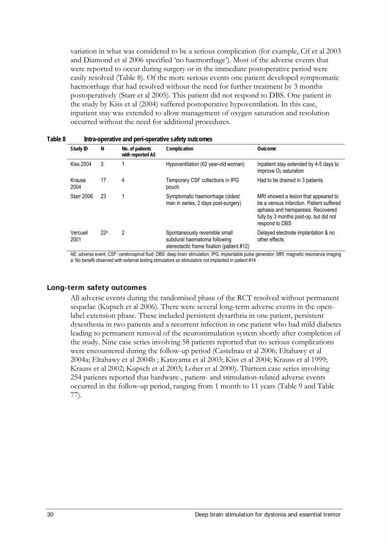
30 Deep brain stimulation for dystonia and essential tremor
variation in what was considered to be a serious complication (for example, Cif et al 2003 and Diamond et al 2006 specified ‘no haemorrhage’). Most of the adverse events that were reported to occur during surgery or in the immediate postoperative period were easily resolved (Table 8). Of the more serious events one patient developed symptomatic haemorrhage that had resolved without the need for further treatment by 3 months postoperatively (Starr et al 2005). This patient did not respond to DBS. One patient in the study by Kiss et al (2004) suffered postoperative hypoventilation. In this case, inpatient stay was extended to allow management of oxygen saturation and resolution occurred without the need for additional procedures.
Table 8 Intra-operative and peri-operative safety outcomes Study ID N No. of patients
with reported AE Complication Outcome
Kiss 2004 3 1 Hypoventilation (62 year-old woman) Inpatient stay extended by 4-5 days to improve O2 saturation
Krause 2004
17 4 Temporary CSF collections in IPG pouch
Had to be drained in 3 patients
Starr 2006 23 1 Symptomatic haemorrhage (oldest man in series, 2 days post-surgery)
MRI showed a lesion that appeared to be a venous infarction. Patient suffered aphasia and hemiparesis. Recovered fully by 3 months post-op, but did not respond to DBS
Vercueil 2001
22a 2 Spontaneously reversible small subdural haematoma following stereotactic frame fixation (patient #12)
Delayed electrode implantation & no other effects
AE: adverse event; CSF: cerebrospinal fluid; DBS: deep brain stimulation; IPG: implantable pulse generator; MRI: magnetic resonance imaging a: No benefit observed with external testing stimulators so stimulators not implanted in patient #14
Long-term safety outcomes All adverse events during the randomised phase of the RCT resolved without permanent sequelae (Kupsch et al 2006). There were several long-term adverse events in the open-label extension phase. These included persistent dysarthria in one patient, persistent dysesthesia in two patients and a recurrent infection in one patient who had mild diabetes leading to permanent removal of the neurostimulation system shortly after completion of the study. Nine case series involving 58 patients reported that no serious complications were encountered during the follow-up period (Castelnau et al 2006; Eltahawy et al 2004a; Eltahawy et al 2004b ; Katayama et al 2003; Kiss et al 2004; Krauss et al 1999; Krauss et al 2002; Kupsch et al 2003; Loher et al 2000). Thirteen case series involving 254 patients reported that hardware-, patient- and stimulation-related adverse events occurred in the follow-up period, ranging from 1 month to 11 years (Table 9 and Table 77).
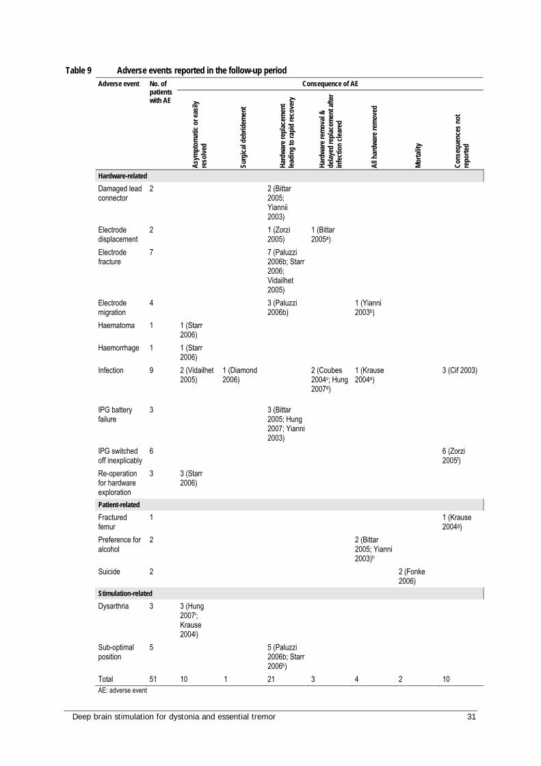
Deep brain stimulation for dystonia and essential tremor 31
Table 9 Adverse events reported in the follow-up period Consequence of AE Adverse event No. of
patients with AE
Asym
ptom
atic
or ea
sily
reso
lved
Surg
ical d
ebrid
emen
t
Hard
ware
repl
acem
ent
leadi
ng to
rapi
d re
cove
ry
Hard
ware
rem
oval
& de
layed
repl
acem
ent a
fter
infe
ctio
n cle
ared
All h
ardw
are r
emov
ed
Morta
lity
Cons
eque
nces
not
re
porte
d
Hardware-related Damaged lead connector
2 2 (Bittar 2005; Yiannii 2003)
Electrode displacement
2 1 (Zorzi 2005)
1 (Bittar 2005a)
Electrode fracture
7 7 (Paluzzi 2006b; Starr 2006; Vidailhet 2005)
Electrode migration
4 3 (Paluzzi 2006b)
1 (Yianni 2003b)
Haematoma 1 1 (Starr 2006)
Haemorrhage 1 1 (Starr 2006)
Infection 9 2 (Vidailhet 2005)
1 (Diamond 2006)
2 (Coubes 2004c; Hung 2007d)
1 (Krause 2004e)
3 (Cif 2003)
IPG battery failure
3 3 (Bittar 2005; Hung 2007; Yianni 2003)
IPG switched off inexplicably
6 6 (Zorzi 2005f)
Re-operation for hardware exploration
3 3 (Starr 2006)
Patient-related Fractured femur
1 1 (Krause 2004g)
Preference for alcohol
2 2 (Bittar 2005; Yianni 2003)h
Suicide 2 2 (Fonke 2006)
Stimulation-related Dysarthria 3 3 (Hung
2007i; Krause 2004j)
Sub-optimal position
5 5 (Paluzzi 2006b; Starr 2006k)
Total 51 10 1 21 3 4 2 10 AE: adverse event
32 Deep brain stimulation for dystonia and essential tremor
a: Patient in ICU until infection cleared, then electrodes replaced b: Both leads removed, triggering further deterioration & admission to ICU with severe cervical dystonia c: IPG infection in a bedridden 6yr old. System removed & reimplanted 6 m later d: Left electrode removed due to infection at 1yr sustained benefits for 2yrs, then declined. Electrode replaced & previous benefit restored e: Transferred to hospital closer to home where IPG became infected and was removed f: One or both IPGs switched off inexplicably leading to cessation of stimulation g: Fracture of femur while patient in state of excitation h: These patients had dystonic symptoms that were in part responsive to alcohol. Improvements from DBS removed the need to suppress symptoms with alcohol, so they requested that the system be removed in preference to decreasing alcohol intake i: Two patients developed moderate dysarthria at optimal stimulation parameters (possibly due to current spread to the internal capsule) j: Without DBS this patient could not talk due to severe dysphonia k: Two leads were too close to the corticobulbar tract & one lead was within the globus pallidus externus rather than the globus pallidus internus.
Hardware-related complications The majority of long-term complications were related to hardware damage, failure, displacement or infection and were often able to be resolved by urgent replacement of the affected hardware which resulted in a return of function (Table 9 and Table 77). The most serious complications were related to infection and sometimes resulted in the removal of the DBS system. All hardware-related complications fully resolved.
Patient-related complications The most serious adverse events reported were two cases of suicide (Table 9). These occurred during the follow-up period after the initiation of DBS in the single study by Fonke et al (2006); however, it is difficult to determine the contribution of DBS, if any, to these suicides as the incidence of suicide in the dystonic population is unknown. Both patients had a history of periods of depression prior to implantation, but were considered psychologically stable by the Centre’s DBS team during the screening period (extensive psychiatric examination was not performed). One patient committed suicide three weeks after surgery, before any follow-up evaluation was performed. The other patient experienced excellent improvement of dystonia following DBS and appeared happy with the results of DBS two weeks before committing suicide, 14 months postoperatively.
Three of the adverse events reported in the follow-up period were related to the patient involved and not to problems with the DBS system. Alcohol as a muscle relaxant can clinically control some dystonic conditions. In two patients with alcohol-responsive dystonic symptoms, the DBS systems were removed upon request by the patients, who preferred to consume alcohol rather than having DBS. One patient suffered from a fractured femur as a result of agitation and dystonic muscle contraction 2 days postoperatively (Krause et al 2004). This patient suffered from very severe PKAN dystonia, with a pre-surgical BFMDRS score of 92. The device continued to be used and a clinically-relevant functional improvement was reported, with a post-DBS follow-up BFMDRS score of 32 seven months after surgery.
Due to the nature of DBS, meningitis and haemorrhage may be considered serious risks of the procedure. Significantly, no incidences of meningitis were reported in any of the included studies.Only one case of haemorrhage was reported, which resolved spontaneously.
Deep brain stimulation for dystonia and essential tremor 33
Stimulation-related complications All stimulation-related complications that occurred in the follow-up period were reversible by adjusting DBS settings (Table 9). In one case of dysarthria where the patient could not talk at baseline, side effects were no worse than at patient baseline status.
Safety concerns for specific patient groups Cif et al (2003) commented on the ‘remarkable tolerance of internal pulse generators in children’ and reported that no complications were encountered due to displacement of hardware with growth. Coubes et al (2004) also commented on suitability for children and suggested that placement of the IPG in the abdominal area seems preferable as the thickness of the skin and fatty tissues contributes to better incision healing and overall outcome. Furthermore it appears that growth does not interfere with stimulation and the implantation of a single 90 mm extension can compensate adequately for the growth of the child.
One paper (Paluzzi et al 2006a) was identified which reported the outcomes during pregnancy of three dystonic patients using DBS (one with post-traumatic secondary dystonia and two with primary dystonia). All three women gave birth to healthy babies (one woman was followed for two successful pregnancies). No device-related adverse events were reported. The Kinetra box was changed two weeks after birth in one woman (approximately 4 years after the initial procedure). This led to discomfort during breastfeeding which lasted for approximately 4 weeks. During this time the discomfort was managed through the use of a breast pump. A second woman was admitted as an emergency in her third trimester of pregnancy in status dystonicus because the IPG batteries had expired 19 months after implantation, which was resolved by uncomplicated battery replacement under general anaesthesia. In this woman labour was induced with prostaglandins at 38 weeks because of intrauterine growth retardation and reduced foetal movement, but there were no other complications or birth defects. These cases indicate that DBS is not a barrier to conception or delivery of a healthy baby and none of the women experienced an exacerbation of symptoms during pregnancy.
Summary: Safety of DBS for dystonia The most serious adverse events reported in any of the DBS studies were two suicides that occurred in the postoperative period in one study; however, the contribution of DBS treatment to these events is unclear. All other adverse events reported in patients who received DBS treatment for dystonia were able to be resolved, often without the need for intervention and did not cause any lasting consequences. Most of the hardware-related complications were resolved by replacement of affected hardware. In two cases complications led to the removal of all hardware but did not result in any further patient complications. Importantly, none of the included studies reported any cases of meningitis and whilst one haemorrhage was reported, the patient had recovered fully by 3 months postoperatively.
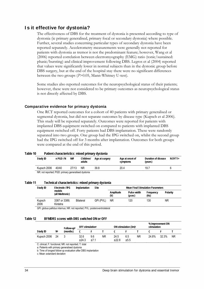
34 Deep brain stimulation for dystonia and essential tremor
Is it effective for dystonia? The effectiveness of DBS for the treatment of dystonia is presented according to type of dystonia (ie primary generalised, primary focal or secondary dystonia) where possible. Further, several studies concerning particular types of secondary dystonia have been reported separately. Accelerometry measurements were generally not reported for patients with dystonia as tremor is not the predominant feature; however, Wang et al (2006) reported correlation between electromyography (EMG) ratio (tonic/sustained: phasic/bursting) and clinical improvement following DBS. Legros et al (2004) reported that values were significantly lower in normal subjects than in the dystonic group before DBS surgery, but at the end of the hospital stay there were no significant differences between the two groups (P>0.05, Mann-Whitney U-test).
Some studies also reported outcomes for the neuropsychological status of their patients; however, these were not considered to be primary outcomes as neuropsychological status is not directly affected by DBS.
Comparative evidence for primary dystonia One RCT reported outcomes for a cohort of 40 patients with primary generalised or segmental dystonia, but did not separate outcomes by disease type (Kupsch et al 2006). This study will be reported separately. Outcomes were reported for patients with implanted DBS equipment switched on compared to patients with implanted DBS equipment switched off. Forty patients had DBS implantation. These were randomly separated into two groups. One group had the IPG switched on, whilst the second group had the IPG switched off for 3 months after implantation. Outcomes for both groups were compared at the end of this period.
Table 10 Patient characteristics: mixed primary dystonia Study ID n PGD / N M/F Children/
adults Age at surgery
Age at onset of symptoms
Duration of disease (years)
N DYT1+
Kupsch 2006 40/40 27/13 NR 39.8 20.4 19.7 6 NR: not reported; PGD: primary generalised dystonia
Table 11 Technical characteristics: mixed primary dystonia Mean Final Stimulation Parameters Study ID Electrode / IPG
models (all Medtronic)
Implantation Site Amplitude (V)
Pulse width (µsec)
Frequency (Hz)
Polarity
Kupsch 2006
3387 or 3389; Kintetra
Bilateral GPi (PVL) NR 120 130 NR
GPi: globus pallidus internus; NR: not reported; PVL: posteroventrolateral
Table 12 BFMDRS scores with DBS switched ON or OFF
OFF stimulationc ON stimulation (3m)c % improvement ON stimulation
Study ID Na Follow-upb (months) C F T C F T C F T
Kupsch 2006 24 3 32.6 ±24.3
9.6 ±7.1
NR 24.5 ±22.8
6.5 ±5.5
NR 24.8% 32.3% NR
C: clinical; F: functional; NR: not reported; T: total a: Patients with primary generalised dystonia b: Time of longest follow-up evaluation after DBS implantation c: Mean ±standard deviation
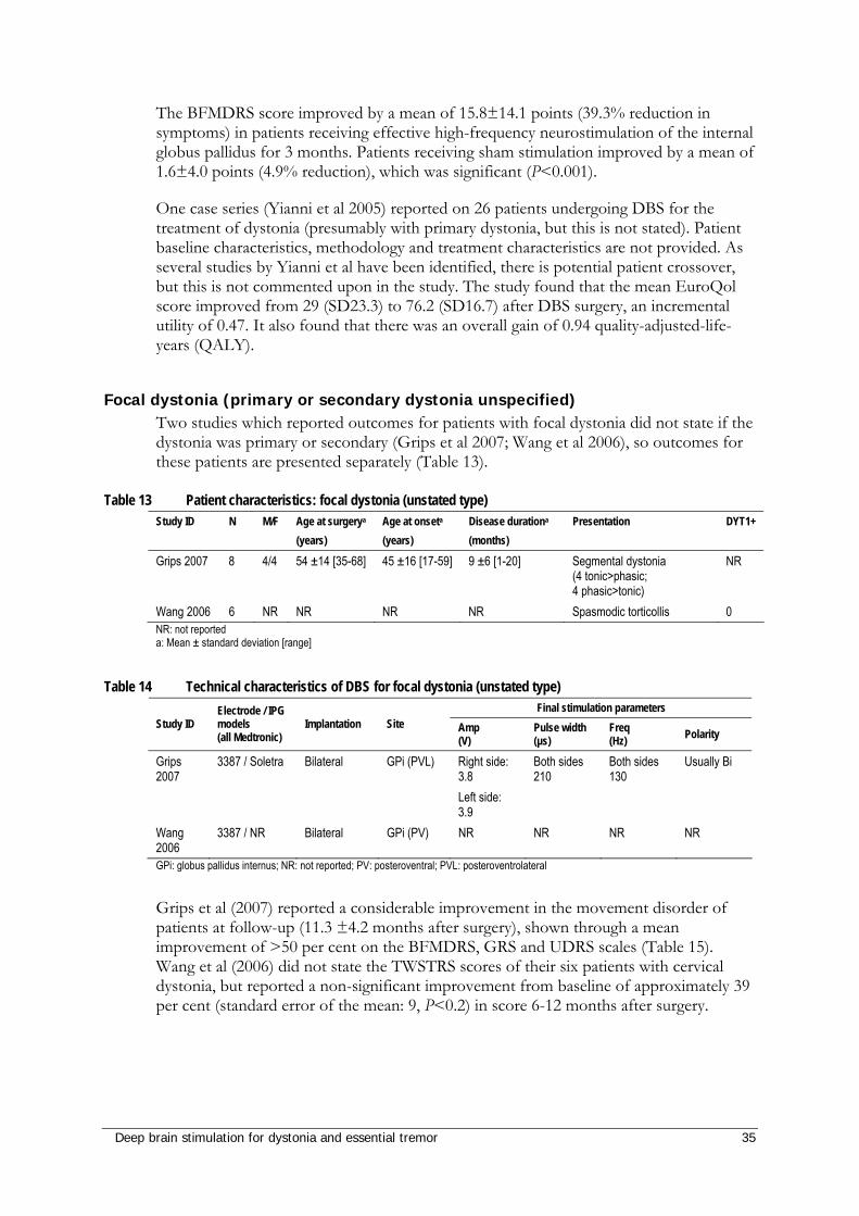
Deep brain stimulation for dystonia and essential tremor 35
The BFMDRS score improved by a mean of 15.8±14.1 points (39.3% reduction in symptoms) in patients receiving effective high-frequency neurostimulation of the internal globus pallidus for 3 months. Patients receiving sham stimulation improved by a mean of 1.6±4.0 points (4.9% reduction), which was significant (P<0.001).
One case series (Yianni et al 2005) reported on 26 patients undergoing DBS for the treatment of dystonia (presumably with primary dystonia, but this is not stated). Patient baseline characteristics, methodology and treatment characteristics are not provided. As several studies by Yianni et al have been identified, there is potential patient crossover, but this is not commented upon in the study. The study found that the mean EuroQol score improved from 29 (SD23.3) to 76.2 (SD16.7) after DBS surgery, an incremental utility of 0.47. It also found that there was an overall gain of 0.94 quality-adjusted-life-years (QALY).
Focal dystonia (primary or secondary dystonia unspecified) Two studies which reported outcomes for patients with focal dystonia did not state if the dystonia was primary or secondary (Grips et al 2007; Wang et al 2006), so outcomes for these patients are presented separately (Table 13).
Table 13 Patient characteristics: focal dystonia (unstated type) Study ID N M/F Age at surgerya
(years) Age at onseta
(years) Disease durationa
(months) Presentation DYT1+
Grips 2007 8 4/4 54 ±14 [35-68] 45 ±16 [17-59] 9 ±6 [1-20] Segmental dystonia (4 tonic>phasic; 4 phasic>tonic)
NR
Wang 2006 6 NR NR NR NR Spasmodic torticollis 0 NR: not reported a: Mean ± standard deviation [range]
Table 14 Technical characteristics of DBS for focal dystonia (unstated type) Final stimulation parameters
Study ID Electrode / IPG models (all Medtronic)
Implantation Site Amp (V)
Pulse width (µs)
Freq (Hz) Polarity
Grips 2007
3387 / Soletra Bilateral GPi (PVL) Right side: 3.8 Left side: 3.9
Both sides 210
Both sides 130
Usually Bi
Wang 2006
3387 / NR Bilateral GPi (PV) NR NR NR NR
GPi: globus pallidus internus; NR: not reported; PV: posteroventral; PVL: posteroventrolateral
Grips et al (2007) reported a considerable improvement in the movement disorder of patients at follow-up (11.3 ±4.2 months after surgery), shown through a mean improvement of >50 per cent on the BFMDRS, GRS and UDRS scales (Table 15). Wang et al (2006) did not state the TWSTRS scores of their six patients with cervical dystonia, but reported a non-significant improvement from baseline of approximately 39 per cent (standard error of the mean: 9, P<0.2) in score 6-12 months after surgery.
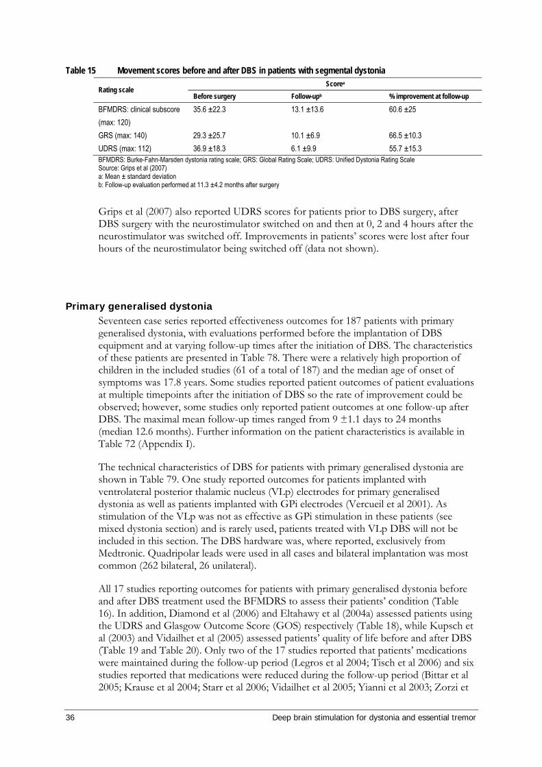
36 Deep brain stimulation for dystonia and essential tremor
Table 15 Movement scores before and after DBS in patients with segmental dystonia Scorea
Rating scale Before surgery Follow-upb % improvement at follow-up
BFMDRS: clinical subscore (max: 120)
35.6 ±22.3 13.1 ±13.6 60.6 ±25
GRS (max: 140) 29.3 ±25.7 10.1 ±6.9 66.5 ±10.3 UDRS (max: 112) 36.9 ±18.3 6.1 ±9.9 55.7 ±15.3 BFMDRS: Burke-Fahn-Marsden dystonia rating scale; GRS: Global Rating Scale; UDRS: Unified Dystonia Rating Scale Source: Grips et al (2007) a: Mean ± standard deviation b: Follow-up evaluation performed at 11.3 ±4.2 months after surgery
Grips et al (2007) also reported UDRS scores for patients prior to DBS surgery, after DBS surgery with the neurostimulator switched on and then at 0, 2 and 4 hours after the neurostimulator was switched off. Improvements in patients’ scores were lost after four hours of the neurostimulator being switched off (data not shown).
Primary generalised dystonia Seventeen case series reported effectiveness outcomes for 187 patients with primary generalised dystonia, with evaluations performed before the implantation of DBS equipment and at varying follow-up times after the initiation of DBS. The characteristics of these patients are presented in Table 78. There were a relatively high proportion of children in the included studies (61 of a total of 187) and the median age of onset of symptoms was 17.8 years. Some studies reported patient outcomes of patient evaluations at multiple timepoints after the initiation of DBS so the rate of improvement could be observed; however, some studies only reported patient outcomes at one follow-up after DBS. The maximal mean follow-up times ranged from 9 ±1.1 days to 24 months (median 12.6 months). Further information on the patient characteristics is available in Table 72 (Appendix I).
The technical characteristics of DBS for patients with primary generalised dystonia are shown in Table 79. One study reported outcomes for patients implanted with ventrolateral posterior thalamic nucleus (VLp) electrodes for primary generalised dystonia as well as patients implanted with GPi electrodes (Vercueil et al 2001). As stimulation of the VLp was not as effective as GPi stimulation in these patients (see mixed dystonia section) and is rarely used, patients treated with VLp DBS will not be included in this section. The DBS hardware was, where reported, exclusively from Medtronic. Quadripolar leads were used in all cases and bilateral implantation was most common (262 bilateral, 26 unilateral).
All 17 studies reporting outcomes for patients with primary generalised dystonia before and after DBS treatment used the BFMDRS to assess their patients’ condition (Table 16). In addition, Diamond et al (2006) and Eltahawy et al (2004a) assessed patients using the UDRS and Glasgow Outcome Score (GOS) respectively (Table 18), while Kupsch et al (2003) and Vidailhet et al (2005) assessed patients’ quality of life before and after DBS (Table 19 and Table 20). Only two of the 17 studies reported that patients’ medications were maintained during the follow-up period (Legros et al 2004; Tisch et al 2006) and six studies reported that medications were reduced during the follow-up period (Bittar et al 2005; Krause et al 2004; Starr et al 2006; Vidailhet et al 2005; Yianni et al 2003; Zorzi et

Deep brain stimulation for dystonia and essential tremor 37
al 2005). Observed improvements in disability scores at follow-up may vary depending on whether patients remained on, or were withdrawn from, medications during this period. A reduction in medication may also, but not necessarily indicate effectiveness of treatment. There were also considerable differences between studies in the severity of patients’ movement disorder at baseline, as reflected by the variability in mean pre-surgical BFMDRS-clinical scores ranging from 38.4 to 103.8.
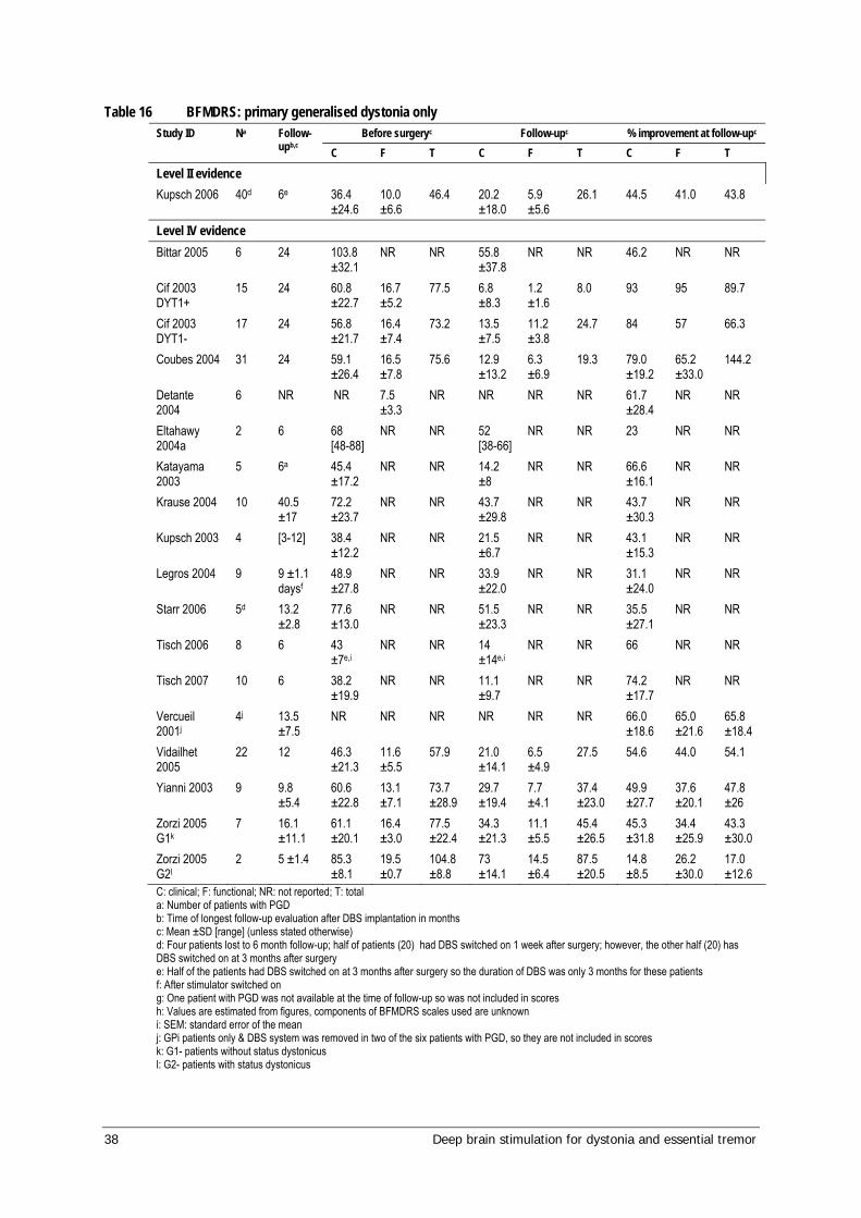
38 Deep brain stimulation for dystonia and essential tremor
Table 16 BFMDRS: primary generalised dystonia only Before surgeryc Follow-upc % improvement at follow-upc Study ID Na Follow-
upb,c C F T C F T C F T
Level II evidence Kupsch 2006 40d 6e 36.4
±24.6 10.0 ±6.6
46.4 20.2 ±18.0
5.9 ±5.6
26.1 44.5 41.0 43.8
Level IV evidence Bittar 2005 6 24 103.8
±32.1 NR NR 55.8
±37.8 NR NR 46.2 NR NR
Cif 2003 DYT1+
15 24 60.8 ±22.7
16.7 ±5.2
77.5 6.8 ±8.3
1.2 ±1.6
8.0 93 95 89.7
Cif 2003 DYT1-
17 24 56.8 ±21.7
16.4 ±7.4
73.2 13.5 ±7.5
11.2 ±3.8
24.7 84 57 66.3
Coubes 2004 31 24 59.1 ±26.4
16.5 ±7.8
75.6 12.9 ±13.2
6.3 ±6.9
19.3 79.0 ±19.2
65.2 ±33.0
144.2
Detante 2004
6 NR NR 7.5 ±3.3
NR NR NR NR 61.7 ±28.4
NR NR
Eltahawy 2004a
2 6 68 [48-88]
NR NR 52 [38-66]
NR NR 23 NR NR
Katayama 2003
5 6a 45.4 ±17.2
NR NR 14.2 ±8
NR NR 66.6 ±16.1
NR NR
Krause 2004 10 40.5 ±17
72.2 ±23.7
NR NR 43.7 ±29.8
NR NR 43.7 ±30.3
NR NR
Kupsch 2003 4 [3-12] 38.4 ±12.2
NR NR 21.5 ±6.7
NR NR 43.1 ±15.3
NR NR
Legros 2004 9 9 ±1.1 daysf
48.9 ±27.8
NR NR 33.9 ±22.0
NR NR 31.1 ±24.0
NR NR
Starr 2006 5d 13.2 ±2.8
77.6 ±13.0
NR NR 51.5 ±23.3
NR NR 35.5 ±27.1
NR NR
Tisch 2006 8 6 43 ±7e,i
NR NR 14 ±14e,i
NR NR 66 NR NR
Tisch 2007 10 6 38.2 ±19.9
NR NR 11.1 ±9.7
NR NR 74.2 ±17.7
NR NR
Vercueil 2001j
4j 13.5 ±7.5
NR NR NR NR NR NR 66.0 ±18.6
65.0 ±21.6
65.8 ±18.4
Vidailhet 2005
22 12 46.3 ±21.3
11.6 ±5.5
57.9 21.0 ±14.1
6.5 ±4.9
27.5 54.6 44.0 54.1
Yianni 2003 9 9.8 ±5.4
60.6 ±22.8
13.1 ±7.1
73.7 ±28.9
29.7 ±19.4
7.7 ±4.1
37.4 ±23.0
49.9 ±27.7
37.6 ±20.1
47.8 ±26
Zorzi 2005 G1k
7 16.1 ±11.1
61.1 ±20.1
16.4 ±3.0
77.5 ±22.4
34.3 ±21.3
11.1 ±5.5
45.4 ±26.5
45.3 ±31.8
34.4 ±25.9
43.3 ±30.0
Zorzi 2005 G2l
2 5 ±1.4 85.3 ±8.1
19.5 ±0.7
104.8 ±8.8
73 ±14.1
14.5 ±6.4
87.5 ±20.5
14.8 ±8.5
26.2 ±30.0
17.0 ±12.6
C: clinical; F: functional; NR: not reported; T: total a: Number of patients with PGD b: Time of longest follow-up evaluation after DBS implantation in months c: Mean ±SD [range] (unless stated otherwise) d: Four patients lost to 6 month follow-up; half of patients (20) had DBS switched on 1 week after surgery; however, the other half (20) has DBS switched on at 3 months after surgery e: Half of the patients had DBS switched on at 3 months after surgery so the duration of DBS was only 3 months for these patients f: After stimulator switched on g: One patient with PGD was not available at the time of follow-up so was not included in scores h: Values are estimated from figures, components of BFMDRS scales used are unknown i: SEM: standard error of the mean j: GPi patients only & DBS system was removed in two of the six patients with PGD, so they are not included in scores k: G1- patients without status dystonicus l: G2- patients with status dystonicus
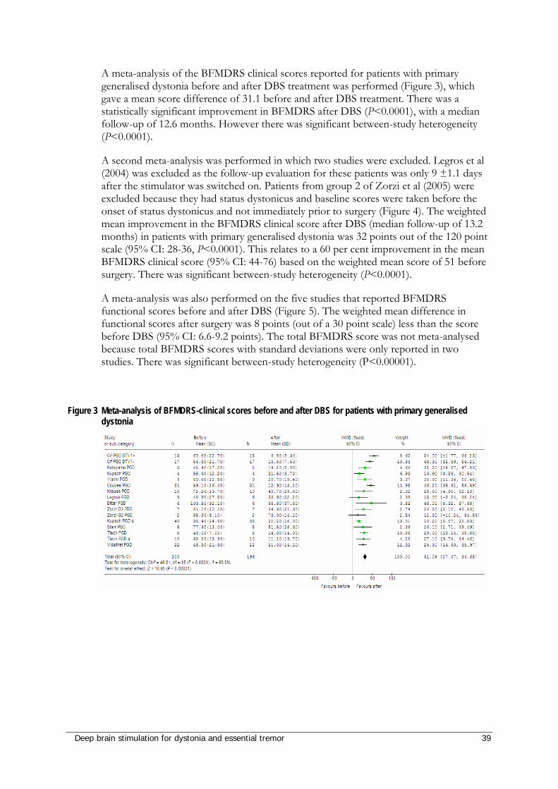
Deep brain stimulation for dystonia and essential tremor 39
A meta-analysis of the BFMDRS clinical scores reported for patients with primary generalised dystonia before and after DBS treatment was performed (Figure 3), which gave a mean score difference of 31.1 before and after DBS treatment. There was a statistically significant improvement in BFMDRS after DBS (P<0.0001), with a median follow-up of 12.6 months. However there was significant between-study heterogeneity (P<0.0001).
A second meta-analysis was performed in which two studies were excluded. Legros et al (2004) was excluded as the follow-up evaluation for these patients was only 9 ±1.1 days after the stimulator was switched on. Patients from group 2 of Zorzi et al (2005) were excluded because they had status dystonicus and baseline scores were taken before the onset of status dystonicus and not immediately prior to surgery (Figure 4). The weighted mean improvement in the BFMDRS clinical score after DBS (median follow-up of 13.2 months) in patients with primary generalised dystonia was 32 points out of the 120 point scale (95% CI: 28-36, P<0.0001). This relates to a 60 per cent improvement in the mean BFMDRS clinical score (95% CI: 44-76) based on the weighted mean score of 51 before surgery. There was significant between-study heterogeneity (P<0.0001).
A meta-analysis was also performed on the five studies that reported BFMDRS functional scores before and after DBS (Figure 5). The weighted mean difference in functional scores after surgery was 8 points (out of a 30 point scale) less than the score before DBS (95% CI: 6.6-9.2 points). The total BFMDRS score was not meta-analysed because total BFMDRS scores with standard deviations were only reported in two studies. There was significant between-study heterogeneity (P<0.00001).
Figure 3 Meta-analysis of BFMDRS-clinical scores before and after DBS for patients with primary generalised dystonia
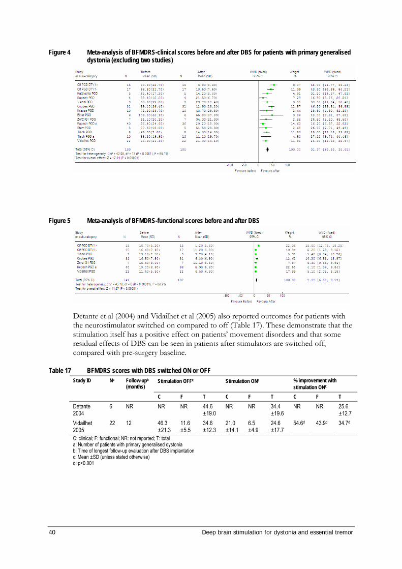
40 Deep brain stimulation for dystonia and essential tremor
Figure 4 Meta-analysis of BFMDRS-clinical scores before and after DBS for patients with primary generalised dystonia (excluding two studies)
Figure 5 Meta-analysis of BFMDRS-functional scores before and after DBS
Detante et al (2004) and Vidailhet et al (2005) also reported outcomes for patients with the neurostimulator switched on compared to off (Table 17). These demonstrate that the stimulation itself has a positive effect on patients’ movement disorders and that some residual effects of DBS can be seen in patients after stimulators are switched off, compared with pre-surgery baseline.
Table 17 BFMDRS scores with DBS switched ON or OFF Stimulation OFFc Stimulation ONc % improvement with
stimulation ONc Study ID Na Follow-upb
(months)
C F T C F T C F T
Detante 2004
6 NR NR NR 44.6 ±19.0
NR NR 34.4 ±19.6
NR NR 25.6 ±12.7
Vidailhet 2005
22 12 46.3 ±21.3
11.6 ±5.5
34.6 ±12.3
21.0 ±14.1
6.5 ±4.9
24.6 ±17.7
54.6d 43.9d 34.7d
C: clinical; F: functional; NR: not reported; T: total a: Number of patients with primary generalised dystonia b: Time of longest follow-up evaluation after DBS implantation c: Mean ±SD (unless stated otherwise) d: p<0.001
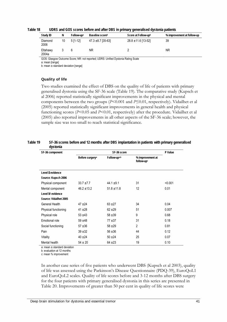
Deep brain stimulation for dystonia and essential tremor 41
Table 18 UDRS and GOS scores before and after DBS in primary generalised dystonia patients Study ID N Follow-upa Baseline scoreb Score at Follow-upb % improvement at follow-up
Diamond 2006
10 5 [1-12] 47.3 ±8.7 [35-63] 28.8 ±11.6 [13-52] 39
Eltahawy 2004a
3 6 NR 2 NR
GOS: Glasgow Outcome Score; NR: not reported; UDRS: Unified Dystonia Rating Scale a: mean [range] b: mean ± standard deviation [range]
Quality of life
Two studies examined the effect of DBS on the quality of life of patients with primary generalised dystonia using the SF-36 scale (Table 19). The comparative study (Kupsch et al 2006) reported statistically significant improvements in the physical and mental components between the two groups (P<0.001 and P≤0.01, respectively). Vidailhet et al (2005) reported statistically significant improvements in general health and physical functioning scores (P<0.05 and P<0.01, respectively) after the procedure. Vidailhet et al (2005) also reported improvements in all other aspects of the SF-36 scale; however, the sample size was too small to reach statistical significance.
Table 19 SF-36 scores before and 12 months after DBS implantation in patients with primary generalised dystonia
SF-36 score SF-36 component
Before surgerya Follow-upa,b % improvement at follow-upc
P Value
Level II evidence Source: Kupsch 2006 Physical component 33.7 ±7.7 44.1 ±9.1 31 <0.001 Mental component 46.2 ±13.2 51.8 ±11.8 12 0.01 Level IV evidence Source: Vidailhet 2005 General Health 47 ±24 63 ±27 34 0.04 Physical functioning 41 ±28 62 ±29 51 0.007 Physical role 53 ±43 58 ±39 9 0.68 Emotional role 59 ±48 77 ±37 31 0.18 Social functioning 57 ±36 58 ±29 2 0.81 Pain 39 ±32 56 ±36 44 0.12 Vitality 40 ±24 50 ±24 25 0.07 Mental health 54 ± 20 64 ±23 19 0.10 a: mean ± standard deviation b: evaluation at 12 months c: mean % improvement
In another case series of five patients who underwent DBS (Kupsch et al 2003), quality of life was assessed using the Parkinson’s Disease Questionnaire (PDQ-39), EuroQoL1 and EuroQoL2 scales. Quality of life scores before and 3-12 months after DBS surgery for the four patients with primary generalised dystonia in this series are presented in Table 20. Improvements of greater than 50 per cent in quality of life scores were
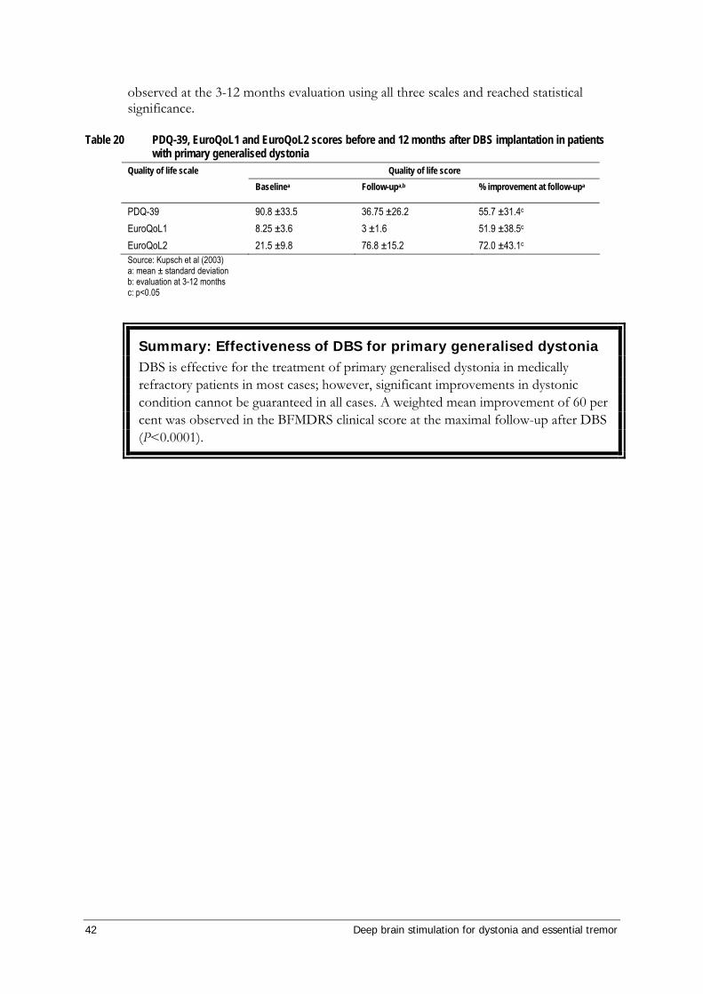
42 Deep brain stimulation for dystonia and essential tremor
observed at the 3-12 months evaluation using all three scales and reached statistical significance.
Table 20 PDQ-39, EuroQoL1 and EuroQoL2 scores before and 12 months after DBS implantation in patients with primary generalised dystonia
Quality of life score Quality of life scale Baselinea Follow-upa,b % improvement at follow-upa
PDQ-39 90.8 ±33.5 36.75 ±26.2 55.7 ±31.4c EuroQoL1 8.25 ±3.6 3 ±1.6 51.9 ±38.5c EuroQoL2 21.5 ±9.8 76.8 ±15.2 72.0 ±43.1c Source: Kupsch et al (2003) a: mean ± standard deviation b: evaluation at 3-12 months c: p<0.05
Summary: Effectiveness of DBS for primary generalised dystonia DBS is effective for the treatment of primary generalised dystonia in medically refractory patients in most cases; however, significant improvements in dystonic condition cannot be guaranteed in all cases. A weighted mean improvement of 60 per cent was observed in the BFMDRS clinical score at the maximal follow-up after DBS (P<0.0001).

Deep brain stimulation for dystonia and essential tremor 43
Primary focal dystonia Outcomes for 51 patients treated with DBS for primary focal dystonia were reported in 11 case series, some of whom were included as a larger cohort of patients. The majority of patients treated with DBS for primary focal dystonia had predominantly cervical dystonia and these shall be reported separately where possible. There were also a number of patients with focal-dystonia affecting other regions, multi-focal-, segmental-, or hemidystonia.
Primary cervical dystonia The majority of patients (39 of 51) included in the primary focal dystonia studies were affected with primary cervical dystonia. The median patient age at surgery was 45.5 years and the median age at onset of disease was 32.5 years. All patients received bilateral DBS of the GPi. Further information on the patient and technical characteristics of the primary cervical dystonia patients is available in Table 21 and Table 22.
Table 21 Patient characteristics: primary cervical dystonia Study ID N M/F Age at
surgerya Age at onseta Disease
durationa
(months)
Presentation (comorbidity & concurrent treatment)
DYT1+
Bittar 2005 6 0/6 39 ±19 [23-68]
31 ±17 [17-59]
8 ±4 [3-16] ST NR
Eltahawy 2004a
3 2/1 50 ±17 [34-67]
40 ±11 [30-52]
10 ±7 [3-15] ST NR
Eltahawy 2004b
3 2/1 47±17.3 37±13.5 10±5.6 1 right ST, 2 left ST NR
Hung 2007 10 5/5 48 ±14.9 [25-67]
38 10.0 ±3.9 [4-17]
ST (3T, 2P, 1T&P, 1T&Tr, 3P&Tr)
NR
Kiss 2004 2 2/0 55.5 ±10.6 [48-63]
46.0 ±4.2 [43-49]
9.5 ±6.4 [5-14]
ST (1 patient suffered from depression. 1 patient trialled C3-4 facet rhizotomy & microvascular compression of the right 11th nerve & upper cervical roots)
NR
Kraus 1999 3 NR [42-53] NR 60-84 Severe complex ST NR Krauss 2002
5 3/2 43 ±9 [28-53]
9 ±8.2 [4-24] 34 ±11.6 [22-47]
ST NR
Yianni 2003 7 1/6 39 ±15 31 ±16 8 ±4 ST 2 Total 39 13/20c Median: 45.5
Median: 32.5 Median: 43.5 2
HD: hemidystonia; MF: multifocal dystonia; NR: not reported; P: phasic; SD: segmental dystonia; ST: spasmodic torticollis; T: tonic; Tr: tremor a: Mean ± standard deviation [range] b: Two patients with primary focal dystonia were included in this study; however, only the patient treated with GPi DBS will be included in this report as VLp DBS is less effective than GPi DBS for dystonia c: Gender of some patients not reported d: Range of mean ages
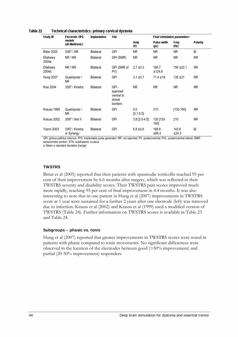
44 Deep brain stimulation for dystonia and essential tremor
Table 22 Technical characteristics: primary cervical dystonia Final stimulation parametersa Study ID Electrode / IPG
models (all Medtronic)
Implantation Site Amp (V)
Pulse width (µs)
Freq (Hz)
Polarity
Bittar 2005 3387 / NR Bilateral GPi NR NR NR Bi Eltahawy 2004a
NR / NR Bilateral GPi (SMR) NR NR NR NR
Eltahawy 2004b
NR / NR Bilateral GPi (SMR of PV)
2.7 ±0.3 166.7 ±124.8
150 ±22.1 NR
Hung 2007 Quadripolar / NR
Bilateral GPi 3.1 ±0.7 71.4 ±18 135 ±21 NR
Kiss 2004 3387 / Kinetra Bilateral GPi -spanned ventral to dorsal borders
NR NR NR NR
Krauss 1999 Quadripolar / NR
Bilateral GPi 4.0 [3.1-5.0]
210 [130-160] NR
Krauss 2002 3387 / Itrel II Bilateral GPi 3.8 [3.0-4.5] 135 [130-145]
210 NR
Yianni 2003 3387 / Kinetra or Synergy
Bilateral GPi 5.8 ±0.6 168.8 ±66.4
143.8 ±24.3
Bi
GPi: globus pallidus internus; IPG: implantable pulse generator; NR: not reported; PV: posteroventral; PVL: posteroventral lateral; SMR: sensorimotor portion; STN: subthalamic nucleus a: Mean ± standard deviation [range]
TWSTRS
Bittar et al (2005) reported that their patients with spasmodic torticollis reached 95 per cent of their improvement by 6.6 months after surgery, which was reflected in their TWSTRS severity and disability scores. Their TWSTRS pain scores improved much more rapidly, reaching 95 per cent of final improvement in 4.4 months. It was also interesting to note that in one patient in Hung et al (2007) improvements in TWSTRS score at 1 year were sustained for a further 2 years after one electrode (left) was removed due to infection. Krauss et al (2002) and Krauss et al (1999) used a modified version of TWSTRS (Table 24). Further information on TWSTRS scores is available in Table 23 and Table 24.
Subgroups – phasic vs. tonic
Hung et al (2007) reported that greater improvements in TWSTRS scores were noted in patients with phasic compared to tonic movements. No significant differences were observed in the location of the electrodes between good (>50% improvement) and partial (20-50% improvement) responders.
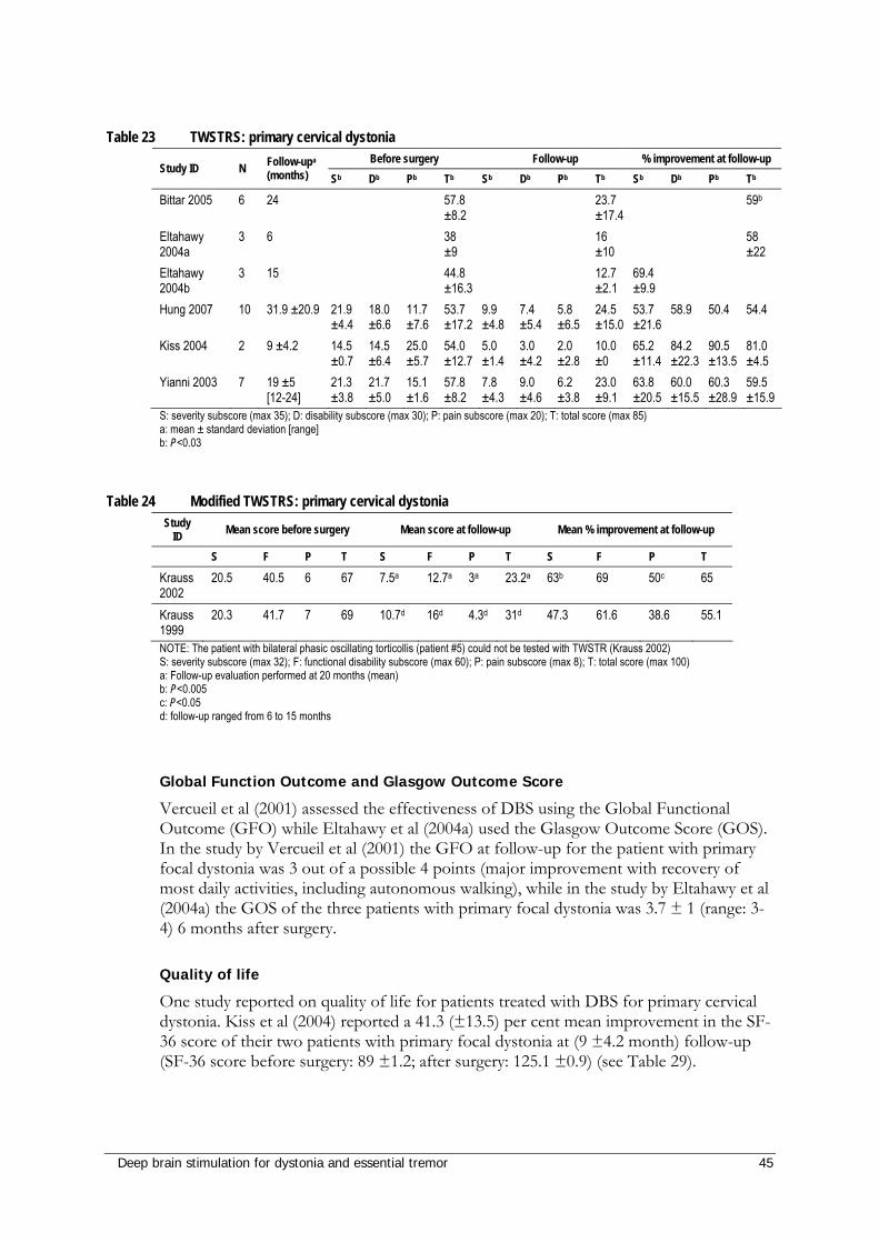
Deep brain stimulation for dystonia and essential tremor 45
Table 23 TWSTRS: primary cervical dystonia
Before surgery Follow-up % improvement at follow-up Study ID N Follow-upa
(months) Sb Db Pb Tb Sb Db Pb Tb Sb Db Pb Tb
Bittar 2005 6 24 57.8 ±8.2
23.7 ±17.4
59b
Eltahawy 2004a
3 6 38 ±9
16 ±10
58 ±22
Eltahawy 2004b
3 15 44.8 ±16.3
12.7 ±2.1
69.4 ±9.9
Hung 2007 10 31.9 ±20.9 21.9 ±4.4
18.0 ±6.6
11.7 ±7.6
53.7 ±17.2
9.9 ±4.8
7.4 ±5.4
5.8 ±6.5
24.5 ±15.0
53.7 ±21.6
58.9 50.4 54.4
Kiss 2004 2 9 ±4.2 14.5 ±0.7
14.5 ±6.4
25.0 ±5.7
54.0 ±12.7
5.0 ±1.4
3.0 ±4.2
2.0 ±2.8
10.0 ±0
65.2 ±11.4
84.2 ±22.3
90.5 ±13.5
81.0 ±4.5
Yianni 2003 7 19 ±5 [12-24]
21.3 ±3.8
21.7 ±5.0
15.1 ±1.6
57.8 ±8.2
7.8 ±4.3
9.0 ±4.6
6.2 ±3.8
23.0 ±9.1
63.8 ±20.5
60.0 ±15.5
60.3 ±28.9
59.5 ±15.9
S: severity subscore (max 35); D: disability subscore (max 30); P: pain subscore (max 20); T: total score (max 85) a: mean ± standard deviation [range] b: P<0.03
Table 24 Modified TWSTRS: primary cervical dystonia Study
ID Mean score before surgery Mean score at follow-up Mean % improvement at follow-up
S F P T S F P T S F P T
Krauss 2002
20.5 40.5 6 67 7.5a 12.7a 3a 23.2a 63b 69 50c 65
Krauss 1999
20.3 41.7 7 69 10.7d 16d 4.3d 31d 47.3 61.6 38.6 55.1
NOTE: The patient with bilateral phasic oscillating torticollis (patient #5) could not be tested with TWSTR (Krauss 2002) S: severity subscore (max 32); F: functional disability subscore (max 60); P: pain subscore (max 8); T: total score (max 100) a: Follow-up evaluation performed at 20 months (mean) b: P<0.005 c: P<0.05 d: follow-up ranged from 6 to 15 months
Global Function Outcome and Glasgow Outcome Score
Vercueil et al (2001) assessed the effectiveness of DBS using the Global Functional Outcome (GFO) while Eltahawy et al (2004a) used the Glasgow Outcome Score (GOS). In the study by Vercueil et al (2001) the GFO at follow-up for the patient with primary focal dystonia was 3 out of a possible 4 points (major improvement with recovery of most daily activities, including autonomous walking), while in the study by Eltahawy et al (2004a) the GOS of the three patients with primary focal dystonia was 3.7 ± 1 (range: 3-4) 6 months after surgery.
Quality of life
One study reported on quality of life for patients treated with DBS for primary cervical dystonia. Kiss et al (2004) reported a 41.3 (±13.5) per cent mean improvement in the SF-36 score of their two patients with primary focal dystonia at (9 ±4.2 month) follow-up (SF-36 score before surgery: 89 ±1.2; after surgery: 125.1 ±0.9) (see Table 29).
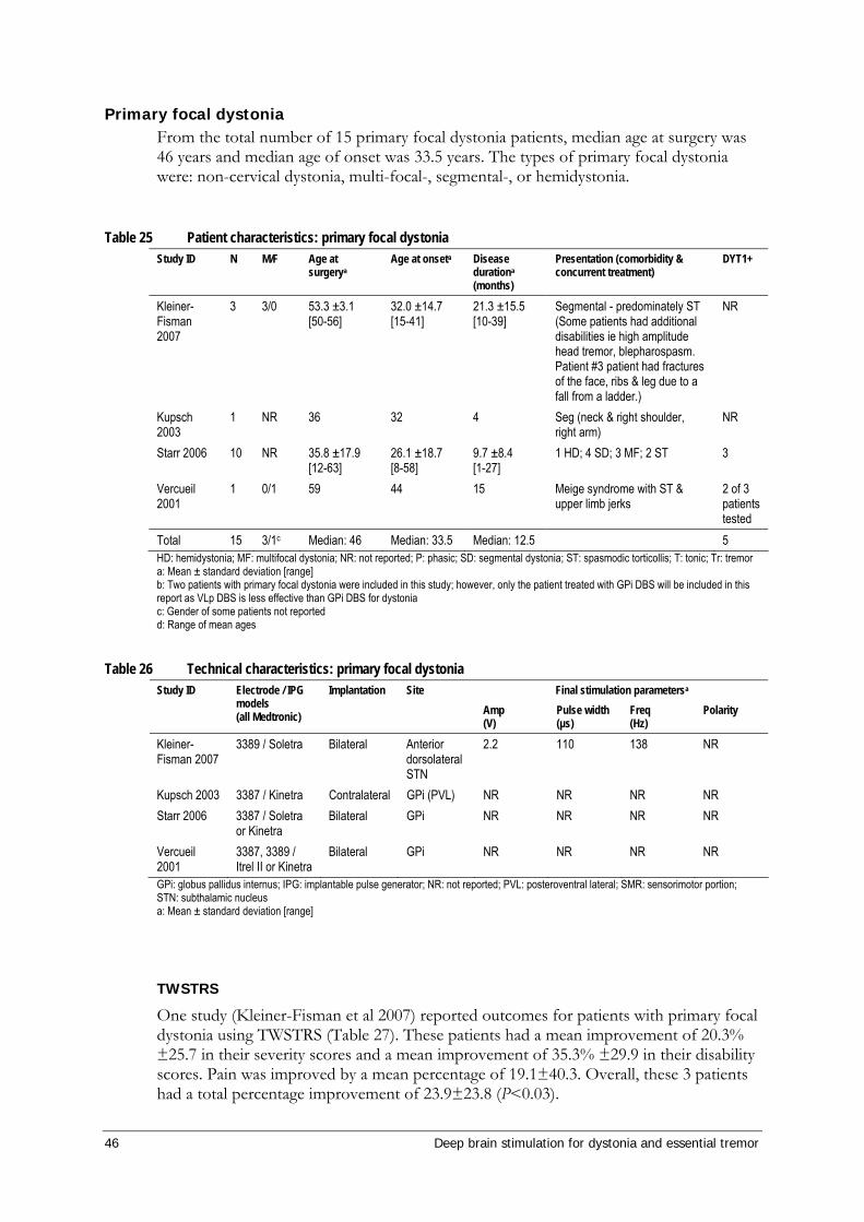
46 Deep brain stimulation for dystonia and essential tremor
Primary focal dystonia From the total number of 15 primary focal dystonia patients, median age at surgery was 46 years and median age of onset was 33.5 years. The types of primary focal dystonia were: non-cervical dystonia, multi-focal-, segmental-, or hemidystonia.
Table 25 Patient characteristics: primary focal dystonia
Study ID N M/F Age at surgerya
Age at onseta Disease durationa
(months)
Presentation (comorbidity & concurrent treatment)
DYT1+
Kleiner-Fisman 2007
3 3/0 53.3 ±3.1 [50-56]
32.0 ±14.7 [15-41]
21.3 ±15.5 [10-39]
Segmental - predominately ST (Some patients had additional disabilities ie high amplitude head tremor, blepharospasm. Patient #3 patient had fractures of the face, ribs & leg due to a fall from a ladder.)
NR
Kupsch 2003
1 NR 36 32 4 Seg (neck & right shoulder, right arm)
NR
Starr 2006 10 NR 35.8 ±17.9 [12-63]
26.1 ±18.7 [8-58]
9.7 ±8.4 [1-27]
1 HD; 4 SD; 3 MF; 2 ST 3
Vercueil 2001
1 0/1 59 44 15 Meige syndrome with ST & upper limb jerks
2 of 3 patients tested
Total 15 3/1c Median: 46 Median: 33.5 Median: 12.5 5 HD: hemidystonia; MF: multifocal dystonia; NR: not reported; P: phasic; SD: segmental dystonia; ST: spasmodic torticollis; T: tonic; Tr: tremor a: Mean ± standard deviation [range] b: Two patients with primary focal dystonia were included in this study; however, only the patient treated with GPi DBS will be included in this report as VLp DBS is less effective than GPi DBS for dystonia c: Gender of some patients not reported d: Range of mean ages
Table 26 Technical characteristics: primary focal dystonia Final stimulation parametersa Study ID Electrode / IPG
models (all Medtronic)
Implantation Site Amp (V)
Pulse width (µs)
Freq (Hz)
Polarity
Kleiner-Fisman 2007
3389 / Soletra Bilateral Anterior dorsolateral STN
2.2 110 138 NR
Kupsch 2003 3387 / Kinetra Contralateral GPi (PVL) NR NR NR NR Starr 2006 3387 / Soletra
or Kinetra Bilateral GPi NR NR NR NR
Vercueil 2001
3387, 3389 / Itrel II or Kinetra
Bilateral GPi NR NR NR NR
GPi: globus pallidus internus; IPG: implantable pulse generator; NR: not reported; PVL: posteroventral lateral; SMR: sensorimotor portion; STN: subthalamic nucleus a: Mean ± standard deviation [range]
TWSTRS
One study (Kleiner-Fisman et al 2007) reported outcomes for patients with primary focal dystonia using TWSTRS (Table 27). These patients had a mean improvement of 20.3% ±25.7 in their severity scores and a mean improvement of 35.3% ±29.9 in their disability scores. Pain was improved by a mean percentage of 19.1±40.3. Overall, these 3 patients had a total percentage improvement of 23.9±23.8 (P<0.03).
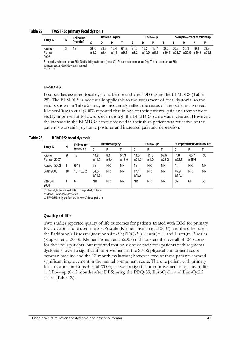
Deep brain stimulation for dystonia and essential tremor 47
Table 27 TWSTRS: primary focal dystonia Before surgery Follow-up % improvement at follow-up
Study ID N Follow-upa (months) S D P T S D P T S D P Tb
Kleiner-Fisman 2007
3 12 26.0 ±5.0
23.3 ±6.4
15.4 ±1.5
64.8 ±9.5
21.0 ±8.2
16.3 ±10.0
12.7 ±6.5
50.0 ±19.5
20.3 ±25.7
35.3 ±29.9
19.1 ±40.3
23.9 ±23.8
S: severity subscore (max 35); D: disability subscore (max 30); P: pain subscore (max 20); T: total score (max 85) a: mean ± standard deviation [range] b: P<0.03
BFMDRS
Four studies assessed focal dystonia before and after DBS using the BFMDRS (Table 28). The BFMDRS is not usually applicable to the assessment of focal dystonia, so the results shown in Table 28 may not accurately reflect the status of the patients involved. Kleiner-Fisman et al (2007) reported that in one of their patients, pain and tremor were visibly improved at follow-up, even though the BFMDRS score was increased. However, the increase in the BFMDRS score observed in their third patient was reflective of the patient’s worsening dystonic postures and increased pain and depression.
Table 28 BFMDRS: focal dystonia Before surgerya Follow-upa % improvement at follow-upa
Study ID N Follow -upa
(months) C F T C F T C F T
Kliener-Fisman 2007
2b 12 44.8 ±11.7
9.5 ±6.4
54.3 ±18.0
44.0 ±21.2
13.5 ±4.9
57.5 ±26.2
-4.6 ±22.5
-60.7 ±55.6
-30
Kupsch 2003 1 6-12 32 NR NR 19 NR NR 41 NR NR Starr 2006 10 13.7 ±8.2 34.5
±11.0 NR NR 17.1
±15.7 NR NR 46.9
±47.6 NR NR
Vercueil 2001
1 6 NR NR NR NR NR NR 66 66 66
C: clinical; F: functional; NR: not reported; T: total a: Mean ± standard deviation b: BFMDRS only performed in two of three patients
Quality of life
Two studies reported quality of life outcomes for patients treated with DBS for primary focal dystonia; one used the SF-36 scale (Kleiner-Fisman et al 2007) and the other used the Parkinson’s Disease Questionnaire-39 (PDQ-39), EuroQoL1 and EuroQoL2 scales (Kupsch et al 2003). Kleiner-Fisman et al (2007) did not state the overall SF-36 scores for their four patients, but reported that only one of their four patients with segmental dystonia showed a significant improvement in the SF-36 physical component score between baseline and the 12-month evaluation; however, two of these patients showed significant improvement in the mental component score. The one patient with primary focal dystonia in Kupsch et al (2003) showed a significant improvement in quality of life at follow-up (6-12 months after DBS) using the PDQ-39, EuroQoL1 and EuroQoL2 scales (Table 29).
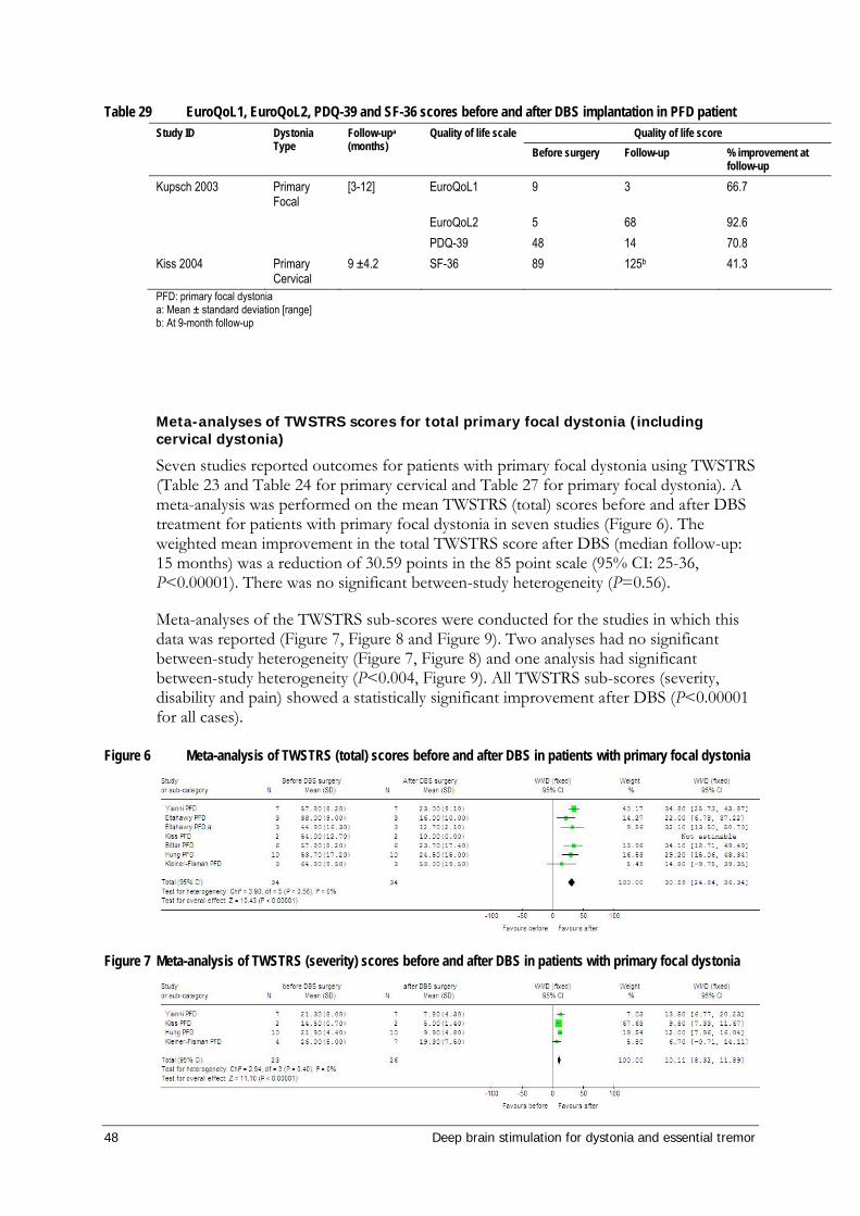
48 Deep brain stimulation for dystonia and essential tremor
Table 29 EuroQoL1, EuroQoL2, PDQ-39 and SF-36 scores before and after DBS implantation in PFD patient Quality of life score Study ID Dystonia
Type Follow-upa (months)
Quality of life scale Before surgery Follow-up % improvement at
follow-up
Kupsch 2003 Primary Focal
[3-12] EuroQoL1 9 3 66.7
EuroQoL2 5 68 92.6 PDQ-39 48 14 70.8 Kiss 2004 Primary
Cervical 9 ±4.2 SF-36 89 125b 41.3
PFD: primary focal dystonia a: Mean ± standard deviation [range] b: At 9-month follow-up
Meta-analyses of TWSTRS scores for total primary focal dystonia (including cervical dystonia)
Seven studies reported outcomes for patients with primary focal dystonia using TWSTRS (Table 23 and Table 24 for primary cervical and Table 27 for primary focal dystonia). A meta-analysis was performed on the mean TWSTRS (total) scores before and after DBS treatment for patients with primary focal dystonia in seven studies (Figure 6). The weighted mean improvement in the total TWSTRS score after DBS (median follow-up: 15 months) was a reduction of 30.59 points in the 85 point scale (95% CI: 25-36, P<0.00001). There was no significant between-study heterogeneity (P=0.56).
Meta-analyses of the TWSTRS sub-scores were conducted for the studies in which this data was reported (Figure 7, Figure 8 and Figure 9). Two analyses had no significant between-study heterogeneity (Figure 7, Figure 8) and one analysis had significant between-study heterogeneity (P<0.004, Figure 9). All TWSTRS sub-scores (severity, disability and pain) showed a statistically significant improvement after DBS (P<0.00001 for all cases).
Figure 6 Meta-analysis of TWSTRS (total) scores before and after DBS in patients with primary focal dystonia
Figure 7 Meta-analysis of TWSTRS (severity) scores before and after DBS in patients with primary focal dystonia
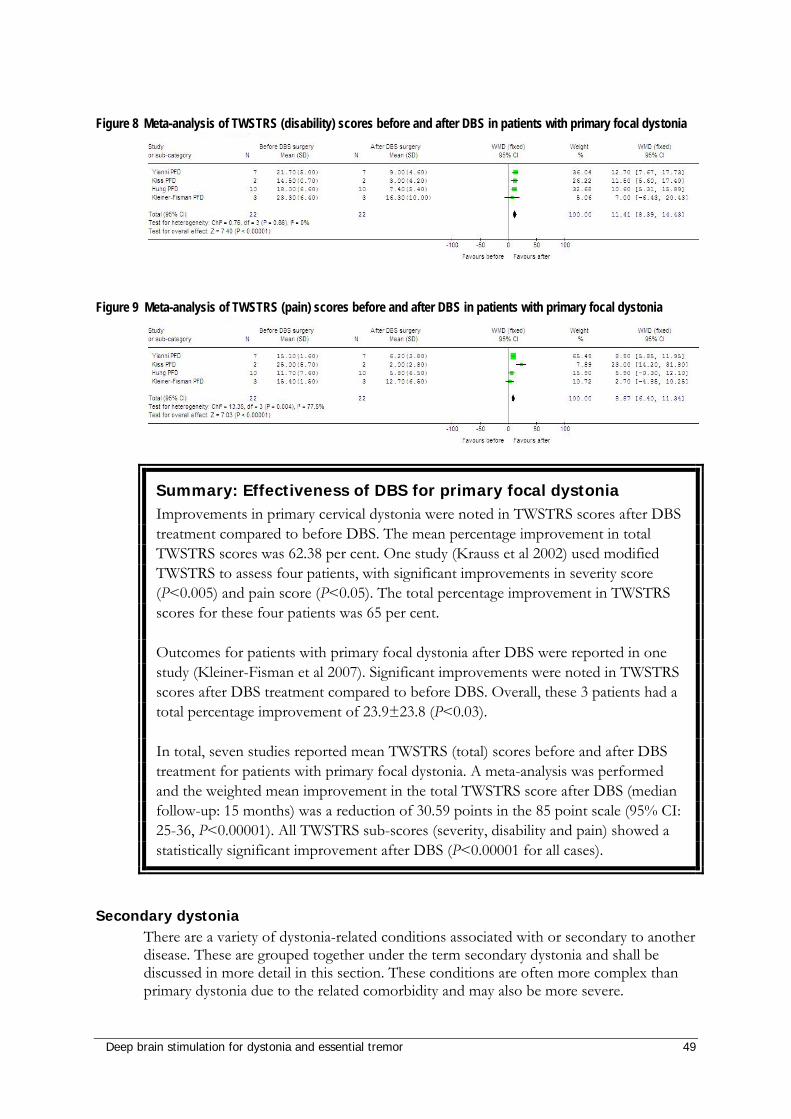
Deep brain stimulation for dystonia and essential tremor 49
Figure 8 Meta-analysis of TWSTRS (disability) scores before and after DBS in patients with primary focal dystonia
Figure 9 Meta-analysis of TWSTRS (pain) scores before and after DBS in patients with primary focal dystonia
Summary: Effectiveness of DBS for primary focal dystonia Improvements in primary cervical dystonia were noted in TWSTRS scores after DBS treatment compared to before DBS. The mean percentage improvement in total TWSTRS scores was 62.38 per cent. One study (Krauss et al 2002) used modified TWSTRS to assess four patients, with significant improvements in severity score (P<0.005) and pain score (P<0.05). The total percentage improvement in TWSTRS scores for these four patients was 65 per cent.
Outcomes for patients with primary focal dystonia after DBS were reported in one study (Kleiner-Fisman et al 2007). Significant improvements were noted in TWSTRS scores after DBS treatment compared to before DBS. Overall, these 3 patients had a total percentage improvement of 23.9±23.8 (P<0.03).
In total, seven studies reported mean TWSTRS (total) scores before and after DBS treatment for patients with primary focal dystonia. A meta-analysis was performed and the weighted mean improvement in the total TWSTRS score after DBS (median follow-up: 15 months) was a reduction of 30.59 points in the 85 point scale (95% CI: 25-36, P<0.00001). All TWSTRS sub-scores (severity, disability and pain) showed a statistically significant improvement after DBS (P<0.00001 for all cases).
Secondary dystonia There are a variety of dystonia-related conditions associated with or secondary to another disease. These are grouped together under the term secondary dystonia and shall be discussed in more detail in this section. These conditions are often more complex than primary dystonia due to the related comorbidity and may also be more severe.
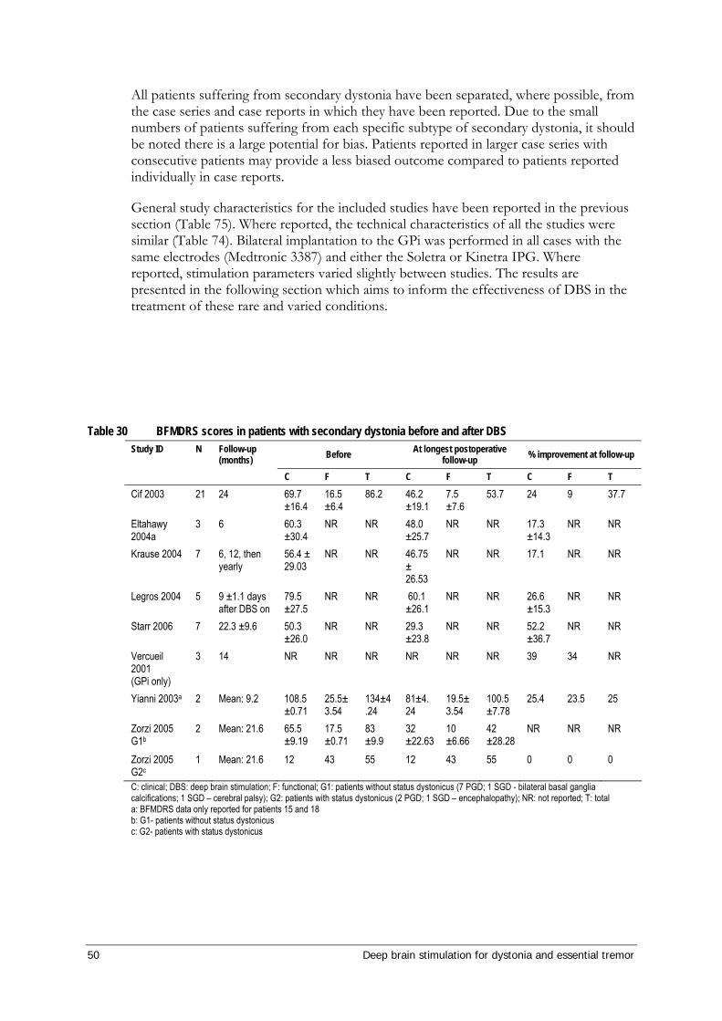
50 Deep brain stimulation for dystonia and essential tremor
All patients suffering from secondary dystonia have been separated, where possible, from the case series and case reports in which they have been reported. Due to the small numbers of patients suffering from each specific subtype of secondary dystonia, it should be noted there is a large potential for bias. Patients reported in larger case series with consecutive patients may provide a less biased outcome compared to patients reported individually in case reports.
General study characteristics for the included studies have been reported in the previous section (Table 75). Where reported, the technical characteristics of all the studies were similar (Table 74). Bilateral implantation to the GPi was performed in all cases with the same electrodes (Medtronic 3387) and either the Soletra or Kinetra IPG. Where reported, stimulation parameters varied slightly between studies. The results are presented in the following section which aims to inform the effectiveness of DBS in the treatment of these rare and varied conditions.
Table 30 BFMDRS scores in patients with secondary dystonia before and after DBS
Before At longest postoperative follow-up % improvement at follow-up Study ID N Follow-up
(months) C F T C F T C F T
Cif 2003 21 24 69.7 ±16.4
16.5 ±6.4
86.2 46.2 ±19.1
7.5 ±7.6
53.7 24 9 37.7
Eltahawy 2004a
3 6 60.3 ±30.4
NR NR 48.0 ±25.7
NR NR 17.3 ±14.3
NR NR
Krause 2004 7 6, 12, then yearly
56.4 ± 29.03
NR NR 46.75 ± 26.53
NR NR 17.1 NR NR
Legros 2004 5 9 ±1.1 days after DBS on
79.5 ±27.5
NR NR 60.1 ±26.1
NR NR 26.6 ±15.3
NR NR
Starr 2006 7 22.3 ±9.6 50.3 ±26.0
NR NR 29.3 ±23.8
NR NR 52.2 ±36.7
NR NR
Vercueil 2001 (GPi only)
3 14 NR NR NR NR NR NR 39 34 NR
Yianni 2003a 2 Mean: 9.2 108.5±0.71
25.5±3.54
134±4.24
81±4.24
19.5±3.54
100.5±7.78
25.4 23.5 25
Zorzi 2005 G1b
2 Mean: 21.6 65.5 ±9.19
17.5 ±0.71
83 ±9.9
32 ±22.63
10 ±6.66
42 ±28.28
NR NR NR
Zorzi 2005 G2c
1 Mean: 21.6 12 43 55 12 43 55 0 0 0
C: clinical; DBS: deep brain stimulation; F: functional; G1: patients without status dystonicus (7 PGD; 1 SGD - bilateral basal ganglia calcifications; 1 SGD – cerebral palsy); G2: patients with status dystonicus (2 PGD; 1 SGD – encephalopathy); NR: not reported; T: total a: BFMDRS data only reported for patients 15 and 18 b: G1- patients without status dystonicus c: G2- patients with status dystonicus
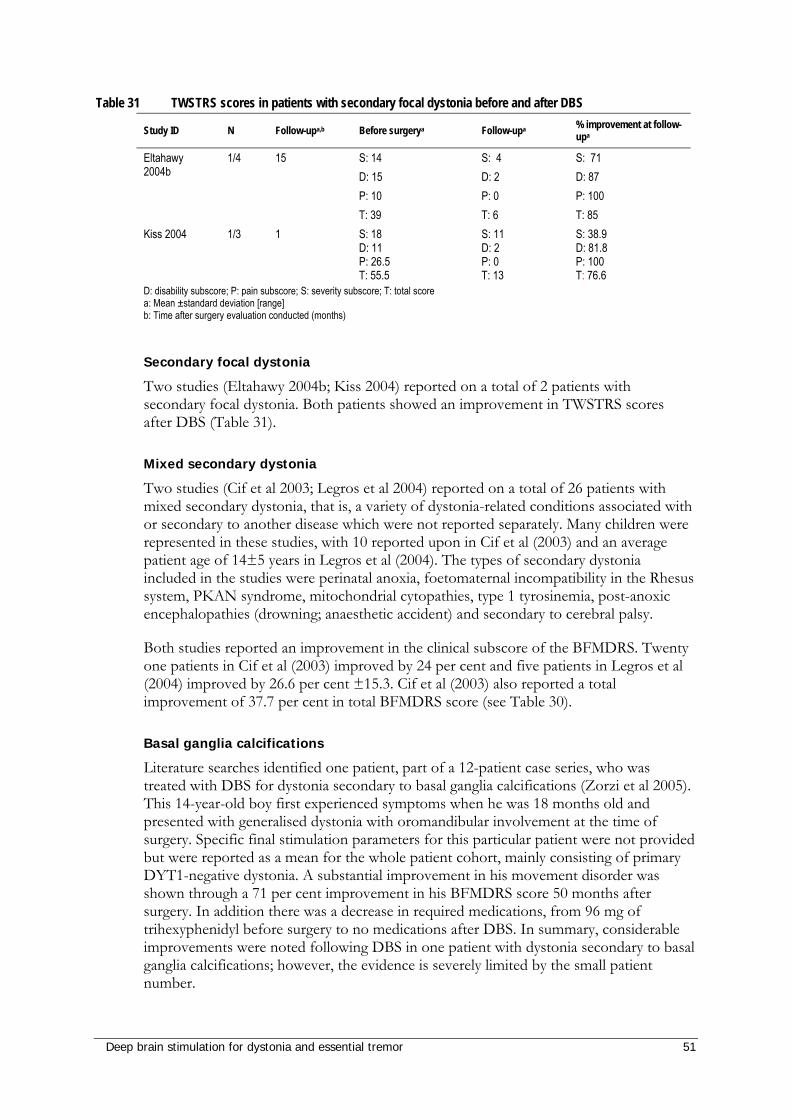
Deep brain stimulation for dystonia and essential tremor 51
Table 31 TWSTRS scores in patients with secondary focal dystonia before and after DBS
Study ID N Follow-upa,b Before surgerya Follow-upa % improvement at follow-upa
Eltahawy 2004b
1/4 15 S: 14 D: 15 P: 10 T: 39
S: 4 D: 2 P: 0 T: 6
S: 71 D: 87 P: 100 T: 85
Kiss 2004 1/3 1 S: 18 D: 11 P: 26.5 T: 55.5
S: 11 D: 2 P: 0 T: 13
S: 38.9 D: 81.8 P: 100 T: 76.6
D: disability subscore; P: pain subscore; S: severity subscore; T: total score a: Mean ±standard deviation [range] b: Time after surgery evaluation conducted (months)
Secondary focal dystonia
Two studies (Eltahawy 2004b; Kiss 2004) reported on a total of 2 patients with secondary focal dystonia. Both patients showed an improvement in TWSTRS scores after DBS (Table 31).
Mixed secondary dystonia
Two studies (Cif et al 2003; Legros et al 2004) reported on a total of 26 patients with mixed secondary dystonia, that is, a variety of dystonia-related conditions associated with or secondary to another disease which were not reported separately. Many children were represented in these studies, with 10 reported upon in Cif et al (2003) and an average patient age of 14±5 years in Legros et al (2004). The types of secondary dystonia included in the studies were perinatal anoxia, foetomaternal incompatibility in the Rhesus system, PKAN syndrome, mitochondrial cytopathies, type 1 tyrosinemia, post-anoxic encephalopathies (drowning; anaesthetic accident) and secondary to cerebral palsy.
Both studies reported an improvement in the clinical subscore of the BFMDRS. Twenty one patients in Cif et al (2003) improved by 24 per cent and five patients in Legros et al (2004) improved by 26.6 per cent ±15.3. Cif et al (2003) also reported a total improvement of 37.7 per cent in total BFMDRS score (see Table 30).
Basal ganglia calcifications
Literature searches identified one patient, part of a 12-patient case series, who was treated with DBS for dystonia secondary to basal ganglia calcifications (Zorzi et al 2005). This 14-year-old boy first experienced symptoms when he was 18 months old and presented with generalised dystonia with oromandibular involvement at the time of surgery. Specific final stimulation parameters for this particular patient were not provided but were reported as a mean for the whole patient cohort, mainly consisting of primary DYT1-negative dystonia. A substantial improvement in his movement disorder was shown through a 71 per cent improvement in his BFMDRS score 50 months after surgery. In addition there was a decrease in required medications, from 96 mg of trihexyphenidyl before surgery to no medications after DBS. In summary, considerable improvements were noted following DBS in one patient with dystonia secondary to basal ganglia calcifications; however, the evidence is severely limited by the small patient number.
52 Deep brain stimulation for dystonia and essential tremor
Cervical dyskinesias and cervical myelopathy
Three patients were identified as having received treatment with DBS for secondary cervical dyskinesia and cervical myelopathy as part of a case series of eight patients (Krauss et al 2002). Two females were wheelchair-bound and presented with severe cervical dyskinesia due to choreoathetoid infantile cerebral palsy. The third patient was a male with secondary cervical myelopathy who had previously received thalamotomy resulting in temporary improvement of the dystonia. Final stimulation parameters were not provided for these three patients (Table 74).
There was an improvement in dystonic posture as scored on the Cervical Dystonia Rating Scale (CDRS) after DBS in all three patients which was maintained at follow-up and the surgery was well tolerated. The authors commented that the improvement in these three patients was less pronounced than in the other patients in the case series who had idiopathic cervical dystonia.
In summary, mean improvements of 40-50 per cent were observed in the CDRS scores of the three patients treated with DBS for secondary cervical dyskinesias; however, evidence for the effectiveness of DBS for this patient group is limited by the small patient number.
Dystonia secondary to basal ganglia haemorrhage
A single female patient treated with DBS for hemidystonia secondary to basal ganglia haemorrhage was identified in a case series of 11 patients (Diamond et al 2006). The patient had a relatively mild dystonia (pre-DBS UDRS score of 18), which improved slightly (UDRS score of 14) after follow-up of 104 days. DBS implantation, parameters and the configuration of polarity were all reported as a mean for the patient cohort as a whole; therefore, the exact technique used for this patient is unknown.
In summary, although slight improvement was noted in the one patient treated with DBS for dystonia secondary to basal ganglia haemorrhage, evidence for effectiveness is limited by the small patient number and lack of reporting of methodology and technique.
Dystonia secondary to cerebral palsy
A single patient who was treated with DBS for dystonia secondary to cerebral palsy was identified in a case series of 12 patients (Zorzi et al 2005). Prior to surgery the patient was wheelchair-bound with generalised dystonia, with a preoperative BFMDRS total score of 90. Following surgery the BFMDRS total score was reduced to 62 (a 31% improvement) with a reduction in medication from 46 mg trihexyphenidyl per day to no medication after implantation. No adverse events were reported.
In summary, the patient studied showed a BFMDRS score improvement of 31 per cent after DBS surgery, but the evidence for the effectiveness of DBS for this patient group is limited by the small patient number.
Dystonia secondary to Huntington’s disease
One patient who was treated with DBS for dystonia secondary to Huntington’s disease was identified as part of a 15-patient case series (Eltahawy et al 2004a). This 34-year-old woman presented with generalised dystonia with disease duration of 9 years at the time of surgery. This patient experienced an improvement of 17 per cent in BFMDRS at six
Deep brain stimulation for dystonia and essential tremor 53
months after bilateral DBS surgery (pre-treatment score: 101; post-treatment score: 84) with a postoperative GOS of 1 (representing mild improvement in movement disorder but no improvement in function).
In summary, only one patient was assessed and the type of machine and stimulation parameters employed were not recorded; hence, this evidence should be used cautiously. The patient’s BFMDRS score had a mild improvement of 17 per cent after DBS surgery, but this was not accompanied by an improvement in function as measured by GOS.
Chorea-neuroacanthocytosis
Outcomes for three patients treated with DBS for secondary dystonia due to chorea-acanthocytosis were presented in two case reports (Burbaud et al 2002; Guehl et al 2007). The patient reported by Burbaud et al (2002) was a 43-year-old male with major hypotonia, oromandibular dystonia and clinical neuropathy, who presented with three general epileptic seizures at the beginning of the disease; the patient was bedridden and totally dependent. Baseline characteristics were only reported for one of the two patients in Guehl et al (2007), a 32-year-old male who first experienced dystonic symptoms at the age of 24. All three patients were refractory to medications and had confirmation of the genetic abnormality causing the chorea-acanthocytosis.
Guehl et al (2007) reported that although a stimulation frequency of 130 Hz resulted in a greater improvement in limb dystonia and bradykinesia in both patients, it unacceptably increased dysarthria and chorea; however, a stimulation frequency of 40 Hz (monopolar) resulted in notable improvement of the condition. The mean UDRS score at 130 Hz was 21.8 (SD 5.3) and at 40 Hz was 10.5 (SD1.4), compared with the off-stimulation score of 13 (SD 3.5).
Burbaud et al (2002) reported considerable improvement in the patient’s movement disorder at three-month follow-up, reflected by a 133 per cent improvement in Barthel index, 71 per cent improvement in EMG and 37 per cent improvement in Mardsen and Schacher Choreoric Score. The patient, who was bedridden and totally dependant before surgery, was able to walk between parallel bars, read and write with a pencil nine months postoperatively, none of which was possible before surgery. His status remained stable at 1 year and his medication was reduced from 25 mg to 15 mg of Haloperidol per day.
In summary, where reported, patients were improved. Burbaud’s patient showed a 133 per cent improvement in their Barthel index and 71 per cent improvement in their Mardsen and Schacher Choreoric Score after DBS. For the patient with baseline characteristics, Guehl et al (2007) reported a notable improvement of UDRS scores at 40Hz. At stimulation off, the total scores were 13±3.5 and at stimulation at 40Hz, the total scores were 10.5±1.4.
Dystonia secondary to multiple sclerosis
One patient, a 51 year-old woman who was treated for dystonia secondary to multiple sclerosis (MS) was reported in a larger 25-patient case series (Yianni et al 2003). This patient presented with spasmodic torticollis and the onset of her disease occurred at the age of 41. This patient may have focal dystonia with a co-existence of MS; however, study reporting did not make this clear so the patient has been considered separately. Her BFMDRS scores 10 months after surgery showed improvements of 22 per cent in clinical scores, 4 per cent in functional scores and 19 per cent in total BFMDRS scores.
54 Deep brain stimulation for dystonia and essential tremor
In summary, a single patient showed slight improvement in clinical, functional and total BFMDRS scores at 10-month follow-up after DBS. The total BFMDRS scores improved by 19 per cent. The evidence of the effectiveness of DBS for dystonia secondary to MS is limited by the small patient number and the fact that this patient may have had focal dystonia with a co-existence of MS, rather than dystonia secondary to MS.
Dystonia secondary to Parkinson’s disease
One study examined the effect of DBS on 16 patients (6 women and 10 men) with advanced idiopathic Parkinson’s disease (Loher et al 2002). The mean age at surgery was 64.9 years (range 49-77 years) with a mean duration of disease of 18.2 years (range 6-30 years). Nine patients had unilateral DBS, three of which had secondary-staged bilateral surgery later on. Seven patients underwent bilateral surgery. Follow-up was 12 months, apart from the three patients who underwent contralateral surgery later on, whose follow up was 9 months and all patients were assessed whilst antiparkinsonian medication was withdrawn. Dystonia did not develop in the follow-up period, if not already present at the preoperative assessment. Dystonia severity was described by the patients according to an ordinal scale ranging from 0 to 4 (0=absent, 1=slight, 2=moderate, 3=pronounced, 4=severe). In patients with unilateral GPi DBS the Unified Parkinson’s Disease Rating Scale (UPDRS) activities of daily living score improved by 34 per cent at 3 months and by 33 per cent at 12 months and the motor score decreased by 38 per cent at 3 and 12 months. These patients also had a significant and sustained improvement of contralateral dystonia (100% improvement, P=0.019) and an improvement in dystonia of 88.2 per cent (P=0.21). Bilateral DBS patients had a reduction in UPDRS activities of daily living score by 36 per cent at 3 months and 34 per cent at 12 months, with motor scores improving by 36 per cent at 3 months and 41 per cent at 12 months. These patients had an improvement in dystonia of 85.7 per cent (P=0.021) and this was sustained throughout the follow up period.
In summary, 16 patients receiving DBS to the GPi for dystonia secondary to Parkinson’s disease experienced significant improvements in dystonia (P=0.021); however, it is not clear whether or not the rating scale used was clinically validated.
Dystonia secondary to striatal necrosis
One patient in the case series by Vercueil et al (2001) was treated with DBS for secondary dystonia due to striatal necrosis. This 30-year-old female had progressive generalised dystonia with severe and painful rigidity which first presented at the age of five. After a trial of VLp stimulation yielded no improvement, GPi stimulation resulted in 31 per cent improvement in her BFMDRS score at 18 months postoperatively (improvement in clinical score: 28%; functional score: 41%) and GFO was 2 (moderate improvement). Postoperative stimulation parameters were adjusted to give maximal benefit with no adverse events, however final stimulation parameters were not provided.
In summary, one patient showed improvement in clinical, functional and total BFMDRS scores after receiving GPi DBS, following failed VLp stimulation. The patient’s total BFMDRS score showed a 31 per cent improvement, yet the evidence of the effectiveness of DBS for dystonia secondary to striatal necrosis is limited by the fact that only one patient was studied.
Deep brain stimulation for dystonia and essential tremor 55
Encephalitic secondary dystonia
Two patients were identified who were treated with DBS for encephalitic secondary dystonia (Eltahawy et al 2004a; Zorzi et al 2005). Both had disease onset at a very young age (5 and 0.5 years). One patient had status dystonicus for two months prior to surgery (Zorzi et al 2005) and both were refractory to medication.
No improvement in the patients’ BFMDRS scores were observed in either study during postoperative follow-up and the GOS score at follow-up for the patient in the study by Eltahawy et al (2004a) was 0 (no improvement). Although the BFMDRS score was not improved after DBS compared to the score before the onset of status dystonicus, the status dystonicus resolved completely within a week of DBS and ventilation and sedation were discontinued (Zorzi et al 2005).
In summary, neither of the two patients with encephalitic secondary dystonia displayed improvements in BFMDRS scores after receiving DBS and one patient (Eltahawy et al 2004a) had no improvement in GOS score at follow-up; however, Zorzi et al (2005) reported that the status dystonicus resolved completely within a week of DBS. The evidence of effectiveness of DBS for encephalitic secondary dystonia is limited by the fact that only two patients were studied.
GM1 Type 3 gangliosidosis secondary dystonia
A single case report reported outcomes for a 24-year-old female patient with secondary dystonia due to GM1 type 3 gangliosidosis (GM1-3g) before and 2 years after DBS implantation (Roze et al 2006). The patient, who had experienced dystonic symptoms for 8 years, presented with severe generalised dystonia with fixed dystonic postures of limb, torticollis and related symptoms and had Akineto-rigid Parkinsonism without significant mental impairment. Although improvements in the BFMDRS (clinical) and UPDRS (akinesia) scores were only 20 per cent and 14 per cent respectively 12 months after surgery, the patient was able to stand up and walk unaided, which she could not do before surgery. Greater improvement was seen initially at three months; the authors suggested that the scores at 12 months were slightly higher due to progression of the disease at a rate similar to that observed before DBS. Additionally, the patient, who was blinded to DBS settings, reported a 30 per cent subjective improvement when the neurostimulator was switched on compared to when it was switched off.
In summary, the evidence of effectiveness of DBS for GM1 type 3 Gangliosidosis secondary dystonia is limited by the fact that only one patient was studied. This patient showed a 20 per cent improvement in BFMDRS clinical scores and a 14 per cent improvement in UPDRS akinesia scores after DBS.
Pantothenate Kinase-Associated Neurodegeneration
One case series and two case reports investigating the use of DBS to treat Pantothenate Kinase-Associated Neurodegeneration (PKAN) were retrieved and included for assessment (Castelnau et al 2006; Krause et al 2006; Umemura et al 2004) (Table 32). Additionally, one case series investigating the use of DBS on dystonia which included one PKAN patient was included (Starr et al 2006). The lack of available evidence may reflect the rarity of PKAN. Where reported, all patients in all the studies had MRI-confirmed PKAN. In addition, where reported, all patients in the included studies had confirmed PANK2 gene mutation. With regard to other patient demographic information, patients were generally relatively young at the time of intervention, with a
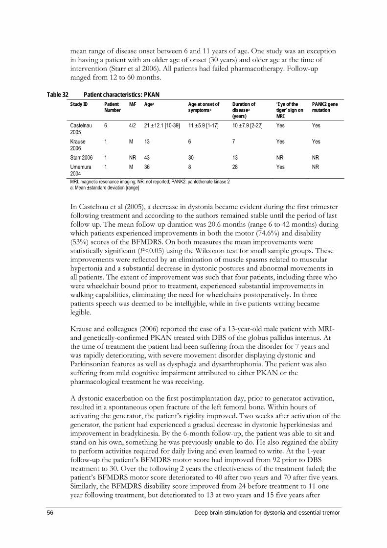
56 Deep brain stimulation for dystonia and essential tremor
mean range of disease onset between 6 and 11 years of age. One study was an exception in having a patient with an older age of onset (30 years) and older age at the time of intervention (Starr et al 2006). All patients had failed pharmacotherapy. Follow-up ranged from 12 to 60 months.
Table 32 Patient characteristics: PKAN Study ID Patient
Number M/F Agea Age at onset of
symptomsa Duration of diseasea (years)
‘Eye of the tiger’ sign on MRI
PANK2 gene mutation
Castelnau 2005
6 4/2 21 ±12.1 [10-39] 11 ±5.9 [1-17] 10 ±7.9 [2-22] Yes Yes
Krause 2006
1 M 13 6 7 Yes Yes
Starr 2006 1 NR 43 30 13 NR NR Umemura 2004
1 M 36 8 28 Yes NR
MRI: magnetic resonance imaging; NR: not reported; PANK2: pantothenate kinase 2 a: Mean ±standard deviation [range]
In Castelnau et al (2005), a decrease in dystonia became evident during the first trimester following treatment and according to the authors remained stable until the period of last follow-up. The mean follow-up duration was 20.6 months (range 6 to 42 months) during which patients experienced improvements in both the motor (74.6%) and disability (53%) scores of the BFMDRS. On both measures the mean improvements were statistically significant (P<0.05) using the Wilcoxon test for small sample groups. These improvements were reflected by an elimination of muscle spasms related to muscular hypertonia and a substantial decrease in dystonic postures and abnormal movements in all patients. The extent of improvement was such that four patients, including three who were wheelchair bound prior to treatment, experienced substantial improvements in walking capabilities, eliminating the need for wheelchairs postoperatively. In three patients speech was deemed to be intelligible, while in five patients writing became legible.
Krause and colleagues (2006) reported the case of a 13-year-old male patient with MRI- and genetically-confirmed PKAN treated with DBS of the globus pallidus internus. At the time of treatment the patient had been suffering from the disorder for 7 years and was rapidly deteriorating, with severe movement disorder displaying dystonic and Parkinsonian features as well as dysphagia and dysarthrophonia. The patient was also suffering from mild cognitive impairment attributed to either PKAN or the pharmacological treatment he was receiving.
A dystonic exacerbation on the first postimplantation day, prior to generator activation, resulted in a spontaneous open fracture of the left femoral bone. Within hours of activating the generator, the patient’s rigidity improved. Two weeks after activation of the generator, the patient had experienced a gradual decrease in dystonic hyperkinesias and improvement in bradykinesia. By the 6-month follow-up, the patient was able to sit and stand on his own, something he was previously unable to do. He also regained the ability to perform activities required for daily living and even learned to write. At the 1-year follow-up the patient’s BFMDRS motor score had improved from 92 prior to DBS treatment to 30. Over the following 2 years the effectiveness of the treatment faded; the patient’s BFMDRS motor score deteriorated to 40 after two years and 70 after five years. Similarly, the BFMDRS disability score improved from 24 before treatment to 11 one year following treatment, but deteriorated to 13 at two years and 15 five years after
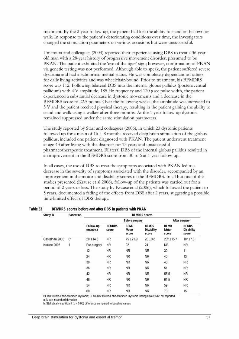
Deep brain stimulation for dystonia and essential tremor 57
treatment. By the 2-year follow-up, the patient had lost the ability to stand on his own or walk. In response to the patient’s deteriorating conditions over time, the investigators changed the stimulation parameters on various occasions but were unsuccessful.
Umemura and colleagues (2004) reported their experience using DBS to treat a 36-year-old man with a 28-year history of progressive movement disorder, presumed to be PKAN. The patient exhibited the ‘eye of the tiger’ sign; however, confirmation of PKAN via genetic testing was not performed. Although able to speak, the patient suffered severe dysarthia and had a subnormal mental status. He was completely dependant on others for daily living activities and was wheelchair-bound. Prior to treatment, his BFMDRS score was 112. Following bilateral DBS into the internal globus pallidus (posteroventral pallidum) with 4 V amplitude, 185 Hz frequency and 120 µsec pulse width, the patient experienced a substantial decrease in dystonic movements and a decrease in the BFMDRS score to 22.5 points. Over the following weeks, the amplitude was increased to 5 V and the patient received physical therapy, resulting in the patient gaining the ability to stand and walk using a walker after three months. At the 1-year follow-up dystonia remained suppressed under the same stimulation parameters.
The study reported by Starr and colleagues (2006), in which 23 dystonic patients followed up for a mean of 16 ± 8 months received deep brain stimulation of the globus pallidus, included one patient diagnosed with PKAN. The patient underwent treatment at age 43 after living with the disorder for 13 years and unsuccessful pharmacotherapeutic treatment. Bilateral DBS of the internal globus pallidus resulted in an improvement in the BFMDRS score from 30 to 6 at 1-year follow-up.
In all cases, the use of DBS to treat the symptoms associated with PKAN led to a decrease in the severity of symptoms associated with the disorder, accompanied by an improvement in the motor and disability scores of the BFMDRS. In all but one of the studies presented (Krause et al 2006), follow-up of the patients was carried out for a period of 2 years or less. The study by Krause et al (2006), which followed the patient to 5 years, documented a fading of the effects from DBS after 2 years, suggesting a possible time-limited effect of DBS therapy.
Table 33 BFMDRS scores before and after DBS in patients with PKAN BFMDRS scores
Before surgery After surgery Study ID Patient no.
Follow–up (months)
BFMDRS score
BFMD Motor score
BFMDS Disability score
BFMD Motor score
BFMDS Disability score
Castelnau 2005 6a 20 ±14.3 NR 75 ±21.9 20 ±9.8 20b ±15.7 10b ±7.8 Pre-surgery NR 92 24 NR NR 12 NR NR NR 30 11 24 NR NR NR 40 13 30 NR NR NR 46 NR 36 NR NR NR 51 NR 42 NR NR NR 55.5 NR 48 NR NR NR 61.5 NR 54 NR NR NR 59 NR
Krause 2006 1
60 NR NR NR 70 15 BFMD: Burke-Fahn-Marsden Dystonia; BFMDRS: Burke-Fahn-Marsden Dystonia Rating Scale; NR: not reported a: Mean ±standard deviation b: Statistically significant (p < 0.05) difference compared to baseline values
58 Deep brain stimulation for dystonia and essential tremor
In summary, the limited evidence available for the use of DBS to treat PKAN reflects the rarity of the disorder. The studies presented demonstrate that DBS is somewhat effective in the treatment of PKAN symptoms, particularly dystonia; they also indicate that the internal sensory motor (posteroventral) globus pallidus may not be completely damaged by PKAN and may preserve functional activity. All nine patients studied showed improvements. While it may not be possible to conduct randomised or even comparative studies given the rarity of this disorder, studies documenting the use of DBS to treat PKAN with longer follow-up periods are required to better assess the potential effectiveness of this therapy for PKAN patients.
Post-anoxic secondary dystonia
Four patients with post-anoxic secondary dystonia were identified in a case series of 19 patients (Vercueil et al 2001); however, only the two patients treated with GPi DBS will be included in this section as VLp DBS was found to be less effective. One patient did not respond to test stimulation and refused the offer of chronic stimulation at a later date. The second patient was treated with DBS for post-anoxic secondary dystonia; the patient was a 38-year-old woman who presented with severe myoclonic dystonia involving both upper limbs after first experiencing symptoms at the age of 37 years. Previously VLp DBS had been trialled and was reported to have little benefit. Improvements of 3 per cent and 16 per cent respectively in the BFMDRS clinical and disability scores were noted at 18 months after surgery.
In summary, two patients with post-anoxic secondary dystonia were treated with GPi DBS. One patient did not improve and refused the offer of chronic stimulation at a later date. The second patient improved by 3 per cent in BFMDRS clinical scores and 16 per cent in BFMDRS disability scores, but the evidence of effectiveness of DBS for post-anoxic secondary dystonia is limited by the fact that only one patient was studied.
Rapid-onset dystonia-Parkinsonism
A single case report was identified in which a patient received DBS for rapid-onset dystonia-Parkinsonism (RDP) (Deutschlander et al 2005). This 23-year-old woman presented with hypomimia, blepharospasm, torticollis, hypophonia, hemidystonia and generalised hypokinesia at the time of surgery, after experiencing dystonic symptoms for 2.3 years. DBS was trialled in this patient for 19 months without any amelioration of symptoms (BFMDRS clinical score remained at 50/120 and BFMDRS disability score remained at 22/30) so the DBS electrodes were removed.
In summary, the effectiveness of DBS for rapid-onset dystonia-Parkinsonism is limited by the fact that only one patient was studied. This patient did not report any improvements in BFMDRS clinical or disability scores and the DBS system was explanted.
Tardive dyskinesia/dystonia
Eighteen patients from three case series and five case reports were treated for tardive dyskinesia/dystonia with DBS. Patients commonly presented with general dystonia induced by neuroleptic treatment which continued even after withdrawal from medication (Table 80). All the patients, where reported, had symptom onset from the age of 27 to 48 years and had failed medical treatment for the dystonia. Age at surgery ranged from 31.5-53 years. Where reported, all were suggested to be caused by neuroleptic
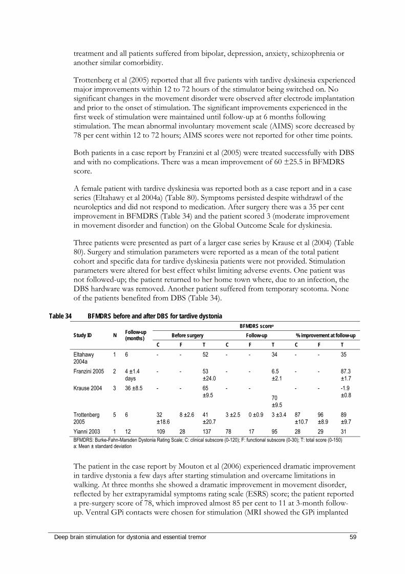
Deep brain stimulation for dystonia and essential tremor 59
treatment and all patients suffered from bipolar, depression, anxiety, schizophrenia or another similar comorbidity.
Trottenberg et al (2005) reported that all five patients with tardive dyskinesia experienced major improvements within 12 to 72 hours of the stimulator being switched on. No significant changes in the movement disorder were observed after electrode implantation and prior to the onset of stimulation. The significant improvements experienced in the first week of stimulation were maintained until follow-up at 6 months following stimulation. The mean abnormal involuntary movement scale (AIMS) score decreased by 78 per cent within 12 to 72 hours; AIMS scores were not reported for other time points.
Both patients in a case report by Franzini et al (2005) were treated successfully with DBS and with no complications. There was a mean improvement of 60 ±25.5 in BFMDRS score.
A female patient with tardive dyskinesia was reported both as a case report and in a case series (Eltahawy et al 2004a) (Table 80). Symptoms persisted despite withdrawl of the neuroleptics and did not respond to medication. After surgery there was a 35 per cent improvement in BFMDRS (Table 34) and the patient scored 3 (moderate improvement in movement disorder and function) on the Global Outcome Scale for dyskinesia.
Three patients were presented as part of a larger case series by Krause et al (2004) (Table 80). Surgery and stimulation parameters were reported as a mean of the total patient cohort and specific data for tardive dyskinesia patients were not provided. Stimulation parameters were altered for best effect whilst limiting adverse events. One patient was not followed-up; the patient returned to her home town where, due to an infection, the DBS hardware was removed. Another patient suffered from temporary scotoma. None of the patients benefited from DBS (Table 34).
Table 34 BFMDRS before and after DBS for tardive dystonia BFMDRS scorea
Before surgery Follow-up % improvement at follow-up Study ID N Follow-up (months)
C F T C F T C F T
Eltahawy 2004a
1 6 - - 52 - - 34 - - 35
Franzini 2005 2 4 ±1.4 days
- - 53 ±24.0
- - 6.5 ±2.1
- - 87.3 ±1.7
Krause 2004 3 36 ±8.5 - - 65 ±9.5
- - 70 ±9.5
- - -1.9 ±0.8
Trottenberg 2005
5 6 32 ±18.6
8 ±2.6 41 ±20.7
3 ±2.5 0 ±0.9 3 ±3.4 87 ±10.7
96 ±8.9
89 ±9.7
Yianni 2003 1 12 109 28 137 78 17 95 28 29 31 BFMDRS: Burke-Fahn-Marsden Dystonia Rating Scale; C: clinical subscore (0-120); F: functional subscore (0-30); T: total score (0-150) a: Mean ± standard deviation
The patient in the case report by Mouton et al (2006) experienced dramatic improvement in tardive dystonia a few days after starting stimulation and overcame limitations in walking. At three months she showed a dramatic improvement in movement disorder, reflected by her extrapyramidal symptoms rating scale (ESRS) score; the patient reported a pre-surgery score of 78, which improved almost 85 per cent to 11 at 3-month follow-up. Ventral GPi contacts were chosen for stimulation (MRI showed the GPi implanted
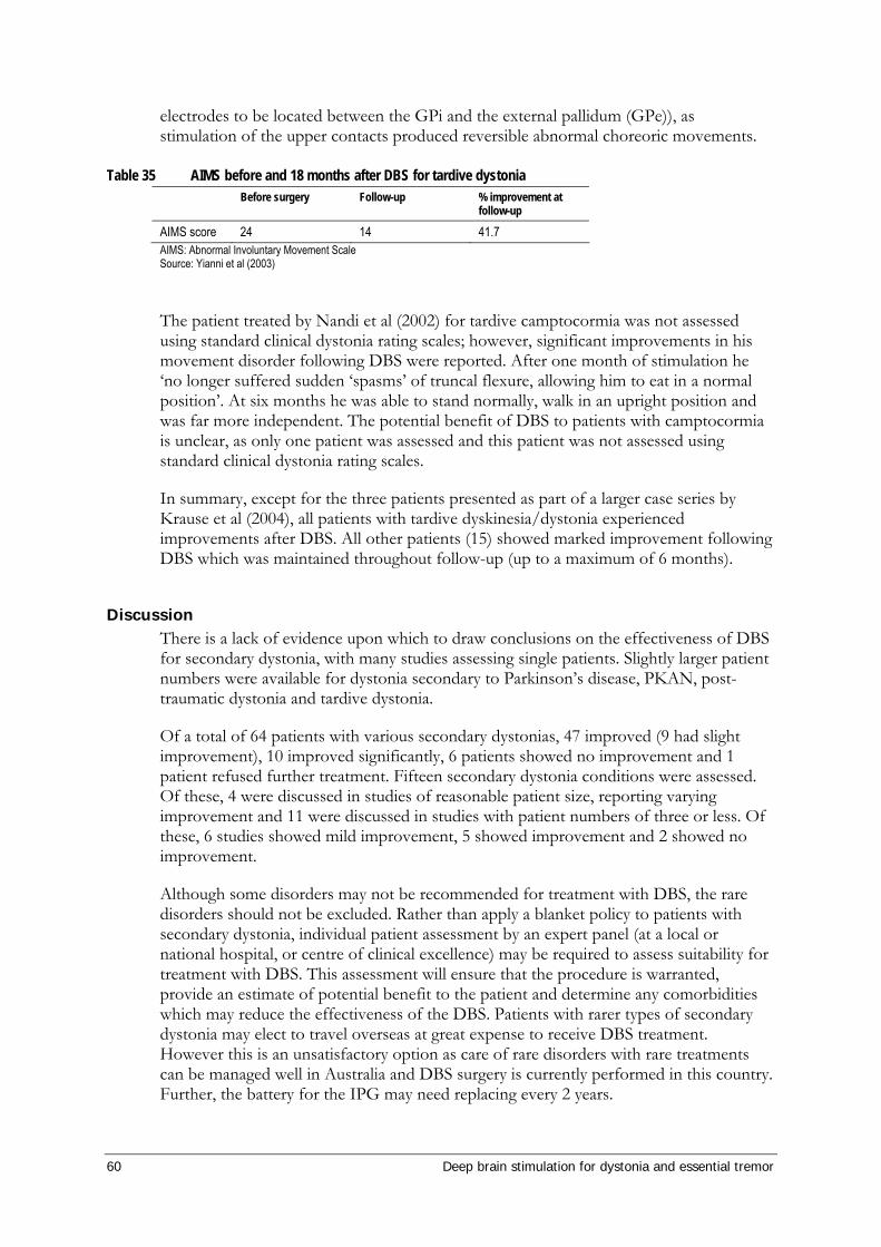
60 Deep brain stimulation for dystonia and essential tremor
electrodes to be located between the GPi and the external pallidum (GPe)), as stimulation of the upper contacts produced reversible abnormal choreoric movements.
Table 35 AIMS before and 18 months after DBS for tardive dystonia Before surgery Follow-up % improvement at
follow-up
AIMS score 24 14 41.7 AIMS: Abnormal Involuntary Movement Scale Source: Yianni et al (2003)
The patient treated by Nandi et al (2002) for tardive camptocormia was not assessed using standard clinical dystonia rating scales; however, significant improvements in his movement disorder following DBS were reported. After one month of stimulation he ‘no longer suffered sudden ‘spasms’ of truncal flexure, allowing him to eat in a normal position’. At six months he was able to stand normally, walk in an upright position and was far more independent. The potential benefit of DBS to patients with camptocormia is unclear, as only one patient was assessed and this patient was not assessed using standard clinical dystonia rating scales.
In summary, except for the three patients presented as part of a larger case series by Krause et al (2004), all patients with tardive dyskinesia/dystonia experienced improvements after DBS. All other patients (15) showed marked improvement following DBS which was maintained throughout follow-up (up to a maximum of 6 months).
Discussion There is a lack of evidence upon which to draw conclusions on the effectiveness of DBS for secondary dystonia, with many studies assessing single patients. Slightly larger patient numbers were available for dystonia secondary to Parkinson’s disease, PKAN, post-traumatic dystonia and tardive dystonia.
Of a total of 64 patients with various secondary dystonias, 47 improved (9 had slight improvement), 10 improved significantly, 6 patients showed no improvement and 1 patient refused further treatment. Fifteen secondary dystonia conditions were assessed. Of these, 4 were discussed in studies of reasonable patient size, reporting varying improvement and 11 were discussed in studies with patient numbers of three or less. Of these, 6 studies showed mild improvement, 5 showed improvement and 2 showed no improvement.
Although some disorders may not be recommended for treatment with DBS, the rare disorders should not be excluded. Rather than apply a blanket policy to patients with secondary dystonia, individual patient assessment by an expert panel (at a local or national hospital, or centre of clinical excellence) may be required to assess suitability for treatment with DBS. This assessment will ensure that the procedure is warranted, provide an estimate of potential benefit to the patient and determine any comorbidities which may reduce the effectiveness of the DBS. Patients with rarer types of secondary dystonia may elect to travel overseas at great expense to receive DBS treatment. However this is an unsatisfactory option as care of rare disorders with rare treatments can be managed well in Australia and DBS surgery is currently performed in this country. Further, the battery for the IPG may need replacing every 2 years.
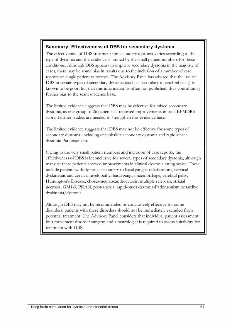
Deep brain stimulation for dystonia and essential tremor 61
Summary: Effectiveness of DBS for secondary dystonia The effectiveness of DBS treatment for secondary dystonia varies according to the type of dystonia and the evidence is limited by the small patient numbers for these conditions. Although DBS appears to improve secondary dystonia in the majority of cases, there may be some bias in results due to the inclusion of a number of case reports on single patient outcomes. The Advisory Panel has advised that the use of DBS in certain types of secondary dystonia (such as secondary to cerebral palsy) is known to be poor, but that this information is often not published, thus contributing further bias to the scant evidence base.
The limited evidence suggests that DBS may be effective for mixed secondary dystonia, as one group of 26 patients all reported improvements in total BFMDRS score. Further studies are needed to strengthen this evidence base.
The limited evidence suggests that DBS may not be effective for some types of secondary dystonia, including encephalitic secondary dystonia and rapid-onset dystonia-Parkinsonism.
Owing to the very small patient numbers and inclusion of case reports, the effectiveness of DBS is inconclusive for several types of secondary dystonia, although many of these patients showed improvements in clinical dystonia rating scales. These include patients with dystonia secondary to basal ganglia calcifications, cervical dyskinesias and cervical myelopathy, basal ganglia haemorrhage, cerebral palsy, Huntington’s Disease, chorea-neuroacanthocytosis, multiple sclerosis, striatal necrosis, GM1-3, PKAN, post-anoxia, rapid-onset dystonia-Parkinsonism or tardive dyskinesia/dystonia.
Although DBS may not be recommended or conclusively effective for some disorders, patients with these disorders should not be immediately excluded from potential treatment. The Advisory Panel considers that individual patient assessment by a movement disorder surgeon and a neurologist is required to assess suitability for treatment with DBS.
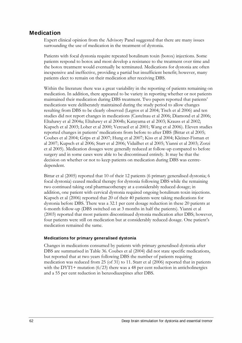
62 Deep brain stimulation for dystonia and essential tremor
Medication Expert clinical opinion from the Advisory Panel suggested that there are many issues surrounding the use of medication in the treatment of dystonia.
Patients with focal dystonia require repeated botulinum toxin (botox) injections. Some patients respond to botox and most develop a resistance to the treatment over time and the botox treatment would eventually be terminated. Medications for dystonia are often inexpensive and ineffective, providing a partial but insufficient benefit; however, many patients elect to remain on their medication after receiving DBS.
Within the literature there was a great variability in the reporting of patients remaining on medication. In addition, there appeared to be variety in reporting whether or not patients maintained their medication during DBS treatment. Two papers reported that patients’ medications were deliberately maintained during the study period to allow changes resulting from DBS to be clearly observed (Legros et al 2004; Tisch et al 2006) and ten studies did not report changes in medications (Castelnau et al 2006; Diamond et al 2006; Eltahawy et al 2004a; Eltahawy et al 2004b; Katayama et al 2003; Krauss et al 2002; Kupsch et al 2003; Loher et al 2000; Vercueil et al 2001; Wang et al 2006). Eleven studies reported changes in patients’ medications from before to after DBS (Bittar et al 2005; Coubes et al 2004; Grips et al 2007; Hung et al 2007; Kiss et al 2004; Kleiner-Fisman et al 2007; Kupsch et al 2006; Starr et al 2006; Vidailhet et al 2005; Yianni et al 2003; Zorzi et al 2005). Medication dosages were generally reduced at follow-up compared to before surgery and in some cases were able to be discontinued entirely. It may be that the decision on whether or not to keep patients on medication during DBS was centre-dependent.
Bittar et al (2005) reported that 10 of their 12 patients (6 primary generalised dystonia; 6 focal dystonia) ceased medical therapy for dystonia following DBS while the remaining two continued taking oral pharmacotherapy at a considerably reduced dosage; in addition, one patient with cervical dystonia required ongoing botulinum toxin injections. Kupsch et al (2006) reported that 20 of their 40 patients were taking medications for dystonia before DBS. There was a 32.1 per cent dosage reduction in these 20 patients at 6-month follow-up (DBS switched on at 3 months in half the patients). Yianni et al (2003) reported that most patients discontinued dystonia medication after DBS; however, four patients were still on medication but at considerably reduced dosage. One patient’s medication remained the same.
Medications for primary generalised dystonia
Changes in medications consumed by patients with primary generalised dystonia after DBS are summarised in Table 36. Coubes et al (2004) did not state specific medications, but reported that at two years following DBS the number of patients requiring medication was reduced from 25 (of 31) to 11. Starr et al (2006) reported that in patients with the DYT1+ mutation (6/23) there was a 48 per cent reduction in anticholinergics and a 55 per cent reduction in benzodiazepines after DBS.
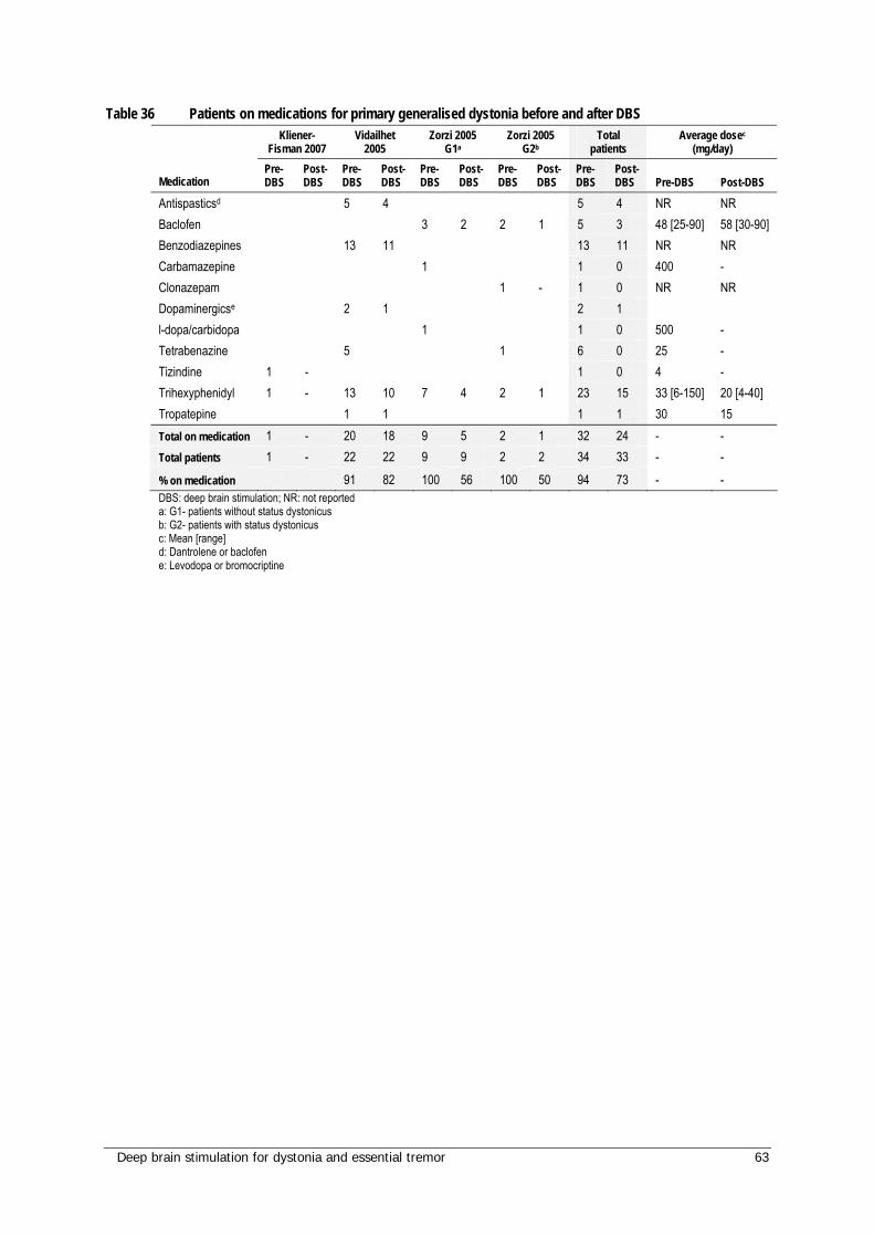
Deep brain stimulation for dystonia and essential tremor 63
Table 36 Patients on medications for primary generalised dystonia before and after DBS Kliener-
Fisman 2007 Vidailhet
2005 Zorzi 2005
G1a Zorzi 2005
G2b Total
patients Average dosec
(mg/day)
Medication Pre-DBS
Post-DBS
Pre-DBS
Post-DBS
Pre-DBS
Post-DBS
Pre-DBS
Post-DBS
Pre-DBS
Post-DBS Pre-DBS Post-DBS
Antispasticsd 5 4 5 4 NR NR Baclofen 3 2 2 1 5 3 48 [25-90] 58 [30-90] Benzodiazepines 13 11 13 11 NR NR Carbamazepine 1 1 0 400 - Clonazepam 1 - 1 0 NR NR Dopaminergicse 2 1 2 1 l-dopa/carbidopa 1 1 0 500 - Tetrabenazine 5 1 6 0 25 - Tizindine 1 - 1 0 4 - Trihexyphenidyl 1 - 13 10 7 4 2 1 23 15 33 [6-150] 20 [4-40] Tropatepine 1 1 1 1 30 15 Total on medication 1 - 20 18 9 5 2 1 32 24 - - Total patients 1 - 22 22 9 9 2 2 34 33 - -
% on medication 91 82 100 56 100 50 94 73 - - DBS: deep brain stimulation; NR: not reported a: G1- patients without status dystonicus b: G2- patients with status dystonicus c: Mean [range] d: Dantrolene or baclofen e: Levodopa or bromocriptine
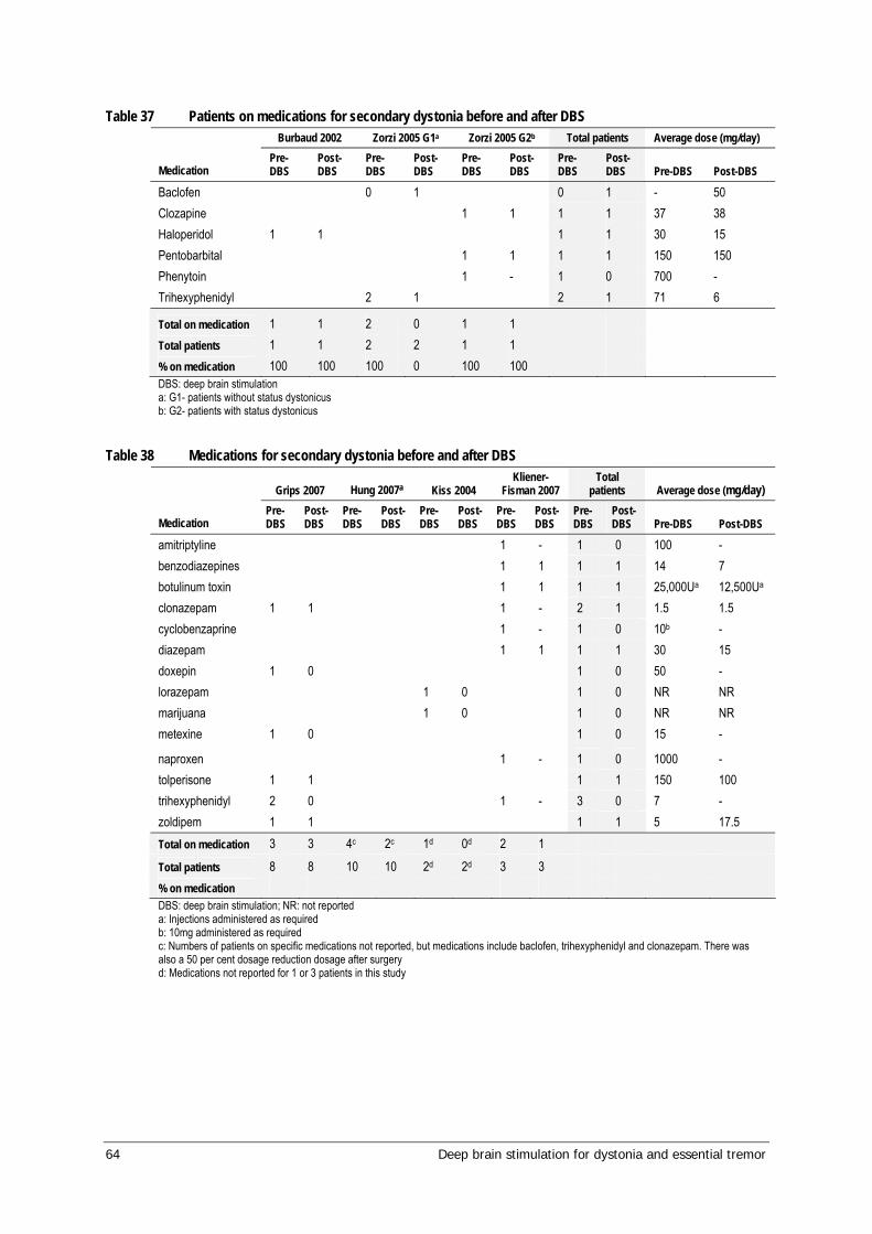
64 Deep brain stimulation for dystonia and essential tremor
Table 37 Patients on medications for secondary dystonia before and after DBS Burbaud 2002 Zorzi 2005 G1a Zorzi 2005 G2b Total patients Average dose (mg/day)
Medication Pre-DBS
Post-DBS
Pre-DBS
Post-DBS
Pre-DBS
Post-DBS
Pre-DBS
Post-DBS Pre-DBS Post-DBS
Baclofen 0 1 0 1 - 50 Clozapine 1 1 1 1 37 38 Haloperidol 1 1 1 1 30 15 Pentobarbital 1 1 1 1 150 150 Phenytoin 1 - 1 0 700 - Trihexyphenidyl 2 1 2 1 71 6
Total on medication 1 1 2 0 1 1 Total patients 1 1 2 2 1 1 % on medication 100 100 100 0 100 100 DBS: deep brain stimulation a: G1- patients without status dystonicus b: G2- patients with status dystonicus
Table 38 Medications for secondary dystonia before and after DBS
Grips 2007 Hung 2007a Kiss 2004 Kliener-
Fisman 2007 Total
patients Average dose (mg/day)
Medication Pre-DBS
Post-DBS
Pre-DBS
Post-DBS
Pre-DBS
Post-DBS
Pre-DBS
Post-DBS
Pre-DBS
Post-DBS Pre-DBS Post-DBS
amitriptyline 1 - 1 0 100 - benzodiazepines 1 1 1 1 14 7 botulinum toxin 1 1 1 1 25,000Ua 12,500Ua clonazepam 1 1 1 - 2 1 1.5 1.5 cyclobenzaprine 1 - 1 0 10b - diazepam 1 1 1 1 30 15 doxepin 1 0 1 0 50 - lorazepam 1 0 1 0 NR NR marijuana 1 0 1 0 NR NR metexine 1 0 1 0 15 -
naproxen 1 - 1 0 1000 - tolperisone 1 1 1 1 150 100 trihexyphenidyl 2 0 1 - 3 0 7 - zoldipem 1 1 1 1 5 17.5 Total on medication 3 3 4c 2c 1d 0d 2 1
Total patients 8 8 10 10 2d 2d 3 3 % on medication DBS: deep brain stimulation; NR: not reported a: Injections administered as required b: 10mg administered as required c: Numbers of patients on specific medications not reported, but medications include baclofen, trihexyphenidyl and clonazepam. There was also a 50 per cent dosage reduction dosage after surgery d: Medications not reported for 1 or 3 patients in this study
Deep brain stimulation for dystonia and essential tremor 65
Results of assessment – essential tremor
Descriptive characteristics of included studies
There were two main groups of studies which reported on the treatment of essential tremor with DBS. The first group of studies reported outcomes of post-surgical testing of the DBS, ie no stimulation (stimulation off) compared to DBS stimulation (on). This group of studies may be useful in informing the effectiveness of the stimulation separately from that of the DBS surgery. They are considered by the Advisory Panel to provide the most information regarding the effectiveness of DBS for essential tremor; hence, these studies have been reported separately. The second group of studies reported outcomes before and after surgery for DBS and may provide evidence on the overall safety and effectiveness of the treatment, including of the surgical interventions (ie the microthalamotomy effect).
No randomised controlled trials or comparative studies as defined by the NHMRC levels of evidence were identified. Therefore all the included studies are level IV evidence, in which before/after, or on/off data are provided for the same cohort of patients. As such the evidence is subject to a degree of bias and should be interpreted accordingly.
Studies assessing essential tremor with DBS switched on compared to off
The basic characteristics for the essential tremor on/off studies are included in Table 39. Twelve studies, with a total of 270 patients, were identified in which the outcomes of DBS treatment were reported during testing when stimulation was switched on and when stimulation was switched off. There were 72 losses to follow-up reported by Bryant et al (2003), Koller et al (1999), Koller et al (2001), Lyons et al (1998), Pahwa et al (2006) and Sydow et al (2003). Many of these patients were accounted for, but several were not adequately reported upon, particularly those lost from Pahwa et al (2006) and Koller et al (1999).
Quality of included studies
Various outcomes were reported when the IPG was switched off (ie no stimulation) and when the IPG was switched on. Due to their nature these types of studies are subject to bias as they do not report on the complete procedure, including surgical implantation. In addition, the patient cohorts were often described as those patients who had responded most successfully to the treatment and therefore would not include poor responders. However, these studies can inform the absolute effectiveness of the stimulation itself on the disorder, separate from the surgical procedure which may in itself have an effect on the disorder. Seven of the studies reported a pre-operative baseline, which can further inform the effectiveness of the surgical procedure. These studies may be regarded as higher quality than those which did not, as they may provide a clearer estimate of the effect of the DBS stimulation and demonstrate the possible microthalomotomy effect of the DBS implantation. Those studies which did not clearly nominate a pre-operative baseline may be subject to more bias through possible mistaken estimation of the effect of the DBS stimulation and also through their vague reporting.
66 Deep brain stimulation for dystonia and essential tremor
The studies featured common inclusion criteria of clinically diagnosed essential tremor, with or without further qualifiers such as medically-refractory. The mean age at surgery ranged from 61.8 to 73.8 with a range of anti-tremor medications taken, most commonly primidone or propranolol. Age at onset of symptoms was reported by only two studies. Carpenter et al (1998) reported onset after age 45 for all patients, whilst Pahwa et al (1999) reported onset at mean age of 38.4 years (range 20-58). All patients presented with essential tremor. Bryant et al (2003) reported on the tremor locations at presentation, including upper extremity, head and truncal tremor and Carpenter et al (1998) reported that patients suffered with voice symptoms when DBS was off. Three studies reported patients with comorbidities. One patient had a thalamotomy contralateral to implant (Bryant et al 2003), one patient had atrial fibrillation and congestive cardiac failure (Pahwa et al 1999), one patient had lung cancer (Putzke et al 2004) and one patient had colon cancer (Putzke et al 2004).
Characteristics of included studies
The technical characteristics of the studies reporting outcomes for patients with essential tremor for stimulation on and off are presented in Table 81 and the patient characteristics are presented in Table 40. In all cases, Medtronic hardware was used and the electrodes were implanted primarily into the ventralis intermedius (VIM) nucleus (the target for implantation in patients with essential tremor), where reported. Exceptions were Lyons et al (1998) who reported implantation into the thalamus and Ushe et al (2006) who reported implantation into the left VIM. Ten of these studies evaluated patients using the FTM tremor rating scale, or subsets thereof (Bryant et al 2003; Koller et al 1999; Koller et al 2001; Lyons et al 1998; Obwegeser et al 2000; Pahwa et al 1999; Pahwa et al 2006; Putzke et al 2004; Sydow et al 2003; and Ushe et al 2006). Six studies used the ADL scale (Bryant et al 2003; Lyons et al 1998; Pahwa et al 1999; Pahwa et al 2006; Putzke et al 2004; Sydow et al 2003). Less commonly used assessments included voice measures (Carpenter et al 1998), speech evaluation for dysarthria (Pahwa et al 1999), a health questionnaire (Bryant et al 2003), global disability ratings (Pahwa et al 1999) and accelerometry (Ushe et al 2006; Vaillancourt et al 2003). One study (Putzke et al 2004) presented possible errors in data, stating that the on stimulation was statistically significant versus on stimulation.
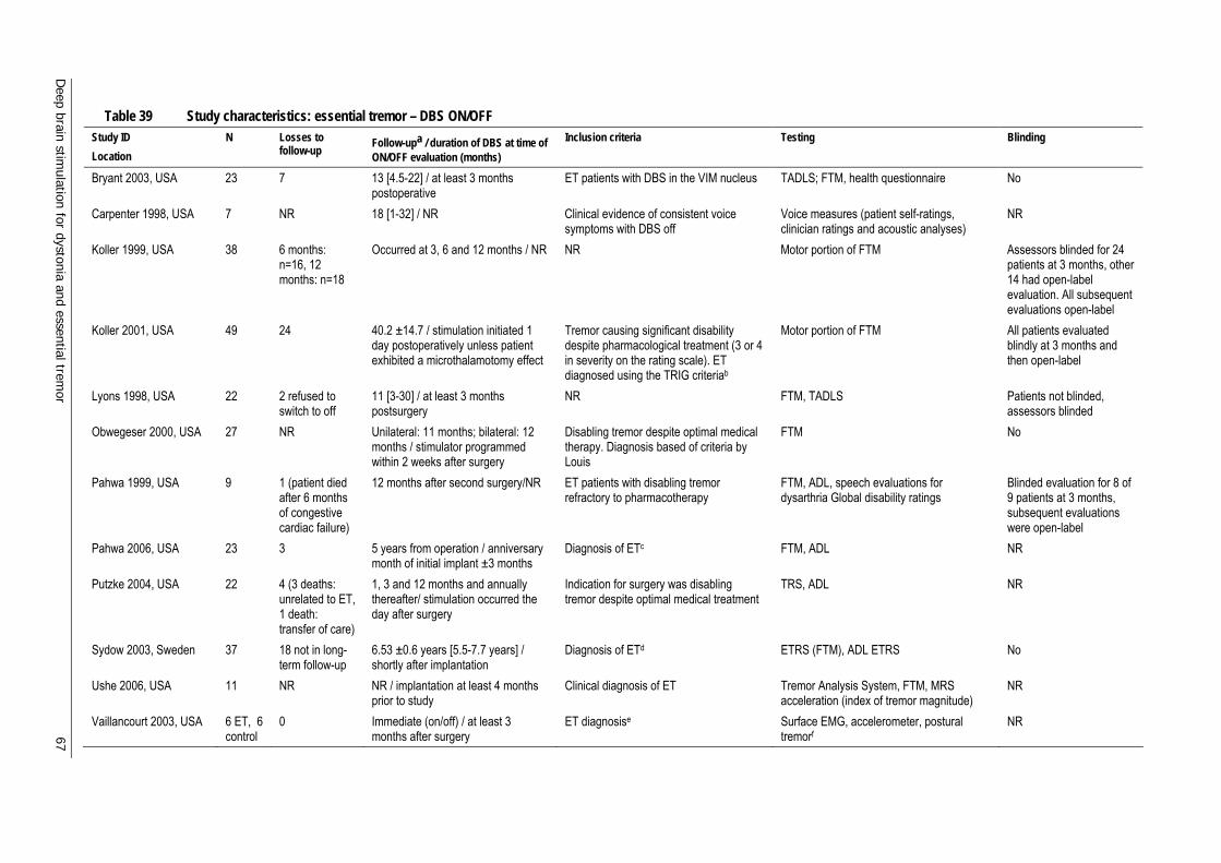
Deep brain stim
ulation for dystonia and essential tremor
67
Table 39 Study characteristics: essential tremor – DBS ON/OFF Study ID Location
N Losses to follow-up Follow-upa / duration of DBS at time of
ON/OFF evaluation (months) Inclusion criteria Testing Blinding
Bryant 2003, USA 23 7 13 [4.5-22] / at least 3 months postoperative
ET patients with DBS in the VIM nucleus TADLS; FTM, health questionnaire No
Carpenter 1998, USA 7 NR 18 [1-32] / NR Clinical evidence of consistent voice symptoms with DBS off
Voice measures (patient self-ratings, clinician ratings and acoustic analyses)
NR
Koller 1999, USA 38 6 months: n=16, 12 months: n=18
Occurred at 3, 6 and 12 months / NR NR Motor portion of FTM Assessors blinded for 24 patients at 3 months, other 14 had open-label evaluation. All subsequent evaluations open-label
Koller 2001, USA 49 24 40.2 ±14.7 / stimulation initiated 1 day postoperatively unless patient exhibited a microthalamotomy effect
Tremor causing significant disability despite pharmacological treatment (3 or 4 in severity on the rating scale). ET diagnosed using the TRIG criteriab
Motor portion of FTM All patients evaluated blindly at 3 months and then open-label
Lyons 1998, USA 22 2 refused to switch to off
11 [3-30] / at least 3 months postsurgery
NR FTM, TADLS Patients not blinded, assessors blinded
Obwegeser 2000, USA 27 NR Unilateral: 11 months; bilateral: 12 months / stimulator programmed within 2 weeks after surgery
Disabling tremor despite optimal medical therapy. Diagnosis based of criteria by Louis
FTM No
Pahwa 1999, USA 9 1 (patient died after 6 months of congestive cardiac failure)
12 months after second surgery/NR ET patients with disabling tremor refractory to pharmacotherapy
FTM, ADL, speech evaluations for dysarthria Global disability ratings
Blinded evaluation for 8 of 9 patients at 3 months, subsequent evaluations were open-label
Pahwa 2006, USA 23 3 5 years from operation / anniversary month of initial implant ±3 months
Diagnosis of ETc FTM, ADL NR
Putzke 2004, USA 22 4 (3 deaths: unrelated to ET, 1 death: transfer of care)
1, 3 and 12 months and annually thereafter/ stimulation occurred the day after surgery
Indication for surgery was disabling tremor despite optimal medical treatment
TRS, ADL NR
Sydow 2003, Sweden 37 18 not in long-term follow-up
6.53 ±0.6 years [5.5-7.7 years] / shortly after implantation
Diagnosis of ETd ETRS (FTM), ADL ETRS No
Ushe 2006, USA 11 NR NR / implantation at least 4 months prior to study
Clinical diagnosis of ET Tremor Analysis System, FTM, MRS acceleration (index of tremor magnitude)
NR
Vaillancourt 2003, USA 6 ET, 6 control
0 Immediate (on/off) / at least 3 months after surgery
ET diagnosise Surface EMG, accelerometer, postural tremorf
NR

68 D
eep brain stimulation for dystonia and essential trem
or
ADL: Activities of Daily Living; DBS: deep brain stimulation; EMG: electromyography; ET: essential tremor; ETRS: Essential Tremor Rating Scale; FTM: Fahn-Tolosa-Marin scale; MRS: mean-root-square; NR: not reported; TADLS: Tremor Activities of Daily Living Scale; TRS: tremor rating scale; VIM: ventral intermediate nucleus of the thalamus a: Mean ±standard deviation [range] (unless otherwise stated) b: Presence of a postural tremor without other neurologic signs c: DBS of VIM, participation in 1997 tremor control study with initial surgery occurring between 1993 and 1997, willingness to sign an informed consent form and to return for as many as five annual follow-up visits at original investigative site d: Tremor present during a major part of the day, inadequately controlled under maximum tolerated doses of primidone, propranolol and/or benzodiazepines. Tremor disabling, with a score of between 3 and 4 on a 5 point tremor scale (0 no tremor; 4 severe tremor), ability to abide by the protocol and to operate the pulse generator e: Consistent with guidelines in Consensus Statement of the Movement Disorder Society on Tremor f: Surface EMG: measure neuromuscular activity in extensor digitorum communis and flexor digitorum profundus. Calibrated Coulbourn type V 94-41 miniature solid-state piezoresistive accelerometer taped to hand (2 cm proximal to middle of first metacarpophalangeal joint). Postural tremor examined under five loading conditions (1000 g, 500 g, 250 g, 100 g and 0 g)
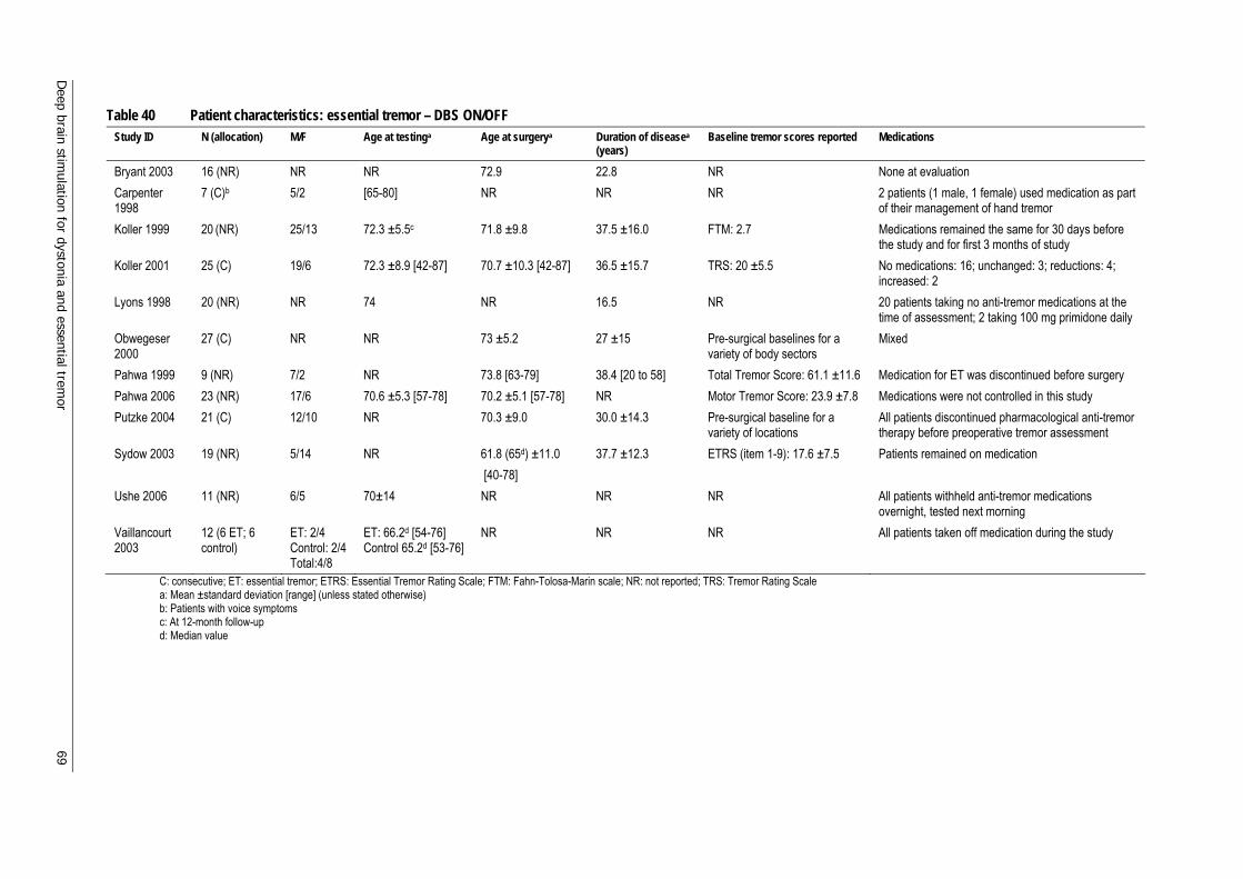
Deep brain stim
ulation for dystonia and essential tremor
69
Table 40 Patient characteristics: essential tremor – DBS ON/OFF Study ID N (allocation) M/F Age at testinga Age at surgerya Duration of diseasea
(years) Baseline tremor scores reported Medications
Bryant 2003 16 (NR) NR NR 72.9 22.8 NR None at evaluation Carpenter 1998
7 (C)b 5/2 [65-80] NR NR NR 2 patients (1 male, 1 female) used medication as part of their management of hand tremor
Koller 1999 20 (NR) 25/13 72.3 ±5.5c 71.8 ±9.8 37.5 ±16.0 FTM: 2.7 Medications remained the same for 30 days before the study and for first 3 months of study
Koller 2001 25 (C) 19/6 72.3 ±8.9 [42-87] 70.7 ±10.3 [42-87] 36.5 ±15.7 TRS: 20 ±5.5 No medications: 16; unchanged: 3; reductions: 4; increased: 2
Lyons 1998 20 (NR) NR 74 NR 16.5 NR 20 patients taking no anti-tremor medications at the time of assessment; 2 taking 100 mg primidone daily
Obwegeser 2000
27 (C) NR NR 73 ±5.2 27 ±15 Pre-surgical baselines for a variety of body sectors
Mixed
Pahwa 1999 9 (NR) 7/2 NR 73.8 [63-79] 38.4 [20 to 58] Total Tremor Score: 61.1 ±11.6 Medication for ET was discontinued before surgery Pahwa 2006 23 (NR) 17/6 70.6 ±5.3 [57-78] 70.2 ±5.1 [57-78] NR Motor Tremor Score: 23.9 ±7.8 Medications were not controlled in this study Putzke 2004 21 (C) 12/10 NR 70.3 ±9.0 30.0 ±14.3 Pre-surgical baseline for a
variety of locations All patients discontinued pharmacological anti-tremor therapy before preoperative tremor assessment
Sydow 2003 19 (NR) 5/14 NR 61.8 (65d) ±11.0 [40-78]
37.7 ±12.3 ETRS (item 1-9): 17.6 ±7.5 Patients remained on medication
Ushe 2006 11 (NR) 6/5 70±14 NR NR NR All patients withheld anti-tremor medications overnight, tested next morning
Vaillancourt 2003
12 (6 ET; 6 control)
ET: 2/4 Control: 2/4 Total:4/8
ET: 66.2d [54-76] Control 65.2d [53-76]
NR NR NR All patients taken off medication during the study
C: consecutive; ET: essential tremor; ETRS: Essential Tremor Rating Scale; FTM: Fahn-Tolosa-Marin scale; NR: not reported; TRS: Tremor Rating Scale a: Mean ±standard deviation [range] (unless stated otherwise) b: Patients with voice symptoms c: At 12-month follow-up d: Median value
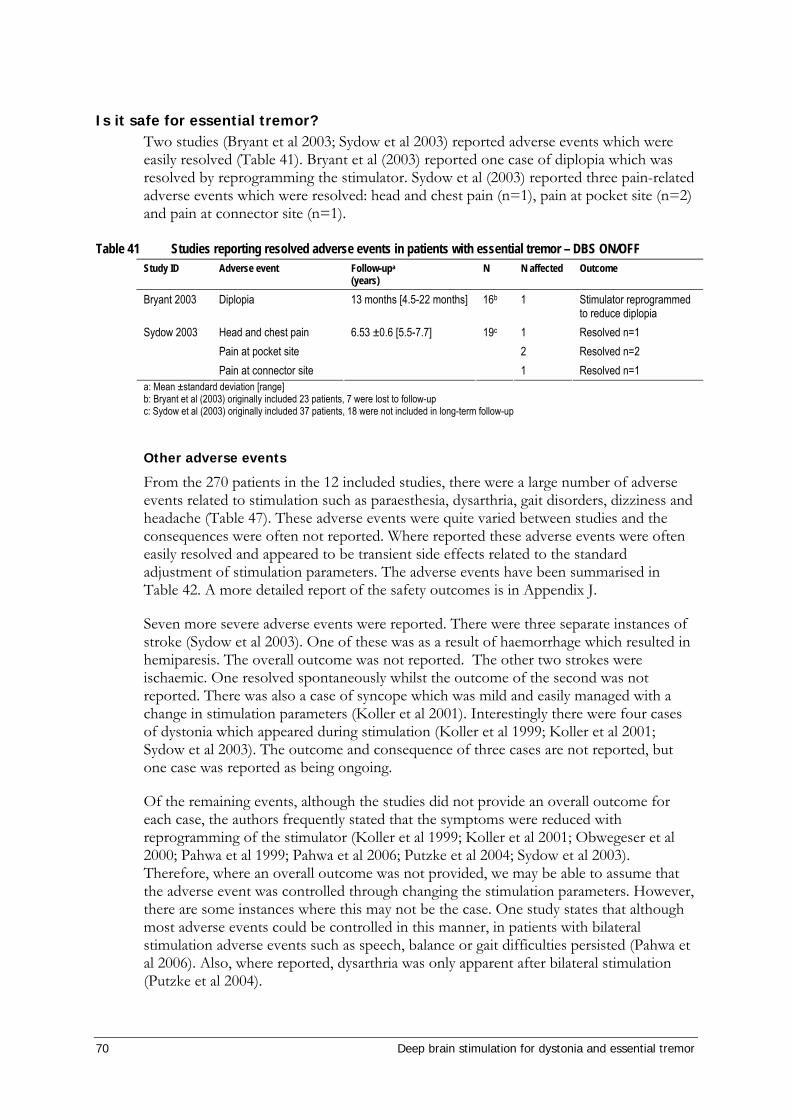
70 Deep brain stimulation for dystonia and essential tremor
Is it safe for essential tremor? Two studies (Bryant et al 2003; Sydow et al 2003) reported adverse events which were easily resolved (Table 41). Bryant et al (2003) reported one case of diplopia which was resolved by reprogramming the stimulator. Sydow et al (2003) reported three pain-related adverse events which were resolved: head and chest pain (n=1), pain at pocket site (n=2) and pain at connector site (n=1).
Table 41 Studies reporting resolved adverse events in patients with essential tremor – DBS ON/OFF Study ID Adverse event Follow-upa
(years) N N affected Outcome
Bryant 2003 Diplopia 13 months [4.5-22 months] 16b 1 Stimulator reprogrammed to reduce diplopia
Head and chest pain 6.53 ±0.6 [5.5-7.7] 19c 1 Resolved n=1 Pain at pocket site 2 Resolved n=2
Sydow 2003
Pain at connector site 1 Resolved n=1 a: Mean ±standard deviation [range] b: Bryant et al (2003) originally included 23 patients, 7 were lost to follow-up c: Sydow et al (2003) originally included 37 patients, 18 were not included in long-term follow-up
Other adverse events
From the 270 patients in the 12 included studies, there were a large number of adverse events related to stimulation such as paraesthesia, dysarthria, gait disorders, dizziness and headache (Table 47). These adverse events were quite varied between studies and the consequences were often not reported. Where reported these adverse events were often easily resolved and appeared to be transient side effects related to the standard adjustment of stimulation parameters. The adverse events have been summarised in Table 42. A more detailed report of the safety outcomes is in Appendix J.
Seven more severe adverse events were reported. There were three separate instances of stroke (Sydow et al 2003). One of these was as a result of haemorrhage which resulted in hemiparesis. The overall outcome was not reported. The other two strokes were ischaemic. One resolved spontaneously whilst the outcome of the second was not reported. There was also a case of syncope which was mild and easily managed with a change in stimulation parameters (Koller et al 2001). Interestingly there were four cases of dystonia which appeared during stimulation (Koller et al 1999; Koller et al 2001; Sydow et al 2003). The outcome and consequence of three cases are not reported, but one case was reported as being ongoing.
Of the remaining events, although the studies did not provide an overall outcome for each case, the authors frequently stated that the symptoms were reduced with reprogramming of the stimulator (Koller et al 1999; Koller et al 2001; Obwegeser et al 2000; Pahwa et al 1999; Pahwa et al 2006; Putzke et al 2004; Sydow et al 2003). Therefore, where an overall outcome was not provided, we may be able to assume that the adverse event was controlled through changing the stimulation parameters. However, there are some instances where this may not be the case. One study states that although most adverse events could be controlled in this manner, in patients with bilateral stimulation adverse events such as speech, balance or gait difficulties persisted (Pahwa et al 2006). Also, where reported, dysarthria was only apparent after bilateral stimulation (Putzke et al 2004).
Deep brain stimulation for dystonia and essential tremor 71
One patient who developed depression after DBS suffered from neurosis preoperatively (Sydow et al 2003).
Summary: Safety of DBS for essential tremor In summary, from this group of studies, DBS is a relatively safe treatment for essential tremor. Most adverse events were mild and could be treated through changing the stimulation parameters. The more severe events were relatively rare and may not affect long-term outcomes. However, the studies poorly report the overall long-term outcomes related to these events.
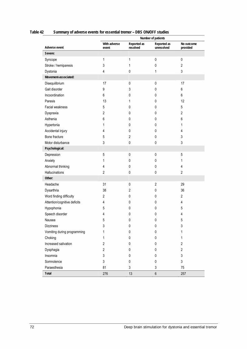
72 Deep brain stimulation for dystonia and essential tremor
Table 42 Summary of adverse events for essential tremor – DBS ON/OFF studies Number of patients
Adverse event With adverse event
Reported as resolved
Reported as unresolved
No outcome provided
Severe: Syncope 1 1 0 0 Stroke / hemiparesis 3 1 0 2 Dystonia 4 0 1 3 Movement-associated: Disequilibrium Gait disorder Incoordination
17 9 6
0 3 0
0 0 0
17 6 6
Paresis Facial weakness
13 5
1 0
0 0
12 5
Dyspraxia 2 0 0 2 Asthenia 6 0 0 6 Hypertonia 1 0 0 1 Accidental injury 4 0 0 4 Bone fracture 5 2 0 3 Motor disturbance 3 0 0 3 Psychological: Depression 5 0 0 5 Anxiety 1 0 0 1 Abnormal thinking 4 0 0 4 Hallucinations 2 0 0 2 Other: Headache 31 0 2 29 Dysarthria 38 2 0 36 Word finding difficulty 2 0 0 2 Attention/cognitive deficits 4 0 0 4 Hypophonia 5 0 0 5 Speech disorder 4 0 0 4 Nausea 5 0 0 5 Dizziness 3 0 0 3 Vomiting during programming 1 0 0 1 Choking 1 0 0 1 Increased salivation 2 0 0 2 Dysphagia 2 0 0 2 Insomnia 3 0 0 3 Somnolence 3 0 0 3 Paraesthesia 81 3 3 75 Total 276 13 6 257
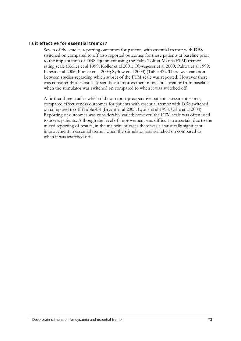
Deep brain stimulation for dystonia and essential tremor 73
Is it effective for essential tremor? Seven of the studies reporting outcomes for patients with essential tremor with DBS switched on compared to off also reported outcomes for these patients at baseline prior to the implantation of DBS equipment using the Fahn-Tolosa-Marin (FTM) tremor rating scale (Koller et al 1999; Koller et al 2001; Obwegeser et al 2000; Pahwa et al 1999; Pahwa et al 2006; Putzke et al 2004; Sydow et al 2003) (Table 43). There was variation between studies regarding which subset of the FTM scale was reported. However there was consistently a statistically significant improvement in essential tremor from baseline when the stimulator was switched on compared to when it was switched off.
A further three studies which did not report preoperative patient assessment scores, compared effectiveness outcomes for patients with essential tremor with DBS switched on compared to off (Table 43) (Bryant et al 2003; Lyons et al 1998; Ushe et al 2004). Reporting of outcomes was considerably varied; however, the FTM scale was often used to assess patients. Although the level of improvement was difficult to ascertain due to the mixed reporting of results, in the majority of cases there was a statistically significant improvement in essential tremor when the stimulator was switched on compared to when it was switched off.
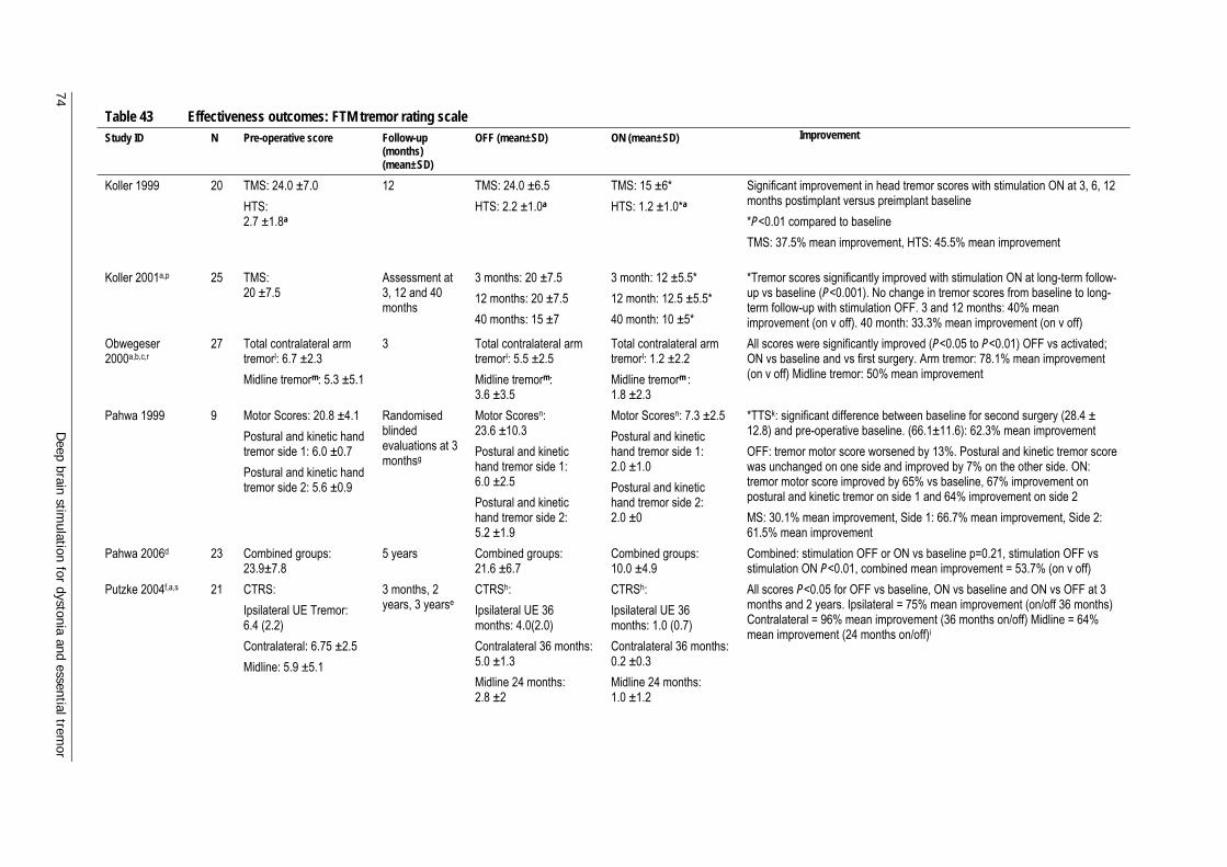
74 D
eep brain stimulation for dystonia and essential trem
or
Table 43 Effectiveness outcomes: FTM tremor rating scale Study ID N Pre-operative score Follow-up
(months) (mean±SD)
OFF (mean±SD) ON (mean±SD) Improvement
Koller 1999 20 TMS: 24.0 ±7.0 HTS: 2.7 ±1.8a
12 TMS: 24.0 ±6.5 HTS: 2.2 ±1.0a
TMS: 15 ±6* HTS: 1.2 ±1.0*a
Significant improvement in head tremor scores with stimulation ON at 3, 6, 12 months postimplant versus preimplant baseline *P<0.01 compared to baseline TMS: 37.5% mean improvement, HTS: 45.5% mean improvement
Koller 2001a,p 25 TMS: 20 ±7.5
Assessment at 3, 12 and 40 months
3 months: 20 ±7.5 12 months: 20 ±7.5 40 months: 15 ±7
3 month: 12 ±5.5* 12 month: 12.5 ±5.5* 40 month: 10 ±5*
*Tremor scores significantly improved with stimulation ON at long-term follow-up vs baseline (P<0.001). No change in tremor scores from baseline to long-term follow-up with stimulation OFF. 3 and 12 months: 40% mean improvement (on v off). 40 month: 33.3% mean improvement (on v off)
Obwegeser 2000a,b,c,r
27 Total contralateral arm tremorl: 6.7 ±2.3 Midline tremorm: 5.3 ±5.1
3 Total contralateral arm tremorl: 5.5 ±2.5 Midline tremorm: 3.6 ±3.5
Total contralateral arm tremorl: 1.2 ±2.2 Midline tremorm : 1.8 ±2.3
All scores were significantly improved (P<0.05 to P<0.01) OFF vs activated; ON vs baseline and vs first surgery. Arm tremor: 78.1% mean improvement (on v off) Midline tremor: 50% mean improvement
Pahwa 1999 9 Motor Scores: 20.8 ±4.1 Postural and kinetic hand tremor side 1: 6.0 ±0.7 Postural and kinetic hand tremor side 2: 5.6 ±0.9
Randomised blinded evaluations at 3 monthsg
Motor Scoresn: 23.6 ±10.3 Postural and kinetic hand tremor side 1: 6.0 ±2.5 Postural and kinetic hand tremor side 2: 5.2 ±1.9
Motor Scoresn: 7.3 ±2.5 Postural and kinetic hand tremor side 1: 2.0 ±1.0 Postural and kinetic hand tremor side 2: 2.0 ±0
*TTSk: significant difference between baseline for second surgery (28.4 ± 12.8) and pre-operative baseline. (66.1±11.6): 62.3% mean improvement OFF: tremor motor score worsened by 13%. Postural and kinetic tremor score was unchanged on one side and improved by 7% on the other side. ON: tremor motor score improved by 65% vs baseline, 67% improvement on postural and kinetic tremor on side 1 and 64% improvement on side 2 MS: 30.1% mean improvement, Side 1: 66.7% mean improvement, Side 2: 61.5% mean improvement
Pahwa 2006d 23 Combined groups: 23.9±7.8
5 years Combined groups: 21.6 ±6.7
Combined groups: 10.0 ±4.9
Combined: stimulation OFF or ON vs baseline p=0.21, stimulation OFF vs stimulation ON P<0.01, combined mean improvement = 53.7% (on v off)
Putzke 2004f,a,s 21 CTRS: Ipsilateral UE Tremor: 6.4 (2.2) Contralateral: 6.75 ±2.5 Midline: 5.9 ±5.1
3 months, 2 years, 3 yearse
CTRSh: Ipsilateral UE 36 months: 4.0(2.0) Contralateral 36 months: 5.0 ±1.3 Midline 24 months: 2.8 ±2
CTRSh: Ipsilateral UE 36 months: 1.0 (0.7) Contralateral 36 months: 0.2 ±0.3 Midline 24 months: 1.0 ±1.2
All scores P<0.05 for OFF vs baseline, ON vs baseline and ON vs OFF at 3 months and 2 years. Ipsilateral = 75% mean improvement (on/off 36 months) Contralateral = 96% mean improvement (36 months on/off) Midline = 64% mean improvement (24 months on/off)i

Deep brain stim
ulation for dystonia and essential tremor
75
Sydow 2003q 19 ETRS (item 1-19): All: 17.6 (7.5) Head Tremor (item 4): All: 1.2 (1.5) Hand Function (item 10-14): All: 26.1 (6.4)
6 years ETRS: All: 19.4 (9.2) Head tremor: All: 1.2 (2.1) Hand function: All: 25.6 (7.7)
ETRS: All: 10.4 (5.4) **~~ Head tremor: All: 0.5 (1.1) *~ Hand function: All: 16.4 (6.4) **~~
Increase in tremor score (items 1-9) from 17.6 to 19.4 points from baseline to OFF Total tremor was reduced significantly from ON to OFF at 1 year (P<0.001) and at 6 years (P<0.001) *P<0.05 vs baseline; ~P<0.05 vs OFF **P<0.001 vs baseline; ~~P<0.001 vs OFF ETRS: 46.5% mean improvement Head tremor: 48.3% mean improvement Hand function: 35.9% mean improvement
Studies which did not report a pre-operative baseline Bryant 2003 16 NR 13 [4.5-22] 32.7 21.6 33.9%m,o Lyons 1998 20 NR 11 [3-30] 20.1 ±6.7 12.2 ±4.3 39.3% improvement, P<0.001 Ushe 2006j 11 NR NR 65.2 ±12.7 [47-83] 24.4 ±13.3 [4-44] Represents a mean 62.8%±19.8% reduction (range 26.3% - 93%)
Abbreviations: BL, baseline; CTRS: Clinical Tremor Rating Scale; ETRS: Essential Tremor Rating Scale; FTM: Fahn-Tolosa-Marin scale; HTS: Head Tremor Score; N: total patient cohort; n: patient subgroup; NR: not reported; Pre-op: pre-operative; TMS: Total Motor Score; TTS: Total Tremor Score; UE: upper extremity a: approximated from figures, not specified in text; b: contralateral, midline and ipsilateral scores are provided for unilateral and bilateral stimulation. Also head, voice, face and tongue posture reported c: percentage of adjustments for tremor control was significantly decreased (66% vs 89%) when comparing bilateral to unilateral d: Mean Tremor Score (postural or kinetic tremor) is also provided e: mean duration between placement of the first & second lead= 223 days, most being undertaken ≤5m (n=17, 77%) following initial surgery. Mean time between initial lead placement and last available follow up= 29 months f: head, voice, tongue, face and trunk scores are also provided g: performed in 8/9 patients using the motor subscale of the TRS h: data is provided 3 months after first implantation and 3 months after second implantation; scores are provided for a given time after the second implantation i: correlations not generated for 36 month postoperative bilateral stimulation interval due to small sample size j: the DBS OFF condition was defined as the baseline condition and was used to normalise all other conditions k: items 1-21, max score 116 l: items 5 or 6 on rating scale m: items 1 through 4 and 7 n: motor scores (items 1-10) o: high patient-clinician correlation of rs=0.91, FTM scores were highly correlated with patient rated TADLS (rs=0.80 on, rs=0.78 off) and clinician rated TADLS (rs=0.88 on, rs=0.86 off) p: 11 patients did not return for long-term follow-up. Average follow-up of 11 months and outcomes comparable to those in the long-term follow-up group (baseline tremor score 24.1, follow-up stimulation on 9.0 and stimulation off 18.0, p<0.001) q: reported data for various locations of tremor, including voice and head tremor, lower limb action tremor and hand function for both unilateral and bilateral stimulation. Only the significant results are included in this table (see attached word document for the rest of the data) r: reported data on several tremor locations, including head-posture, voice, tongue-posture, face and trunk-posture. Only significant data are included in this table s: some patients had two sets of surgery (one unilateral, one bilateral) and the authors provide data for unilateral patients at 1 and 3 months. For consistency, the tables examine these same follow-up periods for bilateral (ie 1 and 3 months). Year 3 is the furthest follow-up and has been included to ascertain the full clinical effect at long-term follow up. Data which returned statistically significant differences at 3 months and at latest follow-up (either 36 or 24 months) are noted in this table, other data are available.
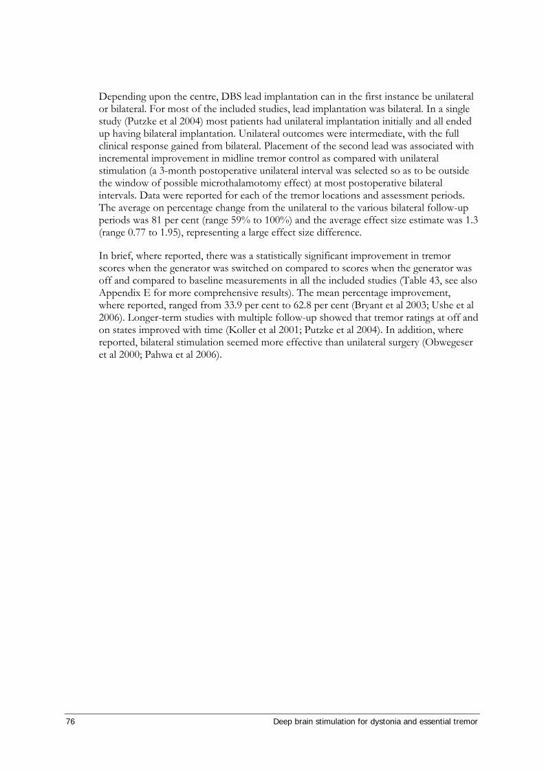
76 Deep brain stimulation for dystonia and essential tremor
Depending upon the centre, DBS lead implantation can in the first instance be unilateral or bilateral. For most of the included studies, lead implantation was bilateral. In a single study (Putzke et al 2004) most patients had unilateral implantation initially and all ended up having bilateral implantation. Unilateral outcomes were intermediate, with the full clinical response gained from bilateral. Placement of the second lead was associated with incremental improvement in midline tremor control as compared with unilateral stimulation (a 3-month postoperative unilateral interval was selected so as to be outside the window of possible microthalamotomy effect) at most postoperative bilateral intervals. Data were reported for each of the tremor locations and assessment periods. The average on percentage change from the unilateral to the various bilateral follow-up periods was 81 per cent (range 59% to 100%) and the average effect size estimate was 1.3 (range 0.77 to 1.95), representing a large effect size difference.
In brief, where reported, there was a statistically significant improvement in tremor scores when the generator was switched on compared to scores when the generator was off and compared to baseline measurements in all the included studies (Table 43, see also Appendix E for more comprehensive results). The mean percentage improvement, where reported, ranged from 33.9 per cent to 62.8 per cent (Bryant et al 2003; Ushe et al 2006). Longer-term studies with multiple follow-up showed that tremor ratings at off and on states improved with time (Koller et al 2001; Putzke et al 2004). In addition, where reported, bilateral stimulation seemed more effective than unilateral surgery (Obwegeser et al 2000; Pahwa et al 2006).
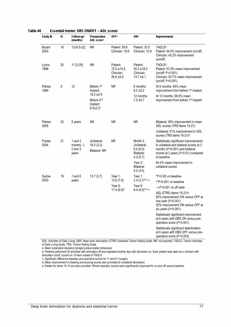
Deep brain stimulation for dystonia and essential tremor 77
Table 44 Essential tremor: DBS ON/OFF – ADL scores Study ID N Follow-upa
(months) Preoperative ADL scorea
OFFa
ONa Improvements
Bryant 2003
16 13 [4.5-22] NR Patient: 59.8 Clinician: 19.6
Patient: 33.5 Clinician: 10.8
TADLSb: Patient: 44.0% improvement (on/off) Clinician: 45.2% improvement (on/off)
Lyons 1998
20 11 [3-30] NR Patient: 72.0 ±15.2, Clinician: 29.6 ±5.5
Patient: 30.3 ±18.3 Clinician: 13.7 ±4.1
TADLSb: Patient: 57.9% mean improvement (on/off; P<0.001) Clinician: 53.7% mean improvement (on/off; P<0.001)
Pahwa 1999
9 12 Before 1st implant: 18.2 ±2.9 Before 2nd implant: 9.0±3.2c
NR 6 months: 6.2 ±5.2 12 months: 7.9 ±5.7
At 6 months: 65% mean improvement from before 1st implant At 12 months: 56.6% mean improvement from before 1st implant
Pahwa 2006
23 5 years
NR NR NR Bilateral: 36% improvement in mean ADL scores (TRS Items 15-21) Unilateral: 51% improvement in ADL scores (TRS Items 15-21)d
Putzke 2004
21 1 and 3 months, 1, 2 and 3 years
Unilateral: 18.0 (3.3) Bilateral: NR
NR
Month 3: Unilateral: 6.4 (6.0) Bilateral: 4.3 (5.7) Year 2: Bilateral: 5.5 (4.4)
Statistically significant improvements in unilateral and bilateral scores at 3 months (P<0.001) and bilateral scores at 2 years (P<0.01) compared to baseline 64.4% mean improvement in unilateral scores
Sydow 2003
19 1 and 6 years
13.7 (3.7) Year 1: 13.6 (7.9) Year 6: 17.4 (6.8)*
Year 1: 2.4 (2.7)**~~ Year 6: 8.4 (6.0)**~~
*P<0.05 vs baseline **P<0.001 vs baseline ~~P<0.001 vs off state ADL ETRS (items 15-21)e: 82% improvement ON versus OFF at one year (P<0.001) 52% improvement ON versus OFF at six years (P<0.001) Statistically significant improvement at 6 years with DBS ON versus pre-operative score (P<0.001) Statistically significant deterioration at 6 years with DBS OFF versus pre-operative score (P=0.003)
ADL: Activities of Daily Living; DBS: deep brain stimulation; ETRS: Essential Tremor Rating Scale; NR: not reported; TADLS: Tremor Activities of Daily Living Scale; TRS: Tremor Rating Scale a: Mean ±standard deviation [range] (unless stated otherwise) b: Patients performed 30 activities with stimulator off and repeated another day with stimulator on. Each patient was seen by a clinician with stimulator on/off, scored on 10-item subset of TADLS c: Significant difference between pre-operative scores for 1st and 2nd surgery d: Mean improvement in drawing and pouring scores also provided for unilateral stimulation e: Details for items 10-14 are also provided. Where reported, scores were significantly improved for on and off versus baseline.

78 Deep brain stimulation for dystonia and essential tremor
Table 45 Essential tremor: DBS ON/OFF – other scores Study ID N Follow-up
(months) Other assessment scales
OFF measurementsa
ON measurementsa Improvementsa
Carpenter 2003
7 18 [1-32]a
Mean patient Severity Mean clinician Severity Acoustic analysesb,c
Patient severity: 2.6 Clinician severity: 2.6 Mean acoustic rate/sec: 3.5 Mean amplitude: 61.3%
Patient severity: -1.75 Clinician severity: -1.67 Mean acoustic rate/sec: -0.1 Mean amplitude: -23%
Patient severity: 33% mean improvement Clinician severity: 35.8% mean improvement, 1- to 3- point change on severity scale 24 to 60% difference in relative amplitude Improvement in voice restricted to patients who demonstrated more severe symptoms in DBS-OFF condition
Pahwa 1999 9 12 GDR: disability ratings before surgery: Marked disability: n=6 Severely disabled: n=3
NR NR Disability ratings at 12 months: No disability: n=3 Mild disability: n=4 Marked disability: n=1
Ushe 2006 11 NR Tremor Analysis Systemd
65.2 ±12.7 [47-38] 24.4 ±13.3 [4-44] Mean reduction: 62.8% ±19.8% [26.3%-93%] Accelerometry: tremor decreased 85.2% ±4.9%
Vaillancourt 2003e
6 ≥3 EMGf, accelerometer taped to handg, Postural tremor h,i
Control measurements: RMS displacement (cm): 0 ±0 Frequency (Hz): 6.1 ±0.2 Approx. entropy: 0.57 ±0.02 EMG: 0.15 ±0.02
RMS displacement (cm): 4.6 ±1.0 Frequency (Hz): 7.9 ±0.3 Approx. entropy: 0.73 ±0.02 EMG: 0.29 ±0.06
RMS displacement (cm): 0.3 ±0.1 Frequency (Hz): 7.0 ±0.2 Approx. entropy: 0.63 ±0.02 EMG: 0.39 ±0.08
For all values tremor was decreased favouring DBS ON RMS displacement: 93% mean improvement (P<0.05) Frequency: 11% mean improvement (P<0.05) Approx. entropy: 13% mean improvement (P<0.05) EMG: 25% mean improvement (P<0.05)
DBS: deep brain stimulation; EMG: electromyography; GDR: global disability ratings: patient rated disability on scale of 0-4 (0=no disability, 1=1-25% disabled, 2=26-50% disabled, 3=51-75% disabled, 4=76-100% disabled); NR: not reported; RMS: root-mean-square a: Mean ±standard deviation [range] b: measures included both rate of tremor (cycles per second) and mean relative amplitude of tremor c: hand tremor scores also provided d: Tremor Analysis System (0-144 scale) uniaxial accelerometer connected to laptop via computer interface for data collection and online visualisation (mean-root-square acceleration used as the index of tremor magnitude)
e: approximations from figure, not specified in text f: surface EMG used to measure neuromuscular activity in the extensor digitorum communis (EDC) and the flexor digitorum profundus (FDP) g: calibrated Coulbourn type V 94-41 miniature solid-state piezoresistive accelerometer was taped to the hand (2 cm proximal to the middle of the first metacarpophalangeal joint) h: subjects performed three trials for each postural tremor condition and order of loaded conditions was randomised i: postural tremor reported for 1000g, 500g, 250g, 100g and 0g – 0g loading reported here.
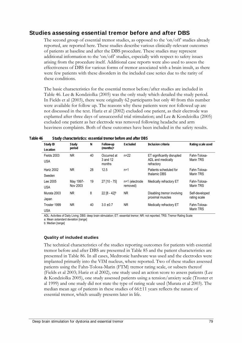
Deep brain stimulation for dystonia and essential tremor 79
Studies assessing essential tremor before and after DBS The second group of essential tremor studies, as opposed to the ‘on/off’ studies already reported, are reported here. These studies describe various clinically-relevant outcomes of patients at baseline and after the DBS procedure. These studies may represent additional information to the ‘on/off’ studies, especially with respect to safety issues arising from the procedure itself. Additional case reports were also used to assess the effectiveness of DBS for various forms of tremor associated with a brain insult, as there were few patients with these disorders in the included case series due to the rarity of these conditions.
The basic characteristics for the essential tremor before/after studies are included in Table 46. Lee & Kondziolka (2005) was the only study which detailed the study period. In Fields et al (2003), there were originally 62 participants but only 40 from this number were available for follow up. The reasons why these patients were not followed up are not discussed in the text. Hariz et al (2002) excluded one patient, as their electrode was explanted after three days of unsuccessful trial stimulation; and Lee & Kondziolka (2005) excluded one patient as her electrode was removed following headache and arm heaviness complaints. Both of these outcomes have been included in the safety results.
Table 46 Study characteristics: essential tremor before and after DBS Study ID Location
Study period
N Follow-up (months)a
Excluded Inclusion criteria Rating scale used
Fields 2003 USA
NR 40 Occurred at 3 and 12 months
n=22 ET significantly disrupted ADL and medically refractory
Fahn-Tolosa-Marin TRS
Hariz 2002 Sweden
NR 28 12.5 n=1 Patients scheduled for thalamic DBS
Fahn-Tolosa-Marin TRS
Lee 2005 USA
May 1997-Nov 2003
19 27 [10 - 75] n=1 (electrode removed)
Medically refractory ET Fahn-Tolosa-Marin TRS
Murata 2003 Japan
NR 8 22 [8 - 42]b NR Disabling tremor involving proximal muscles
Self-developed rating scale
Troster 1999 USA
NR 40 3.0 ±0.7 NR Medically refractory ET Fahn-Tolosa-Marin TRS
ADL: Activities of Daily Living; DBS: deep brain stimulation; ET: essential tremor; NR: not reported; TRS: Tremor Rating Scale a: Mean ±standard deviation [range] b: Median [range]
Quality of included studies
The technical characteristics of the studies reporting outcomes for patients with essential tremor before and after DBS are presented in Table 85 and the patient characteristics are presented in Table 86. In all cases, Medtronic hardware was used and the electrodes were implanted primarily into the VIM nucleus, where reported. Two of these studies assessed patients using the Fahn-Tolosa-Marin (FTM) tremor rating scale, or subsets thereof (Fields et al 2003; Hariz et al 2002), one study used an action score to assess patients (Lee & Kondziolka 2005), one study assessed patients using a tension/anxiety scale (Troster et al 1999) and one study did not state the type of rating scale used (Murata et al 2003). The median mean age of patients in these studies of 66±11 years reflects the nature of essential tremor, which usually presents later in life.
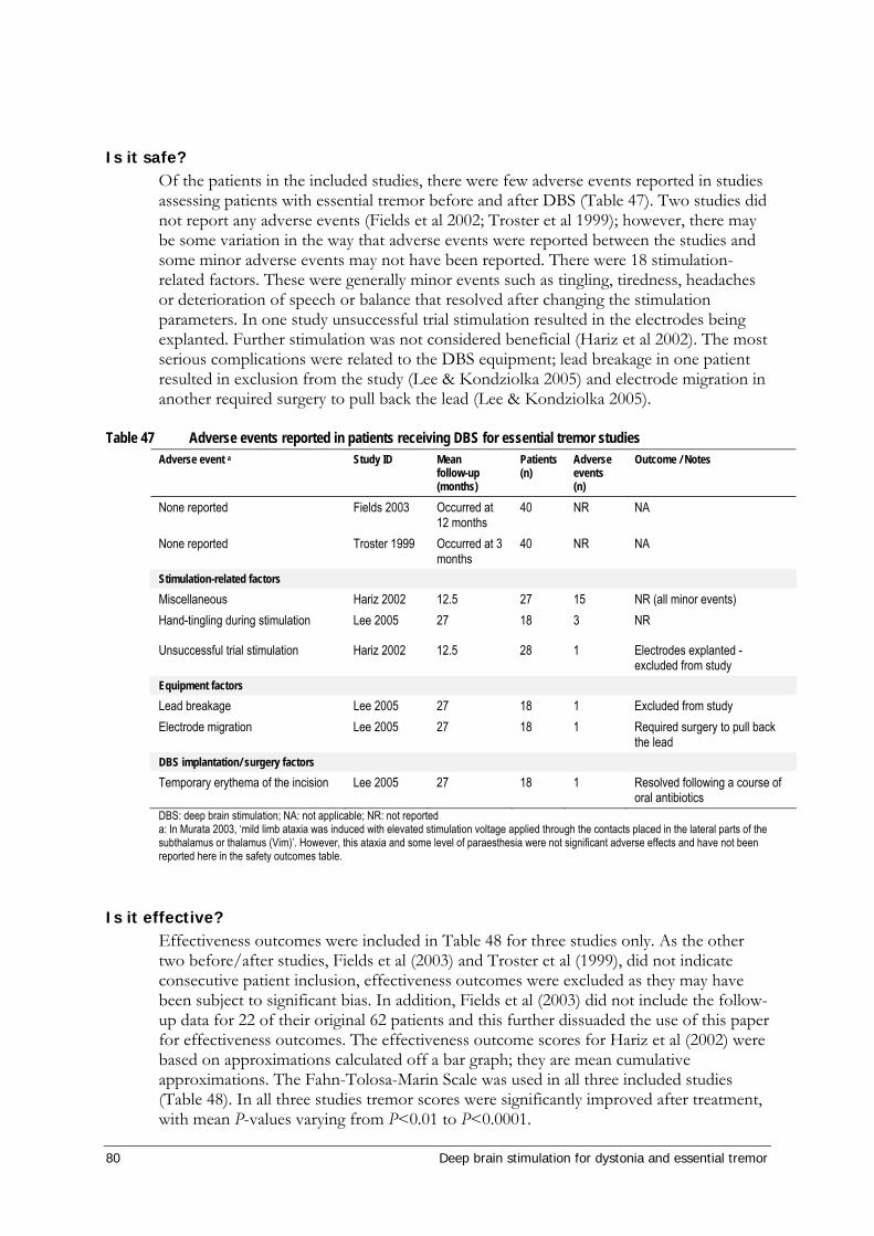
80 Deep brain stimulation for dystonia and essential tremor
Is it safe? Of the patients in the included studies, there were few adverse events reported in studies assessing patients with essential tremor before and after DBS (Table 47). Two studies did not report any adverse events (Fields et al 2002; Troster et al 1999); however, there may be some variation in the way that adverse events were reported between the studies and some minor adverse events may not have been reported. There were 18 stimulation-related factors. These were generally minor events such as tingling, tiredness, headaches or deterioration of speech or balance that resolved after changing the stimulation parameters. In one study unsuccessful trial stimulation resulted in the electrodes being explanted. Further stimulation was not considered beneficial (Hariz et al 2002). The most serious complications were related to the DBS equipment; lead breakage in one patient resulted in exclusion from the study (Lee & Kondziolka 2005) and electrode migration in another required surgery to pull back the lead (Lee & Kondziolka 2005).
Table 47 Adverse events reported in patients receiving DBS for essential tremor studies Adverse event a Study ID Mean
follow-up (months)
Patients (n)
Adverse events (n)
Outcome / Notes
None reported Fields 2003 Occurred at 12 months
40 NR NA
None reported Troster 1999 Occurred at 3 months
40 NR NA
Stimulation-related factors Miscellaneous Hariz 2002 12.5 27 15 NR (all minor events) Hand-tingling during stimulation Lee 2005 27 18 3 NR
Unsuccessful trial stimulation Hariz 2002 12.5 28 1 Electrodes explanted - excluded from study
Equipment factors Lead breakage Lee 2005 27 18 1 Excluded from study Electrode migration Lee 2005 27 18 1 Required surgery to pull back
the lead DBS implantation/ surgery factors Temporary erythema of the incision Lee 2005 27 18 1 Resolved following a course of
oral antibiotics DBS: deep brain stimulation; NA: not applicable; NR: not reported a: In Murata 2003, ‘mild limb ataxia was induced with elevated stimulation voltage applied through the contacts placed in the lateral parts of the subthalamus or thalamus (Vim)’. However, this ataxia and some level of paraesthesia were not significant adverse effects and have not been reported here in the safety outcomes table.
Is it effective? Effectiveness outcomes were included in Table 48 for three studies only. As the other two before/after studies, Fields et al (2003) and Troster et al (1999), did not indicate consecutive patient inclusion, effectiveness outcomes were excluded as they may have been subject to significant bias. In addition, Fields et al (2003) did not include the follow-up data for 22 of their original 62 patients and this further dissuaded the use of this paper for effectiveness outcomes. The effectiveness outcome scores for Hariz et al (2002) were based on approximations calculated off a bar graph; they are mean cumulative approximations. The Fahn-Tolosa-Marin Scale was used in all three included studies (Table 48). In all three studies tremor scores were significantly improved after treatment, with mean P-values varying from P<0.01 to P<0.0001.
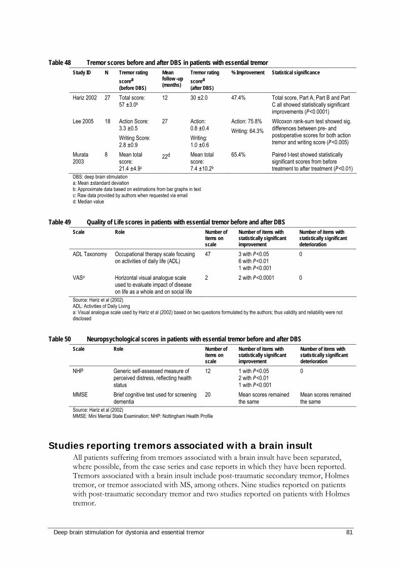
Deep brain stimulation for dystonia and essential tremor 81
Table 48 Tremor scores before and after DBS in patients with essential tremor
Study ID N Tremor rating scorea (before DBS)
Mean follow -up (months)
Tremor rating scorea (after DBS)
% Improvement Statistical significance
Hariz 2002 27 Total score: 57 ±3.0b
12 30 ±2.0 47.4% Total score, Part A, Part B and Part C all showed statistically significant improvements (P<0.0001)
Lee 2005 18 Action Score: 3.3 ±0.5 Writing Score: 2.8 ±0.9
27 Action: 0.8 ±0.4 Writing: 1.0 ±0.6
Action: 75.8% Writing: 64.3%
Wilcoxon rank-sum test showed sig. differences between pre- and postoperative scores for both action tremor and writing score (P<0.005)
Murata 2003
8 Mean total score: 21.4 ±4.9c
22d Mean total score: 7.4 ±10.2b
65.4% Paired t-test showed statistically significant scores from before treatment to after treatment (P<0.01)
DBS: deep brain stimulation a: Mean ±standard deviation b: Approximate data based on estimations from bar graphs in text c: Raw data provided by authors when requested via email d: Median value
Table 49 Quality of Life scores in patients with essential tremor before and after DBS Scale Role Number of
items on scale
Number of items with statistically significant improvement
Number of items with statistically significant deterioration
ADL Taxonomy Occupational therapy scale focusing on activities of daily life (ADL)
47 3 with P<0.05 6 with P<0.01 1 with P<0.001
0
VASa Horizontal visual analogue scale used to evaluate impact of disease on life as a whole and on social life
2 2 with P<0.0001 0
Source: Hariz et al (2002) ADL: Activities of Daily Living a: Visual analogue scale used by Hariz et al (2002) based on two questions formulated by the authors; thus validity and reliability were not disclosed
Table 50 Neuropsychological scores in patients with essential tremor before and after DBS Scale Role Number of
items on scale
Number of items with statistically significant improvement
Number of items with statistically significant deterioration
NHP Generic self-assessed measure of perceived distress, reflecting health status
12 1 with P<0.05 2 with P<0.01 1 with P<0.001
0
MMSE Brief cognitive test used for screening dementia
20 Mean scores remained the same
Mean scores remained the same
Source: Hariz et al (2002) MMSE: Mini Mental State Examination; NHP: Nottingham Health Profile
Studies reporting tremors associated with a brain insult All patients suffering from tremors associated with a brain insult have been separated, where possible, from the case series and case reports in which they have been reported. Tremors associated with a brain insult include post-traumatic secondary tremor, Holmes tremor, or tremor associated with MS, among others. Nine studies reported on patients with post-traumatic secondary tremor and two studies reported on patients with Holmes tremor.
82 Deep brain stimulation for dystonia and essential tremor
Due to the small numbers of patients suffering from each specific subtype of tremor, it should be noted there is a large potential for bias. Patients reported in larger case series with consecutive patients may provide a less biased outcome compared to patients reported individually in case reports.
General study characteristics for the included studies have been reported in Table 87. Where reported, the technical characteristics of all the studies were similar (Table 89). Bilateral implantation to the GPi was performed in all cases with the same electrodes (Medtronic 3387) and either the Soletra or Kinetra IPG. Where reported, stimulation parameters varied slightly between studies. The results are presented in the following section which aims to inform the effectiveness of DBS in the treatment of these rare and varied conditions.
Post-traumatic secondary tremor
Nine patients were identified who were treated with DBS for post-traumatic secondary tremor (Capelle et al 2006; Chang et al 2002; Eltahawy et al 2004b; Kiss et al 2004; Loher et al 2000; Paluzzi et al 2006a; Parkin et al 2001; Starr et al 2006; Vercueil et al 2001). Age of onset varied from 9 to 43 years with the condition, where reported, being caused by head injury in most cases and a peripheral foot injury in one case (Table 88).
Outcomes for these patients were varied and were measured using the BFMDRS, TWSTRS and GFO scales as well as by observation. Overall, there was considerable clinical improvement in movement disorder (≥70%) reported for all patients where reported. Movement scores were not reported by Loher et al (2000), however, they reported that there was a ‘marked improvement of the left-sided hemidystonia evident and pain disappeared completely two months after DBS’. They also reported that after a sustained improvement four years postoperatively, an increase of amplitude from 1 to 1.23 V led to further improvements, however this was accompanied by tonic retraction of the upper lip. As finer adjustments could not be made on the Itrel I, a decision was made to replace the pulse generator with an Itrel II when the batteries ran down. In one patient, it was necessary to use the higher contacts due to the side-effect of flashes of light with the lower contacts (Eltahawy et al 2004b).
The female patient in the study by Paluzzi et al (2006a) has been discussed previously in the in pregnancy section of the dystonia safety section. In brief, the procedure was uneventful and successful, with the woman having two healthy babies while still receiving DBS. She showed a marked (100%) improvement in TWSTRS scores after DBS.
The patient of Vercueil et al (2001) was treated with unilateral DBS with electrodes targeting both the VLp and the GPi. There was a 70 per cent improvement in total BFMDRS (score not provided). The VLp electrode was switched off and no changes in BFMDRS improvement scores were evident. A GFO of 3 was recorded at 12 months (major improvement with recovery of most daily activities, including autonomous walking).
In the study by Chang et al (2002), the patient was reported to have a mild right-sided hemiparesis. Specific pre- and postoperative outcome scores were not recorded. An immediate but short-term improvement was seen following lesion of the left GPi which resolved after 7 days. Following implantation of the IPG there was a clinically relevant functional improvement at up to 12-month follow-up with no reported side-effects.
Deep brain stimulation for dystonia and essential tremor 83
Kiss et al 2004 reported that TWSTRS scores all showed a reduction at the 1-month follow-up.
Starr et al (2006) reported that the DBS device was well-tolerated and there was a slight improvement in BFMDRS score at follow-up of 32 months.
The patient in Parkin et al (2001) experienced an immediate reduction in neck pain and discontinued analgesics 48 hours after the initiation of DBS. Her voluntary head movements, tremor and jerks improved one week later and at two months she was able to date and go shopping alone; and obtained paid employment.
The patient in Capelle et al (2006) had peripherally-induced tremor. No changes in her condition or improvements in her BFMDRS or UDRS scores were observed in the 10-month period following DBS implantation, during which neurostimulation was trialled in the VLp and GPi at pulse width ranging from 180 to 240 µsec, frequencies ranging from 130 to 145 HZ and up to 6 V in amplitude. DBS hardware was removed after a fall when the patient reported electrical sensations in her arm and face.
Two of the patients were assessed for BFMDRS scores. One patient showed a 72 per cent improvement in total BFMDRS and when the VLp electrode was switched off no changes in BFMDRS improvement scores were evident. The second patient showed an 8 per cent improvement in clinical BFMDRS scores. Three patients were assessed for TWSTRS scores. Two showed improvements in pain, disability and severity subscores and all three showed improvements in total TWSTRS scores, with percentage improvements including 79.5 per cent ±4.1, 85 per cent and 100 per cent. One patient recorded a GFO of 3 at 12 months, which represents a major improvement with recovery of most daily activities, including autonomous walking. One patient showed no improvement in her condition and the DBS hardware was removed after a fall.
In summary, nine patients with post-traumatic secondary tremor were studied. Of these, seven showed a good improvement, with four improving greater than 70 per cent (Paulizzi et al 2006; Kiss et al 2004; Vercueil et al 2001; Eltahawy et al 2004b). One patient showed a slight improvement; and one showed no improvement. There was one adverse event of a mild right-sided hemiparesis.
Holmes tremor
Two studies were identified in which patients received DBS for Holmes tremor, which is a condition secondary to brain insult (Foote et al 2005; Nikkah et al 2004). Three patients, one male and two female, presented with Holmes tremor. The median age was 34.3 years, ranging from 24 to 47 years. The two patients in Nikkah et al (2004) both received DBS to the ventral intermediate nucleus of the thalamus (VIM), whilst the patient in Foote et al (2005) received DBS to the VIM and after 2 months of VIM stimulation, the ventralis oralis anterior (VOA)/ventralis oralis posterior (VOP) stimulator was activated.
All patients appear to have benefited from the DBS. At 12-month follow-up, one patient’s left upper extremity tremor (total of resting, postural and action tremor) scores had decreased from 9 at baseline to 3 with both VIM and VOA/VOP stimulation activated, to 5 with VIM stimulation only and to 4 with VOA/VOP stimulation only (Foote et al 2005). This patient was able to return to gainful employment. Two patients received DBS in the VIM (Nikkah et al 2004). For one of these patients, chronic high-
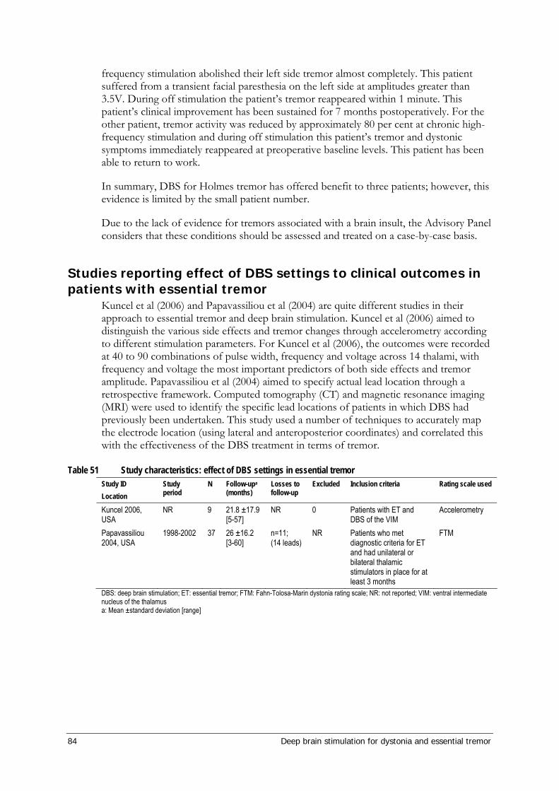
84 Deep brain stimulation for dystonia and essential tremor
frequency stimulation abolished their left side tremor almost completely. This patient suffered from a transient facial paresthesia on the left side at amplitudes greater than 3.5V. During off stimulation the patient’s tremor reappeared within 1 minute. This patient’s clinical improvement has been sustained for 7 months postoperatively. For the other patient, tremor activity was reduced by approximately 80 per cent at chronic high-frequency stimulation and during off stimulation this patient’s tremor and dystonic symptoms immediately reappeared at preoperative baseline levels. This patient has been able to return to work.
In summary, DBS for Holmes tremor has offered benefit to three patients; however, this evidence is limited by the small patient number.
Due to the lack of evidence for tremors associated with a brain insult, the Advisory Panel considers that these conditions should be assessed and treated on a case-by-case basis.
Studies reporting effect of DBS settings to clinical outcomes in patients with essential tremor
Kuncel et al (2006) and Papavassiliou et al (2004) are quite different studies in their approach to essential tremor and deep brain stimulation. Kuncel et al (2006) aimed to distinguish the various side effects and tremor changes through accelerometry according to different stimulation parameters. For Kuncel et al (2006), the outcomes were recorded at 40 to 90 combinations of pulse width, frequency and voltage across 14 thalami, with frequency and voltage the most important predictors of both side effects and tremor amplitude. Papavassiliou et al (2004) aimed to specify actual lead location through a retrospective framework. Computed tomography (CT) and magnetic resonance imaging (MRI) were used to identify the specific lead locations of patients in which DBS had previously been undertaken. This study used a number of techniques to accurately map the electrode location (using lateral and anteroposterior coordinates) and correlated this with the effectiveness of the DBS treatment in terms of tremor.
Table 51 Study characteristics: effect of DBS settings in essential tremor Study ID Location
Study period
N Follow-upa (months)
Losses to follow-up
Excluded Inclusion criteria Rating scale used
Kuncel 2006, USA
NR 9 21.8 ±17.9 [5-57]
NR 0 Patients with ET and DBS of the VIM
Accelerometry
Papavassiliou 2004, USA
1998-2002 37 26 ±16.2 [3-60]
n=11; (14 leads)
NR Patients who met diagnostic criteria for ET and had unilateral or bilateral thalamic stimulators in place for at least 3 months
FTM
DBS: deep brain stimulation; ET: essential tremor; FTM: Fahn-Tolosa-Marin dystonia rating scale; NR: not reported; VIM: ventral intermediate nucleus of the thalamus a: Mean ±standard deviation [range]
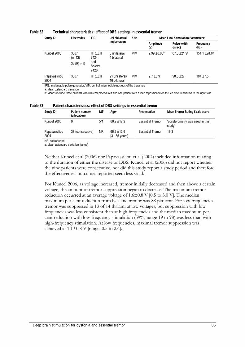
Deep brain stimulation for dystonia and essential tremor 85
Table 52 Technical characteristics: effect of DBS settings in essential tremor Mean Final Stimulation Parametersa Study ID Electrodes IPG Uni / bilateral
implantation Site
Amplitude (V)
Pulse width (µsec)
Frequency (Hz)
Kuncel 2006 3387 (n=13) 3389(n=1)
ITREL II 7424 and Soletra 7426
5 unilateral/ 4 bilateral
VIM 2.99 ±0.86b 87.8 ±21.9b 151.1 ±24.0b
Papavassiliou 2004
3387 ITREL II 21 unilateral/ 16 bilateral
VIM 2.7 ±0.9 98.5 ±27 184 ±7.5
IPG: implantable pulse generator; VIM: ventral intermediate nucleus of the thalamus a: Mean ±standard deviation b: Means include three patients with bilateral procedures and one patient with a lead repositioned on the left side in addition to the right side
Table 53 Patient characteristics: effect of DBS settings in essential tremor Study ID Patient number
(allocation) M/F Agea Presentation Mean Tremor Rating Scale score
Kuncel 2006 9 5/4 66.9 ±17.2 Essential Tremor ‘accelerometry was used in this study’
Papavassiliou 2004
37 (consecutive) NR 66.2 ±13.6 [31-85 years]
Essential Tremor 19.3
NR: not reported a: Mean ±standard deviation [range]
Neither Kuncel et al (2006) nor Papavassiliou et al (2004) included information relating to the duration of either the disease or DBS. Kuncel et al (2006) did not report whether the nine patients were consecutive, nor did this study report a study period and therefore the effectiveness outcomes reported seem less valid.
For Kuncel 2006, as voltage increased, tremor initially decreased and then above a certain voltage, the amount of tremor suppression began to decrease. The maximum tremor reduction occurred at an average voltage of 1.6±0.8 V [0.5 to 3.0 V]. The median maximum per cent reduction from baseline tremor was 88 per cent. For low frequencies, tremor was suppressed in 13 of 14 thalami at low voltages, but suppression with low frequencies was less consistent than at high frequencies and the median maximum per cent reduction with low-frequency stimulation (59%, range 19 to 98) was less than with high-frequency stimulation. At low frequencies, maximal tremor suppression was achieved at 1.1±0.8 V [range, 0.5 to 2.6].
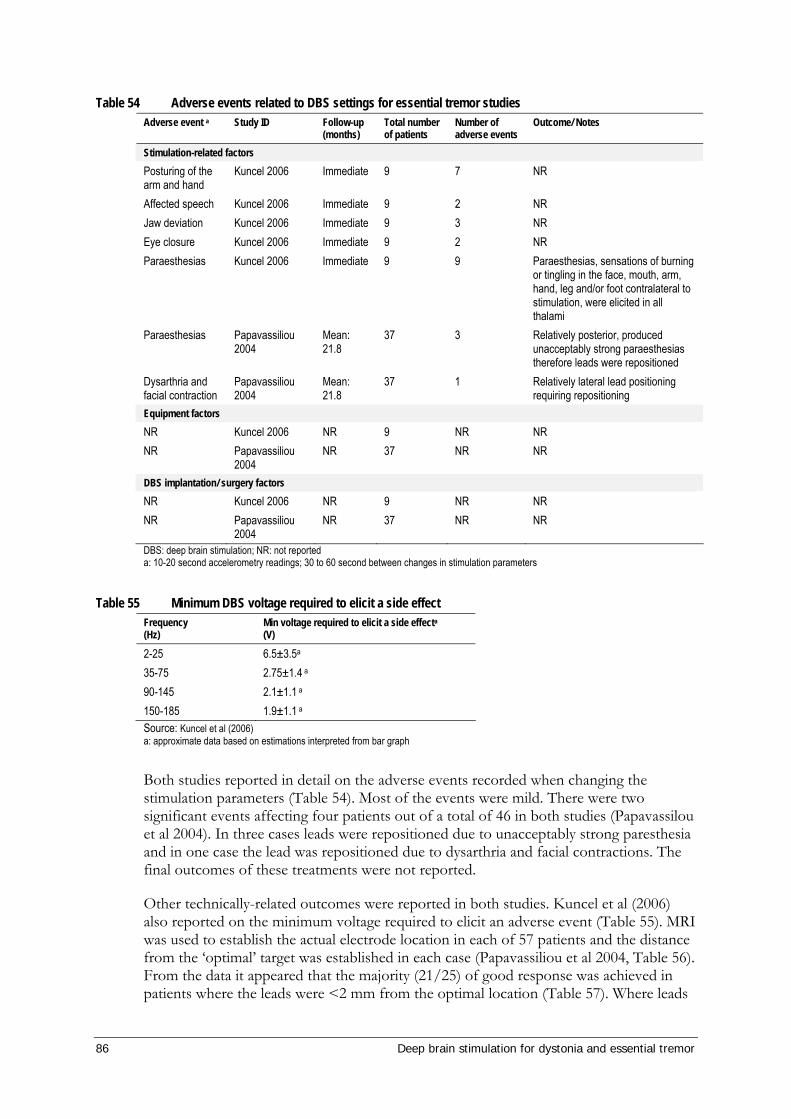
86 Deep brain stimulation for dystonia and essential tremor
Table 54 Adverse events related to DBS settings for essential tremor studies Adverse event a Study ID Follow-up
(months) Total number of patients
Number of adverse events
Outcome/ Notes
Stimulation-related factors Posturing of the arm and hand
Kuncel 2006 Immediate 9 7 NR
Affected speech Kuncel 2006 Immediate 9 2 NR Jaw deviation Kuncel 2006 Immediate 9 3 NR Eye closure Kuncel 2006 Immediate 9 2 NR Paraesthesias Kuncel 2006 Immediate 9 9 Paraesthesias, sensations of burning
or tingling in the face, mouth, arm, hand, leg and/or foot contralateral to stimulation, were elicited in all thalami
Paraesthesias Papavassiliou 2004
Mean: 21.8
37 3 Relatively posterior, produced unacceptably strong paraesthesias therefore leads were repositioned
Dysarthria and facial contraction
Papavassiliou 2004
Mean: 21.8
37 1 Relatively lateral lead positioning requiring repositioning
Equipment factors NR Kuncel 2006 NR 9 NR NR NR Papavassiliou
2004 NR 37 NR NR
DBS implantation/ surgery factors NR Kuncel 2006 NR 9 NR NR NR Papavassiliou
2004 NR 37 NR NR
DBS: deep brain stimulation; NR: not reported a: 10-20 second accelerometry readings; 30 to 60 second between changes in stimulation parameters
Table 55 Minimum DBS voltage required to elicit a side effect Frequency (Hz)
Min voltage required to elicit a side effecta (V)
2-25 6.5±3.5a 35-75 2.75±1.4 a 90-145 2.1±1.1 a 150-185 1.9±1.1 a Source: Kuncel et al (2006) a: approximate data based on estimations interpreted from bar graph
Both studies reported in detail on the adverse events recorded when changing the stimulation parameters (Table 54). Most of the events were mild. There were two significant events affecting four patients out of a total of 46 in both studies (Papavassilou et al 2004). In three cases leads were repositioned due to unacceptably strong paresthesia and in one case the lead was repositioned due to dysarthria and facial contractions. The final outcomes of these treatments were not reported.
Other technically-related outcomes were reported in both studies. Kuncel et al (2006) also reported on the minimum voltage required to elicit an adverse event (Table 55). MRI was used to establish the actual electrode location in each of 57 patients and the distance from the ‘optimal’ target was established in each case (Papavassiliou et al 2004, Table 56). From the data it appeared that the majority (21/25) of good response was achieved in patients where the leads were <2 mm from the optimal location (Table 57). Where leads
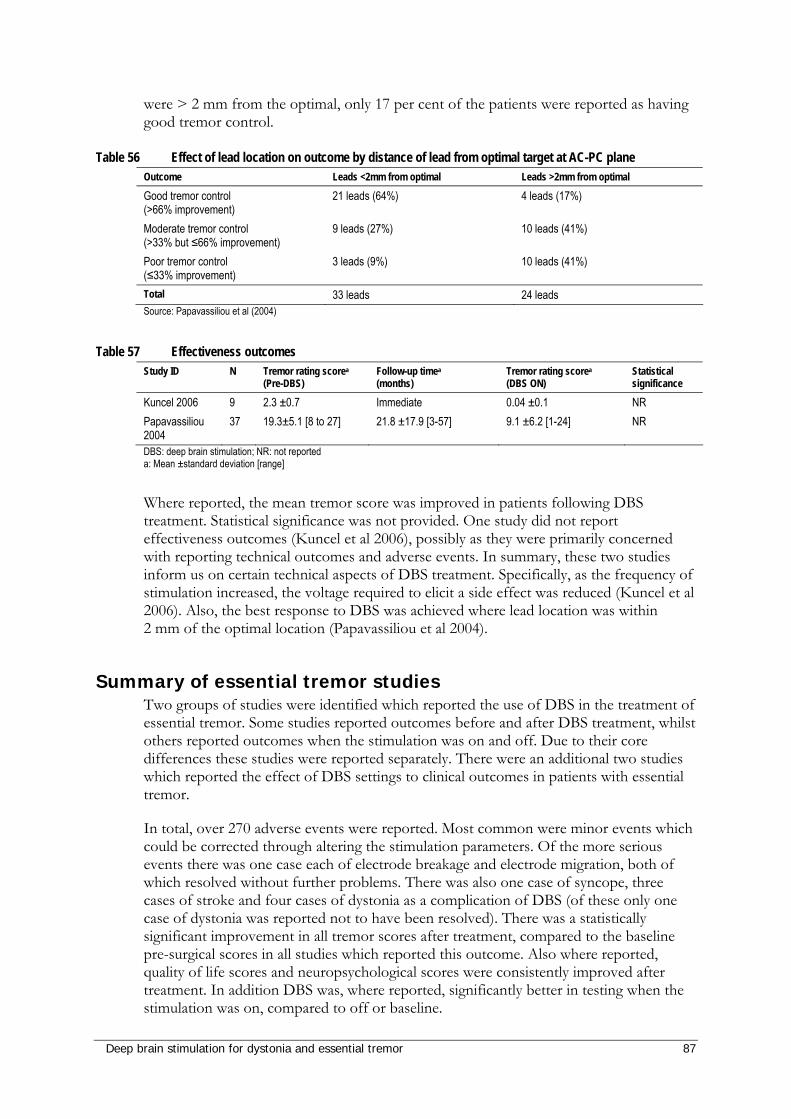
Deep brain stimulation for dystonia and essential tremor 87
were > 2 mm from the optimal, only 17 per cent of the patients were reported as having good tremor control.
Table 56 Effect of lead location on outcome by distance of lead from optimal target at AC-PC plane Outcome Leads <2mm from optimal Leads >2mm from optimal
Good tremor control (>66% improvement)
21 leads (64%) 4 leads (17%)
Moderate tremor control (>33% but ≤66% improvement)
9 leads (27%) 10 leads (41%)
Poor tremor control (≤33% improvement)
3 leads (9%) 10 leads (41%)
Total 33 leads 24 leads Source: Papavassiliou et al (2004)
Table 57 Effectiveness outcomes Study ID N Tremor rating scorea
(Pre-DBS) Follow-up timea (months)
Tremor rating scorea (DBS ON)
Statistical significance
Kuncel 2006 9 2.3 ±0.7 Immediate 0.04 ±0.1 NR Papavassiliou 2004
37 19.3±5.1 [8 to 27] 21.8 ±17.9 [3-57] 9.1 ±6.2 [1-24] NR
DBS: deep brain stimulation; NR: not reported a: Mean ±standard deviation [range]
Where reported, the mean tremor score was improved in patients following DBS treatment. Statistical significance was not provided. One study did not report effectiveness outcomes (Kuncel et al 2006), possibly as they were primarily concerned with reporting technical outcomes and adverse events. In summary, these two studies inform us on certain technical aspects of DBS treatment. Specifically, as the frequency of stimulation increased, the voltage required to elicit a side effect was reduced (Kuncel et al 2006). Also, the best response to DBS was achieved where lead location was within 2 mm of the optimal location (Papavassiliou et al 2004).
Summary of essential tremor studies Two groups of studies were identified which reported the use of DBS in the treatment of essential tremor. Some studies reported outcomes before and after DBS treatment, whilst others reported outcomes when the stimulation was on and off. Due to their core differences these studies were reported separately. There were an additional two studies which reported the effect of DBS settings to clinical outcomes in patients with essential tremor.
In total, over 270 adverse events were reported. Most common were minor events which could be corrected through altering the stimulation parameters. Of the more serious events there was one case each of electrode breakage and electrode migration, both of which resolved without further problems. There was also one case of syncope, three cases of stroke and four cases of dystonia as a complication of DBS (of these only one case of dystonia was reported not to have been resolved). There was a statistically significant improvement in all tremor scores after treatment, compared to the baseline pre-surgical scores in all studies which reported this outcome. Also where reported, quality of life scores and neuropsychological scores were consistently improved after treatment. In addition DBS was, where reported, significantly better in testing when the stimulation was on, compared to off or baseline.

88 Deep brain stimulation for dystonia and essential tremor
Studies which have reported tremor outcomes other than the FTM are presented in Table 44 and Table 45. Comprehensive study information is provided in Appendix J. Many studies used the Tremor Activities of Daily Living Scale (TADLS), which are separated into patient and clinician scales. Some studies use sub-scores of the TADLS, such as the ADL (Activities of Daily Living). Where reported, there was a statistically significant improvement for all studies in tremor scores for ON vs OFF and ON vs baseline. Improvement in overall scores ranged from 36 per cent (Pahwa et al 2006; Sydow et al 2003) to 82 per cent (Ushe et al 2006), depending on the type of scoring system used.
The TADLS system involves two scores; one rated by the patient and the other rated by the clinician (Table 44). The improvements to both scales were similar, with improvements of between 33 per cent and 57.9 per cent overall. The ADL scale was used by six included studies (Table 44). One study reported unilateral and bilateral implantation separately (Pahwa et al 2006). Unilateral improvement showed the larger improvement. In two studies which reported ADL scores at different time points, the improvement in tremor was reduced over time (Pahwa et al 1999; Sydow et al 2003), for example improvement dropped from 82 per cent at one year to 62 per cent at 6 years.
Accelerometry measurements were reported in two studies (Table 45, Ushe et al 2006; Vaillancourt et al 2003). In both cases, an improvement was observed between on and off states. Accelerometry readings were reduced by 85.2 per cent and RMS displacement was reduced by 93 per cent in the on state.
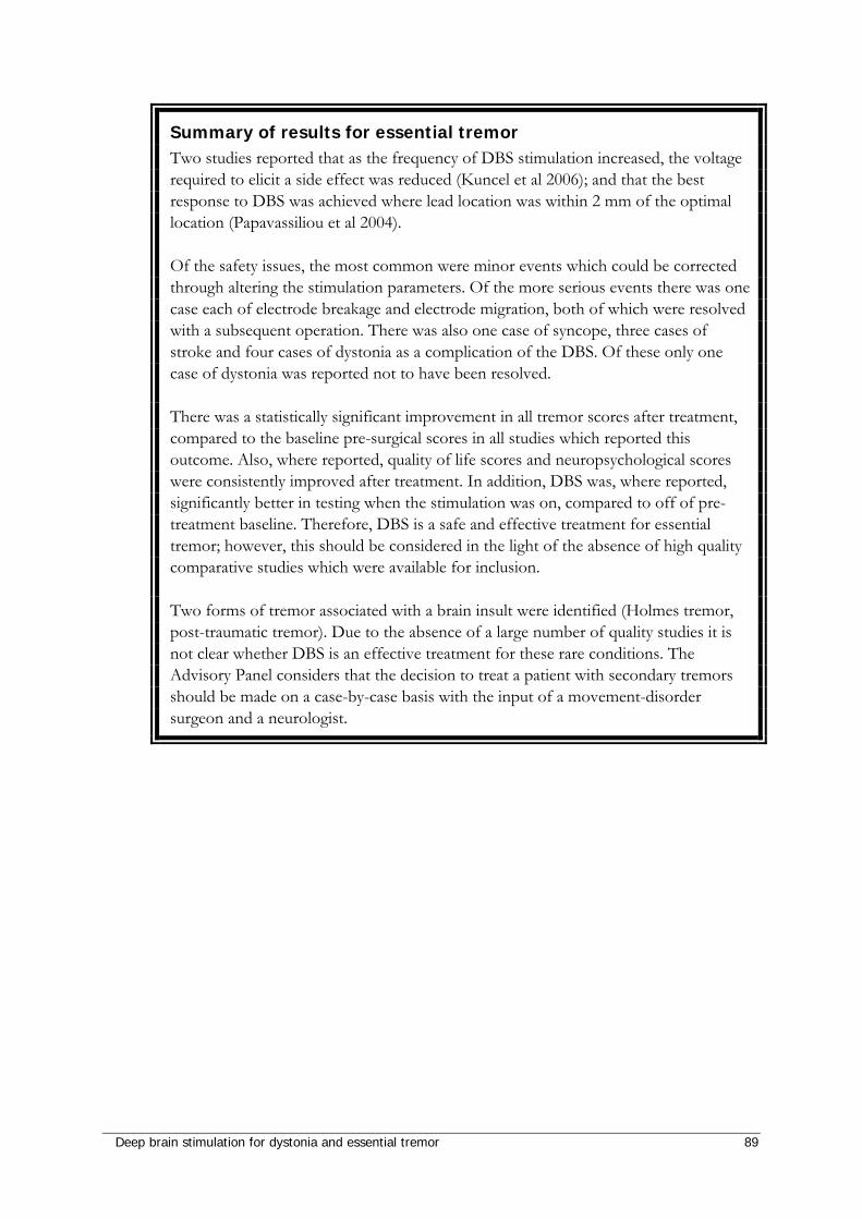
Deep brain stimulation for dystonia and essential tremor 89
Summary of results for essential tremor Two studies reported that as the frequency of DBS stimulation increased, the voltage required to elicit a side effect was reduced (Kuncel et al 2006); and that the best response to DBS was achieved where lead location was within 2 mm of the optimal location (Papavassiliou et al 2004).
Of the safety issues, the most common were minor events which could be corrected through altering the stimulation parameters. Of the more serious events there was one case each of electrode breakage and electrode migration, both of which were resolved with a subsequent operation. There was also one case of syncope, three cases of stroke and four cases of dystonia as a complication of the DBS. Of these only one case of dystonia was reported not to have been resolved.
There was a statistically significant improvement in all tremor scores after treatment, compared to the baseline pre-surgical scores in all studies which reported this outcome. Also, where reported, quality of life scores and neuropsychological scores were consistently improved after treatment. In addition, DBS was, where reported, significantly better in testing when the stimulation was on, compared to off of pre-treatment baseline. Therefore, DBS is a safe and effective treatment for essential tremor; however, this should be considered in the light of the absence of high quality comparative studies which were available for inclusion.
Two forms of tremor associated with a brain insult were identified (Holmes tremor, post-traumatic tremor). Due to the absence of a large number of quality studies it is not clear whether DBS is an effective treatment for these rare conditions. The Advisory Panel considers that the decision to treat a patient with secondary tremors should be made on a case-by-case basis with the input of a movement-disorder surgeon and a neurologist.
90 Deep brain stimulation for dystonia and essential tremor
What are the economic considerations?
Background Economic evaluation of new healthcare technologies is important when determining whether the new initiative offers additional benefits and at what cost. When the new initiative is dominated by the existing technology, the costs are higher and the effectiveness is less. When the new initiative dominates the existing technology, the costs are lower and the effectiveness is greater. Economic evaluation is particularly important where the new initiative offers health benefits at additional costs. Within a constrained healthcare budget, determining the additional cost that would be paid for a given health gain is important when ascertaining whether such incremental costs represent value for money.
The usual process for an economic evaluation is first to determine the incremental effectiveness, which is the additional benefits associated with the new technology relative to current practice. The next step is to determine the incremental costs, which is the difference in costs between the new initiative and the comparator. Finally the incremental cost-effectiveness ratio (ICER) can be calculated using the following ratio:
To allow comparison of effectiveness in one area with effectiveness in another, it is preferable for an economic evaluation to undertake a cost-utility analysis. A cost-utility analysis generates an ICER as described above, using a generic outcome measure, defined as one which can be utilised in different areas of healthcare. The most common generic outcome measure is the quality-adjusted life year (QALY). This is a measure of effectiveness which combines morbidity and mortality dimensions into one composite measure of outcome. The use of cost-utility analysis, while preferable to disease-specific outcome measures, is reliant on the existence of appropriate published data. This includes generic quality of life measures, such as the SF-6D, the SF-36, or the EQ-5D. However, in the absence of good quality outcome data suitable for economic evaluation, it is instructive to consider both the cost per patient of the intervention and the total cost burden to society.
Assumptions for DBS The population for dystonia is generalised rather than focal; however, the costs are likely to be comparable between the two groups.
The comparator is assumed to be ‘no DBS’ since the population is made up of those who have not responded to medical therapy. This means that, in the base case, there is no cost-offset associated with reduced pharmaceutical use.
In the base case we have used a health service perspective, that is, we only consider cost implications that accrue to the system. We do, however, acknowledge the potential for significant productivity effects as individuals return to work, particularly in patients with
Cost New – Cost Comparator Effectiveness New – Effectiveness Comparator
ICER =
Deep brain stimulation for dystonia and essential tremor 91
dystonia who tend to be younger. The effect of this issue on the base case result is investigated in a sensitivity analysis.
Our time horizon is 10 years. Thus, only costs and outcomes accrued over this period are considered. We will discuss the likely effects of extending the time horizon after we present the base case results.
Future events are discounted at 5 per cent per annum. This means that an event which occurs in one year’s time is valued at 1/1.05 of the value if it occurred immediately.
Battery life is assumed to be 5 years for patients with ET and 2 years for those with dystonia. This differential battery life is a consequence of the need to keep the unit turned on during sleep in patients with dystonia.
Search strategies and existing literature As described in the Approach to Assessment, a search strategy was developed to systematically identify studies in which DBS was used in the management of essential tremor or dystonia. Databases of peer-reviewed literature including Medline, PubMed, CINAHL and Cochrane have been searched. The bibliographies of all retrieved publications were hand searched for any relevant references missing in the database search. In addition to the search terms described in the Approach to Assessment section, Cost$ or Econ$ were added, to identify any published cost-effectiveness analyses. The inclusion and exclusion criteria remained the same. Under this approach, we identified one suitable economic evaluation (Yianni et al 2005). This British paper investigates the costs and outcomes associated with deep brain stimulation in a dystonia population. The authors undertake a cost-benefit analysis and a cost-utility analysis, using a sample of 26 individuals. Their results suggest an improvement in Euroqol EQ-5D scores for individuals of 0.472 and a cost per quality-adjusted life year (QALY) of £33,980 (as of February 2008, equivalent to $74,557). This is based on the assumption that the benefit remains for 2 years and then ends. This would usually be considered highly equivocal evidence for the use of the intervention in this population. There are a number of issues which have to be considered before this result can be useful in an Australian context.
Regarding the use of the EQ-5D, there is insufficient description of the method used to elicit the values they provide. Indeed, the clustering of EQ-5D values around multiples of 0.05 suggests inconsistencies since the values of the states described by the EQ-5D do not cluster in this way. Indeed, some of the values (such as 0.95) are impossible under the British algorithm (Dolan et al 1996). Whether this leads to an overestimate or underestimate of cost-effectiveness is uncertain since no EQ-5D evidence could be identified in existing literature for either essential tremor or dystonia.
The cost-benefit analysis is also open to dispute since the tool used to elicit willingness to pay (WTP) might have led respondents to particular values. It gives the cost of various non-health goods, the most expensive of which is a mansion with a swimming pool, valued at one million pounds. It is noteworthy that 7 of the 26 individuals in the analysis suggested deep brain stimulation was worth exactly one million pounds to them. Indeed, the skewed WTP data means that the cost of the intervention exceeds the WTP in 14 of the 26 individuals, yet the mean difference between the two is £291,000.
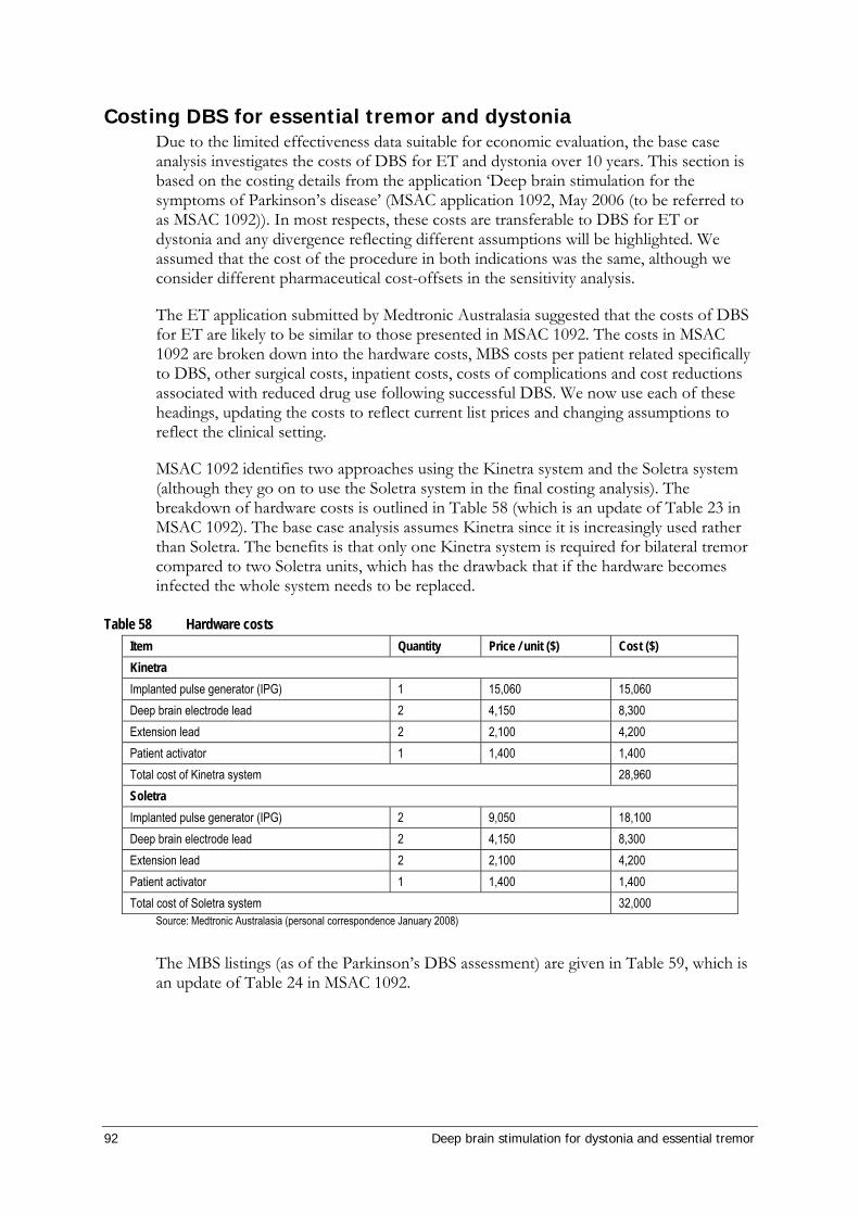
92 Deep brain stimulation for dystonia and essential tremor
Costing DBS for essential tremor and dystonia Due to the limited effectiveness data suitable for economic evaluation, the base case analysis investigates the costs of DBS for ET and dystonia over 10 years. This section is based on the costing details from the application ‘Deep brain stimulation for the symptoms of Parkinson’s disease’ (MSAC application 1092, May 2006 (to be referred to as MSAC 1092)). In most respects, these costs are transferable to DBS for ET or dystonia and any divergence reflecting different assumptions will be highlighted. We assumed that the cost of the procedure in both indications was the same, although we consider different pharmaceutical cost-offsets in the sensitivity analysis.
The ET application submitted by Medtronic Australasia suggested that the costs of DBS for ET are likely to be similar to those presented in MSAC 1092. The costs in MSAC 1092 are broken down into the hardware costs, MBS costs per patient related specifically to DBS, other surgical costs, inpatient costs, costs of complications and cost reductions associated with reduced drug use following successful DBS. We now use each of these headings, updating the costs to reflect current list prices and changing assumptions to reflect the clinical setting.
MSAC 1092 identifies two approaches using the Kinetra system and the Soletra system (although they go on to use the Soletra system in the final costing analysis). The breakdown of hardware costs is outlined in Table 58 (which is an update of Table 23 in MSAC 1092). The base case analysis assumes Kinetra since it is increasingly used rather than Soletra. The benefits is that only one Kinetra system is required for bilateral tremor compared to two Soletra units, which has the drawback that if the hardware becomes infected the whole system needs to be replaced.
Table 58 Hardware costs Item Quantity Price / unit ($) Cost ($) Kinetra Implanted pulse generator (IPG) 1 15,060 15,060 Deep brain electrode lead 2 4,150 8,300 Extension lead 2 2,100 4,200 Patient activator 1 1,400 1,400 Total cost of Kinetra system 28,960 Soletra Implanted pulse generator (IPG) 2 9,050 18,100 Deep brain electrode lead 2 4,150 8,300 Extension lead 2 2,100 4,200 Patient activator 1 1,400 1,400 Total cost of Soletra system 32,000
Source: Medtronic Australasia (personal correspondence January 2008)
The MBS listings (as of the Parkinson’s DBS assessment) are given in Table 59, which is an update of Table 24 in MSAC 1092.
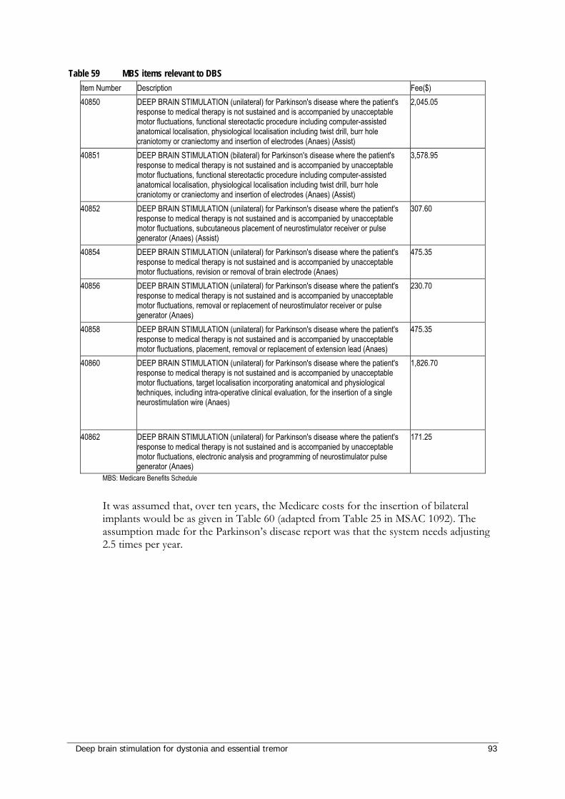
Deep brain stimulation for dystonia and essential tremor 93
Table 59 MBS items relevant to DBS Item Number Description Fee($) 40850 DEEP BRAIN STIMULATION (unilateral) for Parkinson's disease where the patient's
response to medical therapy is not sustained and is accompanied by unacceptable motor fluctuations, functional stereotactic procedure including computer-assisted anatomical localisation, physiological localisation including twist drill, burr hole craniotomy or craniectomy and insertion of electrodes (Anaes) (Assist)
2,045.05
40851 DEEP BRAIN STIMULATION (bilateral) for Parkinson's disease where the patient's response to medical therapy is not sustained and is accompanied by unacceptable motor fluctuations, functional stereotactic procedure including computer-assisted anatomical localisation, physiological localisation including twist drill, burr hole craniotomy or craniectomy and insertion of electrodes (Anaes) (Assist)
3,578.95
40852 DEEP BRAIN STIMULATION (unilateral) for Parkinson's disease where the patient's response to medical therapy is not sustained and is accompanied by unacceptable motor fluctuations, subcutaneous placement of neurostimulator receiver or pulse generator (Anaes) (Assist)
307.60
40854 DEEP BRAIN STIMULATION (unilateral) for Parkinson's disease where the patient's response to medical therapy is not sustained and is accompanied by unacceptable motor fluctuations, revision or removal of brain electrode (Anaes)
475.35
40856 DEEP BRAIN STIMULATION (unilateral) for Parkinson's disease where the patient's response to medical therapy is not sustained and is accompanied by unacceptable motor fluctuations, removal or replacement of neurostimulator receiver or pulse generator (Anaes)
230.70
40858 DEEP BRAIN STIMULATION (unilateral) for Parkinson's disease where the patient's response to medical therapy is not sustained and is accompanied by unacceptable motor fluctuations, placement, removal or replacement of extension lead (Anaes)
475.35
40860 DEEP BRAIN STIMULATION (unilateral) for Parkinson's disease where the patient's response to medical therapy is not sustained and is accompanied by unacceptable motor fluctuations, target localisation incorporating anatomical and physiological techniques, including intra-operative clinical evaluation, for the insertion of a single neurostimulation wire (Anaes)
1,826.70
40862 DEEP BRAIN STIMULATION (unilateral) for Parkinson's disease where the patient's response to medical therapy is not sustained and is accompanied by unacceptable motor fluctuations, electronic analysis and programming of neurostimulator pulse generator (Anaes)
171.25
MBS: Medicare Benefits Schedule
It was assumed that, over ten years, the Medicare costs for the insertion of bilateral implants would be as given in Table 60 (adapted from Table 25 in MSAC 1092). The assumption made for the Parkinson’s disease report was that the system needs adjusting 2.5 times per year.
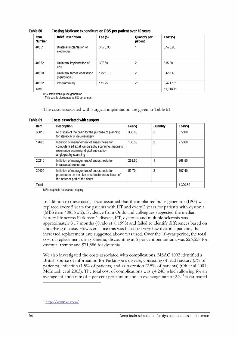
94 Deep brain stimulation for dystonia and essential tremor
Table 60 Costing Medicare expenditure on DBS per patient over 10 years Item Number
Brief Description Fee ($) Quantity per patient
Cost ($)
40851 Bilateral implantation of electrodes
3,578.95 1 3,578.95
40852 Unilateral implantation of IPG
307.60 2 615.20
40860 Unilateral target localisation (neurologist)
1,826.70 2 3,653.40
40862 Programming 171.25 25 3,471.16* Total 11,318.71
IPG: implantable pulse generator * This cost is discounted at 5% per annum
The costs associated with surgical implantation are given in Table 61.
Table 61 Costs associated with surgery Item Description Fee($) Quantity Cost($) 63010 MRI scan of the brain for the purpose of planning
for stereotactic neurosurgery 336.00 2 672.00
17625 Initiation of management of anaesthesia for computerised axial tomography scanning, magnetic resonance scanning, digital subtraction angiography scanning
136.30 2 272.60
20210 Initiation of management of anaesthesia for intracranial procedures
268.50 1 268.50
20400 Initiation of management of anaesthesia for procedures on the skin or subcutaneous tissue of the anterior part of the chest
53.70 2 107.40
Total 1,320.50 MRI: magnetic resonance imaging
In addition to these costs, it was assumed that the implanted pulse generator (IPG) was replaced every 5 years for patients with ET and every 2 years for patients with dystonia (MBS item 40856 x 2). Evidence from Ondo and colleagues suggested the median battery life across Parkinson’s disease, ET, dystonia and multiple sclerosis was approximately 31.7 months (Ondo et al 1998) and failed to identify differences based on underlying disease. However, since this was based on very few dystonia patients, the increased replacement rate suggested above was used. Over the 10-year period, the total cost of replacement using Kinetra, discounting at 5 per cent per annum, was $26,558 for essential tremor and $71,586 for dystonia.
We also investigated the costs associated with complications. MSAC 1092 identified a British source of information for Parkinson’s disease, consisting of lead fracture (5% of patients), infection (1.5% of patients) and skin erosion (2.5% of patients) (Oh et al 2001; McIntosh et al 2003). The total cost of complications was £4,246, which allowing for an average inflation rate of 3 per cent per annum and an exchange rate of 2.241 is estimated
1 http://www.xe.com/
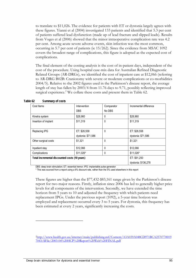
Deep brain stimulation for dystonia and essential tremor 95
to translate to $11,026. The evidence for patients with ET or dystonia largely agrees with these figures. Yianni et al (2004) investigated 133 patients and identified that 5.3 per cent of patients suffered lead dysfunction (made up of lead fracture and slipped leads). Results from Voges et al (2006) showed that the minor intraoperative complication rate was 4.2 per cent. Among acute severe adverse events, skin infection was the most common, occurring in 5.7 per cent of patients (ie 15/262). Since the evidence from MSAC 1092 covers the broadest range of complications, this figure is adopted as the expected cost of complications.
The final element of the costing analysis is the cost of in-patient days, independent of the cost of the procedure. Using hospital case-mix data for Australian Refined Diagnostic Related Groups (AR-DRGs), we identified the cost of inpatient care at $12,066 (referring to AR-DRG B02B: Craniotomy with severe or moderate complications or co-morbidities 2004/5). Relative to the 2002 figures used in the Parkinson’s disease report, the average length of stay has fallen by 2003/4 from 11.76 days to 9.71, possibly reflecting improved surgical experience.2 We collate these costs and present them in Table 62.
Table 62 Summary of costs Cost Items Intervention
DBS Comparator No DBS
Incremental difference
Kinetra system $28,960 0 $28,960 Insertion of implant $11,319 0 $11,319
Replacing IPG ET: $26,558 dystonia: $71,586
0 ET: $26,558 dystonia: $71,586
Other surgical costs $1,321 0 $1,321
Inpatient stay $12,066 0 $12,066 Complications $11,026* 0 $11,026* Total incremental discounted costs (10 years) ET: $91,250
dystonia: $136,278 DBS: deep brain stimulation; ET: essential tremor; IPG: implantable pulse generator * This was sourced from a report using a 6% discount rate, rather than the 5% used elsewhere in this report
These figures are higher than the $77,432-$83,161 range given by the Parkinson’s disease report for two major reasons. Firstly, inflation since 2006 has led to generally higher price levels for all components of the intervention. Secondly, we have extended the time horizon from 5 years to 10 and adjusted the frequency with which patients need replacement IPGs. Under the previous report (1092), a 5-year time horizon was employed and replacement occurred every 3 to 5 years. For dystonia, this frequency has been estimated at every 2 years, significantly increasing the costs.
2http://www.health.gov.au/internet/main/publishing.nsf/Content/A3A039A048CD971BCA25707700197043/$File/2003-04%20HCP%20Report%20Web%20FINAL.pdf
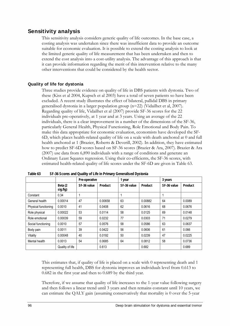
96 Deep brain stimulation for dystonia and essential tremor
Sensitivity analysis This sensitivity analysis considers generic quality of life outcomes. In the base case, a costing analysis was undertaken since there was insufficient data to provide an outcome suitable for economic evaluation. It is possible to extend the costing analysis to look at the limited generic quality of life measurement that has been undertaken and then to extend the cost analysis into a cost-utility analysis. The advantage of this approach is that it can provide information regarding the merit of this intervention relative to the many other interventions that could be considered by the health sector.
Quality of life for dystonia Three studies provide evidence on quality of life in DBS patients with dystonia. Two of these (Kiss et al 2004, Kupsch et al 2003) have a total of seven patients so have been excluded. A recent study illustrates the effect of bilateral, pallidal DBS in primary generalised dystonia in a larger population group (n=22) (Vidailhet et al, 2007). Regarding quality of life, Vidailhet et al (2007) provide SF-36 scores for the 22 individuals pre-operatively, at 1 year and at 3 years. Using an average of the 22 individuals, there is a clear improvement in a number of the dimensions of the SF-36, particularly General Health, Physical Functioning, Role Emotional and Body Pain. To make this data appropriate for economic evaluation, economists have developed the SF-6D, which places health-related quality of life on a scale with death anchored at 0 and full health anchored at 1 (Brazier, Roberts & Deverill, 2002). In addition, they have estimated how to predict SF-6D scores based on SF-36 scores (Brazier & Ara, 2007). Brazier & Ara (2007) use data from 6,890 individuals with a range of conditions and generate an Ordinary Least Squares regression. Using their co-efficients, the SF-36 scores, with estimated health-related quality of life scores under the SF-6D are given in Table 63.
Table 63 SF-36 Scores and Quality of Life in Primary Generalised Dystonia Pre-operative 1 year 3 years Beta (2
sig.fig) SF-36 value Product SF-36 value Product SF-36 value Product
Constant 0.34 1 1 1 General health 0.00014 47 0.00658 63 0.00882 64 0.0089 Physical functioning 0.0010 41 0.0408 62 0.0616 68 0.0676 Role physical 0.00022 53 0.0114 58 0.0125 69 0.0148 Role emotional 0.00039 59 0.0232 77 0.0303 71 0.0279 Social functioning 0.0010 57 0.0576 58 0.0586 63 0.0637 Body pain 0.0011 39 0.0422 56 0.0606 61 0.066 Vitality 0.00048 40 0.0192 50 0.0239 47 0.0225 Mental health 0.0013 54 0.0685 64 0.0812 58 0.0736 Quality of life 0.613 0.682 0.689
This estimates that, if quality of life is placed on a scale with 0 representing death and 1 representing full health, DBS for dystonia improves an individuals level from 0.613 to 0.682 in the first year and then to 0.689 by the third year.
Therefore, if we assume that quality of life increases to the 1-year value following surgery and then follows a linear trend until 3 years and then remains constant until 10 years, we can estimate the QALY gain (assuming conservatively that mortality is 0 over the 5-year
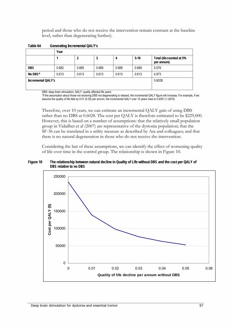
Deep brain stimulation for dystonia and essential tremor 97
period and those who do not receive the intervention remain constant at the baseline level, rather than degenerating further).
Table 64 Generating Incremental QALY’s Year
1 2 3 4 5-10 Total (discounted at 5% per annum)
DBS 0.682 0.685 0.689 0.689 0.689 5.576
No DBS* 0.613 0.613 0.613 0.613 0.613 4.973
Incremental QALY’s 0.6028
DBS: deep brain stimulation; QALY: quality affected life years *If the assumption about those not receiving DBS not degenerating is relaxed, the incremental QALY figure will increase. For example, if we assume the quality of life falls by 0.01 (0.02) per annum, the incremental QALY over 10 years rises to 0.9351 (1.2674)
Therefore, over 10 years, we can estimate an incremental QALY gain of using DBS rather than no DBS at 0.6028. The cost per QALY is therefore estimated to be $229,000. However, this is based on a number of assumptions: that the relatively small population group in Vidailhet et al (2007) are representative of the dystonia population; that the SF-36 can be translated in a utility measure as described by Ara and colleagues; and that there is no natural degeneration in those who do not receive the intervention.
Considering the last of these assumptions, we can identify the effect of worsening quality of life over time in the control group. The relationship is shown in Figure 10.
Figure 10 The relationship between natural decline in Quality of Life without DBS and the cost per QALY of DBS relative to no DBS
0
50000
100000
150000
200000
250000
0 0.01 0.02 0.03 0.04 0.05 0.06
Quality of life decline per annum without DBS
Cost
per
QAL
Y ($
)
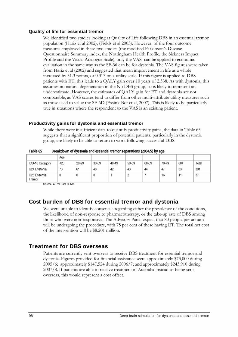
98 Deep brain stimulation for dystonia and essential tremor
Quality of life for essential tremor We identified two studies looking at Quality of Life following DBS in an essential tremor population (Hariz et al 2002), (Fields et al 2003). However, of the four outcome measures employed in these two studies (the modified Parkinson’s Disease Questionnaire Summary index, the Nottingham Health Profile, the Sickness Impact Profile and the Visual Analogue Scale), only the VAS can be applied to economic evaluation in the same way as the SF-36 can be for dystonia. The VAS figures were taken from Hariz et al (2002) and suggested that mean improvement in life as a whole increased by 31.3 points, or 0.313 on a utility scale. If this figure is applied to DBS patients with ET, this leads to a QALY gain over 10 years of 2.538. As with dystonia, this assumes no natural degeneration in the No DBS group, so is likely to represent an underestimate. However, the estimates of QALY gain for ET and dystonia are not comparable, as VAS scores tend to differ from other multi-attribute utility measures such as those used to value the SF-6D (Essink-Bot et al, 2007). This is likely to be particularly true in situations where the respondent to the VAS is an existing patient.
Productivity gains for dystonia and essential tremor While there were insufficient data to quantify productivity gains, the data in Table 65 suggests that a significant proportion of potential patients, particularly in the dystonia group, are likely to be able to return to work following successful DBS.
Table 65 Breakdown of dystonia and essential tremor separations (2004/5) by age Age ICD-10 Category <20 20-29 30-39 40-49 50-59 60-69 70-79 80+ Total G24 Dystonia 73 61 48 42 43 44 47 33 391 G25 Essential Tremor
0 0 0 1 2 7 16 11 37
Source: AIHW Data Cubes
Cost burden of DBS for essential tremor and dystonia We were unable to identify consensus regarding either the prevalence of the conditions, the likelihood of non-response to pharmacotherapy, or the take-up rate of DBS among those who were non-responsive. The Advisory Panel expect that 80 people per annum will be undergoing the procedure, with 75 per cent of these having ET. The total net cost of the intervention will be $8.201 million.
Treatment for DBS overseas Patients are currently sent overseas to receive DBS treatment for essential tremor and dystonia. Figures provided for financial assistance were approximately $73,000 during 2005/6; approximately $147,524 during 2006/7; and approximately $243,910 during 2007/8. If patients are able to receive treatment in Australia instead of being sent overseas, this would represent a cost offset.
Deep brain stimulation for dystonia and essential tremor 99
Discussion
Deep brain stimulation (DBS) has been used widely to treat a variety of movement disorders. DBS is currently MBS-listed for the treatment of Parkinson’s disease. This review reports on the use of DBS in treating two other debilitating conditions, essential tremor and dystonia. Depending on the specific indication patients can receive numerous medications, including botulinum toxin, in treatment of these disorders. However, these treatments have limited success and can become ineffective over time. For medication-refractory patients alternative treatments are limited to pallidotomy and thalamotomy, which are severe interventions that are not recommended for use in Australia. Consequently DBS may in effect be described as an ‘orphan procedure’ for which there is no directly relevant comparator.
The DBS procedure involves two main stages: the implantation of electrodes into the brain (the locations are most often the GPi for dystonia and the VIM for essential tremor); followed hours or days later by the implantation of the implantable pulse generator (IPG). It is the IPG which regulates the stimulation to the brain. The IPG contains a battery and may be switched off. Depending on the intensity of stimulation required the battery needs to be replaced at intervals of between 2 (for dystonia) and 5 (for essential tremor) years.
Many issues for discussion were identified during the completion of this report. Firstly, there was a great variety in the manner in which studies reported the use of DBS for movement disorders. Many studies reported a combination of disorders together (such as Parkinson’s, dystonia and essential tremor). Some studies reported outcomes pre- and post-intervention, while others reported outcomes of stimulation compared to no stimulation (ie the IPG switched off). Where possible, clinically-relevant conditions were reported separately.
Secondly, the quality of the available evidence was limited. One RCT was identified in which patients with primary dystonia were divided into two groups after device implantation. In this study, one group had the IPG switched on, while in the second group the IPG remained off. Outcomes were reported at 3 months. In the absence of high quality evidence, case series were used to assess the safety and effectiveness of DBS. Single case reports of patients with dystonia were included to assess the effectiveness of DBS for dystonia if there was insufficient evidence for specific patient subgroups, such as for the different types of secondary dystonia. Reporting of some of the rarer conditions may have been subject to a degree of bias. In total, 44 studies were included to assess the safety and/or effectiveness of DBS in patients with dystonia and 17 studies were included to assess the safety and/or effectiveness of DBS in patients with essential tremor. There were many studies concerning the use of DBS for essential tremor and dystonia; however, many of these were mixed studies which also considered other conditions such as Parkinson’s disease. The quality of the evidence may limit the generalisability of the results.
Finally, there were numerous issues surrounding the nature of the conditions investigated. Patients with essential tremor or dystonia generally experience a low rate of mortality but a high level of morbidity and decreased quality of life. Their caregivers are also affected and may be unable to work, although the literature on this aspect of the diseases was severely limited. Dystonia and essential tremor may also represent an economic burden on the welfare and hospital systems. Additionally, the Government will presently pay for people to travel overseas (namely France) to receive DBS treatment. Currently it is understood that two individuals are being treated in this manner. Both are children and are twins suffering from severe dystonia. Several patients (especially children) may be unsuitable for this travel which is necessarily
100 Deep brain stimulation for dystonia and essential tremor
frequent as the IPG battery needs replacing every 2 years. This is an unsatisfactory option, as care of rare disorders with rare treatments can be managed successfully within Australia.
For essential tremor and primary dystonia there are patients who are likely to benefit from DBS. DBS should be considered a low volume and invasive procedure, which will not be chosen lightly by patients. Most patients will endure symptoms until they have significant impairment in quality of life (ie the patient is unable to independently feed or toilet). At this point the patient will have failed all alternative treatments, including multiple courses of medication and botulinum toxin in the case of focal dystonia. In some instances, such as for secondary dystonia and tremor as a result of brain insult, the Advisory Panel considered that treatment with DBS should be assessed on a case-by-case basis. An expert committee, comprising a movement disorder surgeon and a neurologist, can assess the extent of disability and the likelihood of benefit.
Deep brain stimulation for dystonia and essential tremor 101
Conclusions
Safety The safety of DBS was assessed from one RCT and 28 studies of level IV evidence for dystonia and from 19 studies of level IV evidence for essential tremor. There was large inter-study variation in the reporting of adverse events; some studies detailed adverse events including side effects experienced during stimulation testing, while others only reported serious adverse events or did not report them at all.
The great majority of adverse events were minor and were resolved simply by changing the stimulation parameters. The most serious adverse events reported in any of the DBS studies were two suicides of dystonia patients that occurred in the postoperative period in one study; however, the contribution of DBS treatment to these events is unclear. Importantly, there were no reported incidences of meningitis. There were two reported cases of haemorrhage, one of which resolved spontaneously (dystonia), whilst the second resulted in mild hemiparesis (essential tremor). There were also two cases of ischaemic stroke in patients with essential tremor. One of these resolved spontaneously, while the outcome of the second was not reported in the study. Reporting upon three dystonia patients who used DBS during pregnancy indicated that DBS is not a barrier to conception or delivery of a healthy baby. None of the women experienced an exacerbation of symptoms during pregnancy.
From the available evidence DBS is a relatively safe treatment for essential tremor and dystonia. Most adverse events are mild and can be resolved completely with or without minor intervention, such as changing the stimulation parameters. Most of the hardware-related complications were resolved by treatment of the local infection or replacement of the affected hardware. In two cases complications led to the removal of all hardware but did not result in any further patient complications. The more severe events are relatively rare and may not affect long-term outcomes; however, many of the studies poorly reported the overall long-term outcomes related to these events.
Effectiveness The effectiveness of DBS was assessed from one RCT and 28 studies of level IV evidence for dystonia and from 19 studies of level IV evidence for essential tremor. The assessment of the effectiveness of DBS for the treatment of dystonia and essential tremor was limited by the relatively small number of individuals who have been analysed, the paucity of high level evidence and the variety of studies included. Evidence was best for primary generalised dystonia, primary focal dystonia and for essential tremor.
Primary generalised dystonia: For a total number of 200 patients with primary generalised dystonia a weighted mean improvement of 60 per cent was observed in the BFMDRS clinical score at the maximal follow-up after DBS of up to 12.6 months (P<0.0001).
Primary focal dystonia: Patients with primary focal dystonia also appeared to benefit from DBS. Seven studies reported mean TWSTRS (total) scores before and after DBS treatment and a meta-analysis revealed that the weighted mean improvement in the total TWSTRS score after DBS (median follow-up: 15 months) was a reduction of 30 points in the 85 point scale (95% CI: 25-36, p<0.00001). All TWSTRS sub-scores (severity,
102 Deep brain stimulation for dystonia and essential tremor
disability and pain) showed a statistically significant improvement after DBS (P<0.00001 for all cases). Patients with primary cervical dystonia noted improvements in TWSTRS scores after DBS treatment compared to before DBS, with a mean percentage improvement in total TWSTRS scores of 62 per cent.
Secondary dystonia: The effectiveness of DBS treatment for secondary dystonia appeared to vary between the different types of dystonia. The evidence was very limited by the small patient numbers for these conditions. Although DBS appears to improve secondary dystonia in the majority of cases, there may be some bias in results due to the inclusion of a number of case reports of single patient outcomes. The limited evidence suggests that DBS may be effective for mixed secondary dystonia, as one group of 26 patients all reported improvements in total BFMDRS score. Although DBS may not be conclusively effective for some disorders, patients with these disorders should not be immediately excluded from potential treatment. The Advisory Panel considered that the final decision to treat a patient with DBS should be made on a case-by-case basis through discussion with a movement disorder surgeon and a neurologist.
Essential tremor: In total, 270 patients were included for essential tremor. For all rating scales used (including the Fahn-Tolosa-Marin tremor rating scale and the activities of daily living) there was a statistically significant improvement in outcomes following DBS compared to baseline pre-surgical scores in all studies. In addition DBS was, where reported, significantly better in testing when the stimulation was on, compared to off or baseline. Meta-analysis of the overall outcomes was not possible as in many cases studies did not clearly define the specific sub-scores which were used.
Certain tremors were also identified which are associated with brain insult (Holmes tremor, post traumatic tremor and tremor secondary to multiple sclerosis). As with secondary dystonia, the evidence was limited to a small number of case reports therefore a conclusive statement on the effectiveness of DBS in the treatment of these conditions is not possible. The Advisory Panel considered that the final decision to treat a patient suffering from tremor as a result of brain insult with DBS should be made on a case-by-case basis through discussion with a movement disorder surgeon and a neurologist.
In summary, DBS is an effective treatment for essential tremor and for primary generalised and primary focal dystonia. For secondary dystonia and secondary tremor, some patients are likely to benefit, but there is no good systematic evidence. It is the Advisory Panel’s opinion that a mechanism should be in place to assess these rarer conditions for treatment with DBS on a case-by-case basis.
Cost-effectiveness Due to limited effectiveness data, the base case in this analysis considers only the resource use of deep brain stimulation (DBS) for essential tremor (ET) and dystonia patients. In the sensitivity analysis, the introduction of the limited existing generic quality of life data is investigated. Productivity benefits associated with return to work are likely to be substantial.
Using a 10-year time horizon, the DBS cost per patient is $91,250 for essential tremor and $136,278 for dystonia. The reason for divergence is because dystonia patients need more frequent battery replacement as the unit is turned on for a greater period of time per day. Using estimates of the total burden of disease in Australia (ie 60 patients per year

Deep brain stimulation for dystonia and essential tremor 103
for ET and 20 patients per year for dystonia), the total cost of DBS in this population is estimated to be $8.201 million.
104 Deep brain stimulation for dystonia and essential tremor
Advice
MSAC has considered the safety, effectiveness and cost effectiveness of deep brain stimulation as end stage treatment for primary and secondary dystonia and essential tremor.
This treatment is indicated where other therapies are insufficient and the patient has severe disability including inability to feed or toilet independently.
DBS is relatively safe in the context of the clinical condition and the net benefit of the treatment.
MSAC considers the treatment is sufficiently effective in these conditions.
Robust information on cost effectiveness is unlikely to emerge but the total cost is acceptable.
MSAC recommends public funding of DBS for primary and secondary dystonia and essential tremor in patients where other therapies are insufficient and the patient has severe disability including inability to feed or toilet independently.
The Minister for Health and Ageing noted MSAC’s advice on 28 August 2008.
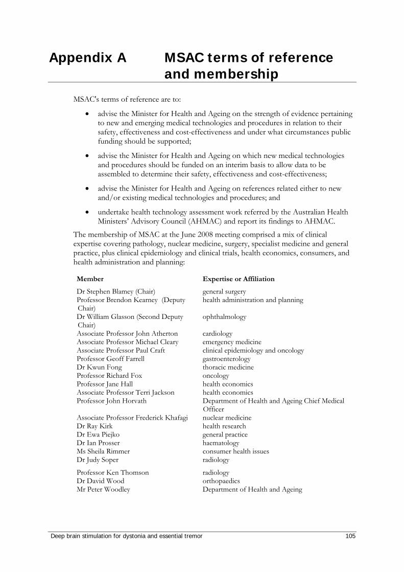
Deep brain stimulation for dystonia and essential tremor 105
Appendix A MSAC terms of reference and membership
MSAC's terms of reference are to:
• advise the Minister for Health and Ageing on the strength of evidence pertaining to new and emerging medical technologies and procedures in relation to their safety, effectiveness and cost-effectiveness and under what circumstances public funding should be supported;
• advise the Minister for Health and Ageing on which new medical technologies and procedures should be funded on an interim basis to allow data to be assembled to determine their safety, effectiveness and cost-effectiveness;
• advise the Minister for Health and Ageing on references related either to new and/or existing medical technologies and procedures; and
• undertake health technology assessment work referred by the Australian Health Ministers’ Advisory Council (AHMAC) and report its findings to AHMAC.
The membership of MSAC at the June 2008 meeting comprised a mix of clinical expertise covering pathology, nuclear medicine, surgery, specialist medicine and general practice, plus clinical epidemiology and clinical trials, health economics, consumers, and health administration and planning:
Member Expertise or Affiliation
Dr Stephen Blamey (Chair) general surgery Professor Brendon Kearney (Deputy Chair)
health administration and planning
Dr William Glasson (Second Deputy Chair)
ophthalmology
Associate Professor John Atherton cardiology Associate Professor Michael Cleary emergency medicine Associate Professor Paul Craft clinical epidemiology and oncology Professor Geoff Farrell gastroenterology Dr Kwun Fong thoracic medicine Professor Richard Fox oncology Professor Jane Hall health economics Associate Professor Terri Jackson health economics Professor John Horvath Department of Health and Ageing Chief Medical
Officer Associate Professor Frederick Khafagi nuclear medicine Dr Ray Kirk health research Dr Ewa Piejko general practice Dr Ian Prosser haematology Ms Sheila Rimmer consumer health issues Dr Judy Soper radiology
Professor Ken Thomson radiology Dr David Wood orthopaedics Mr Peter Woodley Department of Health and Ageing
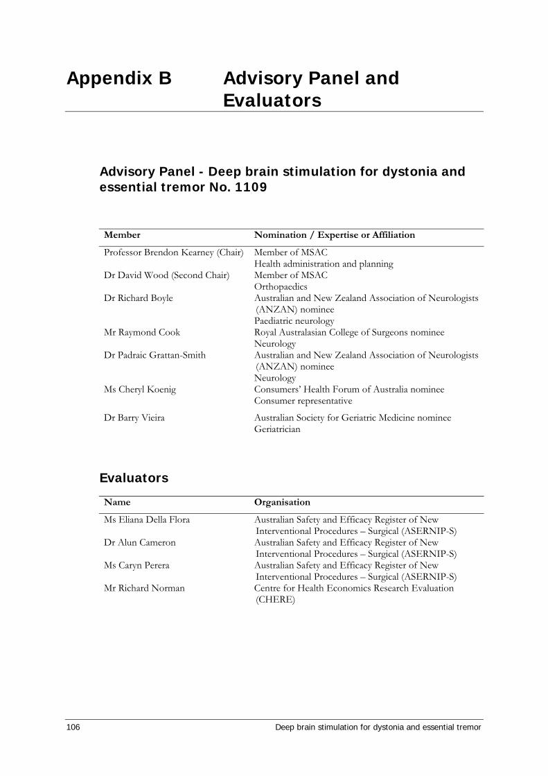
106 Deep brain stimulation for dystonia and essential tremor
Appendix B Advisory Panel and Evaluators
Advisory Panel - Deep brain stimulation for dystonia and essential tremor No. 1109
Member Nomination / Expertise or Affiliation
Professor Brendon Kearney (Chair)
Member of MSAC Health administration and planning
Dr David Wood (Second Chair) Member of MSAC Orthopaedics
Dr Richard Boyle Australian and New Zealand Association of Neurologists (ANZAN) nominee Paediatric neurology
Mr Raymond Cook Royal Australasian College of Surgeons nominee Neurology
Dr Padraic Grattan-Smith Australian and New Zealand Association of Neurologists (ANZAN) nominee Neurology
Ms Cheryl Koenig Consumers’ Health Forum of Australia nominee Consumer representative
Dr Barry Vieira Australian Society for Geriatric Medicine nominee Geriatrician
Evaluators
Name Organisation
Ms Eliana Della Flora Australian Safety and Efficacy Register of New Interventional Procedures – Surgical (ASERNIP-S)
Dr Alun Cameron Australian Safety and Efficacy Register of New Interventional Procedures – Surgical (ASERNIP-S)
Ms Caryn Perera Australian Safety and Efficacy Register of New Interventional Procedures – Surgical (ASERNIP-S)
Mr Richard Norman Centre for Health Economics Research Evaluation (CHERE)
Deep brain stimulation for dystonia and essential tremor 107
Appendix C Studies included in the review
Bittar RG, Yianni J et al, 2005 ‘Deep brain stimulation for generalised dystonia and spasmodic torticollis’, Journal of Clinical Neuroscience, 12(1), 12-16.
Bryant JA, De Salles A et al, 2003. ‘The impact of thalamic stimulation on activities of daily living for essential tremor’, Surgical Neurology, 59(6), 479-484.
Burbaud P, Rougier A et al, 2002. ‘Improvement of severe trunk spasms by bilateral high-frequency stimulation of the motor thalamus in a patient with chorea-acanthocytosis’, Movement Disorders, 17(1), 204-207.
Capelle HH, Grips E et al, 2006. ‘Posttraumatic peripherally-induced dystonia and multifocal deep brain stimulation, case report’, Neurosurgery, 59(3), E702.
Carpenter MA, Pahwa R et al, 1998. ‘Reduction in voice tremor under thalamic stimulation’, Neurology, 50(3), 796-798.
Castelnau P, Cif L et al, 2005. ‘Pallidal stimulation improves pantothenate kinase-associated neurodegeneration’, Annals of Neurology, 57(5), 738-741.
Chang JW, Choi JY et al, 2002. ‘Unilateral globus pallidus internus stimulation improves delayed onset post-traumatic cervical dystonia with an ipsilateral focal basal ganglia lesion’, Journal of Neurology, Neurosurgery & Psychiatry, 73(5), 588-590.
Cif L, El Fertit H et al, 2003. ‘Treatment of dystonic syndromes by chronic electrical stimulation of the internal globus pallidus’, Journal of Neurosurgical Sciences, 47(1), 52-55.
Coubes P, Cif L et al, 2004. ‘Electrical stimulation of the globus pallidus internus in patients with primary generalized dystonia, long-term results’, Journal of Neurosurgery, 101(2), 189-194.
Detante O, Vercueil L et al, 2004. ‘Globus pallidus internus stimulation in primary generalized dystonia, a H215O PET study’, Brain, 127(Pt 8), 1899-1908.
Deutschlander A, Asmus F et al, 2005. ‘Sporadic rapid-onset dystonia-parkinsonism syndrome, failure of bilateral pallidal stimulation’, Movement Disorders, 20(2), 254-257.
Diamond A, Shahed J et al, 2006. ‘Globus pallidus deep brain stimulation in dystonia’, Movement Disorders, 21(5), 692-695.
Eltahawy HA, Saint-Cyr J et al, 2004a. ‘Primary dystonia is more responsive than secondary dystonia to pallidal interventions, outcome after pallidotomy or pallidal deep brain stimulation’, Neurosurgery, 54(3), 613-619.
Eltahawy HA, Saint-Cyr J et al, 2004b. ‘Pallidal deep brain stimulation in cervical dystonia, clinical outcome in four cases’, Canadian Journal of Neurological Sciences, 31(3), 328-332.
108 Deep brain stimulation for dystonia and essential tremor
Fields JA, Troster AI et al, 2003. ‘Neuropsychological and quality of life outcomes 12 months after unilateral thalamic stimulation for essential tremor’, Journal of Neurology, Neurosurgery & Psychiatry, 74(3), 305-311.
Foncke EM, Schuurman PR & Speelman JD, 2006. ‘Suicide after deep brain stimulation of the internal globus pallidus for dystonia’, Neurology, 66(1), 142-143.
Foote KD & Okun MS, 2005. ‘Ventralis intermedius plus ventralis oralis anterior and posterior deep brain stimulation for posttraumatic Holmes tremor, two leads may be better than one, technical note’, Neurosurgery, 56(2, Suppl), E445.
Franzini A, Marras C et al, 2005. ‘Long-term high-frequency bilateral pallidal stimulation for neuroleptic-induced tardive dystonia. Report of two cases’, Journal of Neurosurgery, 102(4), 721-725.
Grips E, Blahak C et al, 2007. ‘Patterns of reoccurrence of segmental dystonia after discontinuation of deep brain stimulation’, Journal of Neurology, Neurosurgery & Psychiatry, 78(3), 318-320.
Guehl D, Cuny E et al, 2007. ‘Deep brain pallidal stimulation for movement disorders in neuroacanthocytosis’, Neurology, 68(2), 160-161.
Hariz GM, Lindberg M & Bergenheim AT, 2002. ‘Impact of thalamic deep brain stimulation on disability and health-related quality of life in patients with essential tremor’, Journal of Neurology, Neurosurgery & Psychiatry, 72(1), 47-52.
Hung SW, Hamani C et al, 2007. ‘Long-term outcome of bilateral pallidal deep brain stimulation for primary cervical dystonia’, Neurology, 68(6), 457-459.
Katayama Y, Fukaya C et al, 2003. ‘Chronic stimulation of the globus pallidus internus for control of primary generalized dystonia’, Acta Neurochirurgica – Supplement, 87, 125-128.
Kiss ZH, Doig K et al, 2004. ‘The Canadian multicenter trial of pallidal deep brain stimulation for cervical dystonia, preliminary results in three patients’, Neurosurgical Focus, 17(1), E5.
Kleiner-Fisman G, Liang GS et al, 2007. ‘Subthalamic nucleus deep brain stimulation for severe idiopathic dystonia, impact on severity, neuropsychological status, and quality of life’, Journal of Neurosurgery, 107(1), 29-36.
Koller WC, Lyons KE et al, 1999. ‘Efficacy of unilateral deep brain stimulation of the VIM nucleus of the thalamus for essential head tremor’, Movement Disorders, 14(5), 847-850.
Koller WC, Lyons KE et al, 2001. ‘Long-term safety and efficacy of unilateral deep brain stimulation of the thalamus in essential tremor’, Movement Disorders, 16(3), 464-468.
Krause M, Fogel W et al, 2004. ‘Pallidal stimulation for dystonia’, Neurosurgery, 55(6), 1361-1368.
Krauss JK, Loher TJ et al, 2002. ‘Pallidal deep brain stimulation in patients with cervical dystonia and severe cervical dyskinesias with cervical myelopathy’, Journal of Neurology, Neurosurgery & Psychiatry, 72(2), 249-256.
Deep brain stimulation for dystonia and essential tremor 109
Krauss JK, Pohle T et al, 1999. ‘Bilateral stimulation of globus pallidus internus for treatment of cervical dystonia’, Lancet 354(9181), 837-838.
Kuncel AM, Cooper SE et al, 2006. ‘Clinical response to varying the stimulus parameters in deep brain stimulation for essential tremor’, Movement Disorders 21(11), 1920-1928.
Kupsch A, Benecke R et al, 2006. ‘Pallidal deep-brain stimulation in primary generalized or segmental dystonia’, New England Journal of Medicine, 355(19), 1978-1990.
Kupsch A, Klaffke S et al, 2003. ‘The effects of frequency in pallidal deep brain stimulation for primary dystonia’. erratum appears in J Neurol. 2004 Aug; 251(8), 1031, Journal of Neurology, 250(10), 1201-1205.
Lee JY & Kondziolka D, 2005. ‘Thalamic deep brain stimulation for management of essential tremor’, Journal of Neurosurgery, 103(3), 400-403.
Legros A, Diakonova N et al, 2004. ‘Accelerometric measurement of involuntary movements during pallidal deep brain stimulation of patients with generalized dystoni’, Brain Research Bulletin, 64(4), 363-369.
Loher TJ, Hasdemir MG et al, 2000. ‘Long-term follow-up study of chronic globus pallidus internus stimulation for posttraumatic hemidystonia’, Journal of Neurosurgery, 92(3), 457-460.
Lyons KE, Pahwa R et al, 1998. ‘Improvements in daily functioning after deep brain stimulation of the thalamus for intractable tremor’, Movement Disorders, 13(4), 690-692.
Mouton S, Xie-Brustolin J et al, 2006. ‘Chorea induced by globus pallidus externus stimulation in a dystonic patient’, Movement Disorders, 21(10), 1771-1773.
Murata J, Kitagawa M et al, 2003. ‘Electrical stimulation of the posterior subthalamic area for the treatment of intractable proximal tremor’, Journal of Neurosurgery, 99(4), 708-715.
Nandi D, Parkin S et al, 2002. ‘Camptocormia treated with bilateral pallidal stimulation, case report’, Neurosurgical Focus, 12(2), ECP2
Nikkhah G, Prokop T et al, 2004. ‘Deep brain stimulation of the nucleus ventralis intermedius for Holmes (rubral) tremor and associated dystonia caused by upper brainstem lesions. Report of two cases’, Journal of Neurosurgery, 100(6), 1079-1083.
Obwegeser AA, Uitti RJ et al, 2000. ‘Thalamic stimulation for the treatment of midline tremors in essential tremor patients’, Neurology, 54(12), 2342-2344.
Pahwa R, Lyons KE et al, 2006. ‘Long-term evaluation of deep brain stimulation of the thalamus’, Journal of Neurosurgery, 104(4), 506-512.
Pahwa R, Lyons KL et al, 1999. ‘Bilateral thalamic stimulation for the treatment of essential tremor’, Neurology, 53(7), 1447-1450.
Paluzzi A, Bain PG et al, 2006a. ‘Pregnancy in dystonic women with in situ deep brain stimulators’, Movement Disorders, 21(5), 695-698.

110 Deep brain stimulation for dystonia and essential tremor
Paluzzi A, Belli A et al, 2006b. ‘Operative and hardware complications of deep brain stimulation for movement disorders’, British Journal of Neurosurgery, 20(5), 290-295.
Papavassiliou E, Rau G et al, 2004. ‘Thalamic deep brain stimulation for essential tremor, relation of lead location to outcome’, Neurosurgery, 54(5), 1120-1129.
Parkin S, Aziz T et al, 2001. ‘Bilateral internal globus pallidus stimulation for the treatment of spasmodic torticollis’, Movement Disorders, 16(3), 489-493.
Putzke JD, Wharen RE et al, 2004. ‘Thalamic deep brain stimulation for essential tremor, recommendations for long-term outcome analysis’, Canadian Journal of Neurological Sciences, 31(3), 333-342.
Roze E, Navarro S et al, 2006. ‘Deep brain stimulation of the globus pallidus for generalized dystonia in GM1 Type 3 gangliosidosis, technical case report’, Neurosurgery, 59(6), E1340.
Starr PA, Turner RS et al, 2006. ‘Microelectrode-guided implantation of deep brain stimulators into the globus pallidus internus for dystonia, techniques, electrode locations, and outcomes’, Journal of Neurosurgery, 104(4), 488-501.
Sydow O, Thobois S et al, 2003 ‘Multicentre European study of thalamic stimulation in essential tremor, A six year follow up’, Journal of Neurology, Neurosurgery and Psychiatry, 74(10), 1387-1391.
Tisch S, Limousin P et al, 2006. ‘Changes in forearm reciprocal inhibition following pallidal stimulation for dystonia’, Neurology, 66(7), 1091-1093.
Tisch S, Rothwell JC et al, 2007. ‘Pallidal stimulation modifies after-effects of paired associative stimulation on motor cortex excitability in primary generalised dystonia’, Experimental Neurology, 206(1), 80-85.
Troster AI, Fields JA et al, 1999. ‘Neuropsychological and quality of life outcome after thalamic stimulation for essential tremor’, Neurology, 53(8), 1774-1780.
Trottenberg T, Volkmann J et al, 2005. ‘Treatment of severe tardive dystonia with pallidal deep brain stimulation’, Neurology, 64(2), 344-346.
Umemura A, Jaggi JL et al, 2004. ‘Pallidal deep brain stimulation for longstanding severe generalized dystonia in Hallervorden-Spatz syndrome, Case report’, Journal of Neurosurgery, 100(4), 706-709.
Ushe M, Mink JW et al, 2006. ‘Postural tremor suppression is dependent on thalamic stimulation frequency’, Movement Disorders, 21(8), 1290-1292.
Vaillancourt DE, Sturman MM et al, 2003. ‘Deep brain stimulation of the VIM thalamic nucleus modifies several features of essential tremor’, Neurology, 61(7), 919-925.
Vercueil L, Pollak P et al, 2001. ‘Deep brain stimulation in the treatment of severe dystonia’, Journal of Neurology, 248(8), 695-700.
Vidailhet M, Vercueil L et al, 2005. ‘Bilateral deep-brain stimulation of the globus pallidus in primary generalized dystonia’, New England Journal of Medicine, 352(5), 459-467.

Deep brain stimulation for dystonia and essential tremor 111
Wang S, Liu X et al, 2006. ‘Use of surface electromyography to assess and select patients with idiopathic dystonia for bilateral pallidal stimulation’, Journal of Neurosurgery, 105(1), 21-25.
Yianni J, Bain P et al, 2003. ‘Globus pallidus internus deep brain stimulation for dystonic conditions, a prospective audit’, Movement Disorders, 18(4), 436-442.
Zorzi G, Marras C et al, 2005. ‘Stimulation of the globus pallidus internus for childhood-onset dystonia’, Movement Disorders 20(9), 1194-1200.
112 Deep brain stimulation for dystonia and essential tremor
Appendix D Studies excluded from the review
Patient overlap with included studies
Coubes P, Roubertie A et al, 2000. ‘Treatment of DYT1-generalised dystonia by stimulation of the internal globus pallidus’, Lancet, 355(9222), 2220-2221.
Eltahawy HA, Feinstein A et al, 2004c. ‘Bilateral globus pallidus internus deep brain stimulation in tardive dyskinesia, a case report’, Movement Disorders, 19(8), 969-972.
Hariz GM, Bergenheim AT et al, 1998. ‘Assessment of ability/disability in patients treated with chronic thalamic stimulation for tremor’, Movement Disorders, 13(1), 78-83.
Krause M, Fogel W et al, 2006. ‘Long-term benefit to pallidal deep brain stimulation in a case of dystonia secondary to pantothenate kinase-associated neurodegeneration’, Movement Disorders, 21(12), 2255-2257.
Liu X, Tailor J et al, 2004. ‘Reversal of hypertonic co-contraction after bilateral pallidal stimulation in generalised dystonia: a clinical and electromyogram case study’, Movement Disorders 19(3), 336-340.
Pillon B, Ardouin C et al, 2006. ‘Preservation of cognitive function in dystonia treated by pallidal stimulation’, Neurology 66(10), 1556-1558.
Trottenberg T, Paul G et al, 2001. ‘Pallidal and thalamic neurostimulation in severe tardive dystonia’, Journal of Neurology, Neurosurgery & Psychiatry 70(4), 557-559.
Yianni J, Bain PG et al, 2003. ‘Post-operative progress of dystonia patients following globus pallidus internus deep brain stimulation’, European Journal of Neurology 10(3), 239-247.
Outcomes reported for mixed conditions other than dystonia and essential tremor
Arnulf I, Bejjani BP et al, 2000. ‘Effect of low and high frequency thalamic stimulation on sleep in patients with Parkinson's disease and essential tremor’, Journal of Sleep Research 9(1), 55-62.
Benabid AL, Pollak P et al, 1991. ‘Long-term suppression of tremor by chronic stimulation of the ventral intermediate thalamic nucleus’, Lancet 337(8738), 403-406.
Blond S, Caparros-Lefebvre D et al, 1992. ‘Control of tremor and involuntary movement disorders by chronic stereotactic stimulation of the ventral intermediate thalamic nucleus’, Journal of Neurosurgery 77(1), 62-68.
Capelle HH, Simpson RK et al, 2005. ‘Long-term deep brain stimulation in elderly patients with cardiac pacemakers’, Journal of Neurosurgery 102(1), 53-59.
Deep brain stimulation for dystonia and essential tremor 113
Halbig TD, Gruber D et al, 2005. ‘Pallidal stimulation in dystonia, effects on cognition, mood, and quality of life’, Journal of Neurology, Neurosurgery & Psychiatry 76(12), 1713-1716.
Hooper J, Taylor R et al, 2002. ‘A prospective study of thalamic deep brain stimulation for the treatment of movement disorders in multiple sclerosis’, British Journal of Neurosurgery 16(2), 102-109.
Hubble JP, Busenbark KL et al, 1997. ‘Effects of thalamic deep brain stimulation based on tremor type and diagnosis’, Movement Disorders 12(3), 337-341.
Koller W, Lang A et al, 1996. ‘Multicenter trial of deep brain stimulation for the treatment of essential tremor and Parkinson's disease’, Neurology 46(2), 49002-49002.
Koller W, Pahwa R et al, 1997. ‘High-frequency unilateral thalamic stimulation in the treatment of essential and parkinsonian tremor’, Annals of Neurology 42(3), 292-299.
Kumar K, Kelly M et al, 1999. ‘Deep brain stimulation of the ventral intermediate nucleus of the thalamus for control of tremors in Parkinson's disease and essential tremor’, Stereotactic & Functional Neurosurgery 72(1), 47-61.
Kumar R, Lozano AM et al, 2003. ‘Long-term follow-up of thalamic deep brain stimulation for essential and parkinsonian tremor’, Neurology 61(11), 1601-1604.
Limousin P, Speelman JD et al, 1999. ‘Multicentre European study of thalamic stimulation in parkinsonian and essential tremor’, Journal of Neurology Neurosurgery and Psychiatry 66(3), 289-296.
Loher TJ, Burgunder J-M et al, 2002. ‘Effect of chronic pallidal deep brain stimulation on off period dystonia and sensory symptoms in advanced Parkinson's disease’, Journal of Neurology Neurosurgery and Psychiatry 73(4), 395-399.
Okun MS, Tagliati M et al, 2005. ‘Management of referred deep brain stimulation failures - A retrospective analysis from 2 movement disorders centers’, Archives of Neurology 62(8), 1250-1255.
Vesper J, Chabardes S et al, 2002. Dual channel deep brain stimulation system (Kinetra) for Parkinson's disease and essential tremor: A prospective multicentre open label clinical study. Journal of Neurology Neurosurgery and Psychiatry 73(3), 275-280.
Non-clinically-relevant outcomes
Anderson TR, Hu B et al, 2006. ‘Selective attenuation of afferent synaptic transmission as a mechanism of thalamic deep brain stimulation-induced tremor arrest’, Journal of Neuroscience 26(3), 841-850.
Bin-Mahfoodh M, Hamani C et al, 2003. ‘Longevity of batteries in internal pulse generators used for deep brain stimulation’, Stereotactic and Functional Neurosurgery 80(1-4), 56-60.
Ceballos-Baumann AO, Boecker H et al, 2001. ‘Thalamic stimulation for essential tremor activates motor and deactivates vestibular cortex’, Neurology 56(10), 1347-1354.
114 Deep brain stimulation for dystonia and essential tremor
Chen CC, Kuhn AA et al, 2006. ‘Oscillatory pallidal local field potential activity correlates with involuntary EMG in dystonia’, Neurology 66(3), 418-420.
Coubes P, Vayssiere N et al, 2002. ‘Deep brain stimulation for dystonia. Surgical technique’, Stereotactic & Functional Neurosurgery 78(3-4), 183-191.
Ferroli P, Franzini A et al, 2004. ‘A simple method to assess accuracy of deep brain stimulation electrode placement: Pre-operative stereotactic CT + postoperative MR image fusion’, Stereotactic and Functional Neurosurgery 82(1), 14-19.
Fields JA & Troster AI, 2001. ‘Electrical stimulation of thalamus for the treatment of essential tremor is related to changes in verbal fluency’, Archives of Clinical Neuropsychology 16(8), 724-724.
Goto S, Mita S et al, 2002. ‘Bilateral pallidal stimulation for cervical dystonia. An optimal paradigm from our experiences’, Stereotactic & Functional Neurosurgery 79(3-4), 221-227.
Gruber D, Haelbig TD et al, 2006. ‘Neuropsychological outcome after combined bilateral pallidal and thalamic stimulation in patients with dystonia and myoclonus dystonia syndrome’, Movement Disorders 21, S685-S685.
Hemm S, Vayssiere N et al, 2004. ‘Evolution of Brain Impedance in Dystonic Patients Treated by GPi Electrical Stimulation’, Neuromodulation 7(2), 67-75.
Kumar R, Dagher A et al, 1999. ‘Globus pallidus deep brain stimulation for generalized dystonia: clinical and PET investigation’, Neurology 53(4), 871-874.
Pralong E, Debatisse D et al, 2003. ‘Effect of deep brain stimulation of GPI on neuronal activity of the thalamic nucleus ventralis oralis in a dystonic patient’, Neurophysiologie Clinique 33(4), 169-173.
Tisch S, Limousin P et al, 2006. ‘Changes in blink reflex excitability after globus pallidus internus stimulation for dystonia’, Movement Disorders 21(10), 1650-1655.
Vayssiere N, Hemm S et al, 2002. ‘Comparison of atlas- and magnetic resonance imaging-based stereotactic targeting of the globus pallidus internus in the performance of deep brain stimulation for treatment of dystonia’, Journal of Neurosurgery 96(4), 673-679.
Vayssiere N, van der GN et al, 2004. ‘Deep brain stimulation for dystonia confirming a somatotopic organization in the globus pallidus internus.[see comment]’, Journal of Neurosurgery 101(2), 181-188.
Yianni J, Bradley K et al, 2005. ‘Effect of GPi DBS on functional imaging of the brain in dystonia’, Journal of Clinical Neuroscience 12(2), 137-141.
Primary dystonia case reports
Alterman RL, Shils JL et al, 2007. ‘Lower stimulation frequency can enhance tolerability and efficacy of pallidal deep brain stimulation for dystonia’, Movement Disorders 22(3), 366-368.
Deep brain stimulation for dystonia and essential tremor 115
Andaluz N, Taha JM et al, 2001. ‘Bilateral pallidal deep brain stimulation for cervical and truncal dystonia’, Neurology 57, 557-558.
Angelini L, Nardocci N et al, 2000. ‘Life-threatening dystonia-dyskinesias in a child: successful treatment with bilateral pallidal stimulation’, Movement Disorders 15(5), 1010-1012.
Boockvar JA, Telfeian A et al, 2000. 'Long-term deep brain stimulation in a patient with essential tremor: clinical response and postmortem correlation with stimulator termination sites in ventral thalamus. Case report’, Journal of Neurosurgery 93,140-144.
Capelle HH, Weigel R et al, 2003. ‘Bilateral pallidal stimulation for blepharospasm-oromandibular dystonia (Meige syndrome)’, Neurology 60, 2017-2018.
Chou KL, Hurtig HI et al, 2005. ‘Bilateral subthalamic nucleus deep brain stimulation in a patient with cervical dystonia and essential tremor’, Movement Disorders 20, 377-380.
Cif L, Valente EM et al, 2004. ‘Deep brain stimulation in myoclonus-dystonia syndrome’, Movement Disorders 19(6), 724-727.
Escamilla-Sevilla F, Minguez-Castellanos A et al, 2002. Unilateral pallidal stimulation for segmental cervical and truncal dystonia: which side? Movement Disorders 17, 1383-1385.
Garcia RP, Muniz DI et al, 2001. ‘Deep brain stimulation holidays in essential tremor’, Journal of Neurology 248, 725-726.
Goto S & Yamada K, 2004. ‘Long term continuous bilateral pallidal stimulation produces stimulation independent relief of cervical dystonia’, Journal of Neurology, Neurosurgery & Psychiatry 75, 1506-1507.
Gross RE, Jones EG et al, 2004. ‘Histological analysis of the location of effective thalamic stimulation for tremor. Case report’, Journal of Neurosurgery 100, 547-552.
Houser M & Waltz T, 2005. ‘Meige syndrome and pallidal deep brain stimulation’, Movement Disorders 20, 1203-1205.
Ilekel S, Zileli M et al, 1999. ‘Unilateral pallidal stimulation in cervical dystonia’, Stereotactic & Functional Neurosurgery 72, 248-252.
Kenney C, Alterman RL et al, 2007. ‘Lower stimulation frequency can enhance tolerability and efficacy of pallidal deep brain stimulation for dystonia’, Movement Disorders 22(3), 366-368.
Katayama Y, Kano T et al, 2005. ‘Difference in surgical strategies between thalamotomy and thalamic deep brain stimulation for tremor control’, Journal of Neurology 252, Suppl-IV22.
Kenney C, Diamond A et al, 2006. ‘Hyperhidrosis due to thalamic deep brain stimulation in a patient with essential tremor’, Movement Disorders 21, S667-S667.
Kronenbuerger M, Fromm C et al, 2006. ‘On-demand deep brain stimulation for essential tremor: a report on four cases’, Movement Disorders 21(3), 401-405.
116 Deep brain stimulation for dystonia and essential tremor
Kronenbuerger M, Fromm C et al, 2004. ‘On demand deep brain stimulation for essential tremor: An approach to avoid tolerance?’, Movement Disorders 19, S292-S293.
Kulisevsky J, Lleo A et al, 2000. ‘Bilateral pallidal stimulation for cervical dystonia: dissociated pain and motor improvement’, Neurology 55(11), 1754-1755.
Lenders MW, Vergouwen MD et al, 2006. ‘Two cases of autosomal recessive generalized dystonia in childhood: 5 year follow-up and bilateral globus pallidus stimulation results’, European Journal of Paediatric Neurology 10(1), 5-9.
Marras C & Broggi G, 2006. ‘Deep brain stimulation of the globus pallidus for generalized dystonia in GM1 type 3 gangliosidosis: Technical case report – Commentary’, Neurosurgery 59(6), E1340.
Muta D, Goto S et al, 2001. ‘Bilateral pallidal stimulation for idiopathic segmental axial dystonia advanced from Meige syndrome refractory to bilateral thalamotomy’, Movement Disorders 16(4), 774-777.
Racette BA, Dowling J et al, 2001. ‘Thalamic stimulation for primary writing tremor’, Journal of Neurology 248(5), 380-382.
Romito LM, Franzini A et al, 2007. ‘Fixed dystonia unresponsive to pallidal stimulation improved by motor cortex stimulation’, Neurology 68(11), 875-876.
Schrader C, Peschel T et al, 2004. ‘Unilateral deep brain stimulation of the internal globus pallidus alleviates tardive dyskinesia’, Movement Disorders 19(5), 583-585.
Sellal F, Hirsch E et al, 1993. ‘A case of symptomatic hemidystonia improved by ventroposterolateral thalamic electrostimulation’, Movement Disorders 8(4), 515-518.
Tann B, Dias L et al, 2006. ‘Utilization of deep brain stimulation as adjunct treatment for control of truncal dystonia in cerebral palsy: a case report’, Developmental Medicine & Child Neurology 48, 46-46.
Tronnier VM, & Fogel W, 2000. ‘Pallidal stimulation for generalized dystonia. Report of three cases’, Journal of Neurosurgery 92(3), 453-456.
Vesper J, Klostermann F et al, 2002. ‘Deep brain stimulation of the globus pallidus internus (GPI) for torsion dystonia--a report of two cases’, Acta Neurochir Suppl 79, 83-88.
Yoon MS, Munz M et al, 1999. ‘Vocal tremor reduction with deep brain stimulation’, Stereotactic & Functional Neurosurgery 72(2-4), 241-244.
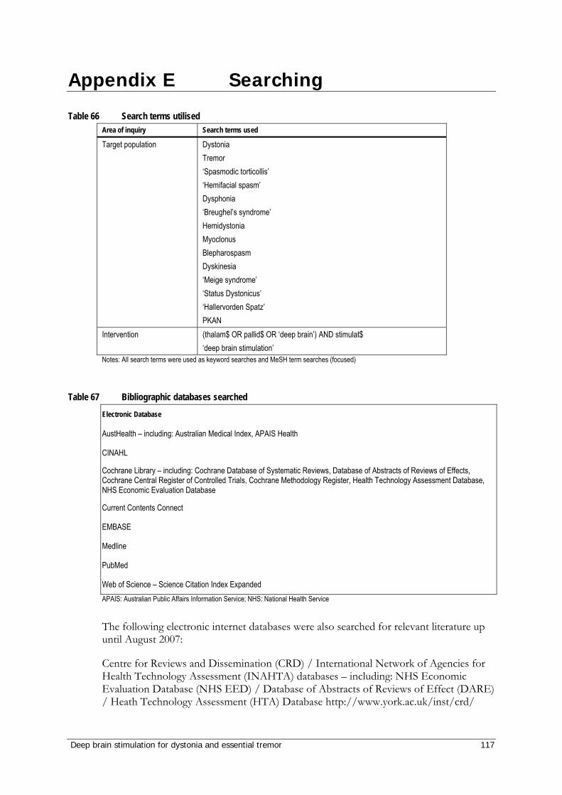
Deep brain stimulation for dystonia and essential tremor 117
Appendix E Searching
Table 66 Search terms utilised Area of inquiry Search terms used
Target population Dystonia Tremor ‘Spasmodic torticollis’ ‘Hemifacial spasm’ Dysphonia ‘Breughel’s syndrome’ Hemidystonia Myoclonus Blepharospasm Dyskinesia ‘Meige syndrome’ ‘Status Dystonicus’ ‘Hallervorden Spatz’ PKAN
Intervention (thalam$ OR pallid$ OR ‘deep brain’) AND stimulat$ ‘deep brain stimulation’
Notes: All search terms were used as keyword searches and MeSH term searches (focused)
Table 67 Bibliographic databases searched Electronic Database
AustHealth – including: Australian Medical Index, APAIS Health
CINAHL
Cochrane Library – including: Cochrane Database of Systematic Reviews, Database of Abstracts of Reviews of Effects, Cochrane Central Register of Controlled Trials, Cochrane Methodology Register, Health Technology Assessment Database, NHS Economic Evaluation Database
Current Contents Connect
EMBASE
Medline
PubMed
Web of Science – Science Citation Index Expanded APAIS: Australian Public Affairs Information Service; NHS: National Health Service
The following electronic internet databases were also searched for relevant literature up until August 2007:
Centre for Reviews and Dissemination (CRD) / International Network of Agencies for Health Technology Assessment (INAHTA) databases – including: NHS Economic Evaluation Database (NHS EED) / Database of Abstracts of Reviews of Effect (DARE) / Heath Technology Assessment (HTA) Database http://www.york.ac.uk/inst/crd/
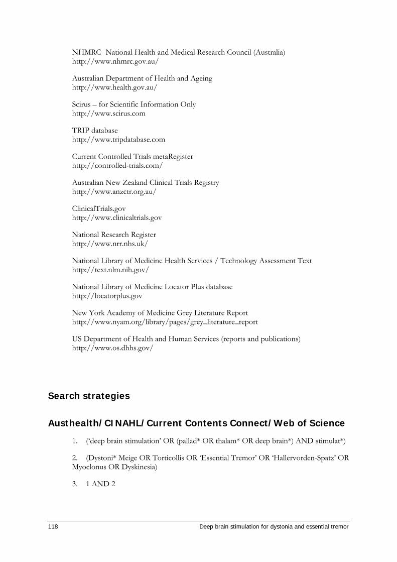
118 Deep brain stimulation for dystonia and essential tremor
NHMRC- National Health and Medical Research Council (Australia) http://www.nhmrc.gov.au/
Australian Department of Health and Ageing http://www.health.gov.au/
Scirus – for Scientific Information Only http://www.scirus.com
TRIP database http://www.tripdatabase.com
Current Controlled Trials metaRegister http://controlled-trials.com/
Australian New Zealand Clinical Trials Registry http://www.anzctr.org.au/
ClinicalTrials.gov http://www.clinicaltrials.gov
National Research Register http://www.nrr.nhs.uk/
National Library of Medicine Health Services / Technology Assessment Text http://text.nlm.nih.gov/
National Library of Medicine Locator Plus database http://locatorplus.gov
New York Academy of Medicine Grey Literature Report http://www.nyam.org/library/pages/grey_literature_report
US Department of Health and Human Services (reports and publications) http://www.os.dhhs.gov/
Search strategies
Austhealth/CINAHL/Current Contents Connect/Web of Science
1. (‘deep brain stimulation’ OR (pallad* OR thalam* OR deep brain*) AND stimulat*)
2. (Dystoni* Meige OR Torticollis OR ‘Essential Tremor’ OR ‘Hallervorden-Spatz’ OR Myoclonus OR Dyskinesia)
3. 1 AND 2
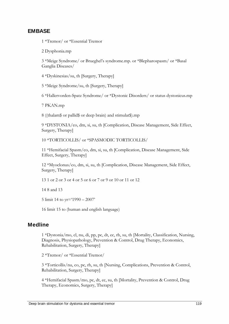
Deep brain stimulation for dystonia and essential tremor 119
EMBASE
1 *Tremor/ or *Essential Tremor
2 Dysphonia.mp
3 *Meige Syndrome/ or Brueghel’s syndrome.mp. or *Blepharospasm/ or *Basal Ganglia Diseases/
4 *Dyskinesias/su, th [Surgery, Therapy]
5 *Meige Syndrome/su, th [Surgery, Therapy]
6 *Hallervorden-Spatz Syndrome/ or *Dystonic Disorders/ or status dystonicus.mp
7 PKAN.mp
8 ((thalam$ or pallid$ or deep brain) and stimulat$).mp
9 *DYSTONIA/co, dm, si, su, th [Complication, Disease Management, Side Effect, Surgery, Therapy]
10 *TORTICOLLIS/ or *SPASMODIC TORTICOLLIS/
11 *Hemifacial Spasm/co, dm, si, su, th [Complication, Disease Management, Side Effect, Surgery, Therapy]
12 *Myoclonus/co, dm, si, su, th [Complication, Disease Management, Side Effect, Surgery, Therapy]
13 1 or 2 or 3 or 4 or 5 or 6 or 7 or 9 or 10 or 11 or 12
14 8 and 13
5 limit 14 to yr=‘1990 – 2007’
16 limit 15 to (human and english language)
Medline
1 *Dystonia/mo, cl, nu, di, pp, pc, dt, ec, rh, su, th [Mortality, Classification, Nursing, Diagnosis, Physiopathology, Prevention & Control, Drug Therapy, Economics, Rehabilitation, Surgery, Therapy]
2 *Tremor/ or *Essential Tremor/
3 *Torticollis/nu, co, pc, rh, su, th [Nursing, Complications, Prevention & Control, Rehabilitation, Surgery, Therapy]
4 *Hemifacial Spasm/mo, pc, dt, ec, su, th [Mortality, Prevention & Control, Drug Therapy, Economics, Surgery, Therapy]
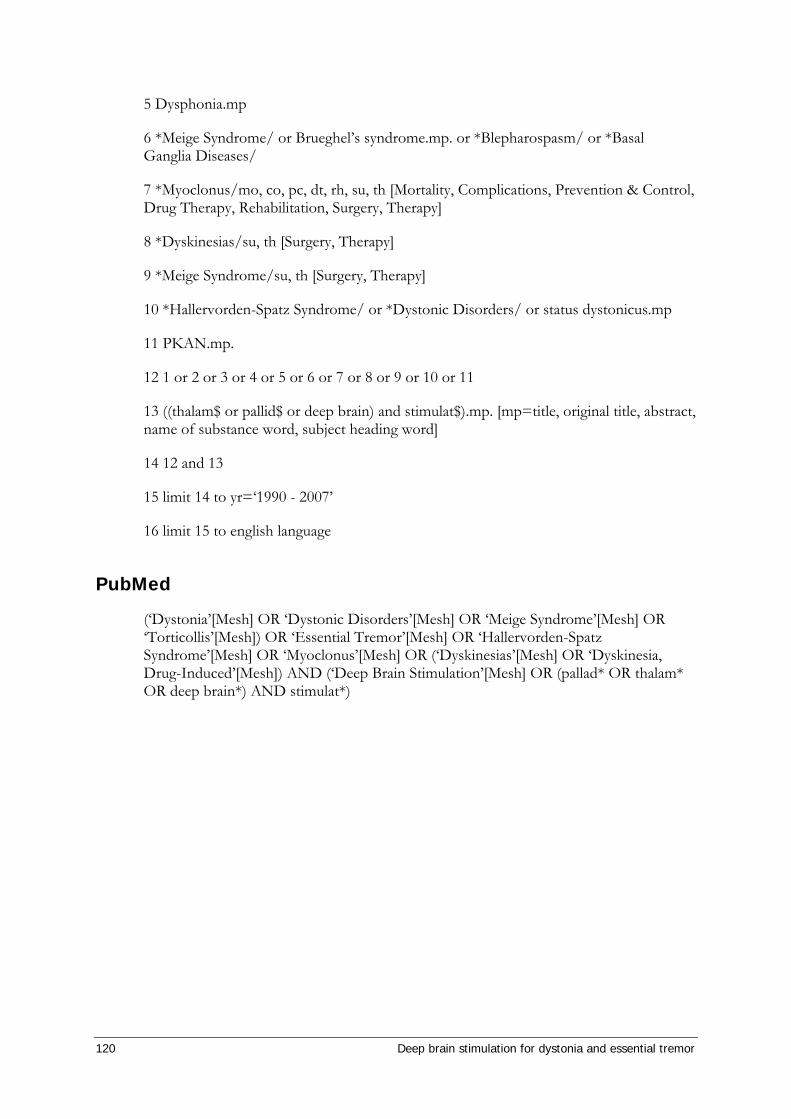
120 Deep brain stimulation for dystonia and essential tremor
5 Dysphonia.mp
6 *Meige Syndrome/ or Brueghel’s syndrome.mp. or *Blepharospasm/ or *Basal Ganglia Diseases/
7 *Myoclonus/mo, co, pc, dt, rh, su, th [Mortality, Complications, Prevention & Control, Drug Therapy, Rehabilitation, Surgery, Therapy]
8 *Dyskinesias/su, th [Surgery, Therapy]
9 *Meige Syndrome/su, th [Surgery, Therapy]
10 *Hallervorden-Spatz Syndrome/ or *Dystonic Disorders/ or status dystonicus.mp
11 PKAN.mp.
12 1 or 2 or 3 or 4 or 5 or 6 or 7 or 8 or 9 or 10 or 11
13 ((thalam$ or pallid$ or deep brain) and stimulat$).mp. [mp=title, original title, abstract, name of substance word, subject heading word]
14 12 and 13
15 limit 14 to yr=‘1990 - 2007’
16 limit 15 to english language
PubMed
(‘Dystonia’[Mesh] OR ‘Dystonic Disorders’[Mesh] OR ‘Meige Syndrome’[Mesh] OR ‘Torticollis’[Mesh]) OR ‘Essential Tremor’[Mesh] OR ‘Hallervorden-Spatz Syndrome’[Mesh] OR ‘Myoclonus’[Mesh] OR (‘Dyskinesias’[Mesh] OR ‘Dyskinesia, Drug-Induced’[Mesh]) AND (‘Deep Brain Stimulation’[Mesh] OR (pallad* OR thalam* OR deep brain*) AND stimulat*)
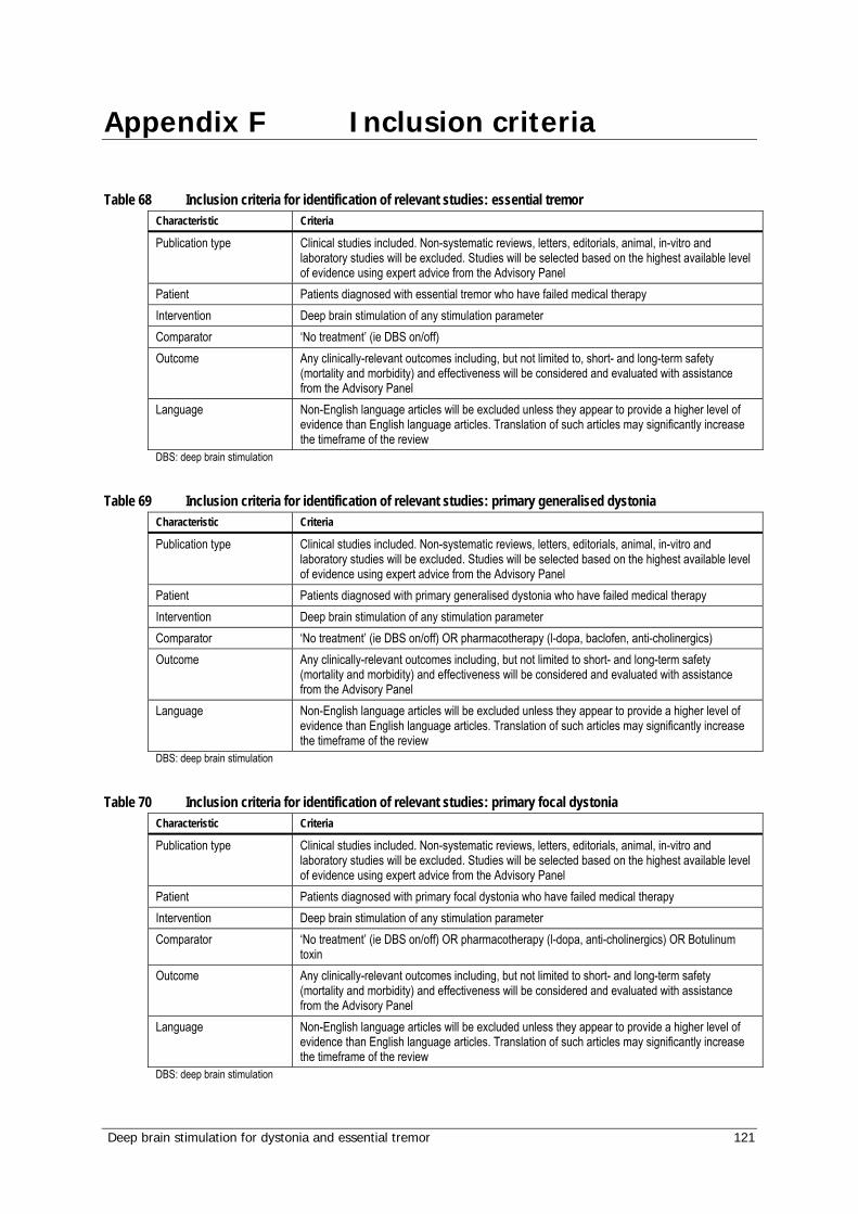
Deep brain stimulation for dystonia and essential tremor 121
Appendix F Inclusion criteria
Table 68 Inclusion criteria for identification of relevant studies: essential tremor
Characteristic Criteria
Publication type Clinical studies included. Non-systematic reviews, letters, editorials, animal, in-vitro and laboratory studies will be excluded. Studies will be selected based on the highest available level of evidence using expert advice from the Advisory Panel
Patient Patients diagnosed with essential tremor who have failed medical therapy Intervention Deep brain stimulation of any stimulation parameter Comparator ‘No treatment’ (ie DBS on/off) Outcome Any clinically-relevant outcomes including, but not limited to, short- and long-term safety
(mortality and morbidity) and effectiveness will be considered and evaluated with assistance from the Advisory Panel
Language Non-English language articles will be excluded unless they appear to provide a higher level of evidence than English language articles. Translation of such articles may significantly increase the timeframe of the review
DBS: deep brain stimulation
Table 69 Inclusion criteria for identification of relevant studies: primary generalised dystonia Characteristic Criteria
Publication type Clinical studies included. Non-systematic reviews, letters, editorials, animal, in-vitro and laboratory studies will be excluded. Studies will be selected based on the highest available level of evidence using expert advice from the Advisory Panel
Patient Patients diagnosed with primary generalised dystonia who have failed medical therapy Intervention Deep brain stimulation of any stimulation parameter Comparator ‘No treatment’ (ie DBS on/off) OR pharmacotherapy (l-dopa, baclofen, anti-cholinergics) Outcome Any clinically-relevant outcomes including, but not limited to short- and long-term safety
(mortality and morbidity) and effectiveness will be considered and evaluated with assistance from the Advisory Panel
Language Non-English language articles will be excluded unless they appear to provide a higher level of evidence than English language articles. Translation of such articles may significantly increase the timeframe of the review
DBS: deep brain stimulation
Table 70 Inclusion criteria for identification of relevant studies: primary focal dystonia Characteristic Criteria
Publication type Clinical studies included. Non-systematic reviews, letters, editorials, animal, in-vitro and laboratory studies will be excluded. Studies will be selected based on the highest available level of evidence using expert advice from the Advisory Panel
Patient Patients diagnosed with primary focal dystonia who have failed medical therapy Intervention Deep brain stimulation of any stimulation parameter Comparator ‘No treatment’ (ie DBS on/off) OR pharmacotherapy (l-dopa, anti-cholinergics) OR Botulinum
toxin Outcome Any clinically-relevant outcomes including, but not limited to short- and long-term safety
(mortality and morbidity) and effectiveness will be considered and evaluated with assistance from the Advisory Panel
Language Non-English language articles will be excluded unless they appear to provide a higher level of evidence than English language articles. Translation of such articles may significantly increase the timeframe of the review
DBS: deep brain stimulation
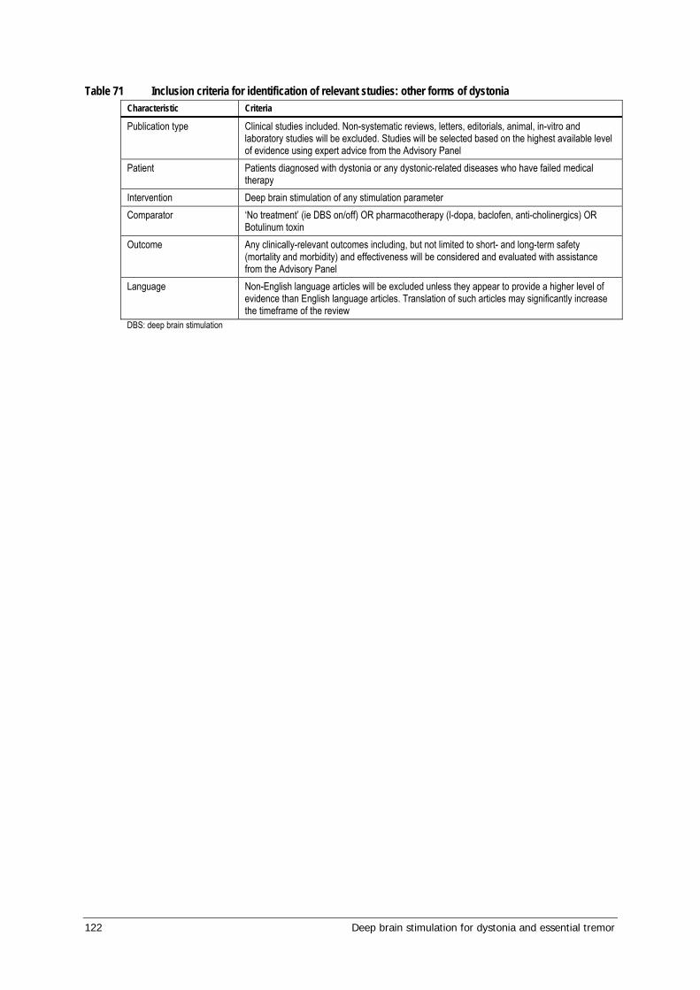
122 Deep brain stimulation for dystonia and essential tremor
Table 71 Inclusion criteria for identification of relevant studies: other forms of dystonia Characteristic Criteria
Publication type Clinical studies included. Non-systematic reviews, letters, editorials, animal, in-vitro and laboratory studies will be excluded. Studies will be selected based on the highest available level of evidence using expert advice from the Advisory Panel
Patient Patients diagnosed with dystonia or any dystonic-related diseases who have failed medical therapy
Intervention Deep brain stimulation of any stimulation parameter Comparator ‘No treatment’ (ie DBS on/off) OR pharmacotherapy (l-dopa, baclofen, anti-cholinergics) OR
Botulinum toxin Outcome Any clinically-relevant outcomes including, but not limited to short- and long-term safety
(mortality and morbidity) and effectiveness will be considered and evaluated with assistance from the Advisory Panel
Language Non-English language articles will be excluded unless they appear to provide a higher level of evidence than English language articles. Translation of such articles may significantly increase the timeframe of the review
DBS: deep brain stimulation
Deep brain stimulation for dystonia and essential tremor 123
Appendix G New studies
Towards the completion of this review a new case series was identified which studied the safety and efficacy of GPi-DBS in patients with medically-refractory cervical dystonia. This study was not discovered as a result of a second systematic search, therefore it is acknowledged that there may be other recently-published studies which were not identified subsequent to the literature search of this review (dated August 2007).
The study (Kiss et al 2007) was a multicentre Canadian study which consecutively recruited patients with clinically diagnosed cervical dystonia. Ten patients (median age 57.5 years, range 47-64 years) were studied, with a median disease duration of 16.5 years (range 19-51 years). Three patients had received previous surgery which was ineffective. Specific stimulation parameters were not defined for use at each centre. Mean and standard deviation DBS parameters at 12 months were: frequency 170 (SD 20) Hz, pulse width 204 (SD 49) µs and amplitude 3.3 (SD 0.7) V.
The primary outcome measure was TWSTRS severity score. This improved from a mean (SD) of 14.7 (4.2) before surgery to 10.6 (4.8) at 6 months and 8.4 (4.4) at 12 months post-operatively (P=0.003). The main secondary outcome measures were TWSTRS disability and pain scores. The disability scores improved from 14.9 (3.8) before surgery to 5.4 (7.0) at 12 months post-surgery (P<0.001) and the pain scores improved from 26.6 (3.6) before surgery to 9.2 (13.1) at 12 months post-operatively (P<0.001).
Further secondary outcomes included Beck depression and quality of life scores (SF-36). Beck depression scores improved from 14.2 (7.2) at baseline to 6.0 (3.5) at 12 months (P<0.001). SF-36 scores improved from 90.9 (11.3) at baseline to 112.9 (19.0) at 12 months (P=0.003).
All adverse events resolved rapidly without permanent morbidity. One patient experienced postoperative dysphagia, which subsided with a decrease in stimulation amplitude yet was still detected at 1 year. One patient suffered from dysarthria which resolved with a change in stimulation parameters. One patient had a transient facial weakness immediately post-operatively; however, this resolved within 3 months. One patient developed shingles after the first side surgery, delaying her second side implant by 1 month. This patient also suffered a chronic subdural fluid collection at the second side surgery (which drained during that procedure) and a very subtle hemiparesis that had resolved completely by 12 months. Two patients had mild swallowing difficulties, while one patient had a decline in phenomic fluency and another in verbal memory.
In summary, with regard to the primary outcome of TWSTRS scores, the mean improvement in dystonia was 43% compared with pre-operative scores. This difference was significant (P=0.003). QOL scores were also significantly improved at 12 months (P=0.003). No significant long-term complications were reported. When combined with patient reported pain and disability scores, the total TWSTRS improved by 59%.
124 Deep brain stimulation for dystonia and essential tremor
Appendix H Current ongoing trials
Websites of clinical trials agencies were searched to identify all relevant ongoing or unpublished clinical trials. These included the Australian Clinical Trials Registry, ClinicalTrials.gov, the National Research Register (UK) and Controlled-Trials.com.
The following six trials may in the future inform the research questions of this report:
Trial identifier: N0263104908 Principal investigator: Dr M Jahanshahi, Institute of Neurology, Queen Square, London, UK Title: Neuropsychological investigation of the impact of deep brain stimulation on executive function, mood, motivation, personality, quality of life and speed of movement in Parkinson's disease and dystonia Suggested end date: September 2007 Comment: This study may inform on the quality of life of dystonia patients.
Trial identifier: N0176113578 Principal investigator: Professor Tipu Z Aziz, Department of Neurosurgery, Radcliffe Infirmary, Oxford, UK Title: Deep Brain Stimulation for Dystonia Suggested end date: April 2007 Comment: This study is recorded as being a randomised controlled trial, although no further detail is provided regarding study methodology such as comparator procedure.
Trial identifier: NCT00331669 Principal investigator: Andreas R Kupsch, MD, Dept. of Neurology, Berlin, Charite, Campus Virchow, Germany Title: Efficacy and Safety of DBS of the GPi in Patients With Tardive Dystonia Suggested end date: December 2010 Comment: With an estimated enrolment of 60 patients, this is possibly a continuation of the study published by Kupsch et al (2006).
Trial identifier: NCT00132340 Principal investigator: J Jankovic, National Institute of Neurological Disorders and Stroke (NINDS), Bethesda, Maryland, United States Title: Deep brain stimulation to treat cervical dystonia Suggested end date: July 2006 Comment: Electrodes will be placed either in the globus pallidus interna (GPi) or in the subthalamic nucleus (STN) to investigate which anatomical site provides optimal improvement.
Trial identifier: NCT00142259 Principal investigator: Jens Volkmann, MD, PhD, Dept. Neurology, UKSH Campus, Kiel, Germany Title: Efficacy and Safety of DBS of the GPi in Patients With Primary Generalized and Segmental Dystonia Suggested end date: August 2009 Comment: A randomised, placebo-controlled trial with an estimated enrolment of 40 participants.

Deep brain stimulation for dystonia and essential tremor 125
Trial identifier: NCT00142259 Principal investigator: Hans Speelman, PhD, Academisch Medisch Centrum - Universiteit van Amsterdam, The Netherlands Title: Bilateral Internal Pallidum Stimulation in Primary Generalized Dystonia Suggested end date: December 2007 Comment: A randomised, placebo-controlled, multi-centre study, which current records show is recruiting patients.
Overall there were many ongoing trials investigating the use of DBS in Parkinson’s disease. Many of the identified trials were not comparative in nature and therefore will not add significantly to the currently available evidence.
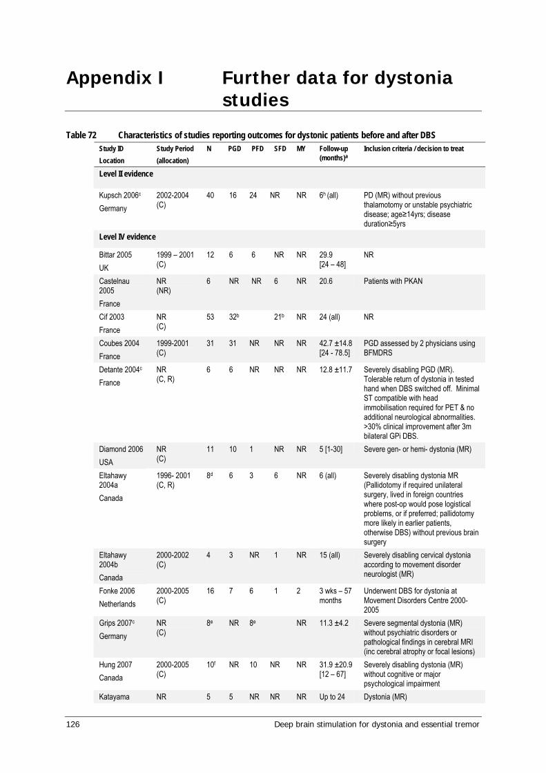
126 Deep brain stimulation for dystonia and essential tremor
Appendix I Further data for dystonia studies
Table 72 Characteristics of studies reporting outcomes for dystonic patients before and after DBS Study ID Location
Study Period (allocation)
N PGD PFD SFD MY Follow-up (months)a
Inclusion criteria / decision to treat
Level II evidence
Kupsch 2006c Germany
2002-2004 (C)
40 16 24 NR NR 6h (all) PD (MR) without previous thalamotomy or unstable psychiatric disease; age≥14yrs; disease duration≥5yrs
Level IV evidence
Bittar 2005 UK
1999 – 2001 (C)
12 6 6 NR NR 29.9 [24 – 48]
NR
Castelnau 2005 France
NR (NR)
6 NR NR 6 NR 20.6 Patients with PKAN
Cif 2003 France
NR (C)
53 32b 21b NR 24 (all) NR
Coubes 2004 France
1999-2001 (C)
31 31 NR NR NR 42.7 ±14.8 [24 - 78.5]
PGD assessed by 2 physicians using BFMDRS
Detante 2004c
France NR (C, R)
6 6 NR NR NR 12.8 ±11.7 Severely disabling PGD (MR). Tolerable return of dystonia in tested hand when DBS switched off. Minimal ST compatible with head immobilisation required for PET & no additional neurological abnormalities. >30% clinical improvement after 3m bilateral GPi DBS.
Diamond 2006 USA
NR (C)
11 10 1 NR NR 5 [1-30] Severe gen- or hemi- dystonia (MR)
Eltahawy 2004a Canada
1996- 2001 (C, R)
8d 6 3 6 NR 6 (all) Severely disabling dystonia MR (Pallidotomy if required unilateral surgery, lived in foreign countries where post-op would pose logistical problems, or if preferred; pallidotomy more likely in earlier patients, otherwise DBS) without previous brain surgery
Eltahawy 2004b Canada
2000-2002 (C)
4 3 NR 1 NR 15 (all) Severely disabling cervical dystonia according to movement disorder neurologist (MR)
Fonke 2006 Netherlands
2000-2005 (C)
16 7 6 1 2 3 wks – 57 months
Underwent DBS for dystonia at Movement Disorders Centre 2000-2005
Grips 2007c Germany
NR (C)
8e NR 8e NR 11.3 ±4.2 Severe segmental dystonia (MR) without psychiatric disorders or pathological findings in cerebral MRI (inc cerebral atrophy or focal lesions)
Hung 2007 Canada
2000-2005 (C)
10f NR 10 NR NR 31.9 ±20.9 [12 – 67]
Severely disabling dystonia (MR) without cognitive or major psychological impairment
Katayama NR 5 5 NR NR NR Up to 24 Dystonia (MR)
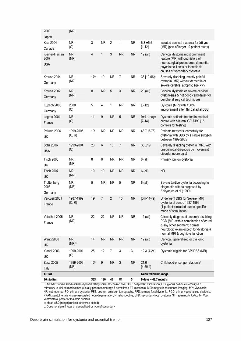
Deep brain stimulation for dystonia and essential tremor 127
2003 Japan
(NR)
Kiss 2004 Canada
NR (C)
3 NR 2 1 NR 6.3 ±5.5 [1-12]
Isolated cervical dystonia for ≥5 yrs (MR) (part of larger 10 patient study)
Kleiner-Fisman 2007 USA
NR (NR)
4 1 3 NR NR 12 (all) Cervical dystonia most prominent feature (MR) without history of neurosurgical procedures, dementia, psychiatric illness or identifiable causes of secondary dystonia
Krause 2004 Germany
NR (NR)
17g 10 NR 7 NR 36 [12-66]g Severely disabling, mostly painful dystonia (MR) without dementia or severe cerebral atrophy; age <75
Krauss 2002 Germany
NR (NR)
8 NR
5
3
NR
20 (all) Cervical dystonia or severe cervical dyskinesias & not good candidates for peripheral surgical techniques
Kupsch 2003 Germany
2000 (C)
5 4 1 NR NR [3-12] Dystonia (MR) with ≥30% improvement after 1hr palladial DBS
Legros 2004 France
NR (C)
11 9 NR 5 NR 9±1.1 days [7-14]
Dystonic patients treated in medical centre with bilateral GPi DBS (+5 controls for testing)
Paluzzi 2006 UK
1999-2005 (C, R)
19i NR NR NR NR 43.7 [6-78] Patients treated successfully for dystonia with DBS by a single surgeon between 1999-2005
Starr 2006 USA
1999-2004 (C)
23 6 10 7 NR 35 ±19 Severely disabling dystonia (MR), with unequivocal diagnosis by movement disorder neurologist
Tisch 2006 UK
NR (NR)
8 8 NR NR NR 6 (all) Primary torsion dystonia
Tisch 2007 UK
NR (NR)
10 10 NR NR NR 6 (all) NR
Trottenberg 2005 Germany
NR (NR)
5 NR NR 5 NR 6 (all) Severe tardive dystonia according to diagnostic criteria proposed by Adityanjee et al (1999)
Vercueil 2001 France
1987-1999 (C, R)
19j 7 2 10 NR [6m-11yrs] Underwent DBS for Severe (MR) dystonia at centre 1987-1999 (1 patient excluded due to specific mode of stimulation)
Vidailhet 2005 France
NR (NR)
22 22 NR NR NR 12 (all) Clinically diagnosed severely disabling PGD (MR) with a combination of crural & any other segment; normal neurologic exam except for dystonia & normal MRI & cognitive function
Wang 2006 UK
NR (NR)b
14 NR NR NR NR 12 (all) Cervical, generalised or dystonic dystonia
Yianni 2003 UK
1999-2001 (C)
25 12 7 3 3 12.3 [4-24] Dystonia eligible for GPi DBS (MR)
Zorzi 2005 Italy
1999-2003 (NR)
12k 9 NR 3 NR 21.6 [4-50.4]
Childhood-onset gen dystoniak
TOTAL Mean follow-up range 26 studies 353 180 45 84 5 9 days – 43.7 months BFMDRS: Burke-Fahn-Marsden dystonia rating scale; C: consecutive; DBS: deep brain stimulation; GPi: globus pallidus internus; MR: refractory to trialled medications (usually pharmacotherapy & sometimes BT injections); MRI: magnetic resonance imaging; MY; Myoclonic; NR: not reported; PD: primary dystonia; PET: positron emission tomography; PFD: primary focal dystonia; PGD: primary generalised dystonia; PKAN: pantothenate kinase-associated neurodegeneration; R: retrospective; SFD: secondary focal dystonia; ST: spasmodic torticollis; VLp: ventrolateral posterior thalamic nucleus a: Mean ±SD [range] (unless otherwise stated) b: Does not state if focal or generalised or type of secondary

128 Deep brain stimulation for dystonia and essential tremor
c: Also examines outcomes for patients with DBS switched on vs. off d: Plus 7 patients who underwent pallidotomy e: All patients had focal dystonia but does not state if primary or secondary f: The first four patients in this study were also reported in Eltahawy et al (2004a) g: Patient #12 came from foreign country and was transferred to a hospital closer to home so was not included in all outcomes h: In half the patients (20) DBS was switched on after 1 wk, but was switched on after 3 m in the other half (20) i: This study was included for safety only. 19 dystonic patients were part of a larger cohort of 96 patients j: Not performed in 2 GPi adverse & 5 VLp k: 2 patients with PGD & 1 patient with SGD had status dystonicus
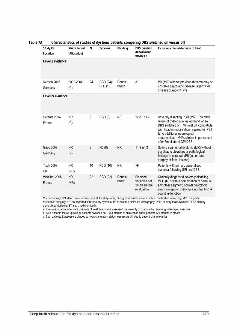
Deep brain stimulation for dystonia and essential tremor 129
Table 73 Characteristics of studies of dystonic patients comparing DBS switched on versus off Study ID Location
Study Period (Allocation)
N Type (n) Blinding DBS duration at evaluation (months)
Inclusion criteria/ decision to treat
Level II evidence
Kupsch 2006 Germany
2002-2004 (C)
24 PGD (24); PFD (16)
Double-blinda
3b PD (MR) without previous thalamotomy or unstable psychiatric disease; age≥14yrs; disease duration≥5yrs
Level IV evidence
Detante 2004 France
NR (C)
6 PGD (6) NR 12.8 ±11.7 Severely disabling PGD (MR). Tolerable return of dystonia in tested hand when DBS switched off. Minimal ST compatible with head immobilisation required for PET & no additional neurological abnormalities. >30% clinical improvement after 3m bilateral GPi DBS.
Grips 2007 Germany
NR (C)
8 FD (8) NR 11.3 ±4.2 Severe segmental dystonia (MR) without psychiatric disorders or pathological findings in cerebral MRI (ie cerebral atrophy or focal lesions)
Tisch 2007 UK
NR (NR)
10 PDG (10) NR >6 Patients with primary generalised dystonia following GPi and DBS
Vidailhet 2005 France
NR (NR)
22 PGD (22) Double-blindc
Electrical variables set 10 hrs before evaluation
Clinically diagnosed severely disabling PGD (MR) with a combination of crural & any other segment; normal neurologic exam except for dystonia & normal MRI & cognitive function
C: continuous; DBS: deep brain stimulation; FD: focal dystonia; GPi: globus pallidus internus; MR: medication refractory; MRI: magnetic resonance imaging; NR: not reported; PD: primary dystonia; PET: positron emission tomography; PFD: primary focal dystonia; PGD: primary generalised dystonia; ST: spasmodic torticollis a: Two investigators who were unaware of treatment status assessed the severity of dystonia by reviewing videotaped sessions b: Also 6-month follow-up with all patients switched on - ie 3 months of stimulation sham patients & 6 months in others c: Both patients & assessors blinded to neurostimulation status. Assessors blinded to patient characteristics
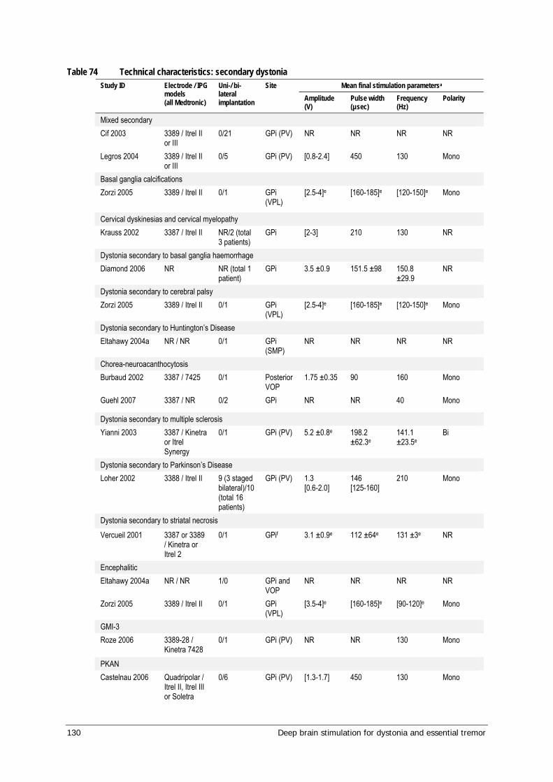
130 Deep brain stimulation for dystonia and essential tremor
Table 74 Technical characteristics: secondary dystonia Mean final stimulation parametersa Study ID Electrode / IPG
models (all Medtronic)
Uni-/ bi-lateral implantation
Site Amplitude (V)
Pulse width (µsec)
Frequency (Hz)
Polarity
Mixed secondary Cif 2003 3389 / Itrel II
or III 0/21 GPi (PV) NR NR NR NR
Legros 2004 3389 / Itrel II or III
0/5 GPi (PV) [0.8-2.4] 450 130 Mono
Basal ganglia calcifications Zorzi 2005
3389 / Itrel II 0/1 GPi (VPL)
[2.5-4]e [160-185]e [120-150]e Mono
Cervical dyskinesias and cervical myelopathy Krauss 2002 3387 / Itrel II NR/2 (total
3 patients) GPi [2-3] 210 130 NR
Dystonia secondary to basal ganglia haemorrhage Diamond 2006 NR NR (total 1
patient) GPi 3.5 ±0.9 151.5 ±98 150.8
±29.9 NR
Dystonia secondary to cerebral palsy Zorzi 2005 3389 / Itrel II 0/1 GPi
(VPL) [2.5-4]e [160-185]e [120-150]e Mono
Dystonia secondary to Huntington’s Disease Eltahawy 2004a NR / NR 0/1 GPi
(SMP) NR NR NR NR
Chorea-neuroacanthocytosis Burbaud 2002 3387 / 7425 0/1 Posterior
VOP 1.75 ±0.35 90 160 Mono
Guehl 2007 3387 / NR 0/2 GPi NR NR 40 Mono
Dystonia secondary to multiple sclerosis Yianni 2003 3387 / Kinetra
or Itrel Synergy
0/1 GPi (PV) 5.2 ±0.8e 198.2 ±62.3e
141.1 ±23.5e
Bi
Dystonia secondary to Parkinson’s Disease Loher 2002 3388 / Itrel II 9 (3 staged
bilateral)/10 (total 16 patients)
GPi (PV) 1.3 [0.6-2.0]
146 [125-160]
210 Mono
Dystonia secondary to striatal necrosis
Vercueil 2001 3387 or 3389 / Kinetra or Itrel 2
0/1 GPif 3.1 ±0.9e 112 ±64e 131 ±3e NR
Encephalitic Eltahawy 2004a NR / NR 1/0 GPi and
VOP NR NR NR NR
Zorzi 2005 3389 / Itrel II 0/1 GPi (VPL)
[3.5-4]e [160-185]e [90-120]e Mono
GMI-3 Roze 2006 3389-28 /
Kinetra 7428 0/1 GPi (PV) NR NR 130 Mono
PKAN Castelnau 2006 Quadripolar /
Itrel II, Itrel III or Soletra
0/6 GPi (PV) [1.3-1.7] 450 130 Mono
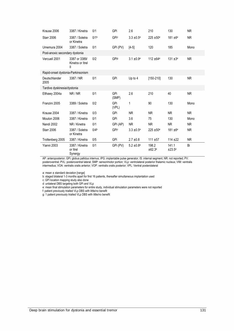
Deep brain stimulation for dystonia and essential tremor 131
Krause 2006 3387 / Kinetra 0/1 GPi 2.6 210 130 NR
Starr 2006 3387 / Soletra or Kinetra
0/1b GPic 3.3 ±0.5e 225 ±50e 181 ±6e NR
Umemura 2004 3387 / Soletra 0/1 GPi (PV) [4-5] 120 185 Mono Post-anoxic secondary dystonia Vercueil 2001 3387 or 3389/
Kinetra or Itrel II
0/2 GPig 3.1 ±0.9e 112 ±64e 131 ±3e NR
Rapid-onset dystonia-Parkinsonism Deutschlander 2005
3387 / NR 0/1 GPi Up to 4 [150-210] 130 NR
Tardive dyskinesia/dystonia Elthawy 2004a NR / NR 0/1 GPi
(SMP) 2.6 210 40 NR
Franzini 2005 3389 / Soletra 0/2 GPi (VPL)
1 90 130 Mono
Krause 2004 3387 / Kinetra 0/3 GPi NR NR NR NR Mouton 2006 3387 / Kinetra 0/1 GPi 3.6 75 130 Mono Nandi 2002 NR / Kinetra 0/1 GPi (AP) NR NR NR NR Starr 2006 3387 / Soletra
or Kinetra 0/4b GPic 3.3 ±0.5e 225 ±50e 181 ±6e NR
Trottenberg 2005 3387 / Kinetra 0/5 GPi 2.7 ±0.8 111 ±57 114 ±22 NR Yianni 2003 3387 / Kinetra
or Itrel Synergy
0/1 GPi (PV) 5.2 ±0.8e 198.2 ±62.3e
141.1 ±23.5e
Bi
AP: anteroposterior; GPi: globus pallidus internus; IPG: implantable pulse generator; IS: internal segment; NR: not reported; PV: posteroventral; PVL: posteroventral lateral; SMP: sensorimotor portion; VLp: ventrolateral posterior thalamic nucleus; VIM: ventralis intermedius; VOA: ventralis oralis anterior; VOP: ventralis oralis posterior; VPL: Ventral posterolateral a: mean ± standard deviation [range] b: staged bilateral 1-3 months apart for first 16 patients, thereafter simultaneous implantation used c: GPi location mapping study also done d: unilateral DBS targeting both GPi and VLp e: mean final stimulation parameters for entire study, individual stimulation parameters were not reported f: patient previously trialled VLp DBS with little/no benefit g: 1 patient previously trialled VLp DBS with little/no benefit
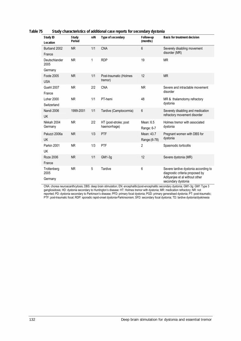
132 Deep brain stimulation for dystonia and essential tremor
Table 75 Study characteristics of additional case reports for secondary dystonia Study ID Location
Study Period
n/N Type of secondary Follow-up
(months) Basis for treatment decision
Burband 2002 France
NR 1/1 CNA 6 Severely disabling movement disorder (MR)
Deutschlander 2005 Germany
NR 1 RDP 19 MR
Foote 2005 USA
NR 1/1 Post-traumatic (Holmes tremor)
12 MR
Guehl 2007 France
NR 2/2 CNA NR Severe and intractable movement disorder
Loher 2000 Switzerland
NR 1/1 PT-hemi 48 MR & thalamotomy refractory dystonia
Nandi 2006 UK
1999-2001 1/1 Tardive (Camptocormia) 6 Severely disabling and medication refractory movement disorder
Nikkah 2004 Germany
NR 2/2 HT (post-stroke; post haemorrhage)
Mean: 6.5 Range: 6-7
Holmes tremor with associated dystonia
Paluzzi 2006a UK
NR 1/3 PTF Mean: 43.7 Range:(6-78)
Pregnant women with DBS for dystonia
Parkin 2001 UK
NR 1/3 PTF 2 Spasmodic torticollis
Roze 2006 France
NR 1/1 GM1-3g 12 Severe dystonia (MR)
Trottenberg 2005 Germany
NR 5 Tardive 6 Severe tardive dystonia according to diagnostic criteria proposed by Adityanjee et al without other secondary dystonia
CNA: chorea neuroacanthcytosis; DBS: deep brain stimulation; EN: encephalitic/post-encephalitic secondary dystonia; GM1-3g: GM1 Type 3 gangliosidosis; HD: dystonia secondary to Huntington’s disease: HT: Holmes tremor with dystonia; MR: medication refractory; NR: not reported; PD: dystonia secondary to Parkinson’s disease; PFD: primary focal dystonia; PGD: primary generalised dystonia; PT: post-traumatic; PTF: post-traumatic focal; RDP: sporadic rapid-onset dystonia-Parkinsonism; SFD: secondary focal dystonia; TD: tardive dystonia/dyskinesia
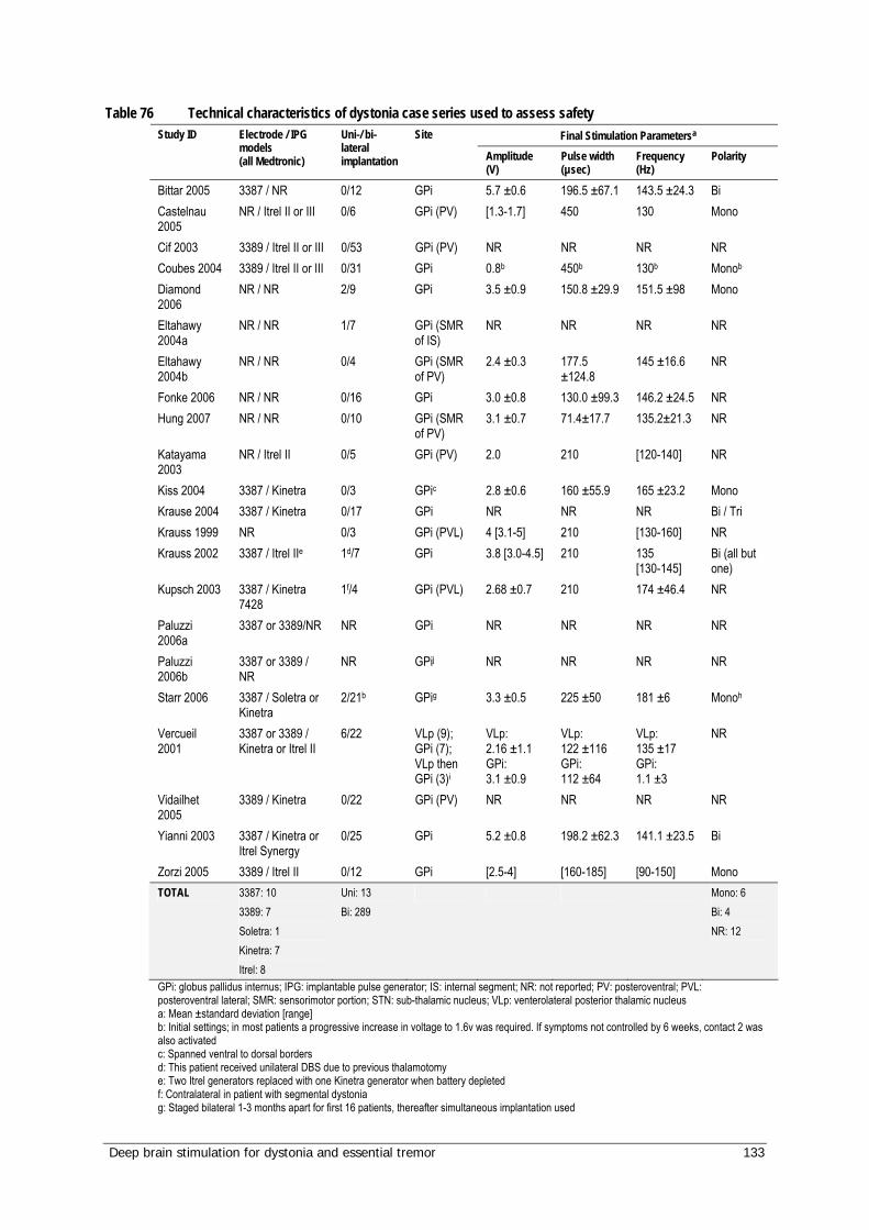
Deep brain stimulation for dystonia and essential tremor 133
Table 76 Technical characteristics of dystonia case series used to assess safety Final Stimulation Parametersa Study ID Electrode / IPG
models (all Medtronic)
Uni-/ bi-lateral implantation
Site
Amplitude (V)
Pulse width (µsec)
Frequency (Hz)
Polarity
Bittar 2005 3387 / NR 0/12 GPi 5.7 ±0.6 196.5 ±67.1 143.5 ±24.3 Bi Castelnau 2005
NR / Itrel II or III 0/6 GPi (PV) [1.3-1.7] 450 130 Mono
Cif 2003 3389 / Itrel II or III 0/53 GPi (PV) NR NR NR NR Coubes 2004 3389 / Itrel II or III 0/31 GPi 0.8b 450b 130b Monob Diamond 2006
NR / NR 2/9 GPi 3.5 ±0.9 150.8 ±29.9 151.5 ±98 Mono
Eltahawy 2004a
NR / NR 1/7 GPi (SMR of IS)
NR NR NR NR
Eltahawy 2004b
NR / NR 0/4 GPi (SMR of PV)
2.4 ±0.3 177.5 ±124.8
145 ±16.6 NR
Fonke 2006 NR / NR 0/16 GPi 3.0 ±0.8 130.0 ±99.3 146.2 ±24.5 NR Hung 2007 NR / NR 0/10 GPi (SMR
of PV) 3.1 ±0.7 71.4±17.7 135.2±21.3 NR
Katayama 2003
NR / Itrel II 0/5 GPi (PV) 2.0 210 [120-140] NR
Kiss 2004 3387 / Kinetra 0/3 GPic 2.8 ±0.6 160 ±55.9 165 ±23.2 Mono Krause 2004 3387 / Kinetra 0/17 GPi NR NR NR Bi / Tri Krauss 1999 NR 0/3 GPi (PVL) 4 [3.1-5] 210 [130-160] NR Krauss 2002 3387 / Itrel IIe 1d/7 GPi 3.8 [3.0-4.5] 210 135
[130-145] Bi (all but one)
Kupsch 2003 3387 / Kinetra 7428
1f/4 GPi (PVL) 2.68 ±0.7 210 174 ±46.4 NR
Paluzzi 2006a
3387 or 3389/NR NR GPi NR NR NR NR
Paluzzi 2006b
3387 or 3389 / NR
NR GPij NR NR NR NR
Starr 2006 3387 / Soletra or Kinetra
2/21b GPig 3.3 ±0.5 225 ±50 181 ±6 Monoh
Vercueil 2001
3387 or 3389 / Kinetra or Itrel II
6/22 VLp (9); GPi (7); VLp then GPi (3)i
VLp: 2.16 ±1.1 GPi: 3.1 ±0.9
VLp: 122 ±116 GPi: 112 ±64
VLp: 135 ±17 GPi: 1.1 ±3
NR
Vidailhet 2005
3389 / Kinetra 0/22 GPi (PV) NR NR NR NR
Yianni 2003 3387 / Kinetra or Itrel Synergy
0/25 GPi 5.2 ±0.8 198.2 ±62.3 141.1 ±23.5 Bi
Zorzi 2005 3389 / Itrel II 0/12 GPi [2.5-4] [160-185] [90-150] Mono TOTAL 3387: 10
3389: 7 Soletra: 1 Kinetra: 7 Itrel: 8
Uni: 13 Bi: 289
Mono: 6 Bi: 4 NR: 12
GPi: globus pallidus internus; IPG: implantable pulse generator; IS: internal segment; NR: not reported; PV: posteroventral; PVL: posteroventral lateral; SMR: sensorimotor portion; STN: sub-thalamic nucleus; VLp: venterolateral posterior thalamic nucleus a: Mean ±standard deviation [range] b: Initial settings; in most patients a progressive increase in voltage to 1.6v was required. If symptoms not controlled by 6 weeks, contact 2 was also activated c: Spanned ventral to dorsal borders d: This patient received unilateral DBS due to previous thalamotomy e: Two Itrel generators replaced with one Kinetra generator when battery depleted f: Contralateral in patient with segmental dystonia g: Staged bilateral 1-3 months apart for first 16 patients, thereafter simultaneous implantation used

134 Deep brain stimulation for dystonia and essential tremor
h: 28 leads unipolar single electrodes; 15: unipolar 2 adjacent electrodes i: In these 3 patients electrodes were implanted into the VLp initially, but a second operation performed implanting electrodes into the GPi due to lack of effectiveness in the VLp j: Dystonia (19): GPi. ET (4): Vop, VIM, ZI)
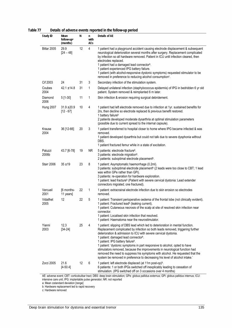
Deep brain stimulation for dystonia and essential tremor 135
Table 77 Details of adverse events reported in the follow-up period Study ID Mean
follow-upa (months)
N n with AEs
Details of AE
Bittar 2005 29.9 [24 – 48]
12 4 1 patient had a playground accident causing electrode displacement & subsequent neurological deterioration several months after surgery. Replacement complicated by infection so all hardware removed. Patient in ICU until infection cleared, then electrodes replaced. 1 patient had a damaged lead connectorb. 1 patient experienced IPG battery failure. 1 patient (with alcohol-responsive dystonic symptoms) requested stimulator to be removed in preference to reducing alcohol consumptionc.
Cif 2003 24 31 3 Secondary infection of the stimulation system. Coubes 2004
42.1 ±14.8 31 1 Delayed unilateral infection (staphylococcus epidermis) of IPG in bedridden 6 yr old patient. System removed & reimplanted 6 m later.
Diamond 2006
5 [1-30] 11 1 Skin infection & erosion requiring surgical debridement.
Hung 2007 31.9 ±20.9 [12 - 67]
10 4 1 patient had left electrode removed due to infection at 1yr. sustained benefits for 2rs, then decline so electrode replaced & previous benefit restored. 1 battery failureb
2 patients developed moderate dysarthria at optimal stimulation parameters (possible due to current spread to the internal capsule).
Krause 2004
36 [12-66] 20 3 1 patient transferred to hospital closer to home where IPG became infected & was removed. 1 patient developed dysarthria but could not talk due to severe dysphonia without DBS. 1 patient fractured femur while in a state of excitation.
Paluzzi 2006b
43.7 [6-78] 19 NR 5 patients: electrode fractureb. 3 patients: electrode migrationb. 2 patients: suboptimal electrode placementb.
Starr 2006 35 ±19 23 8 1 patient: Asymptomatic haemorrhage (0.2ml). 3 patients: suboptimal electrode placementb (2 leads were too close to CBT; 1 lead was within GPe rather than GPi). 3 patients: re-operation for hardware exploration. 1 patient: lead fractureb (Patient with severe cervical dystonia: Lead extender connectors migrated; one fractured).
Vercueil 2001
[6 months-11 years]
22 1 1 patient: extracrainal electrode infection due to skin erosion so electrodes removed.
Vidailhet 2005
12 22 5 1 patient: Transient perioperative oedema of the frontal lobe (not clinically evident). 1 patient: Fractured leadb (leaking current). 1 patient: Cutaneous necrosis of the scalp at site of resolved skin infection near connector. 1 patient: Localised skin infection that resolved. 1 patient: Haematoma near the neurostimulator.
Yianni 2003
12.3 [34-24]
25 4 1 patient: slipping of DBS lead which led to deterioration in mental function. Replacement complicated by infection so both leads removed, triggering further deterioration & admission to ICU with severe cervical dystonia. 1 patient: damaged lead connectorb. 1 patient: IPG battery failureb. 1 patient: ‘dystonic symptoms in part responsive to alcohol, opted to have stimulators removed, because the improvements in neurological function had removed the need to suppress his symptoms with alcohol. He requested that the system be removed in preference to decreasing his level of alcohol intake.’
Zorzi 2005 21.6 [4-50.4]
12 6 1 patient: left electrode displaced (at 11m post-op)b. 6 patients: 1 or both IPGs switched off inexplicably leading to cessation of stimulation. (IPG switched off on 3 occasions over 4 months)
AE: adverse event; CBT: corticobulbar tract; DBS: deep brain stimulation; GPe: globus pallidus externus; GPi: globus pallidus internus; ICU: intensive care unit; IPG: implantable pulse generator; NR: not reported a: Mean ±standard deviation [range] b: Hardware replacement led to rapid recovery c: Hardware removed
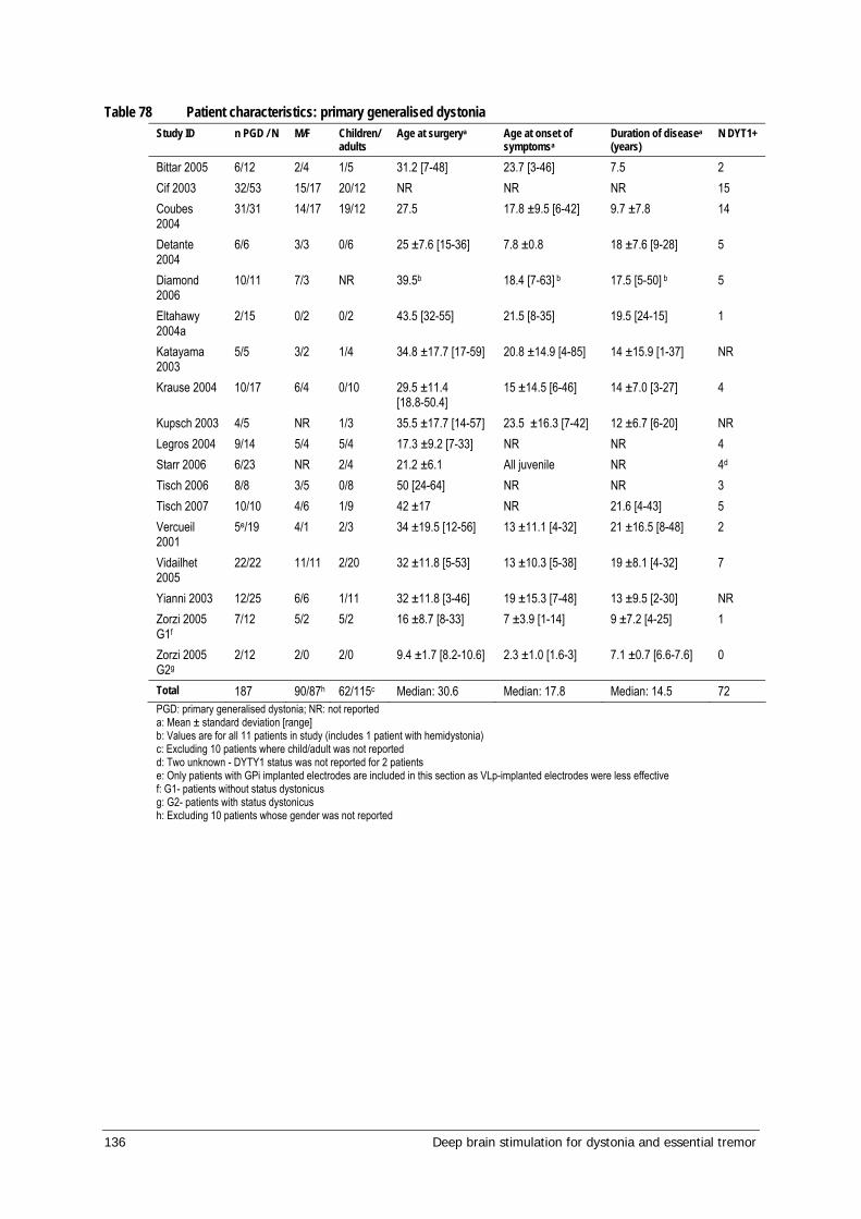
136 Deep brain stimulation for dystonia and essential tremor
Table 78 Patient characteristics: primary generalised dystonia Study ID n PGD / N M/F Children/
adults Age at surgerya Age at onset of
symptomsa Duration of diseasea (years)
N DYT1+
Bittar 2005 6/12 2/4 1/5 31.2 [7-48] 23.7 [3-46] 7.5 2 Cif 2003 32/53 15/17 20/12 NR NR NR 15 Coubes 2004
31/31 14/17 19/12 27.5 17.8 ±9.5 [6-42] 9.7 ±7.8 14
Detante 2004
6/6 3/3 0/6 25 ±7.6 [15-36] 7.8 ±0.8 18 ±7.6 [9-28] 5
Diamond 2006
10/11 7/3 NR 39.5b 18.4 [7-63] b 17.5 [5-50] b 5
Eltahawy 2004a
2/15 0/2 0/2 43.5 [32-55] 21.5 [8-35] 19.5 [24-15] 1
Katayama 2003
5/5 3/2 1/4 34.8 ±17.7 [17-59] 20.8 ±14.9 [4-85] 14 ±15.9 [1-37] NR
Krause 2004 10/17 6/4 0/10 29.5 ±11.4 [18.8-50.4]
15 ±14.5 [6-46] 14 ±7.0 [3-27] 4
Kupsch 2003 4/5 NR 1/3 35.5 ±17.7 [14-57] 23.5 ±16.3 [7-42] 12 ±6.7 [6-20] NR Legros 2004 9/14 5/4 5/4 17.3 ±9.2 [7-33] NR NR 4 Starr 2006 6/23 NR 2/4 21.2 ±6.1 All juvenile NR 4d Tisch 2006 8/8 3/5 0/8 50 [24-64] NR NR 3 Tisch 2007 10/10 4/6 1/9 42 ±17 NR 21.6 [4-43] 5 Vercueil 2001
5e/19 4/1 2/3 34 ±19.5 [12-56] 13 ±11.1 [4-32] 21 ±16.5 [8-48] 2
Vidailhet 2005
22/22 11/11 2/20 32 ±11.8 [5-53] 13 ±10.3 [5-38] 19 ±8.1 [4-32] 7
Yianni 2003 12/25 6/6 1/11 32 ±11.8 [3-46] 19 ±15.3 [7-48] 13 ±9.5 [2-30] NR Zorzi 2005 G1f
7/12 5/2 5/2 16 ±8.7 [8-33] 7 ±3.9 [1-14] 9 ±7.2 [4-25] 1
Zorzi 2005 G2g
2/12 2/0 2/0 9.4 ±1.7 [8.2-10.6] 2.3 ±1.0 [1.6-3] 7.1 ±0.7 [6.6-7.6] 0
Total 187 90/87h 62/115c Median: 30.6 Median: 17.8 Median: 14.5 72 PGD: primary generalised dystonia; NR: not reported a: Mean ± standard deviation [range] b: Values are for all 11 patients in study (includes 1 patient with hemidystonia) c: Excluding 10 patients where child/adult was not reported d: Two unknown - DYTY1 status was not reported for 2 patients e: Only patients with GPi implanted electrodes are included in this section as VLp-implanted electrodes were less effective f: G1- patients without status dystonicus g: G2- patients with status dystonicus h: Excluding 10 patients whose gender was not reported
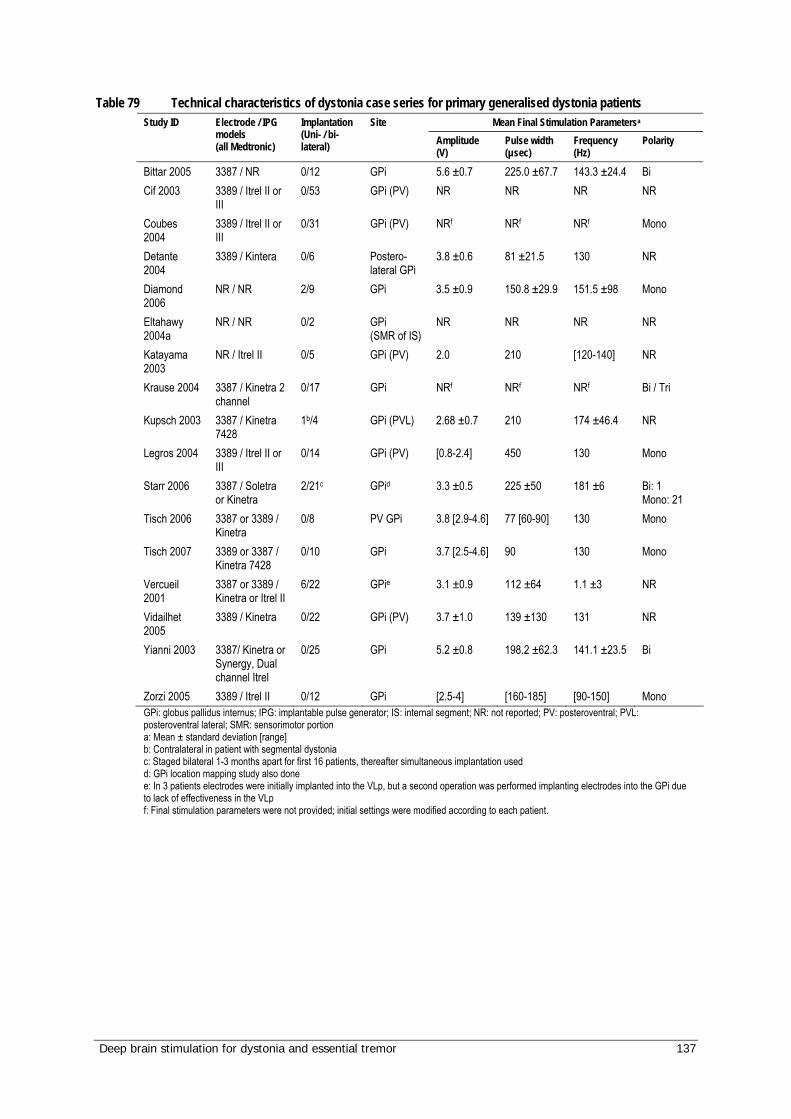
Deep brain stimulation for dystonia and essential tremor 137
Table 79 Technical characteristics of dystonia case series for primary generalised dystonia patients Mean Final Stimulation Parametersa Study ID Electrode / IPG
models (all Medtronic)
Implantation (Uni- / bi-lateral)
Site Amplitude (V)
Pulse width (µsec)
Frequency (Hz)
Polarity
Bittar 2005 3387 / NR 0/12 GPi 5.6 ±0.7 225.0 ±67.7 143.3 ±24.4 Bi Cif 2003 3389 / Itrel II or
III 0/53 GPi (PV) NR NR NR NR
Coubes 2004
3389 / Itrel II or III
0/31 GPi (PV) NRf NRf NRf Mono
Detante 2004
3389 / Kintera 0/6 Postero-lateral GPi
3.8 ±0.6 81 ±21.5 130 NR
Diamond 2006
NR / NR 2/9 GPi 3.5 ±0.9 150.8 ±29.9 151.5 ±98 Mono
Eltahawy 2004a
NR / NR 0/2 GPi (SMR of IS)
NR NR NR NR
Katayama 2003
NR / Itrel II 0/5 GPi (PV) 2.0 210 [120-140] NR
Krause 2004 3387 / Kinetra 2 channel
0/17 GPi NRf NRf NRf Bi / Tri
Kupsch 2003 3387 / Kinetra 7428
1b/4 GPi (PVL) 2.68 ±0.7 210 174 ±46.4 NR
Legros 2004 3389 / Itrel II or III
0/14 GPi (PV) [0.8-2.4] 450 130 Mono
Starr 2006 3387 / Soletra or Kinetra
2/21c GPid 3.3 ±0.5 225 ±50 181 ±6 Bi: 1 Mono: 21
Tisch 2006 3387 or 3389 / Kinetra
0/8 PV GPi 3.8 [2.9-4.6] 77 [60-90] 130 Mono
Tisch 2007 3389 or 3387 / Kinetra 7428
0/10 GPi 3.7 [2.5-4.6] 90 130 Mono
Vercueil 2001
3387 or 3389 / Kinetra or Itrel II
6/22 GPie 3.1 ±0.9 112 ±64 1.1 ±3 NR
Vidailhet 2005
3389 / Kinetra 0/22 GPi (PV) 3.7 ±1.0 139 ±130 131 NR
Yianni 2003 3387/ Kinetra or Synergy, Dual channel Itrel
0/25 GPi 5.2 ±0.8 198.2 ±62.3 141.1 ±23.5 Bi
Zorzi 2005 3389 / Itrel II 0/12 GPi [2.5-4] [160-185] [90-150] Mono GPi: globus pallidus internus; IPG: implantable pulse generator; IS: internal segment; NR: not reported; PV: posteroventral; PVL: posteroventral lateral; SMR: sensorimotor portion a: Mean ± standard deviation [range] b: Contralateral in patient with segmental dystonia c: Staged bilateral 1-3 months apart for first 16 patients, thereafter simultaneous implantation used d: GPi location mapping study also done e: In 3 patients electrodes were initially implanted into the VLp, but a second operation was performed implanting electrodes into the GPi due to lack of effectiveness in the VLp f: Final stimulation parameters were not provided; initial settings were modified according to each patient.
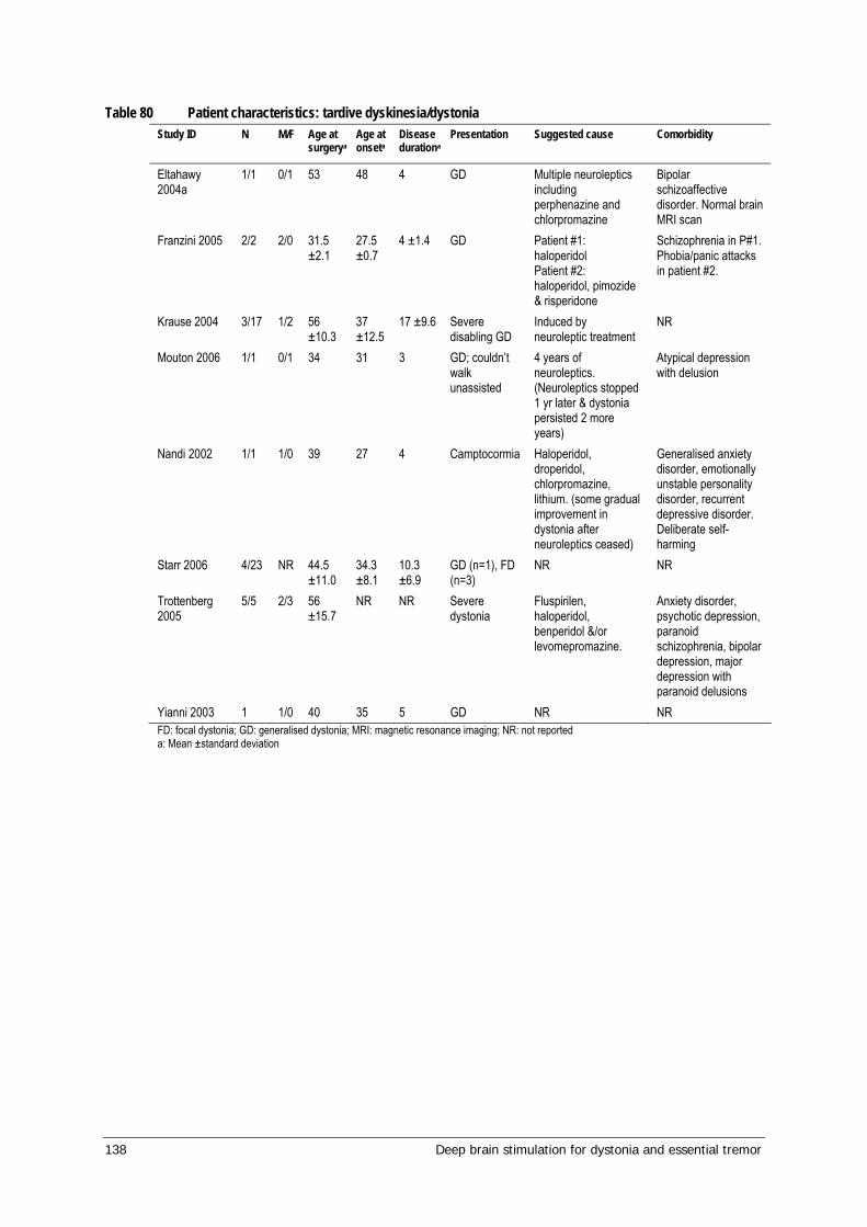
138 Deep brain stimulation for dystonia and essential tremor
Table 80 Patient characteristics: tardive dyskinesia/dystonia Study ID N M/F Age at
surgerya Age at onseta
Disease durationa
Presentation Suggested cause Comorbidity
Eltahawy 2004a
1/1 0/1 53 48 4 GD Multiple neuroleptics including perphenazine and chlorpromazine
Bipolar schizoaffective disorder. Normal brain MRI scan
Franzini 2005 2/2 2/0 31.5 ±2.1
27.5 ±0.7
4 ±1.4 GD Patient #1: haloperidol Patient #2: haloperidol, pimozide & risperidone
Schizophrenia in P#1. Phobia/panic attacks in patient #2.
Krause 2004 3/17 1/2 56 ±10.3
37 ±12.5
17 ±9.6 Severe disabling GD
Induced by neuroleptic treatment
NR
Mouton 2006 1/1 0/1 34 31 3 GD; couldn’t walk unassisted
4 years of neuroleptics. (Neuroleptics stopped 1 yr later & dystonia persisted 2 more years)
Atypical depression with delusion
Nandi 2002 1/1 1/0 39 27 4 Camptocormia Haloperidol, droperidol, chlorpromazine, lithium. (some gradual improvement in dystonia after neuroleptics ceased)
Generalised anxiety disorder, emotionally unstable personality disorder, recurrent depressive disorder. Deliberate self-harming
Starr 2006 4/23 NR 44.5 ±11.0
34.3 ±8.1
10.3 ±6.9
GD (n=1), FD (n=3)
NR NR
Trottenberg 2005
5/5 2/3 56 ±15.7
NR NR Severe dystonia
Fluspirilen, haloperidol, benperidol &/or levomepromazine.
Anxiety disorder, psychotic depression, paranoid schizophrenia, bipolar depression, major depression with paranoid delusions
Yianni 2003 1 1/0 40 35 5 GD NR NR FD: focal dystonia; GD: generalised dystonia; MRI: magnetic resonance imaging; NR: not reported a: Mean ±standard deviation

Deep brain stim
ulation for dystonia and essential tremor 139
Appendix J Further data for essential tremor studies
Table 81 Technical characteristics: essential tremor – DBS ON/OFF Mean final stimulation parametersa Study ID Electrode / IPG models
(all Medtronic) Uni/ bilateral implantation
Time from DBS ON/OFF to assessment Amplitude (V) Pulse width (µsec) Frequency (Hz) Polarity
Bryant 2003 3287 / NR 12b/4 Minimum 30 minutes 2.74 ±0.99 119 ±44.0 160 ±28.2 All mono/bi electrode permutations tested until best results found
Carpenter 1998
3382 / ITREL II 7424 5/2 Minimum 1 minute between on, off and repeat of on
Most common: 3 Most common: 60 Most common: 150 NR
Koller 1999 NR / ITREL II 38/0c NR Baseline: 3.5 ±1.0 Baseline: 86.1 ±37.7 Baseline: 155.9 ±29.4 NR Koller 2001 3387 / ITREL II 7432 25/0d NR At 40 months:
3.6 ±1.3, p=0.430 At 40 months: 99.6 ±45.7, p=0.140
At 40 months: 161.1 ±25.5, p=0.365
NR
Lyons 1998 NR 22/0 On/off ratings performed within same week
NR NR NR NR
Obwegeser 2000
3387 / NR 14/13 Stimulator deactivated minimum 1 hour before tremor assessment off. Assessed on when maximal tremor control without side effects reached
Bilateral: 2.5 ±0.9 Unilateral: 2.8 ±0.7
NR NR NR
Pahwa 1999 NR 0/9 NR NR NR NR NR Pahwa 2006 3387 / IPG 7424 ITREL
II 18/8 If on, switched to off, evaluated 30
minutes after At 5 years: Unilateral: 3.6 Bilateral side one: 3.6 Bilateral side two: 3.2
At 5 years: Unilateral: 111 Bilateral side one: 111 Bilateral side two: 129
At 5 year follow up: Unilateral 158 Bilateral side one: 155 Bilateral side two: 153
19% unilateral patients monopolar; 100% bilateral patients bipolar
Putzke 2004 3387 / NR 20e/22f Deactivated overnight, patient examined in morning. If not deactivated overnight, was deactivated for minimum 1 hour prior to assessment
At 3 months: Unilateral: 2.8 (1.1) At 3 years: Bilateral side one: 2.8 (1.0) Bilateral side two: 2.4 (0.7)
At 3 months: Unilateral: 80.9 (33.0) At 3 years: Bilateral side one: 97.5 (26.6) Bilateral side two: 97.5 (31.1)
At 3 months: Unilateral: 158.3(30.8) At 3 years: Bilateral side one: 168.1 (19.8) Bilateral side two: 158.8 (19.4)
NR
Sydow 2003 Permanent quadripolar stimulation electrode / NR
15/4 NR At 6 years: 2.6 (0.7); n=25g
At 6 years: 88.8 (37.2); n=25g
At 6 years: 172.6 (19.7); n=25g
18 mono; 7 bi
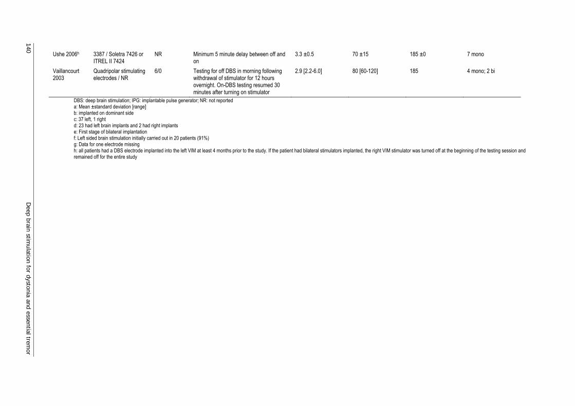
140 D
eep brain stimulation for dystonia and essential trem
or
Ushe 2006h 3387 / Soletra 7426 or ITREL II 7424
NR Minimum 5 minute delay between off and on
3.3 ±0.5 70 ±15 185 ±0 7 mono
Vaillancourt 2003
Quadripolar stimulating electrodes / NR
6/0 Testing for off DBS in morning following withdrawal of stimulator for 12 hours overnight. On-DBS testing resumed 30 minutes after turning on stimulator
2.9 [2.2-6.0] 80 [60-120] 185 4 mono; 2 bi
DBS: deep brain stimulation; IPG: implantable pulse generator; NR: not reported a: Mean ±standard deviation [range] b: implanted on dominant side c: 37 left, 1 right d: 23 had left brain implants and 2 had right implants e: First stage of bilateral implantation f: Left sided brain stimulation initially carried out in 20 patients (91%) g: Data for one electrode missing h: all patients had a DBS electrode implanted into the left VIM at least 4 months prior to the study. If the patient had bilateral stimulators implanted, the right VIM stimulator was turned off at the beginning of the testing session and remained off for the entire study
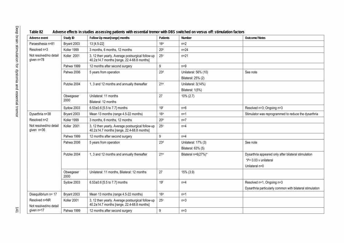
Deep brain stim
ulation for dystonia and essential tremor 141
Table 82 Adverse effects in studies assessing patients with essential tremor with DBS switched on versus off: stimulation factors Adverse event Study ID Follow Up mean[range] months Patients Number Outcome/ Notes
Bryant 2003 13 [4.5-22] 16a n=2 Koller 1999 3 months, 6 months, 12 months 20b n=24 Koller 2001 3, 12 then yearly. Average postsurgical follow-up
40.2±14.7 months [range, 22.4-68.8 months] 25c n=21
Pahwa 1999 12 months after second surgery 9 n=9 Pahwa 2006 5 years from operation 23d Unilateral: 56% (10)
Bilateral: 25% (2) See note
Putzke 2004 1, 3 and 12 months and annually thereafter 21e Unilateral: 3(14%) Bilateral: 1(5%)
Obwegeser 2000
Unilateral: 11 months Bilateral: 12 months
27 10% (2.7)
Paraesthesia n=81 Resolved n=3 Not resolved/no detail given n=78
Sydow 2003 6.53±0.6 [5.5 to 7.7] months 19f n=6 Resolved n=3; Ongoing n=3 Bryant 2003 Mean 13 months (range 4.5-22 months) 16a n=1 Stimulator was reprogrammed to reduce the dysarthria Koller 1999 3 months, 6 months, 12 months 20b n=7 Koller 2001 3, 12 then yearly. Average postsurgical follow-up
40.2±14.7 months [range, 22.4-68.8 months] 25c n=4
Pahwa 1999 12 months after second surgery 9 n=4 Pahwa 2006 5 years from operation 23d Unilateral: 17% (3)
Bilateral: 63% (5) See note
Putzke 2004 1, 3 and 12 months and annually thereafter 21e Bilateral n=6(27%)* Dysarthria appeared only after bilateral stimulation *P= 0.03 v unilateral Unilateral n=0
Obwegeser 2000
Unilateral: 11 months, Bilateral: 12 months 27 15% (3.9)
Dysarthria n=38 Resolved n=2 Not resolved/no detail given n=36
Sydow 2003 6.53±0.6 [5.5 to 7.7] months 19f n=4 Resolved n=1, Ongoing n=3 Dysarthria particularly common with bilateral stimulation
Bryant 2003 Mean 13 months (range 4.5-22 months) 16a n=1 Koller 2001 3, 12 then yearly. Average postsurgical follow-up
40.2±14.7 months [range, 22.4-68.8 months] 25c n=3
Disequilibrium n= 17 Resolved n=NR Not resolved/no detail given n=17 Pahwa 1999 12 months after second surgery 9 n=3
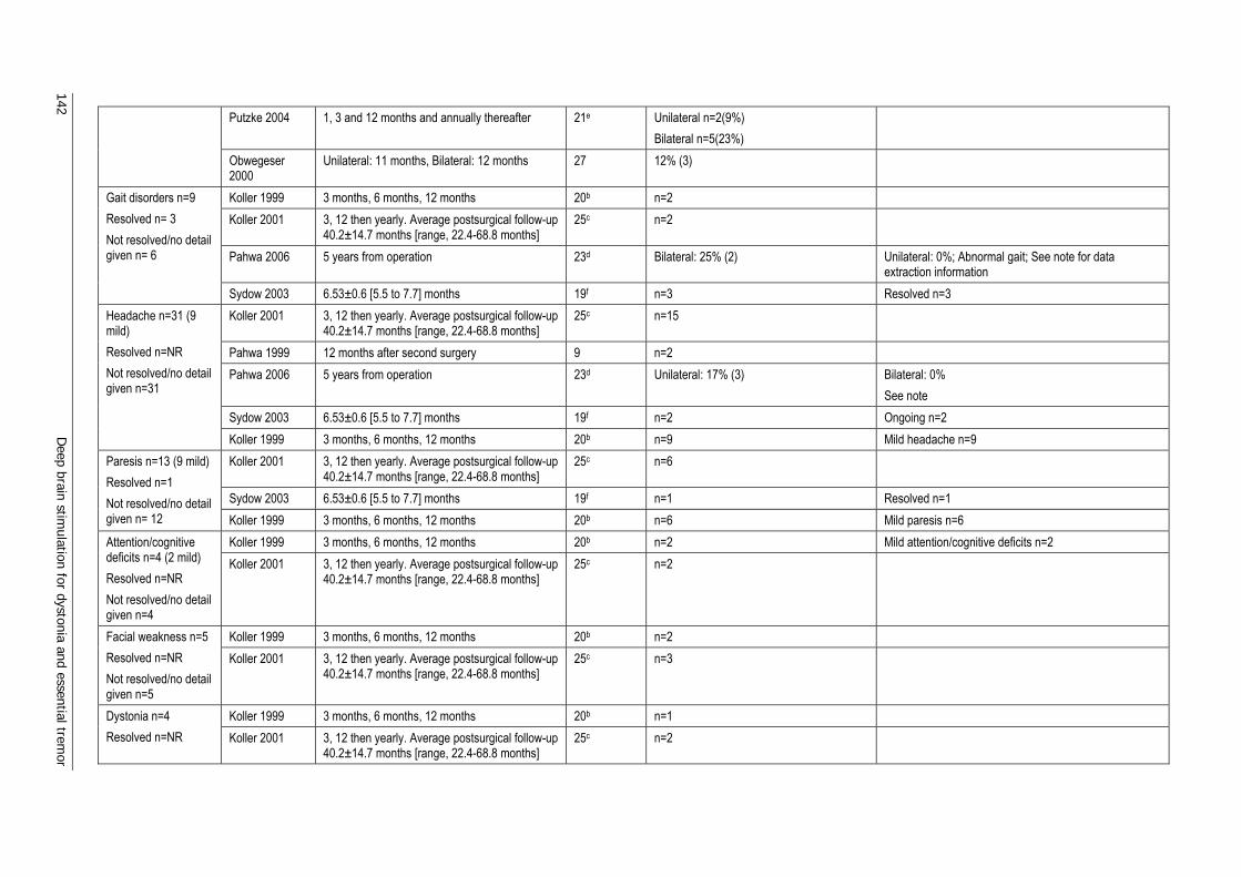
142 D
eep brain stimulation for dystonia and essential trem
or
Putzke 2004 1, 3 and 12 months and annually thereafter 21e Unilateral n=2(9%) Bilateral n=5(23%)
Obwegeser 2000
Unilateral: 11 months, Bilateral: 12 months 27 12% (3)
Koller 1999 3 months, 6 months, 12 months 20b n=2 Koller 2001 3, 12 then yearly. Average postsurgical follow-up
40.2±14.7 months [range, 22.4-68.8 months] 25c n=2
Pahwa 2006 5 years from operation 23d Bilateral: 25% (2) Unilateral: 0%; Abnormal gait; See note for data extraction information
Gait disorders n=9 Resolved n= 3 Not resolved/no detail given n= 6
Sydow 2003 6.53±0.6 [5.5 to 7.7] months 19f n=3 Resolved n=3 Koller 2001 3, 12 then yearly. Average postsurgical follow-up
40.2±14.7 months [range, 22.4-68.8 months] 25c n=15
Pahwa 1999 12 months after second surgery 9 n=2 Pahwa 2006 5 years from operation 23d Unilateral: 17% (3) Bilateral: 0%
See note Sydow 2003 6.53±0.6 [5.5 to 7.7] months 19f n=2 Ongoing n=2
Headache n=31 (9 mild) Resolved n=NR Not resolved/no detail given n=31
Koller 1999 3 months, 6 months, 12 months 20b n=9 Mild headache n=9 Koller 2001 3, 12 then yearly. Average postsurgical follow-up
40.2±14.7 months [range, 22.4-68.8 months] 25c n=6
Sydow 2003 6.53±0.6 [5.5 to 7.7] months 19f n=1 Resolved n=1
Paresis n=13 (9 mild) Resolved n=1 Not resolved/no detail given n= 12 Koller 1999 3 months, 6 months, 12 months 20b n=6 Mild paresis n=6
Koller 1999 3 months, 6 months, 12 months 20b n=2 Mild attention/cognitive deficits n=2 Attention/cognitive deficits n=4 (2 mild) Resolved n=NR Not resolved/no detail given n=4
Koller 2001 3, 12 then yearly. Average postsurgical follow-up 40.2±14.7 months [range, 22.4-68.8 months]
25c n=2
Koller 1999 3 months, 6 months, 12 months 20b n=2 Facial weakness n=5 Resolved n=NR Not resolved/no detail given n=5
Koller 2001 3, 12 then yearly. Average postsurgical follow-up 40.2±14.7 months [range, 22.4-68.8 months]
25c n=3
Koller 1999 3 months, 6 months, 12 months 20b n=1 Dystonia n=4 Resolved n=NR Koller 2001 3, 12 then yearly. Average postsurgical follow-up
40.2±14.7 months [range, 22.4-68.8 months] 25c n=2
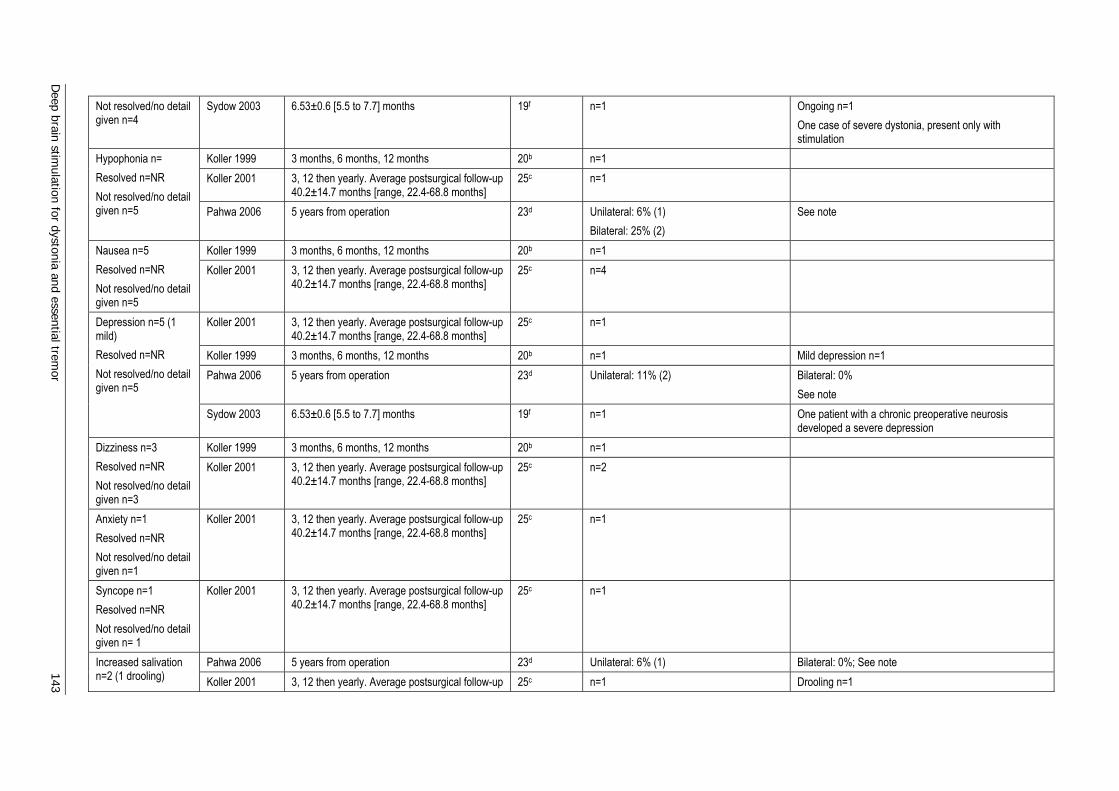
Deep brain stim
ulation for dystonia and essential tremor 143
Not resolved/no detail given n=4
Sydow 2003 6.53±0.6 [5.5 to 7.7] months 19f n=1 Ongoing n=1 One case of severe dystonia, present only with stimulation
Koller 1999 3 months, 6 months, 12 months 20b n=1 Koller 2001 3, 12 then yearly. Average postsurgical follow-up
40.2±14.7 months [range, 22.4-68.8 months] 25c n=1
Hypophonia n= Resolved n=NR Not resolved/no detail given n=5 Pahwa 2006 5 years from operation 23d Unilateral: 6% (1)
Bilateral: 25% (2) See note
Koller 1999 3 months, 6 months, 12 months 20b n=1 Nausea n=5 Resolved n=NR Not resolved/no detail given n=5
Koller 2001 3, 12 then yearly. Average postsurgical follow-up 40.2±14.7 months [range, 22.4-68.8 months]
25c n=4
Koller 2001 3, 12 then yearly. Average postsurgical follow-up 40.2±14.7 months [range, 22.4-68.8 months]
25c n=1
Koller 1999 3 months, 6 months, 12 months 20b n=1 Mild depression n=1 Pahwa 2006 5 years from operation 23d Unilateral: 11% (2)
Bilateral: 0% See note
Depression n=5 (1 mild) Resolved n=NR Not resolved/no detail given n=5
Sydow 2003 6.53±0.6 [5.5 to 7.7] months 19f n=1 One patient with a chronic preoperative neurosis developed a severe depression
Koller 1999 3 months, 6 months, 12 months 20b n=1 Dizziness n=3 Resolved n=NR Not resolved/no detail given n=3
Koller 2001 3, 12 then yearly. Average postsurgical follow-up 40.2±14.7 months [range, 22.4-68.8 months]
25c n=2
Anxiety n=1 Resolved n=NR Not resolved/no detail given n=1
Koller 2001 3, 12 then yearly. Average postsurgical follow-up 40.2±14.7 months [range, 22.4-68.8 months]
25c n=1
Syncope n=1 Resolved n=NR Not resolved/no detail given n= 1
Koller 2001 3, 12 then yearly. Average postsurgical follow-up 40.2±14.7 months [range, 22.4-68.8 months]
25c n=1
Pahwa 2006 5 years from operation 23d Unilateral: 6% (1) Bilateral: 0%; See note Increased salivation n=2 (1 drooling) Koller 2001 3, 12 then yearly. Average postsurgical follow-up 25c n=1 Drooling n=1
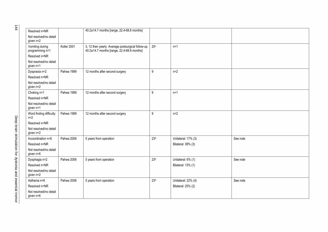
144 D
eep brain stimulation for dystonia and essential trem
or
Resolved n=NR Not resolved/no detail given n=2
40.2±14.7 months [range, 22.4-68.8 months]
Vomiting during programming n=1 Resolved n=NR Not resolved/no detail given n=1
Koller 2001 3, 12 then yearly. Average postsurgical follow-up 40.2±14.7 months [range, 22.4-68.8 months]
25c n=1
Dyspraxia n=2 Resolved n=NR Not resolved/no detail given n=2
Pahwa 1999 12 months after second surgery 9 n=2
Choking n=1 Resolved n=NR Not resolved/no detail given n=1
Pahwa 1999 12 months after second surgery 9 n=1
Word finding difficulty n=2 Resolved n=NR Not resolved/no detail given n=2
Pahwa 1999 12 months after second surgery 9 n=2
Incoordination n=6 Resolved n=NR Not resolved/no detail given n=6
Pahwa 2006 5 years from operation 23d Unilateral: 17% (3) Bilateral: 38% (3)
See note
Dysphagia n=2 Resolved n=NR Not resolved/no detail given n=2
Pahwa 2006 5 years from operation 23d Unilateral: 6% (1) Bilateral: 13% (1)
See note
Asthenia n=6 Resolved n=NR Not resolved/no detail given n=6
Pahwa 2006 5 years from operation 23d Unilateral: 22% (4) Bilateral: 25% (2)
See note
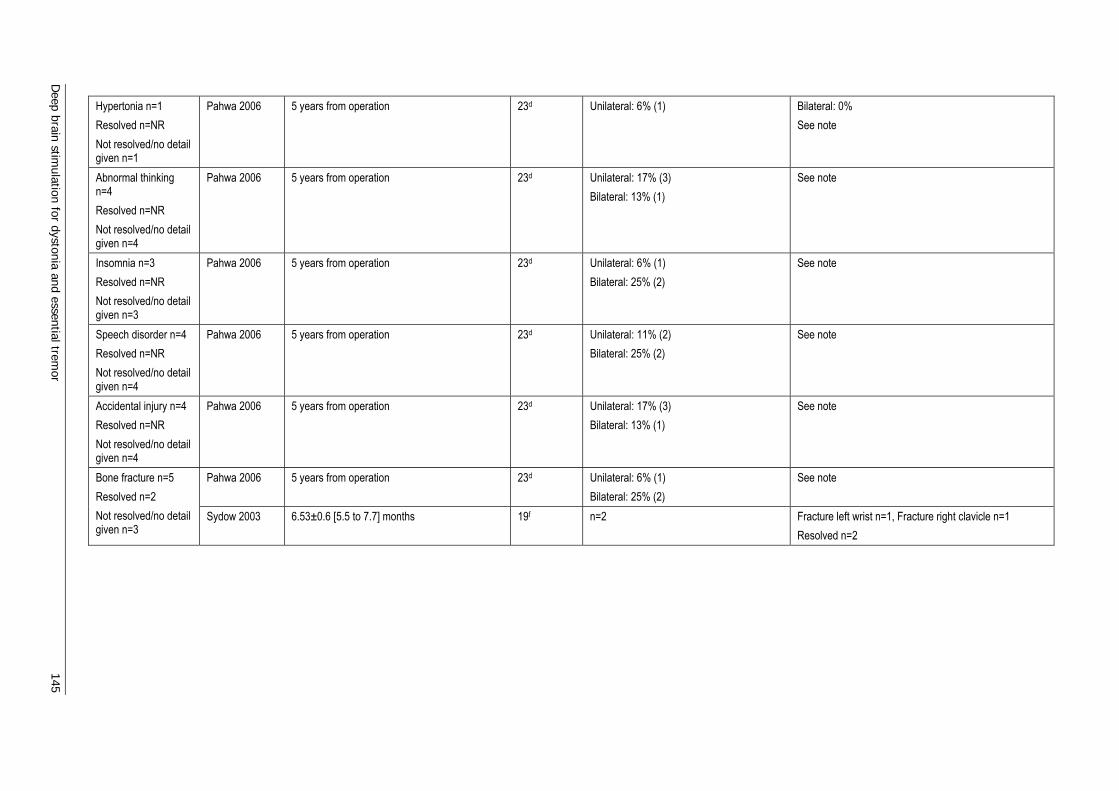
Deep brain stim
ulation for dystonia and essential tremor 145
Hypertonia n=1 Resolved n=NR Not resolved/no detail given n=1
Pahwa 2006 5 years from operation 23d Unilateral: 6% (1)
Bilateral: 0% See note
Abnormal thinking n=4 Resolved n=NR Not resolved/no detail given n=4
Pahwa 2006 5 years from operation 23d Unilateral: 17% (3) Bilateral: 13% (1)
See note
Insomnia n=3 Resolved n=NR Not resolved/no detail given n=3
Pahwa 2006 5 years from operation 23d Unilateral: 6% (1) Bilateral: 25% (2)
See note
Speech disorder n=4 Resolved n=NR Not resolved/no detail given n=4
Pahwa 2006 5 years from operation 23d Unilateral: 11% (2) Bilateral: 25% (2)
See note
Accidental injury n=4 Resolved n=NR Not resolved/no detail given n=4
Pahwa 2006 5 years from operation 23d Unilateral: 17% (3) Bilateral: 13% (1)
See note
Pahwa 2006 5 years from operation 23d Unilateral: 6% (1) Bilateral: 25% (2)
See note Bone fracture n=5 Resolved n=2 Not resolved/no detail given n=3
Sydow 2003 6.53±0.6 [5.5 to 7.7] months 19f n=2 Fracture left wrist n=1, Fracture right clavicle n=1 Resolved n=2
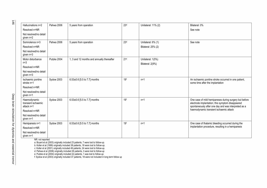
146 D
eep brain stimulation for dystonia and essential trem
or
Hallucinations n=2 Resolved n=NR Not resolved/no detail given n=2
Pahwa 2006 5 years from operation 23d Unilateral: 11% (2)
Bilateral: 0% See note
Somnolence n=3 Resolved n=NR Not resolved/no detail given n=3
Pahwa 2006 5 years from operation 23d Unilateral: 6% (1) Bilateral: 25% (2)
See note
Motor disturbance n=3 Resolved n=NR Not resolved/no detail given n=3
Putzke 2004 1, 3 and 12 months and annually thereafter 21e Unilateral: 1(5%) Bilateral: 2(9%)
Ischaemic pontine stroke n=1 Resolved n=NR Not resolved/no detail given n=1
Sydow 2003 6.53±0.6 [5.5 to 7.7] months 19f n=1 An ischaemic pontine stroke occurred in one patient, some time after the implantation
Haemodynamic transient ischaemic attack n=1 Resolved n=NR Not resolved/no detail given n=1
Sydow 2003 6.53±0.6 [5.5 to 7.7] months 19f n=1 One case of mild hemipareses during surgery but before electrode implantation; this symptom disappeared spontaneously after one day and was interpreted as a haemodynamic transient ischaemic attack
Hemiparesis n=1 Resolved n=NR Not resolved/no detail given n=1
Sydow 2003 6.53±0.6 [5.5 to 7.7] months 19f n=1 One case of thalamic bleeding occurred during the implantation procedure, resulting in a hemiparesis
NR: not reported a: Bryant et al (2003) originally included 23 patients, 7 were lost to follow-up. b: Koller et al (1999) originally included 38 patients, 18 were lost to follow-up. c: Koller et al (2001) originally included 49 patients, 24 were lost to follow-up. d: Pahwa et al (2006) originally included 26 patients, 3 were lost to follow-up e: Putzke et al (2004) originally included 22 patients, 1 was lost to follow-up f: Sydow et al (2003) originally included 37 patients, 18 were not included in long term follow up
Deep brain stim
ulation for dystonia and essential tremor 147
Note: Pahwa et al (2006) appears to have rounded percentages of adverse effects up to the nearest whole per cent, rather than report actual patient numbers. Statistics reported in these tables are based upon whole patient numbers derived from the %). Notes regarding adverse events: Koller et al (1999): Stimulation complications were mild and well controlled with stimulation parameter adjustments Obwegeser et al (2000) Adjustments of the pulse generator improved tremor control in 91 cases and in 13 of these side effects were eliminated at the same time Koller et at (2001): Stimulation adverse events were mild and easy to manage with parameter-setting adjustments Pahwa et al (1999) Adverse effects were mild and resolved with adjustment to stimulation parameters Pahwa et al (2006) Most of the adverse effects were mild and were reduced with changes in stimulation parameters. In patients with bilateral stimulation, adverse events such as dysarthria and other speech difficulties, disequilibrium or balance difficulties and abnormal gait persisted despite optimization of the stimulation parameters Putzke et al (2004) Adverse side effects related to stimulation were generally mild and easily altered by adjusting stimulus parameters Sydow et al (2003) Most adverse events were related to stimulation and thus disappeared when the pulse generator was turned off
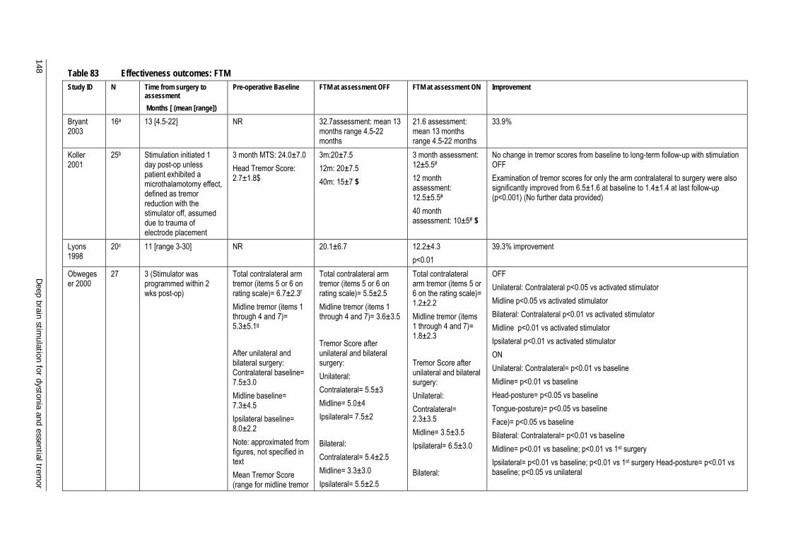
148 D
eep brain stimulation for dystonia and essential trem
or
Table 83 Effectiveness outcomes: FTM Study ID N Time from surgery to
assessment Months [ (mean [range])
Pre-operative Baseline FTM at assessment OFF FTM at assessment ON Improvement
Bryant 2003
16a 13 [4.5-22] NR 32.7assessment: mean 13 months range 4.5-22 months
21.6 assessment: mean 13 months range 4.5-22 months
33.9%
Koller 2001
25b Stimulation initiated 1 day post-op unless patient exhibited a microthalamotomy effect, defined as tremor reduction with the stimulator off, assumed due to trauma of electrode placement
3 month MTS: 24.0±7.0 Head Tremor Score: 2.7±1.8$
3m:20±7.5 12m: 20±7.5 40m: 15±7 $
3 month assessment: 12±5.5# 12 month assessment: 12.5±5.5# 40 month assessment: 10±5# $
No change in tremor scores from baseline to long-term follow-up with stimulation OFF Examination of tremor scores for only the arm contralateral to surgery were also significantly improved from 6.5±1.6 at baseline to 1.4±1.4 at last follow-up (p<0.001) (No further data provided)
Lyons 1998
20c 11 [range 3-30] NR 20.1±6.7 12.2±4.3 p<0.01
39.3% improvement
Obwegeser 2000
27 3 (Stimulator was programmed within 2 wks post-op)
Total contralateral arm tremor (items 5 or 6 on rating scale)= 6.7±2.3f Midline tremor (items 1 through 4 and 7)= 5.3±5.1g After unilateral and bilateral surgery: Contralateral baseline= 7.5±3.0 Midline baseline= 7.3±4.5 Ipsilateral baseline= 8.0±2.2 Note: approximated from figures, not specified in text Mean Tremor Score (range for midline tremor
Total contralateral arm tremor (items 5 or 6 on rating scale)= 5.5±2.5 Midline tremor (items 1 through 4 and 7)= 3.6±3.5 Tremor Score after unilateral and bilateral surgery: Unilateral: Contralateral= 5.5±3 Midline= 5.0±4 Ipsilateral= 7.5±2 Bilateral: Contralateral= 5.4±2.5 Midline= 3.3±3.0 Ipsilateral= 5.5±2.5
Total contralateral arm tremor (items 5 or 6 on the rating scale)= 1.2±2.2 Midline tremor (items 1 through 4 and 7)= 1.8±2.3 Tremor Score after unilateral and bilateral surgery: Unilateral: Contralateral= 2.3±3.5 Midline= 3.5±3.5 Ipsilateral= 6.5±3.0 Bilateral:
OFF Unilateral: Contralateral p<0.05 vs activated stimulator Midline p<0.05 vs activated stimulator Bilateral: Contralateral p<0.01 vs activated stimulator Midline p<0.01 vs activated stimulator Ipsilateral p<0.01 vs activated stimulator ON Unilateral: Contralateral= p<0.01 vs baseline Midline= p<0.01 vs baseline Head-posture= p<0.05 vs baseline Tongue-posture)= p<0.05 vs baseline Face)= p<0.05 vs baseline Bilateral: Contralateral= p<0.01 vs baseline Midline= p<0.01 vs baseline; p<0.01 vs 1st surgery Ipsilateral= p<0.01 vs baseline; p<0.01 vs 1st surgery Head-posture= p<0.01 vs baseline; p<0.05 vs unilateral
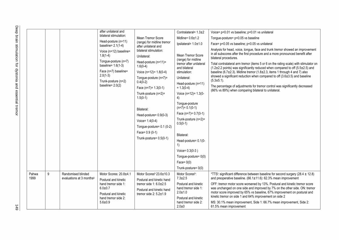
Deep brain stim
ulation for dystonia and essential tremor 149
after unilateral and bilateral stimulation: Head-posture (n=11) baseline= 2.1(1-4) Voice (n=12) baseline= 1.8(1-4) Tongue-posture (n=7) baseline= 1.6(1-3) Face (n=7) baseline= 2.0(1-3) Trunk-posture (n=2) baseline= 2.0(2)
Mean Tremor Score (range) for midline tremor after unilateral and bilateral stimulation: Unilateral: Head-posture (n=11)= 1.6(0-4) Voice (n=12)= 1.8(0-4) Tongue-posture (n=7)= 0.4(0-2) Face (n=7)= 1.3(0-1) Trunk-posture (n=2)= 1.5(0-1) Bilateral: Head-posture= 0.9(0-3) Voice= 1.4(0-4) Tongue-posture= 0.1 (0-2) Face= 0.9 (0-1) Trunk-posture= 0.5(0-1)
Contralateral= 1.3±2 Midline= 0.6±1.2 Ipsilateral= 1.0±1.0 Mean Tremor Score (range) for midline tremor after unilateral and bilateral stimulation: Unilateral: Head-posture (n=11) = 1.3(0-4) Voice (n=12)= 1.3(0-4) Tongue-posture (n=7)= 0.1(0-1) Face (n=7)= 0.7(0-1) Trunk-posture (n=2)= 0.5(0-1) Bilateral: Head-posture= 0.1(0-1) Voice= 0.3(0-3 ) Tongue-posture= 0(0) Face= 0(0) Trunk-posture= 0(0)
Voice= p<0.01 vs baseline; p<0.01 vs unilateral Tongue-posture= p<0.05 vs baseline Face= p<0.05 vs baseline; p<0.05 vs unilateral Analysis for head, voice, tongue, face and trunk tremor showed an improvement in all subscores after the first procedure and a more pronounced benefit after bilateral procedures. Total contralateral arm tremor (items 5 or 6 on the rating scale) with stimulator on (1.2±2.2 points) was significantly reduced when compared to off (5.5±2.5) and baseline (6.7±2.3). Midline tremor (1.8±2.3, items 1 through 4 and 7) also showed a significant reduction when compared to off (3.6±3.5) and baseline (5.3±5.1). The percentage of adjustments for tremor control was significantly decreased (66% vs 89%) when comparing bilateral to unilateral.
Pahwa 1999
9 Randomised blinded evaluations at 3 monthsg
Motor Scores: 20.8±4.1 Postural and kinetic hand tremor side 1: 6.0±0.7 Postural and kinetic hand tremor side 2: 5.6±0.9
Motor Scoresh 23.6±10.3 Postural and kinetic hand tremor side 1: 6.0±2.5 Postural and kinetic hand tremor side 2: 5.2±1.9
Motor Scoresh: 7.3±2.5 Postural and kinetic hand tremor side 1: 2.0±1.0 Postural and kinetic hand tremor side 2: 2.0±0
*TTSi: significant difference between baseline for second surgery (28.4 ± 12.8) and preoperative baseline. (66.1±11.6): 62.3% mean improvement OFF: tremor motor score worsened by 13%. Postural and kinetic tremor score was unchanged on one side and improved by 7% on the other side. ON: tremor motor score improved by 65% vs baseline, 67% improvement on postural and kinetic tremor on side 1 and 64% improvement on side 2 MS: 30.1% mean improvement, Side 1: 66.7% mean improvement, Side 2: 61.5% mean improvement
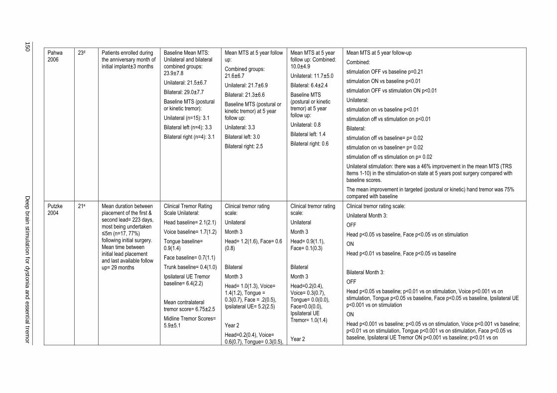
150 D
eep brain stimulation for dystonia and essential trem
or
Pahwa 2006
23d Patients enrolled during the anniversary month of initial implant±3 months
Baseline Mean MTS: Unilateral and bilateral combined groups: 23.9±7.8 Unilateral: 21.5±6.7 Bilateral: 29.0±7.7 Baseline MTS (postural or kinetic tremor): Unilateral (n=15): 3.1 Bilateral left (n=4): 3.3 Bilateral right (n=4): 3.1
Mean MTS at 5 year follow up: Combined groups: 21.6±6.7 Unilateral: 21.7±6.9 Bilateral: 21.3±6.6 Baseline MTS (postural or kinetic tremor) at 5 year follow up: Unilateral: 3.3 Bilateral left: 3.0 Bilateral right: 2.5
Mean MTS at 5 year follow up: Combined: 10.0±4.9 Unilateral: 11.7±5.0 Bilateral: 6.4±2.4 Baseline MTS (postural or kinetic tremor) at 5 year follow up: Unilateral: 0.8 Bilateral left: 1.4 Bilateral right: 0.6
Mean MTS at 5 year follow-up Combined: stimulation OFF vs baseline p=0.21 stimulation ON vs baseline p<0.01 stimulation OFF vs stimulation ON p<0.01 Unilateral: stimulation on vs baseline p<0.01 stimulation off vs stimulation on p<0.01 Bilateral: stimulation off vs baseline= p= 0.02 stimulation on vs baseline= p= 0.02 stimulation off vs stimulation on p= 0.02 Unilateral stimulation: there was a 46% improvement in the mean MTS (TRS Items 1-10) in the stimulation-on state at 5 years post surgery compared with baseline scores. The mean improvement in targeted (postural or kinetic) hand tremor was 75% compared with baseline
Putzke 2004
21e Mean duration between placement of the first & second lead= 223 days, most being undertaken ≤5m (n=17, 77%) following initial surgery. Mean time between initial lead placement and last available follow up= 29 months
Clinical Tremor Rating Scale Unilateral: Head baseline= 2.1(2.1) Voice baseline= 1.7(1.2) Tongue baseline= 0.9(1.4) Face baseline= 0.7(1.1) Trunk baseline= 0.4(1.0) Ipsilateral UE Tremor baseline= 6.4(2.2) Mean contralateral tremor score= 6.75±2.5 Midline Tremor Scores= 5.9±5.1
Clinical tremor rating scale: Unilateral Month 3 Head= 1.2(1.6), Face= 0.6 (0.8) Bilateral Month 3 Head= 1.0(1.3), Voice= 1.4(1.2), Tongue = 0.3(0.7), Face = .2(0.5), Ipsilateral UE= 5.2(2.5) Year 2 Head=0.2(0.4), Voice= 0.6(0.7), Tongue= 0.3(0.5),
Clinical tremor rating scale: Unilateral Month 3 Head= 0.9(1.1), Face= 0.1(0.3) Bilateral Month 3 Head=0.2(0.4), Voice= 0.3(0.7), Tongue= 0.0(0.0), Face=0.0(0.0), Ipsilateral UE Tremor= 1.0(1.4) Year 2
Clinical tremor rating scale: Unilateral Month 3: OFF Head p<0.05 vs baseline, Face p<0.05 vs on stimulation ON Head p<0.01 vs baseline, Face p<0.05 vs baseline Bilateral Month 3: OFF Head p<0.05 vs baseline; p<0.01 vs on stimulation, Voice p<0.001 vs on stimulation, Tongue p<0.05 vs baseline, Face p<0.05 vs baseline, Ipsilateral UE p<0.001 vs on stimulation ON Head p<0.001 vs baseline; p<0.05 vs on stimulation, Voice p<0.001 vs baseline; p<0.01 vs on stimulation, Tongue p<0.001 vs on stimulation, Face p<0.05 vs baseline, Ipsilateral UE Tremor ON p<0.001 vs baseline; p<0.01 vs on
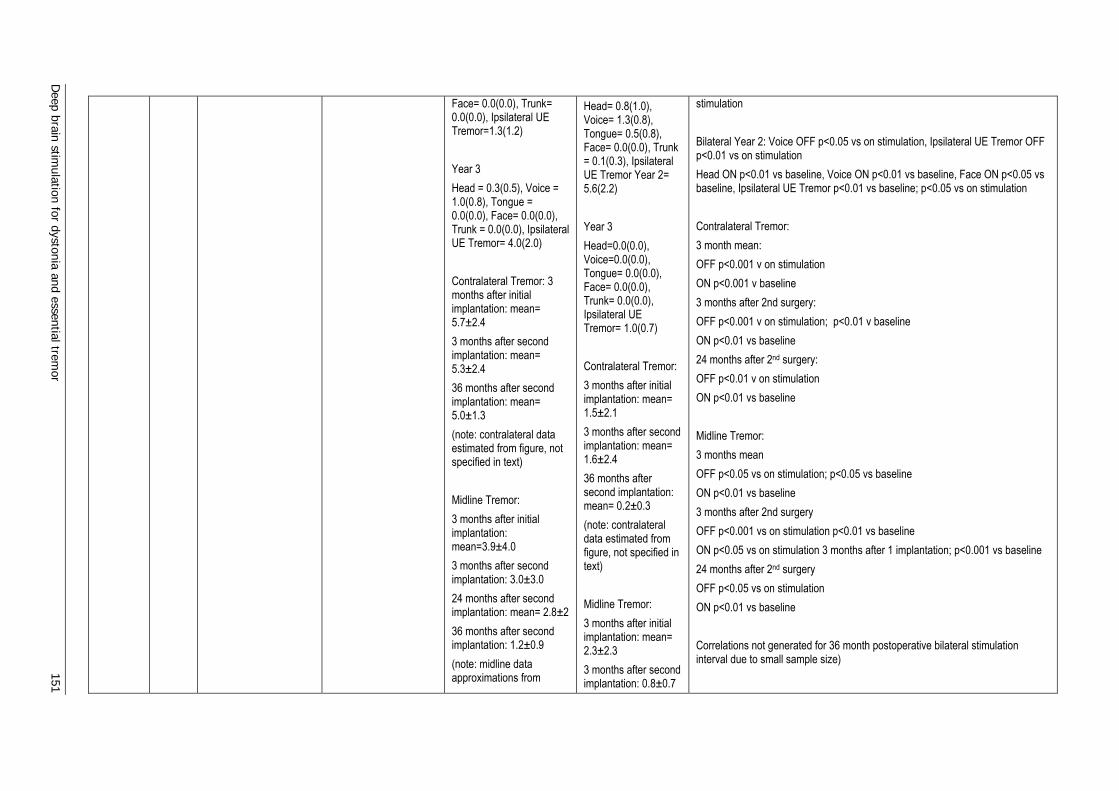
Deep brain stim
ulation for dystonia and essential tremor 151
Face= 0.0(0.0), Trunk= 0.0(0.0), Ipsilateral UE Tremor=1.3(1.2) Year 3 Head = 0.3(0.5), Voice = 1.0(0.8), Tongue = 0.0(0.0), Face= 0.0(0.0), Trunk = 0.0(0.0), Ipsilateral UE Tremor= 4.0(2.0) Contralateral Tremor: 3 months after initial implantation: mean= 5.7±2.4 3 months after second implantation: mean= 5.3±2.4 36 months after second implantation: mean= 5.0±1.3 (note: contralateral data estimated from figure, not specified in text) Midline Tremor: 3 months after initial implantation: mean=3.9±4.0 3 months after second implantation: 3.0±3.0 24 months after second implantation: mean= 2.8±2 36 months after second implantation: 1.2±0.9 (note: midline data approximations from
Head= 0.8(1.0), Voice= 1.3(0.8), Tongue= 0.5(0.8), Face= 0.0(0.0), Trunk = 0.1(0.3), Ipsilateral UE Tremor Year 2= 5.6(2.2) Year 3 Head=0.0(0.0), Voice=0.0(0.0), Tongue= 0.0(0.0), Face= 0.0(0.0), Trunk= 0.0(0.0), Ipsilateral UE Tremor= 1.0(0.7) Contralateral Tremor: 3 months after initial implantation: mean= 1.5±2.1 3 months after second implantation: mean= 1.6±2.4 36 months after second implantation: mean= 0.2±0.3 (note: contralateral data estimated from figure, not specified in text) Midline Tremor: 3 months after initial implantation: mean= 2.3±2.3 3 months after second implantation: 0.8±0.7
stimulation Bilateral Year 2: Voice OFF p<0.05 vs on stimulation, Ipsilateral UE Tremor OFF p<0.01 vs on stimulation Head ON p<0.01 vs baseline, Voice ON p<0.01 vs baseline, Face ON p<0.05 vs baseline, Ipsilateral UE Tremor p<0.01 vs baseline; p<0.05 vs on stimulation Contralateral Tremor: 3 month mean: OFF p<0.001 v on stimulation ON p<0.001 v baseline 3 months after 2nd surgery: OFF p<0.001 v on stimulation; p<0.01 v baseline ON p<0.01 vs baseline 24 months after 2nd surgery: OFF p<0.01 v on stimulation ON p<0.01 vs baseline Midline Tremor: 3 months mean OFF p<0.05 vs on stimulation; p<0.05 vs baseline ON p<0.01 vs baseline 3 months after 2nd surgery OFF p<0.001 vs on stimulation p<0.01 vs baseline ON p<0.05 vs on stimulation 3 months after 1 implantation; p<0.001 vs baseline 24 months after 2nd surgery OFF p<0.05 vs on stimulation ON p<0.01 vs baseline Correlations not generated for 36 month postoperative bilateral stimulation interval due to small sample size)
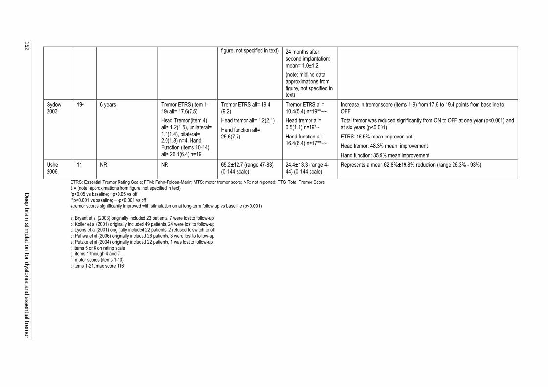
152 D
eep brain stimulation for dystonia and essential trem
or
figure, not specified in text) 24 months after second implantation: mean= 1.0±1.2 (note: midline data approximations from figure, not specified in text)
Sydow 2003
19z 6 years Tremor ETRS (item 1-19) all= 17.6(7.5) Head Tremor (item 4) all= 1.2(1.5), unilateral= 1.1(1.4), bilateral= 2.0(1.8) n=4. Hand Function (items 10-14) all= 26.1(6.4) n=19
Tremor ETRS all= 19.4 (9.2) Head tremor all= 1.2(2.1) Hand function all= 25.6(7.7)
Tremor ETRS all= 10.4(5.4) n=19**~~ Head tremor all= 0.5(1.1) n=19*~ Hand function all= 16.4(6.4) n=17**~~
Increase in tremor score (items 1-9) from 17.6 to 19.4 points from baseline to OFF Total tremor was reduced significantly from ON to OFF at one year (p<0.001) and at six years (p<0.001) ETRS: 46.5% mean improvement Head tremor: 48.3% mean improvement Hand function: 35.9% mean improvement
Ushe 2006
11 NR NR 65.2±12.7 (range 47-83) (0-144 scale)
24.4±13.3 (range 4-44) (0-144 scale)
Represents a mean 62.8%±19.8% reduction (range 26.3% - 93%)
ETRS: Essential Tremor Rating Scale; FTM: Fahn-Tolosa-Marin; MTS: motor tremor score; NR: not reported; TTS: Total Tremor Score $ = (note: approximations from figure, not specified in text) *p<0.05 vs baseline; ~p<0.05 vs off **p<0.001 vs baseline; ~~p<0.001 vs off #tremor scores significantly improved with stimulation on at long-term follow-up vs baseline (p<0.001) a: Bryant et al (2003) originally included 23 patients, 7 were lost to follow-up b: Koller et al (2001) originally included 49 patients, 24 were lost to follow-up c: Lyons et al (2001) originally included 22 patients, 2 refused to switch to off d: Pahwa et al (2006) originally included 26 patients, 3 were lost to follow-up e: Putzke et al (2004) originally included 22 patients, 1 was lost to follow-up f: items 5 or 6 on rating scale g: items 1 through 4 and 7 h: motor scores (items 1-10) i: items 1-21, max score 116
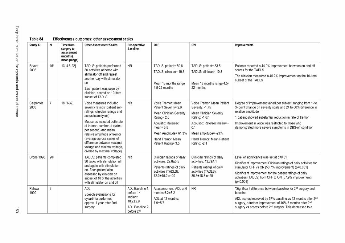
Deep brain stim
ulation for dystonia and essential tremor 153
Table 84 Effectiveness outcomes: other assessment scales Study ID N Time from
surgery to assessment (months) mean [range]
Other Assessment Scales Pre-operative Baseline
OFF
ON Improvements
Bryant 2003
16a 13 [4.5-22] TADLS: patients performed 30 activities at home with stimulator off and repeat another day with stimulator on Each patient was seen by clinician, scored on 10-item subset of TADLS
NR TADLS: patient= 59.8 TADLS: clinician= 19.6 Mean 13 months range 4.5-22 months
TADLS: patient= 33.5 TADLS: clinician= 10.8 Mean 13 months range 4.5-22 months
Patients reported a 44.0% improvement between on and off scores for the TADLS The clinician measured a 45.2% improvement on the 10-item subset of the TADLS
Carpenter 2003
7 18 [1-32] Voice measures included severity ratings (patient self-ratings, clinician ratings and acoustic analyses) Measures included both rate of tremor (number of cycles per second) and mean relative amplitude of tremor (average across cycles of difference between maximal voltage and minimal voltage, divided by maximal voltage)
NR Voice Tremor: Mean Patient Severity= 2.6 Mean Clinician Severity Rating= 2.6 Acoustic: Rate/sec mean= 3.5 Mean Amplitude= 61.3% Hand Tremor: Mean Patient Rating= 3.5
Voice Tremor: Mean Patient Severity: -1.75 Mean Clinician Severity Rating: -1.67 Acoustic: Rate/sec mean= -0.1 Mean amplitude= -23% Hand Tremor: Mean Patient Rating: -2.1
Degree of improvement varied per subject, ranging from 1- to 3- point change on severity scale and 24 to 60% difference in relative amplitude 1 patient showed substantial reduction in rate of tremor Improvement in voice was restricted to those who demonstrated more severe symptoms in DBS-off condition
Lyons 1998 20b TADLS: patients completed 30 tasks with stimulation off and again with stimulation on. Each patient also assessed by clinician on subset of 10 of the activities with stimulator on and off
NR Clinician ratings of daily activities: 29.6±5.5 Patients ratings of daily activities (TADLS): 72.0±15.2 n=20
Clinician ratings of daily activities: 13.7±4.1 Patients ratings of daily activities (TADLS): 30.3±18.3 n=20
Level of significance was set at p<0.01 Significant improvement Clinician ratings of daily activities for stimulator OFF vs ON (53.7% improvement) (p<0.001) Significant improvement for the patient ratings of daily activities (TADLS) from OFF to ON (57.9% improvement) (p<0.001)
Pahwa 1999
9 ADL Speech evaluations for dysarthria performed approx. 1 year after 2nd surgery
ADL Baseline 1: before 1st implant: 18.2±2.9 ADL Baseline 2: before 2nd
At assessment: ADL at 6 months:6.2±5.2 ADL at 12 months: 7.9±5.7
NR *Significant difference between baseline for 2nd surgery and baseline ADL scores improved by 57% baseline vs 12 months after 2nd surgery, a further improvement of 40% 6 months after 2nd surgery vs scores before 2nd surgery. This decreased to a
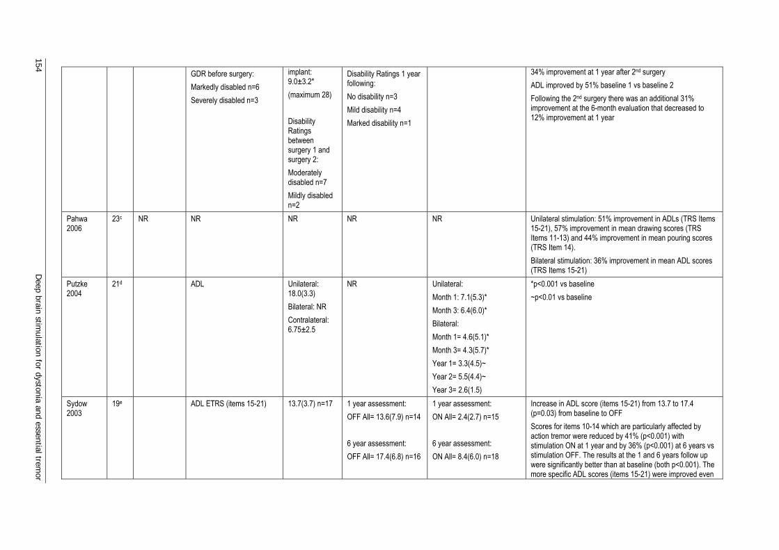
154 D
eep brain stimulation for dystonia and essential trem
or
GDR before surgery: Markedly disabled n=6 Severely disabled n=3
implant: 9.0±3.2* (maximum 28) Disability Ratings between surgery 1 and surgery 2: Moderately disabled n=7 Mildly disabled n=2
Disability Ratings 1 year following: No disability n=3 Mild disability n=4 Marked disability n=1
34% improvement at 1 year after 2nd surgery ADL improved by 51% baseline 1 vs baseline 2 Following the 2nd surgery there was an additional 31% improvement at the 6-month evaluation that decreased to 12% improvement at 1 year
Pahwa 2006
23c NR NR NR NR NR Unilateral stimulation: 51% improvement in ADLs (TRS Items 15-21), 57% improvement in mean drawing scores (TRS Items 11-13) and 44% improvement in mean pouring scores (TRS Item 14). Bilateral stimulation: 36% improvement in mean ADL scores (TRS Items 15-21)
Putzke 2004
21d ADL
Unilateral: 18.0(3.3) Bilateral: NR Contralateral: 6.75±2.5
NR
Unilateral: Month 1: 7.1(5.3)* Month 3: 6.4(6.0)* Bilateral: Month 1= 4.6(5.1)* Month 3= 4.3(5.7)* Year 1= 3.3(4.5)~ Year 2= 5.5(4.4)~ Year 3= 2.6(1.5)
*p<0.001 vs baseline ~p<0.01 vs baseline
Sydow 2003
19e ADL ETRS (items 15-21)
13.7(3.7) n=17 1 year assessment: OFF All= 13.6(7.9) n=14 6 year assessment: OFF All= 17.4(6.8) n=16
1 year assessment: ON All= 2.4(2.7) n=15 6 year assessment: ON All= 8.4(6.0) n=18
Increase in ADL score (items 15-21) from 13.7 to 17.4 (p=0.03) from baseline to OFF Scores for items 10-14 which are particularly affected by action tremor were reduced by 41% (p<0.001) with stimulation ON at 1 year and by 36% (p<0.001) at 6 years vs stimulation OFF. The results at the 1 and 6 years follow up were significantly better than at baseline (both p<0.001). The more specific ADL scores (items 15-21) were improved even
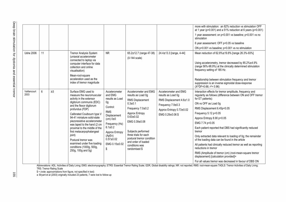
Deep brain stim
ulation for dystonia and essential tremor 155
more with stimulation: an 82% reduction vs stimulation OFF at 1 year (p<0.001) and a 51% reduction at 6 years (p<0.001) 1 year assessment: on p<0.001 vs baseline; p<0.001 vs no stimulation 6 year assessment: OFF p<0.05 vs baseline ON p<0.001 vs baseline; p<0.001 vs no stimulation
Ushe 2006 11 Tremor Analysis System (uniaxial accelerometer connected to laptop via computer interface for data collection and online visualisation) Mean-root-square acceleration used as the index of tremor magnitude
NR 65.2±12.7 (range 47-38) (0-144 scale)
24.4±13.3 [range, 4-44] Mean reduction of 62.8%±19.8% [range 26.3%-93%] Using accelerometry, tremor decreased by 85.2%±4.9% (range 56%-98.9%) at the clinically determined stimulation frequency setting of 185 Hz. Relationship between stimulation frequency and tremor suppression is an inverse sigmoidal dose-response (X2/DF=0.66; r2= 0.96)
Vaillancourt 2003
6 ≥3 Surface EMG used to measure the neuromuscular activity in the extensor digitorum communis (EDC) and the flexor digitorum profundus (FDP) Calibrated Coulbourn type V 94-41 miniature solid-state piezoresistive accelerometer was taped to the hand (2 cm proximal to the middle of the first metacarpophalangeal joint) Postural tremor was examined under five loading conditions (1000g, 500g, 250g, 100g and 0g)
Accelerometer and EMG results as Load 0g Control: RMS Displacement (cm) 0±0 Frequency (Hz) 6.1±0.2 Approx Entropy (ApEn) 0.57±0.02 EMG 0.15±0.02 $
Accelerometer and EMG results as Load 0g RMS Displacement 0.3±0.1 Frequency 7.0±0.2 Approx Entropy 0.63±0.02 EMG 0.39±0.08 Subjects performed three trials for each postural tremor condition and order of loaded conditions was randomised $
Accelerometer and EMG results as Load 0g RMS Displacement 4.6±1.0 Frequency 7.9±0.3 Approx Entropy 0.73±0.02 EMG 0.29±0.06 $
Interaction effects for tremor amplitude, frequency and regularity as follows (difference between ON and OFF tremor for ET patients): ON vs OFF as Load 0g RMS Displacement 6.45p<0.05 Frequency 5.12 p<0.05 Approx Entropy 8.90 p<0.05 EMG 7.74 p<0.05 Each patient reported that DBS had significantly reduced tremor Only extracted data relevant to loading of 0g; the remainder of the loading data can be found in the article All patients had clinically reduced tremor as well as reporting reductions in tremor RMS (Amplitude of tremor (cm) (root-mean-square tremor displacement) [calculation provided]= For all values tremor was decreased in favour of DBS ON
Abbreviations: ADL: Activities of Daily Living; EMG: electromyography; ETRS: Essential Tremor Rating Scale; GDR, Global disability ratings; NR: not reported; RMS: root-mean-square TADLS: Tremor Activities of Daily Living; TRS: Tremor Rating Scale $ = (note: approximations from figure, not specified in text) a: Bryant et al (2003) originally included 23 patients, 7 were lost to follow-up

156 D
eep brain stimulation for dystonia and essential trem
or
b: Lyons et al (1998) originally included 22 patients, 2 refused to switch to off c: Pahwa et al (2006) originally included 26 patients, 3 were lost to follow-up d: Putzke et al (2004) originally included 22 patients, 1 was lost to follow-up e: Sydow et al (2003) originally included 37 patients, 18 were not included in long term follow up
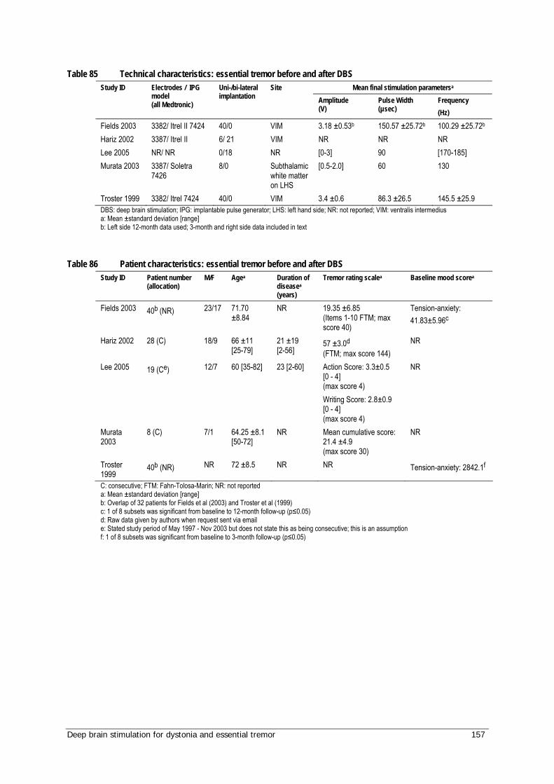
Deep brain stimulation for dystonia and essential tremor 157
Table 85 Technical characteristics: essential tremor before and after DBS Mean final stimulation parametersa Study ID Electrodes / IPG
model (all Medtronic)
Uni-/bi-lateral implantation
Site Amplitude (V)
Pulse Width (µsec)
Frequency (Hz)
Fields 2003 3382/ Itrel II 7424 40/0 VIM 3.18 ±0.53b 150.57 ±25.72b 100.29 ±25.72b Hariz 2002 3387/ Itrel II 6/ 21 VIM NR NR NR Lee 2005 NR/ NR 0/18 NR [0-3] 90 [170-185] Murata 2003 3387/ Soletra
7426 8/0 Subthalamic
white matter on LHS
[0.5-2.0] 60 130
Troster 1999 3382/ Itrel 7424 40/0 VIM 3.4 ±0.6 86.3 ±26.5 145.5 ±25.9 DBS: deep brain stimulation; IPG: implantable pulse generator; LHS: left hand side; NR: not reported; VIM: ventralis intermedius a: Mean ±standard deviation [range] b: Left side 12-month data used; 3-month and right side data included in text
Table 86 Patient characteristics: essential tremor before and after DBS Study ID Patient number
(allocation) M/F Agea Duration of
diseasea (years)
Tremor rating scalea Baseline mood scorea
Fields 2003 40b (NR) 23/17 71.70 ±8.84
NR 19.35 ±6.85 (Items 1-10 FTM; max score 40)
Tension-anxiety: 41.83±5.96c
Hariz 2002 28 (C) 18/9 66 ±11 [25-79]
21 ±19 [2-56]
57 ±3.0d (FTM; max score 144)
NR
Lee 2005 19 (Ce) 12/7 60 [35-82] 23 [2-60] Action Score: 3.3±0.5 [0 - 4] (max score 4) Writing Score: 2.8±0.9 [0 - 4] (max score 4)
NR
Murata 2003
8 (C) 7/1 64.25 ±8.1 [50-72]
NR Mean cumulative score: 21.4 ±4.9 (max score 30)
NR
Troster 1999
40b (NR) NR 72 ±8.5 NR NR Tension-anxiety: 2842.1f
C: consecutive; FTM: Fahn-Tolosa-Marin; NR: not reported a: Mean ±standard deviation [range] b: Overlap of 32 patients for Fields et al (2003) and Troster et al (1999) c: 1 of 8 subsets was significant from baseline to 12-month follow-up (p≤0.05) d: Raw data given by authors when request sent via email e: Stated study period of May 1997 - Nov 2003 but does not state this as being consecutive; this is an assumption f: 1 of 8 subsets was significant from baseline to 3-month follow-up (p≤0.05)
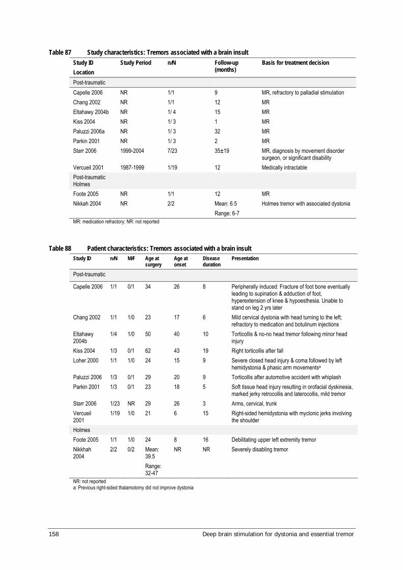
158 Deep brain stimulation for dystonia and essential tremor
Table 87 Study characteristics: Tremors associated with a brain insult Study ID Location
Study Period n/N Follow-up (months)
Basis for treatment decision
Post-traumatic Capelle 2006 NR 1/1 9 MR, refractory to palladial stimulation Chang 2002 NR 1/1 12 MR Eltahawy 2004b NR 1/ 4 15 MR Kiss 2004 NR 1/ 3 1 MR Paluzzi 2006a NR 1/ 3 32 MR Parkin 2001 NR 1/ 3 2 MR Starr 2006 1999-2004 7/23 35±19 MR, diagnosis by movement disorder
surgeon, or significant disability Vercueil 2001 1987-1999 1/19 12 Medically intractable Post-traumatic Holmes
Foote 2005 NR 1/1 12 MR Nikkah 2004 NR 2/2 Mean: 6.5
Range: 6-7 Holmes tremor with associated dystonia
MR: medication refractory; NR: not reported
Table 88 Patient characteristics: Tremors associated with a brain insult Study ID n/N M/F Age at
surgery Age at onset
Disease duration
Presentation
Post-traumatic
Capelle 2006 1/1 0/1 34 26 8 Peripherally induced: Fracture of foot bone eventually leading to supination & adduction of foot, hyperextension of knee & hypoesthesia. Unable to stand on leg 2 yrs later
Chang 2002 1/1 1/0 23 17 6 Mild cervical dystonia with head turning to the left; refractory to medication and botulinum injections
Eltahawy 2004b
1/4 1/0 50 40 10 Torticollis & no-no head tremor following minor head injury
Kiss 2004 1/3 0/1 62 43 19 Right torticollis after fall Loher 2000 1/1 1/0 24 15 9 Severe closed head injury & coma followed by left
hemidystonia & phasic arm movementsa Paluzzi 2006 1/3 0/1 29 20 9 Torticollis after automotive accident with whiplash Parkin 2001 1/3 0/1 23 18 5 Soft tissue head injury resulting in orofacial dyskinesia,
marked jerky retrocollis and laterocollis, mild tremor Starr 2006 1/23 NR 29 26 3 Arms, cervical, trunk Vercueil 2001
1/19 1/0 21 6 15 Right-sided hemidystonia with myclonic jerks involving the shoulder
Holmes Foote 2005 1/1 1/0 24 8 16 Debilitating upper left extremity tremor Nikkhah 2004
2/2 0/2 Mean: 39.5 Range: 32-47
NR NR Severely disabling tremor
NR: not reported a: Previous right-sided thalamotomy did not improve dystonia
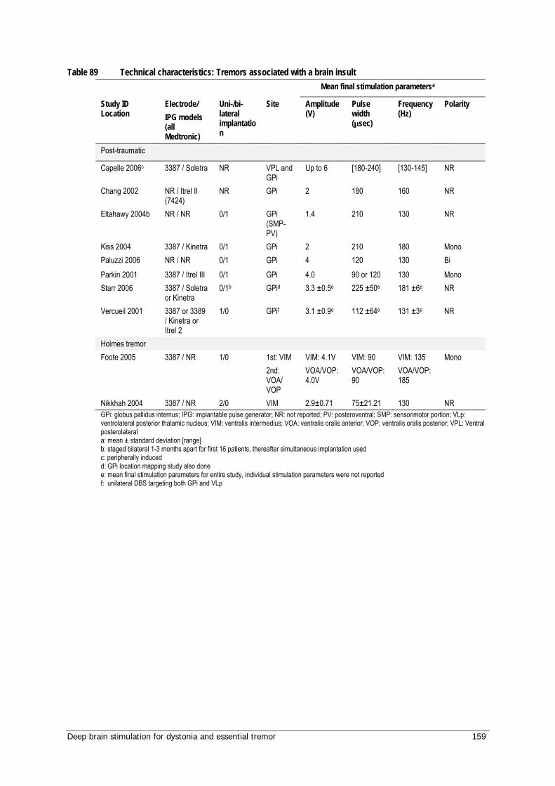
Deep brain stimulation for dystonia and essential tremor 159
Table 89 Technical characteristics: Tremors associated with a brain insult Mean final stimulation parametersa
Study ID Location
Electrode/ IPG models (all Medtronic)
Uni-/bi-lateral implantation
Site Amplitude (V)
Pulse width (µsec)
Frequency (Hz)
Polarity
Post-traumatic
Capelle 2006c 3387 / Soletra NR VPL and GPi
Up to 6 [180-240] [130-145] NR
Chang 2002 NR / Itrel II (7424)
NR GPi 2 180 160 NR
Eltahawy 2004b NR / NR 0/1 GPi (SMP-PV)
1.4 210 130 NR
Kiss 2004 3387 / Kinetra 0/1 GPi 2 210 180 Mono Paluzzi 2006 NR / NR 0/1 GPi 4 120 130 Bi Parkin 2001 3387 / Itrel III 0/1 GPi 4.0 90 or 120 130 Mono Starr 2006 3387 / Soletra
or Kinetra 0/1b GPid 3.3 ±0.5e 225 ±50e 181 ±6e NR
Vercueil 2001 3387 or 3389 / Kinetra or Itrel 2
1/0 GPif 3.1 ±0.9e 112 ±64e 131 ±3e NR
Holmes tremor Foote 2005 3387 / NR 1/0 1st: VIM
2nd: VOA/ VOP
VIM: 4.1V VOA/VOP: 4.0V
VIM: 90 VOA/VOP: 90
VIM: 135 VOA/VOP: 185
Mono
Nikkhah 2004 3387 / NR 2/0 VIM 2.9±0.71 75±21.21 130 NR GPi: globus pallidus internus; IPG: implantable pulse generator; NR: not reported; PV: posteroventral; SMP: sensorimotor portion; VLp: ventrolateral posterior thalamic nucleus; VIM: ventralis intermedius; VOA: ventralis oralis anterior; VOP: ventralis oralis posterior; VPL: Ventral posterolateral a: mean ± standard deviation [range] b: staged bilateral 1-3 months apart for first 16 patients, thereafter simultaneous implantation used c: peripherally induced d: GPi location mapping study also done e: mean final stimulation parameters for entire study, individual stimulation parameters were not reported f: unilateral DBS targeting both GPi and VLp
160 Deep brain stimulation for dystonia and essential tremor
Abbreviations
ADL Activities of Daily Living
AHMAC Australian Health Ministers’ Advisory Council
AIHW Australian Institute of Health and Welfare
AIMS abnormal involuntary movement scale
APPG Australian Prescription Products Guide
AR-DRGs Australian Refined Diagnostic Related Groups
BFMDRS Burke-Fahn-Marsden Dystonia Rating Scale
Botox botulinum toxin
CDRS Cervical Dystonia Rating Scale
CI confidence interval
CNS central nervous system
CT computed tomography
CTRS Clinical Tremor Rating Scale
DBS deep brain stimulation
EMG electromyography
EQ-5D EuroQol, a utility measure commonly used for economic evaluation
ESRS extrapyramidal symptoms rating scale
ET essential tremor
ETRS Essential Tremor Rating Scale
FTM Fahn-Tolosa-Marin Scale
GABA gamma-aminobutyric acid
GDR Global Disability Ratings
GFO Global Functional Outcome
GM1-3g GM1 type 3 gangliosidosis
GOS Glasgow Outcome Score
GPe external pallidum
Deep brain stimulation for dystonia and essential tremor 161
GPi globus pallidus internus
GDS Global Dystonia Rating Scale
GOS Glasgow Outcome Score
GRS Global Rating Scale
HTS Head Tremor Score
ICD International Classification of Diseases
ICER incremental cost-effectiveness ratio
IPG implantable pulse generator
MBS Medicare Benefits Schedule
MRI magnetic resonance imaging
MS multiple sclerosis
MSAC Medical Services Advisory Committee
MTS Motor Tremor Score
NHMRC National Health and Medical Research Council
MMSE Mini Mental State Examination
NHP Nottingham Health Profile
PANK2 pantothenate kinase 2
PDQ-39 Parkinson’s Disease Questionnaire
PKAN pantothenate kinase-associated neurodegeneration
PD Parkinson’s disease
PET positron emission tomography
PFD primary focal dystonia
PGD primary generalised dystonia
PV posteroventral
PVL posteroventrolateral
QALY quality affected life years
QoL quality of life
162 Deep brain stimulation for dystonia and essential tremor
RCT randomised controlled trial
RDP rapid-onset dystonia-Parkinsonism
SD standard deviation
SEM standard error of the mean
SF-36 Short Form-36 health-related quality of life survey
SMR sensorimotor portion
ST spasmodic torticollis
STN subthalamic nucleus
TADLS Tremor Activities of Daily Living Scale
TGA Therapeutic Goods Administration
TMS Total Motor Score
TRS Tremor Rating Scale
TTS Total Tremor Score
TWSTRS Toronto Western Spasmodic Torticollis Rating Scale
UDRS Unified Dystonia Rating Scale
UPDRS Unified Parkinson’s Disease Rating Scale
VAS Visual Analogue Scale
VIM ventralis intermedius
VLp venterolateral posterior thalamic nucleus
VOA ventralis oralis anterior
VOP ventralis oralis posterior
WTP willingness to pay
Deep brain stimulation for dystonia and essential tremor 163
References
Adityanjee, Aderibigbe YA et al, 1999. ‘The current status of tarditive dystonia’, Biological Psychiatry, 45(6), 715-730.
Albanese A, Barnes MP et al, 2006. ‘A systematic review on the diagnosis and treatment of primary (idiopathic) dystonia and dystonia plus syndromes, report of an EFNS/MDS-ES Task Force’, European Journal of Neurology, 13(5), 433-444.
Angelini L, Nardocci N et al, 2000. ‘Life-threatening dystonia-dyskinesias in a child, successful treatment with bilateral pallidal stimulation’, Movement Disorders, 15(5), 1010-1012.
Balas I, Kovacs N & Hollody K, 2006. ‘Staged bilateral stereotactic pallidothalamotomy for life-threatening dystonia in a child with Hallervorden-Spatz disease’, Movement Disorders, 21(1), 82-85.
Benabid AL, Pollak P et al, 1991. ‘Long-term suppression of tremor by chronic stimulation of the ventral intermediate thalamic nucleus’, Lancet, 337(8738), 403-406.
Benabid AL, Benazzous A & Pollak P, 2002. ‘Mechanisms of deep brain stimulation’, Movement Disorders, 17 Suppl 3, S73-S74.
Berk C, Carr J et al, 2002. ‘Thalamic deep brain stimulation for the treatment of tremor due to multiple sclerosis, a prospective study of tremor and quality of life’, Journal of Neurosurgery, 97(4), 815-820.
Bittar RG, Yianni J et al, 2005. ‘Deep brain stimulation for generalized dystonia and spasmodic torticollis’, Journal of Clinical Neuroscience, 12(1), 12-16.
Brazier J & Ara R, 2007. ‘Predicting SF-6D preference-based utilities using mean SF-6D health dimension scores: approximating health state utilities when patient level data are not available’ Quality of Life Research Supplement, A-65 Abstract 82/1392.
Brazier J, Roberts J, Deverill, M, 2002. ‘The estimation of a preference-based measure of health from the SF-36’ Journal of Health Economics, 21(2), 271-292.
Broggi G, Brock S et al, 1993. ‘A case of posttraumatic tremor treated by chronic stimulation of the thalamus’, Movement Disorders, 8(2), 206-208.
Bryant JA, De Salles A et al, 2003. ‘The impact of thalamic stimulation on activities of daily living for essential tremor’, Surgical Neurology, 59(6), 479-484.
Burbaud P, Rougier A et al, 2002. ‘Improvement of severe trunk spasms by bilateral high-frequency stimulation of the motor thalamus in a patient with chorea-acanthocytosis’, Movement Disorders, 17(1), 204-207.
Capelle HH, Grips E et al, 2006. ‘Posttraumatic peripherally-induced dystonia and multifocal deep brain stimulation, case report’, Neurosurgery, 59(3), E702.
Carpenter MA, Pahwa R et al, 1998. ‘Reduction in voice tremor under thalamic stimulation’, Neurology, 50(3), 796-798.
164 Deep brain stimulation for dystonia and essential tremor
Castelnau P, Cif L et al, 2005. ‘Pallidal stimulation improves pantothenate kinase-associated neurodegeneration’, Annals of Neurology, 57(5), 738-741.
Chang JW, Choi JY et al, 2002. ‘Unilateral globus pallidus internus stimulation improves delayed onset post-traumatic cervical dystonia with an ipsilateral focal basal ganglia lesion’, Journal of Neurology, Neurosurgery & Psychiatry, 73(5), 588-590.
Chen R & Hallett M, 1998. ‘Focal dystonia and repetitive motion disorders’, Clinical Orthopaedics & Related Research, 351, 102-106.
Chou KL, Hurtig HI et al, 2005. ‘Bilateral subthalamic nucleus deep brain stimulation in a patient with cervical dystonia and essential tremor’, Movement Disorders, 20(3), 377-380.
Cif L, El Fertit H et al, 2003. ‘Treatment of dystonic syndromes by chronic electrical stimulation of the internal globus pallidus’, Journal of Neurosurgical Sciences, 47(1), 52-55.
Cif L, Valente EM et al, 2004. ‘Deep brain stimulation in myoclonus-dystonia syndrome’, Movement Disorders, 19(6), 724-727.
Cohen DB, Oh MY et al, 2007. ‘Long-term follow-up of thalamic deep brain stimulation for essential tremor’, Stereotactic and Functional Neurosurgery, 85(1), 32-32.
Coubes P, Cif L et al, 2004. ‘Electrical stimulation of the globus pallidus internus in patients with primary generalized dystonia, long-term results’, Journal of Neurosurgery, 101(2), 189-194.
Coubes P, Roubertie A et al, 2000. ‘Treatment of DYT1-generalised dystonia by stimulation of the internal globus pallidus’, Lancet, 355(9222), 2220-2221.
Coubes P, Vayssiere N et al, 2002. ‘Deep brain stimulation for dystonia. Surgical technique’, Stereotactic & Functional Neurosurgery, 78(3-4), 183-191.
Defazio G, Abbruzzese G et al, 2004. ‘Epidemiology of primary dystonia’, Lancet Neurology, 3(11), 673-678.
Detante O, Vercueil L et al, 2004. ‘Globus pallidus internus stimulation in primary generalized dystonia, a H215O PET study’, Brain, 127(Pt 8), 1899-1908.
Deutschlander A, Asmus F et al, 2005. ‘Sporadic rapid-onset dystonia-parkinsonism syndrome, failure of bilateral pallidal stimulation’, Movement Disorders, 20(2), 254-257.
Diamond A, Shahed J et al, 2006. ‘Globus pallidus deep brain stimulation in dystonia’, Movement Disorders, 21(5), 692-695.
Dogu O, Sevim S et al, 2003. ‘Prevalence of essential tremor, door-to-door neurological exams in Mersin Province, Turkey’, Neurology, 61, 1804-1807.
Dolan P, Gudex C et al, 1996. ‘The time trade-off method: results from a general population study’, Health Economics, 5(2), 141-154.
Deep brain stimulation for dystonia and essential tremor 165
Eltahawy HA, Saint-Cyr J et al, 2004a. ‘Primary dystonia is more responsive than secondary dystonia to pallidal interventions, outcome after pallidotomy or pallidal deep brain stimulation’, Neurosurgery, 54(3), 613-619.
Eltahawy HA, Saint-Cyr J et al, 2004b. ‘Pallidal deep brain stimulation in cervical dystonia, clinical outcome in four cases’, Canadian Journal of Neurological Sciences, 31(3), 328-332.
Eltahawy HA, Feinstein A et al, 2004c. ‘Bilateral globus pallidus internus deep brain stimulation in tardive dyskinesia, A case report’, Movement Disorders, 19(8), 969-972.
Essink-Bot ML, Stuifbergen MC et al, 2007. ‘Individual differences in the use of the response scale determine valuations of hypothetical health states: an empirical study’, BMC Health Services Research, 7, 62.
EuroQol Group, 1990. ‘EuroQol--a new facility for the measurement of health-related quality of life. The EuroQol Group’, Health Policy, 16(3), 199-208.
Evidente VGH, Lyons MK et al, 2007. ‘First case of X-linked dystonia-parkinsonism (‘Lubag’) to demonstrate a response to bilateral pallidal stimulation’, Movement Disorders, 22(12), 1790-1793.
Ferreira J & Cristina Sampaio C, 2007. ‘Essential tremor’, BMJ Clinical Evidence, 2007.
Fields JA, Troster AI et al 2003. ‘Neuropsychological and quality of life outcomes 12 months after unilateral thalamic stimulation for essential tremor’, Journal of Neurology, Neurosurgery & Psychiatry, 74(3), 305-311.
Foncke EM, Schuurman PR & Speelman JD, 2006. ‘Suicide after deep brain stimulation of the internal globus pallidus for dystonia’, Neurology, 66(1), 142-143.
Foote KD & Okun MS 2005. ‘Ventralis intermedius plus ventralis oralis anterior and posterior deep brain stimulation for posttraumatic Holmes tremor, two leads may be better than one, technical note’, Neurosurgery, 56(2, Suppl), E445.
Foote KD, Seignourel P et al, 2006. ‘Dual electrode thalamic deep brain stimulation for the treatment of posttraumatic and multiple sclerosis tremor’, Neurosurgery, 58(4 Suppl 2), ONS-5.
Franzini A, Marras C et al, 2005. ‘Long-term high-frequency bilateral pallidal stimulation for neuroleptic-induced tardive dystonia. Report of two cases’, Journal of Neurosurgery, 102(4), 721-725.
Garratt A, Schmidt L et al, 2002. ‘Quality of life measurement: bibliographic study of patient assessed health outcome measures’, British Journal of Medicine, 324 (7351), 1417.
Ghika J, Villemure JG et al, 2002. ‘Postanoxic generalized dystonia improved by bilateral Voa thalamic deep brain stimulation’, Neurology, 58(2), 311-313.
Greene P, 2005. ‘Deep-brain stimulation for generalized dystonia’, New England Journal of Medicine, 352(5), 498-500.
166 Deep brain stimulation for dystonia and essential tremor
Grips E, Blahak C et al, 2007. ‘Patterns of reoccurrence of segmental dystonia after discontinuation of deep brain stimulation’, Journal of Neurology, Neurosurgery & Psychiatry, 78(3), 318-320.
Guehl D, Cuny E et al, 2007. ‘Deep brain pallidal stimulation for movement disorders in neuroacanthocytosis’, Neurology, 68(2), 160-161.
Halbig TD, Gruber D et al, 2005. ‘Pallidal stimulation in dystonia, effects on cognition, mood, and quality of life’, Journal of Neurology, Neurosurgery & Psychiatry 76(12), 1713-1716.
Hariz GM, Bergenheim AT et al, 1998. ‘Assessment of ability/disability in patients treated with chronic thalamic stimulation for tremor’, Movement Disorders, 13(1), 78-83.
Hariz GM, Lindberg M & Bergenheim AT, 2002. ‘Impact of thalamic deep brain stimulation on disability and health-related quality of life in patients with essential tremor’, Journal of Neurology, Neurosurgery & Psychiatry, 72(1), 47-52.
Hariz MI, 2002. ‘Complications of deep brain stimulation surgery’, Movement Disorders, 17(Suppl 3), S162-S166.
Hayflick SJ, 2003. ‘Unraveling the Hallervorden-Spatz syndrome, pantothenate kinase-associated neurodegeneration is the name’, Current Opinions in Pediatrics, 15(6), 572-577.
Holloway KL, Baron MS et al, 2006. ‘Deep brain stimulation for dystonia, A meta-analysis’, Neuromodulation, 9(4), 253-261.
Hubble JP, Busenbark KL et al, 1996. ‘Deep brain stimulation for essential tremor’, Neurology, 46(4), 1150-1153.
Hung SW, Hamani C et al, 2007. ‘Long-term outcome of bilateral pallidal deep brain stimulation for primary cervical dystonia’, Neurology, 68(6), 457-459.
Hurvitz EA, Ellenberg MR et al, 1988. ‘Orthotic technique for dystonia musculorum deformans’, Archives of Physical Medicine and Rehabilitation, 69(10), 892-4.
Jankovic J, 2006. ‘Treatment of dystonia’, Lancet Neurology, 5(10), 864-872.
Joint C, Nandi D et al, 2002. ‘Hardware-related problems of deep brain stimulation’, Movement Disorders, 17(Suppl 3), S175-S180.
Kapoor S, Hortnagel K et al, 2005. ‘Pantothenate kinase associated neurodegeneration (Hallervorden-Spatz syndrome)’, Indian Journal of Pediatrics, 72(3), 261-263.
Kartha N, 2006. ‘Dystonia’, Clinical Geriatric Medicine, 22(4), 899-914, vii.
Katayama Y, Fukaya C et al, 2003. ‘Chronic stimulation of the globus pallidus internus for control of primary generalized dystonia’, Acta Neurochirurgica – Supplement, 87, 125-128.
Katayama Y, Kano T et al, 2005. ‘Difference in surgical strategies between thalamotomy and thalamic deep brain stimulation for tremor control’, Journal of Neurology 252, Suppl-IV22.
Deep brain stimulation for dystonia and essential tremor 167
Kimber TE & Thompson PD, 2000. ‘Increased blink rate in advanced Parkinson’s disease: a form of ‘off’-period dystonia?’, Movement Disorders, 15(5), 982-985.
Kiss ZH, Doig K et al, 2004. ‘The Canadian multicenter trial of pallidal deep brain stimulation for cervical dystonia, preliminary results in three patients’, Neurosurgical Focus, 17(1), E5.
Kleiner-Fisman G, Liang GS et al, 2007. ‘Subthalamic nucleus deep brain stimulation for severe idiopathic dystonia, impact on severity, neuropsychological status, and quality of life’, Journal of Neurosurgery, 107(1), 29-36.
Koller WC, Lyons KE et al, 1999. ‘Efficacy of unilateral deep brain stimulation of the VIM nucleus of the thalamus for essential head tremor’, Movement Disorders, 14(5), 847-850.
Koller WC, Lyons KE et al, 2001. ‘Long-term safety and efficacy of unilateral deep brain stimulation of the thalamus in essential tremor’, Movement Disorders, 16(3), 464-468.
Kosel M, Sturm V et al, 2007. ‘Mood improvement after deep brain stimulation of the internal globus pallidus for tardive dyskinesia in a patient suffering from major depression’, Journal of Psychiatric Research, 41(9), 801-803.
Krause M, Fogel W et al, 2004. ‘Pallidal stimulation for dystonia’, Neurosurgery, 55(6), 1361-1368.
Krause M, Fogel W et al, 2006. ‘Long-term benefit to pallidal deep brain stimulation in a case of dystonia secondary to pantothenate kinase-associated neurodegeneration’, Movement Disorders, 21(12), 2255-2257.
Krauss JK, Loher TJ et al, 2002. ‘Pallidal deep brain stimulation in patients with cervical dystonia and severe cervical dyskinesias with cervical myelopathy’, Journal of Neurology, Neurosurgery & Psychiatry, 72(2), 249-256.
Krauss JK, Pohle T et al, 1999. ‘Bilateral stimulation of globus pallidus internus for treatment of cervical dystonia’, Lancet 354(9181), 837-838.
Kuncel AM, Cooper SE et al, 2006. ‘Bilateral stimulation of globus pallidus internus for treatment of cervical dystonia’, Lancet, 354(9181), 837-838.
Kuncel AM, Cooper SE et al, 2006. ‘Clinical response to varying the stimulus parameters in deep brain stimulation for essential tremor’, Movement Disorders 21(11), 1920-1928.
Kupsch A, Benecke R et al, 2006. ‘Pallidal deep-brain stimulation in primary generalized or segmental dystonia’, New England Journal of Medicine, 355(19), 1978-1990.
Kupsch A, Klaffke S et al, 2003. ‘The effects of frequency in pallidal deep brain stimulation for primary dystonia.[erratum appears in J Neurol. 2004 Aug;251(8),1031]’, Journal of Neurology, 250(10), 1201-1205.
Lee JY & Kondziolka D, 2005. ‘Thalamic deep brain stimulation for management of essential tremor’, Journal of Neurosurgery, 103(3), 400-403.
168 Deep brain stimulation for dystonia and essential tremor
Leehey MA, Munhoz RP et al, 2003. ‘The fragile x premutation presenting as essential tremor’, Archives of Neurology, 60(1), 117-121.
Legros A, Diakonova N et al, 2004. ‘Accelerometric measurement of involuntary movements during pallidal deep brain stimulation of patients with generalized dystoni’, Brain Research Bulletin, 64(4), 363-369.
Lim VK, 2007. ‘Health related quality of life in patients with dystonia and their caregivers in New Zealand and Australia’, Movement Disorders, 22(7), 998-1003.
Logroscino G, Livrea P et al, 2003. ‘Agreement among neurologists on the clinical diagnosis of dystonia at different body sites’, Journal of Neurology, Neurosurgery & Psychiatry, 74(3), 348-350.
Loher TJ, Hasdemir MG et al, 2000. ‘Long-term follow-up study of chronic globus pallidus internus stimulation for posttraumatic hemidystonia’, Journal of Neurosurgery, 92(3), 457-460.
Louis ED, 1999. ‘A new twist for stopping the shakes? Revisiting GABAergic therapy for essential tremor’, Archives of Neurology, 56(7), 807-808.
Louis ED, 2001. ‘Clinical practice. Essential tremor’, New England Journal of Medicine, 345(12), 887-891.
Louis ED, 2005. ‘Essential tremor’, Lancet Neurology, 4(2), 100-110.
Louis ED, 2006. ‘Essential tremor’, Clinics in Geriatric Medicine, 22(4), 843-857.
Louis ED, Benito-Leon J, Ottman R & Bermejo-Pareja F (2007). A population-based study of mortality in essential tremor. Neurology 69(21): 1982-1989.
Louis ED, Ottman R & Hauser WA, 1998. ‘How common is the most common adult movement disorder? estimates of the prevalence of essential tremor throughout the world’, Movement Disorders, 13(1), 5-10.
Lyons KE, Pahwa R et al, 1998. ‘Improvements in daily functioning after deep brain stimulation of the thalamus for intractable tremor’, Movement Disorders, 13(4), 690-692.
Marras C & Broggi G, 2006. ‘Deep brain stimulation of the globus pallidus for generalized dystonia in GM1 type 3 gangliosidosis, Technical case report – Commentary’, Neurosurgery, 59(6), E1340.
McIntosh E, Gray A & Aziz T, 2003. ‘Estimating the costs of surgical innovations: the case for subthalamic nucleus stimulation in the treatment of advanced Parkinson’s disease’, Movement Disorders, 18(9), 993-999.
Merello M, Nouzeilles MI et al, 1999. ‘Unilateral radiofrequency lesion versus electrostimulation of posteroventral pallidum, a prospective randomized comparison’, Movement Disorders, 14(1), 50-56.
Moberg-Wolff EA, 1998. ‘An aggressive approach to limb dystonia: a case report’, Archives of Physical Medicine and Rehabilitation, 79(5), 589-90.
Deep brain stimulation for dystonia and essential tremor 169
Mouton S, Xie-Brustolin J et al, 2006. ‘Chorea induced by globus pallidus externus stimulation in a dystonic patient’, Movement Disorders, 21(10), 1771-1773.
Murata J, Kitagawa M et al, 2003. ‘Electrical stimulation of the posterior subthalamic area for the treatment of intractable proximal tremor’, Journal of Neurosurgery, 99(4), 708-715.
Nandi D, Parkin S et al, 2002. ‘Camptocormia treated with bilateral pallidal stimulation, case report’, Neurosurgical Focus, 12(2), ECP2
NHMRC, 1999. ‘A guide to the development, implementation and evaluation of clinical practice guidelines’, National Health and Medical Research Council, Canberra.
NHMRC, 2000. ‘How to use the evidence, assessment and application of scientific evidence’, National Health and Medical Research Council, Canberra.
Nicholson T, Milne R, 1999. ‘Pallidotomy, thalamotomy and deep brain stimulation for severe Parkinson’s disease’, Development and Evaluation Committee Report No. 105 Wessex Institute for Health Research and Development, Southampton.
Nikkhah G, Prokop T et al, 2004. ‘Deep brain stimulation of the nucleus ventralis intermedius for Holmes (rubral) tremor and associated dystonia caused by upper brainstem lesions. Report of two cases’, Journal of Neurosurgery, 100(6), 1079-1083.
Nutt JG, Muenter MD et al, 1988. ‘Epidemiology of dystonia in Rochester, Minnesota’, Advances in Neurology, 50, 361-365.
Obwegeser AA, Uitti RJ et al, 2000. ‘Thalamic stimulation for the treatment of midline tremors in essential tremor patients’, Neurology, 54(12), 2342-2344.
Oh MY, Kim SH et al, 2001. ‘Hardware-related complications of deep brain stimulation in 100 consecutive electrodes’, Journal of Neurosurgery, 94, 412A.
Okun MS, Tagliati M et al, 2005. ‘Management of referred deep brain stimulation failures: a retrospective analysis from 2 movement disorders centers’, Archives of Neurology, 62(8), 1250-1255.
Ondo W, Jankovic J et al, 1998. ‘Unilateral thalamic deep brain stimulation for refractory essential tremor and Parkinson’s disease tremor’, Neurology, 51(4), 1063-1069
Page D, Butler A & Jahanshahi M, 2007. ‘Quality of life in focal, segmental, and generalized dystonia’, Movement Disorders, 22(3), 341-347.
Pahwa R & Lyons KE, 2003. ‘Essential tremor, differential diagnosis and current therapy’, American Journal of Medicine, 115(2), 134-142.
Pahwa R, Lyons KE et al, 2001. ‘Comparison of thalamotomy to deep brain stimulation of the thalamus in essential tremor’, Movement Disorders, 16(1), 140-143.
Pahwa R, Lyons KE et al, 2006. ‘Long-term evaluation of deep brain stimulation of the thalamus’, Journal of Neurosurgery, 104(4), 506-512.
Pahwa R, Lyons KL et al, 1999. ‘Bilateral thalamic stimulation for the treatment of essential tremor’, Neurology, 53(7), 1447-1450.
170 Deep brain stimulation for dystonia and essential tremor
Paluzzi A, Bain PG et al, 2006a. ‘Pregnancy in dystonic women with in situ deep brain stimulators’, Movement Disorders, 21(5), 695-698.
Paluzzi A, Belli A et al, 2006b. ‘Operative and hardware complications of deep brain stimulation for movement disorders’, British Journal of Neurosurgery, 20(5), 290-295.
Papavassiliou E, Rau G et al, 2004. ‘Thalamic deep brain stimulation for essential tremor, relation of lead location to outcome’, Neurosurgery, 54(5), 1120-1129.
Parkin S, Aziz T et al, 2001. ‘Bilateral internal globus pallidus stimulation for the treatment of spasmodic torticollis’, Movement Disorders, 16(3), 489-493.
Plaha P, Patel NJ & Gill SS, 2004. ‘Stimulation of the subthalamic region for essential tremor’, Journal of Neurosurgery, 101 J Neurosurg 101: 48-54
Putzke JD, Wharen RE et al, 2004. ‘Thalamic deep brain stimulation for essential tremor, recommendations for long-term outcome analysis’, Canadian Journal of Neurological Sciences, 31(3), 333-342.
Rincon F & Louis ED, 2005. ‘Benefits and risks of pharmacological and surgical treatments for essential tremor, disease mechanisms and current management’, Expert Opinion on Drug Safety, 4(5), 899-913.
Roze E, Navarro S et al, 2006. ‘Deep brain stimulation of the globus pallidus for generalized dystonia in GM1 Type 3 gangliosidosis, technical case report’, Neurosurgery, 59(6), E1340.
Schrader C, Peschel T et al, 2004. ‘Unilateral deep brain stimulation of the internal globus pallidus alleviates tardive dyskinesia’, Movement Disorders, 19(5), 583-585.
Schuurman PR, Bosch DA et al, 2000. ‘A comparison of continuous thalamic stimulation and thalamotomy for suppression of severe tremor’, New England Journal of Medicine, 342(7), 461-468.
Sellal F, Hirsch E et al, 1993. ‘A case of symptomatic hemidystonia improved by ventroposterolateral thalamic electrostimulation’, Movement Disorders, 8(4), 515-518.
Starr PA, Turner RS et al, 2006. ‘Microelectrode-guided implantation of deep brain stimulators into the globus pallidus internus for dystonia, techniques, electrode locations, and outcomes’, Journal of Neurosurgery, 104(4), 488-501.
Starr PA, Vitek JL & Bakay RAE, 1998. ‘Deep brain stimulation for movement disorders’, Neurosurgery Clinics of North America, 9(2), 381-402.
Sydow O, Thobois S et al, 2003. ‘Multicentre European study of thalamic stimulation in essential tremor, A six year follow up’, Journal of Neurology, Neurosurgery and Psychiatry, 74(10), 1387-1391.
Tann B, Dias L et al, 2006. ‘Utilization of deep brain stimulation as adjunct treatment for control of truncal dystonia in cerebral palsy, a case report’, Developmental Medicine & Child Neurology, 48, 46-46.
Deep brain stimulation for dystonia and essential tremor 171
Tasker RR, 1998. ‘Deep brain stimulation is preferable to thalamotomy for tremor suppression’, Surgical Neurology, 49(2), 145-153.
Tisch S, Limousin P et al, 2006. ‘Changes in forearm reciprocal inhibition following pallidal stimulation for dystonia’, Neurology, 66(7), 1091-1093.
Tisch S, Rothwell JC et al, 2007. ‘Pallidal stimulation modifies after-effects of paired associative stimulation on motor cortex excitability in primary generalised dystonia’, Experimental Neurology, 206(1), 80-85.
Troster AI, Fields JA et al, 1999. ‘Neuropsychological and quality of life outcome after thalamic stimulation for essential tremor’, Neurology, 53(8), 1774-1780.
Trottenberg T, Volkmann J et al, 2005. ‘Treatment of severe tardive dystonia with pallidal deep brain stimulation’, Neurology, 64(2), 344-346.
Umemura A, Jaggi JL et al, 2004. ‘Pallidal deep brain stimulation for longstanding severe generalized dystonia in Hallervorden-Spatz syndrome, Case report’, Journal of Neurosurgery, 100(4), 706-709.
Ushe M, Mink JW et al, 2006. ‘Postural tremor suppression is dependent on thalamic stimulation frequency’, Movement Disorders, 21(8), 1290-1292.
Vaillancourt DE, Sturman MM et al, 2003. ‘Deep brain stimulation of the VIM thalamic nucleus modifies several features of essential tremor’, Neurology, 61(7), 919-925.
Vercueil L, Pollak P et al, 2001. ‘Deep brain stimulation in the treatment of severe dystonia’, Journal of Neurology, 248(8), 695-700.
Vidailhet M, Vercueil L et al, 2005. ‘Bilateral deep-brain stimulation of the globus pallidus in primary generalized dystonia’, New England Journal of Medicine, 352(5), 459-467.
Vidailhet M, Vercueil L et al, 2007. ‘Bilateral, pallidal, deep-brain stimulation in primary generalised dystonia: a prospective 3 year follow-up study’, Lancet Neurology, 6(3), 223-229.
Voges J, Waerzeggers Y et al, 2006. ‘Deep-brain stimulation, long-term analysis of complications caused by hardware and surgery--experiences from a single centre’, Journal of Neurology, Neurosurgery and Psychiatry, 77(7), 868-872.
Wang S, Liu X et al, 2006. ‘Use of surface electromyography to assess and select patients with idiopathic dystonia for bilateral pallidal stimulation’, Journal of Neurosurgery, 105(1), 21-25.
Warner, 2000. ‘A prevalence study of primary dystonia in eight European countries’, Journal of Neurology, 247(10), 787-792.
Yianni J, Bain P et al, 2003. ‘Globus pallidus internus deep brain stimulation for dystonic conditions, a prospective audit’, Movement Disorders, 18(4), 436-442.
Yianni J, Green AL et al, 2005. ‘The Costs and Benefits of Deep Brain Stimulation Surgery for Patients with Dystonia: An Initial Exploration’, Neuromodulation, 8(3), 155-161.

172 Deep brain stimulation for dystonia and essential tremor
Yianni J, Nandi D et al, 2004. ‘Increased risk of lead fracture and migration in dystonia compared with other movement disorders following deep brain stimulation’, Journal of Clinical Neuroscience, 11(3), 243-245.
Zesiewicz T, Elble R et al, 2005. ‘Practice parameter, Therapies for essential tremor. Report of the quality standards subcommittee of the American academy of neurology’, Neurology, 64(12), 2008-2020.
Zhou B, Westaway SK et al, 2001. ‘A novel pantothenate kinase gene (PANK2) is defective in Hallervorden-Spatz syndrome’, Nature Genetics, 28(4), 345-349.
Zorzi G, Marras C et al, 2005. ‘Stimulation of the globus pallidus internus for childhood-onset dystonia’, Movement Disorders 20(9), 1194-1200.


![Successful Pallidal Deep Brain Stimulation Treatment in a ...downloads.hindawi.com/journals/crinm/2019/3154653.pdf · dystonia [2], lower limb dystonia [3], blepharospasm, myo-clonus](https://static.fdocuments.us/doc/165x107/603f7e59c9407644c91de6c2/successful-pallidal-deep-brain-stimulation-treatment-in-a-dystonia-2-lower.jpg)