Deconstructing Postmastectomy...
17
Deconstructing Postmastectomy Syndrome Implications for Physiatric Management Eric Wisotzky, MD a, *, Nicole Hanrahan, MD b , Thomas P. Lione, DO c , Susan Maltser, DO d BACKGROUND Epidemiology of Breast Cancer Breast cancer is the most commonly diagnosed cancer among women in the United States, irrespective of race or ethnicity, accounting for nearly 1 in 3 cancers. 1 Each year, there are more than 230,000 new cases of breast cancer in the United States and more than 40,000 deaths. 2 It is the second leading cause of cancer death among women after lung cancer and the most common cause of death from cancer among Hispanic women according to the Centers for Disease Control and Prevention. One in 8 women will develop invasive breast cancer. 3 Approximately one million new cases The authors have no commercial or financial conflicts of interest. a MedStar National Rehabilitation Network, Georgetown University School of Medicine, 102 Irving Street, Northwest, Washington, DC 20010, USA; b MedStar National Rehabilitation Network, 102 Irving Street, Northwest, Washington, DC 20010, USA; c Department of Physical Medicine and Rehabilitation, Hofstra Northwell School of Medicine, 1554 Northern Boulevard, 4th Floor, Manhasset, NY 11030, USA; d Cancer Rehabilitation, Department of Physical Medi- cine and Rehabilitation, Hofstra Northwell School of Medicine, 1554 Northern Boulevard, 4th Floor, Manhasset, NY 11030, USA * Corresponding author. E-mail address: [email protected] KEYWORDS Postmastectomy Pain Breast cancer Cancer rehabilitation Intercostobrachial KEY POINTS Postmastectomy pain syndrome is a prevalent disorder that negatively impacts the func- tion and quality of life of breast cancer survivors. The key to successful treatment of postmastectomy pain is a thorough evaluation to iden- tify the specific anatomic structures that lead to this pain syndrome. After determining the anatomic cause of postmastectomy pain, there are many potential treatments, including rehabilitation, medications, injections, and other nonpharmacologic treatments. Phys Med Rehabil Clin N Am 28 (2017) 153–169 http://dx.doi.org/10.1016/j.pmr.2016.09.003 pmr.theclinics.com 1047-9651/17/ª 2016 Elsevier Inc. All rights reserved.
Transcript of Deconstructing Postmastectomy...
DeconstructingPostmastectomy Syndrome
Implications for Physiatric ManagementEric Wisotzky, MDa,*, Nicole Hanrahan, MDb,Thomas P. Lione, DOc, Susan Maltser, DOd
KEYWORDS
� Postmastectomy � Pain � Breast cancer � Cancer rehabilitation � Intercostobrachial
KEY POINTS
� Postmastectomy pain syndrome is a prevalent disorder that negatively impacts the func-tion and quality of life of breast cancer survivors.
� The key to successful treatment of postmastectomy pain is a thorough evaluation to iden-tify the specific anatomic structures that lead to this pain syndrome.
� After determining the anatomic cause of postmastectomy pain, there are many potentialtreatments, including rehabilitation, medications, injections, and other nonpharmacologictreatments.
BACKGROUNDEpidemiology of Breast Cancer
Breast cancer is the most commonly diagnosed cancer among women in the UnitedStates, irrespective of race or ethnicity, accounting for nearly 1 in 3 cancers.1 Eachyear, there are more than 230,000 new cases of breast cancer in the United Statesand more than 40,000 deaths.2 It is the second leading cause of cancer death amongwomen after lung cancer and the most common cause of death from cancer amongHispanic women according to the Centers for Disease Control and Prevention. Onein 8 women will develop invasive breast cancer.3 Approximately one million new cases
The authors have no commercial or financial conflicts of interest.a MedStar National Rehabilitation Network, Georgetown University School of Medicine, 102Irving Street, Northwest, Washington, DC 20010, USA; b MedStar National RehabilitationNetwork, 102 Irving Street, Northwest, Washington, DC 20010, USA; c Department of PhysicalMedicine and Rehabilitation, Hofstra Northwell School of Medicine, 1554 Northern Boulevard,4th Floor, Manhasset, NY 11030, USA; d Cancer Rehabilitation, Department of Physical Medi-cine and Rehabilitation, Hofstra Northwell School of Medicine, 1554 Northern Boulevard,4th Floor, Manhasset, NY 11030, USA* Corresponding author.E-mail address: [email protected]
Phys Med Rehabil Clin N Am 28 (2017) 153–169http://dx.doi.org/10.1016/j.pmr.2016.09.003 pmr.theclinics.com1047-9651/17/ª 2016 Elsevier Inc. All rights reserved.
Wisotzky et al154
are diagnosed globally every year, and this number is expected to increase in futuredecades.4 As a result, more women will likely undergo surgical procedures for thetreatment of breast cancer, and the incidence of postoperative complications andpain syndromes is likely to increase.
Surgical Techniques in the Treatment of Breast Cancer
In the past several decades, the standard of care for the treatment of breast cancerinvolved radical mastectomy and total axillary dissection to achieve local tumor con-trol and increase the likelihood of cure.5 Over the years, less invasive surgical ap-proaches have become the standard of care and include modified radicalmastectomy, total (simple) mastectomy, and more recently, skin-sparing mastectomyand nipple-sparing mastectomy.6 These less invasive surgical techniques havecontinued to gain favor over time. These more conservative surgical approacheshave enhanced rates of local control and 5-year survival, in part due to a gradual shiftfrom unimodal to multimodal treatment approaches.Treatment options for patients with early stage breast cancer typically include total
mastectomy or breast-conserving surgery with adjuvant radiation therapy.7 Total mas-tectomy involves removal of skin, nipple, areola, breast tissue, and the fascia of thepectoralis major. Patients who have undergone total mastectomy may be candidatesfor immediate breast reconstruction, which is increasingly used with a skin-sparingmastectomy technique, especially for women with smaller breasts. Regardless of spe-cific technique, all patients with invasive breast cancer should undergo sampling ofaxillary lymph nodes for proper staging and treatment.Until the late 1990s, it was standard of practice for patients to undergo 2- to 3-level
axillary lymph node dissection (ALND) in addition to breast-conserving treatment ormastectomy.7 The extent of lymph node dissection can potentially be limited by theidentification of the axillary sentinel lymph node(s) (SLN) using a radiolabeled isotope.Based on predictable patterns of lymphatic drainage within the breast, SLN identifica-tion limits nodal dissection and may allow for immediate completion ALND in caseswith metastatic disease within the sentinel node.7,8
Finally, women who undergo mastectomy for the treatment or prevention of breastcancer often consider cosmetic reconstruction. Multiple surgical techniques arecurrently used and include single-stage reconstruction, tissue expansion followedby implant, combined autologous tissue/implant reconstruction, and autologous tis-sue reconstruction alone.7 Common approaches to autologous breast reconstructioninclude transverse rectus abdominus muscle flap, latissimus flap, and deep inferiorepigastric perforator flap. Reconstruction is deemed safe from an oncology perspec-tive and can improve appearance, sense of femininity, and self-esteem.7
POSTMASTECTOMY PAIN SYNDROMEDefinition
Postmastectomy pain syndrome (PMPS) refers to persistent pain following any breastsurgery, not just mastectomy.9,10 Pain has been reported following other breast pro-cedures, including lumpectomy, breast reconstruction, augmentation, and reduction,although procedures targeting the upper outer quadrant of the breast or axilla areparticularly prone to pain syndromes.9,11,12 For a diagnosis of PMPS, pain mustpersist for more than 3 months postoperatively when all other causes of pain, suchas infection or tumor recurrence, have been excluded13; however, no specific diag-nostic criteria are universally accepted. Persistent pain after mastectomy was first re-ported during the 1970s and was characterized as a dull, burning, and/or aching

Deconstructing Postmastectomy Syndrome 155
sensation in the anterior chest, arm, and axilla, often worsened by movement of theshoulder.14 There are various potential causes of PMPS, including intraoperative dam-age to the intercostobrachial nerve, axillary nerve, or chest wall; phantom breast pain;incisional pain; musculoskeletal pain; and pain caused by neuroma.9,15 Although tradi-tionally, PMPS has been defined in the literature as a neuropathic pain syndrome, theauthors of this article believe this to be an inaccurate generalization. Many patientswith PMPS have musculoskeletal pain syndromes with no apparent component ofneuropathic pain.
Epidemiology
Reported incidence rates of PMPS have varied significantly, perhaps as a result of theabsence of a formalized definition or diagnostic criteria. Most studies of chronic painfollowing breast surgery report an incidence ranging from 20% to 70%.10,15,16 Onestudy showed 44% of breast cancer survivors still with arm pain more than 4 years af-ter breast surgery.17 It is likely that incidence rates differ across anatomic subsets ofpostmastectomy pain. Therefore, more accurate and clinically relevant incidence es-timates may require broad acceptance and application of subset-specific diagnoses.An important characteristic that distinguishes PMPS subtypes and has direct import
for clinical decision making is the presumed pathophysiology of the pain. Nociceptivepain occurs as a result of surgical injury and typically resolves as damaged tissueheals. Musculoskeletal pain syndromes are common nociceptive causes of PMPS,but for most, epidemiology remains uncharacterized. In contrast, neuropathic pain re-sults from dysfunction of the nervous system and may be difficult to treat.18 Severaldifferent types of neuropathic pain following breast surgery have been described.Phantom breast pain may occur after radical mastectomy or modified radical mastec-tomy and refers to the sensation of a removed breast or nipple that is painful.17 Studieshave estimated the prevalence of phantom breast pain from 13% to 44%.17 Intercos-tobrachial neuralgia refers to pain related to surgical injury to the intercostobrachialnerve, which may occur following axillary dissection and is a commonly discussedcause of PMPS in the literature, although rigorous incidence estimates are lacking.17
Neuromas are another cause of chronic pain after breast surgery and may formfollowing injury to any nerve.17 Neuromas can form within scars following both mas-tectomy and lumpectomy, although pain from neuroma may be more commonfollowing lumpectomy or in patients who have undergone concomitant ALND and ra-diation.19 The prevalence of neuroma pain following breast surgery varies throughoutthe literature and ranges from 20% to 50%.17 Additional causes of neuropathic painare also possible and include damage to the intercostal, thoracodorsal, medial andlateral pectoral nerves, and long thoracic nerves.20
Clinical Presentation
Patients with PMPS typically present with neuropathic or musculoskeletal pain symp-toms. Symptom onset may be in the immediate postoperative period, but can alsooccur several months after surgery and persist beyond the normal timeframe for sur-gical healing.21 Pain is commonly localized to the axilla, operative site, and/or ipsilat-eral arm.16,22 Patients may also experience pain in the chest wall and/or shoulder withaccompanying limitations in range of motion, or decreased handgrip strength.23
Risk Factors
Reasons for the development of persistent pain following a mastectomy are not clearlyunderstood and are likely multifactorial. Risk factors span clinical and demographicdomains, including the severity of acute postoperative pain, adjuvant radiation,
Wisotzky et al156
ALND, and psychosocial factors.10 Severe postoperative pain that becomes chronichas been studied after various surgical procedures. Persistent acute pain is thoughtto activate mechanisms within the central and peripheral nervous systems, leadingto sensitization. This, in turn, leads to allodynia, hyperalgesia, and hyperpathia thatcan affect physical functioning and lead to chronic pain. In a study of 569 patients re-ported by Tasmuth and colleagues,24 patients with significant postoperative pain weremore likely to develop persistent ipsilateral arm pain compared with those whose painwas less severe.
Demographic risk factorsYounger age at diagnosis is associated with increased risk for PMPS.25 Although thereason for this is poorly understood, several mechanisms have been proposed. Thesemechanisms include greater sensitivity to nerve damage in younger people, lower painthresholds, and increased preoperative anxiety.13,26 In addition, younger age may becorrelated with more aggressive treatments, including a more thorough axillarydissection and adjuvant radiotherapy.27
Socioeconomic status has also been associated with PMPS. Patients with lower so-cioeconomic status, including lower annual income and educational level, are predis-posed to develop higher rates of chronic pain. One explanation for this may bediagnosis at later stages of cancer, necessitating more aggressive surgical and adju-vant treatment.28
Treatment- and complication-related risk factorsAlthough it is commonly assumed that PMPS is a sequela of mastectomy, large-scalestudies show no difference in PMPS rates between mastectomy and lumpectomy.10
Rather, it is the extensiveness of ALND, damage to the intercostobrachial nerve,and adjuvant treatment that are presumed to initiate and sustain postoperative andchronic pain.13 Tumors located in the upper outer quadrant may warrant a more exten-sive axillary dissection, leading to greater neuropathic pain.29 Furthermore, the num-ber of axillary lymph nodes dissected as well as the number and location of surgicaldrains may play a role.21 Immediate reconstruction following mastectomy has notbeen found to effect the development of PMPS.30
Adjuvant radiation therapy to the axilla is another potential cause of neuropathicpain in patients with breast cancer. Pain related to radiation can occur months to yearsfollowing treatment, even in those patients who undergo breast conservation surgery.Pain following radiation can present in the breast, chest wall, and ipsilateral arm.31
One study demonstrated that surgical complications, such as cellulitis, and thedevelopment of neuromas and seromas, do not increase the risk of developingchronic pain.
Psychosocial risk factorsIn terms of psychosocial risk factors for the development of PMPS, patients with mod-erate postoperative pain reported significantly higher preoperative levels of depres-sion, anxiety, sleep disturbance, and fatigue as well as lower social well-being andquality of life.16 Almost half of all patients with cancer exhibit some elements of anxietyand depression. In one study of women with breast cancer, 50% were noted to havedepression and anxiety within the first year of diagnosis.32 In a study of 611 patientsundergoing a mastectomy, preoperative catastrophizing was the only independentcontributing factor to predicting clinically significant pain at 2 days after breast sur-gery.29 Catastrophizing and maladaptive coping behavior may mediate postoperativepain by way of central and peripheral sensitization and impaired pain inhibition29
(Box 1).
Box 1
Postmastectomy pain syndrome risk factors
� Severity of acute postmastectomy pain
� Radiation
� ALND
� Younger age
� Low socioeconomic status
� Preoperative and postoperative anxiety and depression, especially catastrophizing
Deconstructing Postmastectomy Syndrome 157
CLINICAL ASSESSMENT OF POSTMASTECTOMY PAIN SYNDROMEHistory
Evaluation of PMPS requires a careful history and physical examination, which shouldfocus on identifying the cause of the pain and the pain generator(s). History shouldinclude the timing of symptom onset, the type of breast surgery (mastectomy vs lump-ectomy), type of reconstruction, and whether the patient had an ALND or SLN biopsy.The clinician should be aware of howmany lymph nodes were removed and howmanywere positive for metastatic disease. In addition, the patient’s tumor stage and gradeshould be noted. Adjuvant treatment should be included in the history, including typeand duration of chemotherapy, radiation therapy, and hormonal therapy.A careful history of the patient’s pain should be taken. The distribution, duration, and
quality of the pain, as well as associated symptoms such as numbness, tingling, andweakness, should be included. Phantom pain/sensations, breast pain, and/or swellingshould be assessed. It is important to determine whether the patient has difficulty withshoulder motion or a history of premorbid shoulder dysfunction. Finally, the patient’spsychosocial status should be evaluated, including mood, coping skills, and extent ofsocial support. Underlying depression and anxiety should be noted and addressed.Functional history, including activities of daily living, as well as vocational and avoca-tional activities, before and after cancer diagnosis should be assessed (Box 2).
Physical Examination
Physical examination includes examination of the breasts, skin, and upper extremitiesfor any signs of cellulitis, radiation-association change, muscle atrophy, scapular
Box 2
Taking a postmastectomy pain syndrome history
� Surgical factors (timing, lumpectomy vs mastectomy, ALND vs SLN biopsy, type ofreconstruction)
� Cancer pathology, stage, grade, estrogen/progesterone receptor status, metastatic disease
� Adjuvant treatments (chemotherapy, radiation, hormone therapy)
� Pain history
� Functional history
� Social status and support system
� Assess for anxiety and depression
Wisotzky et al158
winging, or lymphedema. The surgical incision should be evaluated for signs of woundinfection, adhesions, seromas, and neuromas. The cervical and thoracic spine shouldbe inspected for deformities and range-of-motion testing should be performed. Thetrapezius, serratus anterior, pectoralis muscles, rhomboids, and latissimus musclesshould be palpated for potential asymmetry, trigger points, or other stigmata of myo-fascial dysfunction. For patients who have undergone breast reconstruction, specialattention should be given to the pectoralis and lateral chest wall muscles, as well asthe flap harvest sites, such as the abdomen. The axilla should be palpated for cordingor masses. Shoulder girdle examination should include inspection for asymmetry,passive and active range of motion, as well as palpation and provocative testing ofthe rotator cuff and other musculoskeletal structures. In addition to testing strength,sensation, and deep tendon reflexes of the upper extremity myotomes and derma-tomes, the neurologic examination should focus onmotor testing of themuscles inner-vated by nerves that can be affected by surgery of the breast. These include thethoracodorsal, long thoracic, medial, and lateral pectoral nerves7 (Box 3).
Diagnostic Studies
Diagnostic studies can support and supplement the history and physical examinationand aid the clinician when the diagnosis is unclear. Ultrasound can be helpful in diag-nosing musculoskeletal abnormality. MRI may confirm shoulder, cervical spine, orthoracic spine abnormality.33 A chest computed tomographic scan or MRI can eval-uate potential metastatic disease or brachial plexus injury. PET scanning can alsobe ordered if tumor recurrence or metastatic disease is suspected. Nerve conductionstudies and electromyography can diagnose radiculopathy, plexopathy, mononeuro-pathies, polyneuropathy, or evidence of radiation fibrosis, for example, myokymiaand/or myopathic motor unit action potentials.
Outcome Measures
Although standardized outcomemeasures in the setting of postmastectomy pain havenot been clearly delineated, there are several well-established breast cancer–specificfunctional outcome measures. A prospective surveillance approach with functionalmeasures for breast cancer has been proposed.34 The authors suggested serial func-tional evaluations with a 6-minute walk test, chair stand, shoulder range of motion,hand grip strength, upper extremity functional index or Kwan’s arm problem scale,and functional assessment of cancer therapy-breast or Breast-Q.
Box 3
Performing a postmastectomy pain syndrome physical examination
� Inspecting skin, incision, breast, cervical spine, shoulder girdle (for atrophy, asymmetry, orscapular winging)
� Palpate incision for tenderness, neuromas, and mobility
� Palpate musculature for tender/trigger points
� Palpate axilla and upper extremity for cording
� Range of motion
� Neurologic examination
� Special provocative tests depending on suspected diagnosis (eg, rotator cuff impingementsigns)
Deconstructing Postmastectomy Syndrome 159
POSTMASTECTOMY PAIN SYNDROME DIAGNOSTIC SUBGROUPS
The key to successful treatment of PMPS is to determine the specific pain cause orcauses. The following section details pain generators that can lead to PMPS and sug-gested treatment approaches. Some treatments are evidence based, and somereflect anecdotal experience.
Rotator Cuff Dysfunction
Rotator cuff dysfunction, the leading cause of shoulder pain in the general population,is also a well-recognized phenomenon among breast cancer survivors. The frequencyof rotator cuff–related pain among breast cancer survivors has not been rigorouslyestimated. Altered biomechanics have been implicated in the development of rotatorcuff dysfunction, whether after mastectomy or lumpectomy. Postmastectomy scapu-lothoracic motion has been noted to increase disproportionately to glenohumeral mo-tion, with subsequent rotator cuff microtrauma and tendinopathy. This is due topostoperative glenohumeral range-of-motion limitations and decreased muscle activ-ity in the musculature affecting the scapula (serratus anterior, upper trapezius, pector-alis major, and rhomboid) ipsilateral to the carcinoma.35 The shoulder girdle is placedin a more protracted and inferior position as a result of shortening of the pectoral mus-cles and associated soft tissues, thus narrowing the subacromial space available forthe rotator cuff tendons.36 Symptoms include pain, tightness, decreased function, andweakness. Kyphotic posture can also contribute to pectoralis muscle tightness, result-ing in further rotator cuff impingement. It should be noted that studies have shownaltered scapulothoracic motion contralateral to the cancer, explaining the occurrenceof bilateral shoulder dysfunction, or even unilateral involvement of the unaffected side.Other risk factors may contribute to rotator cuff dysfunction in patients with breast
cancer. Chest wall radiation treatment contributes to muscle and soft tissue tight-ening; fibrosis may be a major factor in this phenomenon. Interestingly, one studydemonstrated that chest wall radiation involving the pectoralis major, regardless ofthe presence of clinically detectable radiation fibrosis, was a significant contributorto shoulder morbidity.37 A review of several studies involving radiotherapy demon-strated that the addition of axillary radiation places patients at increased risk for lateshoulder dysfunction compared with chest wall radiation alone.3 Lymphedema alsocontributes to rotator cuff tendinopathy. The incidence of shoulder pain in lymphe-dema patients is reported as 53% and 71% from 2 separate studies.38,39 Increasedweight of the limb and decreased joint range of motion can aggravate subacromialimpingement, intrinsic tendinopathy, and/or functional overload40 (Fig. 1). In addition,reports have speculated that the increased risk of cellulitis in the lymphedematous armmay decrease healing capacity of tissue and cause pathologic effects on the rotatorcuff, but this has not been validated. Early management of lymphedema has been sug-gested given the association noted between the duration of lymphedema symptomsand, rotator cuff abnormality seen on musculoskeletal ultrasound.37
Anecdotally, conventional rehabilitation techniques for rotator cuff tendinopathy canbe very helpful for this patient population. In addition, injection techniques includingsubacromial corticosteroid injection or percutaneous needle tenotomy may beconsidered.
Intercostobrachial Neuralgia
One of the most common causes of PMPS discussed in the literature is intercostobra-chial neuralgia. The intercostobrachial nerve is a cutaneous branch of T1 and/or T2and provides sensory innervation to the medial upper arm and lateral chest wall.
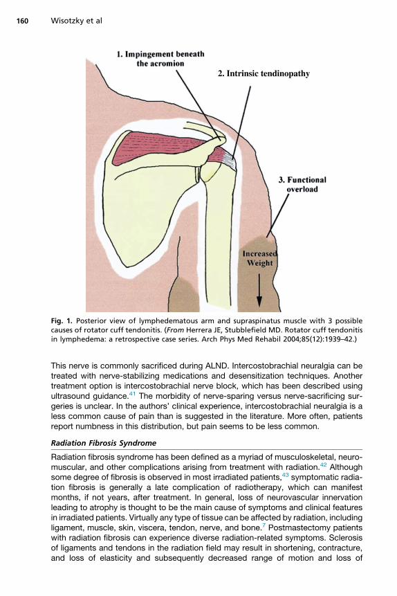
Fig. 1. Posterior view of lymphedematous arm and supraspinatus muscle with 3 possiblecauses of rotator cuff tendonitis. (From Herrera JE, Stubblefield MD. Rotator cuff tendonitisin lymphedema: a retrospective case series. Arch Phys Med Rehabil 2004;85(12):1939–42.)
Wisotzky et al160
This nerve is commonly sacrificed during ALND. Intercostobrachial neuralgia can betreated with nerve-stabilizing medications and desensitization techniques. Anothertreatment option is intercostobrachial nerve block, which has been described usingultrasound guidance.41 The morbidity of nerve-sparing versus nerve-sacrificing sur-geries is unclear. In the authors’ clinical experience, intercostobrachial neuralgia is aless common cause of pain than is suggested in the literature. More often, patientsreport numbness in this distribution, but pain seems to be less common.
Radiation Fibrosis Syndrome
Radiation fibrosis syndrome has been defined as a myriad of musculoskeletal, neuro-muscular, and other complications arising from treatment with radiation.42 Althoughsome degree of fibrosis is observed in most irradiated patients,43 symptomatic radia-tion fibrosis is generally a late complication of radiotherapy, which can manifestmonths, if not years, after treatment. In general, loss of neurovascular innervationleading to atrophy is thought to be the main cause of symptoms and clinical featuresin irradiated patients. Virtually any type of tissue can be affected by radiation, includingligament, muscle, skin, viscera, tendon, nerve, and bone.7 Postmastectomy patientswith radiation fibrosis can experience diverse radiation-related symptoms. Sclerosisof ligaments and tendons in the radiation field may result in shortening, contracture,and loss of elasticity and subsequently decreased range of motion and loss of
Deconstructing Postmastectomy Syndrome 161
function.40 Musculature of the shoulder and axilla can atrophy, leading to weaknessand secondary rotator cuff dysfunction. In addition, irradiated muscles may fibroseto such an extent that a focal myopathy develops.44 Patients with breast cancertreated with radiation also complain of muscle spasms in the chest wall, especiallythe pectoralis major, serratus anterior and latissimus dorsi muscles, thought to bedue to ectopic activity of motor nerves. Irradiated patients may also complain ofskin changes, including thickening, tightness, nipple retraction, and breast edema.45
Radiation fibrosis syndrome is a clinical diagnosis that can be challenging to confirmexclusive of other common neuromusculoskeletal abnormalities.Management of radiation fibrosis is geared toward functional and symptomatic
treatment as well as treatment of the fibrosis. Physical and occupational therapyimprove range of motion, manually release fibrotic tissue, and improve upper extrem-ity function.Nerve-stabilizing medications and opioids have been anecdotally reported in the
literature as effective in radiation fibrosis–associated pain.46 Botulinum toxin injectionhas been examined in the literature as a treatment for the muscle spasms, pain, andneuropathy associated with radiation therapy.40 Pentoxifylline in combination withtocopherol has been validated in the treatment of radiation fibrosis. Pentoxifyllinelimits aberrant transforming growth factor-b activity, thereby limiting fibroblastcollagen proliferation. Anti-inflammatory and immunologic effects have been positedas well. Some studies have reported effectiveness at 1200 mg/d; however, optimaltreatment dose and duration continue to be researched. One study showed promisingresults in patients treated for up to 3 years.41 Tocopherol in doses greater than 400 IU/d (some studies used 400 IU, some 700 IU, and some 1000 IU) has also shown prom-ise in the treatment of radiation fibrosis, although increased all-cause mortality hasalso been noted with similar doses. Tocopherol is thought to scavenge radical oxygenspecies and decrease platelet aggregation, nitric oxide, and superoxide production inmacrophages and neutrophils.41 The combination of pentoxifylline and tocopherol hasbeen established as safe and potentially effective in the treatment of radiationfibrosis.41 Hyperbaric oxygen therapy has also been theorized to be effective for radi-ation fibrosis by stimulating angiogenesis and reducing fibroblast proliferation and tis-sue edema; however, small case series have reported mixed results.41
Chest Wall Pain
Chest wall pain in postmastectomy patients can be related to a multitude of potentialcauses. Pain over incision sites is a common complaint of patients with breast cancer.A study by Skov and colleagues47 noted that 35% of women developed incisional painafter mastectomy, with 92% of these women noting that incisional pain developedwithin the first 3 months. This pain seems to lessen with time; in the same study,only 23% of those who had initially complained of incisional pain still had persistentdiscomfort, and both intensity and duration of pain had diminished as well. Pain isoften due to a hypomobile incision that has adhered to the underlying chest wall. Pain-ful scars can be treated with soft tissue mobilization and scar tissue release with aphysical or occupational therapist. Silicone gel sheeting, triamcinolone cream or injec-tion, or laser treatments may also be helpful adjuncts. If these methods are ineffective,the patient may choose to undergo elective scar resection.
NeuromaNeuroma formation is another cause that can contribute to PMPS. It is hypothesizedthat this pain originates from the T4 and T5 intercostal sensory cutaneous branchesthat arise from the chest wall and enter the breast along with a blood vessel. These
Wisotzky et al162
branches are often cut and cauterized during mastectomy and can cause burning,shooting pain, and point tenderness at the midaxillary line or at the inframammaryfold directly inferior to the nipple.48 Patients may benefit from nerve-stabilizing medi-cations or desensitization techniques in physical or occupational therapy. If thesemethods are ineffective, perineural infiltration of these neuromas with a combinationof anesthetic and corticosteroid has emerged in recent literature as a safe and poten-tially effective treatment. In one study, a small sample of 19 patients was treated withinjections of 0.5% bupivacaine and 4 mg/mL dexamethasone at the point of maximaltenderness. All but 1 of the patients experienced relief after injection, with pain levelson the visual analogue scale decreasing from 8 to 9 to 0 to 1. Most patients requiredonly 1 injection; however, 7 patients required 2 injections, and 1 patient required 3 in-jections to achieve long-term relief.46 Ultrasound guidance can be considered forthese injections to increase safety.
Postreconstruction pain syndromePostreconstruction pain syndrome is a term that some use (coined by Dr MichaelStubblefield) to describe a constellation of symptoms, similar to PMPS, in patients un-dergoing breast reconstruction, whether autologous or with implants. Pain often pre-sents as tightness and spasm of chest wall muscles, typically the pectoralis muscles,serratus anterior, and latissimus dorsi. Studies have shown that patients with breastcancer undergoing reconstruction have pain more frequently than those withoutreconstruction.49 One small case series demonstrated efficacy of botulinum toxininto the pectoralis major in patients with persistent chest wall muscular pain afterbreast reconstruction.50 The authors encourage use of ultrasound guidance for theseinjections, especially in the setting of any underlying implant. If the implant is punc-tured by a needle, the results can be catastrophic and may require open surgery toreplace the implant. One must also bear in mind that those undergoing 2-stage recon-struction with placement of an implant after radiation therapy develop higher rates ofacute and chronic complications, including capsular contracture, as well as pooreraesthetic outcomes due to the nature of the irradiated tissue.51
After diagnosis via appropriate workup and imaging, several palliative treatment op-tions have been proposed for this type of pain, including intercostal and paravertebralnerve blockade, intercostal neurolysis, serratus plane block, and thoracic nerve pulseradiofrequency ablation. For intractable cases, an intrathecal pump delivering opioidand local anesthetic may be placed in the midthoracic region after a successfulscreening trial has established efficacy in terms of pain relief.52
Axillary Web Syndrome
Axillary web syndrome, or cording, is a common phenomenon. Axillary web syndromehas been attributed to sclerosis or thrombosis of axillary lymphatics and/or veins. Thesyndrome can cause discomfort in the axilla and can result in decreased shoulderrange of motion. The natural history is spontaneous resolution, and they may even“pop,” which can be alarming to the patient, but not a cause for clinical concern. Pa-tients should be counseled that cords may pop, so as to decrease alarm if this doesoccur. Resolution may be hastened by manual therapy techniques by a physical oroccupational therapist.53
Other Musculoskeletal Pain Generators
Breast cancer survivors may experience pain from musculoskeletal structures of theneck, shoulder, chest wall, or axilla that are commonly affected irrespective of cancertreatment (Box 4). The causes may include cervical radiculopathy, cervical facet
Box 4
Postmastectomy pain syndrome diagnostic subgroups
� Rotator cuff abnormality
� Intercostobrachial neuralgia
� Chest wall pain (neuroma, postreconstruction pain, incisional pain)
� Axillary web syndrome (cording)
� Phantom breast pain
� Other musculoskeletal pain
Deconstructing Postmastectomy Syndrome 163
arthropathy, myofascial pain syndrome, bicipital tendinopathy, shoulder osteoarthro-sis, and adhesive capsulitis, among others. A recently published study demonstratedcommon regions for myofascial tenderness and hypersensitivity, including the uppertrapezius and pectoralis major insertion.54 A sensible rehabilitation prescription canhelp reduce or eliminate pain due to these anatomic structures. In addition, injectionscan be helpful adjuncts including trigger point injections, glenohumeral injections,bicipital tendon injections, and others, depending on the anatomic trigger of pain.
TREATMENTS FOR POSTMASTECTOMY PAIN SYNDROMERehabilitation
Rehabilitation protocols for patients with PMPS are highly variable. Rehabilitation pre-scriptions should be individualized. Four core techniques are hypothesized to be bene-ficial in the functional rehabilitation of PMPS: (1) restore jointmobility andprevent tendonshorteningwith passivemobilization techniques; (2) reduce painwithmyofascial releaseand sustained trigger point compression; (3) address tightmuscles, such as thepectoralgroup, withmanual stretching and transverse strain; (4) strengthen shoulder girdlemus-cles with active and/or active-assisted mobilization.55 A recent systematic review ofthese core modalities demonstrated that a multimodal approach, involving stretchingand active exercises, was useful for the treatment of breast cancer pain and impairedrange ofmotion in the upper limb. It also suggested that high-quality studies are neededto determine the effectiveness of passive mobilization, stretching, and myofascial ther-apy.49 Physical and occupational therapists may benefit from attending breast cancercourses to become more comfortable with specific manual techniques.Postmastectomy rehabilitation can usually be started in the first week after surgery,
or once drains have been removed and the patient has received clearance from thesurgical team. This rehabilitation includes using the affected limb to perform activitiesof daily living as well as gentle range-of-motion exercises. Patients can begin lifting 1-to 2-pound weights within 4 to 6 weeks postoperatively.50 Of note, there are otherevidence-based precautions during the rehabilitation of postmastectomy patients. Itis important to maintain appropriate skin hygiene and avoid gross trauma to thelimb and breast. Thermal therapy, laser treatment, microwave, and electrical stimula-tion are not recommended due to insufficient evidence supporting their use in thispopulation.56
The safety of modalities among cancer populations has been controversial and re-quires weighing the risks and benefits in discussion with the patient’s care team. Asurvivor’s rehabilitation program should be individualized to target abnormalitiesnoted on examination. This frequently includes scapular stabilization exercises,stretches for the chest wall muscles, and postural activities to optimize alignment.

Wisotzky et al164
In addition to the functional rehabilitation of the patient, a broader, multifactorialapproach should be considered involving diverse health care disciplines as well asthe patient and their caregivers. The authors propose that the rehabilitation teaminclude representation from breast surgery, radiation oncology, medical oncology,physiatry, physical therapy, occupational therapy, speech and language pathology (ifpatient has cognitive symptoms), psychology, and social work. However, the authorsrecognize that these disciplines are rarely colocated outside of tertiary centers. In the-ory, an encompassed and collaborative approach ensures that all parties are attunedto patients’ needs, allowing for the early detection and treatment of impairments.
Pharmacologic
Overall, there is a dearth of high-quality research demonstrating the efficacy of med-ications in the treatment of PMPS. One study noted greater relief of neuropathic PMPSwith venlafaxine than placebo.57 A retrospective review of 89 patients found improve-ment in neuropathic PMPS in 80% of patients on gabapentin.58 In a small study, 8 of15 patients treated with amitriptyline for neuropathic PMPS experienced greater than50% pain reduction. A similarly small study of capsaicin demonstrated 5 of 13 patientsexperiencing greater than 50% pain relief. A randomized trial of 28 patients treatedwith topical lidocaine patches showed no difference between the lidocaine patchand the placebo patch.59 Given the low-quality and limited evidence, research isneeded to rigorously assess pharmacologic treatments for PMPS.
Nonpharmacologic
Transcutaneous electrical nerve stimulation (TENS) is a commonly used modality inthe treatment of somatic and neuropathic pain. One study comparing TENS to a pla-cebo treatment demonstrated no significant improvement over placebo for patientswith PMPS.60 Acupuncture has been studied in patients in the acute postoperativeperiod after mastectomy. Patients receiving acupuncture reported reduced pain levelsand improved range of motion compared with usual care during the acute postoper-ative period.61
PREVENTION OF POSTMASTECTOMY PAIN SYNDROME
Because chronic pain can develop following routine surgery, many perioperative inter-ventions have been studied in an attempt to minimize postoperative pain and preventthe development of chronic pain.62 Because the development of acute postoperativepain is a risk factor for chronic PMPS, perioperative pain has been targeted as a reme-diable contributor. Traditionally, perioperative pain management has focused on non-steroidals, regional analgesia, and opiates, which may undertreat acute neuropathicpain.63 Neuropathic medications that have been studied perioperatively include gaba-pentin, antidepressants, ketamine, N-methyl-D-aspartate (NMDA) antagonists, andlocal anesthetics. One Cochrane Review of the prevention of chronic pain across aspectrum of surgical procedures identified 40 randomized controlled trials (RCTs) ofvarious perioperative pharmacologic interventions. Meta-analysis suggested a statis-tically significant reduction in chronic pain following treatment only with ketamine.64
Several studies have looked at the utility of paravertebral blocks during mastectomy.Although paravertebral blocks have been established as safe and efficacious inreducing acute postoperative pain, their effect on chronic pain is unclear.65,66 In onestudy, patients were treated with venlafaxine 37.5 mg/d versus gabapentin300 mg/d versus placebo for 10 days starting the night before breast surgery.Although both medications reduced postoperative pain, only venlafaxine significantly
Deconstructing Postmastectomy Syndrome 165
reduced the incidence of postmastectomy pain at 6 months.67 In another prospectivestudy, venlafaxine 75 mg for 2 weeks beginning the evening before mastectomy withALNDwas found to be effective in reducing pain in the chest wall and axilla at 6 monthswhen compared with patients treated with placebo.68
Two studies evaluated gabapentin at differing doses, 1200 mg and 600 mg, perio-peratively and found reduced postoperative pain and morphine requirements,although the trials did not demonstrate any long-lasting pain control.69,70 One RCTexamined the NMDA receptor antagonist amantadine in the perioperative setting.No differences were noted between the intervention and placebo group at 1, 3, and6 months.71
One area of interest has been intraoperative preservation of the intercostobrachialnerve to decrease postoperative and chronic pain. Studies to date have been incon-clusive. Two studies showed reduced sensory deficit but no difference in dysthesthe-sia, paresthesia, and shoulder pain.72–74 For this reason, there is a lack of consensusamong breast surgeons regarding the utility of intercostobrachial nerve preservation.Psychological interventions offer techniques for coping with acute pain, and relax-
ation training and counseling to reduce distress. Identifying key risk factors such ascatastrophizing and intervening early may prevent the development of chronic pain,although this has not been studied. Understandably, patients with breast cancer expe-rience anxiety and depression; however, patients with higher levels of mood symp-toms may be at higher risk of PMPS. For this reason, early screening followed byrapid treatment with psychological interventions, including pharmacologic agents orpsychotherapy, offers the potential to improve outcomes.
SUMMARY
Postmastectomy pain is a common, debilitating complication of breast cancer treat-ment. Future study is warranted regarding prevention techniques, rehabilitation proto-cols, and pharmacologic and nonpharmacologic treatments. This review identifiesspecific, common anatomic generators of PMPS and guides clinicians in diagnosingthe causes of pain and developing a sensible and specific treatment plan. Usingthis approach, clinicians will not only treat the pain but also treat its cause.75 It is crit-ical that patients with breast cancer undergo appropriate workup to identify remedi-able causes, before resigning the patient to a long-term analgesic regimen. Ananatomic problem that can potentially be fixed should not be reflexively treated withchronic opioids. Patients with breast cancer deserve preservation of their functionand quality of life as well as optimal cancer treatment.
REFERENCES
1. DeSantis C, Fedewa S, Sauer A, et al. Breast cancer statistics, 2015: conver-gence of incidence rates between black and white women. CA Cancer J Clin2016;66:31–42.
2. Siegel R, Naishadham D, Jemal A. Cancer statistics 2013. CA Cancer J Clin2013;63(1):11.
3. DeSantis C, Ma J, Bryan L, et al. Breast cancer statistics, 2013. CA Cancer J Clin2014;64(1):52–62.
4. Beyaz SG, Ergonenc J, Ergonenc T, et al. Postmastectomy pain: a cross-sectional study of prevalence, pain characteristics, and effects on quality oflife. Chin Med J 2016;129:66–71.
5. Mascaro A, Farina M, Gigli R, et al. Recent advances in the surgical care ofbreast cancer patients. World J Surg Oncol 2010;8:5.
Wisotzky et al166
6. Rahman GA. Breast conserving therapy: a surgical technique where little canmean more. J Surg Tech Case Rep 2011;3(1):1–4.
7. Stubblefield MD, O’Dell MW. Cancer rehabilitation, vol. 8. New York: Demos Med-ical Publishing; 2009. p. 105–13.
8. Cody HS, Borgen PI. State-of-the-art approaches to sentinel node biopsy forbreast cancer: study design, patient selection, technique, and quality control atMemorial Sloan-Kettering Cancer Center. Surg Oncol 1999;8:85–91.
9. Macdonald L, Bruce J, Scott NW, et al. Long-term follow-up of breast cancer sur-vivors with post-mastectomy pain syndrome. Br J Cancer 2005;92(2):225–30.
10. Gartner R, Jensen MB, Nielsen J, et al. Prevalence of and factors associated withpersistent pain following breast cancer surgery. JAMA 2009;302(18):1985–92.
11. Vecht CJ, Van de Brand HJ, Wajer OJ. Post-axillary dissection pain in breast can-cer due to a lesion of the intercostobrachial nerve. Pain 1989;38(2):171.
12. Wong L. Intercostal neuromas: a treatable cause of postoperative breast surgerypain. Ann Plast Surg 2001;46(5):481.
13. Meijuan Y, Zhiyou P, Yuwen T, et al. A retrospective study of postmastectomy painsyndrome: incidence, characteristics, risk factors, and influence on quality of life.ScientificWorldJournal 2013;2013:159732.
14. Wood K. Intercostobrachial nerve entrapment syndrome. South Med J 1978;71:662–3.
15. Vilholm OJ, Cold S, Rasmussen L, et al. The postmastectomy pain syndrome: anepidemiological study on the prevalence of chronic pain after surgery for breastcancer. Br J Cancer 2008;99:604–10.
16. Belfer I, Schreiber KL, Shaffer JR, et al. Persistent postmastectomy pain in breastcancer survivors: analysis of clinical, demographic, and psychosocial factors.J Pain 2013;14(10):1185–95.
17. McCredie MRE, Dite GS, Porter L, et al. Prevalence of self-reported arm morbidityfollowing treatment for breast cancer in the Australian Breast Cancer FamilyStudy. Breast 2001;10(6):515–22.
18. Jung B, Ahrendt GM, Oaklander AL, et al. Neuropathic pain following breast can-cer surgery: proposed classification and research update. Pain 2003;104:1–13.
19. Rosso R, Scelsi M, Carnevali L. Granular cell traumatic neuroma: a lesion occur-ring in mastectomy scars. Arch Pathol Lab Med 2000;124:709–11.
20. Wallace AM, Wallace MS. Postmastectomy and postthoracotomy pain. AnesthClin North Am 1997;15:353–70.
21. Miguel R, Kuhn AM, Shons AR, et al. The effect of sentinel node selective axillarylymphadenectomy on the incidence of postmastectomy pain syndrome. CancerControl 2001;8(5):427–30.
22. Stubblefield MD, Custodio CM. Upper-extremity pain disorders in breast cancer.Arch Phys Med Rehabil 2006;87(3 Suppl 1):S96–9.
23. Rietman JS, Dijkstra PU, Debreczeni R, et al. Impairments, disabilities and healthrelated quality of life after treatment for breast cancer: a follow-up study 2.7 yearsafter surgery. Disabil Rehabil 2004;26(2):78.
24. Tasmuth T, Kataja M, Blomqvist C, et al. Treatment-related factors predisposing tochronic pain in patients with breast cancer–a multivariate approach. Acta Oncol1997;36(6):625.
25. Ip HYV, Abrishami A, Peng PWH, et al. Predictors of postoperative pain and anal-gesic consumption: a qualitative systematic review. Anesthesiology 2009;111(3):657–77.
26. Fecho K, Miller NR, Merritt SA, et al. Acute and persistent postoperative pain afterbreast surgery. Pain Med 2009;10(4):708–15.

Deconstructing Postmastectomy Syndrome 167
27. Steegers MA, Wolters B, Evers AW, et al. Effect of axillary lymph node dissectionon prevalence and intensity of chronic and phantom pain after breast cancer sur-gery. J Pain 2008;9(9):813–22.
28. Miaskowski C, Paul SM, Cooper B, et al. Identification of patient subgroups andrisk factors for persistent arm/shoulder pain following breast cancer surgery. EurJ Oncol Nurs 2014;18(3):242–53.
29. Kudel I, Edwards RR, Kozachik S, et al. Predictors and consequences of multiplepersistent postmastectomy pains. J Pain Symptom Manage 2007;34(6):619–27.
30. Schreiber KL, Martel MO, Shnol H, et al. Persistent pain in postmastectomy pa-tients: comparison of psychophysical, medical, surgical, and psychosocial char-acteristics between patients with and without pain. Pain 2013;154(5):660–8.
31. Tasmuth T, von Smitten K, Hietanen P, et al. Pain and other symptoms afterdifferent treatment modalities of breast cancer. Ann Oncol 1995;6(5):453.
32. Burgess C, Cornelius V, Love S, et al. Depression and anxiety in women with earlybreast cancer: five year observational cohort study. BMJ 2005;330(7493):702.
33. Blunt C, Schmiedel A. Some cases of severe post-mastectomy pain syndromemay be caused by an axillary haematoma. Pain 2004;108(3):294–6.
34. Campbell KL, Pusic AL, Zucker DS, et al. A prospective model of care for breastcancer rehabilitation: function. Cancer 2012;118(S8):2300–11.
35. Shamley D, Srinaganathan R, Oskrochi R, et al. Three-dimensional scapulothora-cic motion following treatment for breast cancer. Breast Cancer Res Treat 2009;118(2):315–22.
36. Stubblefield MD, Keole N. Upper body pain and functional disorders in patientswith breast cancer. PM R 2014;6(2):170–83.
37. Levangie PK, Drouin J. Magnitude of late effects of breast cancer treatments onshoulder function: a systematic review. Breast Cancer Res Treat 2009;116(1):1–15.
38. Jang D-H, Kim MW, Oh SJ, et al. The influence of arm swelling duration on shoul-der pathology in breast cancer patients with lymphedema. PLoS One 2015;10(11):e0142950.
39. Jeong HJ, Sim YJ, Hwang KH, et al. Causes of shoulder pain in women withbreast cancer-related lymphedema: a pilot study. Yonsei Med J 2011;52(4):661–7.
40. Herrera JE, Stubblefield MD. Rotator cuff tendonitis in lymphedema: a retrospec-tive case series. Arch Phys Med Rehabil 2004;85(12):1939–42.
41. Wisotzky E, Saini V, Kao C. Ultrasound-guided intercostobrachial nerve block forintercostobrachial neuralgia in breast cancer patients: a case series. PM R 2015;8(3):273–7.
42. Stubblefield MD, Levine A, Custodio CM, et al. The role of botulinum toxin type Ain the radiation fibrosis syndrome: a preliminary report. Arch Phys Med Rehabil2008;89(3):417–21.
43. Delanian S, Lefaix JL. Current management for late normal tissue injury: radiation-induced fibrosis and necrosis. Semin Radiat Oncol 2007;17(2):99–107.
44. Portlock CS, Boland P, Hays AP, et al. Nemaline myopathy: a possible late compli-cation of Hodgkin’s disease therapy. Hum Pathol 2003;34:816–8.
45. Yi A, Kim HH, Shin HJ, et al. Radiation-induced complications after breast cancerradiation therapy: a pictorial review of multimodality imaging findings. Korean JRadiol 2009;10(5):496–507.
46. Stubblefield MD. Radiation fibrosis syndrome: neuromuscular and musculoskel-etal complications in cancer survivors. PM R 2011;3(11):1041–54.

Wisotzky et al168
47. Skov J, Krøner K, Krebs B, et al. Pain and dysesthesias in the mastectomy scar.Ugeskr Laeger 1990;152(42):3081–4.
48. Tang CJ, Elder SE, Lee DJ, et al. 2013 San Antonio Breast Cancer SymposiumAbstract P3-10-03. Presented December 12, 2013.
49. Wallace MS, Wallace AM, Lee J, et al. Pain after breast surgery: a survey of 282women. Pain 1996;66(2–3):195–205.
50. O’Donnell CJ. Pectoral muscle spasms after mastectomy successfully treatedwith botulinum toxin injections. PM R 2011;3(8):781–2.
51. Kronowitz SJ, Robb GL. Radiation therapy and breast reconstruction: a critical re-view of the literature. Plast Reconstr Surg 2009;124(2):395–408.
52. Gulati A, Shah R, Puttanniah V, et al. A retrospective review and treatment para-digm of interventional therapies for patients suffering from intractable thoracicchest wall pain in the oncologic population. Pain Med 2015;16(4):802–10.
53. Fourie WJ, Robb KA. Physiotherapy management of axillary web syndromefollowing breast cancer treatment: discussing the use of soft tissue techniques.Physiotherapy 2009;95(4):314–20.
54. Caro-Moran E, Fernandez-Lao C, Dıaz-Rodrıguez L, et al. Pressure pain sensi-tivity maps of the neck-shoulder region in breast cancer survivors. Pain Med2016 [pii: pnw064]; [Epub ahead of print].
55. De Groef A, Van Kampen M, Dieltjens E, et al. Effectiveness of postoperativephysical therapy for upper-limb impairments after breast cancer treatment: a sys-tematic review. Arch Phys Med Rehabil 2015;96(6):1140–53.
56. Harris SR, Schmitz KH, Campbell KL, et al. Clinical practice guidelines for breastcancer rehabilitation. Cancer 2012;118(S8):2312–24.
57. Tasmuth T, Hartel B, Kalso E. Venlafaxine in neuropathic pain following treatmentof breast cancer. Eur J Pain 2002;6(1):17–24.
58. De Miguel-Jimeno JM, Forner-Cordero I, Zabalza-Azparren M, et al. Postmastec-tomy pain syndrome in our region: characteristics, treatment, and experiencewith gabapentin. Rev Neurol 2016;62(6):258–66 [in Spanish].
59. Cheville AL, Sloan JA, Northfelt DW, et al. Use of a lidocaine patch in the manage-ment of postsurgical neuropathic pain in patients with cancer: a phase III double-blind crossover study (N01CB). Support Care Cancer 2009;17(4):451–60.
60. Robb KA, Newham DJ, Williams J. Transcutaneous electrical nerve stimulation vs.transcutaneous spinal electroanalgesia for chronic pain associated with breastcancer treatments. J Pain Symptom Manage 2007;33(4):410–9.
61. He JP, Friedrich M, Ertan AK, et al. Pain-relief and movement improvement byacupuncture after ablation and axillary lymphadenectomy in patients with mam-mary cancer. Clin Exp Obstet Gynecol 1998;26(2):81–4.
62. Kehlet H, Jensen TS, Woolf CJ. Persistent postsurgical pain: risk factors and pre-vention. Lancet 2006;367(9522):1618–25.
63. Humble SR, Dalton AJ, Li L. A systematic review of therapeutic interventions toreduce acute and chronic post-surgical pain after amputation, thoracotomy ormastectomy. Eur J Pain 2015;19(4):451–65.
64. Chaparro LE, Smith SA, Moore RA, et al. Pharmacotherapy for the prevention ofchronic pain after surgery in adults. Cochrane Database Syst Rev2013;(7):CD008307.
65. Karmakar MK, Samy W, Li JW, et al. Thoracic paravertebral block and its effectson chronic pain and health-related quality of life after modified radical mastec-tomy. Reg Anesth Pain Med 2014;39(4):289–98.
Deconstructing Postmastectomy Syndrome 169
66. Aufforth R, Jain J, Morreale J, et al. Paravertebral blocks in breast cancer sur-gery: is there a difference in postoperative pain, nausea, and vomiting? AnnSurg Oncol 2012;19(2):548–52.
67. Amr YM, Yousef AA. Evaluation of efficacy of the perioperative administration ofvenlafaxine or gabapentin on acute and chronic postmastectomy pain. Clin JPain 2010;26(5):381–5.
68. Reuben SS, Makari-Judson G, Lurie SD. Evaluation of efficacy of the periopera-tive administration of venlafaxine XR in the prevention of postmastectomy painsyndrome. J Pain Symptom Manage 2004;27(2):133.
69. Grover VK, Mathew PJ, Yaddanapudi S, et al. A single dose of preoperative ga-bapentin for pain reduction and requirement of morphine after total mastectomyand axillary dissection: randomized placebo-controlled double-blind trial.J Postgrad Med 2009;55(4):257–60.
70. Fassoulaki A, Patris K, Sarantopoulos C, et al. The analgesic effect of gabapentinand mexiletine after breast surgery for cancer. Anesth Analg 2002;95(4):985–91.
71. Eisenberg E, Pud D, Koltun L, et al. Effect of early administration of the N-Methyl-D-aspartate receptor antagonist amantadine on the development of postmastec-tomy pain syndrome: a prospective pilot study. J Pain 2007;8(3):223–9.
72. Taira N, Shimozuma K, Ohsumi S, et al. Impact of preservation of the intercosto-brachial nerve during axillary dissection on sensory change and health-relatedquality of life 2 years after breast cancer surgery. Breast Cancer 2014;21(2):183–90.
73. Freeman SRM, Washington SJ, Pritchard T, et al. Long term results of a rando-mised prospective study of preservation of the intercostobrachial nerve. Eur JSurg Oncol 2003;29(3):213–5.
74. Salmon RJ, Ansquer Y, Asselain B. Preservation versus section of intercostal-brachial nerve (IBN) in axillary dissection for breast cancer–a prospective ran-domized trial. Eur J Surg Oncol 1998;24(3):158–61.
75. Haig A, Grabois M. Chronic pain: cure it first, treat it second. PM R 2015;7(11):S324–5.