Decay fungi and associated rates of decay in standing trees killed by
25
Decay fungi and associated rates of decay in standing trees killed by mountain pine beetle Colette Breuil Mountain Pine Beetle Working Paper 2008-11 Department of Wood Science, Faculty of Forestry, University of British Columbia. 4036, 2424 Main Mall, Vancouver, BC, V6T 1Z4. Project leader and coordinator. Phone: 604-822-9738; Fax: 604-822-9104. E-mail address: [email protected] . MPB Project #8.36 Natural Resources Canada Canadian Forest Service Pacific Forestry Centre 506 West Burnside Road Victoria, British Columbia V8Z 1M5 Canada 2008 © Her Majesty the Queen in Right of Canada 2008 Printed in Canada
Transcript of Decay fungi and associated rates of decay in standing trees killed by

Decay fungi and associated rates of decay in standing trees killed by mountain pine beetle
Colette Breuil
Mountain Pine Beetle Working Paper 2008-11
Department of Wood Science, Faculty of Forestry,
University of British Columbia.
4036, 2424 Main Mall, Vancouver, BC, V6T 1Z4. Project leader and coordinator.
Phone: 604-822-9738; Fax: 604-822-9104. E-mail address: [email protected].
MPB Project #8.36
Natural Resources Canada Canadian Forest Service Pacific Forestry Centre
506 West Burnside Road Victoria, British Columbia V8Z 1M5
Canada
2008
© Her Majesty the Queen in Right of Canada 2008 Printed in Canada

Library and Archives Canada Cataloguing in Publication
Breuil, Colette Decay fungi and associated rates of decay in standing trees killed by mountain pine beetle / Colette Breuil.
(Mountain Pine Beetle Initiative working paper 2008-11) "MPBI Project # 8.36". "Mountain Pine Beetle Initiative, Canadian Forest Service". Includes bibliographical references: p. Includes abstract in French. ISBN 978-0-662-48891-0 Cat. no.: Fo143-3/2008-11E
1. Wood-decaying fungi. 2. Basidiomycetes--British Columbia. 3. Lodgepole pine--Diseases and pests--British Columbia. 4. Mountain pine beetle--Ecology--British Columbia. 5. Lodgepole pine--Microbiology. I. Pacific Forestry Centre II. Title. III. Series.
SB945.M78 B73 2008 634.9'75164909711 C2008-980190-3
ii

Abstract
Due to the mountain pine beetle (MPB) epidemic that has been occurring in BC for the past fifteen years, it is important to accurately identify and characterize the fungal species that may potentially damage wood and decrease its market values. While sapstaining fungi do not affect the structural properties of wood, many basidiomycetes (e.g., decay) also vectored by mountain pine beetles or other secondary beetles (e.g., Ips or ambrosia beetles) have a significant impact on the forest industry in British Columbia. This report focused on the characterization of 40 different basidiomycetous fungi that were isolated form 12 different sites across BC. Fungi were isolated from MPB-infested lodgepole pine trees in green, red, and grey stages from each of the 12 sites. The fungal diversity observed could be attributed to the geographic location, extent of the MPB epidemic in the area, and age and moisture content of the lodgepole pines. The decay fungi were identified and their ability to degrade both sapwood and heartwood was examined using the soil jar decay test. We also established the growth rate and lignolytic activity of the major isolates. Some species caused a significant wood weight loss in three months, indicating that wood structural components (cellulose, hemicellulose, and lignin) were affected. The data generated could help foresters make more informed decisions regarding which trees should be rapidly harvested after MPB attack and which trees could be left alone for a specified period of time without reducing the wood structural quality. As a result, the forest industry could reduce the economic losses caused by the MPB epidemic. To further support the decisions made with regards to the harvest management of MPB-infected trees, the decay rates of various decay fungi at different moisture contents and temperatures need to be further characterized. Key words: Mountain pine beetle, basidiomycetes, fungal identification, DNA sequencing, decay fungi, white rot, brown rot, lodgepole pine sapwood and heartwood, decay tests.
iii

Résumé
En raison de l’épidémie de dendroctone du pin ponderosa (DPP) qui sévit en Colombie-Britannique depuis quinze ans, il faut impérativement dresser une liste exacte des espèces fongiques qui pourraient endommager le bois et en diminuer la valeur marchande, et les décrire avec précision. Même si les champignons qui colorent le bois ne modifient pas les propriétés structurales du bois, bon nombre de basidiomycètes (p. ex. la pourriture), également propagés par le DPP ou d’autres coléoptères (p. ex., les scolytes du bois), ont des répercussions considérables sur l’industrie forestière en Colombie-Britannique. Nous nous sommes concentrés sur la caractérisation de 40 champignons basidiomycètes, prélevés dans douze régions de la Colombie-Britannique. Nous avons prélevé les champignons sur des pins tordus, infestés de DPP aux stades vert, rouge et gris, dans chacune de ces douze régions. La diversité fongique observée pourrait s’expliquer par l’emplacement géographique, l’acuité de l’épidémie de DPP dans la région, l’âge et la teneur en eau des pins tordus. Nous avons isolé les champignons de pourriture et avons examiné leur capacité de dégrader l’aubier et le cœur du bois ‘duramen’, à l’aide du test de pourriture en bocal. Nous avons également établi le taux de croissance et l’activité lignolytique des principaux isolats. Certaines espèces ont provoqué une diminution considérable du poids du bois en trois mois, ce qui signifie que les éléments structuraux du bois (cellulose, hémi-cellulose et lignine) sont affectés. Les données obtenues pourraient aider les forestiers à prendre des décisions plus éclairées concernant les arbres à couper rapidement après une attaque de DPP et les arbres à ne pas toucher pendant une période déterminée, sans porter atteinte à la qualité structurale du bois. Par conséquent, l’industrie forestière pourrait réduire les pertes économiques associées à l’épidémie du DPP. Afin de corroborer les décisions prises à l’égard de la gestion de la coupe des arbres infectés par le DPP, il faut caractériser davantage le taux de dégradation causé par les divers champignons de pourriture, à divers degrés d’humidité et températures.
Mots-clés : Dendroctone du pin ponderosa, basidiomycètes, identification fongique, séquençage de l’ADN, champignons de pouriture, pourriture blanche, pourriture brune, aubier et duramen du pin tordu, test de pourriture.
iv
Contents
1 Introduction........................................................................................................................1
2 Material and methods........................................................................................................1 2.1 Sampling .................................................................................................................1 2.2 Fungal Isolation......................................................................................................2 2.3 Morphological and molecular identification of fungal isolates..........................2 2.4 Determination of Wood Decay Rates ...................................................................3
3 Results and Discussion.......................................................................................................3 3.1 Trees characteristics ..............................................................................................3 3.2 Growing and grouping the isolates from 12 sites ................................................3 3.3 DNA analyses and partial identification of the isolates ......................................6 3.5 Decay tests.............................................................................................................10
4 Conclusions .......................................................................................................................13
5 Acknowledgements ..........................................................................................................14
6 Literature cited.................................................................................................................18
v

vi
List of Tables Table 1: Characteristics of the harvested green, red, and grey lodgepole pine trees after MPB attack. Table 2: Number of isolates from the green, red and grey trees from 12 sites. Table 3: Fungi isolated from MPB-infested lodgepole pine trees and identified using DNA sequences. Table 4: Characteristics of the trees used. Table 5: Summary: decay tests on wood blocks from Williams Lake, lignolytic activity and growth rate. Table 6: Decay tests on lodgepole pine from Kamloops.
List of Figures Figure 1: MPB-associated fungal isolates collected in 2003-2004 from ten sites across British Columbia and in 2002 from two sites Kamloops and Williams Lake. Figure 2: Examples of colonies from isolates grown on malt extract. Figure 3: Hyphal morphology of sterile mycelium from Phanerochaete sp. and Trametes versicolor. Figure 4: Sap-rot: Trichaptum abietinum fruiting body Figure 5: LSU phylogenetic tree obtained using Neighbor Joining method. Figure 6: Examples of green trees with heartrot prior to MPB attack Figure 7: Longitudinal and transverse sections of Kamloops and Williams Lake wood blocks. Figure 8: Soil jar decay test. Figure 9: Lodgepole pine wood blocks (2x2x2 cm) after 12 weeks of incubation with different species. Figure 10: Examples of growth and lignolytic activity on 1% MEA with 0.5% tannic acid.

1 Introduction A tree undergoes three main stages after a successful beetle attack. The progression
of these stages is indicated by the tree’s change in foliage colour and can vary depending on the weather and the tree’s physiological conditions. Green, red, and grey attack stages occur 1, 2, and 3 years following the initial MPB attack, respectively (Kim et al. 2005). The fungal staining of the sapwood does not cause significant reduction in strength of the wood; however, uncut MPB-killed trees are more susceptible to fungal decay. The major concern arising from the MPB epidemic is the loss of wood and fibre yield and value over large areas. It has been reported that alteration of nutrients and other toxic chemicals by staining fungi may provide more suitable environments for wood decay fungi. Decay fungi can affect structural wood properties by degrading structural wood components like cellulose, hemicellulose and lignin (Zabel and Morrell 1992). The proposed work is intended to generate information that will enhance the recovery of wood fibers from infested areas. The work presented here will consolidate previous work on staining fungi sampled from lodgepole pines with green, red and grey crowns at 10 different sites across BC (see Fig. 1, map below). During this first extensive survey, we observed a wide range in the ability of different fungal isolates to damage wood rapidly. The nature of MPB-associated decay fungi is still largely unknown. Building a database for basidiomycete decay fungi should allow the industry and the government to make informed decisions on harvesting, handling and utilizing MPB-attacked trees, in order to reduce economic loss by recovering the maximum wood value from infested areas.
2 Material and methods 2.1 Sampling
Fungal isolates were collected from 10 different sites across British Columbia, Canada from June 2003 to September 2004. The geographical location of each site can be seen in Fig. 1. Ten trees of each MPB-attack phase – green, red, and grey – for a total of thirty trees were sampled from each site. The sites were: Manning Park (site 1), Riske Creek (site 2), Radium (site 3), Cranbrook (site 4), Little Fort (site 5), Robson Park (site 6), Monte Lake (site 7), Burns Lake (site 8), Prince George (site 9), and Quesnel (site 10). Two logs from each tree, top and bottom, were collected to characterize fungal isolates. The age and moisture content of each tree were also recorded. Isolates from additional trees collected in 2002-2003, at two other sites, Kamloops (site 11), and Williams Lake (site 12) were also included.
1

2
Figure 1. MPB-associated fungal isolates collected from 12 sites across British Columbia. 2.2 Fungal Isolation
The majority of basidiomycete isolates were collected from wood. A small number of fungal isolates were also collected from either washing beetle body (mountain Pine beetle or Ips beetle) or from the beetle gallery. Wood chips were taken from either the sapwood or heartwood of the log and placed in 2% malt extract agar (MEA, 20g of malt extract, 15 g of agar, and 1000 mL of distilled water) and 2% MEA with benomyl (BMEA; Clubbe and Levy 1977). The fungi were allowed to grow for 7-14 days before being transferred onto new media. 2.3 Morphological and molecular identification of fungal isolates
Fungal isolates were identified to either genera or species level by morphological and molecular techniques. Isolates were allowed to grow on 2% MEA plates and morphological characteristics were compared with reference cultures (Arx and Hennebert 1965, Nobles 1965, Staplers 1978, Upadhyay 1981). Isolates were divided into groups based on morphology and representative isolates from each group were used for DNA analysis.
Fungal isolates were allowed to grow on 2 % MEA overlaid with cellophane, then the fungal mycelium was collected for DNA extraction (Kim et al. 2005). DNA was extracted using a phenol protocol followed by PCR amplification for the 28S rRNA large subunit (LSU) gene and the internal transcribed spacer (ITS) gene. PCR products were purified using the Qiaquick Purification Kit and were sequenced at Macrogen (Seoul, Korea) (Gardes and Burns 1993, White et al. 1990, Adair et al., 2002). The sequences were analyzed and compared against GenBank database. Sequence alignment was performed and a phylogenetic tree was created.
①
⑫②
③④
⑤
⑥
⑦
⑧ ⑨⑩
⑪

2.4 Determination of Wood Decay Rates We set up our decay tests using the soil block decay method (ASTM, 2000). First,
wood blocks were oven dried at 105°C for 24 hours in order to determine the moisture content. Then they were soaked 2-3 hours in sterile distilled water and autoclaved.
About 34 isolates belonging to different genus or species were each placed into autoclaved jars of soil and were allowed to grow on the feeder strip on top of the soil. Once the mycelium has completely covered the feeder strip, two wood blocks per jar were then placed on the feeder strip. Each fungal isolate was tested on sapwood and heartwood blocks with two jars per wood for a total of four replicates each. The decay tests were run for approximately 12 weeks for both sapwood and heartwood, after which the wood blocks were removed and oven dried for measuring wood weight losses. The post-decay test oven dry weight was compared to the oven dry weight recorded before the decay test, and the % weight loss was then determined. 3 Results and Discussion
3.1 Trees characteristics The oldest trees were from Prince George, (average ages 138.2 to 130.9 years).
The average ages of green trees were 59.5 years in Radium, of red trees were 57.1 years in Burns Lake, and of grey trees were 52.6 years in Riske Creek. Lodgepole pine trees are considered as mature after 80 years, and these mature trees are preferentially attacked by the MPB (Government of BC 2001). Sapwood moisture content (MC) for all green trees was over 40%, except for those found in Little Fort (Table 1). At Monte Lake, sapwood MC in green trees was higher than any other site. When comparing green and red trees, significant higher values in sapwood MC can be seen in green trees regardless of the site locations (Table 1). However, when the sapwood MC is compared between red and grey trees, the difference was significant in some locations and not others. Fungi were absent from red and grey trees with MC below 20%. Heartwood MC in the green, red, and grey trees from all the sites ranged from 31.1% to 43.0%, 22.2% to 35.7%, and 14.6% to 35.8%, respectively.
3.2 Growing and grouping the isolates from 12 sites
Initially, we determined that isolates growing on MEA complemented with benomyl were basidiomycetes. However, we also isolated basidiomycetes from 2% MEA that could not grow in the presence of benomyl. We examined 333 isolates from 12 sites. The isolates were grown on 2% MEA, and isolates from each site were grouped by colony morphology, color and growth rate (slow, medium and fast); a few examples are provided in Fig. 2. Morphological characteristics of some fungal hyphae are shown Fig.3. Specimens with similar macro- and micro-morphological characteristics were further group using taxonomic guides and standard procedures (Nobles 1965; Stalpers 1978; Wang and Zabel 1990). For example, 142 isolates from Manning Park comprised potentially 20 different subgroups. This initial identification was complemented by molecular sequence analyses. In artificial media we observed basisia and oedocephaloid conidiophores for S. brinkmannii and Heterobasisium annosum. Fruiting bodies of Trichaptum abietinum have been found on a few dead trees (Fig. 4, and Kim et al. 2005).
3

Based on morphological characteristics, Entomocorticium and Peniophora could only be identified to the genus level.
Figure 2. Examples of different isolates on malt extract agar and hyphae morphology.
Figure 3. Hyphal morphology of sterile mycelium from Phanerochaete sp. (A) and Trametes versicolor (B). a: generative hyphae with clamp connections, b: skeletal hyphae. Bar = 10 μm.
4

Table 1. Characteristics of the harvested green, red, and grey lodgepole pine trees after MPB attack.
(%) Moisture content a Heart rot b Location
Lat. / Long.
Phase
No. of trees
Age (yrs) Sapwood Heartwood Diam (cm) B T
Date sampled
Green 10 79.3 ± 16 66.9 ± 30 32.7 ± 5 21.3 ±3 3 2
Red 10 80.1 ± 25 26.9 ± 9 27.6 ± 4 19.1 ± 5 3 3
Manning Park (site 1)
N 49° 11′ 35″ /
120° 35′ 05″ Grey 10 88.6 ± 14 18.8 ± 8 19.7 ± 5 19.0 ± 4 4 0
June10, 2003
Green 10 60.9 ± 5 79.4 ± 19 33.9 ± 11 22.5 ± 4 0 0
Red 10 70.3 ± 21 19.9 ± 6 24.3 ± 5 27.0 ± 3 0 0
Riske Creek
(site 2)
N 52° 01′ 35.2″ /
122° 31′ 26.6’’ Grey 10 52.6 ± 6 14.2 ± 9 15.5 ± 2 19.5 ± 4 0 1
Aug.12, 2003
Green 10 59.5 ± 5 42.5 ± 12 34.0 ± 2 21.7 ± 3 1 0
Red 10 70.3 ± 8 19.8 ± 9 24.2 ± 4 26.3 ± 5 2 1
Radium
(site 3)
N 50° 40′ 83.7″ /
115° 51′ 91.6″ Grey 10 67.1 ± 11 10.4 ± 3 14.6 ± 5 26.4 ± 5 4 4
Sept. 30,2003
Green 10 75.9 ± 9 55.4 ± 23 33.1 ± 1 26.2 ± 4 0 0 Red 10 81.1 ± 6 19.9 ± 1 24.1 ± 2 21.6 ± 4 3 4
Cranbrook
(site 4)
N 49° 27′ 07.8″ /
115° 43′ 45.4″ Grey 10 69.4 ± 7 15.1 ± 4 17.6 ± 6 21.1 ± 3 2 3
Oct. 1, 2003
Green 10 92.4 ± 8 26.8 ± 2 31.1 ± 2 29.4 ± 4 1 3
Red 10 100.4 ± 6 22.3 ± 3 29.1 ± 7 32.2 ± 5 3 (1) 4
Little Fort (site 5)
N 51° 22' 54.8" / 120° 15' 35.7"
Grey 10 96.8 ± 5 21.7 ± 5 21.8 ± 5 30.9 ± 4 2 (3) 3
Mar. 12,2004
Green 10 78.2 ± 9 64.4 ± 30 33.9 ± 5 27.0 ± 2 1
Red 10 73.5 ± 13 27.5 ± 8 27.3 ± 4 26.2 ± 3 1
Robson Park (site 6) N 53° 01' 50" / 119°
12' 32" Grey 10 72.0 ± 11 22.2 ± 4 26.5 ± 6 29.6 ± 3 1 (1)
Aug. 24, 2004
Green 10 95.8 ± 3 100.6 ± 26 34.4 ± 4 23.6 ± 3
Red 10 92.8 ± 6 28.2 ± 7 29.0 ± 3 25.2 ± 3 2 1
Monte Lake (site 7)
N 50° 31' 5" / 119° 56' 25"
Grey 10 85.1 ± 9 17.7 ± 4 20.3 ± 5 24.4 ± 3 1
Aug. 25,2004
Green 10 61.3 ± 1 41.6 ± 20 33.1 ± 3 21.8 ± 4 3 Red 10 57.1 ± 5 31.6 ± 6 31.8 ± 4 23.9 ± 4 3 (9) 1
Burns Lake (site 8)
N 54° 10' 31.3" / 125° 27' 24.9"
Grey 10 59.8 ± 2 22.6 ± 5 22.6 ± 5 23.4 ± 2 1 (6) 0 (1) Sept. 21, 2004
Green 10 138.2 ± 7 58.3 ± 25 43.0 ± 7 23.8 ± 2 4 3
Red 10 137.0 ± 21 32.0 ± 9 35.7 ± 4 27.5 ± 3 5 (1) 3 (1)
Prince George (site 9)
N 53° 40' 39.1" / 122° 55' 37.0"
Grey 10 130.9 ± 16 31.2 ± 9 35.8 ± 7 27.2 ± 2 5 (1) 6 (1)
Sept. 23,2004
Green 10 86.2 ± 4 50.8 ± 24 40.0 ± 9 23.7 ± 2 1
Red 10 88.1 ± 5 21.7 ± 2 22.2 ± 2 24.7 ± 3 5 (6) 1
Quesnel (site 10) N 52° 59' 12.4" /
123° 4' 22.9"
Grey 10 93.6 ± 10 20.0 ± 2 22.8 ± 6 24.4 ± 5 3 (3) 3
Sept. 24,2004
a Values are mean of ten bottom bolts per tree, including measurements from four pieces of wood per bolt. b Number of the bottom and top bolts with heart rot: B, bottom bolt; T, top bolt. Values in parentheses are number of bolts with sap rot.
5

6
fruiting body 3.3 DNA an
rDNA large subunit (LSU) or internal transcribed spacer (ITS) region (L 005; Lee et al., 2006) have been completed for sam ple was compared agains entified to the species level when the LSU sequence sim nk data was greater than 99% and to the genera level when the similarity was less than 99%. All the nucleotide sequences presented in this work will be deposited at Genbank in the near future. Based on the LSU data, 32 genera were identified and 30 potential species were recognized (Table 3). A preliminary phylogenetic tree of the LSU has been created with the majority of the representative isolates (Fig. 5). However, further work on the ITS rDNA region was necessary to confirm the preliminary results from LSU and to attempt to clarify isolates identified only to the genus level. For a few genera, like Entomocorticium, Ganoderma, Pheniophora and Pholiota, we were not able to identify the isolates to the species level. For most isolates, the LSU phylogenetic tree confirmed the initial morphological identification. Fungal isolates like Fomitopsis pinicola and Trichaptum abietinum were easily identified to the species level due to their close sequence match with related taxa from GenBank. Table 2. Number of isolates from the green, red and grey trees from 12 sites
Figure 4. Sap-rot: Trichaptum abietinum
alyses and partial identification of the isolatesSequence analyses of the partial 28
im et al., 2005; Kim et al., 2ples from the 12 sites (Table 2). The sequence of each sam
t the Genbank database. Isolates were idilarity to Genba
Manning Park
Riske Creek
Radium Cran-brook
Little Fort
Robson Park
Monte Lake
Burns Lake
Prince George
Quesnel Kam-loops
William total s lake
Total Isolates
142
13
30
36
33
12
10
32
28
32
58
49
333
Isolates for DNA work
20
8
9
13
13
7
3
18
14
17
10
15
147
Isolates for decay Test
11
2
7
10
5
3
2
10
9
12
1
2
74

Table 3. Fungi isolated from MPB-infested lodgepole pine trees and identified using DNA sequences; 270 were identified at the genus or species level.
Species Site 1
Site 2
Site 3
Site 4
Site 5
Site 6
Site 7
Site 8
Site 9
Site 10
Site 11
Site 12 Total
Acanthophysellum lividocoeruleum 1 1 Amylostereum chailletii 1 1 4 1 7 Athelia epiphylla 2 2 Byssomerulium corium 1 3 4 8 Ceriporia sp. 2 2 Clavulina cristata 1 1 Coniophora olivaceae 2 1 3 Coprinellus xanthothrix 1 1 Cylindrobasidium sp. 1 1 2 Diplomitoporus crustulinus 1 1 Entomocorticium sp.A 3 2 2 1 2 27 9 46 Entomocorticium sp.B 6 5 13 11 1 4 40 Fomitopsis pinicola 3 2 1 1 3 10 Ganoderma sp. 3 3 Gloeocystidiellum clavuligerum 1 1 Heterobasidion annosum 5 2 2 9 Hypholoma fasciculare 1 1 Lentinellus omphalodes 1 1 Metulodontia sp. 1 3 4 Oligoporus placenta 1 9 10 Oligoporus rennyi 1 2 3 Panus rudis 1 1 2 Peniophora sp. 2 1 1 4 1 1 1 3 14 Phanerochaete sp. 2 2 Phellinus ferreus 1 1 Phellinus pini or chrysoloma 1 3 1 1 1 7 Phlebia centrifuga 2 2 Phlebia queletii 1 1 2 Phlebia radiata 1 1 2 Phlebia subserialis 1 1 Phlebia tremellosa 1 1 2 4 Phlebia uda 1 1 Pholiota sp. 1 1 Pleurotus pulmonarius 1 1 Sarcomyxa serotina 1 1 Scytinostroma sp. 1 4 5 Sistotrema brinkmannii 3 1 1 2 1 2 17 13 40 Stereum sanguinolentum 1 1 1 7 10 Trametes versicolor 1 1 2 Trichaptum abietinum 1 1 1 1 3 1 1 7 16 Total 270
7
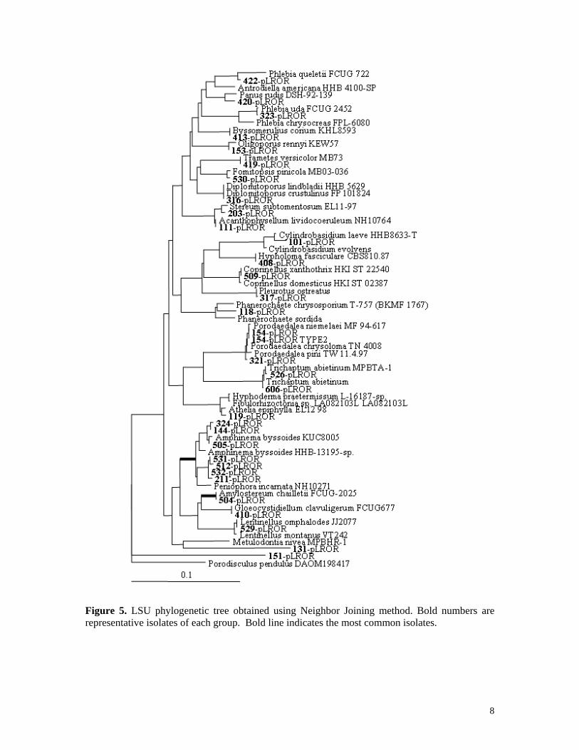
Figure 5. LSU phylogenetic tree obtained using Neighbor Joining method. Bold numbers are representative isolates of each group. Bold line indicates the most common isolates.
8

9
3.4 Fungal diversity at the different sites Basidiomycetes were isolated both from the bottom and top billets and from the
three types of trees: green, red and grey. With the exception of the Radium site, the basidiomycetes diversity seems slightly higher at the bottom than at the top of the tree. In the green trees, often basidiomycete isolations were from the beetle galleries, including galleries from MPB, Ips, or ambrosia.
Some of the species were present at only one or two sites while other species were present at six or eight sites. For example, Acanthophysellum lividocoeruleum and Metulodontia sp. (a heartrot) were only isolated at one site while Pheniophora sp., Sistotrema brinkmannii and T. abietinum were found at eight of the 12 sites. Entomocorticium species, commonly found in the beetle galleries, were also frequently isolated from the sapwood in contact with the beetle gallery. Entomocorticium species A was found at seven sites while species B was isolated at six sites. The abundance of each species was similar. Overall, Entomocorticium was the most frequently isolated genus; it is well known that species from this genus grow very slowly and do not cause major fiber degradation. Species of this genus have mutualistic relationships with insect species (Hsiau and Harrington 2003: Whitney et al. 1987) and have also been reported to be a nutrient source for the MPB.
The highest basidiomycete diversity was observed at Manning Park (15 species), Radium (13 species), Little Port (10 species) and Williams Lake (10 species) while the lowest diversity was at Monte Lake (3 species) and Riske Creek (5 species) (Table 3). Some of the species are well-known white rot (WR) while others are brown rot (BR). Ganoderma species (WR) were only isolated at Quesnel, Heterobasidium annosum, was isolated at three of the sites: Manning Park, Radium and Cranbrook. Phellinus pini, a pine decaying fungus, was present at five sites, including Robson Park, a site with a low frequency of basidiomycete fungi. Peniophora species, were present at 8 of the 12 sites. These were not isolated at Canbrook, BurnsLake, Kamloops and Williams Lake. Similarly, the sap rot T. abietinum was present at 8 of the 12 sites, it was not found at Riske Creek, Radium Monte Lake and Kamloops.
Figure 6. Examples of green trees with heartrot prior to MPB attack .
It is important to note that decay, especially heartrot, was present prior or after MPB attack. The importance of the decay allows the differentiation between both types. Fig. 6 shows examples of heartrot.

3.5 Decay tests
3.5.1 Lodgepole pine wood from Williams Lake (lower density) We harvested a 97-year-old tree from Williams Lake and a 118-year-old tree from
Kamloops (Table 4). Sapwood and heartwood from the trees were separated and processed into boards that were dried at room temperature before being further processed into small blocks. Processing and drying took three months. The difference between the Williams Lake and Kamloops woodblocks can be seen in their ring density (Fig. 5) and specific gravity measurements (Table 4). We had enough wood blocks from Williams Lake to carry out decay tests on most of the fungi.
Table 4. Characteristics of the trees used.
Specific Gravity (SG)*
Location Age (year)
Diameter (cm) Sapwood Heartwood
Williams Lake 97 26 0.46 ± 0.014 0.46 ± 0.021 Kamloops 118 21.5 0.55 ± 0.034 0.51 ± 0.037
*Means of five replicates
Figure 7. Longitudinal and transverse sections of Kamloops and Williams Lake woodblocks. The longitudinal (left) and transverse (right) faces of the sapwood (S) and heartwood (H) woodblocks obtained from Kamloops (K) and Williams Lake (W) lodgepole pine trees show significantly different ring density.
The moisture content (MC) of the wood was determined on a representative
number of samples (Table 4). Sapwood blocks from Williams Lake (MC: 56%) had slightly higher moisture contents than those from Kamloops (MC: 40%), while the moisture content of the hardwood (MC: 20-21%) was similar for the two locations. Then, plugs of fungi grown on 2% MEA were inoculated on a wood strip that rested on the soil in a jar (Fig. 8). After two to three weeks, when fungal growth was established on the soil and wood strip, two small wood blocks per jar were carefully placed on the top of each wood strip. The inoculated jars were incubated at 20°C in the dark for another 12 weeks (Fig. 8). After the incubation period, the wood blocks were oven dried and weighted. The difference before and after incubation was calculated and the average percentage weight loss was determined. When possible, several strains of the same species were tested.
10

A B C
D
Figure 8. Soil jar decay test. Fungal inoculation of Amylostereum chailletii at the edge of the feeder strip (A); Incubation of two sapwood blocks (B); incubation of two heartwood blocks (C); same as C but closer picture (D).
The decay potential of 34 species was assessed. For some of the species we have only one isolate, while for others we tested up to five isolates. The results are shown Table 5 Examples of wood block decay after 12 weeks incubation are shown Fig. 9.
Figure 9. Lodgepole pine wood blocks (2 x 2 x 2 cm) after 12 weeks of incubation with different species. From left to right: heartwood (longitudinal & transverse), sapwood (longitudinal & transverse). A. Fomitopsis pinicola. B. Heterobasidion annosum. C. Trichaptum abietinum. D. Stereum sanguinolentum. E. Ganoderma species.
Wood blocks
A B
C D
E
11
Some species, like Entomocorticium sp. and Sistotrema brinkmannii, caused only slight weight losses and these species were probably not affecting the wood structure. It is likely that the weight losses in all these species are due to the removal of non-structural wood components (e.g., triglycerides, fatty acids). The Entomocorticium species can be a source of nutrient for the mountain pine beetle and they do not appear to damage the wood fibres. Although reported as wood-rotting basidiomycetes, Sistrotrema brinkmanii, an aggregate of biological species, did not cause a substantial wood weight loss. If this species degrades wood fibers, the degradation process is probably very slow. Three other rare species, Coprinellus xanthothrix, Cylindrobasidium sp., and Panus rudis did not appear to cause decay. For these species both sapwood and heartwood weight losses were below 2%.
Species, like Amylostereum chailleti, Peniophora sp., Phlebia tremellosa, Oligoporus rennyi, Oligoporus placenta, Stereum sanguinolentum and Phellinus pini decayed the sapwood preferentially while others e.g., Ganoderma sp., Fomitopsis pinicola, Coniophora olivaceae and Trichaptum abietinum, degraded both the sapwood and heartwood. None of the species degraded, only the heartwood. F. pinicola, T. abietinum and Ganoderma sp. degraded the sapwood faster than the heartwood, while C. olivaceae seemed to degrade the sapwood and heartwood at the same rate. It is likely that C. olivaceae is less affected by the extractive contents present in the heartwood than other decay fungi. Among all the species tested, F. pinicola, Ganoderma sp. and Metulodontia sp. cause the most damage, reducing the wood weight by almost 50% or more after 12 weeks of incubation at 20°C. The well-known root rot, Heterobasidium annosum did not affect the heartwood. Two isolates of this species showed a moderate degradation of the sapwood (between 10-17%) while the third isolate which had a slow growth did not affect the wood weight. Trametes versicolor, another well-known decay fungus, caused only moderate wood weight losses of the sapwood and very small heartwood weight losses.
Among the isolates of the same species we noticed some variability in the overall decay ability. For example, among four isolates of A. chailletii, three caused less than 10% weight losses of the sapwood while one caused a reduction of 17%. This species is a well-known mycangial fungus of wood wasps (Slippers et al. 2003). While all the T. abietinum isolates degraded the sapwood, only four of the five isolates degraded the heartwood. 3.5.1 Lodgepole pine wood from Kamloops (high density)
We also examined the decay ability of a few species on lodgepole pine with high
wood density (Table 6). The fungal species tested were species used with the Williams Lake wood decay test. A. chailletii showed a slight reduction in its ability to degrade sapwood with high density, while both isolates of C. olivaceae were more effective in degrading both sapwood and heartwood of the denser wood tree. For this fungal species, the wood weight losses were similar between the sapwood and heartwood. F. pinicola and Ganoderma sp. cause more weight losses of the sapwood than the heartwood, for both high and low wood density. Similarly to our results with lodgepole wood from Williams Lake, T. abietinum (isolate 606) did not cause any weight loss of the
12

heartwood, while the other isolate (927) degraded heartwood and showed a 16% weight loss. 3.5.2 Lignolytic activity
To differentiate white rot from brown rot fungi, we examined the production of lignolytic enzyme activity on media containing tannic acid. Brown rot fungi utilized mainly the wood carbohydrates (cellulose and hemicellulose), while white rot degrade lignin and carbohydrates. The fungal isolates that we used in the decay test were incubated on 2% MEA containing tannic acid (Fig. 10). When phenolic oxidases or ligninases are secreted by the fungi, a red reaction zone is formed (Noble 1965). Tannic acid seems to induce different lignolytic activities (laccase, phenol oxidases…) by white-rot fungi. Brown rot fungi that do not degrade lignin do not produce these enzymes. Some of the white rot species showed a strong lignolytic activity while others showed weak or no reaction (Table 5). A few species also were not able to grow on media containing tannic acid.
Most of the fungal species causing substantial wood weight losses were white rot and brown rot fungi. Many white rot species produced strong reactions in the tannic media, indicating the presence of lignolytic activity. As expected, Fomitopsis pinicola, Coniophora olivaceae, Oligoporus rennyi and placenta did not produce any reaction Confirming that these fungi were brown rot and did not degrade lignin. F. pinicola is one of the most frequently occurring decay fungi in BC and one of the most damaging in old growth forest (Allen et al. 1996). Overall, during this survey we isolated more white rot fungi than brown rot.
Figure 10. Examples of growth and lignolytic activity on 1% MEA with 0.5% tannic acid. Ganoderma sp. showed strong reaction. Phlebia uda showed weak reaction. Oligoporus placenta showed no reaction. Coprinellus xanthothrix showed no growth. 4 Conclusions The proposed work was a proof of concept to establish whether decay fungi were present in MPB-killed trees. For the first time, information about the basidiomycetes present in green, red, and grey lodgepole pines resulting from the mountain pine beetle attack in BC has been generated on 12 sites across the province. Although a true symbiotic association has not been shown between MPB and decay fungi, we showed that in an epidemic situation the beetle could carry and seed decay fungi into new trees. Decay fungi were
13

present at all the sites surveyed; however, some sites showed a higher fungal frequency and diversity. We identified the basidiomycetes at the genera or species level and found about 40 different basidiomycetes. We performed decay tests with different isolates from the important genera or species and we showed that some of these species were aggressive decay fungi and could degrade 50% of the wood fibers in 3 months at 20°C. Having established the first basic information about decay fungi associated with the MPB epidemic, we could develop tests to rapidly identify the most damaging fungi present in wood and more reliably predict how rapidly these species would decay wood with time. This would allow government and industry to be more effective in deciding which trees should be left standing and which trees should be processed rapidly. 5 Acknowledgements This project was funded by the Government of Canada through the Mountain Pine Beetle Initiative, a Program administered by Natural Resources Canada, Canadian Forest Service. Publication does not necessarily signify that the contents of this report reflect the views or policies of Natural Resources Canada – Canadian Forest Service.
14

Table 5: Summary: decay tests on wood blocks from Williams Lake,
lignolytic activity and growth rate.
Percentage Weight Loss a Genus Species ID
Heartwood Sapwood
Ligno-lytic Acti-h vity b
Growth Rate c mm/day
Acanthophysellum lividocoeruleum 111 1.33 ± 0.63 4.21 ± 1.81 S 4.0 Amylostereum Chailletii 139 2.32 ± 0.51 10.59 ± 1.59 W 2.4 Amylostereum Chailletii 514 1.64 ± 0.76 17.76 ± 1.49 W 2.4 Amylostereum Chailletii 428 1.64 ± 0.56 9.26 ± 1.66 S 4.0 Amylostereum Chailletii 329 - - S 5.8 Amylostereum Chailletii 417 0.80 ± 0.15 7.53 ± 2.58 W 3.4 Athelia Epiphylla 112 - - S 1.0 Athelia Epiphylla 112 0.33 ± 0.07 0.60 ± 0.11 S 1.0 Athelia Epiphylla 119 33.81 ± 2.88 1.23 ± 0.16 S 1.4 Byssomerulium Corium 413 0.88 ± 0.13 4.17 ± 1.41 ND 4.7 Byssomerulium Corium 912 1.47 ± 0.18 27.20 ± 5.60 W 6.2 Byssomerulium Corium 909 1.096 ± 0.096 20.82 ± 3.51 W/NG 10.6 Byssomerulium Corium 815 2.80 ± 2.67 26.92 ± 6.85 W 12.9 Clavulina Cristata 926 0.47 ± 0.19 1.31 ± 0.31 W/NG 3.0 Coniophora Olivaceae 818 33.20 ± 7.23 34.62 ± 11.88 NR 4.6 Coniophora Olivaceae 831 29.30 ± 13.87 10.66 ± 1.35 NR 11.6 Coprinellus xanthothrix 509 0.58 ± 0.10 0.63 ± 0.23 NG 8.4 Cylindrobasidium sp. 101 0.68 ± 0.24 0.83 ± 0.29 ND 2.2 Cylindrobasidium sp. 401 0.93 ± 0.29 0.69 ± 0.31 ND 2.2 Entomocorticium A 324 0.46 ± 0.24 0.74 ± 0.36 S 1.8 Diplomitoporus crustulinus 316 5.03 ± 2.26 35.24 ± 4.97 S 14.7 Fomitopsis Pinicola 817 15.73 ± 9.57 55.95 ± 8.10 NR 3.5 Fomitopsis Pinicola 810 23.37 ± 15.50 32.63 ± 17.95 NR 4.3 Fomitopsis Pinicola 530 38.90 ± 12.01 61.48 ± 0.95 NR 2.2 Fomitopsis Pinicola 607 31.64 ± 6.66 60.99 ± 2.21 NR 0.2 Ganoderma sp. 1004 24.39 ± 3.82 31.82 ± 12.54 S 9.4 Ganoderma sp. 1005 16.20 ± 2.77 46.10 ± 21.15 S 9.3 Ganoderma sp. 1006 35.56 ± 14.0 49.43 ± 28.63 S 7.4 Gloeocystidiellum clavuligerum 410 2.31 ± 0.85 7.14 ± 0.71 W 0.2
15

Table 5: decay tests continued
Percentage Weight Loss a Genus Species ID Heartwood Sapwood
Ligno- lytic
Activity b
Growth Rate c
mm/day
Heterobasidion Annosum 312 0.90 ± 0.28 10.61 ± 3.08 S 10.7
Heterobasidium Annosum 936 1.071 ± 0.046 0.77 ± 0.23 S 6.4 Heterobasidium Annosum 1017 1.22 ± 0.33 17.27±2.34 S 11.4 Hypholoma Fasciculare 408 5.62 ± 2.27 26.91±1.53 S 1.6 Lentinellus omphalodes 529 5.82 ± 2.65 5.59 ± 1.22 S 3.4 Metulodontia sp. 131 43.55 ± 3.61 67.04± 3.43 S 7.5 Oligoporus Rennyi 153 38.53 ± 4.74 28.93±2.32 NR 1.4 Oligoporus Placenta 816 26.94 ± 4.61 35.56 ± 7.90 NR 8.9 Panus Rudis 420 0.37 ± 0.23 1.42 ± 0.23 W 1.2 Peniophora sp. 930 - - S 10.3 Peniophora sp. 211 3.11 ± 0.91 9.57 ± 0.79 S 4.6 Peniophora sp. 616 1.45 ± 0.11 8.93 ± 0.98 S 4.6 Peniophora sp. 1010 1.63 ± 0.69 8.67 ± 0.88 S 6.1 Phanerochaete sp. 118 0.059 ± 0.052 4.59 ± 1.32 W 8.0 Phanerochaete sp. 149 2.53 ± 1.12 24.8 ± 0.95 W 8.0 Phellinus Ferreus 713 1.94 ± 0.23 13.08 ± 5.0 S 2.5
Phellinus Pini /chrysoloma 154 3.42 ± 0.93 9.05 ± 2.65 S 0.8
Phellinus pini/ chrysoloma 321 4.60 ± 1.70 18.86 ± 3.15 S 0.8
Phellinus pini/ chrysoloma 925 - - S 3.3
Phellinus pini/ chrysoloma 421 7.28 ± 1.18 17.8 ± 1.36 S 2.9
Phlebia Radiata 933 3.92 ± 0.73 23.4 ± 4.58 S 9.0 Phlebia Radiata 1022 5.81 ± 1.11 18.9 ± 2.6 S 9.5 Phlebia Subserialis 802 2.12 ± 2.21 12.0 ± 2.6 NG 10.8 Phlebia Subserialis 1013 2.48 ± 1.49 14.8 ± 2.87 W 6.8 Phlebia Tremellosa 714 1.48 ± 0.42 23.95 ± 2.3 S 11.1 Phlebia Tremellosa 803 0.47 ± 0.15 18.4 ± 3.43 S 15.5 Phlebia Tremellosa 1011 1.79 ± 1.58 25.8 ± 1.47 W 17.5 Phlebia Tremellosa 1016 3.33 ± 0.95 23.4 ± 2.59 S 14.0 Phlebia Uda 323 0.661 ± 0.085 12.2 ± 2.44 W 4.8
16

Table 5: decay tests continued
Percentage Weight Loss a Genus Species ID
Heartwood Sapwood
Ligno lytic Activity b
Growth Rate c
mm /day Phlebia queletii 422 0.61 ± 0.38 3.26 ± 0. 77 W 1.4 Pleurotus plumonarius 317 0.67 ± 0.13 22.43 ± 9.06 NG 3.0 Sarcomyxa serotina 1019 1.04 ± 0.36 5.98 ± 1.09 S 5.6 Sistotrema brinkmannii Klp - 2.6 ± 0.75 NG Sistotrema brinkmannii 934 0.54 ± 0.15 1.86 ± 0.18 NG 4.7 Stereum sanguinolentum 203 1.08 ± 0.13 1.35 ± 0.62 S 3.2 Stereum sanguinolentum 524 5.32 ± 1.33 18.08 ± 1.40 S 3.2 Stereum sanguinolentum 914 4.76 ± 0.91 12.28 ± 0.57 S 7.6 Stereum sanguinolentum 829 1.06 ± 0.17 12.35 ± 2.48 S 7.4 Stereum sanguinolentum WL 20.8 ± 2.27 S Trametes versicolor 419 3.62 ± 0.66 12.88 ± 1.91 S 5.5 Trametes versicolor 929 5.92 ± 1.58 12.06 ± 1.91 S 12.8 Trichaptum abietinum 120B 13.96 ± 1.9 31.57 ± 3.25 S 0.9 Trichaptum abietinum 606 3.72 ± 1.29 28.07 ± 3.38 S 0.9 Trichaptum abietinum 1001 13.79 ± 2.0 36.32 ± 12.98 S 8.2 Trichaptum abietinum 927 21.2 ± 4.75 38.73 ± 3.94 S 5.9 Trichaptum abietinum 807 12.3 ± 5.65 27.45 ± 12.87 S 2.8 Trichaptum abietinum WL 24.6 ± 7 S
a Mean ± standard deviation (4 replicates) b lignolytic activity; abbreviations: S = strong, W = weak, NR = no reaction, NG = no growth, ND = not done c Growth rate measured on 1 % MEA = colony diameter (mm/day)
Kpl = Kamploops; WL = Williams Lake Table 6: Decay tests on lodgepole pine from Kamloops.
Fungi were incubated 12 weeks at 20°C Fungal Species ID Heartwood
Weight loss (%) Sapwoood
Weight loss (%) Amylostereum chailletii 139 3.11 ± 1.66 7.11 ± 0.45 Amylostereum chailletii 514 5.02 ± 2.66 12.07 ± 1.36 Coniophora olivaceae 818 50.27 ± 4.53 52.42 ± 10.59 Coniophora olivaceae 831 41.35 ± 3.35 40.20 ± 4.23 Fomitopsis pinicola 530 36.31 ± 5.33 60.35 ± 1.92
Fomitopsis pinicola 607 26.77 ± 8.86 59.55 ± 2.38 Trichaptum abietinum 927 16.18 ± 12.86 39.67 ± 2.43 Trichaptum abietinum 606 3.30 ± 2.72 24.16 ± 2.19 Ganoderma sp. 1004 43.49 ± 3.30 51.74 ± 6.54 Ganoderma sp. 1006 28.23 ± 16.33 45.25 ± 6.82
17

6 Literature cited Adair, S.; Kim, S.H.; Breuil, C. 2002. A molecular approach for early monitoring of decay basidiomycetes in wood chips. FEMS Microbiology Letters. 211: 117-122. Allen E.; Morrison, D.; Wallis, G. 1996. Common tree Diseases of British Columbia. Natural Resources Canada, Canadian Forest Service, Pacific Forestry Centre, Victoria, BC. 178 p. Arx von, J.A. 1981. The genera of fungi sporulating in pure culture. J. Cramer, Vaduz, Germany. ASTM, 2000. Annual book of ASTM standards. ASTM Internaional Press, West Conshohocken, Pennsylvania. Clubbe, C.P.; Levy, J.F. 1977. Isolation and identification of the fungal flora in treated wood. Revised technique. International Research Group on Wood Preservation. Document no. IRG/WP/159. Gardes, M.; Burns, T.D. 1993. ITS primers with enhanced specificity for basidiomycetes: application to the identification of mycorrhizae and rusts. Molecular Ecology. 2: 113-118. Government of British Columbia. 2001. Mountain Pine Beetles in British Columbia – Beetle Facts. http://www.for.gov.bc.ca/hfp/mountain%Fpine%5Fbeetle/fats.htm (cited December 11, 2006). Hsiau, P.T.W.; Harrington, T.C. 2003. Phylogenetics and adaptations of basidiomycetous fungi fed upon bark beetles (Coleoptera: Scolytidae). Symbiosis 34:111-131. Kim, J.J; Allen, E.A.; Humble, L.M.; Breuil, C. 2005. Ophiostomatoid and basidiomycetous fungi associated with green, red, and grey lodgepole pines after mountain pine beetle (Dendroctonus ponderosae) infestation. Canadian Journal of Forest Research. 35: 274-284. Lee, S.; Kim, J.-J.; Breuil, C. 2006. Fungal diversity associated with the mountain pine beetle, Dendroctonus poderosae and infested lodgepole pines in British Columbia. Fungal Diversity 22:91-105. Lim, Y.W.; Kim, J.J.; Lu, M.; Breuil, C. 2005. Determining fungal diversity on Dendroctonus ponderosae and Ips pini affecting lodgepole pine using cultural and molecular methods. Fungal Diversity 19:79-84. Nobles, M.K. 1965. Identification of cultures of wood inhabiting Hymenomycetes. Can. J. Bot. 43: 1097-1139. Upadhyay, H.P. 1981. A monograph of Ceratocystis and Ceratocystiopsis. The University of Georgia Press, Athens, GA. Slippers B.; Coutinho, T.A.; Wingfield, B.D.; Slippers, M.J. 2003. A review of thegenus Amylostereum andits association with woodwasps. South African Journal Science. 99: 70-74. Staplers, J.A. 1978. Identification of wood-inhabiting fungi in pure culture. Studies In Mycology. 16:1-248.
18

19
Wang, C.J.K. and R.A. Zabel. 1990. Identification manual for fungi from utility poles in the eastern united states. Allen Press Inc., Lawrence, KS, USA. White, T.J.; Burns, T.D.; Lee, S.; Taylor, J.W. 1990. Amplification and direct sequencing of fungal ribosomal RNA genes for phylogenetics. In M.A. Innis, D.H. Gelfand, J.J. Sninsky and T.J White, eds. PCR protocols: a guide to methods and applications. Academic Press, Inc. San Diego, pp. 315-322. Whitney H.S.; Bandoni, R.J.; Oberwinkler, F. 1987. Entomocorticium dendroctoni et sp. nov. (Basidiomycotina), a possible nutritional symbiote of the mountain pine beetle in lodgepole pine in British Columbia. Canadian Journal of Botany. 65: 95-102. Zabel, R.A.; Morrell, J.J. 1992. Wood microbiology. Decay and its prevention. Academic Press, Inc., London.