Dear Participants, - PTBH · Dear Participants, It is my great ... Katarzyna...
25
Transcript of Dear Participants, - PTBH · Dear Participants, It is my great ... Katarzyna...

Dear Participants,
It is my great pleasure to welcome you to our biennial conference on biogenic amines and their system constituents. We meet for the 16th time. I must confess that, when we are talking about biogenic amines, histamine is of course the first one which comes to my mind. And, while we are on the subject of histamine, this year we have a good reason to be happy and to congratulate our French colleagues of Jean-Charles Schwartz’s Group as winners a long lasting but finally successful battle. It is already over twenty years (23) after they described the presence of H3- a new histamine receptor in the central nervous system. Now, Pitolisant, an antagonist of the H3 receptor, is entering the clinic as a drug against narcolepsy, registered in Europe under the commercial name of Wakix. The development of some other selective ligands of H3 receptor is thus warranted and certainly for different indications as well . It is worth noting that a few months ago, in June of this year, a positive outcome was announced in phase 2a of a clinical study in patients with moderate to severe atopic dermatitis, treated with H4 receptor antagonist, ZPL-389 (ZIARCO Group Ltd). Hopefully, we may soon celebrate an implementation of H4 receptor antagonist to the therapy. H4 receptor was discovered in 1999/2000. There is still another event we may be happy with - Associate Professor Dariusz Szukiewicz, Vice President of the Polish Histamine Research Society, has been approved for Full Professor nomination , which is soon to be granted by President of Poland.. I believe, everybody here will join me in congratulations to our Nominee. During this Conference, we shall have 12 lectures, delivered by scientists of the top international level , and 24 presentations, provided mostly by young investigators. I want to express my sincere thanks to all the Speakers for undertaking their tasks, especially to the Invited Speakers who have responded to our invitation from abroad, including European countries but also countries from Asia and North America. I am sure that even during such a short period of time we shall be able to discuss and learn a lot, while simultaneously entering and strengthening scientific cooperation and personal friendship. The planned social events should be very helpful to find new, interesting contacts and maintain to-date’s relationships.
Welcome!
W. Agnieszka Fogel
President Polish Histamine Research Society
Scientific Programme
Thursday, October 27th 2016
14.00 Arrival, accommodationAmbasador Centrum Hotel, Piłsudskiego 29 St., 90-307 Lodz
16.00 – 19.00 Registration: Ambasador Centrum Hotel, Reception Hall
17.00 – 19.00 Poster mounting, Conference room B
19.00 Opening:Prof. Dr. W. Agnieszka Fogel, President of the Polish Histamine Research Society
Prof. Dr. Jurek OlszewskiDean of the Faculty of Military Medicine, the Medical University of Lodz
Conference Lecture:JNJ-54175446: A P2X7 RECEPTOR ANTAGONIST CLINICAL CANDIDATE FOR MAJOR DEPRESSIVE DISORDERSNicholas I. CarruthersJanssen Research & Development, LLC; San Diego, USA
20.00 – 22.00 Welcome Reception Ambasador Centrum Hotel, Restaurant
Friday, October 28th 2016
9.00 – 10.30 Session I, chaired by Dariusz Szukiewicz and Anna Stasiak
9.00 – 9.30L1
NEW TARGETS FOR MODIFYING MAST CELL ACTIVATION IN ASTHMA Madeleine Ennis Centre for Experimental Medicine, School of Medicine, Dentistry and Biomedical Sciences, Queens University Belfast, Belfast, United Kingdom
9.30 – 9.35 P1
CATHELICIDIN LL-37 REGULATES TLR EXPRESSION IN THE MATURE TISSUE MAST CELLSJustyna Agier, Paulina Żelechowska, Elżbieta Kozłowska, Ewa Brzezińska-BłaszczykDepartment of Experimental Immunology, Medical University of Lodz, Lodz, Poland
9.35 – 9.40P2
SERUM CATHELICIDIN LL-37 LEVELS IN PATIENTS WITH PULMONARY INFECTIOUS DISEASESPaulina Żelechowska, Karol Majewski, Elżbieta Kozłowska, Justyna Agier, Ewa Brzezińska-BłaszczykDepartment of Experimental Immunology, Medical University of Lodz, Lodz, Poland
9.40 – 10.10L2
IgE-INDEPENDENT ACTIVATION OF MAST CELLS – AN EXPERIMENTAL CONTACT ALLERGEN, 1-FLUORO-2,4-DINITROBENZENE IS A MAST CELL SECRETAGOGUESatoshi Tanaka Department of Immunobiology, Graduate School of Medicine, Dentistry and Pharmaceutical Sciences, Okayama University, Japan
10.10 – 10.30O1
THE ROLE OF INFLAMMATORY CYTOKINES IN THE DEVELOPMENT OF TUMOR VESSELS IN GYNECOLOGICAL CANCERSGrzegorz Szewczyk1, 2, Dariusz Szukiewicz1
1Department of General and Experimental Pathology, Medical University of Warsaw, Poland; 2Department of Obstetrics and Gynecology, Institute of Mother and Child, Warsaw, Poland
10.30 – 10.50 Coffee /tea Discussions by Posters
10.50 – 12.55 Session II, chaired by Madeleine Ennis and Krzysztof Wąsowicz
10.50 – 11.20L3
MAST CELL AND HYPOXIA Jarosław DastychLaboratory of Cellular Immunology, Institute of Medical Biology of the Polish Academy of Sciences, Lodz, Poland
11.20 – 11.40O2
HYPOXIA MEDIATED UPREGULATION OF EXPRESSION OF PRO-INFLAMMATORY MEDIATORS IN HUMAN LAD2 MAST CELLS Aurelia Walczak-Drzewiecka, Joanna Pastwińska, Anna Sałkowska, Marcin Ratajewski, Jarosław Dastych Laboratory of Cellular Immunology, Institute of Medical Biology of the Polish Academy of Sciences, Lodz, Poland
11.40 – 12.00O3
NEUROPEPTIDE Y AND THE CENTRAL CARDIOVASCULAR REGULATION IN HAEMORRHAGIC HYPOTENSION IN RATSJerzy JochemDepartment of Physiology, Medical University of Silesia, Katowice, Poland
12.00 – 12.20O4
ROLE OF COMPENSATORY MECHANISMS IN RESPONSE TO MILD ANEMIA DURING PREGNANCY Aleksandra Stangret, Marta Skoda, Michał Pyzlak, Dariusz SzukiewiczDepartment of General & Experimental Pathology with Centre for Preclinical Research and Technology (CEPT), Medical University of Warsaw, Warsaw, Poland
12.20 – 12.25 P3
ANALGESIC AND ANTI-INFLAMMATORY ACTION OF ESCULETIN IN ACUTE AND CHRONIC INFLAMMATIONPrzemysław Rzodkiewicz1,2, Katarzyna Romanowska-Próchnicka1,3, Emilia Gasinska4, Magdalena Bujalska-Zadrozny4, Sławomir Maslinski1,Dariusz Szukiewicz1
1Department of General and Experimental Pathology, CEPT Laboratory, Medical University of Warsaw, Warsaw, Poland; 2Department of Gerontology and Public Health and 3Department and Polyclinic of Systemic Connective Tissue Diseases, National Institute of Geriatrics, Gerontology and Rehabilitation, Warsaw, Poland; 4Department of Pharmacodynamics, CEPT laboratory, Medical University of Warsaw, Warsaw, Poland
12.25 – 12.30P4
THE SERUM TNFa LEVELS ARE INFLUENCED BY SALSOLINOL GIVEN INTRAPERITONEALLYMagdalena Kurnik-Łucka, Andrzej Bugajski, Krzysztof GilDepartment of Pathophysiology, Jagiellonian University Medical College, Krakow, Poland
12.30 – 12.35 P5
IDENTIFICATION OF miRNA IN CELLS TREATED WITH ADALIMUMAB IN THE CONTEXT OF HISTAMINERGIC SYSTEMAleksandra Skubis1, Dominika Wcisło-Dziadecka2, Bartosz Sikora1, Beniamin Grabarek1, Klaudia Simka1, Bartłomiej Skowronek1, Celina Kruszniewska-Rajs1, Joanna Gola1, Urszula Mazurek1
1Department of Molecular Biology, 2Department of Skin Structural Studies, School of Pharmacy with Division of Laboratory Medicine in Sosnowiec, Medical University of Silesia, Katowice, Poland
12.35 – 12.40P6
EXPRESSION PROFILE OF GENES ASSOCIATED WITH THE HISTAMINERGIC SYSTEM IN NORMAL HUMAN DERMAL FIBROBLAST (NHDF) CELLS TREATED WITH ADALIMUMABBeniamin Grabarek1, Dominika Wcisło-Dziadecka2, Bartosz Sikora1, Aleksandra Skubis1, Celina Kruszniewska-Rajs1, Joanna Gola1, Andrzej Plewka3, Klaudia Simka1, Bartłomiej Skowronek1, Urszula Mazurek1
1Department of Molecular Biology, 2Department of Skin Structural Studies, 3Department of Proteomics, School of Pharmacy with Division of Laboratory Medicine in Sosnowiec, Medical University of Silesia, Katowice, Poland
12.40 – 12.45P7
EXPRESSION PROFILE OF GENES ASSOCIATED WITH THE HISTAMINERGIC SYSTEM IN PATIENTS WITH PSORIASIS VULGARIS DURING ANTI-TNF THERAPHY: ADALIMUMABDominika Wcisło-Dziadecka1, Beniamin Grabarek2, Agata Kaźmierczak1, Celina Kruszniewska-Rajs2, Joanna Gola2, Urszula Mazurek2
1Department of Skin Structural Studies, 2Department of Molecular Biology, School of Pharmacy with Division of Laboratory Medicine in Sosnowiec, Medical University of Silesia, Katowice, Poland

12.45 – 12.50 P8
IDENTIFICATION OF miRNA IN PORCINE ENDOGENOUS RETROVIRUS (PERV) INFECTION MODELBartosz Sikora, Krzysztof Łopata, Emilia Wojdas, Aleksandra Skubis, Celina Kruszniewska-Rajs, Joanna Gola, Urszula MazurekDepartment of Molecular Biology, School of Pharmacy with Division of Laboratory Medicine in Sosnowiec, Medical University of Silesia, Katowice, Poland
12.50 – 12.55P9
INVOLVEMENT OF miRNAs IN THE REGULATION OF TNF AND SEROTONIN INDUCED PATHWAYS IN RPTEC CELLS TREATED WITH AmB-Cu2+ AND AmB
Joanna Gola1, Barbara Strzałka-Mrozik1, Celina Kruszniewska-Rajs1, Jolanta Adamska1 Grzegorz Czernel2, Mariusz Gagoś3, Urszula Mazurek1 1Department of Molecular Biology, School of Pharmacy with the Division of Laboratory Medicine in Sosnowiec, Medical University of Silesia in Katowice, Poland; 2Department of Cell Biology, Institute of Biology and Biotechnology, Maria Curie-Skłodowska University, Lublin, Poland; 3Department of Biophysics, University of Life Sciences in Lublin, Poland
13.00 – 14.15 Lunch
14.15 – 15.20 Session III, chaired by Mercedes Unzeta and Jarosław Dastych
14.15 – 14.45L4
SPERMINE ENZYMATIC OXIDATION PRODUCTS INDUCE MITOCHONDRIAL MEDIATED CYTOTOXICITY IN HUMAN CANCER CELLS DETECTED BY SILAC-BASED MASS SPECTROSCOPY ANALYSISEnzo Agostinelli1, Carlos Barrero2, Shinji Ohkubo1, Silvia Grancara1, Antonio Toninello3, Salim Merali21Department of Biochemical Sciences ‘A. Rossi Fanelli’, SAPIENZA University of Rome, Rome, Italy; 2Temple University School of Pharmacy, Philadelphia, USA; 3Department of Biomedical Sciences, University of Padova, Padova, Italy
14.45 – 14.50P10
MicroRNA PROFILE IN THE REGULATION OF GENE EXPRESSION ASSOCIATED WITH SEROTONIN PATHWAY IN ENDOMETRIAL CANCER Małgorzata Kimsa-Furdzik1, Tomasz Francuz1, Nikola Zmarzły3, Agnieszka Jęda-Golonka2, Andrzej Witek2, Celina Kruszniewska-Rajs3, Joanna Gola3, Tomasz Janikowski3, Urszula Mazurek3
1Department of Biochemistry, 2Department of Gynecology and Obstetrics School of Medicine in Katowice, 3Department of Molecular Biology, School of Pharmacy with the Division of Laboratory Medicine in Sosnowiec, Medical University of Silesia, Katowice, Poland
14.50 – 14.55P11
MELATONIN: THE POTENTIAL RELATIONSHIP WITH CELL DEATH IN ENDOMETRIAL CANCER Andrzej Witek1, Katarzyna Szczepanek1, Agnieszka Jęda-Golonka1, Nikola Zmarzły3, Tomasz Janikowski3, Michał Baliś1, Celina Kruszniewska-Rajs3, Joanna Gola3, Małgorzata Kimsa-Furdzik2, Tomasz Francuz2, Urszula Mazurek3
1Department of Gynecology and Obstetrics; 2Department of Biochemistry, School of Medicine in Katowice; 3Department of Molecular Biology, School of Pharmacy with Division of Laboratory Medicine in Sosnowiec, Medical University of Silesia, Katowice, Poland
14.55 – 15.00P12
THE POTENTIAL FUNCTIONS OF MELATONIN IN REGULATION OF THE CELL CYCLE IN ENDOMETRIAL CANCEREwelina Hermyt1, Agnieszka Jęda-Golonka1, Michał Baliś1 Katarzyna Szczepanek1, Nikola Zmarzły3, Tomasz Janikowski3, Celina Kruszniewska-Rajs3, Joanna Gola3, Andrzej Witek1, Małgorzata Kimsa-Furdzik2, Tomasz Francuz2 , Urszula Mazurek3
1Department of Gynecology and Obstetrics, 2Department of Biochemistry, School of Medicine in Katowice, 3Department of Molecular Biology, School of Pharmacy with the Division of Laboratory Medicine in Sosnowiec, Medical University of Silesia, Katowice, Poland
15.00 – 15.05P13
EXPRESSION PROFILE OF DOPAMINE-RELATED GENES IN ENDOMETRIAL CANCERNikola Zmarzły1, Agnieszka Jęda-Golonka2, Andrzej Witek2, Celina Kruszniewska-Rajs1, Joanna Gola1, Tomasz Janikowski1, Urszula Mazurek1
1Department of Molecular Biology, School of Pharmacy with Division of Laboratory Medicine in Sosnowiec; 2Department of Gynecology and Obstetrics, School of Medicine in Katowice, Medical University of Silesia, Katowice, Poland
15.05 – 15.10P14
EXPRESSION PROFILE OF GENES ASSOCIATED WITH HISTAMINERGIC SYSTEM IN COLORECTAL CANCER CELLS TREATED WITH BETULIN DERIVATIVESAgnieszka Lubczyńska1, Ewa Bębenek1, Celina Kruszniewska-Rajs2, Urszula Mazurek2, Stanisław Boryczka1
1Department of Organic Chemistry, 2Department of Molecular Biology, Medical University of Silesia in Katowice, Sosnowiec, Poland
15.10 – 15.15P15
INFLUENCE OF BACTERIAL AND/OR RETROVIRAL INFECTIONS ON EXPRESSION HISTAMINE RECEPTORS Krzysztof Łopata1, Emilia Wojdas1, Bartosz Sikora1, Aleksandra Skubis1, Nikola Zmarzły1, Katarzyna Łopata1, Marzena Gruszka1, Magdalena Kimsa-Dudek2, Urszula Mazurek1
1Department of Molecular Biology, 2Department of Food and Nutrition, School of Pharmacy with Division of Laboratory Medicine in Sosnowiec, Medical University of Silesia, Katowice, Poland
15.15 – 15.20P16
ISOLATION OF PURE POPULATION OF UNTOUCHED HUMAN NEUTROPHILS IN PATIENTS WITH SYSTEMIC SCLEROSISKatarzyna Romanowska-Próchnicka1,2, Przemysław Rzodkiewicz1,3, Marzena Olesińska2, Dariusz Szukiewicz1
1Department of General and Experimental Pathology, CEPT laboratory, Medical University of Warsaw, Warsaw, Poland; 2Department and Polyclinic of Systemic Connective Tissue Diseases and 3Department of Gerontology and Public Health, National Institute of Geriatrics, Gerontology and Rehabilitation, Warsaw, Poland
15.20 – 16.15 Coffee/tea Discussions by Posters
17:10 Visit to Muzeum of the Factory - Manufaktura by tram, meeting point: Reception
19:00 Dinner Galicja Restaurant
Saturday, October 29th 2016
9.00 – 10.50 Session IV, chaired by Barbara Skrzydło-Radomańska and Krzysztof Walczyński
9.00 – 9.30L5
INTERACTIONS OF THE HISTAMINERGIC AND DOPAMINERGIC SYSTEMS IN THE BRAIN: EVIDENCE FOR DOPAMINERGIC REGULATION OF HISTAMINERGIC NEURON DEVELOPMENTPertti PanulaDepartment of Anatomy and Neuroscience Center, University of Helsinki, Helsinki, Finland
9.30 – 10.00L6
HISTAMINE H3 RECEPTORS: MULTIPLE TARGETING WITH THE H3R PHAMACOPHORHolger StarkHeinrich-Heine-Universitaet Duesseldorf, Institut fuer Pharmazeutische und Medizinische Chemie, Duesseldorf, Germany
10.00 – 10.05P17
THE DETERMINATION OF ADME-TOX PARAMETERS IN VITRO OF THE NEW HISTAMINE H3R ANTAGONISTS WITH ANTICONVULSANT ACTIVITY IN MALE ADULT RATSGniewomir Latacz, Annamaria Lubelska, Agnieszka Olejarz, Tadeusz Karcz, Katarzyna Kieć-Kononowicz Department of Technology and Biotechnology of Drugs, Jagiellonian University Medical College, Krakow, Poland
10.05 – 10.10P18
THE SEARCH FOR HISTAMINE H4 RECEPTOR LIGANDS AMONG UREA/THIOU-REA DERIVATIVESEnrique Domínguez-Álvarez1, Dorota Łażewska1, Stephan Schwed2, Holger Stark2, Katarzyna Kieć-Kononowicz1
1Department of Technology and Biotechnology of Drugs, Jagiellonian University Medical College, Krakow, Poland; 2Heinrich-Heine-Universitaet Duesseldorf, Institut fuer Pharmazeutische und Medizinische Chemie, Duesseldorf, Germany

10.10 – 10.15P19
PHARMACOLOGICAL PROFILING OF THE NEWLY SYNTHESIZED HISTAMINE H4 LIGANDS; EFFECT ON HUMAN EOSINOPHIL ADHESION TO ENDOTHELIUM Marek Grosicki1,2, Stefan Chłopicki2,3, Dorota Łażewska1, Małgorzata Więcek1, Katarzyna Kieć-Kononowicz1
1Department of Technology and Biotechnology of Drugs, Jagiellonian University Medical College, Cracow, Poland; 2Jagiellonian Centre for Experimental Therapeutics (JCET), Cracow, Poland; 3Chair of Pharmacology, Jagiellonian University Medical College, Cracow, Poland
10.15 – 10.20P20
INHIBITION OF MONOAMINE OXIDASE B BY NOVEL ANALOGS AND DERIVATIVES OF PIPERIDINYL-PROPOXY BENZENEAgnieszka Olejarz, Urszula Cichoń, Dorota Łażewska, Tadeusz Karcz, Katarzyna Kieć-KononowiczDepartment of Technology and Biotechnology of Drugs, Jagiellonian University Medical College, Krakow, Poland
10.20 – 10.50L7 ALLOSTERIC AND BIASED AGONIST – FUTURE OF OPIOIDS
Dariusz Matosiuk, Agnieszka A. Kaczor, Damian BartuziDepartment of Synthesis and Chemical Technology of Pharmaceutical Sciences, Faculty of Pharmacy, Medical University of Lublin
10.50 – 11.20 Coffee/TeaDiscussions by Posters
11.20 – 13.20 Session V, chaired by W. Agnieszka Fogel and Andrzej Pilc
11.20 – 11.50L8
L-DOPA AND DOPAMINE RELEASE: IS THERE A ROLE FOR HISTAMINERGIC NEURONS BEHIND SEROTONERGIC AND NORADRENERGIC FIBRES?Philippe De DeurwaerdèreInstitute of Neurodegenerative Diseases, University of Bordeaux, CNRS UMR 5293, France
11.50 – 12.20L9
A MULTITARGET-DIRECTED DONEPEZIL-LIKE LIGAND AS A THERAPEUTIC APPROACH FOR STROKE AND CAA-ADPing Sun, Montserrat Solé, Mercedes UnzetaInstitut de Neurociències i Departament de Bioquímica i Biologia Molecular, Edifici M, Facultat de Medicina, Universitat Autònoma de Barcelona, Bellaterra, Barcelona, Spain
12.20 – 12.50L10
HISTAMINERGIC NEURONS PARTICIPATE IN THE GUT-BRAIN AXISM. Beatrice PassaniDepartment of Health Sciences, University of Florence, Firenze, Italy
12.50 – 13.20L11
THE ANTIPSYCHOTIC LIKE ACTIVITY OF mGlu RECEPTOR AGENTS; FOCUS ON NOVEL ALLOSTERIC VS. ORTHOSTERIC AGONISTS of mGlu4 RECEPTORSJoanna M. Wieronska, Piotr Brański, Andrzej PilcDepartment of Neurobiology, Institute of Pharmacology, Polish Academy of Sciences, Krakow, Poland
13.20 Closing Ceremony of the XVI-th Conference of the Polish Histamine Research Society
13.30 Lunch
18.15 Transfer to the Grand Theater Lodz
19.00 Opera: Piotr Czajkowski “Eugene Onegin”, premiere
21.30 DinnerAmbasador Centrum Hotel, Restaurant

This report discloses the discovery of the clinical candidate JNJ-54175446 and the SAR of other 1H-[1,2,3]triazolo[4,5-c]pyridin-5(4H)-yl derived P2X7 receptor (P2X7R) antagonists. The P2X7 receptor is an ATP-gated ion channel expressed abundantly on microglial cells in the CNS. Activation of P2X7R by increased levels of ATP results in the secretion of IL-1b and other pro-inflammatory cytokines. Literature reports support that antagonists of P2X7R would reduce central IL-1b levels and could function as a useful treatment for depression. Although a few clinical trials of P2X7R antagonists for immune mediated disorders have appeared, none of those compounds are reported to have CNS penetration. This presentation will focus on efforts that led to the discovery of highly selective, potent, brain penetrant P2X7R antagonists. It will describe the evolution of a series of 1H-[1,2,3]triazolo[4,5-c]pyridin-5(4H)-yl compounds from early SAR through to key tool compounds and ending with a description of the clinical candidate JNJ-54175446
Nicholas I. Carruthers
Janssen Research & Development, LLC; 3210 Merryfield Row, San Diego, CA 92121, USA
JNJ-54175446: A P2X7 RECEPTOR ANTAGONIST CLINICAL CANDIDATE FOR MAJOR DEPRESSIVE DISORDERS
In 2006, I published a review with the above title. My review ended with a very hopeful paragraph: For many years the only new drugs coming onto the market for asthma were variations on a theme e.g. long acting β2 agonists, different formulations of products etc. We now have the leukotriene antagonists and anti-IgE on the market. There are several new drugs further in the pipeline such as the A2B antagonists and phosphodiesterase inhibitors. Other approaches described would involve testing drugs that are already available for other purposes e.g. the statins and ambroxol. In contrast, some of the agents described in this review are simply ideas that need to be followed up e.g. the H4 receptor antagonist and work with TLR ligands. This is an exciting time to investigate new methods to treat asthma and there are many possibilities. Now 6 years later, I will examine how the potential drugs and agents have fared in the clinical arena. I will also discuss other agents which were not included in the review such as the recently published study on the use of an antagonist of prostaglandin D2 receptor 2 in asthma as well as other biological agents.
Madeleine Ennis
Centre for Experimental Medicine, The Wellcome-Wolfson Building, School of Medicine, Dentistry and Biomedical Sciences, Queens University Belfast, 97 Lisburn Road, Belfast, BT9 7BL, United Kingdom; correspondence: [email protected]
NEW TARGETS FOR MODIFYING MAST CELL ACTIVATION IN ASTHMA
10 11
Accumulating evidence suggests that mast cells should be involved in a wide variety of immune responses in addition to IgE-dependent immediate reactions. However, it remains to be fully clarified how mast cells are activated in an IgE-independent manner. Signaling pathways involved in mast cell degranulation have been classified into two types; one is IgE/FcɛRI-mediated where tyrosine phosphorylation of various signaling molecules plays critical roles, and the other is mediated by trimeric Gi family proteins. The target G protein-coupled receptors (GPCRs) involved in the latter pathway remained unknown, but recent studies have indicated that Mas-related gene family should be the possible candidate that induces Gi-mediated degranulation of mast cells [1, 2]. Dudeck et al. demonstrated that several experimental contact allergens, such as 1-fluoro-2,4-dinitrobenzene (DNFB) and fluorescein isothiocyanate, can induce degranulation of murine cutaneous mast cells, although its mechanism remains unknown [3]. We investigated the potentials of various experimental contact allergens for induction of mast cell degranulation and found that DNFB can directly induce degranulation of rat peritoneal mast cells and a murine mast cell line, C57, but not that of murine bone marrow-derived cultured mast cells. DNFB-mediated degranulation was suppressed by treatment with pertussis toxin, U73122 (a phospholipase C inhibitor), and BAPTA-AM (a cell permeable Ca2+ chelator), indicating that DNFB should induce Gi-mediated degranulation. We then performed a chemical screening of DNFB derivatives and found a reverse correlation between electron density of the C1 carbon and the degree of degranulation. Recently, Inoue et al. established a novel assay system for GPCR, TGF-α shedding assay [4]. DNFB was found to activate endogenous unknown GPCRs in HEK293 cells to induce TGF-α shedding, which was augmented by forced expression of Gαi protein. These findings might raise the possibility that DNFB function as a potent contact allergen through induction of cutaneous mast cell degranulation, because previous studies demonstrated that degranulation of mast cells promoted dendritic cell migration to the lymph node.
References
1. Tatemoto K et al. Biochem Biophys Res Commun 2006; 349: 1322-1328.2. McNeil BD et al. Nature 2016; 519: 237-241.3. Dudeck A et al. Immunity 2011; 34: 973-984.4. Inoue A et al. Nat Meth 2012; 9: 1021-1029.
Satoshi Tanaka
Department of Immunobiology, Graduate School of Medicine, Dentistry, and Pharmaceutical Sciences, Okayama University, Japan; correspondence: [email protected]
IgE-INDEPENDENT ACTIVATION OF MAST CELLS – AN EXPERIMENTAL CONTACT ALLERGEN, 1-FLUORO-2,4-DINITROBENZENE IS A MAST CELL SECRETAGOGUE Mast cells are immune cells important for inflammatory processes include allergic
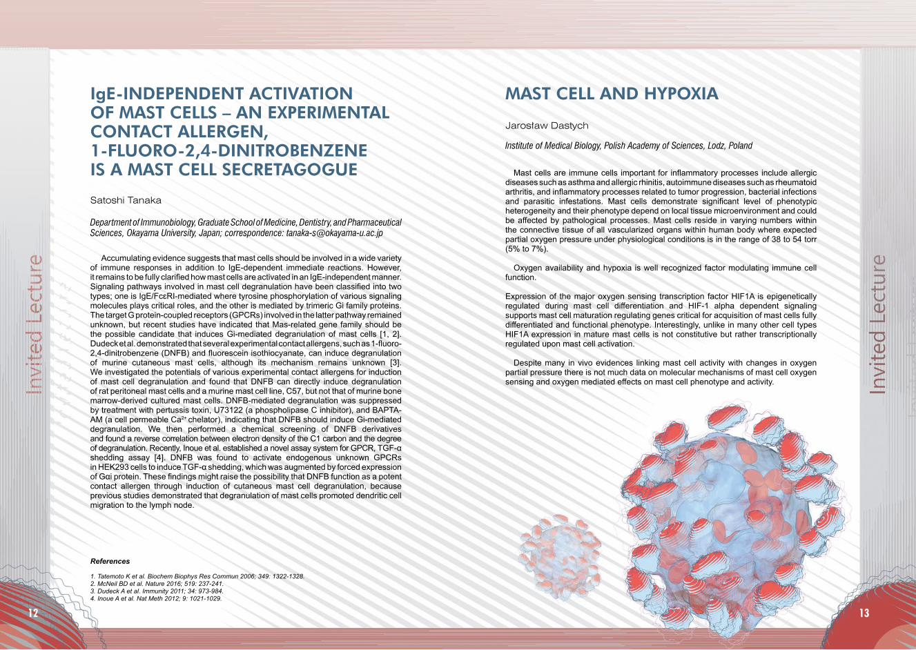
diseases such as asthma and allergic rhinitis, autoimmune diseases such as rheumatoid arthritis, and inflammatory processes related to tumor progression, bacterial infections and parasitic infestations. Mast cells demonstrate significant level of phenotypic heterogeneity and their phenotype depend on local tissue microenvironment and could be affected by pathological processes. Mast cells reside in varying numbers within the connective tissue of all vascularized organs within human body where expected partial oxygen pressure under physiological conditions is in the range of 38 to 54 torr (5% to 7%).
Oxygen availability and hypoxia is well recognized factor modulating immune cell function.
Expression of the major oxygen sensing transcription factor HIF1A is epigenetically regulated during mast cell differentiation and HIF-1 alpha dependent signaling supports mast cell maturation regulating genes critical for acquisition of mast cells fully differentiated and functional phenotype. Interestingly, unlike in many other cell types HIF1A expression in mature mast cells is not constitutive but rather transcriptionally regulated upon mast cell activation.
Despite many in vivo evidences linking mast cell activity with changes in oxygen partial pressure there is not much data on molecular mechanisms of mast cell oxygen sensing and oxygen mediated effects on mast cell phenotype and activity.
Jarosław Dastych
Institute of Medical Biology, Polish Academy of Sciences, Lodz, Poland
MAST CELL AND HYPOXIA
12 13

Enzo Agostinelli1, Carlos Barrero2, Shinji Ohkubo1, Silvia Grancara1, Antonio Toninello3, Salim Merali2
1Department of Biochemical Sciences ‘A. Rossi Fanelli’, SAPIENZA University of Rome, Piazzale Aldo Moro 5, 00185 Rome, Italy; 2Temple University School of Pharmacy 3307 N Broad Street Room 552 Philadelphia, PA 19140, USA; 3Department of Biomedical Sciences, University of Padova, via U. Bassi 58/B, 35131 Padova, Italy; correspondence: [email protected], Okayama University, Japan; correspondence: [email protected]
SPERMINE ENZYMATIC OXIDATION PRODUCTS INDUCE MITOCHONDRIAL MEDIATED CYTOTOXICITY IN HUMAN CANCER CELLS DETECTED BY SILAC-BASED MASS SPECTROSCOPY ANALYSIS
Several studies have demonstrated alterations in the brain histaminergic system in Parkinson’s disease, but the mechanisms for increased histamine have remained unknown. Histamine is an important transmitter in the brain, and zebrafish is a useful model to study its role in behaviour and interactions with other systems. We knocked down one of the two tyrosine hydroxylases (th1 or th2) in larval zebrafish, and studied the development of histaminergic and hypocretin neurons in the absence of this enzyme. We first verified with HPLC that removal of either one of the enzymes reduced brain dopamine levels by approximately 50% and this verified that both enzymes contribute to dopamine synthesis. We then counted the numbers of histaminergic and hypocretin neurons in the hypothalamus. The number of histaminergic neurons was significantly higher in th2 knockdown fish than in control fish. The number of neurons expressing hdc and histamine levels were also higher in th2 knockdown fish than in control fish. To verify that this increase is related to dopaminergic signaling, we then exposed developing normal larvae to either L-DOPA (dopamine precursor) or dopamine receptor agonists quinpirole or SKF38393. All these substances caused a decrease in the number of histaminergic neurons. Dopamine receptor antagonists haloperidol or SCH23390 prevented the decreases induced by agonists, respectively, but alone did not alter cell numbers.
Since we have previously shown that histamine is a key regulator of the number of hypocretin neurons, we counted also the numbers of hypocretin neurons in th2 knockdown fish. There was a significant increase in hypocretin neurons compared with control fish injected with control morpholino oligonucleotide.These results suggest that dopaminergic signaling is one of the factors which determine the number of histaminergic neurons in the brain, and histamine in turn regulates the number of hypocretin neurons.
The results may suggest mechanisms relevant for human Parkinson’s disease and narcolepsy, in which abnormalities have been found in the histaminergic system.
Pertti Panula
Department of Anatomy and Neuroscience Center, University of Helsinki, Helsinki, Finland
INTERACTIONS OF THE HISTAMINERGIC AND DOPAMINERGIC SYSTEMS IN THE BRAIN: EVIDENCE FOR DOPAMINERGIC REGULATION OF HISTAMINERGIC NEURON DEVELOPMENT
In situ formation of cytotoxic metabolites by an enzyme-catalyzed reaction is a recent approach in cancer chemotherapy. By a clonogenic cell survival assay we demonstrate that multidrug resistant human colon adenocarcinoma cells (LoVo DX) are more sensitive than the corresponding wild type cells (LoVo WT) to hydrogen peroxide and aldehydes, the products of bovine serum amine oxidase (BSAO)-catalyzed oxidation of spermine (SPM). Transmission electron microscopy investigations showed that BSAO and SPM induced evident mitochondrial alterations, more pronounced in LoVo DX than in LoVo WT cells. The mitochondrial activity was checked by flow cytometry studies, labelling cells with the probe JC1, that displayed a basal hyperpolarized status of the mitochondria in multidrug-resistant cells. After treatment with amine oxidase in the presence of polyamine-SPM, the cells showed a marked increase in mitochondrial membrane depolarization higher in LoVo DX than in LoVo WT cells. The above effect was mainly due to H2O2 formed by the enzymatic reaction. In order to obtain an unbiased global view on the effect of BSAO in presence of SPM in tumor cells, stable isotope labelling of amino acids in cell culture (SILAC) proteomics approach was performed using LoVo cells, WT and DX, and prostate cancer (LNCaP) cells treated for 1 hr with and without BSAO/SPM. In total 721 unique proteins were identified of which 40 were differentially expressed by more than 1.3 folds. Bioinformatics analysis on the differentially expressed proteins was performed using Ingenuity Pathway Analysis. The diseases and bio-functions analysis in the heat map show an increasing in cell death of tumor cells. The canonical pathways that exhibited the largest differences between BSAO treated and untreated cells in the presence of SPM include the mitochondrial dysfunction and eIF-2 signaling. Interestingly, complex I was up-regulated while complexes III, IV and V were down regulated in cells treated with BSAO/SPM as compared to untreated cells. Mitochondrial alterations were also detected in the organelles isolated from tumor cells and demonstrating mitochondrial permeability transition, induction and release of pro-apoptotic factors. Therefore, we conclude that the mechanism of the cytotoxicity of BSAO/SPM is partly related to mitochondrial dysfunction. Our findings suggest that toxic oxidation products formed from SPM and BSAO could be a powerful tool in the development of new anticancer treatments, mainly against LoVo DX tumor cells.
14 15
Histamine receptor subtypes have been prominent therapeutic targets for some decades [1]. Whereas antagonists at histamine H1 and H2 have been blockbuster drugs, in 2016 the histamine H3 receptor (H3R) antagonist pitolisant has received market approval as drug against an orphan disease which is narcolepsy with and without cataplexy (Wakix®). Since additional therapeutic indications are in clinical phase for this and other H3R antagonists, the need for additional pharmacological properties may optimize some therapeutic approaches with new ligands.
In this multiple targeting approach, we and others have worked on the combination of different pharmacological targets to the H3R ligands [2-4]. Recently we have discovered that the reference H3R antagonist ciproxifan possesses some properties in inhibiting both monamine oxidase isoenzymes, MAO A and MAO B. In a continuation of that discovery and extension of some co-operation a H3R antagonist with simultaneous irreversible inhibitory potencies at MAO A and MAO B has been developed. This compound, named contilisant, contains also inhibitory potency on reactive oxygen species (ROS) and inhibitory potencies at acetylcholine and butyrylcholine esterases.
These combined properties in a small molecule raise hopes for an optimized drug for neurodegenerative diseases like Alzheimer´s disease or Parkinson´s disease.
References
1. Panula P, Chazot PL, Cowart M, Gutzmer R, Leurs R, Liu WLS, Stark H, Thurmond RL, Haas HL. International Union of Basic and Clinical Pharmacology. XCVIII. Histamine Receptors. Pharmacol Rev 2015; 67: 601-655.2. Nikolic K, Agbaba D, Stark H. Pharmacophore Modeling, Drug Design and Virtual Screening on Multi-Targeting Procognitive Agents Approaching Histaminergic Pathways. J Taiwan Inst Chem E 2015; 46: 15-29.3. Nikolic K, Mavridis L, Bautista-Aguilera OM, Marco-Contelles J, Stark H, do Carmo Carreiras M, Rossi I, Massarelli P, Agbaba D, Ramsay RR, Mitchell JB. Predicting Targets of Multipotent Compounds against Neurological Diseases Using Cheminformatic Methodology. J Comp Aided Mol Des 2015; 29: 183-198.4. Khanfar MA, Affini A, Lutsenko K, Nikolic K, Butini S, Stark H. Multiple Targeting Approaches on Histamine H3 Receptor Antagonists. Front Neurosci 2016; 10: 201; doi: 10.3389/fnins.2016.00201 (open access).
Holger Stark
Heinrich-Heine-Universitaet Duesseldorf, Institut fuer Pharmazeutische und Medizinische Chemie, Universitaetsstr. 1, 40225 Duesseldorf, Germany; correspondence: [email protected]
HISTAMINE H3 RECEPTORS: MULTIPLE TARGETING WITH THE H3R PHAMACOPHOR
Allosterism is known and considered as useful tool in elucidation of the alternative pathways of the cellular signals transduction for quite long time. As a target for new and more selective ligands/drugs it became a point of interest in late 90’. Till now several such ligands for many the CNS GPCR’s have been elaborated and are currently in the final stages of the clinical trials [1]. Story which started with barbiturates and benzodiazepines are currently reaching muscarinic, dopaminergic and metabotropic glutaminergic receptors, from which the last two are recognized as a potential targets in schizophrenia, anxiety and depression. Biased agonism is a completely new idea considered interesting since beta-arestine pathway of GPCR’s signaling, separate from the G proteins activation was recognized. Interest in such agonists started from beginning of 21 century. So far only few biased agonists for GPCR’s were discovered. One of them are biased agonists for MOP and DOP published just recently [2].
Search for ligands of the opioid receptors with antinociceptive activity was of the interest in our Department for many years. But just recently some new and very interesting groups of imidazoline derivatives with antinociceptive activity, exhibiting in addition interesting enhancement of the morphine activity at the low, non-active doses, was reported.
The results of the experiments supported by molecular modeling, which suggests dual allo-orthosteric way of activity and possible biased agonism will be presented and discussed.
References 1. May LT, Leach K, Sexton PM, Christopoulos A. Ann Rev Pharmacol Toxicol 2007; 47: 1-51. 2. Manglik A et al. Nature 2016; doi: 10.1038/nature19112.
Dariusz Matosiuk, Agnieszka A. Kaczor, Damian Bartuzi
Department of Synthesis and Chemical Technology of Pharmaceutical Sciences, Faculty of Pharmacy, Medical University of Lublin; correspondence: [email protected]
ALLOSTERIC AND BIASED AGONIST – FUTURE OF OPIOIDS
16 17

The therapeutic benefit of L-DOPA is commonly attributed to restoration of dopamine (DA) extracellular levels in the striatum of Parkinsonian patients. The DA effects of L-DOPA overwhelm the striatum favouring the idea that the other monoaminergic neurons, serotonin (5-hydroxytryptamine, 5-HT), noradrenalin (NA) or histamine (HA) that have a wider distribution in the brain, participate to the release of DA induced by exogenous L-DOPA. This presentation will recall how 5-HT and NA neurons participate in the mechanism of action of L-DOPA and will discuss theoretically the participation of HA neurons.
In a series of experiments conducted in the 6-hydroxydopamine rat model of Parkinson’s disease, using multi-site intracerebral microdialysis coupled to high performance liquid chromatography, we found that L-DOPA dose-dependently (1-100 mg/kg + 15 mg/kg benserazide, the peripheral aromatic amino acid decarboxylase -AADC- inhibitor) enhanced DA release in the striatum, the prefrontal cortex, the hippocampus and the substantia nigra pars reticulata. In rats bearing an additional lesion of 5-HT neurons, treatment with L-DOPA was no longer able to enhance DA release. In rats bearing an additional lesion of NA neurons, L-DOPA enhanced DA release normally in the striatum, and more in the other brain regions. It meant that 5-HT neurons are totally responsible for L-DOPA-induced DA release and that extracellular DA is taken up into NA neurons in regions enriched in NA fibres (not the striatum).
HA neurons would have no specific role in the ability of L-DOPA to release DA in the brain. Nonetheless, even if most HA neurons do not express AADC, they express histidine decarboxylase which is capable of converting L-DOPA into DA with a higher Km compared to AADC. Moreover, HA neurons express VMAT2 and the Km of VMAT2 is considerably lower for DA compared to HA. Based on these facts, the hypothesis can be posed that the conversion of L-DOPA into DA inside HA neurons is a limiting step compared to the other aminergic systems. The participation of HA neurons in L-DOPA-induced DA release could, however, be increased after chronic treatment. Two facts would justify further experiments. First, the concentration of histidine decarboxylase is surprisingly high in the striatum. Second, after chronic treatment with L-DOPA, L-DOPA-induced DA release is diminished in several regions but is quite preserved (lower decrease) in the striatum. In any case, the decarboxylation of L-DOPA to DA inside HA cells might have repercussion on central HA transmission.
In conclusion, the mechanism of action of L-DOPA leading to enhance DA release in the brain involves 5-HT neurons as origin, and NA neurons as a regulator of DA extracellular levels. Speculatively, HA neurons might have a role in L-DOPA-induced DA release upon chronic treatment, in addition to a role of HA itself in modulating via its receptors the effects of L-DOPA.
[De Deurwaerdère et al., 2016; Prog Neurobiol ; in press]
Philippe De Deurwaerdère
Institute of Neurodegenerative Diseases, University of Bordeaux, CNRS UMR 5293, France
L-DOPA AND DOPAMINE RELEASE: IS THERE A ROLE FOR HISTAMINERGIC NEURONS BEHIND SEROTONERGIC AND NORADRENERGIC FIBRES?
Increasing evidences suggest that the cerebrovascular system plays an important role in the onset and progression of neurological disorders like Alzheimer’s disease (AD). In this context it has been widely reported that β-amyloid is able to accumulate in cerebral vessels inducing the Cerebral Amyloid Angiopathy (CAA) that constitutes the 80% of the AD diagnosed. Moreover Stroke is a vascular disorder that induces cerebral hypoperfusion, atherosclerosis, oxidative stress, vascular Aβ deposition that can disturb the cerebrovascular function contributing to cognitive decline and dementia. Actually, the fact that a high percentage of patients having suffered stroke, subsequently develop AD evidences that both are related pathologies in which the cerebrovascular system plays an important role.
The multifactorial etiology of these neurological disorders has driven the design of new Multitarget Directed Ligands (MTDL), as a more effective therapy able to interact with the different targets involved. Taking into account that the stroke can trigger the progression to AD, the aim of this work was to analyse whether the DPH-4, a novel multitarget-directed ligand (MTDL), based on the hybridisation of moieties from donepezil, propargylamine and 8-hydroxyquinoline would be able to exhibit a protective effect on in vitro models of AD and cerebral ischemia using in the last case human cerebral microvascular endothelial cells expressing the human Semicarbazide Sensitive Amine Oxidase and Vascular Adhesion protein-1 (SSAO/VAP-1), (HcMEC/D3 hSSAO/VAP-1) as a model of the BBB. The protective effect of DPH-4 could be mediated among other targets by the inhibition of different Amine Oxidases, both MAO isoforms and the SSAO-VAP-1. In the context of the close relationship between AD and Stroke and the involvement of SSAO-VAP-1 in both disorders, DPH-4 could be considered as a promising multivalent drug with potential therapeutic interest in both pathologies.
Ping Sun, Montserrat Solé, Mercedes Unzeta
Institut de Neurociències i Departament de Bioquímica i Biologia Molecular, Edifici M, Facultat de Medicina, Universitat Autònoma de Barcelona (UAB), (08193) Bellaterra, Barcelona, Spain
A MULTITARGET-DIRECTED DONEPEZIL-LIKE LIGAND AS A THERAPEUTIC APPROACH FOR STROKE AND CAA-AD
18 19

The central nervous system and viscera constitute a functional ensemble, the gut-brain axis, that allows bidirectional information flow that contributes to the control of feeding behaviour appetite, memory and response to stress and pain. Recent research on the gut-brain axis has reveiled the contribution of extensive neuronal networks and chemical factors among which a lipid compound synthesised in the intestine upon ingestion of dietary fat, the anandamide monounsatured analogue, oleoylethanolamide (OEA). OEA affects homeostatic and cognitive function mainly via activation of the vagus nerve. We recently found that the central neurotransmitter systems recruited by peripheral OEA to inhibit food intake include also histaminergic neurons (Provensi et al., 2014 PNAS). Using different behavioural settings, we observed that OEA induced a hypophagic effect that was significantly attenuated in mice lacking the gene encoding for histidine decarboxylase or acutely depleted of histamine through i.c.v. infusions of the HDC blocker α-fluoromethylhistidine (α-FMHis).
OEA improves memory retention in the inhibitory avoidance and the Morris water maze tests [Campolongo et al., 2009 PNAS], presumably to optimize food searching and the ability to remember the context associated with food availability. We found that OEA induced an exaggerated emotional response in another aversively motivated task the contextual fear conditioning paradigm. This effect was abrogated by the inhibition of histaminergic neurotransmission or the local blockade of either H1 or H2 receptors in the BLA. Our findings are discussed in light of recent studies on PTSD patients.
In this regard, markers of histaminergic dysregulation are found in several neuropsychiatric disorders characterized by repetitive behaviours, thoughts and stereotypies [Castellan Baldan et al, 2014, Neuron]. Therefore, we analyzed the effect of acute brain histamine depletion on the temporal organization of motor sequences of CD1 mice behaviour in the open-field test with a dedicated software. We found that histamine deficiency is related with a general enhancement of behavioral pattern complexity, suggesting a putative involvement of histamine in the pathophysiology of tics and related disorders. Systemic OEA reverted the effects of histamine depletion, suggesting a potential role in the treatment of such diseases .
In conclusion, we are beginning to unravel unsuspected functions of the brain histaminergic system as part of the complex gut-brain axis.
M. Beatrice Passani
Department of Health Sciences, University of Florence, Viale Pieraccini 6, 50139 Firenze, Italy
HISTAMINERGIC NEURONS PARTICIPATE IN THE GUT-BRAIN AXIS
The metabotropic glutamate 4 (mGlu4) receptor is the most studied among group III mGlu receptors. The antipsychotic activity of the non-selective orthosteric agonist of mGlu4/mGlu8 receptors, ACPT-I was demonstrated. The activity of the compound was evident in the models’ predictive of positive symptoms, moreover, it was shown that ACPT-I dose-dependently inhibited spontaneous EPSC evoked by DOI administration in rat frontal cortex. Similar results were obtained for the second orthosteric agonist of mGlu receptors, LSP1-2111. Similar results were obtained with selective mGlu4 receptor PAMs Lu AF21934 or ADX88178. These compounds showed an antipsychotic-like activity in animal models, albeit the efficacy of the former compound was stronger than that for the latter one. The actions of Lu AF21934 were robust and evident in animal models of positive, negative and cognitive symptoms. The administration of the selective 5-HT1A antagonist WAY100635 fully reversed the action of both LSP1-2111 (orthosteric agonist) and Lu AF21934 (positive allosteric modulator) in preclinical models considered as mirroring positive, negative and cognitive symptoms of schizophrenia. Simultaneously, the administration of sub-effective doses of the ligands induced clear antipsychotic-like effects not observed for each compounds separately. Therefore it can be speculated that the combined treatment based on the mGlu4-5-HT1A agonism may be regarded as a potentially effective new antipsychotic treatment. Moreover histamine displayed certain properties of the positive allosteric modulator of mGlu4 receptors.
Supported by Maestro grant no 12/06/A/NZ7/00014 to AP.
Joanna M. Wieronska, Piotr Brański, Andrzej Pilc Institute of Pharmacology, Polish Academy of Sciences, Krakow, Poland
THE ANTIPSYCHOTIC LIKE ACTIVITY OF mGlu RECEPTOR AGENTS; FOCUS ON NOVEL ALLOSTERIC VS. ORTHOSTERIC AGONISTS of mGlu4 RECEPTORS
20 21

22 23
Background. Cathelicidins exhibit direct antimicrobial activities against a broad spectrum of microbes and they are important effector molecules in the innate immunity mechanisms. More and more data indicate that these peptides possess also various immunomodulatory activities.
Aim. The aim of this study was to determine the effect of cathelicidin LL-37 on expression of TLRs, both cell surface and intracellularly, in tissue fully mature mast cells (MCs).
Materials and methods. All experiments were carried out in vitro on freshly isolated fully mature peritoneal MCs obtained from female albino Wistar rats. We used qRT-PCR and flow cytometry technique to study the constitutive and LL-37-induced gene and protein expression of TLR2, TLR3, TLR4, TLR5, TLR7, and TLR9.
Results. We demonstrated that naive mature MCs cells express mRNAs for all the studied receptors. We found that exposure of MCs to LL-37 resulted in upregulation of TLR2 and TLR5 mRNA expression. We noticed that TLR2, TLR4, and TLR5 molecules are expressed on MC surface while TLR3, TLR7, and TLR9 proteins are located both on the cell surface and intracellularly. Also, we assessed that stimulation of MCs with LL-37 affects considerable increase in TLR4, TLR5, and intracellular TLR9 expression.
Conclusion. Our findings indicate that MCs can respond to LL-37 by altering their TLR expression. Thus, LL-37 may modulate MCs sensitivity to TLR ligands.
This work was supported by the Medical University of Lodz (Grant No 503/6-164-01/503-61-001).
Justyna Agier, Paulina Żelechowska, Elżbieta Kozłowska, Ewa Brzezińska-Błaszczyk
Department of Experimental Immunology, Medical University of Lodz, Lodz, Poland
CATHELICIDIN LL-37 REGULATES TLR EXPRESSION IN THE MATURE TISSUE MAST CELLS
Background. It is well established that the antimicrobial peptide cathelicidin LL-37 is up-regulated in infection. However, the role of LL-37 in bacterial lung infection is not entirely clear.
Aim. The aim of this study was to determine LL-37 concentrations in the serum of pulmonary tuberculosis (TB) patients, patients with pneumonia caused by Gram-positive bacteria and Gram-negative bacteria and to compare them with those of healthy patients.
Materials and methods. Thirty TB patients, 30 patients with pneumonia caused by Gram-positive bacteria, 30 patients with pneumonia caused by Gram-negative bacteria, and 30 healthy subjects were enrolled in the study. Serum LL-37 concentration was evaluated using an enzyme-linked immunosorbent assay (ELISA) kit.
Results. The means ± standard error of the mean [SEM] serum LL-37 levels in TB patients, patients with Gram-positive bacteria-induced pneumonia, patients with Gram-negative bacteria-induced pneumonia, and healthy subjects were 13.94 ± 5.13 ng/mL, 7.87 ± 4.58 ng/mL, 10.27 ± 3.60 ng/mL, and 1.75 ± 0.71 ng/mL, respectively. The mean LL-37 concentration in patients with TB was significantly higher than that in patients with Gram-positive bacteria-induced pneumonia (P = 0.00077), in patients with Gram-negative-induced pneumonia (P = 0.00730), and in control healthy subjects (P = 0.00004).
Conclusion. Our data suggest that cathelicidin LL-37 is an important element of host defense in the course of bacterial diseases within the respiratory tract, especially when the infection is caused by the intracellular pathogen.
This work was supported by the Medical University of Lodz (Grant No 503/6-164-01/503-61-001).
Paulina Żelechowska, Karol Majewski, Elżbieta Kozłowska, Justyna Agier, Ewa Brzezińska-Błaszczyk
Department of Experimental Immunology, Medical University of Lodz, Lodz, Poland
SERUM CATHELICIDIN LL-37 LEVELS IN PATIENTS WITH PULMONARY INFECTIOUS DISEASES

24 25
Angiogenesis is a significant event in the pathogenesis of some gynecological diseases including endometriosis and malignant tumors. According to the Folkman’s theory, growth of each tumor over 1-2 mm3 must be associated with a direct access to vascular network. The physiological process of angiogenesis is controlled by balancing between pro- and anti-angiogenic factors, and in the pathological conditions (e.g. malignant tumors) a dominant shift to proangiogenic pathway is observed. The best known stimulator of angiogenesis is hypoxia, which acts mainly through hypoxia-inducible factor-1α (HIF-1α) and directly activates expression of pro-angiogenic proteins: vascular endothelial growth factor (VEGF), vascular endothelial growth factor receptor-1 (VEGFR-1), TIE-2 receptor and others. There are many proofs that not hypoxia alone but also inflammation plays a regulatory role in angiogenesis. Not surprisingly, as inflammation accompanies tumors in a primary site as well as in metastases. The interaction between tumor cells, inflammatory process and angiogenesis can overlap each other, thus it is not so easy to discuss them separately. Tumor-associated macrophages, neutrophils and mast cells create the microenvironment affecting the vascular network by release of numerous proangiogenic cytokines as it can be observed in the pathogenesis of ovarian or cervix carcinoma. This complex interaction between tumor and inflammatory cells can be recognized as inflammation-associated angiogenesis in gynecological cancers.
Grzegorz Szewczyk1, 2, Dariusz Szukiewicz1
1Department of General and Experimental Pathology, Medical University of Warsaw, Poland; 2Department of Obstetrics and Gynecology, Institute of Mother and Child, Warsaw, Poland
THE ROLE OF INFLAMMATORY CYTOKINES IN THE DEVELOPMENT OF TUMOR VESSELS IN GYNECOLOGICAL CANCERS
Mast cells are source of multiple mediators including cytokines, enzymes and peptides, capable to regulate inflammatory process. The characteristic feature of inflammatory sites is local hypoxia.
We investigated the effect of hypoxia on profile of gene expression in human mast cells. To this end LAD2 mast cells were maintained for 5 days under standard conditions or under hypoxic conditions (1% O2) and RNA obtained from mast cells was employed in RNA sequencing. Numbers of copies of each transcript were analyzed and genes showing statistically significant differences in expression under hypoxic conditions were identified.
We have observed changes in expression of 179 among analyzed 23 616 genes. Hypoxia upregulated expression of certain pro-inflammatory mediators including cytokines IL-17 and IL-18 and proteases cathepsin D, cathepsin F, disintegrin ADAM8. Interestingly, there was significant upregulation of expression of adrenomedullin known to downregulate expression of proinflammatory cytokines and alpha-2-Macroglobulin capable to inhibits multiple extracellular proteases.
In summary, pattern of changes in human mast cell gene expression under hypoxic conditions suggests that hypoxia is changing capacities of human mast cell to promote and support local inflammatory process.
Aurelia Walczak-Drzewiecka, Joanna Pastwińska, Anna Sałkowska, Marcin Ratajewski, Jarosław Dastych
Laboratory of Cellular Immunology, Institute of Medical Biology of the Polish Academy of Sciences, Lodz, Poland
HYPOXIA MEDIATED UPREGULATION OF EXPRESSION OF PRO-INFLAMMATORY MEDIATORS IN HUMAN LAD2 MAST CELLS
26 27
Neuropeptide Y (NPY) is a 36-amino acid peptide belonging to the pancreatic polypeptide family, widely distributed both in the central and peripheral nervous systems. Acting as a neuromodulator, it affects the secretion of classical neurotransmitters and influences many functions of the central nervous system, such as learning and memory, food intake, circadian rhythms, neuroendocrine control and the central cardiovascular regulation. NPY acts through a series of membrane receptors (Y1, Y2, Y4, Y5 and Y6); three of them ( Y1, Y2 and Y5) are present in the mammalian brain. We present here (1) central cardiovascular effects of NPY in hemorrhage-shocked rats, (2) an involvement of Y1 an Y5 receptors in this action and (3) regulatory mechanisms activated after central administration of Y1 and Y5 receptor antagonists. Studies were carried out in male Wistar rats anaesthetized with ketamine and xylazine. An irreversible model of hemorrhagic shock was induced by intermittent bleeding until mean arterial pressure (MAP) was stabilized at the level of 20-25 mmHg, which resulted in the death of all control animals within 30 min. NPY (2 and 5 µg) administered intracerebroventricularly (icv) at 5 min of critical hypotension caused dose-dependent significant decreases in MAP and the survival time. Blockage of Y1 receptors with BIBO3304 (22,7 µg, icv) led to an increase in MAP, pulse pressure (PP) and renal blood flow (RBF), with a 100% survival at 2 h in shocked rats. These effects were inhibited by intravenous pre-treatments with α1-, α2- and β-adrenoceptor antagonist prazosin (0,5 mg/kg), yohimbine (0,5 mg/kg) and propranolol (1,0 mg/kg), respectively, as well as by angiotensin type 1 receptor blocker ZD 7155 (0,5 mg/kg) and vasopressin receptor V1a antagonist [β-merkapto-β,β-cyklopentametylenopropionylo-O-metylo-Tyr,Arg]AVP (10 µg/kg). Y5 receptor antagonist L-152,804 (30 µg, icv) given at 5 min of critical hypotension also induced an increase in MAP and survival rate. MAP changes evoked by L-152,804 were inhibited by prasosin, yohimbine, V1a receptor blocker and ZD 7155. In conclusion, (1) NPY inhibits the activity of the circulatory center in hemorrhage-shocked rats, (2) the action of NPY is mediated via Y1 and Y5 receptors and (3) the effectory mechanisms activated after Y1 and Y5 receptor blockade include the sympathetic nervous system, renin-angiotensin system and vasopressin.
Jerzy Jochem
Department of Physiology, Medical University of Silesia, Katowice, Poland; H. Jordana 19, 41-808 Zabrze; correspondence: [email protected]
NEUROPEPTIDE Y AND THE CENTRAL CARDIOVASCULAR REGULATION IN HAEMORRHAGIC HYPOTENSION IN RATS
Aim. The study was designed to assess maternal hematological status and placental compensatory mechanisms that prevent anemia. Lowered hemoglobin (Hb) concentration implies decreased oxygen carrying capacity of the blood and is defined as anemia. This condition can result in hypoxia, which is a common factor stimulating new blood vessels formation. Therefore, the aim of this research was to determine whether the lowered Hb concentration and hematocrit (Ht) values during pregnancy may upregulate vascular growth factor receptors expression such as VEGFR-1 (flt-1) and VEGFR-2 (flk-1/KDR).
Materials and methods. 43 specimens of term placentas obtained from normal course pregnancies delivered at term were included in the study. The expression of flt-1 and flk-1/KDR receptors was analyzed by immunohistochemical staining combined with quantitative computer morphometry. Interpretation of flt-1 and flk-1 expressions was performed in respect of some clinical data (birth weight, placental-weight ratio). Nonparametric Mann-Whitney U-test and Spearman’s rank correlation were used to compare the various parameters and their differences between groups.
Results. Statistically significant increased expression of flt-1 and flk-1/KDR under conditions of decreased Hb concentration (9.7g/dl ≤ Hb ≤ 10.8) and Ht values (28.7% ≤ Ht ≤ 32%) was observed. The highest birthweight was found among women with increased expression of flt-1 and Hb concentration within the range between 9.7 and 10.8 g%.
Conclusion. Mild maternal anemia might be an important factor that stimulates expression of flt-1. The dynamics of placental flt-1 expression may be essential for the proper development of this organ.
Aleksandra Stangret, Marta Skoda, Michał Pyzlak, Dariusz Szukiewicz
Department of General & Experimental Pathology with Centre for Preclinical Research and Technology (CEPT), Medical University of Warsaw, Pawinskiego 3C Str., 02-106 Warsaw, Poland
ROLE OF COMPENSATORY MECHANISMS IN RESPONSE TO MILD ANEMIA DURING PREGNANCY

28 29
Esculetin (6,7-dihydroxycoumarin) is natural coumarin with anti-oxidant, anti-inflammatory and antinociceptive activity. It acts as a potent inhibitor of lipooxygenases (5-LOX and 12-LOX) and decreases production of matrix metalloproteinases (MMP-1, MMP-3 and MMP-9). Because both inhibition of lipooxygenases and inhibition of metalloproteinases are effective strategies in the treatment of rheumatoid arthritis, we investigated whether esculetin may be effective in adjuvant-induced arthritis in rats.The study was performed on male Lewis rats, in the chronic inflammatory model (Adjuvant-induced arthritis-AIA) and compared to acute inflammatory model (Carrageenan-induced inflammation-CII). Rats were treated with esculetin [10 mg/kg ip.] (ESC) and treated with indomethacin [1 mg/kg ip.] (IND). To evaluate pain- nociceptive response towards mechanical stimulation was assessed (Randall-Selitto Test), edema formation was evaluated with a plethysmometer. Concentrations of LTB-4 in plasma and histamine level in whole blood was also determined. Total blood luminol-induced chemiluminescence was evaluated to assess antioxidative properties of analyzed compounds.
The LTB4 level in plasma of rats with AIA or CII treated with esculetin was smaller than in control group. Histamine level in blood of rats with CII treated with esculetin was smaller than in control group. Significantly lower oxidative metabolism measured as chemiluminescence of neutrophils was observed in rats treated with esculetin. Esculetin treatment decreased hyperalgesia and edema in CII and AIA rats, however, it was less effective than indomethacin.
The decreased LTB4 level in plasma, decreased oxidative metabolism of neutrophils and slight anti-edematous and anti-nociceptive effect produced in rats with AIA suggest that treatment with esculetin may produce beneficial effects to patients with rheumatoid arthritis.
Przemysław Rzodkiewicz1,2, Katarzyna Romanowska-Próchnicka1,3, Emilia Gasinska4, Magdalena Bujalska-Zadrozny4, Sławomir Maslinski1, Dariusz Szukiewicz1
1Department of General and Experimental Pathology, CEPT Laboratory, Medical University of Warsaw, Banacha 1B Str., 02-097 Warsaw, Poland; 2Department of Gerontology and Public Health and 3Department and Polyclinic of Systemic Connective Tissue Diseases, National Institute of Geriatrics, Gerontology and Rehabilitation, Spartanska 1 Str., 02-637 Warsaw, Poland; 4Department of Pharmacodynamics, CEPT laboratory, Medical University of Warsaw, Banacha 1B Str., 02-097 Warsaw, Poland; correspondence: [email protected]
ANALGESIC AND ANTI-INFLAMMATORY ACTION OF ESCULETIN IN ACUTE AND CHRONIC INFLAMMATION
Salsolinol (1-methyl-6,7- dihydroxy-1,2,3,4-tetrahydroisoquinoline; SAL) is thought to regulate dopaminergic neurons and to act as a mediator in the neuroendocrine system. We reported that exogenous SAL evokes enteric neuronal cell death. Interestingly, it was also suggested that SAL can exhibit opposing biological actions depending on its concentration, either neuroprotective or pro-apoptotic. Thus, prolonged exposure to its high concentration might cause apoptotic nerve cell death. Since TNFα is believed to be a link between neuroinflammation and excitotoxicity, characteristic for neurodegenerative diseases, we decided to measure its serum levels after SAL administration under different experimental conditions. Male Wistar rats were randomly divided into the following groups: 1) continuous intraperitoneal (i.p.) dosing of SAL - 200 mg/kg (S1 group) or 300 mg/kg (S2 group) in total with ALZET osmotic mini-pumps for 4 weeks with normal diet; 2) single i.p. injection od SAL - 200 mg/kg with either normal (S3 group) or high-fat diet (SF3 group) and decapitated after 2 weeks; 3) appropriate control groups. Serum samples were assayed for TNFα by the ELISA method, according to the manufactures’ instructions (eBioscience Affymetrix). We also assessed fasting serum glucose levels and lipid profile using a chemistry immune-analyzer (Olympus AU600). TNFα serum levels were significantly different between S1 and S2 groups (21.65 pg/ml ± 7.9 vs. 42.43 pg/ml ± 7.6, p=0.029; C=25.87 pg/ml ± 11.5). Single injections of SAL did not evoke any difference in TNFα serum levels between salsolinol-treated and control rats. Serum glucose level was lower in rats injected with salsolinol (S4–normal diet) in comparison to control rats (4.28 mmol/l ± 0.2 vs. 5.39 mmol/l ± 0.1, p = 0.004). The LDL/ HDL ratio was lower in rats injected with salsolinol (SF4–high-fat diet) in comparison to control rats (0.185 mmol/l ± 0.04 vs 0.264 mmol/l ± 0.04, p=0.033).
Our results suggest that the biological action of exogenous salsolinol might be indeed dose and time dependent.
Magdalena Kurnik-Łucka, Andrzej Bugajski, Krzysztof Gil
Department of Pathophysiology, Jagiellonian University Medical College, Krakow, Poland
THE SERUM TNFa LEVELS ARE INFLUENCED BY SALSOLINOL GIVEN INTRAPERITONEALLY

30 31
MicroRNAs (miRNAs) are endogenous, nonprotein-coding, regulatory RNAs with important roles in health and disease. miRNAs are present in the circulation in a stable form and their levels are altered in diseases. There is still not enough information about regulation of genes through miRNA in patients with psoriasis. Adalimumab (Humira) belongs to the class of biologic medicines and it is approved for the treatment of psoriasis, psoriatic arthritis and rheumatoid arthritis. Adalimumab’s molecular mechanism of action is connected with tumour necrosis factor (TNF) suppression.
Histamine is a mediator of inflammation and immune responses, exerting its many actions through four G protein-coupled receptors that signal through distinct intracellular pathways and have different therapeutic potentials.
The objectives of the study is to determine whether histamine related genes are regulated by miRNAs in cells treated with adalimumab.
Human fibroblasts (NHDF) were cultured with or without the presence of 8 µg adalimumab by 2, 8, 24 hours. Total RNA was extracted from NHDFs using the TRIzol reagent (Invitrogen Life Technologies, Kalifornia, USA). The expression profile of miRNA related to the histaminergic system was appointed with the use of miRNA microarrays (GeneChip® miRNA 2.0 Array, Affymetrix). Data analysis was performed with the use of miRNAQC Tool version 1.1.10 (Affymetrix) and Transcriptome Analysis Console 2.0 (Affymetrix).
The level of circulating miRNAs is altered in cells treated with Humira. We observed 20 differentiating miRNA in cells treated with Adalimumab 2 hours and 9 miRNA after 8 h of culture with medicine. After 24 hours in culture data showed that only 3 miRNA were statistically significant in comparison to control. We also noticed that two of them (mir-132, mir-505) are not express in shorter culture with adalimumab.
Aleksandra Skubis1, Dominika Wcisło-Dziadecka2, Bartosz Sikora1, Beniamin Grabarek1, Klaudia Simka1, Bartłomiej Skowronek1, Celina Kruszniewska-Rajs1, Joanna Gola1, Urszula Mazurek1
1Department of Molecular Biology, 2Department of Skin Structural Studies, School of Pharmacy with Division of Laboratory Medicine in Sosnowiec, Medical University of Silesia, Katowice, Poland
IDENTIFICATION OF miRNA IN CELLS TREATED WITH ADALIMUMAB IN THE CONTEXT OF HISTAMINERGIC SYSTEM
Background. Adalimumab is a fully human monoclonal antibody which neutralises free and membrane-bound tumor necrosis factor α (TNF-α). This drug is used in treatment both psoriasis vulgaris and psoriasis arthritis. Histamine is a biogenic amine that exerts many biological processes, such as immune response, neurotransmission, inflammatory response. This compound can modulate immune-inflammatory process and it can be assumed that adalimumab might have also an impact on concentration of histamine.
Aim. The aim of this study was to evaluate influence of adalimumab on histaminergic system related genes expression in Normal Human Dermal Fibroblast (NHDF) cells stimulated with 8 µg of adalimumab by 2, 8, 24 hours and the identification of differentially expressed genes whose transcriptional activity significantly differs.
Materials and methods. Human fibroblasts (NHDF) were cultured with or without the presence of 8 µg adalimumab by 2, 8, 24 hours. Total RNA was extracted from NHDFs using the TRIzol reagent (Invitrogen Life Technologies, Kalifornia, USA). The expression profile of genes related to the histaminergic system was appointed with the use of oligonucleotide microarrays HG-U133A (Affymetrix). Data analysis was performed with the use of GeneSpring 12.0 platform (Agilent Technologies).
Results. Among 22283 ID mRNA, 65 are associated with histaminergic system. There were obtained 11 genes differentiating NHDFs cultures with adalimumab from control (2 h vs C: VAMP2, HNMT, HRH1, HRH1, DIAPH1, HNMT; 8 h vs C: EDNRA, EDNRA, EDN1; 24 h vs C: BTK, DRD2).
Conclusion. Adalimumab changes expression profile of genes associated with histaminergic system. Further research on the effects of adalimumab on the molecular mechanisms associated with the induction and development of inflammation is essential to better understand interaction between the drug and the changes taking place under his administration.
Beniamin Grabarek1, Dominika Wcisło-Dziadecka2, Bartosz Sikora1, Aleksandra Skubis1, Celina Kruszniewska-Rajs1, Joanna Gola1, Andrzej Plewka3, Klaudia Simka1, Bartłomiej Skowronek1, Urszula Mazurek1
1Department of Molecular Biology, 2Department of Skin Structural Studies, 3Department of Proteomics, School of Pharmacy with Division of Laboratory Medicine in Sosnowiec, Medical University of Silesia, Katowice, Poland
EXPRESSION PROFILE OF GENES ASSOCIATED WITH THE HISTAMINERGIC SYSTEM IN NORMAL HUMAN DERMAL FIBROBLAST (NHDF) CELLS TREATED WITH ADALIMUMAB

32 33
Background. Histamine is known as a transmitter in the nervous system and a signaling molecule mainly in the immune system. The inflammatory process is strongly associated with higher concentration of cytokines, for example: tumor necrosis factor alpha (TNF-α) and biogenic amine, such as histamine. The anti-TNF therapy is gaining in importance in the treatment of psoriasis vulgaris and psoriasis arthritis. Nowadays three TNF-antagonists are used in therapy psoriasis vulgaris and psoriasis arthritis: adalimumab, infliximab, enteracept.
Aim. The aim of this study was to evaluate influence of adalimumab on histaminergic system related genes expression in blood from patients with psoriasis vulgaris.
Material and methods. Material was whole blood from patients with psoriasis vulgaris treated with adalimumab. This drug was administered according to product characteristics. Whole blood from healthy volunteers was used as a control.Total RNA was extracted from whole blood using the Fenozol (A&A Biotechnology, Gdańsk, Poland) reagent. The analysis of the expression profile of genes related to histaminergic signal transduction pathway was performed using oligonucleotide microarrays of HG-U133A (Affymetrix, Santa Clara, CA). Data analysis was performed with the use of GeneSpring 12.0 platform (Agilent Technologies).
Results. Among 22283 ID mRNA, 65 are associated with histaminergic system. There was obtained 1 gene differentiating study group from the control group (H0 vs C: GABRB3).
Conclusion. Adalimumab changes expression profile of genes associated with histaminergic system in patients treated with adalimumab. Further analysis of the biological interactions of adalimumab and the molecular mechanisms associated with inflammation will be helpful for better understanding the link between the drug and the changes that take place under its administration.
Dominika Wcisło-Dziadecka1, Beniamin Grabarek2, Agata Kaźmierczak1, Celina Kruszniewska-Rajs2, Joanna Gola2, Urszula Mazurek2
1Department of Skin Structural Studies, 2Department of Molecular Biology, School of Pharmacy with Division of Laboratory Medicine in Sosnowiec, Medical University of Silesia, Katowice, Poland
EXPRESSION PROFILE OF GENES ASSOCIATED WITH THE HISTAMINERGIC SYSTEM IN PATIENTS WITH PSORIASIS VULGARIS DURING ANTI-TNF THERAPHY: ADALIMUMAB
MicroRNAs (miRNAs) are small, noncoding RNAs that regulate the expression of target mRNAs. Thousands of miRNAs have been identified, few have been functionally linked to specific biological pathways. Microarray-based expression analysis is a good strategy for identifying miRNAs. Histamine is an amine that is produced as part of a local immune response to cause inflammation. It is produced by basophils and by mast cells found in nearby connective tissues. Mast cells produce also serotonin (5HT).
The aim of this study was to describe the changes in the miRNA regulated genes related to TNF and serotonin signal transduction pathways stimulated with LPS in PERV-infected human fibroblasts.
Human fibroblasts (NHDF) were co-cultured with normal epithelial porcine kidney cells (PK15 cell line) in the presence of lipopolysaccharide (LPS). Total RNA was extracted with the use of phenol-chloroform method. The expression profile of genes related to the TNF and 5-HT signal transduction pathways was appointed with the use of oligonucleotide microarrays HG-U133A 2.0 (Affymetrix). Then we analyse the expression profile of miRNA with the use of miRNA microarrays (GeneChip® miRNA 2.0 Array, Affymetrix). Data analysis was performed with the use of miRNAQC Tool version 1.1.10 (Affymetrix) and Transcriptome Analysis Console 2.0 (Affymetrix). There are differences in the miRNAs expression in cells with LPS in PERV-infected human fibroblasts. We observed different miRNAs expression depending on cell culture conditions. This suggest that miRNA expression is altered according to the origin of infection.
This research was financed by the Medical University of Silesia, Katowice, no. KNW-2-053/D/6/N.
Bartosz Sikora, Krzysztof Łopata, Emilia Wojdas, Aleksandra Skubis, Celina Kruszniewska-Rajs, Joanna Gola, Urszula Mazurek
Department of Molecular Biology, School of Pharmacy with Division of Laboratory Medicine in Sosnowiec, Medical University of Silesia, Katowice, Poland
IDENTIFICATION OF miRNA IN PORCINE ENDOGENOUS RETROVIRUS (PERV) INFECTION MODEL

Background. Amphotericin B (AmB) is one of the basic antifungal drugs, unfortunately it can induce severe renal injury. To decrease AmB toxicity, new form of the drug has been developed - AmB complex with copper (II) ions (AmB–Cu2+). One suggested mechanism of renal injury during AmB treatment is the activation of TNF (Tumor Necrosis Factor) expression, which subsequently leads to the induction of inflammation. The neurotransmitter - serotonin (5-hydroxytryptamine, 5-HT) modulates many physiological functions, including modulation of immune response. Through particular subtypes of 5-HT receptors serotonin can block TNFR1-induced NFkB activation. On the other hand, downregulation of genes involved in the intracellular signalization could be miRNA-dependent.
Aim. The objective of the study was to determine the potential links between changes in the profile of miRNAs and transcriptomes of genes related to TNF and serotonin signal transduction pathways in Human Renal Proximal Tubule Cells (RPTEC) treated with AmB–Cu2+ or AmB.
Methods. RPTEC cells were treated with 0,5mg of Cu2+ or AmB per ml of medium. Total RNA was extracted using phenol-chlorophorm method. The miRNA profile was appointed with the use of miRNA microarrays (miRNA 2.0, Affymetrix). The mRNA profile was determined using oligonucleotide microarrays (HG-U133A 2.0, Affymetrix). Appointment of differentiating genes was performed with the use of GeneSpring 13.0 and PL-Grid platform. Differentiating miRNAs were appointed with the use of Trancriptome Analysis Console 3.1 (Affymetrix). miRNA targets were found using MicroRNA Target prediction (miRTar) tool.
Results. In AmB-Cu2+-treated cells eight miRNAs were upregulated and nine mRNAs were downregulated. Five of differentiating miRNAs were predicted to potentially down-regulate expression of seven mRNAs involved in TNF induced signalization. None of them was linked to the regulation of mRNAs involved in serotonin-induced pathways. In AmB-treated cells only three upregulated miRNAs have been linked to downregulated mRNAs. The only gene involved in serotonin-induced pathway was PLCB1.
Conclusion. Both AmB-Cu2+ and AmB change the miRNA profile in RPTEC cells. Most of downregulated mRNAs - potential targets of upregulated miRNAs – are involved in TNF-induced pathways. This might indicate that both mechanism – immunomodulation by serotonin and downregulation by miRNA – can be involved in the overcoming of the induction of proinflammatory signalization.
This research was financed by the National Science Centre of Poland on the basis of decision no. DEC-2012/05/B/NZ1/00037 and KNW-1-029/N/6/0. This research was supported in part by PL-Grid Infrastructure.
Joanna Gola1, Barbara Strzałka-Mrozik1, Celina Kruszniewska-Rajs1, Jolanta Adamska1 Grzegorz Czernel2, Mariusz Gagoś3, Urszula Mazurek1
1Department of Molecular Biology, School of Pharmacy with the Division of Laboratory Medicine in Sosnowiec, Medical University of Silesia in Katowice, Poland; 2Department of Cell Biology, Institute of Biology and Biotechnology, Maria Curie-Skłodowska University, Lublin, Poland; 3Department of Biophysics, University of Life Sciences in Lublin, Poland
INVOLVEMENT OF miRNAs IN THE REGULATION OF TNF AND SEROTONIN INDUCED PATHWAYS IN RPTEC CELLS TREATED WITH AmB-Cu2+ AND AmB
Aim. Serotonin may affect the development of cancer, and its adverse impact is mainly due to abnormal intracellular signaling pathways. MicroRNAs play an important role in the negative regulation of gene expression in many cellular processes. These molecules can also have a potential clinical implications for personalized therapy. Therefore, this study focused on the determination of correlation between changes of expression profiles of selected miRNAs and their corresponding genes associated with serotonin pathway in endometrial cancer samples of various histological grades and in the control group.
Materials and methods. Total RNA, including miRNA, was extracted from endometrial samples with the use of TRIzol reagent (Invitrogen, Carlsbad, CA) according to the manufacturer’s instruction. The expression profile of 84 serotonin pathway-related transcripts was analyzed using commercially available oligonucleotide microarrays of HG-U133A (Affymetrix, Santa Clara, CA). In the next stage, in the same material, the expression profile of microRNAs was evaluated using the miRNA 2.0 microarray technique (Affymetrix, Santa Clara, CA). mRNA and miRNA microarray data analysis was performed with the use of Affymetrix Expression Console 1.4.1, Affymetrix Transcriptome Analysis Console 3.0 and miRTar tool (http://mirtar.mbc.nctu.edu.tw/human/).
Results. When the G1 samples were compared to the controls, differentiating transcripts were not found, but 3 miRNAs demonstrated significant expression differences (One-Way ANOVA, p < 0.05, FC > 2.0). In the G2 samples, 9 mRNA transcripts (One-Way ANOVA, p < 0.05), including 3 transcripts with FC > 2.0, and 52 miRNAs were differentially expressed compared to the controls (One-Way ANOVA, p < 0.05, FC > 2.0). In turn, in the G3 samples, 7 transcripts demonstrated significant differences (One-Way ANOVA, p < 0.05, FC > 1.1) and only 1 transcript had FC > 2.0. Moreover, regulatory relationships between selected miRNAs and mRNAs in G2 samples were identified.
Conclusions. Our study suggests a role of selected miRNAs in endometrial cancer development. The molecular details of regulation of gene expression associated with serotonin pathway in endometrial cancer may provide guidelines for the development of novel treatment strategies.
Małgorzata Kimsa-Furdzik1, Tomasz Francuz1, Nikola Zmarzły3, Agnieszka Jęda-Golonka2, Andrzej Witek2, Celina Kruszniewska-Rajs3, Joanna Gola3, Tomasz Janikowski3, Urszula Mazurek3
1Department of Biochemistry, 2Department of Gynecology and Obstetrics School of Medicine in Katowice, 3Department of Molecular Biology, School of Pharmacy with the Division of Laboratory Medicine in Sosnowiec, Medical University of Silesia, Katowice, Poland
MicroRNA PROFILE IN THE REGULATION OF GENE EXPRESSION ASSOCIATED WITH SEROTONIN PATHWAY IN ENDOMETRIAL CANCER
34 35

36 37
Aim. Normal and cancer cells show differential patterns of expression of proteins that researches are trying to explore in order to find new drugs which could be of benefit for cancer patients. The majority of experimental studies suggest that melatonin inhibits initiation and growth of hormone-dependent tumors by decreasing both the expression of estrogen receptors and aromatase activity or by inducing apoptosis in estrogen-responsive cells. Endometrial cancer is the most common gynecologic malignancy in Poland with over 6,000 new cases a year. Risk factors for endometrial cancer include factors that increase unopposed estrogen exposure, including obesity, postmenopausal hormone use, nulliparity, older age at first birth, early menarche, and late menopause. This study aimed to identify potential relationship melatonin and cell death in endometrial cancer using oligonucleotide microarrays of HG-U133A 2.0 (Affymetrix, Santa Clara, CA).
Materials and methods. The analyses were made on endometrial cancer samples collected from patients during surgery, from the central part of the tumor. The control was taken from the normal endometrium. From all of this samples total RNA was extracted by the use of TRIZOL® reagent (Invitrogen, Carlsbad). The obtained cRNA was hybridized with HG-U133A microarrays. This research was supported in part by PL-Grid Infrastructure (http://www.plgrid.pl/en).
Results. We selected from the NetAffxTM 703 melatonin-related genes that may regulate apoptosis and analyzed them statistically by use Gene Spring 11.5 software (Agilent Technologies). We indicated 10 transcripts (MAP2K5, BAK1, MAP2K4, BCL2, MAOB, BAX, TP53, MAP2K2, MAP2K3, CASP2) which distinguished endometrioid endometrial cancer from normal endometrium. Genes were considered differentiating when p-value 0,05 and FC1.
Conclusion. Evidence for a relationship between melatonin production and cell death is accumulating from several more recent nested case-control studies and is further supported by indirect evidence from observational studies of night workers, in whom a higher endometrial cancer risk has been described.
This study was supported by grant No. KNW-1-158/K/3/0 and KNW-1-205/N/5/0 from Medical University of Silesia, Katowice, Poland.
Andrzej Witek1, Katarzyna Szczepanek1, Agnieszka Jęda-Golonka1, Nikola Zmarzły3, Tomasz Janikowski3, Michał Baliś1, Celina Kruszniewska-Rajs3, Joanna Gola3, Małgorzata Kimsa-Furdzik2, Tomasz Francuz2, Urszula Mazurek3
1Department of Gynecology and Obstetrics; 2Department of Biochemistry, School of Medicine in Katowice; 3Department of Molecular Biology, School of Pharmacy with Division of Laboratory Medicine in Sosnowiec, Medical University of Silesia, Katowice, Poland
MELATONIN: THE POTENTIAL RELATIONSHIP WITH CELL DEATH IN ENDOMETRIAL CANCER
Aim. Melatonin (N-acetyl-5-metoxy-tryptamine) (MLT) is a hormone mainly synthesized in the pineal gland but also in other parts of the body. Some publications indicate that melatonin strongly inhibits the growth of cancer cells in vitro and in vivo. The oncostatic properties of thin molecule has been considered on different kinds of tumors especially on estrogen- dependent breast cancer. Although evidence supporting a direct inhibitory action of melatonin on human cancer cell proliferation exists in the literature, the molecular and cellular signaling mechanisms involved are largely undefined on endometrial cancer. Therefore, the present study aimed to select mRNA connected with melatonin and involved in cell cycle regulation in normal endometrial tissue and endometrial cancer.
Material and methods. Human endometrial samples were obtained at surgery in the Department of Gynecology Silesian University Hospital in Katowice. All patients underwent hysterectomy for gynecological disorders, including uterine leiomyoma, uterine prolapse and uterine cancer. Samples had been collected after whole uterus removal by incision of the uterus wall in a middle line from fundus to the cervical giving a full view of the endometrial tissue. The samples collected for the molecular analysis were stored accordingly to the protocol for RNA-later. In this study was analyzed RNA isolated from 22 samples of endometrial cancer and 11 samples of normal endometrium. The molecular analysis of the expression genes connected with melatonin and involved in cell cycle regulation was performed using oligonucleotide microarrays HG-U133A 2.0 (Affymetrix, Santa Clara, CA). This research was supported in part by PL-Grid Infrastructure (http:plgrid.pl/en).
Results. Differentiating genes were determined using GeneSpring 11.5, at p-value<0.5 and FC(log2)>1.5. Among 703 ID mRNA of genes of connected with melatonin and cell cycle, MAP2K5, MAP2K4, JUN, TP53, MAP2K2, MAP2K3 distinguished the endometrial cancer specimens from normal endometrium.
Conclusion. Melatonin may induce the differentiation of endometrial cells via the MAPK-ERK pathway, which plays a crucial role in cell differentiation. The intracellular signaling pathways depending on the cell type and functional status may be the base of the apparently controversial action of melatonin in normal and endometrial cancer cells.
This study was supported by grant No. KNW-1-158/K/3/0 and KNW-1-205/N/5/0 from Medical University of Silesia, Katowice, Poland.
Ewelina Hermyt1, Agnieszka Jęda-Golonka1, Michał Baliś1 Katarzyna Szczepanek1, Nikola Zmarzły3, Tomasz Janikowski3, Celina Kruszniewska-Rajs3, Joanna Gola3, Andrzej Witek1 Małgorzata Kimsa-Furdzik2, Tomasz Francuz2 , Urszula Mazurek3
1Department of Gynecology and Obstetrics, 2Department of Biochemistry, School of Medicine in Katowice, 3Department of Molecular Biology, School of Pharmacy with the Division of Laboratory Medicine in Sosnowiec, Medical University of Silesia, Katowice, Poland
THE POTENTIAL FUNCTIONS OF MELATONIN IN REGULATION OF THE CELL CYCLE IN ENDOMETRIAL CANCER

38 39
Background. Dopaminergic system releases very important neurotransmitters which transfer signals between nervous and immune system cells via their receptors and cause various effects. Dopamine is an endogenous catecholamine that exerts widespread effects both in neuronal and non-neuronal tissues. It promotes proliferation of non-transformed cells but has antiproliferative effects in cancer cells. Endometrial cancer is one of the most commonly diagnosed gynecologic cancer, affecting mainly postmenopausal women. However, the exact cause of this cancer is still unknown.
Aim. The aim of the study was to evaluate transcriptional activity of genes associated with dopamine in endometrial cancer.
Material and methods. The research included samples of the endometrium histopathologically confirmed as endometrial cancer, further divided according to the tumor grade G1, G2, G3 and normal endometrium samples as the control group. Total RNA was extracted from endometrial samples using TRIzol reagent (Invitrogen, Carlsbad, CA) according to the manufacturer’s protocol. The analysis of the expression profile of genes related to dopamine activity was performed using HG-U133A oligonucleotide microarrays (Affymetrix, Santa Clara, CA). Data analysis was performed with the use of GeneSpring 12.0 platform (Agilent Technologies).
Results. Typing of differentially expressed genes was performed in a panel of 175 transcripts of 92 genes encoding proteins associated with dopamine. The changed expression of 18 genes was identified compared to control: G1 vs C (3 genes: ARVCF, PTPN11, GNAQ), G2 vs C (15 genes: ABL1, ARVCF, C5, CAV2, CXCL12, FLNA, GNA11, GNB1, HTT, MAOB, PALM, PTPN11, SLC18A2, SLC22A3, SNCAIP), G3 vs C (8 genes: ABL1, ARVCF, CXCL12, FLNA, GNA11, SLC18A2, SNCA, SNCAIP).
Conclusion. In this study, a significant down-regulation of dopamine-related genes involved in processes such as immune surveillance, inflammation response and protein binding was observed. Genes associated with proliferation, migration and oncogenic transformation were up-regulated in endometrial cancer compared to control. The results of this study may lead to a better understanding of the importance of dopamine-related genes and their influence on cancer induction and progression.
Nikola Zmarzły1, Agnieszka Jęda-Golonka2, Andrzej Witek2, Celina Kruszniewska-Rajs1, Joanna Gola1, Tomasz Janikowski1, Urszula Mazurek1
1Department of Molecular Biology, School of Pharmacy with Division of Laboratory Medicine in Sosnowiec; 2Department of Gynecology and Obstetrics, School of Medicine in Katowice, Medical University of Silesia, Katowice, Poland
EXPRESSION PROFILE OF DOPAMINE-RELATED GENES IN ENDOMETRIAL CANCER
Background. Betulin is a pentacyclic triterpenoid lupine type with different bioactivities. The most important are antiviral, anti-inflammatory and antitumor properties. Structure modifications led to obtain derivatives with higher bioactivity. Molecular pathobiology of colorectal cancer implicates pro-inflammatory conditions in every stage of tumour development. Histamine can have different effects on cancerous cells and it plays a significant role in colorectal cancer development.Aim. The aim of the study was to determine changes in expression profile of genes associated
with histaminergic system in two colorectal cell lines.
Methods. Caco-2 and HT-29 cells were treated with 28-O-propynoylbetulin 1 in concentration 4 µg/ml and 3.1 µg/ml respectively, and with 28-O-propargyloxycarbonylbetulin 2 in concentration 2.5 µg/ml and 1.5 µg/ml respectively. Total RNA was extracted with the use TRIZOL, according to manufacturer protocol. Gene expression profile was evaluated by oligonucleotide microarray HG-U133A 2 (Affymetrix). Comparative analysis included 65 ID of genes mRNA related with histaminergic system.
Results. Analysis showed 3 differentially expressed genes. In Caco-2 cells analysis showed only 1 statistically significant ID mRNA in cells treated with compound 1. We observed down-regulation EDN1 ID mRNA, which encodes protein endothelin 1. In HT-29 cells we noted changes in expression genes only in cells treated with compound 2. Up-regulated was: EDNRA and LYN, while down-regulation we observed for VAMP-2.
Conclusion. Changes in gene expression that occur under the influence of betulin derivatives are closely related with type of cells. Each compound causes different changes in expression genes. These results suggest selectivity betulin derivatives in histaminergic system regulation.
Agnieszka Lubczyńska1, Ewa Bębenek1, Celina Kruszniewska-Rajs2, Urszula Mazurek2, Stanisław Boryczka1
1Department of Organic Chemistry, 1Medical University of Silesia in Katowice, Jagiellońska Street 4, 41-200 Sosnowiec, Poland; 2Department of Molecular Biology, Medical University of Silesia in Katowice, Jedności Street 8, 41-200 Sosnowiec, Poland
EXPRESSION PROFILE OF GENES ASSOCIATED WITH HISTAMINERGIC SYSTEM IN COLORECTAL CANCER CELLS TREATED WITH BETULIN DERIVATIVES
40 41
Background. Histamine plays a significant role in cellular events and immune response to foreign pathogens. It acts through four receptors HRH1 (Histamine Receptor H1), HRH2, HRH3, HRH4. During infections the expression profile of host cell genes are changed. A special type of infections are retroviral infections.
Aim. The aim of this study was to assess an influence of bacterial and/or retroviral infections on expression histamine receptors in NHDF (Normal Human Dermal Fibroblasts).
Materials and Methods. The research was performed on NHDF cells at standard and inflammation conditions, on the following groups: untreated and uninfected NHDF cells (control cells), untreated and PERVs (Porcine Endogenous Retrovirus) - infected NHDF cells, LPS (Lipopolysaccharide) - treated and uninfected NHDF cells and LPS-treated and PERVs-infected NHDF cells. Total RNA was extracted from NHDF cells using TRIzol reagents (Invitrogen, Carlsbad, CA, USA) according to manufacturer protocol. The expression profile of histamine receptors genes was evaluated in NHDF cells using oligonucleotide microarrays (Affymetrix, Santa Clara, CA).
Results. The expression level of histamine receptors genes were different depending on culture conditions. Among 7 IDmRNA (Probe Set ID) representing histamine receptors, the level of 2 IDmRNA was changed (p<0,05) between the analyzed groups. In LPS-treated and PERV-infected NHDF cells HRH1 gen was up-regulated and HRH3 gen was down-regulated. In untreated PERV-infected NHDF cells HRH1 gen was up-regulated. In LPS-treated and uninfected NHDF cells there was not statistically significant expression level of histamine genes comparing to controls cells.
Conclusion. The expression profile of histamine receptors genes can be used as a complementary marker to differentiate between mono-infection and co-infection caused by PERVs and/or Gram-negative bacteria.
This research was financed by the Medical University of Silesia, Katowice, no. KNW-2-052/D/6/N.
Krzysztof Łopata1, Emilia Wojdas1, Bartosz Sikora1, Aleksandra Skubis1, Nikola Zmarzły1, Katarzyna Łopata1, Marzena Gruszka1, Magdalena Kimsa-Dudek2, Urszula Mazurek1
1Department of Molecular Biology, 2Department of Food and Nutrition, School of Pharmacy with Division of Laboratory Medicine in Sosnowiec, Medical University of Silesia, Katowice, Poland
INFLUENCE OF BACTERIAL AND/OR RETROVIRAL INFECTIONS ON EXPRESSION HISTAMINE RECEPTORS
Neutrophils are first line defense of organism. These cells express wide range of receptors that are easy to activate that makes them vulnerable. Activation of neutrophils in process of their isolation limits possibility to perform functional experiments with these cells. Most commonly used method of density gradient centrifugation is time consuming and has another weakness - high risk of contamination.
In this study we present optimized method for isolation of untouched neutrophils. The method was used for isolation of clear population of neutrophils from blood of healthy donors and patients with systemic sclerosis. Neutrophils were isolated with the MACSxpress Neutrophil Isolation Kit and then lysis of erythrocytes was performed. Purity and viability of cells was assessed with FACS Calibur cytometer after staining with specific antibodies (CD14 FITC and CD15 PE).
The purity and viability of cells was 97% and 95% respectively. Number of isolated neutrophils correlates with leukocytosis. Presented method of isolation may be an interesting tool for pharmacological studies on novel compounds targeting neutrophils. The method can be also used to determine the expression of specific neutrophil proteins.
Katarzyna Romanowska-Próchnicka1,2, Przemysław Rzodkiewicz1,3, Marzena Olesińska2, Dariusz Szukiewicz1
1Department of General and Experimental Pathology, CEPT laboratory, Medical University of Warsaw, Banacha 1B Str., 02-097 Warsaw, Poland; 2Department and Polyclinic of Systemic Connective Tissue Diseases and 3Department of Gerontology and Public Health, National Institute of Geriatrics, Gerontology and Rehabilitation, Spartanska 1 Str., 02-637 Warsaw, Poland; correspondence: [email protected]
ISOLATION OF PURE POPULATION OF UNTOUCHED HUMAN NEUTROPHILS IN PATIENTS WITH SYSTEMIC SCLEROSIS

42 43
In the modern drug discovery and development process is required to evaluate not only the pharmacological properties but also the ADME-Tox parameters together as early as possible. To this study we have chosen five compounds with high affinity towards hH3R and with promising protective anticonvulsant effects in vivo subsequent to acute systemic administration in STR-, PTZ-, and MES-induced seizure model, respectively [1].
The H3R ligands were screened to identify beneficial and adverse ADME-Tox properties using in vitro methods imitating in vivo conditions and based on the eukaryotic and prokaryotic cell culture growths, microsomes, and bioluminescent enzymatic assays. The metabolic stability was tested by commercial, pooled, human (adult male & female) liver microsomes. The CYP3A4 and CYP2D6 P450-Glo™ bioluminescent assays based on the conversion of the beetle D-luciferin derivative into D-luciferin by recombinant human CYPs were used to determine the potential drug-drug interactions DDI. The preliminary evaluation of the safety profile was performed by the formazan dye-based EZ4U assay, which allows to determine the influence of the examined compounds on the proliferation of eukaryotic cell lines, and by Ames microplate fluctuation protocol (MPF) assay to evaluate the risk of genotoxic effect.
As a results, the metabolic stability and main metabolic pathways were evaluated for all tested H3R ligands. No genotoxic effect in comparison to the reference mutagen NQNO and weak antiproliferative effect in comparison to the reference drug doxorubicin were shown. Moreover, very weak influence on CYP3A4 and strong influence on CYP2D6, but only in the highest concentrations were observed. Unlike the H3R ligands with long aliphatic chain, for the lead structure DL77 no antiproliferative and no mutagenic effects, as well as no influence on examined CYPs were observed.
References 1.Sadek B, Saad A, Latacz G, Olejarz A, Karcz T, Kuder K, Kieć-Kononowicz K. Nonimidazole-based histamine H3R antagonists with anticonvulsant activity in different seizure models in male adult rats. Drug Des Devel Ther 2106 – accepted
Acknowledgements The project was financed from the resources of National Science Centre granted on the basis of decision: No DEC-2011/02/A/NR4/00031 and partly supported by K/ZDS/004689
Gniewomir Latacz, Annamaria Lubelska, Agnieszka Olejarz, Tadeusz Karcz, Katarzyna Kieć-Kononowicz
Department of Technology and Biotechnology of Drugs, Jagiellonian University Medical College, Medyczna 9, 30-688 Krakow, Poland; correspondence: [email protected]
THE DETERMINATION OF ADME-TOX PARAMETERS IN VITRO OF THE NEW HISTAMINE H3R ANTAGONISTS WITH ANTICONVULSANT ACTIVITY IN MALE ADULT RATS
The histamine H4 receptor (H4R), discovered only 16 years ago by several groups independently, is considered a promising target for drug development. Results of pharmacological studies suggest that the H4R plays a crucial role in immunological and inflammatory processes [1]. A growing number of scientific publications and patent applications shows that both pharmaceutical industry and academia have performed an intensive and productive search for new ligands [2,3]. 4-Methylpiperazine carboxyamide derivatives of fused azoles, such as e.g. indoles (e.g. JNJ7777120) or benzimidazoles (e.g. JNJ10191584/VUF6002) are known histamine H4R ligands [2,3]. So far, urea or thiourea moieties in histamine H4R ligands were only present in imidazole-containing compounds e.g. thioperamide, or burimamide. The aim of this study was to synthesize urea or thiourea derivatives of 4-methylpiperazine and to evaluate their affinity for human histamine H4R. The performed radioligand binding assays, in the model of Sf9 insect cells transiently expressing the recombinant human histamine H4R, showed affinities of some compounds in micromolar range. The most potent compound in this series was N-(3,4-dichlorophenyl)-4-methylpiperazine-1-carboxamide with a Ki value of 1.75 μM.
Partly supported by National Science Center, on the basis of decision No DEC-2011/02/A/NZ4/00031, and grant K/ZDS/004689.
References1. Tiligada E et al. Expert Opin Investig Drugs 2009; 18: 1519-1531.2. Kiss R, Reserű GM. Expert Opin Ther Pat 2014; 24: 1185-1197.3. Łażewska D et al. Curr Med Chem 2016; 23: 1870-1925.
Enrique Domínguez-Álvarez1, Dorota Łażewska1, Stephan Schwed2, Holger Stark2, Katarzyna Kieć-Kononowicz1
1Department of Technology and Biotechnology of Drugs, Jagiellonian University Medical College, Kraków, Poland; 2Institute of Pharmaceutical and Medicinal Chemistry, Heinrich Heine University Duesseldorf, Universitätsstraße 1, 40225 Düsseldorf, Germany
THE SEARCH FOR HISTAMINE H4 RECEPTOR LIGANDS AMONG UREA/THIOUREA DERIVATIVES

44 45
Histamine, an biogenic amine is involved in many different aspects of human physiology and pathophysiology. Its pleiotropic function originate from histamine ability to react with four distinct histamine receptors. As histamine receptors are involved in different clinical conditions, many antihistamines compounds have been developed as an effective drug candidates. Recently special attention has been focused on development of the new potent ligands against histamine H4 receptor. This receptor is an interesting target against chronic inflammatory conditions like: allergy, asthma, chronic pruritus, autoimmune diseases and arthritic diseases.
Therefore the aim of presented study was to investigate the effect of preselected newly synthesized histamine H4 receptor ligands on histamine dependent human eosinophils adhesion to endothelium.
In the study, highly purified eosinophils population has been isolated from human peripheral blood, using immunomagnetic cell sorting methods. The compounds effects on eosinophils adhesion to endothelium was evaluated during eosinophils co-culture with human Ea.hy.926 endothelium cell lines, under static condition. During the adhesion assays cells were exposed to: histamine and selective histamine H4 receptor ligands, including: JNJ7777120, KP-9D, TR-DL-49, TR-DL-20, TR-DL-45, DL-76, MWJ-3, TR-18, TR-7, JN-35, JN-25, PHY-1 and JNJ10191584.
Histamine significantly and in dose dependent manner upregulated the number of adherent eosinophils to endothelium. The reference histamine H4 receptor antagonist - JNJ7777120 decreased the number of adherent cells in presence of 1 µM histamine. Newly synthesized histamine H4 receptor ligands had different effects on eosinophils adhesion. The most potent compounds affecting adhesion were: KP-9D, JN-35, TR-DL-49 and JNJ10191584. This study proven that introduced adhesion assay is suitable for pharmacological investigation of histamine receptors ligands.
Acknowledgements Study was supported by NCN grants: DEC/2014/13/N/NZ7/00897 and EC/2011/02/A/NZ4/00031
Marek Grosicki1,2, Stefan Chłopicki2,3, Dorota Łażewska1, Małgorzata Więcek1, Katarzyna Kieć-Kononowicz1
1Department of Technology and Biotechnology of Drugs, Jagiellonian University Medical College, Faculty of Pharmacy, Medyczna 9, 30-688 Cracow, Poland; 2Jagiellonian Centre for Experimental Therapeutics (JCET), Bobrzyńskiego 14, 30-348 Cracow, Poland; 3Chair of Pharmacology, Jagiellonian University Medical College, Grzegórzecka 16, 31-531 Cracow, Poland
PHARMACOLOGICAL PROFILING OF THE NEWLY SYNTHESIZED HISTAMINE H4 LIGANDS – COMPOUNDS EFFECT ON HUMAN EOSINOPHILS ADHESION TO ENDOTHELIUM
Monoamine oxidase A and B are two izoenzymes that catalyze deamination of molecules like dopamine and serotonin in the presence of oxygen [1, 2]. Monoamine oxidase B (MAO-B) is known for its crucial role in neurodegenerative diseases. MAO-B inhibitors, such as selegiline and rasagiline are drugs registered in the treatment of Parkinson’s disease. We investigated the group of novel analogs and derivatives of piperidinyl-propoxy benzene for the activity towards MAO-B.
Compounds were investigated for inhibition of human recombinant MAO-B using Amplex Red® Monoamine Oxidase kit (LifeTechnologies). Inhibition activity was measured in presence of the reference substrate, p-tyramine (200 µM) and was compared to the activity of the reference inhibitors: pargyline and rasagiline.
First, compounds were screened in one concentration (1 µM). Compounds, that exhibited more than 50% of the maximum inhibition activity (presented by pargyline in 10 µM conc.) were chosen for further investigation. IC50 values for the most active compounds ranged between 10.4 nM and 2848.0 nM. The structure-activity relationship can be helpful in drug development in this group of compounds.
Data were calculated in GraphPad Prism 5 free trial. Instant JChem was used for structure database management, search and prediction.
Support of K/ZDS/004689 and Polish National Science Center grant (decision no. DEC-2011/02/A/NZ4/00031) are kindly acknowledged.
References1. Stoßel A et al. J Med Chem 2013; 56: 4580-4596. 2. Zhou M, Panchuk-Voloshina N. Anal Biochem 1997; 253: 169-174. 3. Koch P et al. Bioorg. Med Chem 2013; 21: 7435-7452.
Agnieszka Olejarz, Urszula Cichoń, Dorota Łażewska, Tadeusz Karcz, Katarzyna Kieć-Kononowicz
Department of Technology and Biotechnology of Drugs, Faculty of Pharmacy, Jagiellonian University Medical College, Krakow, Poland; correspondence: [email protected]
INHIBITION OF MONOAMINE OXIDASE B BY NOVEL ANALOGS AND DERIVATIVES OF PIPERIDINYL-PROPOXY BENZENE

MUZEUM FABRYKI | FABRIcUM.pl