Pontential Germicidal Soap of Mangifera Indica Linn. Mango Fruit Peel Extract

De-novo assembly of mango fruit peeltranscriptome reveals mechanisms of mangoresponse to hot water treatmentLuria et al.
Luria et al. BMC Genomics 2014, 15:957http://www.biomedcentral.com/1471-2164/15/957

Luria et al. BMC Genomics 2014, 15:957http://www.biomedcentral.com/1471-2164/15/957
RESEARCH ARTICLE Open Access
De-novo assembly of mango fruit peeltranscriptome reveals mechanisms of mangoresponse to hot water treatmentNeta Luria1†, Noa Sela2†, Mor Yaari1, Oleg Feygenberg1, Ilana Kobiler1, Amnon Lers1 and Dov Prusky1*
Abstract
Background: The mango belongs to the genus Mangifera, consisting of numerous tropical fruiting trees in theflowering plant family, Anacardiaceae. Postharvest treatment by hot water brushing (HWB) for 15–20 s wasintroduced commercially to improve fruit quality and reduce postharvest disease. This treatment enabled successfulstorage for 3–4 weeks at 12°C, with improved color and reduced disease development, but it enhanced lenticeldiscoloration on the fruit peel. We investigated global gene expression induced in fruit peel by HWB treatment, andidentified key genes involved in mechanisms potentially associated with fruit resistance to pathogens, peel colorimprovement, and development of lenticel discoloration; this might explain the fruit’s phenotypic responses.
Results: The mango transcriptome assembly was created and characterized by application of RNA-seq to fruit-peelsamples. RNA-seq-based gene-expression profiling identified three main groups of genes associated with HWB treatment:1) genes involved with biotic and abiotic stress responses and pathogen-defense mechanisms, which were highlyexpressed; 2) genes associated with chlorophyll degradation and photosynthesis, which showed transient and lowexpression; and 3) genes involved with sugar and flavonoid metabolism, which were highly expressed.
Conclusions: We describe a new transcriptome of mango fruit peel of cultivar Shelly. The existence of three main groupsof genes that were differentially expressed following HWB treatment suggests a molecular basis for the biochemical andphysiological consequences of the postharvest HWB treatment, including resistance to pathogens, improved colordevelopment, and occurrence of lenticel discoloration.
Keywords: Mangifera indica, Transcription profiling, RNA-seq, Induced resistance, Sugar metabolism, Lenticel discoloration,Chlorophyll metabolism, Postharvest diseases
BackgroundMango (Mangifera indica) belongs to the plant familyAnacardiaceae, which includes numerous tropical fruit-ing trees; it is native to South Asia, from where it hasbeen distributed worldwide to become one of the mostcultivated fruits in the tropics, with significant economicimportance [1,2]. High-quality fruits should be free of ex-ternal damage, bruises, latex or sap injury, and decay. Thestorage life of mangoes is limited to 3 or 4 weeks at 10 −15°C [1,2], but production and postharvest practices, aswell as novel technologies and packinghouse management,
* Correspondence: [email protected]†Equal contributors1Department of Postharvest Science of Fresh Produce, ARO, the VolcaniCenter, Bet Dagan 50250, IsraelFull list of author information is available at the end of the article
© 2014 Luria et al.; licensee BioMed Central LtCommons Attribution License (http://creativecreproduction in any medium, provided the orDedication waiver (http://creativecommons.orunless otherwise stated.
contribute greatly to retention of the fruit’s external qual-ity throughout the worldwide supply chain [2,3].Mango losses after harvest are caused by: harvesting at
inappropriate stages of fruit maturity, mechanical dam-age during harvesting or through improper field hand-ling, sap burn, discoloration of lenticels, fruit softening,chilling injury, and/or disease development and pestdamage [4-8]. Two main factors affecting fruit qualityare lenticel discoloration and postharvest disease [9].Lenticel discoloration is a superficial blemish that af-
fects some cultivars, imparting a speckled appearance tothe fruits, which then are regarded as less desirable andare downgraded, although the speckled appearance doesnot affect fruit internal quality. Blemish development islimited to the lenticel perimeter and the immediately
d. This is an Open Access article distributed under the terms of the Creativeommons.org/licenses/by/4.0), which permits unrestricted use, distribution, andiginal work is properly credited. The Creative Commons Public Domaing/publicdomain/zero/1.0/) applies to the data made available in this article,

Figure 1 Characterization of de-novo assembly of mangotranscriptome. All distinct gene sequences that had BLASTannotations within the non-redundant protein database with acut-off E-value ≤10−5 were analyzed for: (A) transcript length; (B)E-value distribution; and (C) species distribution.
Luria et al. BMC Genomics 2014, 15:957 Page 2 of 14http://www.biomedcentral.com/1471-2164/15/957
adjacent area, and does not extend deeper than theoutermost layers of the rind [10].Postharvest diseases reduce fruit quality and result in
severe losses [11]. In Israel, the main postharvest diseaseis alternaria black spot (ABS), caused by the fungusAlternaria alternata, which penetrates the fruit duringits growth and is affected by the relative humidity inthe orchard [12,13]; following penetration, the fungusremains quiescent until fruit harvest and ripening[14,15]. Stem-end rots that occur following long pe-riods of storage are caused in Israel, mainly by Pho-mopsis mangiferae [12,16].Control of postharvest development of side rots caused
by A. alternata, and stem-end rot caused by P. mangiferae,has been achieved by adopting a series of postharvest man-agement practices, including hot-water brushing (HWB)for 15–20 s [2] and, in some cases, application of acid pro-chloraz and waxing with a polyethylene emulsion [2]. Thistreatment enables commercially successful storage for 3–4weeks at 12°C and ripening for an additional week at 20°C.We recently demonstrated that HWB stresses the
fruits and activates processes that could reduce fruitquality after storage [9]. The objective of the presentstudy was to use gene-expression profiling to elucidatethe biological mechanisms activated in mango by HWBand that mediate fruit quality and resistance to post-harvest diseases. Mango fruits of cultivar Shelly weresubjected to a commercial HWB procedure followedby a detailed transcriptomic analysis that used next-generation sequencing platforms. The differential gene-expression profiles of treated fruits indicated several tran-sient HWB-regulated mechanisms, including: expressionof host-resistance to pathogens related genes; transient de-crease in the expression of chlorophyll catabolism- andphotosynthesis; and a late decrease in the expression ofgenes that modulate processes related to glucose and fla-vonoid metabolism. The present data suggest fine controlof the fruit response by the HWB exogenous treatment as-sociated with packinghouse handling that may stronglymodulate fruit quality during storage.
Results and discussionCharacterization of mango transcriptome assemblyThe mango is a member of the family Anacardiaceaeand is an allotetraploid (2n =40) fruit tree with a smallgenome size of about 450 Mbp [17]. A new mango tran-scriptome was assembled from 8.6-Gbp sequence data(coverage of 190-fold) by using Trinity [18] software,which generated 57,544 contigs with N50 of 1,598 basesand an average length of 863.3 bases (Figure 1A). Toidentify the putative functions of assembled transcripts,a sequence-similarity search was conducted against theNCBI non-redundant (NR) database by using a BLASTxsearch with a cut-off E value of 10−5. A total of 35,719
transcripts (62.07%) showed significant similarity toknown proteins in the NR database. Based on the NRannotations, 53.12% of the annotated sequences showedvery high homology (E-value <10−60), and 20.9% showedhigh homology (10−60 < E-value <10−30). An additional25.9% showed homology (10−30 < E-value <10−5) to avail-able plant sequences (Figure 1B). With respect to spe-cies, 35.9 and 14.2% of the unique sequences had topmatches to sequences from Theobroma cacao and Vitisvinifera, respectively, with additional hits to Ricinuscommunis (12.1%), Populus trichocarpa (12%), Prunuspersica (7.5%), Fragaria vesca (2.3%(, Glycine max (2.2%)and Cucumis sativus (1.8%) (Figure 1C). Gene ontology

Luria et al. BMC Genomics 2014, 15:957 Page 3 of 14http://www.biomedcentral.com/1471-2164/15/957
(GO) [19] was used to classify the functions of the pre-dicted mango genes. Based on sequence homology, atotal of 28,317 transcripts (49.2%) could be categorizedinto one of three main categories: biological process, cel-lular component, and molecular function (Figure 2).
Profiling the expression of mango genes following HWBtreatmentThe variation in gene-expression profiles in mangofruit harvested at the mature-green stage was ana-lyzed comparing the gene expression immediatelyafter HWB (10–20 min considered as time 0), and at4, 17 and 48 h after the treatment with those of un-treated fruits at the same time points. Both HWBand control fruits were stored at 12°C. Two basic cri-teria were used to define differential gene expression: atwofold difference in transcript levels between treated andcontrol fruits, and a P-value <0.05 after false-discovery-rate (FDR) determination (Additional file 1: Table S1,Additional file 2: Table S2, Additional file 3: Table S3,Additional file 4: Table S4).Analysis of gene responses revealed a decrease in the
number of differentially expressed genes from time 0 to48 h: immediately after HWB treatment (10-20 min wasconsidered as 0 h) 827 genes were differentially expressedwhereas 48 h later only 87 genes showed differential
Figure 2 Gene ontology (GO) classification of the Mangifera indica trawithin the GO database into three main categories: cellular component, mnumber of transcripts; the x-axis indicates the GO category.
expression (Table 1). Venn diagrams (Figure 3) of the dif-ferential expression at the four different sampling timesshowed that most of differentially expressed genes areunique to one of the time points.The expression patterns of 1,225 genes that were
differentially expressed in at least one time pointwere subjected to hierarchical clustering, which re-sulted in five main clusters (Figure 4A), which thenwere visualized on a heat map (Figure 4B). The firstcluster contained 343 genes, and clusters 2 through 5contained 370, 120, 245, and 147 genes, respectively(Figures 4A,B). The expression patterns shown in Fig-ures 4A and 4B indicate that, compared with theircontrols: cluster 1 showed increased transcript abun-dance at times 0 and 4 h and decreased abundance attimes 17 and 48 h; cluster 2 was characterized by atransient increase in transcript abundance at time 0 hand decreases to almost no change at times 4, 17 and48 h; cluster 3 showed transient up-regulation at 0 hand down-regulation at 17 h; cluster 4 was character-ized by down-regulation of transcript abundance at 0and 4 h, less marked down-regulation at 17 h, and al-most returned to equal expression in treated and con-trol fruits at 48 h after the treatment; cluster 5showed an increased gene expression from time 0 to4 which is maintained through 48 h.
nscripts. Out of 57,544 transcripts, 28,317 sequences were annotatedolecular function, and biological process. The y-axis indicates the
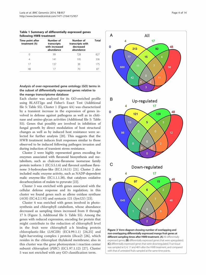
Table 1 Summary of differentially expressed genesfollowing HWB treatment
Time point aftertreatment (h)
Number oftranscripts
with increasedabundance
Number oftranscripts with
decreasedabundance
Total
0 99 728 827
4 141 195 336
17 137 38 175
48 17 70 87
Figure 3 Venn diagram showing number of overlapping andnon-overlapping differentially expressedmango fruit genes atdifferent sampling times after HWB treatment. (A)All differentiallyexpressed genes; (B) differentially expressed genes that were upregulated;(C) differentially expressed genes that were downregulated. Fruit tissuewas sampled at 0, 4, 17 and 48 h after the HWB treatment, and comparedwith that of untreated fruits sampled at the same time points.
Luria et al. BMC Genomics 2014, 15:957 Page 4 of 14http://www.biomedcentral.com/1471-2164/15/957
Analysis of over-represented gene ontology (GO) terms inthe subset of differentially expressed genes relative tothe mango transcriptome databaseEach cluster was analyzed for its GO-enriched profileusing BLAST2go and Fisher’s Exact Test (Additionalfile 5: Table S5). Cluster 1 (Figure 4A) was characterizedby a transient increase in the expression of genes in-volved in defense against pathogens as well as in chiti-nase and amino-glycan activities (Additional file 5: TableS5). Genes that possibly are involved in inhibition offungal growth by direct modulation of host structuralchanges as well as by induced host resistance were se-lected for further analysis [20]. This suggests that theHWB treatment induces fruit responses similar to thoseobserved to be induced following pathogen invasion andduring induction of transient stress resistance.Cluster 2 were highly represented genes encoding for
enzymes associated with flavanoid biosynthesis and me-tabolism, such as chalcone-flavanone isomerase familyprotein isoform 1 (EC:5.5.1.6) and flavonol synthase flava-none 3-hydroxylase-like (EC:1.14.11) [21]. Cluster 2 alsoincluded malic enzyme activity, such as NADP-dependentmalic enzyme-like (EC:1.1.1.38), that catalyzes oxidativedecarboxylation of malate to pyruvate [22].Cluster 3 was enriched with genes associated with the
cellular defense response and its regulation; in thiscluster we found genes such as allene oxidase synthase(AOS) (EC:4.2.1.92) and syntaxin 121 (Syn121) [23].Cluster 4 was enriched with genes involved in photo-
synthesis and chlorophyll catabolism, whose abundancedecreased as sampling times increased from 0 through17 h (Figure 3, Additional file 5: Table S5). Among thegenes with reduced expression, encoding for protein thatmight contribute to the reduction of chlorophyll levelin the fruit were chlorophyll a-b binding proteinchloroplastic-like (LHCIIb) (EC4.99.1.1) [24,25] andlight-harvesting complex i protein (lhca2) [26], whichresides in the chloroplast thylakoid membrane; also inthis cluster was the gene photosystem i reaction centersubunit chloroplast (PIRC) (EC1.97.1.12) [27]. Cluster5 was not enriched with any GO classification term.

Figure 4 Heat-map diagram showing the five clusters of differentially expressed genes following HWB treatment. (A) Plots of theexpression profiles of 1,225 differentially expressed genes. Gray lines mark the various gene profiles; the green, red, blue, pink and light-blue linesrepresent the average expression profiles of clusters 1–5, respectively. (B) Heat map showing relative expression of 1,225 fruit genes at the foursampling times (0, 4, 17 and 48 h). Color key represents relative expression on a log 2 scale.
Luria et al. BMC Genomics 2014, 15:957 Page 5 of 14http://www.biomedcentral.com/1471-2164/15/957
Validation of differential gene expressionTo validate the differential expression of specific genesidentified by the RNA-seq analysis, quantitative (q) RT-PCR analyses were performed for key genes of interest.Mango genes belonging to the five different clusters ofgenes that were differentially expressed following HWBtreatment were analyzed for their expression levels(Figure 5). Chitinase 7 (EC:3.2.1.14) and phenylalanine am-monia lyase (PAL) (EC:4.3.1.25; EC:4.3.1.24) genes involvedin chitinase activity and response to wounding were repre-sented in cluster 1 (Figures 5A,B). Oxysterol binding pro-tein (OxyBP) (EC:2.7.11.9) and inositol-tetrakisphosphate1-kinase 2-like (IT1K2) (EC:2.7.1.159; EC:2.7.1.134) genes
involved in jasmonic acid stimulus were represented incluster 2 (Figure 5C,D). AOS and Syn 121 genes involvedin negative regulation of cellular defense responses wererepresented in cluster 3 (Figure 5E,F). LHCIIb and Ent-kaurene oxidase (Entkox) (EC:1.14.13.78) genes involvedin photosynthesis and heme binding were represented incluster 4 (Figure 5G,H). Salicylate o-methyltransferase(salometh) (EC 2.1.1.274) and peroxidase 15-like (perox-idase15) (EC 1.11.1.7) genes were represented in cluster5 (Figure 5I,J). Comparison with the results of the qRT-PCR analysis showed expression patterns that were signifi-cantly and consistently similar to those of the RNA-seqanalysis.

Figure 5 Validation of RNA-seq results by means of qRT-PCR. Ten differentially expressed genes (two from each of clusters 1 to 5) wereexamined by RNA-seq and qRT-PCR at four different time points after HWB treatment: A, B (cluster 1); C, D (cluster 2); E, F (cluster 3); G, H(cluster 4); and I, J (cluster 5). Values were normalized to the values obtained with untreated mango fruit samples at 0 h and the proportionalfold-change (FC) was calculated. Expression data are means of two replicates.
Luria et al. BMC Genomics 2014, 15:957 Page 6 of 14http://www.biomedcentral.com/1471-2164/15/957
The relationships among mango fruit HWB-inducedresponses, disease resistance to A. alternata and thedifferential expression of genes of the different clustersHWB treatment of freshly harvested fruits reduced theincidence of natural A. alternata infestation on fruits ofcvs. Palmer, Kent, Tommy Atkins, Keitt, Lilly and Shelly(Figure 6A). Hot water brushing treatment reduced theincidence of decay observed after 21 days of storage at12°C by 64–84% (Figure 6A), as also observed in citrus[28-30] and in peaches [31]. Several genes (Figure 6C) thatare known to modulate the host pathogen-resistancemechanism, related to JA and SA, showed upregulation[32-35]. Syn121 gene showed significant differential ex-pression (Figure 5F). This gene is a member of the SNAREprotein family that contributes to defense against fungalpenetration [36] and might be modulated by abiotic andbiotic stress responses [37,38]. It acts as a regulator of SA,
and may contribute to host resistance in the fruit. SAcontributes to systemic acquired resistance (SAR) [39]through processes that activate the hypersensitive reac-tion response and increased production of reactiveoxygen species [40-42]. A second upregulated genefamily in this group was that encoding glutaredoxin(EC 1.20.4.1). Glutaredoxin gene family is regarded ascandidates for controlling the redox state of regulatoryproteins [43]; they interact with TGA-transcriptionfactors which are bZip plant transcription factors.These transcription factors contain a palindromicmotif that is present in several plant promoters that aretranscriptionally activated in response to elevated SAlevels, and that negatively regulate the JA-inducibleexpression of defensin-like protein 16 (PDF1.2).Gutaredoxinis commonly used as a marker for JA-dependent defenseresponses [44].
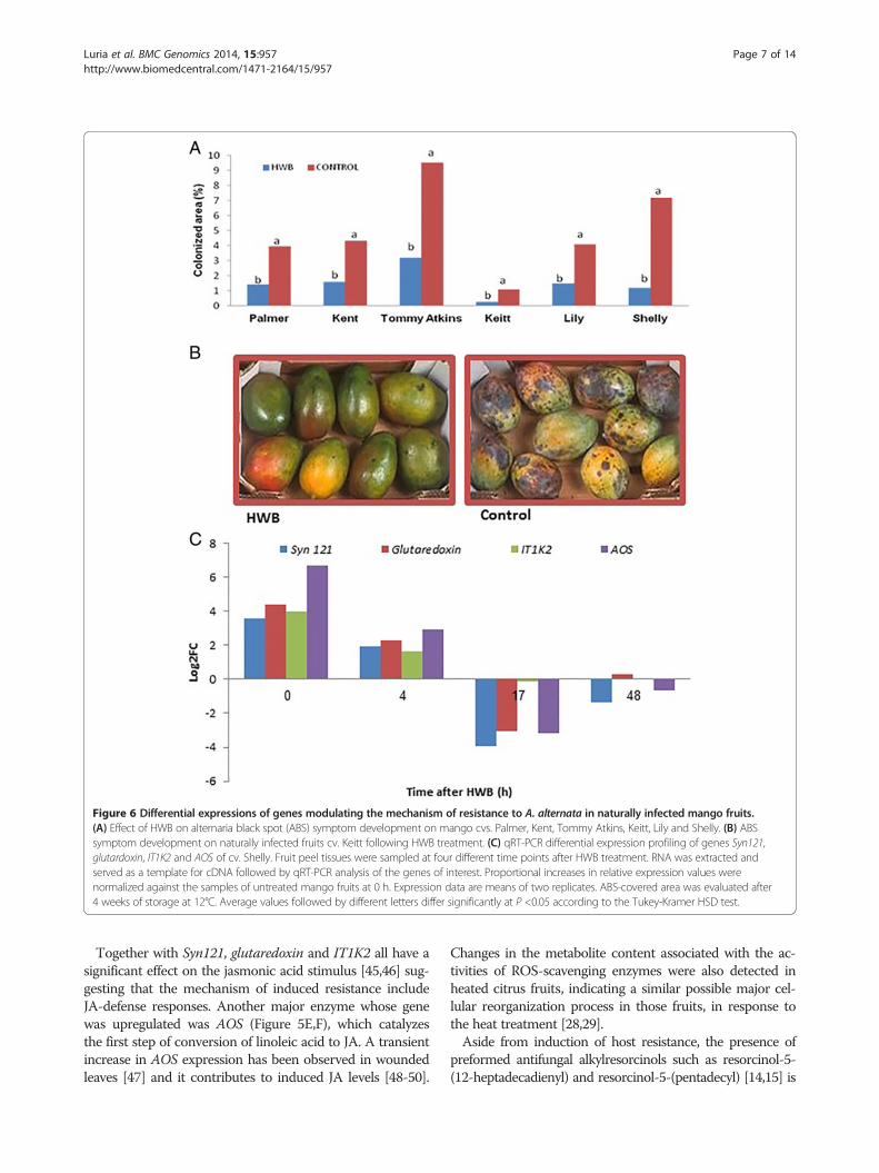
Figure 6 Differential expressions of genes modulating the mechanism of resistance to A. alternata in naturally infected mango fruits.(A) Effect of HWB on alternaria black spot (ABS) symptom development on mango cvs. Palmer, Kent, Tommy Atkins, Keitt, Lily and Shelly. (B) ABSsymptom development on naturally infected fruits cv. Keitt following HWB treatment. (C) qRT-PCR differential expression profiling of genes Syn121,glutardoxin, IT1K2 and AOS of cv. Shelly. Fruit peel tissues were sampled at four different time points after HWB treatment. RNA was extracted andserved as a template for cDNA followed by qRT-PCR analysis of the genes of interest. Proportional increases in relative expression values werenormalized against the samples of untreated mango fruits at 0 h. Expression data are means of two replicates. ABS-covered area was evaluated after4 weeks of storage at 12°C. Average values followed by different letters differ significantly at P <0.05 according to the Tukey-Kramer HSD test.
Luria et al. BMC Genomics 2014, 15:957 Page 7 of 14http://www.biomedcentral.com/1471-2164/15/957
Together with Syn121, glutaredoxin and IT1K2 all have asignificant effect on the jasmonic acid stimulus [45,46] sug-gesting that the mechanism of induced resistance includeJA-defense responses. Another major enzyme whose genewas upregulated was AOS (Figure 5E,F), which catalyzesthe first step of conversion of linoleic acid to JA. A transientincrease in AOS expression has been observed in woundedleaves [47] and it contributes to induced JA levels [48-50].
Changes in the metabolite content associated with the ac-tivities of ROS-scavenging enzymes were also detected inheated citrus fruits, indicating a similar possible major cel-lular reorganization process in those fruits, in response tothe heat treatment [28,29].Aside from induction of host resistance, the presence of
preformed antifungal alkylresorcinols such as resorcinol-5-(12-heptadecadienyl) and resorcinol-5-(pentadecyl) [14,15] is

Luria et al. BMC Genomics 2014, 15:957 Page 8 of 14http://www.biomedcentral.com/1471-2164/15/957
also a key factor modulating fruit reisitance to postharvestpathogens. These compounds are fatty-acid derivatives ob-tained with specialized type III polyketide synthases (referredto as ‘alkylresorcinol synthases’), which catalyze the forma-tion of 5-alkylresorcinols by using fatty acyl-CoA starterunits and malonyl-CoA extension units. The polyketide syn-thase (PKS) enzymes involved in the biosynthesis of aromaticring-containing intermediates as the resorcinol mainly usean aldol-condensation-based mechanism (stilbene synthase-type) or a Claisen-condensation-basedmechanism (chalconesynthase-type) for ring folding [51]. A chalcone flavone isom-erase involved in fatty-acid biosynthesis showed high expres-sion at time 0 h after HWB, suggesting possible activation ofthis process by the inducing treatment.
Effect of HWB on lenticel discolorationLenticel discoloration results from stress induced bythe HWB treatment, which leads to anthocyanin accu-mulation [9] (Figure 7). Four genes encoding UDP-glucose flavonoid 3-o-glucosyltransferase 3-like (Ugft3)(EC 2.4.1.91) – PAL, chalcone-flavanone isomerase-like
Figure 7 Effects of HWB treatment on the expression of flavonoid biodiscoloration on mango fruit cv. Shelly. (A) qRT-PCR profile of differentithe flavonoid biosynthesis process, naringenin-chalcone synthase activity, adiscoloration of HWB-treated and control fruits, and (C) lenticel discoloratioqRT-PCR values were normalized to the values obtained in samples from ureplicates. Lenticel discoloration was evaluated following 2 weeks of storagsignificantly at P <0.05 according to the Tukey-Kramer HSD test.
protein (CFIL) (EC 5.5.1.6) and chalcone synthase (CHS)(EC:2.3.1.74) – related to the anthocyanin accumulation,phenylpropanoid and flavonoid biosynthesis pathwaysfound in cluster 2 were tested by qRT-PCR (Figure 7A).Increased expression of these genes was clearly observed atthe 0 and 4 h time points, followed by decreased expression at17 and 48 h after the induction treatment. 1) ugft3, was de-scribed to control anthocyanin synthesis in grapes [52]; 2)PAL, encodes one of the major enzymes involved in flavonoidand phenylpropanoid biosynthesis in plants, and converts L-phenylalanine to ammonia and trans-cinnamic acid, which isthe precursor of the polyphenol compounds [53,54]; 3) CFILis the second committed enzyme of the flavonoid biosyntheticpathway which enhances flavonoid production and pigmenta-tion [55] 4) CHS, encoding chalcone synthase, which belongsto the PKSs and is also known as a type III PKS [56]; it cata-lyzes the initial step of flavonoid biosynthesis by converting 4-coumaroyl-CoA and malonyl-CoA to naringenin chalcone[57]. Although these genes peaked relatively early (4 h) afterthe HWB treatment, we envision that they activate relevantprocesses modulating fruit-resistance at later stages of fruit
synthesis-related genes and the occurrence of red lenticelally expressed genes Ugft3, PAL, CFIL and CHSï, which are related tond the phenylpropanoid biosynthesis pathway. (B) level of lenticeln symptoms on mango fruits, cv. Shelly following HWB treatment.ntreated mango fruits at 0 h. Expression data are means of twoe at 12°C [9]. Average values followed by different letters differ

Luria et al. BMC Genomics 2014, 15:957 Page 9 of 14http://www.biomedcentral.com/1471-2164/15/957
life. Similar phenylpropanoid pathway-expression patternwere described in heat treated peaches [58], suggesting a widemechanism of fruit responses to heat treatment.
Effect of HWB treatment on skin color changeOne of the significant consequences of HWB treatment isthe improved and enhanced color development onthe fruit skin, which results from both anthocyanin produc-tion and inhibition of chlorophyll accumulation. The fruitcolor index of HWB-treated fruits was higher during all theperiod of fruit storage at 12 and 20°C (Figure 8). The index inuntreated fruits increased from values of 2.8, 16 days afterharvest to 3.4, 8 days later. During the same period theHWB-treated fruits showed an increase in color index to 4.6(Figure 8C), indicating induction of a 31% increase in colorlevel by the HWB treatment. The decreased expression ofchlorophyll and anthocyanin biosynthesis-related genes sug-gested that HWB played a role in the modulation of thoseprocesses (Figure 8A). Reduced expression of LHCIIb encod-ing for chlorophyll a/b-binding protein, one of the mostabundant proteins in the chloroplast, which is important inthe structure of photosynthesis reaction centers [24,25,59,60]indicates a reduction in chlorophyll levels. Other key genesthat showed reduced expression included those encoding:the oxygen-evolving enhancer protein chloroplastic (Oxepch)(EC1.3.1.74) [61]; PIRC; and thioredoxin-like 1-chloroplastic(Thl1ch) (EC 1.1.1.49), in which the chloroplast thioredoxinshave been suggested as mediators in the light-dependentregulation of chloroplast enzyme activity [62].Two genes related to anthocyanin accumulation – antho-
cyanin 5-aromatic (anthocyanin5a) (EC:2.3.1.144) and UDP-glycosyltransferase 85a2-like (85A2) (EC:2.4.1.115) – showedincreased expression at the first two time points (0 and 4 h)after HWB, as detected by qRT-PCR (Figure 8B). The antho-cyanin5a modulates anthocyanin by aromatic acylation [63]and 85A2 affects the level of glucosyl anthocyanidins in redfruit during ripening [52]. This suggests that different path-ways are activated by HWB to modulate anthocyanin accu-mulation and to reduce processes associated with chlorophyllaccumulation [64], thereby enhancing color development.
ConclusionsOverall, gene-expression profiling inmango skin revealed simi-larities in heat responses to those found in citrus and peachfruits [30,58], with three major effects following HWB treat-ment: 1) a transient increase in expression of the stress- andpathogen-defense mechanisms-related genes; 2) a transient re-duction in the expression of chlorophyll-related genes; and 3)increased expression of sugar and flavonoidmetabolism relatedgenes 4 h after treatment. These threemain trends indicated bythe observed HWB-induced modulation of gene expressioncan account for the major results of postharvest HWB treat-ment including: 1) induced resistance toA. alternata, attributedto the transient increase in the expression of genes involved in
immune response and host resistance (Figure 6); 2) improvedcolor development observed after HWB which can be attrib-uted to decreased photosynthesis, including reduction ofchlorophyll accumulation after the treatment (Figure 8) andincreased abundance of genes of flavonoid metabolism; and 3)enhanced lenticel discoloration that is also correlated with up-regulation of flavanoidmetabolism (Figure 7).In light of the physiological changes described, the im-
proved fruit quality acquired following heat stress is probablya result of various stress-response mechanisms that act coor-dinately to improve the fruit quality, prevent pathogen devel-opment, prevent cell damage and re-establish cellularhomeostasis. Genes identified in the present study that aremodified in mango fruits following heat treatment, couldhave similar functions in other fruits such as citrus or peach.
MethodsFruit, and storage conditionsFreshly harvested mango fruits (Mangifera indica L. cv.Shelly) were obtained from trees in commercial orchards inthe north of Israel. Several experiments to determine thephenotypic response of the fruits to HWB were carried outin each season during three consecutive years. The pheno-typic responses obtained in the various years were similar,therefore results from only 1 year are presented. Each treat-ment comprised 6 replications, each with 15 fruits.Fruits were harvested at the commercial mature-green
stage, treated on the same or the following day, and trans-ferred to simulated export conditions. They were stored forabout 4 weeks (depending on the experiment) at 12°C, ca.90% RH [9]. Control, untreated fruits were stored under thesame conditions, immediately upon arrival from the orchard.
Postharvest packinghouse treatments in semi-commercialmango experimentsPostharvest treatments were carried out at the Department ofPostharvest Science of theVolcani Center in Bet Dagan, Israel.HWB treatment at 55°C was applied on the packing line, as aspray (nozzle pressure of 2 atm) above brushes revolving at60 g, a rate of 100–120 L min−1 and at a nozzle pressure of2 atm [2]. Fruits were passed over five to seven transverselyoriented, 12-cm-diameter plastic brushes for 15–20 s.
Fruit ripening, development of red lenticels, black spotand stem-end rots during storageDisease severity, measured as the percentage of the fruitsurface covered by black lesions, was recorded for 90fruits (15 fruits from each of six replicates) after about4 weeks of storage at 12°C and 3 days at 20°C. Fruitswere regarded as unmarketable when more than 1% oftheir surface area exhibited black spots.Lenticel spotting was assessed visually at the end of
storage, on a scale of 0 to 3, with values based on a

Figure 8 Shelly. Effect of HWBondifferential expression of chlorophyll and anthocyanin accumulation-related genes and colordevelopment inmango cv. Shelly. (A, B) qRT-PCR gene-expression profiles of genes related to (A) chlorophyll accumulation (Thl1ch, LHCIIb, Oxepch and PIRC)and (B) anthocyanin synthesis (85A2 and Anthocyanin5). The expression profile comprises data taken from samples ofmango tissues sampled from cv. Shelly atfour different timepoints after HWB treatment. (C)Changes in color index after 16 days of storage at 12°C followedby 8 days at 20°C. Vertical bars indicate SDof fivereplicates. qRT-PCR valueswere normalized to the values obtainedwith untreatedmango fruit samples at 0 h. Expression data are themeans of two replicates.
Luria et al. BMC Genomics 2014, 15:957 Page 10 of 14http://www.biomedcentral.com/1471-2164/15/957
combination of degree of lenticel discoloration and areacovered by the symptoms [9].Skin color development was assessed visually at the
end of storage and a color index was calculated on ascale of 1 (green) to 5 (full color) [9].
Total RNA extractionTotal RNA was extracted according to Yang et al. [65], withminor changes: about 1- to 2-g aliquots of HWB-treatedand control tissues were sampled from pools composed offive different fruits from the same tree. The samples were

Luria et al. BMC Genomics 2014, 15:957 Page 11 of 14http://www.biomedcentral.com/1471-2164/15/957
ground to a fine powder in liquid nitrogen and transferredinto 50-mL centrifuge tubes with 10 mL of CTAB RNA ex-traction buffer (100 mM Tris-borate pH 8, 2 M NaCl,25 mM ethylenediaminetetraacetic acid pH 8, 2% (w/v)CTAB, 2% (w/v) polyvinylpolypyrrolidone and 2% (v/v) β-mercaptoethanol). The mixture was shaken for 3 min andthen incubated at 65°C for 15 min. Samples were extractedtwice with an equal volume of chloroform:isoamyl alcohol[24:1 (v/v)], and the phases were separated by centrifuga-tion at 10,000 g for 10 min. Following centrifugation, lith-ium chloride was added to a final concentration of 2.5 Mand RNA was allowed to precipitate overnight at 4°C. RNAwas pelleted at 4°C for 30 min at 10,000 g, washed with70% ethanol, and re-suspended at 65°C for 3 min in SSTEbuffer (10 mM Tris pH 8, 1 M NaCl, 1 mM EDTA pH 8and 0.5% (w/v) SDS). Samples were extracted with an equalvolume of chloroform:isoamyl alcohol (24:1), and with anequal volume of chloroform:isoamyl alcohol:water-satu-rated phenol (24:1:25), and the phases were separated bycentrifugation at 10,000 g for 10 min. The RNA wasethanol-precipitated overnight, and resuspended in diethyl-pyrocarbonate-treated water. RNA was further treated withTurbo DNAse (Ambion, Austin, TX, USA).
Table 2 Primers used for qRT-PCR analysis
Gene Forward 5‘→ 3’
Actin CATTGTGCTCAGTGGTGGT
FSL TGGGAGCATATGTTAGGGTAT
PAL AATGGAAATGCGGCGATT
CHS GAAAGATGTTCCCGGGTTG
CFIL GCAGTGATCCCACCACTTG
Oxybp CAATTCGACGGAACCGAA
IT1K2 AACTATTTCCCTGGGTATGGG
Chitinase 7 CATTCAAGACTGCCCTGTGG
Hl1ch TGCAACACCAAAGCAACAA
PIRCS AGTCTAACGCTGACAGGAAG
LHCIIb GGGCTATGCTTGCTGTTCC
85A2 CAAGCTTCATGAGAGTCACTG
Entkox TGGCAGTGAGATTGCCGTA
Salometh ACTCACCATCACCAAGCGA
Syn121 TGCGGTCCAGGATCTTCAT
Glutaredoxin GGGAAGATGGTTTCCGAGA
AOS AGAGCAGAGGAGTTCGTGG
Peroxidase15 TGCCCAGGAGTTGTCTCTTG
Ugft3 CTCATCTGCAATCCAGAAAT
Thl1ch TGCAACACCAAAGCAACAA
Oxepch GCCACGTCTCGAACATTAA
Anthocyanin5 TTTCTCACTTCCCTGCTTTG
Analysis by qRT-PCRSingle-stranded cDNA was synthesized from 1 μg of totalRNA by means of the Verso cDNA synthesis kit (ThermoFisher Scientific, Waltham, MA, USA). The synthesizedcDNA was used as a template for qRT-PCR analysis, to esti-mate the expression level of the selected genes. The cDNAsamples were diluted 1:10 (v/v) to the final template concen-tration for qRT-PCR. Real-time qRT-PCR was performedwith a RotorGene 3000 system (Corbett Research, Sydney,Australia). PCR amplification was run with 3.5 μL of cDNAtemplate in 10 μL of reaction mixture containing 5 μL abso-lute blue qPCR SYBR green ROX mix (Thermo Scientific)and 300 nMprimers. PCR conditions were: initial denaturingfor 15 min at 94°C; 40 denaturing cycles of 10 s at 94°C;annealing at 60°C for 15 s; extension at 72°C for 20 s (cyclingA), 77°C for 6 s (cycling B), or 80°C for 6 s (cycling C),and melting at 72–99°C. The samples were subjectedto melting-curve analysis with the RotorGene pro-gram. All samples were normalized to actin gene levelsin the same qRT-PCR, and the values were expressedas increase or decrease in level relative to a calibrationsample. The forward and reverse primers for all of thegenes are listed in Table 2.
Reverse 5‘→ 3’
T TTGGAGCAAGTGCAGTGATT
TGG TCTCATCGCCTCATAATTCAAGAA
C TCCCAGCTCTTCCCTCACAA
A AGTCGTTGATGCCGATTGG
A TCATGCATTCGTACATCTGTAGCA
TC CCGTTCCCTACCAAGTCGTTT
AA CTGACTCTGTTTCAGGCCCAA
TA GGCTCGAATAGTTGCACCAAA
AC TCCCACCATCTAAGAGACCTACTG
GAA AATCCACCAGCGAACTTGGA
A AGCCCACTCTTGAGCCTTTACC
ATG GAAGATTTCAAGCAGTCGTGTGAT
A CAGGCCTCCACACGTTAGGAT
AA TTGTGTAATCTCAGTCCGATCAATC
C TCCATCGAGTCCTGCAACTTC
AT AGCAAACGCTTCACCACATG
C TGGTCCGTTTGACCACAGC
T CAGGTTTGACCTCCATCCAAA
CG AATCGAGTGTCGCGGTTTG
AC TCCCACCATCTAAGAGACCTACTG
GC TACACTGCCGTCGTCTTCTTGA
G TCCGGCGTCATTACAGATGA
Luria et al. BMC Genomics 2014, 15:957 Page 12 of 14http://www.biomedcentral.com/1471-2164/15/957
Bioinformatics analysis of RNA-seq dataThe transcriptome of M. indica cv. Shelly was sequencedaccording to Illumina Hiseq2000 and Trueseq protocols,at the Crown Institute for Genomics, The Nancy andStephen Grand Israel National Center for PersonalizedMedicine at the Weizmann Institute of Science, Reho-vot, Israel. Eight libraries with total single-end RNA-seqreads 100 nucleotides in length were generated. Theeight libraries contained the following sequences: 1) con-trol RNA-seq of mango peel at time 0 h with 20,025,080reads; 2) RNA-seq of mango peel treated by HWB attime 0 h with 20,202,891 reads; 3) control RNA-seq ofmango peel at time 4 h with 21,622,332 reads; 4) RNA-seq of mango peel 4 h after HWB with 20,744,762 reads;5) control RNA-seq of mango peel at 17 h with20,916,444 reads; 6) RNA-seq of mango peel 17 h afterHWB with 21,410,839 reads; 7) control RNA-seq ofmango peel at 48 h with 20,687,358 reads; and 8) RNA-seq of mango peel 48 h after HWB with 21,230,696reads. The transcriptome datasets are available in theNCBI Sequence Read Archive (SRA) under accessionnumber SRX375390 and BioProject accessionPRJNA227243. A new transcriptome was assembled fromthe 8.6-Gbp sequences by using Trinity software [66], gen-erating 57.544 contigs with N50 of 1,598 bp. This Tran-scriptome Shotgun Assembly project has been depositedat DDBJ/EMBL/GenBank under accession no.GBCV00000000. The version described in the presentpaper is the first version, GBCV01000000.Tophat [67], Bowtie2 [68] and Cufflink packages [67]
were used to align the RNA-seq with the transcriptomeand to calculate differentially expressed genes. The li-braries were aligned with the mango transcriptome atalignment rates (mapped reads/total reads) of 90.84,90.02, 89.48, 89.77, 90.70, 90.11, 90.25 and 90.35% forsamples 1 to 8, respectively.The genes of M. indica cv. Shelly were annotated by
using BLASTx [69], after which their GO term [70]was assigned by combining both BLASTx data andinterproscan analysis [71] by means of the BLAST2gosoftware pipeline [72]. GO-enrichment analysis wasperformed by using Fisher’s exact test with multipletesting correction of FDR. Heat mapping and cluster-ing of the genes were performed with the R softwareggplots2 package [73].
Additional files
Additional file 1: Table S1. Table of differentially expressed genes andtheir annotations after HWB treatment versus control at time 0 h.
Additional file 2: Table S2. Table of differentially expressed genes andtheir annotations after HWB treatment versus control at time 4 h.
Additional file 3: Table S3. Table of differentially expressed genes andtheir annotations after HWB treatment versus control at time 17 h.
Additional file 4: Table S4. Table of differentially expressed genes andtheir annotations after HWB treatment versus control at time 48 h.
Additional file 5: Table S5. GO enrichment analysis of gene clusters [74].
AbbreviationsHWB: Hot-water brushing; OxyBP: Oxysterol-binding protein; IT1K2: Inositol-tetrakisphosphate 1-kinase 2-like; AOS: Allene oxide synthase;Syn121: Syntaxin-121-like; LHCIIb: Chlorophyll a/b-binding proteinchloroplastic-like; PIRCS: Photosystem I reaction center subunit chloroplastic-like; 85A2: UDP-glycosyltransferase 85a2-like; PAL: Phenylalanine ammonialyase; CFIL: Chalcone-flavanone isomerase-like protein; CHS: Chalconesynthase; Entkox: Ent-kaurene oxidase; Salometh: Salicylate o-methyltransferase; Ugft3: UDP-glucose flavonoid 3-o-glucosyltransferase;Thl1ch: Thioredoxin-like 1- chloroplastic; Oxepch: Oxygen-evolving enhancerprotein chloroplastic.
Competing interestsThe authors declare that they have no competing interests.
Authors’ contributionsNL performed the experiments and drafted the manuscript. NS performedthe bioinformatics analyses and drafted the manuscript. MY performed theRNA extractions and initial qPCR quantification. OF and IK performed theHWB and sampling of fruits. AL helped with the analysis and writing of themanuscript. DP conceived the project, supervised the project, and draftedthe manuscript. All authors have read and approved the final manuscript.
AcknowledgmentsThis study was supported in part by a grant from the Chief Scientist of theIsraeli Ministry of Agriculture and Rural Development.
Author details1Department of Postharvest Science of Fresh Produce, ARO, the VolcaniCenter, Bet Dagan 50250, Israel. 2Department of Plant Pathology and WeedResearch, ARO, the Volcani Center, Bet Dagan 50250, Israel.
Received: 13 February 2014 Accepted: 23 October 2014Published: 5 November 2014
References1. Brecht JK, Sargent SA, Kader AA, Mitcham EJ, Maul F, Brecht PE, Menocal O:
Mango Postharvest Best Management Practices Manual. Gainesville: Univ.of Fla. Horticultural Sciences Department; 2010. Publ. HS1185. 78 pp.
2. Prusky D, Fuchs Y, Kobiler I, Roth I, Weksler A, Shalom Y, Fallik E, ZaubermanG, Pesis E, Akerman M: Effect of hot water brushing, prochloraz treatmentand waxing on the incidence of black spot decay caused by Alternariaalternata in mango fruits. Postharvest Biol Technol 1999, 15(2):165–174.
3. Kader A, Mitcham B: Optimum procedures for ripening mangoes. In FruitRipening and Ethylene Management, Univ. Calif. Postharvest TechnologyResearch and Information Center Publication Series, Volume 9; 2008.
4. Pesis E, Aharoni D, Aharon Z, Ben-Arie R, Aharoni N, Fuchs Y: Modifiedatmosphere and modified humidity packaging alleviates chilling injurysymptoms in mango fruit. Postharvest Biol Technol 2000, 19(1):93–101.
5. Prusky D, Fuchs Y, Yanko U: Assessment of latent infections as a basis forcontrol of postharvest disease of mango. Plant Dis 1983, 67(7):816–818.
6. Prusky D, Fuchs Y, Zauberman G: A method for pre‐harvest assessment oflatent infections in fruits. Ann Appl Biol 1981, 98(1):79–85.
7. Prusky D, Shalom Y, Kobiler I, Akerman M, Fuchs Y: The level of quiescentinfection of Alternaria alternata in mango fruits at harvest determinesthe postharvest treatment applied for the control of rots during storage.Postharvest Biol Technol 2002, 25(3):339–347.
8. Yahia EM: Modified and controlled atmospheres for tropical fruits. StewartPostharvest Rev 2006, 2(5):1–10.
9. Feygenberg O, Keinan A, Kobiler I, Pesisa E, Lersa A, Prusky D, FeygenbergO, Keinan A, Kobiler I, Pesisa E, Lersa A, Prusky D: Improved managementof mango fruits by orchard and in packing house treatments for thereduction of lenticels discoloration and decay prevention. Postharvest BiolTechnol 2014. in press.
Luria et al. BMC Genomics 2014, 15:957 Page 13 of 14http://www.biomedcentral.com/1471-2164/15/957
10. Du Plooy W, Combrinck S, Botha B, Van der Merwe C, Regnier T:Development of discolouration in mango lenticels. Acta Horticult 2006,820:665–672.
11. Prusky D: Pathogen quiescence in postharvest diseases. Annu RevPhytopathol 1996, 34(1):413–434.
12. Kobiler I, Shalom Y, Roth I, Akerman M, Vinokour Y, Fuchs Y, Prusky D:Effect of 2,4-dichlorophenoxyacetic acid on the incidence of side andstem-end rots in mango fruits. Postharvest Biol Technol 2001, 23(1):23–32.
13. Prusky D, Eshel D, Kobiler I, Yakoby N, Beno-Moualem D, Ackerman M,Zuthji Y, Ben Arie R: Postharvest chlorine treatments for the control ofthe persimmon black spot disease caused by Alternaria alternata.Postharvest Biol Technol 2001, 22(3):271–277.
14. Droby S, Prusky D, Jacoby B: Induction of an antifungal agent in unripemango fruits to demonstrate their involvement in latent infections ofAlternaria alternata. Physiol Mol Plant Pathol 1987, 30:285–292.
15. Droby S, Prusky D, Jacoby B, Goldman A: Presence of an antifungalcompound and its relation to the latency of Alternaria alternata inunripe peel of mango fruits. Physiol Mol Plant Pathol 1986,29:173–183.
16. Davidzon M, Kobiler I, Alkan N, Prusky D: Acidification of fruit environmentby gluconic acid during decay development of Phomopsis mangiferae.Postharvest Biol Technol 2009, 55(2):71–77.
17. Singh N: A Draft Genome of the King of Fruit, Mango (Mangiferaindica L). In Plant and Animal Genome XXII Conference, Plant and AnimalGenome; 2014.
18. Grabherr MG, Haas BJ, Yassour M, Levin JZ, Thompson DA, Amit I, AdiconisX, Fan L, Raychowdhury R, Zeng Q: Full-length transcriptome assemblyfrom RNA-Seq data without a reference genome. Nat Biotechnol 2011,29(7):644–652.
19. Ashburner M, Ball CA, Blake JA, Botstein D, Butler H, Cherry JM, Davis AP,Dolinski K, Dwight SS, Eppig JT: Gene ontology: tool for the unification ofbiology. Nat Genet 2000, 25(1):25–29.
20. Howe GA, Schilmiller AL: Oxylipin metabolism in response to stress. CurrOpin Plant Biol 2002, 5(3):230–236.
21. Winkel-Shirley B: Flavonoid biosynthesis. A colorful model for genetics,biochemistry, cell biology, and biotechnology. Plant Physiol 2001,126(2):485–493.
22. Mitsch MJ, Voegele RT, Cowie A, Osteras M, Finan TM: Chimeric structureof the NAD (P) + −and NADP + −dependent malic enzymes ofRhizobium(Sinorhizobium) meliloti. J Biol Chem 1998, 273(15):9330–9336.
23. Maekawa S, Inada N, Yasuda S, Fukao Y, Fujiwara M, Sato T, Yamaguchi J:The C/N regulator ATL31 controls papilla formation in response topowdery mildew fungi penetration by interacting with SNARE SYP121 inArabidopsis. Plant Physiol 2014, 113:230995.
24. Paulsen H, Dockter C, Volkov A, Jeschke G: Folding and Pigment Binding ofLight-harvesting Chlorophyll a/b Protein (LHCIIb). In: The Chloroplast. Springer;2010:231–244.
25. Szabó I, Bergantino E, Giacometti GM: Light and oxygenic photosynthesis:energy dissipation as a protection mechanism against photo-oxidation.EMBO Rep 2005, 6(7):629–634.
26. Wientjes E, Croce R: The light-harvesting complexes of higher-plantPhotosystem I: Lhca1/4 and Lhca2/3 form two red-emittingheterodimers. Biochem J 2011, 433:477–485.
27. Bengis C, Belson N: Subunit structure of chloroplast photosystem Ireaction center. J Biol Chem 1977, 252(13):4564–4569.
28. Perotti VE, Del Vecchio HA, Sansevich A, Meier G, Bello F, Cocco M, GarránSM, Anderson C, Vázquez D, Podestá FE: Proteomic, metabalomic, andbiochemical analysis of heat treated Valencia oranges during storage.Postharvest Biol Technol 2011, 62(2):97–114.
29. Sapitnitskaya M, Maul P, McCollum GT, Guy CL, Weiss B, Samach A, Porat R:Postharvest heat and conditioning treatments activate differentmolecular responses and reduce chilling injuries in grapefruit. J Exp Bot2006, 57(12):2943–2953.
30. Yun Z, Gao H, Liu P, Liu S, Luo T, Jin S, Xu Q, Xu J, Cheng Y, Deng X:Comparative proteomic and metabolomic profiling of citrus fruit withenhancement of disease resistance by postharvest heat treatment.BMC Plant Biol 2013, 13:44.
31. Lauxmann MA, Brun B, Borsani J, Bustamante CA, Budde CO, Lara MV,Drincovich MF: Transcriptomic profiling during the post-harvest ofheat-treated Dixiland Prunus persica fruits: common and distinctresponse to heat and cold. PLoS One 2012, 7(12):e51052.
32. Dixon RA, Harrison MJ, Lamb CJ: Early events in the activation of plantdefense responses. Annu Rev Phytopathol 1994, 32(1):479–501.
33. Jones JD, Dangl JL: The plant immune system. Nature 2006,444(7117):323–329.
34. Keen NT: Gene-for-gene complementarity in plant-pathogen interactions.Annu Rev Genet 1990, 24:447–463.
35. Ponce De León I, Montesano M: Activation of defense mechanismsagainst pathogens in mosses and flowering plants. Int J Mol Sci 2013,14(2):3178–3200.
36. Shukla S, Srivastava P, Choubey SK: Comparative analysis and structureelucidation of Syntaxin-A novel component tends to defense mechanismin plant proteomics. Saurabh Shukla1, Prashant Srivastava1, SanjayKumar Choubey1 and Gomase VS 2. 2010.
37. Kunkel BN, Brooks DM: Cross talk between signaling pathways inpathogen defense. Curr Opin Plant Biol 2002, 5(4):325–331.
38. Wasternack C, Hause B: Jasmonates and octadecanoids: signals in plantstress responses and development. Prog Nucleic Acid Res Mol Biol 2002,72:165–221.
39. Ryals JA, Neuenschwander UH, Willits MG, Molina A, Steiner H-Y, Hunt MD:Systemic acquired resistance. Plant Cell 1996, 8(10):1809.
40. Mur LAJ, Bi YM, Darby RM, Firek S, Draper J: Compromising early salicylicacid accumulation delays the hypersensitive response and increases viraldispersal during lesion establishment in TMV-infected tobacco. Plant J1997, 12(5):1113–1126.
41. Mur LAJ, Brown IR, Darby RM, Bestwick CS, Bi YM, Mansfield JW, Draper J:A loss of resistance to avirulent bacterial pathogens in tobacco isassociated with the attenuation of a salicylic acid-potentiated oxidativeburst. Plant J 2000, 23(5):609–621.
42. Shirasu K, Nakajima H, Rajasekhar VK, Dixon RA, Lamb C: Salicylic acidpotentiates an agonist-dependent gain control that amplifies pathogensignals in the activation of defense mechanisms. Plant Cell Online 1997,9(2):261–270.
43. Lemaire SD, Guillon B, Le Maréchal P, Keryer E, Miginiac-Maslow M,Decottignies P: New thioredoxin targets in the unicellular photosyntheticeukaryote Chlamydomonas reinhardtii. Proc Natl Acad Sci U S A 2004,101(19):7475–7480.
44. Brown RL, Kazan K, McGrath KC, Maclean DJ, Manners JM: A role for theGCC-box in jasmonate-mediated activation of the PDF1.2 gene ofArabidopsis. Plant Physiol 2003, 132(2):1020–1032.
45. Mosblech A, Thurow C, Gatz C, Feussner I, Heilmann I: Jasmonic acidperception by COI1 involves inositol polyphosphates in Arabidopsisthaliana. Plant J 2011, 65(6):949–957.
46. Sheard LB, Tan X, Mao H, Withers J, Ben-Nissan G, Hinds TR, Kobayashi Y,Hsu F-F, Sharon M, Browse J: Jasmonate perception by inositol-phosphate-potentiated COI1-JAZ co-receptor. Nature 2010,468(7322):400–405.
47. Laudert D, Pfannschmidt U, Lottspeich F, Holländer-Czytko H, Weiler EW:Cloning, molecular and functional characterization of Arabidopsisthaliana allene oxide synthase (CYP 74), the first enzyme of theoctadecanoid pathway to jasmonates. Plant Mol Biol 1996, 31(2):323–335.
48. Hamberg M: Biosynthesis of 12-oxo-10, 15 (Z)-phytodienoic acid:identification of an allene oxide cyclase. Biochem Biophys Res Commun1988, 156(1):543–550.
49. Laudert D, Hennig P, Stelmach BA, Müller A, Andert L, WE W: Analysis of12-oxo-phytodienoic acid enantiomers in biological samples by capillarygas chromatography–mass spectrometry using cyclodextrin stationaryphases. Anal Biochem 1997, 246(2):211–217.
50. Vick BA, Zimmerman DC: Lipoxygenase, hydroperoxide isomerase, andhydroperoxide cyclase in young cotton seedlings. Plant Physiol 1981,67(1):92–97.
51. Baerson SR, Schroder J, Cook D, Rimando AM, Pan Z, Dayan FE, Noonan BP,Duke SO: Alkylresorcinol biosynthesis in plants: new insights from anancient enzyme family? Plant Signal Behav 2010, 5(10):1286–1289.
52. Ford CM, Boss PK, Hoj PB: Cloning and characterization of Vitis viniferaUDP-glucose:flavonoid 3-O-glucosyltransferase, a homologue of theenzyme encoded by the maize Bronze-1 locus that may primarily serve toglucosylate anthocyanidins in vivo. J Biol Chem 1998, 273(15):9224–9233.
53. Camm EL, Towers G: Phenylalanine ammonia lyase. Phytochemistry 1973,12(5):961–973.
54. Jones DH: Phenylalanine ammonia-lyase: regulation of its induction, andits role in plant development. Phytochemistry 1984, 23(7):1349–1359.

Luria et al. BMC Genomics 2014, 15:957 Page 14 of 14http://www.biomedcentral.com/1471-2164/15/957
55. Morita Y, Takagi K, Fukuchi‐Mizutani M, Ishiguro K, Tanaka Y, Nitasaka E,Nakayama M, Saito N, Kagami T, Hoshino A: A chalcone isomerase‐likeprotein enhances flavonoid production and flower pigmentation. Plant J2014, 78(2):294–304.
56. Austin MB, Noel JP: The chalcone synthase superfamily of type IIIpolyketide synthases. Nat Prod Rep 2003, 20(1):79–110.
57. Ferrer JL, Jez JM, Bowman ME, Dixon RA, Noel JP: Structure of chalconesynthase and the molecular basis of plant polyketide biosynthesis.Nat Struct Biol 1999, 6(8):775–784.
58. Lauxmann MA, Borsani J, Osorio S, Lombardo VA, Budde CO, Bustamante CA,Monti LL, Andreo CS, Fernie AR, Drincovich MF: Deciphering the metabolicpathways influencing heat and cold responses during post‐harvestphysiology of peach fruit. Plant Cell Environ 2014, 37(3):601–616.
59. Bellafiore S, Barneche F, Peltier G, Rochaix JD: State transitions and lightadaptation require chloroplast thylakoid protein kinase STN7. Nature2005, 433(7028):892–895.
60. Niyogi KK, Li XP, Rosenberg V, Jung HS: Is PsbS the site of non-photochemicalquenching in photosynthesis? J Exp Bot 2005, 56(411):375–382.
61. Mayfield SP, Rahire M, Frank G, Zuber H, Rochaix J-D: Expression of thenuclear gene encoding oxygen-evolving enhancer protein 2 is requiredfor high levels of photosynthetic oxygen evolution in Chlamydomonasreinhardtii. Proc Natl Acad Sci 1987, 84(3):749–753.
62. Buchanan BB, Wolosiuk RA, Schürmann P: Thioredoxin and enzymeregulation. Trends Biochem Sci 1979, 4(4):93–96.
63. Fujiwara H, Tanaka Y, Fukui Y, Nakao M, Ashikari T, Kusumi T: Anthocyanin5‐aromatic acyltransferase from gentiana triflora. Eur J Biochem 1997,249(1):45–51.
64. Ishida S, Takabayashi A, Ishikawa N, Hano Y, Endo T, Sato F: A novelnuclear-encoded protein, NDH-dependent cyclic electron flow 5, isessential for the accumulation of chloroplast NAD(P)H dehydrogenasecomplexes. Plant Cell Physiol 2009, 50(2):383–393.
65. Yang G, Zhou R, Tang T, Shi S: Simple and efficient isolation of high-quality total RNA from Hibiscus tiliaceus, a mangrove associate andits relatives. Prep Biochem Biotechnol 2008, 38(3):257–264.
66. Haas BJ, Papanicolaou A, Yassour M, Grabherr M, Blood PD, Bowden J,Couger MB, Eccles D, Li B, Lieber M: De novo transcript sequencereconstruction from RNA-seq using the Trinity platform for referencegeneration and analysis. Nat Protoc 2013, 8(8):1494–1512.
67. Trapnell C, Roberts A, Goff L, Pertea G, Kim D, Kelley DR, Pimentel H,Salzberg SL, Rinn JL, Pachter L: Differential gene and transcript expressionanalysis of RNA-seq experiments with TopHat and Cufflinks. Nat Protoc2012, 7(3):562–578.
68. Langmead B, Salzberg SL: Fast gapped-read alignment with Bowtie 2.Nat Methods 2012, 9(4):357–359.
69. Altschul SF, Gish W, Miller W, Myers EW, Lipman DJ: Basic local alignmentsearch tool. J Mol Biol 1990, 215(3):403–410.
70. Gene Ontology C, Blake JA, Dolan M, Drabkin H, Hill DP, Li N, Sitnikov D,Bridges S, Burgess S, Buza T, McCarthy F, Peddinti D, Pillai L, Carbon S,Dietze H, Ireland A, Lewis SE, Mungall CJ, Gaudet P, Chrisholm RL, Fey P,Kibbe WA, Basu S, Siegele DA, McIntosh BK, Renfro DP, Zweifel AE, Hu JC,Brown NH, Tweedie S, et al: Gene ontology annotations and resources.Nucleic Acids Res 2013, 41(Database issue):D530–D535.
71. Hunter S, Apweiler R, Attwood TK, Bairoch A, Bateman A, Binns D, Bork P,Das U, Daugherty L, Duquenne L, Finn RD, Gough J, Haft D, Hulo N, Kahn D,Kelly E, Laugraud A, Letunic I, Lonsdale D, Lopez R, Madera M, Maslen J,McAnulla C, McDowall J, Mistry J, Mitchell A, Mulder N, Natale D, Orengo C,Quinn AF, et al: InterPro: the integrative protein signature database.Nucleic Acids Res 2009, 37(Database issue):D211–D215.
72. Conesa A, Gotz S, Garcia-Gomez JM, Terol J, Talon M, Robles M: Blast2GO:a universal tool for annotation, visualization and analysis in functionalgenomics research. Bioinformatics 2005, 21(18):3674–3676.
73. R Development Core Team: R: A Language and Environment for StatisticalComputing. Vienna, Austria: R Foundation for Statistical Computing; 2009.
74. Benjamini Y, Hochberg Y: Controlling the false discovery rate: a practicaland powerful approach to multiple testing. J R Stat Soc Ser B (Methodol)1995, 57(1):289–300.
doi:10.1186/1471-2164-15-957Cite this article as: Luria et al.: De-novo assembly of mango fruit peeltranscriptome reveals mechanisms of mango response to hot watertreatment. BMC Genomics 2014 15:957.
Submit your next manuscript to BioMed Centraland take full advantage of:
• Convenient online submission
• Thorough peer review
• No space constraints or color figure charges
• Immediate publication on acceptance
• Inclusion in PubMed, CAS, Scopus and Google Scholar
• Research which is freely available for redistribution
Submit your manuscript at www.biomedcentral.com/submit