DDX3X: structure, physiologic functions and cancer
20
REVIEW Open Access DDX3X: structure, physiologic functions and cancer Jie Mo 1,2,3† , Huifang Liang 1,2,3† , Chen Su 1,2,3† , Pengcheng Li 1,2,3 , Jin Chen 1,2,3* and Bixiang Zhang 1,2,3,4* Abstract The DEAD-box helicase family member DDX3X (DBX, DDX3) functions in nearly all stages of RNA metabolism and participates in the progression of many diseases, including virus infection, inflammation, intellectual disabilities and cancer. Over two decades, many studies have gradually unveiled the role of DDX3X in tumorigenesis and tumour progression. In fact, DDX3X possesses numerous functions in cancer biology and is closely related to many well- known molecules. In this review, we describe the function of DDX3X in RNA metabolism, cellular stress response, innate immune response, metabolic stress response in pancreatic β cells and embryo development. Then, we focused on the role of DDX3X in cancer biology and systematically demonstrated its functions in various aspects of tumorigenesis and development. To provide a more intuitive understanding of the role of DDX3X in cancer, we summarized its functions and specific mechanisms in various types of cancer and presented its involvement in cancer-related signalling pathways. Keywords: DDX3X, RNA metabolism, Cancer Background The DEAD (Asp-Glu-Ala-Asp)-box helicase family is the largest helicase family in eukaryotes and functions in nearly all aspects of eukaryotic RNA metabolism [1, 2]. DDX3 is one of the members of the DEAD-box helicase family. The human genome encodes two types of DDX3 genes: DDX3X and its homologue DDX3Y [3]. DDX3X (DBX, DDX3) is located on p11.3–11.23 on the X chromo- some and escapes X-inactivation in females [4, 5]. It is expressed ubiquitously in human tissues and participates in many biological processes [2, 6–8]. DDX3Y (DBY) is lo- cated on the Y-chromosomal AZFa region [9]. Unlike its multifunctional homologue, it is only expressed in sper- matocytes by translation control and is crucial for sperm- atogenesis [9, 10]. The two proteins share 92% similarity in protein sequence identity. Although their range of ex- pression and function seems quite different, several lines of evidence indicate that DDX3X and DDX3Y might be interchangeable in some circumstances [11, 12]. In this re- view, we mainly discuss the structure, localization and functions of DDX3X. The DDX3 subfamily of DEAD-box helicases includes human DDX3X, yeast Ded1p, Xenopus An3, mouse PL10 and Drosophila Belle. The structures of these homologous proteins are highly conserved, indicat- ing their crucial role in biological processes in life [13]. As an outstanding member of the DEAD-box family, DDX3X is able to regulate nearly all stages of RNA metabolism, in- cluding transcription, pre-mRNA splicing, RNA export and translation [14–20]. Based on its function in RNA metabolism, DDX3X has a major effect on many bio- logical processes. Dysfunction of this helicase plays a vital role in various diseases, including viral infection, inflam- mation, intellectual disability and cancer [7, 8, 21–23]. Over the years, DDX3X has become a molecule of interest in cancer biology. Many studies in over 10 types of cancers gradually uncovered its functions in the pro- gression of malignancies [23–32]. In fact, DDX3X has a wide range of functions, ranging from tumorigenesis to © The Author(s). 2021 Open Access This article is licensed under a Creative Commons Attribution 4.0 International License, which permits use, sharing, adaptation, distribution and reproduction in any medium or format, as long as you give appropriate credit to the original author(s) and the source, provide a link to the Creative Commons licence, and indicate if changes were made. The images or other third party material in this article are included in the article's Creative Commons licence, unless indicated otherwise in a credit line to the material. If material is not included in the article's Creative Commons licence and your intended use is not permitted by statutory regulation or exceeds the permitted use, you will need to obtain permission directly from the copyright holder. To view a copy of this licence, visit http://creativecommons.org/licenses/by/4.0/. The Creative Commons Public Domain Dedication waiver (http://creativecommons.org/publicdomain/zero/1.0/) applies to the data made available in this article, unless otherwise stated in a credit line to the data. * Correspondence: [email protected]; [email protected] † Jie Mo, Huifang Liang and Chen Su contributed equally to this work. 1 Hubei Key Laboratory of Hepato-Pancreato-Biliary Diseases, Wuhan, Hubei 430030, People’s Republic of China Full list of author information is available at the end of the article Mo et al. Molecular Cancer (2021) 20:38 https://doi.org/10.1186/s12943-021-01325-7
Transcript of DDX3X: structure, physiologic functions and cancer

REVIEW Open Access
DDX3X: structure, physiologic functions andcancerJie Mo1,2,3†, Huifang Liang1,2,3†, Chen Su1,2,3†, Pengcheng Li1,2,3, Jin Chen1,2,3* and Bixiang Zhang1,2,3,4*
Abstract
The DEAD-box helicase family member DDX3X (DBX, DDX3) functions in nearly all stages of RNA metabolism andparticipates in the progression of many diseases, including virus infection, inflammation, intellectual disabilities andcancer. Over two decades, many studies have gradually unveiled the role of DDX3X in tumorigenesis and tumourprogression. In fact, DDX3X possesses numerous functions in cancer biology and is closely related to many well-known molecules. In this review, we describe the function of DDX3X in RNA metabolism, cellular stress response,innate immune response, metabolic stress response in pancreatic β cells and embryo development. Then, wefocused on the role of DDX3X in cancer biology and systematically demonstrated its functions in various aspects oftumorigenesis and development. To provide a more intuitive understanding of the role of DDX3X in cancer, wesummarized its functions and specific mechanisms in various types of cancer and presented its involvement incancer-related signalling pathways.
Keywords: DDX3X, RNA metabolism, Cancer
BackgroundThe DEAD (Asp-Glu-Ala-Asp)-box helicase family is thelargest helicase family in eukaryotes and functions innearly all aspects of eukaryotic RNA metabolism [1, 2].DDX3 is one of the members of the DEAD-box helicasefamily. The human genome encodes two types of DDX3genes: DDX3X and its homologue DDX3Y [3]. DDX3X(DBX, DDX3) is located on p11.3–11.23 on the X chromo-some and escapes X-inactivation in females [4, 5]. It isexpressed ubiquitously in human tissues and participatesin many biological processes [2, 6–8]. DDX3Y (DBY) is lo-cated on the Y-chromosomal AZFa region [9]. Unlike itsmultifunctional homologue, it is only expressed in sper-matocytes by translation control and is crucial for sperm-atogenesis [9, 10]. The two proteins share 92% similarityin protein sequence identity. Although their range of ex-pression and function seems quite different, several lines
of evidence indicate that DDX3X and DDX3Y might beinterchangeable in some circumstances [11, 12]. In this re-view, we mainly discuss the structure, localization andfunctions of DDX3X. The DDX3 subfamily of DEAD-boxhelicases includes human DDX3X, yeast Ded1p, XenopusAn3, mouse PL10 and Drosophila Belle. The structures ofthese homologous proteins are highly conserved, indicat-ing their crucial role in biological processes in life [13]. Asan outstanding member of the DEAD-box family, DDX3Xis able to regulate nearly all stages of RNA metabolism, in-cluding transcription, pre-mRNA splicing, RNA exportand translation [14–20]. Based on its function in RNAmetabolism, DDX3X has a major effect on many bio-logical processes. Dysfunction of this helicase plays a vitalrole in various diseases, including viral infection, inflam-mation, intellectual disability and cancer [7, 8, 21–23].Over the years, DDX3X has become a molecule of
interest in cancer biology. Many studies in over 10 typesof cancers gradually uncovered its functions in the pro-gression of malignancies [23–32]. In fact, DDX3X has awide range of functions, ranging from tumorigenesis to
© The Author(s). 2021 Open Access This article is licensed under a Creative Commons Attribution 4.0 International License,which permits use, sharing, adaptation, distribution and reproduction in any medium or format, as long as you giveappropriate credit to the original author(s) and the source, provide a link to the Creative Commons licence, and indicate ifchanges were made. The images or other third party material in this article are included in the article's Creative Commonslicence, unless indicated otherwise in a credit line to the material. If material is not included in the article's Creative Commonslicence and your intended use is not permitted by statutory regulation or exceeds the permitted use, you will need to obtainpermission directly from the copyright holder. To view a copy of this licence, visit http://creativecommons.org/licenses/by/4.0/.The Creative Commons Public Domain Dedication waiver (http://creativecommons.org/publicdomain/zero/1.0/) applies to thedata made available in this article, unless otherwise stated in a credit line to the data.
* Correspondence: [email protected]; [email protected]†Jie Mo, Huifang Liang and Chen Su contributed equally to this work.1Hubei Key Laboratory of Hepato-Pancreato-Biliary Diseases, Wuhan, Hubei430030, People’s Republic of ChinaFull list of author information is available at the end of the article
Mo et al. Molecular Cancer (2021) 20:38 https://doi.org/10.1186/s12943-021-01325-7
metastasis [24, 33–36]. In these processes, DDX3X isalso closely associated with many other well-known mol-ecules in cancer-related pathways, including P53, β-catenin and KRAS [25, 26]. However, whether it func-tions as an oncogene or a tumour suppressor has alwaysbeen controversial. In this review, we first describe itsfunctions in RNA metabolism and other biological pro-cesses. Then, we focus on the role of DDX3X in cancerbiology and systematically demonstrate its functions invarious aspects of tumorigenesis and development. Toprovide a more comprehensive understanding of its rolein cancer, we summarized the role of DDX3X and thespecific mechanisms in various types of cancer and pre-sented its involvement in cancer-related signallingpathways.
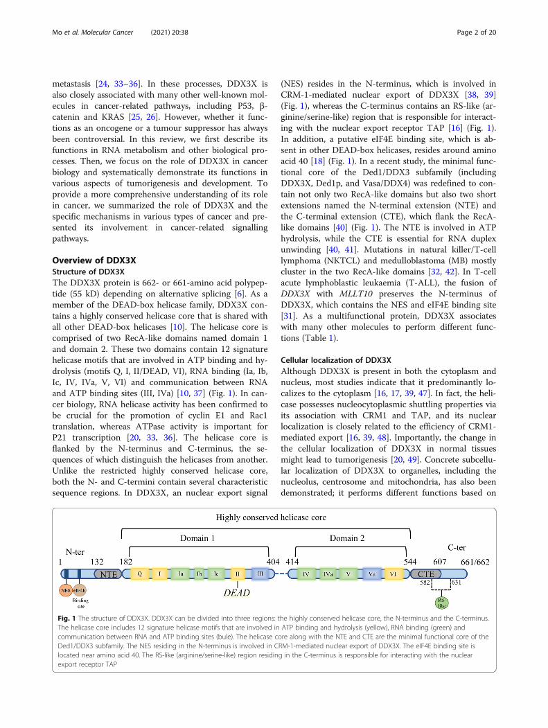
Overview of DDX3XStructure of DDX3XThe DDX3X protein is 662- or 661-amino acid polypep-tide (55 kD) depending on alternative splicing [6]. As amember of the DEAD-box helicase family, DDX3X con-tains a highly conserved helicase core that is shared withall other DEAD-box helicases [10]. The helicase core iscomprised of two RecA-like domains named domain 1and domain 2. These two domains contain 12 signaturehelicase motifs that are involved in ATP binding and hy-drolysis (motifs Q, I, II/DEAD, VI), RNA binding (Ia, Ib,Ic, IV, IVa, V, VI) and communication between RNAand ATP binding sites (III, IVa) [10, 37] (Fig. 1). In can-cer biology, RNA helicase activity has been confirmed tobe crucial for the promotion of cyclin E1 and Rac1translation, whereas ATPase activity is important forP21 transcription [20, 33, 36]. The helicase core isflanked by the N-terminus and C-terminus, the se-quences of which distinguish the helicases from another.Unlike the restricted highly conserved helicase core,both the N- and C-termini contain several characteristicsequence regions. In DDX3X, an nuclear export signal
(NES) resides in the N-terminus, which is involved inCRM-1-mediated nuclear export of DDX3X [38, 39](Fig. 1), whereas the C-terminus contains an RS-like (ar-ginine/serine-like) region that is responsible for interact-ing with the nuclear export receptor TAP [16] (Fig. 1).In addition, a putative eIF4E binding site, which is ab-sent in other DEAD-box helicases, resides around aminoacid 40 [18] (Fig. 1). In a recent study, the minimal func-tional core of the Ded1/DDX3 subfamily (includingDDX3X, Ded1p, and Vasa/DDX4) was redefined to con-tain not only two RecA-like domains but also two shortextensions named the N-terminal extension (NTE) andthe C-terminal extension (CTE), which flank the RecA-like domains [40] (Fig. 1). The NTE is involved in ATPhydrolysis, while the CTE is essential for RNA duplexunwinding [40, 41]. Mutations in natural killer/T-celllymphoma (NKTCL) and medulloblastoma (MB) mostlycluster in the two RecA-like domains [32, 42]. In T-cellacute lymphoblastic leukaemia (T-ALL), the fusion ofDDX3X with MLLT10 preserves the N-terminus ofDDX3X, which contains the NES and eIF4E binding site[31]. As a multifunctional protein, DDX3X associateswith many other molecules to perform different func-tions (Table 1).
Cellular localization of DDX3XAlthough DDX3X is present in both the cytoplasm andnucleus, most studies indicate that it predominantly lo-calizes to the cytoplasm [16, 17, 39, 47]. In fact, the heli-case possesses nucleocytoplasmic shuttling properties viaits association with CRM1 and TAP, and its nuclearlocalization is closely related to the efficiency of CRM1-mediated export [16, 39, 48]. Importantly, the change inthe cellular localization of DDX3X in normal tissuesmight lead to tumorigenesis [20, 49]. Concrete subcellu-lar localization of DDX3X to organelles, including thenucleolus, centrosome and mitochondria, has also beendemonstrated; it performs different functions based on
Fig. 1 The structure of DDX3X. DDX3X can be divided into three regions: the highly conserved helicase core, the N-terminus and the C-terminus.The helicase core includes 12 signature helicase motifs that are involved in ATP binding and hydrolysis (yellow), RNA binding (green) andcommunication between RNA and ATP binding sites (bule). The helicase core along with the NTE and CTE are the minimal functional core of theDed1/DDX3 subfamily. The NES residing in the N-terminus is involved in CRM-1-mediated nuclear export of DDX3X. The eIF4E binding site islocated near amino acid 40. The RS-like (arginine/serine-like) region residing in the C-terminus is responsible for interacting with the nuclearexport receptor TAP
Mo et al. Molecular Cancer (2021) 20:38 Page 2 of 20

its location [4, 48, 50]. Additionally, DDX3X is alsopresent in intracellular RNA/protein bodies such asstress granules [46].
Biological functions of DDX3XRNA metabolismTranscriptionDDX3X enhances transcription by interacting with tran-scription factors to promote their binding to the pro-moter of the target gene [20, 25, 26, 51]. The bestcharacterized mechanism is its cooperation with thetranscription factor SP1. The downstream genes ofDDX3X-SP1-mediated transactivation include P21,KRAS, and MDM2 [20, 25, 26], which are critical forcancer development and progression. DDX3X also inter-acts with YY1 to facilitate the transcription of genes in-volved in WNT/β-catenin signalling [51]. Moreover,DDX3X can directly impinge on E-cadherin and IFN-βpromoters to regulate their transcription without co-operating with any transcription factor [24, 52].
Pre-mRNA splicingDDX3X has been successively identified in affinity-purified human spliceosomes, messenger ribonucleopro-teins (mRNPs) and spliceosomal B complexes [14, 53, 54].MERZ et al. found that the link between DDX3X andmRNPs is achieved by DDX3X binding with exon junctioncomplex (EJC) proteins [14]. However, the specific func-tion of DDX3X in pre-mRNA splicing needs to be furtherelucidated.
RNA exportDDX3X is involved in facilitating Rev./REE-CRM1-dependent export of HIV transcripts [39]. In thisprocess, DDX3X binds with CRM1 and localizes to nu-clear membrane pores [39]. The NES residing in the N-terminus of the helicase is thought to be responsible forbinding DEAD-box helicases with CRM1 as a cargo [55].However, the region responsible for the association be-tween DDX3X and CRM1 is C-terminal residues 260–517 [39]. In addition, the interaction of DDX3X andCRM1 does not require RAN-GTP [39]. Therefore,
DDX3X is a functional element in the complex ratherthan a passenger. DDX3X also interacts with cap-binding protein complex (CBC) and Tip-associated pro-tein (TAP) [16, 56], which are major receptors for bulkmRNA export [57, 58]. Nonetheless, the helicase has lit-tle effect on general mRNA export [16]. TAP is recruitedto mRNPs and is responsible for their export [58]. Con-sidering the roles of DDX3X and TAP in pre-mRNAsplicing, there is a possibility that DDX3X is recruited tomRNPs during splicing, accompanies mRNPs to beexported by TAP and completes its function in RNAmetabolism in the cytoplasm. In addition to interactingwith CRM1 and TAP, DDX3X also participates ineIF4E-mediated mRNA export [56]. However, its actualrole in the process needs to be further explored.
TranslationEukaryotes possess two translation initiation mecha-nisms: cap-dependent and cap-independent translation.Cap-dependent translation starts via recognition of them7GTP cap and the subsequent recruitment of the 43Spreinitiation complex (PIC) to the mRNA [59]. Thisprocess is facilitated by the eIF4F complex, which con-sists of eIF4E, eIF4G and eIF4A [59]. While the transla-tion of most cellular mRNAs depends on this process[59], some RNA viruses along with several cellular tran-scripts utilize cap-independent translation, which re-quires an internal ribosomal entry site (IRES) on theRNA molecule [59]. DDX3X is involved in both cap-dependent and cap-independent translation initiation toregulate protein synthesis. In liver cancer, DDX3X inhibitsthe eIF4E-eIF4G interaction by binding with eIF4E to re-press global protein synthesis (Fig. 2a). In contrast, it alsofacilitates cap-dependent translation initiation of somespecific RNAs that contain structured 5′UTRs by bindingwith the eIF4F complex [19, 33, 36] (Fig. 2b). In HeLacells, DDX3X may facilitate protein synthesis by interact-ing with eIF3, but the specific role of DDX3X-eIF3 bind-ing in protein synthesis remains unclear (Fig. 2c). Inaddition to its involvement in cap-dependent translationinitiation, DDX3X facilitates IRES-mediated translation ofboth viral RNA and some cellular transcripts through its
Table 1 DDX3X and its binding proteins
Interaction protein Interaction region Function Ref
ALKBH5 211–404 Modulating m6A RNA Demethylation [43]
CK1ε 456–662 Involving in WNT/β-catenin signaling [44]
CRM1 260–517 Promoting Rev./REE-CRM1-dependent export of HIV transcripts [39]
eIF4E 38–44 Involving in translation initiation [18]
GSK3-β 100–662 Forming anti-apoptosis complex [45]
PABP1 227–534 Involving in stress granules assembly [46]
TAP/NXF1 536–662 Transported by TAP with mRNPs [16]
Mo et al. Molecular Cancer (2021) 20:38 Page 3 of 20

unwinding ability and interaction with the eIF4E complex[4, 16, 18, 19, 30]. Another report also stated that DDX3Xcooperates with ribosome protein RPL13 and eIF3 sub-units e and j to facilitate viral IRES-mediated translation[60] (Fig. 2d).DDX3X also participates in specialized translation pro-
grams. In eukaryotes, upstream open reading frames(uORFs) lead to defects in translation and nonsense-mediated decay (NMD) of transcripts, thereby limitingthe expression of key regulators of the stress responseand epithelial-mesenchymal transformation (EMT) [61].DDX3X facilitates the translation of uORF-containingmRNAs by cooperating with the cap-binding proteincomplex (CBC) and eIF3 to enhance the metastatic abil-ity of cancers [29] (Fig. 2e). Hexanucleotide GGGGCCrepeat expansion in the C9ORF72 gene can cause toxic
accumulation of dipeptide repeat (DPR) proteins, whichis a common cause of amyotrophic lateral sclerosis(ALS) and frontotemporal dementia (FTD) [62]. DPRproteins are produced through an unconventional trans-lation method called repeat-associated non-AUG (RAN)translation [63]. DDX3X effectively inhibits RAN trans-lation by directly binding to (GGGGCC) n RNAs [64](Fig. 2f). Therefore, it is a potential therapeutic target forALS/FTD.
MicroRNA expressionDDX3X regulates microRNA (miRNA) levels in a directand an indirect manner [65, 66]. As an RNA bindingprotein (RNP), DDX3X binds with the miR-20a locusand regulates its expression level [65]. Depletion ofDDX3X leads to reductions in miR-20a pri/pre/mature
Fig. 2 DDX3X and Translation. a DDX3X suppresses inhibition by directly binding with eIF4E to inhibit the eIF4E-eIF4G interaction. b DDX3Xfacilitates cap-dependent translation initiation of some RNAs that contain structured 5′UTRs by binding with the eIF4F complex. c DDX3X mayfacilitate translation by interacting with eIF3. d DDX3X facilitates IRES-mediated translation of viral RNAs and some cellular transcripts, the specificmechanism of which remains unclear. e DDX3X interacts with the cap-binding protein complex (CBC) and eIF3 to promote the translation ofuORF-containing mRNAs. f DDX3X directly binds to (GGGGCC) n RNAs to suppress RAN translation
Mo et al. Molecular Cancer (2021) 20:38 Page 4 of 20

species [65], implying that it is involved in pri-miRNAproduction or stability. In liver cancer, DDX3X affectsthe levels of a subset of tumour-suppressive miRNAs byreducing DNMT3A (DNA methyltransferase 3A) bind-ing and hypermethylation on the promoter regions ofthese miRNAs [66].
Cellular stress responseWhen encountering cellular stresses, the cell faces twochoices: survival or death. Under cellular stress, stressgranules (SGs), which are large cytoplasmic foci com-prising RNPs (ribonucleoproteins), are formed to protectcells from death [67]. On the other hand, cellularstressors can activate inflammasomes, which are multi-protein heteromeric complexes, that direct cells to pyr-optosis, a form of programmed cell death [68]. DDX3Xplays a pivotal role in the crosstalk of these two pro-cesses and determines the fate of these cells [69].DDX3X participates in the assembly of SGs, but it alsohas the ability to interact with NLRP3 to activate inflam-masomes. The assembly of SGs detains DDX3X, thusrepressing the activation of the NLRP3 inflammasome.The competition between SGs and NLRP3 for DDX3Xdetermines the ultimate fate of the cell [69].
Innate immune responseDDX3X plays an important role in the TANK-bindingkinase 1 (TBK1)-dependent innate immune response.DDX3X is a substrate of TBK1 [52]. Phosphorylation ofDDX3X by TBK1 leads to DDX3X directly interactingwith the IFN-β promoter to activate its transcription[52]. Moreover, DDX3X can influence the NF-κB signal-ling pathway and affect the production of various inflam-matory cytokines, such as IL12 and IFNγ [70]. Loss ofDDX3X expression in macrophages leads to deficiencyin restricting L. monocytogenes growth [70].
Metabolic stress response in pancreatic β cellsThe transcription factor JUND can promote β cell apop-tosis by regulating pro-oxidant and proinflammatorygenes [71]. During metabolic stress, such as high levelsof glucose and free fatty acids, JUND expression is up-regulated in pancreatic cells via the MEK/ERK/hnRNPKpathway at the posttranscriptional level [72]. DDX3Xbinds with hnRNPK and is essential for efficient transla-tion of JUND [72].
Embryo developmentThe WNT/β-catenin signalling pathway plays an import-ant role in embryonic development [73]. DDX3X partici-pates in this pathway as a regulatory subunit of CK1ε[44]. Under WNT signalling, DDX3X binds to caseinkinase 1 ε (CK1ε) and activates its kinase activity. Acti-vated CK1ε then phosphorylates the scaffold protein
dishevelled (Dvl), thereby ensuring the formation of theWNT/β-catenin signalosome [44]. Lack of DDX3X ex-pression in Xenopus embryogenesis leads to abnormalembryonic development marked by enlarged heads andeyes, shortened tails, and defective melanocyte and eyepigmentation [44]. In mouse embryos, DDX3X is crucialfor both extraembryonic and embryonic development[74]. Deficient expression of DDX3X leads to higherlevels of genome damage and cell cycle arrest duringembryogenesis [74].
DDX3X in cancerDDX3X is closely related to several of the hallmarks ofcancer, including evading growth suppressors, resistingcell death, activating invasion and metastasis, promotinggene instability and mutation and deregulating cellularmetabolism [4, 25, 45, 50, 75, 76]. Here, we first summa-rized DDX3X protein expression and clinical character-istics in multiple cancers. Then, we described itsfunction as it relates to the hallmarks of cancer. A sum-mary of its functions and specific mechanisms in varioustypes of cancers is listed in Table 2.
Protein expression and clinical characteristicsOver two decades, many cohort studies in various can-cers have investigated the expression level of DDX3Xand its connection with the clinical characteristics of tu-mours. However, the results are contradictory [23, 26,49, 85, 89, 90], which might be caused by the use of dif-ferent detection methods, different antibodies or the dif-ferent cut-offs for positivity [23, 26, 49, 85, 89, 90]. Toprovide a more succinct description, we have summa-rized the association of DDX3X expression and the clin-ical characteristics of various tumours in Table 3.Evidence has shown that DDX3X is overexpressed in
glioma, medulloblastoma (MB), meningioma, head andneck squamous cell carcinoma (HNSSC), lung cancer,breast cancer, hepatocellular carcinoma (HCC), gallblad-der carcinoma, pancreatic ductal adenocarcinoma(PDAC), colorectal cancer (CRC), prostate cancer andsarcoma [27–29, 77, 79, 83, 85, 89, 90, 95, 98, 99, 104,105]. Among them, lung cancer, gallbladder carcinomaand the smoking subpopulation of patients with HNSSCshows a correlation between overexpression of DDX3Xand poor prognosis (overall survival (OS)/relapse-freesurvival (RFS)/median survival time) [28, 29, 85, 89].From a pathological point of view, overexpression ofDDX3X is positively correlated with pathological classifi-cation in glioma, meningioma and PDAC [27, 77, 83],indicating that DDX3X has the potential to differentiatethe degrees of pathological classification of tumours.Conversely, a reduction in DDX3X has been reported inHNSSC, lung cancer, HCC, and CRC [20, 23, 26, 49, 92,103]. Low expression of DDX3X is correlated with poor
Mo et al. Molecular Cancer (2021) 20:38 Page 5 of 20

Table 2 Oncogenic/tumor-suppressive role of DDX3X in various cancers
Cancer type Oncogenic/tumor-suppressive
Evidence Mechanism/pathway Ref
Glioma Oncogenic Protein expression; Positivelycorrelated with Snail
- [77]
- [78]
Medulloblastoma (MB) Oncogenic Protein expression - [79]
Mutations Mutations led to alteration ofprotein function
[80–82]
Inhibitor therapy Inhibiting WNT/β-cateninsignaling
[79]
Meningioma Oncogenic Protein expression - [83]
Unknown Mutations - [84]
Head and neck squamouscell carcinoma (HNSSC)
Oncogenic Protein expression - [29, 85]
Tumor-suppressive Protein expression - [49]
Stemness - [86]
Promoting metastasis Cooperating with CBC complexand eIF3 to promote ATF4 translation
[29]
Inhibitor therapy - [86, 87]
Cutaneous squamous cellcarcinoma (cSCC)
Tumor-suppressive Protein expression - [20]
Melanoma Oncogenic Stemness - [88]
Tumor-suppressive Mutations Mutations mostly led to loss of function [30]
Repressing metastasis Promoting MITF translation [30]
Lung cancer Oncogenic Protein expression - [89]
Stemness - [91]
Inhibitor therapy Inhibiting Wnt/β-catenin pathwayactivity; impairing radiation-inducedDNA double-strand break (DSB)
[90]
Tumor-suppressive Protein expression - [26, 92]
Repressing proliferation Synergistically enhancing P53-activatedP21 transcription
[92]
Repressing metastasis Promoting MDM2 transcription to preventE-cadherin degradation
[26]
Mesothelioma Unknown Mutations - [93, 94]
Breast cancer Oncogenic Protein expression - [95]
Inducing tumorigenesis - [24]
Hypoxia responsive Directly regulated by HIF-1α [96]
Promoting proliferation Downregulating KLF4 expression via alteringKLF4 mRNA exon usage; downregulating P21
[24, 97]
Promoting metastasis upregulating E-cadherin expression viainteracting to its promoter
[24]
Inhibitor therapy Targeting mitochondria translation [50]
Hepatocellular carcinoma (HCC) Oncogenic Protein expression - [90]
Inducing tumorigenesis - [90]
Tumor-suppressive Protein expression - [20, 23]
Reducing tumorigenesis Maintaining genome stability [12]
Repressing stemness Upregulating the expression of a subsetof tumor-suppressive miRNAs via reducingDNMT3A activity
[66]
Repressing global protein synthesis Interacting with eIF4E and inhibitingits activity
[18]
Gallbladder carcinoma Oncogenic Protein expression - [28]
Mo et al. Molecular Cancer (2021) 20:38 Page 6 of 20

Table 2 Oncogenic/tumor-suppressive role of DDX3X in various cancers (Continued)
Cancer type Oncogenic/tumor-suppressive
Evidence Mechanism/pathway Ref
Pancreatic ductal adenocarcinoma(PDAC)
Oncogenic Protein expression - [27]
Colorectal carcinoma (CRC) Oncogenic Protein expression - [98, 99]
Promoting metastasis DDX3X/KRAS/ERK/AKT/β-catenin/ZEB1 axis; DDX3X/CK1ε/Dvl2 axis;DDX3X/KRAS/HIF-1α/YAP1/SIX2 axis
[100–102]
Drug resistance DDX3X/YAP1/SIX2 axis [102]
Inhibitor therapy Inhibiting WNT/β-catenin signaling;mitochondrial swelling and increasedROS production
[98, 99]
Tumor-suppressive Protein exprssion - [103]
Repressing metastasis DDX3X/Snail/E-cadherin axis [103]
Prostate cancer Oncogenic Protein expression - [104]
Inhibitor therapy Radiosensitizing prostate cancer cell [104]
Ewing sarcoma Oncogenic Protein expression - [105]
Inhibitor therapy repressing translation of proteinswith conserved biologic functions
[105]
Chronic lymphocytic leukemia (CLL) Unknown - - [106–108]
T-cell acute lymphoblastic leukemia(T-ALL)
Oncogenic Fusion with MLLT10 - [31, 109]
Natural killer/T-cell lymphoma(NKTCL)
Tumor-suppressive Repressing proliferation - [42]
Mutations Abnormal activated NF-κB andMAPK pathways
[42]
Aggressive natural killer-cellleukemia (ANKL)
Unknown Mutations - [110]
Burkitt lymphoma (BL) Unknown Mutations - [161]
Burkitt-like lymphoma with 11qaberration (BLL-11q)
Unknown Mutations - [111]
Various cell lines: Hela, Huh7,HCT116
Tumor-suppressive Repressing proliferation Promoting P21 transcription viainteracting with SP1
[20]
Various cell lines: OVCAR3 ES2,A549 H1437, SUM159, HCC1500,HT1080
Tumor-suppressive Substrate of CMA - [162]
Various cell lines: MDA-MB-231,1321N1, Jurkat, HeLa
Oncogenic Anti-apoptosis Cooperating with GSK3 and cIAP-1to confront with extrinsic apoptosis signaling
[45]
P53 wide-type cell line: MCF-7,SH-SY5Y
Tumor-suppressive Promoting DNAdamage-induced apoptosis
Stabilizing P53 expression viainteracting with it
[112]
P53 non-function or mutationcell line: Hela, MDA-MB-231
Oncogenic Repressing DNA damage-inducedapoptosis
- [112]
Hela cell line Tumor-suppressive Promoting proper chromosomesegregation
Interacting with hCAP-H [35]
Various cell lines: HCT116, U2OS Tumor-suppressive Ensuring bipolar mitosis Colocalizing with P53 in centrosomevia upregulating P53 expression andphosphorylating P53 to inactivateand coalesce excess centrosome
[4]
Various cell lines: N2A, Hela Oncogenic Promoting metastasis DDX3X/Rac1/β-catenin axis [36]
Various cell lines: MKN-45, AGS Oncogenic Facilitating β-catenin signaling Transactivating YY1 in the helpof circ-CTNNB1
[51]
Hela cell line Oncogenic Promoting protein synthesis Interacting with eIF3 [17]
Anti-apoptosis Downregulating P21 expression [113]
Various cell lines: Hela, H1299,A549 and U2OS
Oncogenic Promoting G1/S phase transition Promoting cyclin E1 translation [33]
Mo et al. Molecular Cancer (2021) 20:38 Page 7 of 20

prognosis in lung cancer, CRC and the non-smokingsubpopulation of patients with HNSSC [49, 92, 103]. Itis worth noting that the reduction in DDX3X expressionis closely related to virus infection in lung cancer andHCC [23, 26]. In addition, in HCC, reduced DDX3X ex-pression is more common in males than in females [23]
In many cancers, DDX3X is predominantly present inthe cytoplasm of cancer cells, whereas paired non-tumour tissue expresses little or no DDX3X. Nuclearlocalization of DDX3X has been detected in breast andcolorectal cancer tissues and is correlated with other fac-tors associated with poor prognosis [48, 114]. More
Table 3 Expression of DDX3X in various cancers
Cancer type mRNA/Protein
High/Lowexpression
Percentage Clinical characteristics remarks ref
Glioma mRNAandProtein
High – Positively correlated with WHO Grading;associated with poor median survival
– [77]
Medulloblastoma(MB)
Protein High 55% (31/56)in pediatric67% (6/9) inadult
– Mainly in cytoplasm [79]
Meningioma Protein High – Significantly higher in atypical meningiomas thanin benign meningiomas
Mainly in cytoplasm [83]
Head and necksquamous cellcarcinoma (HNSSC)
ProteinProteinProtein
HighHighLow
71% (15/21)51% (217/423)90% (290/324)
Associated with lymph node metastasis (N value),stage and poor patient survivalAssociated with shorter median survival (HR =1.34, 95%CI = 1.00–1.81) in smokersAssociated with male gender, smoking, alcoholconsumption, betel quid chewing, poor RSF andpoor OS; associated with poorer OS in non-smokers
Mainly in cytoplasmSubtype: 206 OSCC and 217OPSSC;Mainly in cytoplasmSubtype: OSCC;Both in cytoplasm and nucleus
[29][85][49]
Cutaneoussquamous cellcarcinoma (cSCC)
Protein – – – normal epidermis: mainly innuclear; cSCC: mainly in cytoplasm
[20]
Lung cancer ProteinmRNAProtein
highLowLow
66% (63/94)53% (73/138)53% (76/144)
Associated with a shorter survival time (HR = 2.1,95% CI; 1.13–3.93); a predictor of OS-Associated with a shorter median period of OS(HR = 1.61, 95%CI = 1.04–2.48) and RSF (HR = 1.78,95%CI = 1.17–2.69)
Mainly in cytoplasmMore common in E6 positive orP53 mutation samples;Associated with P21 and E-cadherin expression
[89][26][92]
Breast cancer Protein High 35% (127/366)
– Cytoplasm; associated withhypoxia response
[95]
Hepatocellularcarcinoma (HCC)
mRNAProteinmRNA
HighLowLow
64% (29/45)57% (49/86)58% (26/45)
-More common in males and HBV-positivepatients-
---
[90][23][20]
Gallbladdercarcinoma
Protein High 55% (69/126)
Associated with large tumor size, high TNMstage, lymph node metastasis, poor surgicalcurability and OS
Mainly in cytoplasm [28]
Pancreatic ductaladenocarcinoma(PDAC)
Protein High 52% (55/106)
Associated with poor differentiation, surroundingtissue and lymph node metastasis, advancedTMN stage, shorter survival and motility
Mainly in cytoplasm [27]
Colorectal cancer(CRC)
ProteinProteinmRNA
HighHighLow
41% (124/303)53% (28/53)-
--Prognostic predictive indicator (RNA sequencing,HR = 0.53; RNA microarray analysis, HR = 0.72);associated with poor OS and RFS, distantmetastasis
Mainly in cytoplasm--
[98][99][103]
Prostate cancer Protein High – – Both in cytoplasm and nuclear;positively associated with P21,androgen receptor (AR), PHD2,PHD3, CA9
[104]
Sarcoma Protein High 61% (103/170)
– Mainly in cytoplasm [105]
Mo et al. Molecular Cancer (2021) 20:38 Page 8 of 20

importantly, patients with nuclear DDX3X expressionhave a worse prognosis than those without nuclearDDX3X [48].
Cell cycle — evading growth suppressorsThe cell cycle is regulated by cyclins, CDKs (cyclin-dependent kinases) and CKIs (cyclin-dependent kinaseinhibitors) [115]. Depletion of DDX3X induces G1 phasearrest in breast cancer, lung cancer, colorectal cancer,prostate cancer and medulloblastoma [79, 89, 97, 98,104]. This phenomenon might result from a reductionin cyclin E1, which is upregulated by DDX3X at thetranslation level (Fig. 3) [33]. Additionally, DDX3X in-hibits the expression of KLF4 by altering the alternativesplicing of KLF4 mRNA, followed by upregulatedCCNA2 and CDK2 expression [97] (Fig. 3). P21 is a clas-sic CKI that causes cell growth arrest by interacting withcyclin/CDK complexes [116]. P53 transactivates the P21promoter via increased SP1 binding affinity [117]. Inlung cancer, DDX3X transcription is directly regulatedby P53 [92]. More importantly, DDX3X synergisticallyenhances P53-activated P21 transcription by increasingthe interaction between P53 and SP1 and promotingSP1 binding to the P21 promoter [92] (Fig. 3). In E6-positive lung tumours, downregulation of DDX3X ex-pression by P53 inactivation promotes cell proliferationand colony formation via reduced SP1 binding activityon the P21 promoter [92]. However, P21 expression canalso be induced in a P53-independent manner [118]. In
liver cancer, the reduction in P21 is independent of P53status, and DDX3X directly interacts with SP1 to promoteP21 transcription, leading to tumour cell growth inhib-ition [20] (Fig. 3). Nevertheless, reduced P21 expressioncaused by DDX3X overexpression is also observed inbreast cancer [24].
Apoptosis — resisting cell deathApoptosis is a process of programmed cell death thatproceeds via the mitochondrial pathway (intrinsic) orthe death receptor pathway (extrinsic) [119]. Extrinsicapoptosis is induced by the activation of death recep-tors. Stimulation of these receptors by death ligandsresults in the recruitment of FADD (Fas associatedwith death domain protein) and Caspase-8 (orCaspase-10) to form DISC (death-inducing signallingcomplex), thus promoting the activation of down-stream Caspases [120]. DDX3X binds with TRAIL-R2and is cleaved during TRAIL-mediated apoptosis [34].In addition, DDX3X cooperates with GSK3 and cIAP-1 to form an anti-apoptotic complex that caps majordeath receptors before they can be stimulated [45].Stimulated death receptors overcome the anti-apoptotic cap by inactivating GSK3β and cleavingDDX3X and cIAP-1 [45]. Cleavage occurs in the N-terminus of DDX3X, and the truncated protein canstill bind GSK3-β [45]. However, the complex remainsfunctional in cancer cells resistant to death receptorstimulation (Fig. 4). Collectively, an inability of the
Fig. 3 DDX3X and cell cycle. DDX3X facilitates cyclin E1 translation by resolving the secondary structure in its 5′UTR during translation initiation.Moreover, it suppresses KLF4 expression by manipulating KLF4 mRNA alternative splicing. Therefore, by promoting cyclin E1 translation andsuppressing KLF4 expression, DDX3X promotes G1/S transition. In lung cancer, P53 promotes DDX3X transcription. DDX3X synergisticallyenhances p53-activated P21 transcription by increasing the binding affinity of SP1 on the P21 promoter. In liver cancer, DDX3X directly interactswith SP1 to promote P21 transcription in a P53-independent manner, thus leading to tumour cell growth arrest
Mo et al. Molecular Cancer (2021) 20:38 Page 9 of 20

death receptors to disable DDX3X activity may con-tribute to resistance to death receptor-induced apop-tosis in tumours, suggesting that targeting DDX3Xmight be a useful strategy for promoting deathreceptor-induced apoptosis.P53 plays a vital role in DNA damage-induced intrin-
sic apoptosis [121, 122]. DDX3X binds with both wild-type P53 and mutant P53 in tumours. When DNA dam-age occurs, DDX3X can still bind wild-type P53 andstabilize its protein level, thus promoting P53-mediatedapoptosis. However, the association of DDX3X and mu-tant P53 was reduced after DNA damage occurred,which impeded Caspase activation [112]. Disassociationmay contribute to the translocation of DDX3X to deathreceptors, where it attenuates Caspase activation (Fig. 4).Alterations in P21, a target gene of P53, are also observedwhen DDX3X is manipulated [112], thus confirming thatDDX3X functions in intrinsic apoptosis via the DDX3X/P53/P21 axis. However, another report showed that phos-phorylated DDX3X reverses sanguinarine-induced intrin-sic apoptosis by strongly repressing P21 expression [113](Fig. 4). The different results from these two reports mightbe due to different drug models or cell types.
Metastasis — activating invasion and metastasisDDX3X and β-catenin are very closely related in metas-tasis. Chen et al. reported that DDX3X promotes cancercell migration and invasion via the Rac1/β-catenin path-way [36]. Rac1 plays a pivotal role in cell-cell contactsand cell migration [123]. Importantly, it protects β-
catenin from proteasome-dependent degradation by en-hancing β-catenin phosphorylation on S675 [124]. By fa-cilitating the translation of Rac1 mRNAs containing astructured 5′UTR, DDX3X stabilizes β-catenin, thus in-creasing the expression of its downstream transcriptionaltargets involved in tumour metastasis, includingMMP14, Pld1 and Stat3 [36]. DDX3X also modulatescell-cell adhesion by downregulating E-cadherin [36],which might be achieved by enhancing Rac1-dependentE-cadherin endocytosis [125]. Therefore, DDX3X likelypromotes metastasis through both the Rac1/E-cadherinand Rac1/β-catenin pathways (Fig. 5). Of note, E-cadherin is negatively regulated by the transcription fac-tor snail [126]. DDX3X can induce Snail expression tosuppress E-cadherin expression and drive metastasis[78]. In addition, the helicase directly binds to the E-cadherin promoter and represses its transcriptional ac-tivity [24]. E-cadherin, along with the majority of β-catenin, predominantly localizes to the cell membrane.Depletion of E-cadherin induced by DDX3X abolishesthis interaction and releases β-catenin into the cyto-plasm and nucleus [24]. Therefore, the motility and in-vasive properties induced by DDX3X are probablymediated by direct and indirect mechanisms (Fig. 5). Ingastric cancer, DDX3X binds the transcription factorYY1 (yin yang 1) with the help of the circRNA circ-CTNNB1, which results in the transactivation of YY1and the subsequent activation of genes involved inWNT/β-catenin signalling, thereby promoting tumourprogression (Fig. 5) [51].
Fig. 4 DDX3X and apoptosis. In the extrinsic apoptosis pathway, DDX3X binds with GSK3 and cIAP-1, forming an anti-apoptotic complex to capmajor death receptors. After death ligands bind to their receptors, the anti-apoptotic complex is destroyed by inactivation of GSK3β and cleavageof DDX3X and cIAP-1 by caspases. Cleavage occurred in the N-terminus of DDX3X, and the truncated protein can still bind GSK3-β. In the intrinsicapoptosis pathway, DDX3X binds wild-type P53 and mutant P53 in tumours. When encountering DNA damage, DDX3X binds wild-type P53 andstabilizes its protein level, promoting P53/P21 axis-mediated apoptosis. However, DDX3X and mutant P53 are separated after DNA damageoccurs, which impedes Caspase activation in P53-mutant tumours. This disassociation may contribute to the translocation of DDX3X to deathreceptors. However, in HeLa cells, phosphorylated DDX3X reverses sanguinarine (SA)-induced intrinsic apoptosis by strongly repressingP21 expression
Mo et al. Molecular Cancer (2021) 20:38 Page 10 of 20

KRAS is an oncogene that is mutated in more than40% of human colorectal cancer cases [127]. In colorec-tal cancer, DDX3X increases the expression of KRAS bypromoting SP1 binding to the KRAS promoter to facili-tate tumour metastasis [25, 128]. However, the specificmolecular mechanisms by which DDX3X promotes me-tastasis are different in colorectal cancers harbouringwild-type or mutant KRAS. In colorectal cancer har-bouring mutant KRAS, DDX3X-induced expression ofKRAS activates the RAF/MEK/ERK/c-Jun pathway tosuppress the tumour suppressor gene PTEN, a negativeregulator of the PI3K/AKT pathway [100]. GSK3-β playsa crucial role in β-catenin degradation via phosphoryl-ation on the N-terminus of β-catenin [129]. However,the activity of GSK3-β can be suppressed by the MEK/ERK and PI3K/AKT signalling pathways [130, 131].Therefore, DDX3X deactivates GSK3-β to stabilize β-catenin, which then enhances ZEB1 transcription formetastasis [25] (Fig. 6a). Additionally, in colorectal can-cer harbouring mutant KRAS, DDX3X stabilizes β-catenin via the CK1ε/Dvl2 axis to promote invasiveness[101]. Similar to the abovementioned findings, DDX3Xbinds CK1ε and stimulates its activity. Activated CK1εthen phosphorylates Dvl2, which decreases the associ-ation of PP2A with the β-catenin degradation complex,
therefore stabilizing β-catenin [101] (Fig. 6a). In colorec-tal cancer harbouring wild-type KRAS, DDX3X-inducedKRAS elevated the level of ROS, which was followed byincreased HIF1-α expression. HIF-1α in turn directlyupregulates DDX3X expression at the transcriptionallevel, thus generating a cascade feedback loop [128]. Fur-thermore, DDX3X-induced HIF-1α directly binds to theYAP1 promoter to promote its transcription [128].YAP1, a novel oncogene in the Hippo pathway, targetsPTEN by elevating miR-29c expression to activate PI3K/AKT signalling [132]. The activated PI3K/AKT pathwayupregulates the expression of and phosphorylates thetranscription factor c-fos and eventually leads to the in-creased transcription level of SIX2, a gene that sup-presses E-cadherin expression to promote metastasis inbreast cancer [102]. The YAP1/SIX2 axis is responsiblefor DDX3X-induced cell invasiveness in colorectal cancerharbouring wild-type KRAS (Fig. 6b). In addition to pro-moting metastasis, the DDX3X-induced YAP1/SIX2 axismight be responsible for resistance to treatment with theanti-EGFR antibody cetuximab (CTX) in colorectal cancerharbouring wild-type KRAS via enhanced autophagy andanti-apoptotic mechanisms [128]. However, Su et al. re-ported that downregulation of DDX3X expression in colo-rectal cancer leads to upregulation of Snail expression,
Fig. 5 DDX3X and metastasis. Rac1 protects β-catenin from degradation by enhancing β-catenin phosphorylation on S675. DDX3X facilitates Rac1translation to stabilize β-catenin. Additionally, DDX3X might modulate cell-cell adhesions by enhancing Rac1-dependent E-cadherin endocytosis.Moreover, DDX3X directly binds to the E-cadherin promoter to suppress its expression. The decrease in E-cadherin expression causes β-catenin tobe released from the cell membrane. The accumulated β-catenin translocates to the nucleus and interacts with TCF4 to increase the expressionof its downstream target genes. In the nucleus, DDX3X activates the transcription factor YY1 with the help of circRNA circ-CTNNB1, leading tosubsequent activation of genes involved in WNT/β-catenin signalling
Mo et al. Molecular Cancer (2021) 20:38 Page 11 of 20

Fig. 6 DDX3X and metastasis. a In colorectal cancer, DDX3X increases the expression of KRAS by promoting SP1 binding to the KRAS promoter.In CRC harbouring mutant KRAS, DDX3X activates the KRAS/ERK/PTEN/AKT cascade to stabilize β-catenin, which then enhances ZEB1 transcriptionto promote metastasis. In addition, DDX3X binds with and activates CK1ε, which then phosphorylates Dvl2. Phosphorylated Dvl2 causesdissociation of PP2A and the β-catenin degradation complex, therefore stabilizing β-catenin. The accumulated β-catenin translocates into thenucleus and interacts with TCF4 to increase the expression of its downstream target genes. b In CRC harbouring wild-type KRAS, DDX3X/KRAS/HIF1-α generates a cascade feedback loop. HIF-1α binds to the YAP1 promoter to promote YAP1 transcription. YAP1 then targets PTEN byelevating miR-29c expression to activate PI3K/AKT signalling. Phosphorylated AKT activates c-fos and eventually leads to increased levels ofSIX2 transcription
Fig. 7 DDX3X and metastasis. a In HNSCC, DDX3X cooperates with the CBC-eIF3 complex to enhance the translation of ATF4 mRNA. IncreasedATF4 expression results in upregulated expression of ACTA2, CDH2 (N-cadherin), FAP, SNAI2 (Slug), and VIM (vimentin) and downregulatedexpression of CHD1 (E-cadherin), thus triggering EMT. b DDX3X interacts with SP1 to promote MDM2 transcription. In E6-positive lung cancer,P53 inactivation leads to downregulation of DDX3X expression, which suppresses MDM2 expression. MDM2 promotes E-cadherin expression bymediating proteasomal degradation of Slug. Therefore, loss of DDX3X downregulates MDM2 expression, stabilizes Slug and suppresses E-cadherinexpression, which eventually promotes tumour metastasis
Mo et al. Molecular Cancer (2021) 20:38 Page 12 of 20

decreased E-cadherin expression and increased vimentinand N-cadherin expression. Furthermore, knocking downSnail significantly reduced the migration and invasion cap-acities of cells with DDX3X knockdown, indicating thatDDX3X represses colorectal cancer cell metastasis by me-diating the Snail/E-cadherin pathway [103]. These con-flicting results in colorectal cancer are possibly due to theuse of different cell lines. However, the actual role ofDDX3X in colorectal cancer needs to be verified.In head and neck squamous cell carcinoma (HNSCC),
DDX3X cooperates with the CBC-eIF3 complex to en-hance some uORF-containing mRNAs [29]. ATF4 is acrucial gene for EMT [133–135] and is responsible forthe effect of DDX3X overexpression on EMT-relatedgene expression, including upregulation of ACTA2,CDH2 (N-cadherin), FAP, SNAI2 (Slug), and VIM(vimentin) expression and downregulation of CHD1 (E-cadherin) expression. In addition, knockdown of CBC oreIF3 impairs cell invasiveness and decreases the expres-sion of mesenchymal-related genes but increases the ex-pression of E-cadherin [29]. Therefore, there is apossibility that DDX3X acts co-ordinately with the CBC-eIF3 complex to enhance the translation of mRNAscontaining uORFs that together modulate the EMT pro-gram, hence promoting HNSCC metastasis (Fig. 7a). Inlung cancer, loss of DDX3X via P53 inactivation sup-presses MDM2 transcription by decreasing SP1 bindingto the MDM2 promoter [26]. MDM2 promotes E-cadherin expression by mediating proteasomal degrad-ation of Slug [136]. Loss of DDX3X stabilizes Slug ex-pression by suppressing the MDM2-mediated ubiquitinproteasomal pathway and consequently suppresses E-cadherin expression, thus promoting cell invasion [26](Fig. 7b). Melanoma is an aggressive malignancy of me-lanocytes characterized by rapid metastasis [137]. A mu-tational assessment of 864 melanoma tumours identifiedDDX3X mutations, most of which might eventually leadto DDX3X loss of expression. Intriguingly, loss ofDDX3X expression directs a proliferative-to-metastaticphenotypic switch in melanoma cells [30].MITF, which is strongly correlated with a less invasive
and more proliferative expression signature in melanoma[138], was identified as a direct translational target ofDDX3X [30]. Mechanistically, DDX3X promotes MITFmRNA translation via its internal ribosome entry site(IRES) within the 5′UTR.
Gene instability and mutationIn recent years, mutations in DDX3X have been re-ported to be involved in chronic lymphocytic leukaemia(CLL), an incurable disease with variable clinical presen-tation and evolution [106–108, 139]. In 48 CLL cases,10% (5/48) presented with DDX3X mutations, whichwere either nonsense mutations or frameshift indels that
eventually led to truncated production. Moreover,DDX3X is preferentially mutated in males (4/5). Fur-thermore, in two of the five male cases, two independenttruncating mutations were identified. Analysis of thesetwo cases shows that these mutations showed trends in-creased and decreased activity at different time points[106], Additionally, inactivating DDX3X mutations areassociated with unfavourable clinical markers and poorclinical outcomes [106]. A longitudinal analysis in a co-hort of 8 cases of monoclonal B-cell lymphocytosis(MBL) showed that one case possessed a mutation inSF3B1 and two independent mutations in DDX3X. Lon-gitudinal analysis of this patient demonstrates that at thefirst time point, DDX3X mutation I415V was present innearly 50% of the allelic fraction, while the mutationD164G was present in only 10%. However, at the secondtime point, this trend was reversed [140]. The variationin DDX3X mutations in CLL and MBL suggests thepresence of DDX3X mutations in different subcloneswith alternating dominance between the time points.MLLT10 is a moderately common MLL fusion partnerthat predominantly occurs in acute monoblastic leukae-mia (AML) [141]. DDX3X is one of the partners ofMLLT10 in adult and paediatric T-cell acute lympho-blastic leukaemia (T-ALL) [31, 109]. Upon investigating99 patients with adult T-ALL, researchers found that ap-proximately 10% (10/99) of the patients had MLLT10translocations. Among them, 3 cases possessed theDDX3X-MLLT10 fusion. Another biological samplefrom a 4th patient was obtained from a different cohortof 20 adult T-ALL patients. All 4 cases were confirmedto have in-frame DDX3X-MLLT10 transcripts with dif-ferent breakpoints. At the N-terminus, DDX3X containsa nuclear export signal (NES) domain. Three patientsretained the entire eIF4E interacting motif, and 1retained only half of this motif. The MLLT10 leukaemo-genic OM-LZ domain, which induces acute myeloid leu-kaemia in mouse models, was maintained at the C-terminus in all the fusions. In addition, all 4 cases weremales, indicating that the complete absence of a nor-mally functional DDX3X protein might contribute toleukaemogenesis [31]. In natural killer/T-cell lymphoma(NKTCL), whole-exome sequencing in 25 patients andsubsequent target sequencing in 80 patients show thatrecurrent mutations are most frequently located inDDX3X (20.0%, 21/105), followed by P53, STAT3, etc.Most of the mutations in DDX3X affect two highly con-served RacA-like domains. Half of the mutations eventu-ally lead to truncation or loss of the protein, while theother half lead to altered protein function. Indeed,DDX3X with the mutations A404P and E348K exhibitsdecreased RNA-unwinding activity, an impaired abilityto suppress cell cycle progression and abnormally acti-vated NF-κB and MAPK pathways at the transcriptional
Mo et al. Molecular Cancer (2021) 20:38 Page 13 of 20

level. In addition, DDX3X mutations are correlated withadvanced disease stage and poor clinical outcome [42].It is worth noting that DDX3X and P53 are the twogenes most commonly mutated in NKTCL, but they sel-dom overlap with each other, implying that they are in-volved in very closely related biological processes inNKTCL. Mutations in DDX3X have also been discov-ered in aggressive natural killer-cell leukaemia (ANKL),a rare mature NK-cell tumour [110]; Burkitt lymphoma(BL) [161]; and Burkitt-like lymphoma with 11q aberra-tion (BLL-11q), a category similar to Burkitt lymphomabut lacking the MYC rearrangement and containing 11qarm distortion [111].Medulloblastoma (MB) arises in the cerebellum or
medulla/brain stem [142] and is the most commonmalignant childhood brain tumour [143]. In the lastfew years, gene expression profiling of moderate-to-large cohorts of patients with this disease identified 4distinct molecular subgroups: WNT, presenting wntpathway activation; SHH, displaying hedgehog path-way activation; and groups 3 and 4, which are lesswell characterized on the molecular level [32]. Theresults from a wave of medulloblastoma genome-sequencing studies revealed that DDX3X is the sec-ond most frequently mutated gene in medulloblas-toma (8%, 25/300), followed by CTNNB1 (β-catenin).Half of WNT medulloblastoma patients from threecohorts harboured DDX3X variants (50%, 16/32),while the percentage of patients with SHH medullo-blastoma was 11% (7/66) [80–82]. Another genomesequencing analysis of SHH medulloblastoma showedthat DDX3X is mutated in 54% of adult SHH medul-loblastomas (27/50) and 7.2% of paediatric medullo-blastomas (6/83) [144]. In contrast to mutationsfound in blood cancer that contain premature stopcodons, frameshifts, or splice variants, nearly all mu-tations in medulloblastoma were nonsynonymous sin-gle nucleotide variants (SNVs), which were likely tocause alteration of protein function rather than lossof function [80–82]. Indeed, neither wild-type DDX3Xnor mutant DDX3X enhanced the ability of β-cateninto transactivate TCF/LEF in medulloblastoma. How-ever, the majority of DDX3X mutations enhance cellproliferation by potentiating the transactivation cap-acity of mutant β-catenin [81]. Moreover, mutationsin DDX3X are crucial for the proliferation and/ormaintenance of the LRLP lineage, which is believed tobe the cell-of-origin of WNT medulloblastoma [80].The variants of DDX3X appeared to cluster in eitherof the two helicase domains, which are important forcatalytic function [80–82]. Consistently, further func-tional studies on mutations in medulloblastoma re-vealed that DDX3X mutants G302V and G325E haveseverely defective RNA-stimulated ATPase activity
and cannot complement the growth defect in a Ded1p(yeast homologue of DDX3X) temperature-sensitivestrain of fission yeast [41]. Moreover, mutations inDDX3X were confirmed to drive stress granule as-sembly and impair global translation [145]. Wild-typeDDX3X interacts extensively with RNA and ribosomalmachinery to help remodel the translation landscapein response to stress, while DDX3X with the mutationR534H adapts this response to selectively preserve transla-tion involved in chromatin organization [146]. In melan-oma, DDX3X was mutated in 5.8% of the 864 tumours.These mutants included 35% truncating mutations and65% missense mutations, which might eventually lead tothe loss of DDX3X expression. Importantly, 82% of allDDX3X mutations, including all truncating mutations,were detected in male patients, implying that DDX3Xmight play an important role in the progression of melan-oma in males [30]. In addition to the aforementioned ma-lignancies, DDX3X mutations were also found inprogressive/higher grade meningiomas and mesothelio-mas [84, 93, 94]. DDX3X CNVs (copy number variants)were also found in patients with oral squamous cell car-cinoma (OSCC) [147].
Deregulating cellular metabolismMitochondrial localization of DDX3X has been discov-ered in breast cancer and colorectal cancer cells [50, 99].Targeting DDX3X inhibits mitochondrial translation,followed by reduced oxidative phosphorylation(OXPHOS) and increased ROS (reactive oxygen species)production, which ultimately triggers apoptosis andcauses cell death [50, 99]. Cellular stressors, such as ion-izing radiation, can also increase ROS expression. Inaddition, ionizing radiation leads to an increased de-mand for ATP in cancer cells, which needs to be ad-dressed by a large OXPHOS reserve capacity [148, 149].The combination of ionizing radiation and DDX3X in-hibition causes cancer cells to undergo metabolic catas-trophe [50], which is a promising anti-tumourtherapeutic strategy. In breast cancer, DDX3X expres-sion is induced by HIF-1 under hypoxic conditions [96].Its expression is also correlated with other hypoxia-responsive genes [95]. This evidence indicates thatDDX3X plays a role in hypoxia, but the specific functionof DDX3X in these conditions needs to be furtherexplored.
Stemness and immunogenicity — tumourmicroenvironmentCancer stem cells (CSCs) are a subset of cells within atumour that are responsible for the long-term mainten-ance of tumour growth in several cancers [150]. CSCsare characterized by self-renewal, chemoresistance,EMT, motility and CSC expansion, which result in
Mo et al. Molecular Cancer (2021) 20:38 Page 14 of 20

tumour initiation and anti-cancer therapy resistance[151]. Human small cell lung carcinoma, colorectal can-cer, and breast cancer cells with CSC markers express ahigh level of DDX3X, yet normal human tissues onlyfaintly express DDX3X [88]. In liver cancer, well-differentiated cell lines showed higher expression ofDDX3X than did poorly differentiated cell lines. More-over, DDX3X represses the expression of signaturestemness genes, including NANOG, OCT4, c-MYC,SOX2, KLF4, BMI1 and CK19, to prevent the generationof CSCs in liver cancer [66]. Mechanistically, DDX3X re-presses the expression of stemness genes via upregula-tion of the expression of a subset of tumour-suppressivemiRNAs, including miR-200b, miR-200c, miR-122 andmiR-145, by reducing DNMT3A (DNA methyltransfer-ase 3A) binding and hypermethylation on their promoterregions [66] (Fig. 8a). Conversely, in lung adenocarcin-oma cells harbouring an EGFR mutation, DDX3X
overexpression induces a CSC-like phenotype (increasedSox2 and Snail expression and elevated anchorage-independent proliferation) and resistance to EGFR-tyrosine kinase inhibitors (EGFR-TKIs) [91]. Overexpres-sion of DDX3X reduces EGFR signalling but facilitatesWnt/β-catenin signalling [91], which is consistent withthe fact that stem cells can utilize Wnt/β-catenin signal-ling pathways to replace receptor-type tyrosine kinase sig-nalling [152]. In fact, control cells, which are nonadherent,lack EGFR signalling and resist EGFR-TKIs, express highlevels of DDX3X [91], providing further proof thatDDX3X plays a role in inducing a stem cell-like state (Fig.8a). Consistent with the findings in lung cancer, DDX3Xis also upregulated in cisplatin-resistant oral squamouscell carcinoma (OSCC) cells compared to cisplatin-sensitive OSCC cells [86]. Targeting DDX3X impairs theCSC population in cisplatin-resistant cells via decreasedexpression of FOXM1 and NANOG [86], which are
Fig. 8 DDX3X in mitosis and stemness. a During prophase/prometaphase of mitosis, DDX3X translocates in close proximity to the condensingchromosomes and interacts with hCAP-H to promote chromosome segregation. In colon cancer and osteosarcoma, DDX3X upregulates P53expression by promoting IRES-mediated translation of P53, thus preventing DNMTs from hypermethylating the P53 promoter and repressing thebinding of repressive histone markers to the P53 promoter. In addition, DDX3X activates ATM kinase to phosphorylate P53, which leads to thelocalization of P53 to centrosomes. At the centrosome, DDX3X interacts with P53, leading to inactivation and coalescence of excess centrosomes.b In liver cancer, DDX3X upregulates the expression of miR-200b, miR-200c, miR-122 and miR-145 by reducing DNMT3A binding andhypermethylation of their promoter regions. This subset of miRNAs suppresses the expression of signature stemness genes, including SOX2,NANOG, and OCT4, which are responsible for self-renewal, chemoresistance and EMT. In lung adenocarcinoma cells harbouring an EGFRmutation, DDX3X overexpression induces increased Sox2 and Snail expression, anchorage-independent proliferation, resistance to EGFR-TKIs andpromotion of Wnt/β-catenin signalling. In cisplatin-resistant OSCC cells, DDX3X interacts with ALKBH5 and increases its expression. ALKBH5 thenupregulates FOXM1 and NANOG expression by demethylating their methylated mRNAs, which eventually leads to cisplatin resistance
Mo et al. Molecular Cancer (2021) 20:38 Page 15 of 20

important for self-renewal properties and drug resistancein cancers that are upregulated by the m6A demethylaseALKBH5 [43, 153–156]. In cisplatin-resistant OSCC cells,DDX3X interacts with ALKBH5 and increases its expres-sion to upregulate FOXM1 and NANOG expression [86](Fig. 8a). DDX3X is a major immunogenic protein inCD133+ melanoma cells. Inoculation with DDX3X-primed specific T cells exhibits defensive and beneficialantitumour immunity, curing established skin melanoma.DDX3X-primed CD4+ T cells produce tumour-specificIFNγ and IL-17 from CD133+ cells, suggesting thatDDX3X possesses immunogenic MHC class II-restrictedepitopes [88]. Therefore, anti-DDX3X immunotherapy isa promising treatment to eradicate CSCs to cure cancer.
MitosisDuring mitosis, each daughter cell inherits one copy ofevery chromosome. The accuracy of this process isachieved by chromosome segregation, as mediated bycondensin I and II complexes [157]. Defects in chromo-some segregation cause aneuploidy and then cell deathor cancer [158]. DDX3X regulates chromosome segrega-tion by interacting with hCAP-H in HeLa cells [35].hCAP-H is a subunit of condensin I that has been shownto promote proper chromosome segregation in HeLacells [35]. During interphase, DDX3X localizes in cyto-plasmic foci. In prophase/prometaphase, DDX3X trans-locates within close proximity to the condensingchromosomes and interacts with hCAP-H to promotechromosome segregation. Knockdown of DDX3X abol-ishes the robust localization of hCAP-H to mitotic chro-mosomes, leading to an increased incidence of laggingchromosomes [35] (Fig. 8b). In colorectal cancer andosteosarcoma, DDX3X prevents multipolar mitosisthrough inactivation and coalescence of excess centro-somes [4]. Interestingly, the localization of DDX3X tocentrosomes is dependent on P53 expression Moreover,DDX3X promotes IRES-mediated translation of P53,and DDX3X knockdown activates DNMTs to hyper-methylate the P53 promoter and promotes the bindingof repressive histone markers to the P53 promoter.DDX3X also promotes P53 Ser15 phosphorylation by ac-tivating ATM kinase, which eventually leads to thelocalization of P53 centrosomes [4]. Therefore, by regu-lating P53 expression and colocalizing with P53, DDX3Xensures proper mitotic progression and genome stability[4], implying its tumour-suppressive function (Fig. 8b).
TumorigenesisOverexpression of DDX3X in the liver cancer cell lineTong leads to moderate colony formation in soft agar,whereas unadulterated Tong cells per se do not have thisability [90]. BPDE (benzopyrene diol epoxide), a majorcancer-causing compound, induces consistent activation
of DDX3X in the immortalized human breast cell lineMCF10A. Overexpression of DDX3X in MCF10A cellsleads to colony formation in soft agar assays, inductionof EMT and enhanced cell motility and invasive proper-ties [24]. Nonetheless, hepatocyte-specific DDX3X abla-tion promotes the development of hepatocellulartumours in aged female mice, whereas loss of DDX3Xcauses profound ductular reactions and apoptosis,followed by compensatory proliferation in young femalemice. In addition, DNA single-strand break and double-strand break signalling are induced in young female micewith ablated DDX3X expression, indicating that replica-tive stress occurs. Furthermore, DDX3X is found to bindto the promoter regions of DDB2 and XPA, two DNArepair factors, via transcription factor SP1 to maintaingenome stability [12]. Therefore, loss of DDX3X led toaccumulated DNA damage and replication stress andeventually to spontaneous liver tumours and acceleratedDEN-induced (diethylnitrosamine-induced) liver tumori-genesis [12].
Anti-cancer therapyRK-33 is a synthetic DDX3X inhibitor that specificallybinds to the ATP-binding cleft of DDX3X and causesdecreased unwinding activity. In lung cancer, RK-33 in-hibits cell growth and sensitizes lung cancer cells toradiotherapy [89]. The combination of RK33 and radi-ation can effectively promote tumour regression [89].Mechanistically, RK-33 inhibits Wnt/β-catenin pathwayactivity and impairs the response to radiation-inducedDNA double-strand breaks (DSBs) [89]. Along with itseffects on lung cancer, RK33, especially in combinationwith radiation, has been reported to exert inhibitory ef-fects on many other cancers, including breast cancer,prostate cancer, colorectal cancer, medulloblastoma andEwing sarcoma [50, 79, 98, 104, 105]. In addition to RK-33, other compounds, such as doxorubicin and ketorolacsalt, have been confirmed to inhibit DDX3X and areideal drug candidates to treat DDX3X-associated oralcancer [86, 87, 159]. Avenanthramide A (AVNs), an ex-tract of oat bran, can directly bind to the Arg287 andArg294 residues in DDX3X and repress protein expres-sion. AVNs targeting DDX3X lead to mitochondrialswelling and increase ROS production, eventually lead-ing to cell death in colorectal cancer [99].
ConclusionDDX3X possesses numerous functions in the processesof RNA metabolism, viral infection and cancer biology.Its roles in tumours are diverse and cancer-typedependent. There are many factors that influence the ex-pression or functions of DDX3X, including viral infec-tion, sex and cellular localization [12, 20, 23, 26, 49].The expression of DDX3X is correlated with viral
Mo et al. Molecular Cancer (2021) 20:38 Page 16 of 20

infection. In liver cancer, the reduction in DDX3X ispositively linked with hepatitis virus infection, especiallyHBV [23]. In lung cancer, HPV-induced P53 inactivationcauses loss of DDX3X expression [26]. Since DDX3Xcan directly or indirectly affects virus replication [160], itmight participate in the progression of viral infection tooncogenesis. DDX3X is an X-linked gene that escapesX-inactivation in females [5]. This unique property ofDDX3X might explain why cancers caused by a reduc-tion or loss in DDX3X expression mostly occur in malepatients [30, 31]. However, liver tumours induced byhepatocyte-specific DDX3X ablation mostly occurred infemale mice [12], indicating the possibility that DDX3Yin male mice acts as a functional substitute for the lossof DDX3X in some contexts. DDX3X can shuttle be-tween cytoplasm and nuclei. The subcellular localizationof DDX3X seems to determine the fate of the tumour aswell [16, 39, 48]. Most studies have indicated high cyto-plasmic DDX3X expression in tumour tissues, and someof them indicate that this predicts poor prognosis [28,29, 85, 89]. High expression of nuclear DDX3X was alsopresent in colorectal cancer and breast cancer [48].Nevertheless, nuclear DDX3X might also function as asentinel to protect squamous cells from tumorigenesis[20, 49]. However, the concrete relationship betweenDDX3X and these factors needs to be further verified.Numerous studies have uncovered the relationship be-
tween DDX3X and other functional proteins in cancerbiology. P53 is a well-studied, powerful tumour suppres-sor. In lung cancer, P53 is located upstream of DDX3Xand regulates DDX3X by directly activating the DDX3Xpromoter [92]. Moreover, these proteins work togetherto regulate the cell cycle, activation of intrinsic apoptosisand genome stability [4, 33, 92]. DDX3X also has astrong connection with oncogenic β-catenin. In normalmammalian cells, DDX3X activates β-catenin as a sub-unit of CK1ε in a wnt-dependent manner [105]. How-ever, in multiple cancers, DDX3X activates β-cateninthrough a number of pathways to promote tumour pro-gression [25, 36, 101]. Additionally, mutations inDDX3X frequently appear in WNT medulloblastoma,which is characterized by alterations of the WNT signal-ling pathway [80–82].
Future researchThe study of DDX3X in cancer involves in proliferation,metastasis, genome mutation and so on. Regrettably, theresearch of it in angiogenesis, immune destruction,tumour-promoting inflammation and cellular energeticsare still in the initial stage. Although the functions ofDDX3X have been gradually revealed, controversy per-sists as to whether regarding its identity as a tumoursuppressor or oncogene has been ongoing. Thorough re-search is needed to settle this question. However, studies
on DDX3X inhibitors and the achievements made so farhave presented a new potential strategy for the treat-ment of cancer. It may be wise to advance the researchof DDX3X targeted drug in cancer treatment.
AbbreviationsACTA2: Actin alpha 2; ALKBH5: RNA demethylase ALKBH5; ALS: Amyotrophiclateral sclerosis; ATF4: Cyclic AMP-dependent transcription factor ATF-4;ATIS: Alternative translation initiation site; ATP: Adenosine triphosphate;BL: Burkitt lymphoma; BMI1: Polycomb complex protein BMI-1; BPDE: Benzopyrene diol epoxide; CBC: Cap-binding protein complex; CCNA2: Cyclin-A2;CDH2: Cadherin 2; CDK2: Cyclin-dependent kinase 2; CDK: Cyclin dependentkinase; c-fos: Proto-oncogene c-Fos; CHD1: Cadherin 1; cIAP-1: cellularinhibitor of apoptosis protein-1; CK19: Cytokeratin-19; CKI: Cyclin dependentkinase inhibitor; CK1ε: Casein kinase I isoform epsilon; CLL: Chroniclymphocytic leukaemia; CNVs: Copy number variants; CRC: Colorectal cancer;CRM-1: Chromosome region maintenance 1 protein homologue;CSCs: Cancer stem cells; cyclin E1: G1/S-specific cyclin-E1; CTX: Cetuximab;DDB2: DNA damage-binding protein 2; DDX3X (DBX, DDX3): ATP-dependentRNA helicase DDX3X; DDX3Y (DBY): ATP-dependent RNA helicase DDX3X;DISC: Death-inducing signalling complex; DEN: Diethylnitrosamine;DNMT3A: DNA methyltransferase 3A; DSB: Double-strand break;Dvl2: Segment polarity protein dishevelled homologue DVL-2; E-cadherin: Cadherin-1; eIF3: Eukaryotic translation initiation factor 3;eIF4A: Eukaryotic initiation factor 4A-I; eIF4E: Eukaryotic translation initiationfactor 4E; eIF4G: Eukaryotic translation initiation factor 4 gamma 1; EJC: Exonjunction complex; EMT: Epithelial-mesenchymal-like transformation;FADD: Fas associated with death domain protein; FAP: Fibroblast activationprotein alpha; FOXM1: Forkhead box protein M1; FTD: Frontotemporaldementia; GSK3: Glycogen synthase kinase-3; hCAP-H: Condensin complexsubunit 2; HCC: Hepatocellular carcinoma; HIF1-α: Hypoxia-inducible factor 1-alpha; HNSSC: Head and neck squamous cell; HRE: Hypoxia responseelement; IFN-β: Interferon beta; IFNγ: Interferon gamma; IL12: Interleukin-12;IRES: Internal ribosomal entry site; JUND: Transcription factor jun-D;KLF4: Krueppel-like factor 4; KRAS: GTPase KRas; MB: Medulloblastoma;MBL: Monoclonal B-cell lymphocytosis; MDM2: E3 ubiquitin-protein ligaseMdm2; MITF: Microphthalmia-associated transcription factor; MLLT10: ProteinAF-10; Mmp14: Matrix metalloproteinase-14; mRNPs: ribonucleoproteins;MYC: Myc proto-oncogene protein; NANOG: Homeobox protein NANOG;NES: Nuclear export signal; NF-κB: Nuclear factor kappa-B; NKTCL: Naturalkiller/T-cell lymphoma; NLRP3: NACHT, LRR and PYD domains-containing pro-tein 3; NMD: Nonsense-mediated decay; OCT4: Octamer-binding protein 4;OS: Overall survival; OSCC: Squamous cell carcinoma; OXPHOS: Oxidativephosphorylation; P21: Cyclin-dependent kinase inhibitor 1A; PDAC: pancreaticductal adenocarcinoma; Pld1: Phospholipase D1; PTEN: Phosphatidylinositol3,4,5-trisphosphate 3-phosphatase and dual-specificity protein phosphatasePTEN; PIC: Pre-initiation complex (PIC); Rac1: Ras-related C3 botulinum toxinsubstrate 1; RAF: RAF proto-oncogene serine/threonine-protein kinase;RAN: Repeat-associated non-AUG; RFS: Relapse free survival; RNP: RNAbinding protein; ROS: Proto-oncogene tyrosine-protein kinase ROS; RS-like: Arginine−/serine-like; SF3B1: Splicing factor 3B subunit 1; SGs: Stressgranules; SIX2: Homeobox protein SIX2; Snail: Zinc finger protein SNAI1; SNAIL2: Snail family transcriptional repressor 2; SP1: Transcription factor Sp1;Stat3: Signal transducer and activator of transcription 3; SNVs: Singlenucleotide variants; TAP: Nuclear RNA export factor 1; TBK1: TANK-bindingkinase 1; TCF4: Transcription factor 4; T-ALL: T-cell acute lymphoblasticleukaemia; TRAIL: Tumour necrosis factor–related apoptosis-inducing ligand;uORFs: upstream open reading frames; VIM: Vimentin; XPA: DNA repairprotein complementing XP-A cells; YAP1: Transcriptional coactivator YAP1;YY1: Transcriptional repressor protein YY1; ZEB1: Zinc finger E-box-bindinghomeobox 1
AcknowledgementsNot applicable.
Authors’ contributionsJM collected the related papers and was a major contributor in writing themanuscript. HFL generated the data and prepared the figures. CS and PCLrevised the article. BZX and JC initiated the study and revised themanuscript. All authors read and approved the final manuscript.
Mo et al. Molecular Cancer (2021) 20:38 Page 17 of 20

FundingThis work was supported by The National Natural Science Foundation ofChina (No. 81874189 to Bixiang Zhang; No. 82003003 to Jin Chen).
Availability of data and materialsThe datasets used and/or analysed during the current study are availablefrom the corresponding author upon reasonable request.
Ethics approval and consent to participateNot applicable.
Consent for publicationNot applicable.
Competing interestsThe authors declare that they have no competing interests.
Author details1Hubei Key Laboratory of Hepato-Pancreato-Biliary Diseases, Wuhan, Hubei430030, People’s Republic of China. 2Hepatic Surgery Centre, Tongji Hospital,Tongji Medical College, Huazhong University of Science and Technology,Wuhan, Hubei 430030, People’s Republic of China. 3Clinical MedicineResearch Centre for Hepatic Surgery of Hubei Province, Wuhan, Hubei430030, People’s Republic of China. 4Key Laboratory of OrganTransplantation, Ministry of Education, P.R.China; Key Laboratory of OrganTransplantation, National Health Commission, P.R.China; Key Laboratory ofOrgan Transplantation, Chinese Academy of Medical Sciences, Wuhan, China.
Received: 7 October 2020 Accepted: 22 January 2021
References1. Jankowsky A, Guenther UP, Jankowsky E. The RNA helicase database.
Nucleic Acids Res. 2011;39(Database issue):D338–41.2. Linder P, Jankowsky E. From unwinding to clamping - the DEAD box RNA
helicase family. Nat Rev Mol Cell Biol. 2011;12(8):505–16.3. Kim YS, et al. Gene structure of the human DDX3 and chromosome
mapping of its related sequences. Mol Cells. 2001;12(2):209–14.4. Chen WJ, et al. DDX3 localizes to the centrosome and prevents multipolar
mitosis by epigenetically and translationally modulating p53 expression. SciRep. 2017;7(1):9411.
5. Lahn BT, Page DC. Functional coherence of the human Y chromosome.Science. 1997;278(5338):675–80.
6. Soto-Rifo R, Ohlmann T. The role of the DEAD-box RNA helicase DDX3 inmRNA metabolism. Wiley Interdiscip Rev RNA. 2013;4(4):369–85.
7. Bol GM, Xie M, Raman V. DDX3, a potential target for cancer treatment. MolCancer. 2015;14:188.
8. Schroder M. Viruses and the human DEAD-box helicase DDX3: inhibition orexploitation? Biochem Soc Trans. 2011;39(2):679–83.
9. Foresta C, Ferlin A, Moro E. Deletion and expression analysis of AZFa geneson the human Y chromosome revealed a major role for DBY in maleinfertility. Hum Mol Genet. 2000;9(8):1161–9.
10. Hogbom M, et al. Crystal structure of conserved domains 1 and 2 of thehuman DEAD-box helicase DDX3X in complex with the mononucleotideAMP. J Mol Biol. 2007;372(1):150–9.
11. Sekiguchi T, et al. Human DDX3Y, the Y-encoded isoform of RNA helicaseDDX3, rescues a hamster temperature-sensitive ET24 mutant cell line with aDDX3X mutation. Exp Cell Res. 2004;300(1):213–22.
12. Chan CH, et al. DNA damage, liver injury, and tumorigenesis: consequencesof DDX3X loss. Mol Cancer Res. 2019;17(2):555–66.
13. Johnstone O, et al. Belle is a Drosophila DEAD-box protein required forviability and in the germ line. Dev Biol. 2005;277(1):92–101.
14. Merz C, et al. Protein composition of human mRNPs spliced in vitro anddifferential requirements for mRNP protein recruitment. RNA. 2007;13(1):116–28.
15. Geissler R, Golbik RP, Behrens SE. The DEAD-box helicase DDX3 supportsthe assembly of functional 80S ribosomes. Nucleic Acids Res. 2012;40(11):4998–5011.
16. Lai MC, Lee YH, Tarn WY. The DEAD-box RNA helicase DDX3 associates withexport messenger ribonucleoproteins as well as tip-associated protein andparticipates in translational control. Mol Biol Cell. 2008;19(9):3847–58.
17. Lee CS, et al. Human DDX3 functions in translation and interacts with thetranslation initiation factor eIF3. Nucleic Acids Res. 2008;36(14):4708–18.
18. Shih JW, et al. Candidate tumor suppressor DDX3 RNA helicase specificallyrepresses cap-dependent translation by acting as an eIF4E inhibitoryprotein. Oncogene. 2008;27(5):700–14.
19. Soto-Rifo R, et al. DEAD-box protein DDX3 associates with eIF4F to promotetranslation of selected mRNAs. EMBO J. 2012;31(18):3745–56.
20. Chao CH, et al. DDX3, a DEAD box RNA helicase with tumor growth-suppressiveproperty and transcriptional regulation activity of the p21waf1/cip1 promoter, is acandidate tumor suppressor. Cancer Res. 2006;66(13):6579–88.
21. Tantravedi S, et al. Role of DDX3 in the pathogenesis of inflammatorybowel disease. Oncotarget. 2017;8(70):115280–9.
22. Kellaris G, et al. A hypomorphic inherited pathogenic variant in DDX3Xcauses male intellectual disability with additional neurodevelopmental andneurodegenerative features. Hum Genomics. 2018;12(1):11.
23. Chang PC, et al. DDX3, a DEAD box RNA helicase, is deregulated in hepatitisvirus-associated hepatocellular carcinoma and is involved in cell growthcontrol. Oncogene. 2006;25(14):1991–2003.
24. Botlagunta M, et al. Oncogenic role of DDX3 in breast cancer biogenesis.Oncogene. 2008;27(28):3912–22.
25. Wu DW, et al. DDX3 enhances oncogenic KRASinduced tumor invasion incolorectal cancer via the betacatenin/ZEB1 axis. Oncotarget. 2016;7(16):22687–99.
26. Wu DW, et al. DDX3 loss by p53 inactivation promotes tumor malignancyvia the MDM2/slug/E-cadherin pathway and poor patient outcome in non-small-cell lung cancer. Oncogene. 2014;33(12):1515–26.
27. Liang S, et al. The clinical and pathological significance of Nectin-2 andDDX3 expression in pancreatic ductal adenocarcinomas. Dis Markers. 2015;2015:379568.
28. Miao X, et al. Nectin-2 and DDX3 are biomarkers for metastasis and poorprognosis of squamous cell/adenosquamous carcinomas andadenocarcinoma of gallbladder. Int J Clin Exp Pathol. 2013;6(2):179–90.
29. Chen HH, et al. DDX3 activates CBC-eIF3-mediated translation of uORF-containing oncogenic mRNAs to promote metastasis in HNSCC. Cancer Res.2018;78(16):4512–23.
30. Phung B, et al. The X-linked DDX3X RNA helicase dictates translationreprogramming and metastasis in melanoma. Cell Rep. 2019;27(12):3573–3586 e7.
31. Brandimarte L, et al. DDX3X-MLLT10 fusion in adults with NOTCH1 positiveT-cell acute lymphoblastic leukemia. Haematologica. 2014;99(5):64–6.
32. Northcott PA, et al. Medulloblastomics: the end of the beginning. Nat RevCancer. 2012;12(12):818–34.
33. Lai MC, et al. DDX3 regulates cell growth through translational control ofcyclin E1. Mol Cell Biol. 2010;30(22):5444–53.
34. Li Y, et al. Inducible resistance of tumor cells to tumor necrosis factor-relatedapoptosis-inducing ligand receptor 2-mediated apoptosis by generation of ablockade at the death domain function. Cancer Res. 2006;66(17):8520–8.
35. Pek JW, Kai T. DEAD-box RNA helicase belle/DDX3 and the RNA interferencepathway promote mitotic chromosome segregation. Proc Natl Acad Sci U SA. 2011;108(29):12007–12.
36. Chen HH, et al. DDX3 modulates cell adhesion and motility and cancer cellmetastasis via Rac1-mediated signaling pathway. Oncogene. 2015;34(21):2790–800.
37. Sharma D, Jankowsky E. The Ded1/DDX3 subfamily of DEAD-box RNAhelicases. Crit Rev Biochem Mol Biol. 2014;49(4):343–60.
38. Brennan R, et al. Investigating nucleo-cytoplasmic shuttling of the humanDEAD-box helicase DDX3. Eur J Cell Biol. 2018;97(7):501–11.
39. Yedavalli VS, et al. Requirement of DDX3 DEAD box RNA helicase for HIV-1rev-RRE export function. Cell. 2004;119(3):381–92.
40. Floor SN, et al. Autoinhibitory Interdomain interactions and subfamily-specific extensions redefine the catalytic Core of the human DEAD-boxprotein DDX3. J Biol Chem. 2016;291(5):2412–21.
41. Epling LB, et al. Cancer-associated mutants of RNA helicase DDX3X aredefective in RNA-stimulated ATP hydrolysis. J Mol Biol. 2015;427(9):1779–96.
42. Jiang L, et al. Exome sequencing identifies somatic mutations of DDX3X innatural killer/T-cell lymphoma. Nat Genet. 2015;47(9):1061–6.
43. Shah A, et al. The DEAD-box RNA helicase DDX3 interacts with m(6) a RNADemethylase ALKBH5. Stem Cells Int. 2017;2017:8596135.
44. Cruciat CM, et al. RNA helicase DDX3 is a regulatory subunit of caseinkinase 1 in Wnt-beta-catenin signaling. Science. 2013;339(6126):1436–41.
45. Sun M, et al. Identification of an antiapoptotic protein complex at deathreceptors. Cell Death Differ. 2008;15(12):1887–900.
Mo et al. Molecular Cancer (2021) 20:38 Page 18 of 20

46. Shih JW, et al. Critical roles of RNA helicase DDX3 and its interactions witheIF4E/PABP1 in stress granule assembly and stress response. Biochem J.2012;441(1):119–29.
47. Mamiya N, Worman HJ. Hepatitis C virus core protein binds to a DEAD boxRNA helicase. J Biol Chem. 1999;274(22):15751–6.
48. Heerma van Voss MR, et al. Nuclear DDX3 expression predicts poor outcomein colorectal and breast cancer. Onco Targets Ther. 2017;10:3501–13.
49. Lee CH, et al. Low/negative expression of DDX3 might predict poor prognosisin non-smoker patients with oral cancer. Oral Dis. 2014;20(1):76–83.
50. van Voss MRH, et al. Targeting mitochondrial translation by inhibiting DDX3: a novelradiosensitization strategy for cancer treatment. Oncogene. 2018;37(1):63–74.
51. Yang F, et al. Cis-acting circ-CTNNB1 promotes beta-catenin Signaling andCancer progression via DDX3-mediated transactivation of YY1. Cancer Res.2019;79(3):557–71.
52. Soulat D, et al. The DEAD-box helicase DDX3X is a critical component ofthe TANK-binding kinase 1-dependent innate immune response. EMBO J.2008;27(15):2135–46.
53. Zhou Z, et al. Comprehensive proteomic analysis of the humanspliceosome. Nature. 2002;419(6903):182–5.
54. Deckert J, et al. Protein composition and electron microscopy structure ofaffinity-purified human spliceosomal B complexes isolated underphysiological conditions. Mol Cell Biol. 2006;26(14):5528–43.
55. Askjaer P, et al. RanGTP-regulated interactions of CRM1 with nucleoporinsand a shuttling DEAD-box helicase. Mol Cell Biol. 1999;19(9):6276–85.
56. Topisirovic I, et al. Molecular dissection of the eukaryotic initiation factor 4E(eIF4E) export-competent RNP. EMBO J. 2009;28(8):1087–98.
57. Moore MJ. From birth to death: the complex lives of eukaryotic mRNAs.Science. 2005;309(5740):1514–8.
58. Kang Y, Cullen BR. The human tap protein is a nuclear mRNA export factorthat contains novel RNA-binding and nucleocytoplasmic transportsequences. Genes Dev. 1999;13(9):1126–39.
59. Jackson RJ, Hellen CU, Pestova TV. The mechanism of eukaryotic translationinitiation and principles of its regulation. Nat Rev Mol Cell Biol. 2010;11(2):113–27.
60. Han S, et al. Ribosomal Protein L13 Promotes IRES-Driven Translation ofFoot-and-Mouth Disease Virus in a Helicase DDX3-Dependent Manner. JVirol. 2020;94(2):e01679-19.
61. Liu B, Qian SB. Translational reprogramming in cellular stress response.Wiley Interdiscip Rev RNA. 2014;5(3):301–15.
62. DeJesus-Hernandez M, et al. Expanded GGGGCC hexanucleotide repeat innoncoding region of C9ORF72 causes chromosome 9p-linked FTD and ALS.Neuron. 2011;72(2):245–56.
63. Ash PE, et al. Unconventional translation of C9ORF72 GGGGCC expansiongenerates insoluble polypeptides specific to c9FTD/ALS. Neuron. 2013;77(4):639–46.
64. Cheng W, et al. CRISPR-Cas9 screens identify the RNA helicase DDX3X as arepressor of C9ORF72 (GGGGCC) n repeat-associated non-AUG translation.Neuron. 2019;104(5):885–98.
65. Nussbacher JK, Yeo GW. Systematic discovery of RNA binding proteins thatregulate MicroRNA levels. Mol Cell. 2018;69(6):1005–1016 e7.
66. Li HK, et al. DDX3 represses Stemness by epigenetically modulating tumor-suppressive miRNAs in hepatocellular carcinoma. Sci Rep. 2016;6:28637.
67. Anderson P, Kedersha N. Stress granules. Curr Biol. 2009;19(10):R397–8.68. Shi J, Gao W, Shao F. Pyroptosis: Gasdermin-mediated programmed necrotic
cell death. Trends Biochem Sci. 2017;42(4):245–54.69. Samir P, et al. DDX3X acts as a live-or-die checkpoint in stressed cells by
regulating NLRP3 inflammasome. Nature. 2019;573(7775):590–4.70. Szappanos D, et al. The RNA helicase DDX3X is an essential mediator of
innate antimicrobial immunity. PLoS Pathog. 2018;14(11):e1007397.71. Good AL, et al. JUND regulates pancreatic beta cell survival during
metabolic stress. Mol Metab. 2019;25:95–106.72. Good AL, et al. Metabolic stress activates an ERK/hnRNPK/DDX3X pathway
in pancreatic beta cells. Mol Metab. 2019;26:45–56.73. Grigoryan T, et al. Deciphering the function of canonical Wnt signals in
development and disease: conditional loss- and gain-of-function mutationsof beta-catenin in mice. Genes Dev. 2008;22(17):2308–41.
74. Chen CY, et al. Targeted inactivation of murine Ddx3x: essential roles of Ddx3xin placentation and embryogenesis. Hum Mol Genet. 2016;25(14):2905–22.
75. Hanahan D, Weinberg RA. Hallmarks of cancer: the next generation. Cell.2011;144(5):646–74.
76. van Voss MRH, et al. Global effects of DDX3 inhibition on cell cycleregulation identified by a combined phosphoproteomics and single celltracking approach. Transl Oncol. 2018;11(3):755–63.
77. Hueng DY, et al. DDX3X biomarker correlates with poor survival in humanGliomas. Int J Mol Sci. 2015;16(7):15578–91.
78. Sun M, et al. The role of DDX3 in regulating snail. Biochim Biophys Acta.2011;1813(3):438–47.
79. Tantravedi S, et al. Targeting DDX3 in Medulloblastoma using the smallmolecule inhibitor RK-33. Transl Oncol. 2019;12(1):96–105.
80. Robinson G, et al. Novel mutations target distinct subgroups ofmedulloblastoma. Nature. 2012;488(7409):43–8.
81. Pugh TJ, et al. Medulloblastoma exome sequencing uncovers subtype-specific somatic mutations. Nature. 2012;488(7409):106–10.
82. Jones DT, et al. Dissecting the genomic complexity underlyingmedulloblastoma. Nature. 2012;488(7409):100–5.
83. Tsai WC, et al. Applying DDX3X biomarker to discriminate atypical frombenign Meningiomas in tissue microarray. Appl Immunohistochem MolMorphol. 2018;26(4):263–7.
84. Juratli TA, et al. DMD genomic deletions characterize a subset ofprogressive/higher-grade meningiomas with poor outcome. ActaNeuropathol. 2018;136(5):779–92.
85. van Voss MRH, et al. DDX3 has divergent roles in head and neck squamous cellcarcinomas in smoking versus non-smoking patients. Oral Dis. 2015;21(2):270–1.
86. Shriwas O, et al. DDX3 modulates cisplatin resistance in OSCC through ALKBH5-mediated m(6)A-demethylation of FOXM1 and NANOG. Apoptosis. 2020;25:233–46.
87. Samal SK, et al. Ketorolac salt is a newly discovered DDX3 inhibitor to treatoral cancer. Sci Rep. 2015;5:9982.
88. Koshio J, et al. DEAD/H (asp-Glu-Ala-asp/his) box polypeptide 3, X-linked isan immunogenic target of cancer stem cells. Cancer Immunol Immunother.2013;62(10):1619–28.
89. Bol GM, et al. Targeting DDX3 with a small molecule inhibitor for lungcancer therapy. EMBO Mol Med. 2015;7(5):648–69.
90. Huang JS, et al. Diverse cellular transformation capability of overexpressedgenes in human hepatocellular carcinoma. Biochem Biophys Res Commun.2004;315(4):950–8.
91. Nozaki K, et al. DDX3X induces primary EGFR-TKI resistance based onintratumor heterogeneity in lung cancer cells harboring EGFR-activatingmutations. PLoS One. 2014;9(10):e111019.
92. Wu DW, et al. Reduced p21(WAF1/CIP1) via alteration of p53-DDX3 pathwayis associated with poor relapse-free survival in early-stage humanpapillomavirus-associated lung cancer. Clin Cancer Res. 2011;17(7):1895–905.
93. Bueno R, et al. Comprehensive genomic analysis of malignant pleuralmesothelioma identifies recurrent mutations, gene fusions and splicingalterations. Nat Genet. 2016;48(4):407–16.
94. Joseph NM, et al. Genomic profiling of malignant peritoneal mesotheliomareveals recurrent alterations in epigenetic regulatory genes BAP1, SETD2,and DDX3X. Mod Pathol. 2017;30(2):246–54.
95. Bol GM, et al. Expression of the RNA helicase DDX3 and the hypoxiaresponse in breast cancer. PLoS One. 2013;8(5):e63548.
96. Botlagunta M, et al. Expression of DDX3 is directly modulated by hypoxiainducible factor-1 alpha in breast epithelial cells. PLoS One. 2011;6(3):e17563.
97. Cannizzaro E, et al. DDX3X RNA helicase affects breast cancer cell cycleprogression by regulating expression of KLF4. FEBS Lett. 2018;592(13):2308–22.
98. van Voss MRH, et al. Identification of the DEAD box RNA helicase DDX3 as atherapeutic target in colorectal cancer. Oncotarget. 2015;6(29):28312–26.
99. Fu R, et al. Avenanthramide a triggers potent ROS-mediated anti-tumor effectsin colorectal cancer by directly targeting DDX3. Cell Death Dis. 2019;10(8):593.
100. Vasudevan KM, et al. Suppression of PTEN expression is essential forantiapoptosis and cellular transformation by oncogenic Ras. Cancer Res.2007;67(21):10343–50.
101. He TY, et al. DDX3 promotes tumor invasion in colorectal cancer via theCK1epsilon/Dvl2 axis. Sci Rep. 2016;6:21483.
102. Wang CA, et al. Homeoprotein Six2 promotes breast cancer metastasis viatranscriptional and epigenetic control of E-cadherin expression. Cancer Res. 2014;74(24):7357–70.
103. Su CY, et al. DDX3 as a strongest prognosis marker and its downregulationpromotes metastasis in colorectal cancer. Oncotarget. 2015;6(21):18602–12.
104. Xie M, et al. RK-33 Radiosensitizes prostate Cancer cells by blocking the RNAhelicase DDX3. Cancer Res. 2016;76(21):6340–50.
105. Wilky BA, et al. RNA helicase DDX3: a novel therapeutic target in Ewingsarcoma. Oncogene. 2016;35(20):2574–83.
106. Ojha J, et al. Identification of recurrent truncated DDX3X mutations inchronic lymphocytic leukaemia. Br J Haematol. 2015;169(3):445–8.
Mo et al. Molecular Cancer (2021) 20:38 Page 19 of 20

107. Wang L, et al. SF3B1 and other novel cancer genes in chronic lymphocyticleukemia. N Engl J Med. 2011;365(26):2497–506.
108. Takahashi K, et al. Clinical implications of cancer gene mutations in patientswith chronic lymphocytic leukemia treated with lenalidomide. Blood. 2018;131(16):1820–32.
109. Brandimarte L, et al. New MLLT10 gene recombinations in pediatric T-acutelymphoblastic leukemia. Blood. 2013;121(25):5064–7.
110. Dufva O, et al. Aggressive natural killer-cell leukemia mutational landscapeand drug profiling highlight JAK-STAT signaling as therapeutic target. NatCommun. 2018;9(1):1567.
111. Gonzalez-Farre B, et al. Burkitt-like lymphoma with 11q aberration: agerminal center-derived lymphoma genetically unrelated to Burkittlymphoma. Haematologica. 2019;104(9):1822–9.
112. Sun M, et al. DDX3 regulates DNA damage-induced apoptosis and p53stabilization. Biochim Biophys Acta. 2013;1833(6):1489–97.
113. Nguyen CN, et al. Regulation of p21 expression for anti-apoptotic activity ofDDX3 against sanguinarine-induced cell death on intrinsic pathway.Phytomedicine. 2019;65:153096.
114. van Voss MRH, et al. The prognostic effect of DDX3 upregulation in distantbreast cancer metastases. Clin Exp Metastasis. 2017;34(1):85–92.
115. Sherr CJ, Roberts JM. CDK inhibitors: positive and negative regulators of G1-phase progression. Genes Dev. 1999;13(12):1501–12.
116. Dotto GP. p21(WAF1/Cip1): more than a break to the cell cycle? BiochimBiophys Acta. 2000;1471(1):M43–56.
117. El-Deiry WS, et al. WAF1, a potential mediator of p53 tumor suppression.Cell. 1993;75(4):817–25.
118. Marchetti A, et al. p21 RNA and protein expression in non-small cell lungcarcinomas: evidence of p53-independent expression and association withtumoral differentiation. Oncogene. 1996;12(6):1319–24.
119. Peter ME. Programmed cell death: apoptosis meets necrosis. Nature. 2011;471(7338):310–2.
120. Peter ME, Krammer PH. The CD95(APO-1/Fas) DISC and beyond. Cell DeathDiffer. 2003;10(1):26–35.
121. Bieging KT, Attardi LD. Deconstructing p53 transcriptional networks intumor suppression. Trends Cell Biol. 2012;22(2):97–106.
122. Hupp TR, et al. Regulation of the specific DNA binding function of p53. Cell.1992;71(5):875–86.
123. Parri M, Chiarugi P. Rac and rho GTPases in cancer cell motility control. CellCommun Signal. 2010;8:23.
124. Zhu G, et al. A Rac1/PAK1 cascade controls beta-catenin activation in coloncancer cells. Oncogene. 2012;31(8):1001–12.
125. Akhtar N, Hotchin NA. RAC1 regulates adherens junctions throughendocytosis of E-cadherin. Mol Biol Cell. 2001;12(4):847–62.
126. Nieto MA, et al. Emt: 2016. Cell. 2016;166(1):21–45.127. Khan AQ, et al. RAS-mediated oncogenic signaling pathways in human
malignancies. Semin Cancer Biol. 2019;54:1–13.128. Wu DW, et al. The YAP1/SIX2 axis is required for DDX3-mediated tumor
aggressiveness and cetuximab resistance in KRAS-wild-type colorectalcancer. Theranostics. 2017;7(5):1114–32.
129. Cohen P, Frame S. The renaissance of GSK3. Nat Rev Mol Cell Biol. 2001;2(10):769–76.
130. Ding Q, et al. Erk associates with and primes GSK-3beta for its inactivationresulting in upregulation of beta-catenin. Mol Cell. 2005;19(2):159–70.
131. Cross DA, et al. Inhibition of glycogen synthase kinase-3 by insulinmediated by protein kinase B. Nature. 1995;378(6559):785–9.
132. Tumaneng K, et al. YAP mediates crosstalk between the hippo and PI(3)K-TORpathways by suppressing PTEN via miR-29. Nat Cell Biol. 2012;14(12):1322–9.
133. Robichaud N, Sonenberg N. Translational control and the cancer cellresponse to stress. Curr Opin Cell Biol. 2017;45:102–9.
134. Suzuki T, Osumi N, Wakamatsu Y. Stabilization of ATF4 protein is requiredfor the regulation of epithelial-mesenchymal transition of the avian neuralcrest. Dev Biol. 2010;344(2):658–68.
135. Feng YX, et al. Epithelial-to-mesenchymal transition activates PERK-eIF2alpha andsensitizes cells to endoplasmic reticulum stress. Cancer Discov. 2014;4(6):702–15.
136. Wang SP, et al. p53 controls cancer cell invasion by inducing the MDM2-mediated degradation of slug. Nat Cell Biol. 2009;11(6):694–704.
137. Shain AH, Bastian BC. From melanocytes to melanomas. Nat Rev Cancer.2016;16(6):345–58.
138. Hoek KS, et al. Metastatic potential of melanomas defined by specific geneexpression profiles with no BRAF signature. Pigment Cell Res. 2006;19(4):290–302.
139. Zenz T, et al. From pathogenesis to treatment of chronic lymphocyticleukaemia. Nat Rev Cancer. 2010;10(1):37–50.
140. Ojha J, et al. Monoclonal B-cell lymphocytosis is characterized by mutationsin CLL putative driver genes and clonal heterogeneity many years beforedisease progression. Leukemia. 2014;28(12):2395–8.
141. Van Limbergen H, et al. Molecular cytogenetic analysis of 10;11rearrangements in acute myeloid leukemia. Leukemia. 2002;16(3):344–51.
142. Louis DN, et al. The 2016 World Health Organization classification of Tumorsof the central nervous system: a summary. Acta Neuropathol. 2016;131(6):803–20.
143. Ostrom QT, et al. CBTRUS Statistical Report: Primary Brain and Other CentralNervous System Tumors Diagnosed in the United States in 2009–2013.Neuro Oncol. 2016;18(suppl_5):v1–v75.
144. Kool M, et al. Genome sequencing of SHH medulloblastoma predicts genotype-related response to smoothened inhibition. Cancer Cell. 2014;25(3):393–405.
145. Valentin-Vega YA, et al. Cancer-associated DDX3X mutations drive stressgranule assembly and impair global translation. Sci Rep. 2016;6:25996.
146. Oh S, et al. Medulloblastoma-associated DDX3 variant selectively alters thetranslational response to stress. Oncotarget. 2016;7(19):28169–82.
147. India Project Team of the International Cancer Genome, C. Mutationallandscape of gingivo-buccal oral squamous cell carcinoma reveals newrecurrently-mutated genes and molecular subgroups. Nat Commun. 2013;4:2873.
148. Viale A, Corti D, Draetta GF. Tumors and mitochondrial respiration: aneglected connection. Cancer Res. 2015;75(18):3685–6.
149. LeBleu VS, et al. PGC-1alpha mediates mitochondrial biogenesis andoxidative phosphorylation in cancer cells to promote metastasis. Nat CellBiol. 2014;16(10):992–1003.
150. Dick JE. Stem cell concepts renew cancer research. Blood. 2008;112(13):4793–807.
151. Visvader JE, Lindeman GJ. Cancer stem cells in solid tumours: accumulatingevidence and unresolved questions. Nat Rev Cancer. 2008;8(10):755–68.
152. Malanchi I, et al. Cutaneous cancer stem cell maintenance is dependent onbeta-catenin signalling. Nature. 2008;452(7187):650–3.
153. Yang N, et al. FOXM1 recruits nuclear Aurora kinase a to participate in apositive feedback loop essential for the self-renewal of breast cancer stemcells. Oncogene. 2017;36(24):3428–40.
154. Zbinden M, et al. NANOG regulates glioma stem cells and is essentialin vivo acting in a cross-functional network with GLI1 and p53. EMBO J.2010;29(15):2659–74.
155. Zhang C, et al. Hypoxia induces the breast cancer stem cell phenotype byHIF-dependent and ALKBH5-mediated m(6)A-demethylation of NANOGmRNA. Proc Natl Acad Sci U S A. 2016;113(14):E2047–56.
156. Zhang S, et al. M(6) a Demethylase ALKBH5 maintains Tumorigenicity ofGlioblastoma stem-like cells by sustaining FOXM1 expression and cellproliferation program. Cancer Cell. 2017;31(4):591–606 e6.
157. Hirano T. Condensins: organizing and segregating the genome. Curr Biol.2005;15(7):R265–75.
158. Nasmyth K. Segregating sister genomes: the molecular biology ofchromosome separation. Science. 2002;297(5581):559–65.
159. Botlagunta M, et al. In vitro anti-cancer activity of doxorubicin againsthuman RNA helicase, DDX3. Bioinformation. 2016;12(7):347–53.
160. Kukhanova MK, Karpenko IL, Ivanov AV. DEAD-box RNA Helicase DDX3:Functional Properties and Development of DDX3 Inhibitors as Antiviral andAnticancer Drugs. Molecules. 2020;25(4):1015.
161. Schmitz R, Young RM, Ceribelli M, Jhavar S, Xiao W, Zhang M, Wright G,Shaffer AL, Hodson DJ, Buras E, Liu X, Powell J, Yang Y, Xu W, Zhao H,Kohlhammer H, Rosenwald A, Kluin P, Müller-Hermelink HK, Ott G, GascoyneRD, Connors JM, Rimsza LM, Campo E, Jaffe ES, Delabie J, Smeland EB,Ogwang MD, Reynolds SJ, Fisher RI, Braziel RM, Tubbs RR, Cook JR,Weisenburger DD, Chan WC, Pittaluga S, Wilson W, Waldmann TA, Rowe M,Mbulaiteye SM, Rickinson AB, Staudt LM. Burkitt lymphoma pathogenesisand therapeutic targets from structural and functional genomics. Nature.2012;490(7418):116-20.
162. Hao Y, Kacal M, Ouchida AT, Zhang B, Norberg E, Vakifahmetoglu-NorbergH. Targetome analysis of chaperonemediated autophagy in cancer cells.Autophagy. 2019;15(9):1558-71.
Publisher’s NoteSpringer Nature remains neutral with regard to jurisdictional claims inpublished maps and institutional affiliations.
Mo et al. Molecular Cancer (2021) 20:38 Page 20 of 20