DBS in Epilepsy - Neurosurgery Residentneurosurgeryresident.net/E. Epilepsy and Seizures/E27....
13
DBS IN EPILEPSY E27 (1) DBS in Epilepsy Last updated: April 12, 2020 ANTERIOR NUCLEI OF THALAMUS (ANT) ............................................................................................... 1 ANATOMY .............................................................................................................................................. 1 INDICATIONS .......................................................................................................................................... 2 FDA approval ................................................................................................................................... 2 TARGET.................................................................................................................................................. 3 Indirect targeting ................................................................................................................... 3 Direct targeting ...................................................................................................................... 5 Electrode verification – hippocampal electrodes .................................................................. 7 Electrode verification – impedances ..................................................................................... 7 Electrode verification – MER................................................................................................ 8 TRAJECTORIES ....................................................................................................................................... 8 Transventricular Frontal (Precoronal) ................................................................................... 9 Lateral Extraventricular (Transcortical) ................................................................................ 9 Posterior Inferior Parietal ...................................................................................................... 9 HARDWARE (MEDTRONIC)..................................................................................................................... 9 Patient remote ................................................................................................................................... 9 Battery .............................................................................................................................................. 9 Leads ................................................................................................................................................ 9 Extensions ...................................................................................................................................... 10 COMPLICATIONS, SIDE EFFECTS........................................................................................................... 10 PROGRAMMING .................................................................................................................................... 10 OUTCOMES........................................................................................................................................... 10 Stimulation of the Anterior Nucleus of Thalamus for Epilepsy (SANTE) trial .................. 10 MedtrOnic Registry for Epilepsy (MORE) ......................................................................... 12 CENTROMEDIAN NUCLEUS OF THALAMUS (CMT)................................................................................ 12 HIPPOCAMPUS........................................................................................................................................ 12 STN ........................................................................................................................................................ 13 CEREBELLUM......................................................................................................................................... 13 NUCL. ACCUMBENS ................................................................................................................................ 13 POSTERIOR HYPOTHALAMUS ................................................................................................................ 13 DBS is indicated for poorly localized or multiple regions of seizure origin. Comparison of Neuromodulations (RNS, DBS, VNS) – see p. E11 >> ANTERIOR NUCLEI OF THALAMUS (ANT) Möttönen T, Katisko J, Haapasalo J et al. Defining the anterior nucleus of the thalamus (ANT) as a deep brain stimulation target in refractory epilepsy: delineation using 3 T MRI and intraoperative microelectrode recording. Neuroimage Clin . 2015;7:823-829. V. Salanova. Deep brain stimulation for epilepsy. Epilepsy Behav, 88 (2018), pp. 21-24 Bouwens van der Vlis TAM, Schijns O, Schaper F, et al. Deep brain stimulation of the anterior nucleus of the thalamus for drug-resistant epilepsy. Neurosurg Rev. 2018. Cukiert, Lehtimäki et al. Deep brain stimulation targeting for refractory epilepsy. Epilepsia 2017 M.C.H. Li, M.J. Cook. Deep brain stimulation for drug-resistant epilepsy. Epilepsia (2017), pp. 1-18 Lehtimäki K, Möttönen T, Järventausta K, Katisko J, Tähtinen T, Haapasalo J, et al: Outcome based definition of the anterior thalamic deep brain stimulation target in refractory epilepsy. Brain Stimul 9:268–275, 2016 Krishna V, King NK, Sammartino F, et al. Anterior Nucleus Deep Brain Stimulation for Refractory Epilepsy: Insights Into Patterns of Seizure Control and Efficacious Target. Neurosurgery. 2016;78(6):802-811. Kamali A, Zhang CC, Riascos RF, Tandon N, Bonafante- Mejia EE, Patel R, et al: Diffusion tensor tractography of the mammillothalamic tract in the human brain using a high spatial resolution DTI technique. Sci Rep 8:5229, 2018 ANATOMY Central node of Papez circuit
Transcript of DBS in Epilepsy - Neurosurgery Residentneurosurgeryresident.net/E. Epilepsy and Seizures/E27....

DBS IN EPILEPSY E27 (1)
DBS in Epilepsy Last updated: April 12, 2020
ANTERIOR NUCLEI OF THALAMUS (ANT) ............................................................................................... 1 ANATOMY .............................................................................................................................................. 1
INDICATIONS .......................................................................................................................................... 2
FDA approval ................................................................................................................................... 2 TARGET .................................................................................................................................................. 3
Indirect targeting ................................................................................................................... 3 Direct targeting ...................................................................................................................... 5
Electrode verification – hippocampal electrodes .................................................................. 7 Electrode verification – impedances ..................................................................................... 7
Electrode verification – MER ................................................................................................ 8
TRAJECTORIES ....................................................................................................................................... 8 Transventricular Frontal (Precoronal) ................................................................................... 9
Lateral Extraventricular (Transcortical) ................................................................................ 9 Posterior Inferior Parietal ...................................................................................................... 9
HARDWARE (MEDTRONIC) ..................................................................................................................... 9
Patient remote ................................................................................................................................... 9 Battery .............................................................................................................................................. 9
Leads ................................................................................................................................................ 9 Extensions ...................................................................................................................................... 10
COMPLICATIONS, SIDE EFFECTS........................................................................................................... 10
PROGRAMMING .................................................................................................................................... 10 OUTCOMES........................................................................................................................................... 10
Stimulation of the Anterior Nucleus of Thalamus for Epilepsy (SANTE) trial .................. 10 MedtrOnic Registry for Epilepsy (MORE) ......................................................................... 12
CENTROMEDIAN NUCLEUS OF THALAMUS (CMT)................................................................................ 12
HIPPOCAMPUS ........................................................................................................................................ 12 STN ........................................................................................................................................................ 13
CEREBELLUM ......................................................................................................................................... 13
NUCL. ACCUMBENS ................................................................................................................................ 13 POSTERIOR HYPOTHALAMUS ................................................................................................................ 13
DBS is indicated for poorly localized or multiple regions of seizure origin.
Comparison of Neuromodulations (RNS, DBS, VNS) – see p. E11 >>
ANTERIOR NUCLEI OF THALAMUS (ANT)
Möttönen T, Katisko J, Haapasalo J et al. Defining the anterior nucleus of the thalamus (ANT) as a
deep brain stimulation target in refractory epilepsy: delineation using 3 T MRI and intraoperative
microelectrode recording. Neuroimage Clin . 2015;7:823-829.
V. Salanova. Deep brain stimulation for epilepsy. Epilepsy Behav, 88 (2018), pp. 21-24
Bouwens van der Vlis TAM, Schijns O, Schaper F, et al. Deep brain stimulation of the anterior
nucleus of the thalamus for drug-resistant epilepsy. Neurosurg Rev. 2018.
Cukiert, Lehtimäki et al. Deep brain stimulation targeting for refractory epilepsy. Epilepsia 2017
M.C.H. Li, M.J. Cook. Deep brain stimulation for drug-resistant epilepsy. Epilepsia (2017), pp. 1-18
Lehtimäki K, Möttönen T, Järventausta K, Katisko J, Tähtinen T, Haapasalo J, et al: Outcome based
definition of the anterior thalamic deep brain stimulation target in refractory epilepsy. Brain Stimul
9:268–275, 2016
Krishna V, King NK, Sammartino F, et al. Anterior Nucleus Deep Brain Stimulation for Refractory
Epilepsy: Insights Into Patterns of Seizure Control and Efficacious Target. Neurosurgery.
2016;78(6):802-811.
Kamali A, Zhang CC, Riascos RF, Tandon N, Bonafante-
Mejia EE, Patel R, et al: Diffusion tensor tractography of
the mammillothalamic tract in the human brain using a high
spatial resolution DTI technique. Sci Rep 8:5229, 2018
ANATOMY
Central node of Papez circuit

DBS IN EPILEPSY E27 (2)
Fronto-temporal epilepsies may respond best (as opposed to parieto-occipital epilepsies).
Irving Cooper reasoned that due to its location in Papez circuit, ANT could serve as a key location to
disrupt limbic seizures.
dimensions 4 x 10 x 5.5 mm
located at the floor of the lateral ventricle
surrounded by plexus choroideus, the thalamostriatal vein, and the internal cerebral vein.
located at the anterior-superior-medial aspect of the thalamus and constitutes its anterodorsal
border.
partially enveloped (isolated from the rest of thalamus) by a myelin-rich sheath belonging to the
mammillothalamic tract (MTT) and the internal medullary lamina
subnuclei (all have distinct patterns of connectivity): anterodorsal, anteroventral, and anteromedial.
projects to superior frontal and temporal lobe structures commonly involved in seizures.
inputs from the subiculum, the mammillary bodies via the mammillothalamic tract, and the
retrosplenial cortex.
MTT joins ANT at its inferior border slightly anterior to the midpoint of ANT in the anterior–
posterior axis (this junction is close to the border between anterior principal and anteromedial
subnucleus according to the Schaltenbrand–Wahren atlas).
INDICATIONS
most useful in partial epilepsy (with/without secondary generalization).
there is no seizure type that would predict response to DBS.
o according to study by Piacentino et al. (6 patients) ANT DBS was most effective in patients
with epileptic origins strictly in the limbic system who had no discrete anatomical lesions.
o DBS is least effective for FAS (focal aware seizures); however, maybe DBS converts
FUAS (focal unaware seizures) to FAS and gives such false impression?
Available in Europe since 2011.
FDA APPROVAL
May 1, 2018 FDA has granted premarket approval for Medtronic's DBS therapy:
adjunctive therapy for reducing the frequency of seizures
bilateral anterior thalamic nucleus stimulation
18 years of age or older
partial-onset seizures, with or without secondary generalization
refractory to ≥ 3 antiepileptic medications.

DBS IN EPILEPSY E27 (3)
≥ 6 seizures per month over the 3 most recent months (with no more than 30 days between
seizures).
Medtronic has preauthorization request guides and also letter samples for appeals in denial cases.
TARGET
Nucleus (antero)principalis
- superior, anterior part of ANT
- best stim contacts – 2-3 mm above where mammillothalamic tract terminates.
High anatomical variability (more variable coordinates than any other stereotactic target) – direct
targeting is preferable!
INDIRECT TARGETING
AC-PC coordinates (golden coordinates in parentheses):
10-16 (12) mm superior
0-5 (2) mm anterior to MCP or 8 mm anterior to PC
4-7 (5) mm lateral
N.B. individual variations up to 5 mm (even between sides) – need direct targeting!
indirect targeting is particularly challenging in epilepsy as the thalamus is known to atrophy in the
setting of chronic epilepsy.
no characteristic MER signatures.
no side effect profile to guide targeting.
Dr. Lehtimäki targets slightly lateral to prevent lead slipping medially into 3rd ventricle.
Lehtimäki et al. analyzed the placement of 62 contacts in 15 patients, 10 of whom were
responders. Using an ANT-normalized coordinate system, they found that contacts in
responders were placed significantly more anteriorly and superiorly than they were in
nonresponders. They hypothesized that the white matter structures at the inferior and
posterior aspects of the ANT prevented the spread of stimulation current into the ANT,
which limited the utility of electrodes placed in that region. Krishna et al. found similar
results, noting that patients with the most long-term stimulation benefit had electrodes
placed in the anteroventral ANT in close proximity to the mammillothalamic tract.
Schaltenbrand-Warren atlas:
Lehtimäki (2018)
Mai atlas (2008):

DBS IN EPILEPSY E27 (4)
Lehtimäki (2018)
Anatomical variation:
Lehtimäki (2018)
DBS IN EPILEPSY E27 (5)
DIRECT TARGETING
ANT is commonly located more anterior and superior in 3T MRI compared to the target based on SW
atlas sagittal data!
Target - within the anteroventral subdivision of the ANT, superior and slightly posterior to the entry of
the MTT into the ANT.
3T MRI:
a) STIR
b) FGATIR (better and faster than STIR)
c) DTI – some experts say it does not add extra value to FGATIR
Comparison of three imaging protocols in delineating the mammillothalamic tract (arrowheads) and ANT (arrow): A. MP-RAGE acquired at 0.8 mm3 - poor delineation of the mammillothalamic tract and ANT. B. MP-RAGE acquired at 1.2 mm3 - better illustrates the mammillothalamic tract and ANT. C. FGATIR acquired at 0.8 mm3 - superior delineation of the mammillothalamic tract allowing more precise definition of the
ANT.
FGATIR (3T):
Source of picture: Medtronic
MPRAGE (3T):
Source of picture: Buentjen L et al. Direct targeting of the thalamic anteroventral nucleus for deep brain stimulation by T1-weighted
magnetic resonance imaging at 3T. Stereotact Funct Neurosurg. 2014; 92:25-30
STIR (3T, mtt = mammillothalamic tract, Apr + AM = ANT):
Source of picture: Lehtimaki K et al. Outcome based definition of the anterior thalamic deep brain stimulation target in refractory
epilepsy. Brain stim. 2016; 9: 268-275
STIR (1.5T, mtt = mammillothalamic tract, ANT = Anterior nucleus of the thalamus, eml = external medullary lamina):
Source of picture: Jiltsova E et al. Imaging of anterior nucleus of the thalamus using 1.5 T MRI for deep brain stimulation targeting in
refractory epilepsy. Neuromodulation. 2016; 19(8):812-817
DTI (coregistered to FGATIR) of the mammillothalamic tract:

DBS IN EPILEPSY E27 (6)
FGATIR MR images in the axial (A), coronal (B), and sagittal (C) planes. The mammillothalamic tract (arrows) is clearly visualized as a linear hypointensity extending dorsally from the mammillary body (arrowhead) to the anterior thalamus. Cadaveric dissection in the sagittal plane (D) illustrates the course of the mammillothalamic tract (arrow) originating in the
mammillary body (arrowhead) and projecting to the anterior thalamus
ANT DBS electrode (Medtronic 3389) localization coregistered to the preoperative FGATIR MR image. Final electrode localization is shown relative to the mammillothalamic tract (arrows) in the coronal (A) and axial (B) planes. Coronal (C), axial (D), left parasagittal (E), and right parasagittal (F) images show VTAs for the right (blue) and left (red) ANT electrodes relative to the mammillothalamic tract (arrows) and ANT (arrowheads). The VTAs are closely localized to the junction of the mammillothalamic tract and ANT on both sides.
Source of picture: Cukiert, Lehtimäki (2017) >>
Transventricular trajectory:

DBS IN EPILEPSY E27 (7)
Source of picture: Lehtimäki et al. (2018) >>
Extraventricular trajectory (missed ANT):
Source of picture: Lehtimäki et al. (2018) >>
ELECTRODE VERIFICATION – HIPPOCAMPAL ELECTRODES
hippocampal electrodes are placed, and the Medtronic Activa PC+S system used to record ANT
stimulation-induced hippocampal evoked potentials as electrophysiological confirmation of
appropriate placement of the ANT leads. Van Gompel JJ, Klassen BT, Worrell GA, Lee KH, Shin C, Zhao CZ, et al: Anterior nuclear
deep brain stimulation guided by concordant hippocampal recording. Neurosurg Focus
38(6):E9, 2015
ELECTRODE VERIFICATION – IMPEDANCES
impedances in CSF are lower than in parenchyma.

DBS IN EPILEPSY E27 (8)
ELECTRODE VERIFICATION – MER
Kerrigan et al (2004)
Location too MER observations
Posterior Thinner MER cross-section if posterior to target region of ANT - Posterior to
ANT = No neuronal activity (IML)
Anterior Anterior to ANT = No neuronal activity (lateral ventricle) - Cells
representative of ANT if within the nucleus but anterior to target region
Lateral No neuronal activity (IML) - Spiking activity with higher frequency than ANT
(VA nucleus)
Medial No neuronal activity (IML) - Spiking activity with lower spike amplitude and
more regular firing patterns than ANT (DM nucleus)
Inferior (along
electrode
trajectory)
No neuronal activity (IML) - Spiking activity with lower spike amplitude and
more regular firing patterns than ANT (DM nucleus - inferomedial) - Spiking
activity with higher frequency than ANT (VA nucleus - inferolateral)
TRAJECTORIES
SANTE trial – transventricular frontal (recommended for best accuracy)
MORE registry – lateral extraventricular (fails to enter ANT most often of all approaches).
Mayo Clinic – posterior extraventricular.
Trajectory angle should be adapted to align with the individual shape of the ANT!
DDaattaa ffrroomm MMOORREE Lehtimäki K, Coenen VA, Gonçalves Ferreira A, Boon P, Elger C, Taylor RS, et al: The surgical
approach to the anterior nucleus of thalamus in patients with refractory epilepsy: experience
from the international multicenter registry (MORE). Neurosurgery [epub ahead of print], 2018
73 ANT-DBS implants (146 leads) in 17 European centers participating in the MORE registry -
53.4% used an extraventricular (EV) trajectory and 46.6% used a transventricular (TV)
trajectory.
MER appears not to be a crucial factor in successful lead placement in the ANT.
TV EV
at least 1 contact at ANT 90% 71%

DBS IN EPILEPSY E27 (9)
at least 1 contact at ANT bilaterally 84% 58%
leads missing the ANT 10% 30%
mean number of contacts at ANT 1.63 1.40
TRANSVENTRICULAR FRONTAL (PRECORONAL)
~ 60° posterior from an axial plane parallel to AC-PC plane, i.e. ~ 30° anterior from a coronal
plane perpendicular to AC-PC plane.
trajectory typically runs through the narrow vascular window between the superior choroidal vein
and thalamostriate vein (between caudate and ANT); blunt stylet advanced slowly pushes veins
away.
sometimes choroid plexus is on top of ANT (but it is a mobile structure so hemorrhage is rare – Dr.
Lehtimäki goes through it).
MORE study - wo distinct types of misplacements were observed:
a) too deep position of the lead in a trajectory through the ANT
b) deviation of the lead from the trajectory through the ANT - 1 lead deviated medially
and 2 leads were positioned in CSF spaces - related to the penetration of the lateral
ventricle.
TV trajectory traverses ANT with at least 95% probability, the main surgical challenge being
the correct depth of the lead!
LATERAL EXTRAVENTRICULAR (TRANSCORTICAL)
passes through eloquent cortex, such as the operculum.
provides improved mediolateral coverage.
thalamostriatal vein runs at the anterior and lateral aspect of ANT - in order to reach ANT from
frontal EV approach, a very lateral and posterior entry point is needed to pass the thalamostriatal
vein, which is in contrast limited by frontal eloquent cortex; compromise between these anatomical
limits is most likely achieved by adjusting the target inferiorly, laterally, and posteriorly, probably
aiming to stimulate the MTT-ANT junction rather than ANT nucleus per se.
POSTERIOR INFERIOR PARIETAL
greatest anteroposterior coverage
Van Gompel et al.: electrodes are placed along a posterior inferior parietal route, to
avoid intraventricular hemorrhage and lead misplacement associated with
transventricular and lateral transcortical approaches.
HARDWARE (MEDTRONIC)
PATIENT REMOTE
same as for movement disorders but has blue button “Seizure” – it is programmable (e.g. logging
the event, restarting stim cycle).
BATTERY
Activa PC – same as for movement disorders.
List price – 17,000 USD (2019 October)
LEADS
FDA approved:
3387 (used in SANTE trial)
3389 - preferred
use “at target” cannula; “10 mm above” cannula may cause DBS lead to deviate (some experts set
target 8 mm deeper so that “10 mm above” cannula enters the parenchyma).

DBS IN EPILEPSY E27 (10)
Lead 3387 – plan to place contact (3 or 2) inside ANT:
EXTENSIONS
allow 15% length stretch.
COMPLICATIONS, SIDE EFFECTS
Tröster AI et al. Memory and mood outcomes after anterior thalamic stimulation for refractory
partial epilepsy. Seizure. 2017 Feb; 45:133-141.
1. Exacerbating seizures / inducing new seizures (0.5-13% with 74-86% of those occurring around
the time of electrode placement or initiation of stimulation)
review of 2101 electrode placements across 16 reports revealed an incidence of new onset seizures
in up to 13% of patients. At least 74% of seizures occurred around the time of electrode placement,
with many patients experiencing intracranial hemorrhage.
others estimated that DBS is associated with a < 2.4% (95% CI 1.7%–3.3%) risk of seizures and
that the postprocedural risk of seizures from chronic DBS was approximately 0.5% (95% CI
0.02%–1.0%).
separate report examined 161 patients (288 leads) - 4.3% experienced seizures - the vast majority
(86%) of seizures occurred within 48 hours after lead implantation.
2. Psychiatric side effects
SANTE: depression 37.3% (vs 1.8% in controls) – patients need to be watched closely!
changing stim contacts almost always helps.
3. Cognitive side effects
SANTE: subjective memory impairment 27.3% (vs 1.8% in controls); all resolved with no group
differences on objective neuropsychological testing.
At 7 years: no significant cognitive declines or worsening of depression scores were
observed through the blinded phase or at year 7.
Improved scores were observed at 7-years on measures of executive functions and attention.
4. Sleep disruptions with vivid dreams.
Neuropsychological monitoring of memory and mood is recommended in ANT DBS!
PROGRAMMING
Differences from DBS for movement disorders
1) intermittent (vs. continuous) stimulation – “cycling”
2) contact is programmed to be a cathode (negatively charged electrode) and case as anode* – to
cause depolarization block.
*patients may feel tingling at battery site
Phase I (start 2-3 weeks postop) – increasing output
Amplitude – start at 1.5-2.0 V and increase monthly (or even longer intervals – analogy with adjusting
AEDs) by 0.5 V to target 4.5-5.0 V (up to 7.5 V)
- gradual amplitude increase helps to minimize occurrence of side effects
- keep symmetrical between sides.
- corresponds to 4-7 mA
Pulse width 90 msec (this is invariable*)
Frequency 145 Hz (this is invariable*)
*hardware allows to change it but in studies it did not make any difference
Duty cycle: 1 min on, 5 mins off.
Monopolar
- typically not the deepest contact
- usually most centrally located contact
- do not use contacts with low impedances – electrical current will shunt into CSF (not into
parenchyma).
- alternative – wide bipolar stim (anode most distal).
N.B. bipolar stimulation is used to limit current spread into surrounding structures.
Phase II – changing cycling
Decreasing off time from 5 to 3 min.
OUTCOMES
best effects are on disabling, multifocal epilepsy as well as temporal lobe epilepsy (involvement of
Papez circuit).
it takes time for efficacy to build up (vs. DBS in movement disorders).
seizures may intensify upon initiation of stimulation.
STIMULATION OF THE ANTERIOR NUCLEUS OF THALAMUS FOR EPILEPSY (SANTE) TRIAL
Complete set of trial data >> Fisher RS, Salanova V, Witt T et al. and the SANTE Study Group. Electrical stimulation of the
anterior nucleus of thalamus for treatment of refractory epilepsy. Epilepsia. 2010 May;
51(5):899-908
5-year outcome:
Salanova V et al. Long-term efficacy and safety of thalamic stimulation for drug-resistant partial
epilepsy. Neurology. 2015 Mar10; 84(10):1017-25.

DBS IN EPILEPSY E27 (11)
7-year outcome:
Sandok E et al. Long term outcomes of the SANTE Trial: 7-Year Follow-Up. American Epilepsy
Society Annual Meeting. 2016 Abst. 1.298.
level I evidence for medically refractory partial seizures with or without secondary generalization -
positive effects of bilateral stimulation appear to be long-lasting + patients had improved quality of
life.
multicenter, prospective, randomized, double-blind, parallel groups pivotal study – high quality
data.
110 patients who were implanted with a Medtronic DBS system at 17 centers located in the U.S.
blinded phase – 3 months.
patients with ≥ 50% reduction in seizures:
3 months 40.4% vs. 14.5% in placebo
13 months 43% (n=99)
25 months 54% (n=81)
37 months 67% (n=42)
5 years 68%
7 years 75% (18% experienced at least one 6-month seizure-free period, 7% were
seizure-free for the preceding 2 years)
in real life may expect better results than in SANTE, as SANTE investigators did not know the
exact target location.

DBS IN EPILEPSY E27 (12)
statistically significant reduction in seizure frequency only in temporal epilepsies - 44.2%, (vs.
controls - 21.8%)
patients previously implanted with a VNS device* or who underwent resective surgery prior to
DBS had outcomes that were not different from previously nonoperated patients.
*for SANTE trial, patients had VNS explanted because VNS was ineffective
side effects – see above >>
analysis revealed placement outside the ANT in 8.2% of electrodes (vs. 3.6% in DBS for
movement disorders).
MEDTRONIC REGISTRY FOR EPILEPSY (MORE)
>>
- multicenter international registry conducted since October 2011 in 13 countries and using an open
label observational study design to evaluate the long-term effectiveness, safety, and performance of
ANT-DBS .
CENTROMEDIAN NUCLEUS OF THALAMUS (CMT)
CMT, together with the parafascicular nuclei, form the posterior group of the intralaminar nuclei of
the thalamus. The motor cortex provides input to the CMT, as do the globus pallidus interna (GPi).
CMT projects back to the motor cortex as well as the striatum with particular preference for the
putamen and the head of the caudate nucleus proximal to the internal capsule.
N.B. CMT has much more widespread connections than ANT (“only” Papez).
majority of available data support the use of CMT DBS for the treatment of generalized epilepsy,
including patients suffering from Lennox-Gastaut syndrome.
current data is only from level III-IV studies.
imaging – CMT cannot be seen even on 7T MRI (vs. ANT).
placed under general anesthesia with recruiting response.
Outcomes
response rates from 0% (Andrade et al. Neurology 2006;66:1571–1573) to 100% (Cuikert et al. Seizure 18
(2009) 588–592)
o the largest series, published by Son et al. in 2016 reported a 79% response rate (11 of
14 patients), with a mean seizure frequency reduction of 68%; they did not find any
correlation between lead positioning and the magnitude of seizure reduction on
regression analysis.
best responders more anterior and lateral in CM, concentrated in parvocellular portion
less effective in focal epilepsies although it did help with secondary generalization.
causes no change in neuropsychological tests; benefit - improved attention.
Study Velasco et al Cuikert et al Andrade et al
N 13 4 pts s/p CC 2
Pathology LGS IGE 2
LGS 2
SGE 1
Multifocal 1
Targeting Recruiting response Recruiting response
Stim
parameters 130 Hz, 450 µs, 2-3 v 130 Hz, 300µs, 2v
100-185 Hz, 90-
120 µs, 1-10v
Outcome
Sz free 2
87-95% 6
50-80% 3
<50% 1
100 % RR
Av 78%
Initially worsened,
no clear diff in on
and off
Neuropsych
outcome
Improvement related to
Sz Outcome
Improved alertness
(SNAP IV) N/A
Comments
Anterolateral nucleus in
parvocellular best
response
Improvement in
alertness at 0.5v and
sz control at 1.5 v
HIPPOCAMPUS
patients selected from population undergoing invasive electrodes: diagnostic electrodes replaced
with stimulation electrodes at site of seizure focus.
60-100% response rates (some become seizure free).
causes no change in neuropsychological tests.
Study n Randomization Stim
Param
Seizure
Outcome
Neuro-
psych Comment
Velasco et al
Epilepsia ‘07 9
Immediate on
vs 1 mo delay
130 Hz
450 us
cyclic
100% RR
4/9 sz free
No
decline
Absence of MS on
MRI predicts
success
Boon et al
Epilepsia ’07 10 no
130 Hz
450 us
cont
70% RR
1/10 sz free
(+MS)
No
decline
Pts selected based
on dec in spikes
with stim
DBS IN EPILEPSY E27 (13)
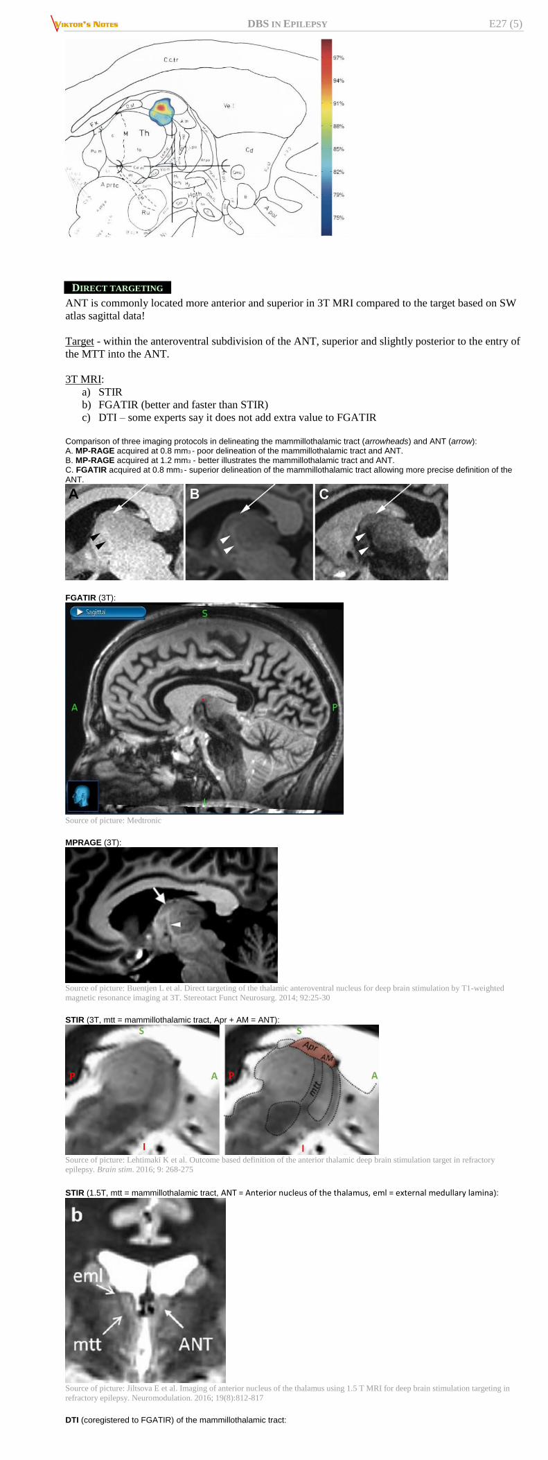
Telez-Zellento
et al Neurol ‘06 4
Alternating 1
mo blocks over
6 mo
190 Hz
90 us
cont
25% RR
¼ sz free
No
decline
Design of
randomiz not
optimal
Cukiert et al (2017) - the results of a prospective, double-blind, randomized controlled trial evaluating
the efficacy of unilateral and bilateral HCP DBS in 16 patients with refractory TLE:
o 2 months after surgery, all patients were randomized to stimulation on or off for a 6-month
blinded period.
o of the 8 patients randomized to the on-stimulation group, 4 became seizure free and 7 were
defined as responders, whereas 1 patient did not respond to DBS therapy.
o the experimental group experienced significantly fewer simple partial and complex partial
seizures than the control group throughout the blinded period.
Vonck et al. reported on 11 patients who underwent bilateral HCP DBS electrode implantation, with
stimulation laterality applied based on seizure localization. After 2.5–3 years of follow-up, patients
who were initially started on unilateral stimulation were converted to bilateral stimulation if seizure
reduction of > 90% had not been achieved. At final follow-up, and after switching to bilateral
stimulation as necessary, 6 patients achieved ≥ 90% seizure reduction, 3 patients achieved seizure
reduction rates ranging from 40% to 70%, and 2 patients achieved < 30% seizure reduction.
Importantly, the authors found that switching from unilateral to bilateral stimulation further improved
seizure outcomes in 3 of 5 patients with unilateral ictal onset. Implementing day-night cycling after
attaining treatment stability did not affect seizure control, and no changes in neuropsychological
testing were noted after DBS therapy.
STN
Author N Localization of epilepsy Outcome
Benabid/Chabardes
2002
3 sensory motor cortex 67-87%
2
< 50%
1
0
Shon (Seoul)
Stereotact Funct
Neurosurg 2005;83:84–90
2 FLE s/p failed resection 87-89%
Handforth (UCLA)
Epilepsia 47(7):1239–
1241, 2006
1 Bitemporal epilepsy 50%
1 Frontal encephalomalacia 33%
Neme (Santiago) 1
> 50%
3
< 50%
CEREBELLUM
while the cerebellum (hemispheres) has the longest history in DBS for the treatment of epilepsy,
results have been mixed. Therefore, stimulation of the cerebellum has fallen out of favor.
NUCL. ACCUMBENS
POSTERIOR HYPOTHALAMUS
BIBLIOGRAPHY for ch. “Epilepsy and Seizures” → follow this LINK
Viktor’s Notes℠ for the Neurosurgery Resident
Please visit website at www.NeurosurgeryResident.net