DALIA KAMAL ELDIEN MOAMMED Aspergillosis Lecture No-10-
27
DALIA KAMAL ELDIEN MOAMMED Aspergillosis Lecture No-10-
-
Upload
ashlynn-harrison -
Category
Documents
-
view
218 -
download
0
Transcript of DALIA KAMAL ELDIEN MOAMMED Aspergillosis Lecture No-10-

DALIA KAMAL ELDIEN MOAMMED
AspergillosisLecture No-10-

introduction
Aspergillosis is a mycotic disease caused by Aspergillus species
Aspergillus is a genus of ubiquitous soil fungi, found as saprophyte in soil, vegetation especially the stored grains, are common contaminants of starchy foods (such as bread and potatoes), and grow in or on many plants and trees, their spore widely distributed in air, hence they are a very common lab contaminant
They are rapid growing non fastidious organism, so can grow in simple media

Main species
There are more than 180 species Aspergillus , of which 34 have been associated with human disease, include:
A. fumigatusA. flavusA. terreusA. nigerA. nidulans

pathogenicity
The clinical manifestations of aspergillosis are determined by the host immune response to Aspergillus spp with the spectrum ranging from a local inappropriate inflammatory response, causing allergy, to local saprophytic lung disease with mycelial balls.
In humans, the major forms of disease are:1- Otomycosis = ear infection2- Brain abscess = brain infection3- Mycetoma = subcutaneous tissue infection4- Endocarditis & myocarditis= heart infection5- Sinuses aspergillosis = nasal sinuses infection6- Aspergillus flavus produces the carcinogenic mycotoxin,
aflatoxin which often contaminates foods such as nuts.

7- Pulmonary aspergillosis = lung infection, have 3 forms:
Allergic bronchopulmonary aspergillosis, which affects patients with respiratory diseases such as asthma, cystic fibrosis, and sinusitis
Acute invasive aspergillosis, a form that grows into surrounding tissue, more common in those with weakened immune systems such as AIDS or chemotherapy patients
Disseminated invasive aspergillosis, an infection spread widely through the body
Aspergilloma, a "fungus ball" that can form within cavities such as the lung
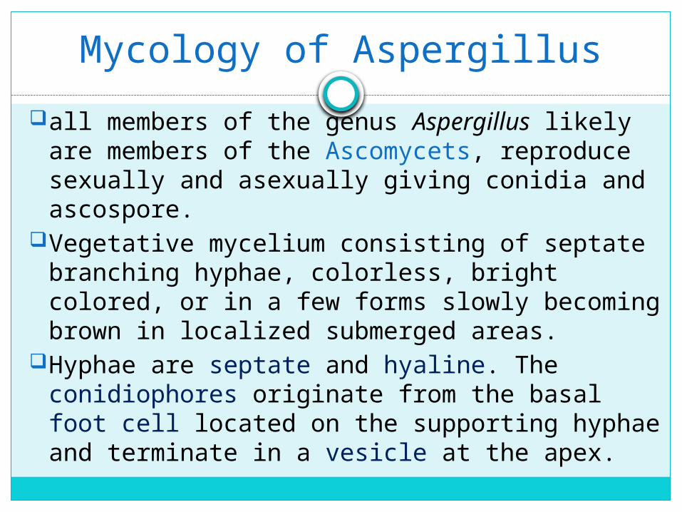
Mycology of Aspergillus
all members of the genus Aspergillus likely are members of the Ascomycets, reproduce sexually and asexually giving conidia and ascospore.
Vegetative mycelium consisting of septate branching hyphae, colorless, bright colored, or in a few forms slowly becoming brown in localized submerged areas.
Hyphae are septate and hyaline. The conidiophores originate from the basal foot cell located on the supporting hyphae and terminate in a vesicle at the apex.

Vesicle is the typical formation for the genus Aspergillus.
The morphology and color of the conidiophore vary from one species to another.
Covering the surface of the vesicle entirely ("radiate" head) or partially only at the upper surface ("columnar" head) are the flask-shaped phialides which are either uniseriate and attached to the vesicle directly or are biseriate and attached to the vesicle via a supporting cell, metula.
Over the phialides are the round conidia (2-5 µm in diameter) forming radial chains.

The typical Aspergillus head showing a conidiophore, swollen vesicle, phialides, and radiating chains of conidia is formed in cultures and is not often seen in uncultured specimens. In the tissue Aspergillus present as branching hyphae

Aspergillus head

Aspergillus head

Aspergillus head of different species
species conidiophores vesicle Strigmata(Phialides)
Arrangement of
conidiophores
A fumigatus
Color lesssmooth
Flask One raw columnar
A. flavus Color lessrough
oval One or two radiation
A. terreus Color lesssmooth
round Two raw Long columnar
A. niger Yellow or brownsmooth
round One or two irregular
A. nidulans
variablesmooth
round Two raw Short columnar

Laboratory diagnosis
specimens :according to the site of the infection in which fungi are
possible to be the etiological agents .Direct microscopy:a wet mount preparation of 10% potassium hydroxide,
which aids in the visualization of hyphal elementsFluorescent techniquesFluorescent dyes—eg, Calcofluor white, U vitex 2B, and
Blankophor, are water soluble color less dyes that selectively bind to beta-glycosidically linked polysaccharides within fungal cell walls. They are not specific for Aspergillus spp, but have the advantages of relatively high sensitivity.
Tissue sections should be stained with H&E or PAS, Note Aspergillus hyphae may be missed in H&E stained sections. Examine specimens for broad segmented hyphae showing the dichotomously branched(45% angle).
Direct immunofluorescence technique: by specific antisera

CultureAspergillus spp can be recovered on most routine
solid and liquid microbiological media (eg, blood agar, chocolate agar, brain heart infusion broth). Plus the fungal medium eg, Sabouraud dextrose agar with chloramphenicol and without cyclohexamide, incubate at 37C aerobically, if A. fumigatus is suspected incubate another plate at 43C
Aspergillus are fast growing after (2-7 days) as white mycelia, then colored according to the spore color
The inoculation on Czapek dox agar and potato dextrose agar and incubated at 25oC, will encourage the sporulation.

Aspergillus molds have a powdery texture. However the color of the mold's surface differs from species to another and can be used to identify the type of Aspergillus, the rate of growth can also be used to identify Aspergillus, with most species growing quite quickly, however A.glaucus and A.nidulans grow more slowly, and the temperature(A. fumigatus grow at 43C).
colonial morphology A. fumigatus = blue green and some time grey green A. flavus = yellow green A. terreus = cinammon brown A. niger = black A. nidulans = deep blue green or buff brown, also have
especial structure: Hülle cell(horse shoes)

Hülle cell
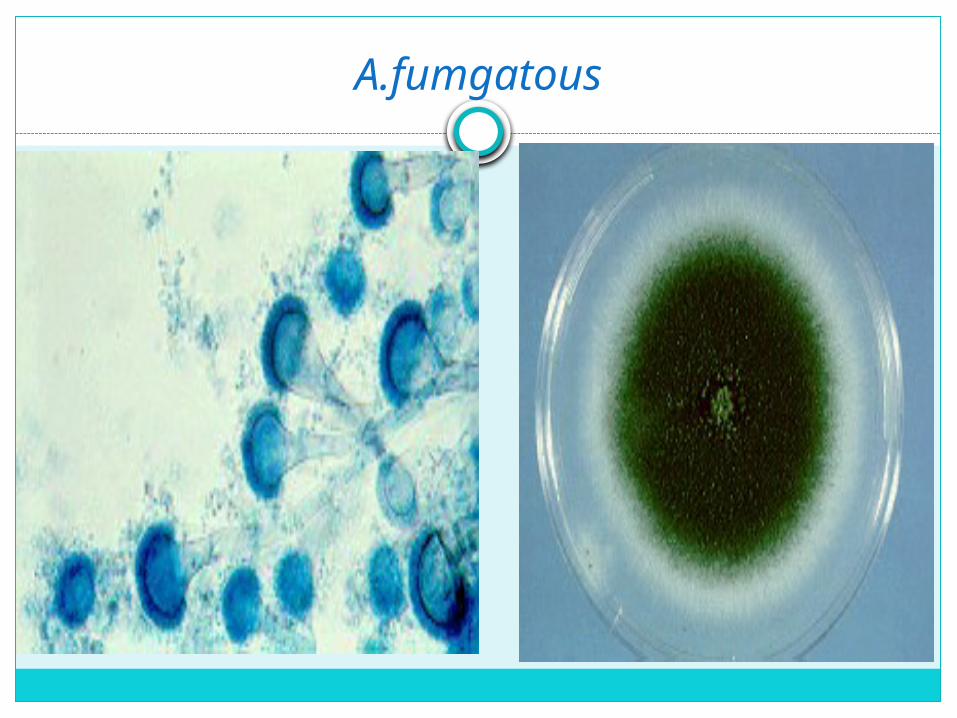
A.fumgatous
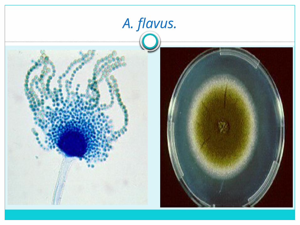
A. flavus.

A.nidulanous

A.niger
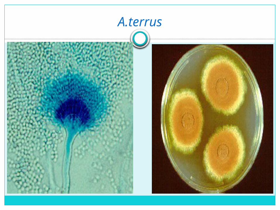
A.terrus
Serological diagnosis
Immunoelectrophoresis is the electrophoresis of a determined antigen mixture in an agarose gel that allows the separation of different antigens along the gel slide, and then the lateral diffusion of an antibody in the gel.
Counter Current Immunoelectrophoresis is a modification of immunoelectrophoresis in which antigen and antibody move in opposite directions and form precipitates in the area between the cells where they meet in concentrations of optimal proportions.

In this method, immuno precipitation occurs when antigen at the cathode is caused to migrate in an electric field through a suitable medium of diffusion against a stream of antibody migrating from the anode because of endosmotic flow. This is one-dimensional double electroimmuno diffusion in which antibody and antigen are moved towards each other by an applied electric field, because the gel is buffered at a pH between the isoelectric points of the antigen and antibody. The antigen and antibody are placed in separate wells in an Agarose gel plate.

MATERIALS: Agarose, Antigen, Test antiserum, Positive antiserum, Assay Buffer, Electrophoresis apparatus, slides.
PROCEDURE: 1.Prepare 10 ml of 1.0% Agarose2. Mark the end of a glass slide as +ve anode and -ve cathode.3.Place the glass slide on a horizontal surface. Pipette and
spread 5 ml of agarose onto slide. Allow to solidify for 15 minutes
4. Cut wells of according to the template using gel puncher. 5. Add 10µl of antigen in each of the two wells towards
cathode (Negative electrode) and 10µl of positive control antiserum and test antisera in wells towards anode
6. Connect the power7. Observe for precipitin line between the antigen and antisera
wells.

Counter Current Immunoelectrophoresis

Molecular techniques Polymerase chain reaction and other
techniques are highly sensitive and specific