
dADAR, a Drosophila double-stranded RNA-specific adenosine ...
15
dADAR,a Drosophila double-stranded RNA-specific adenosine deaminase is highly developmentally regulated and is itself a target for RNA editing MICHAEL J. PALLADINO, 1 LIAM P. KEEGAN, 2 MARY A. O’CONNELL, 2 and ROBERT A. REENAN 1 1 Department of Genetics and Developmental Biology , University of Connecticut Health Center , Farmington, Connecticut 06030, USA 2 MRC Human Genetics Unit, Western General Hospital, Edinburgh EH4 2XU, United Kingdom ABSTRACT We have identified a homolog of the ADAR (a denosine d ea minases that act on R NA) class of RNA editases from Drosophila, dADAR. The dADAR locus has been localized to the 2B6–7 region of the X chromosome and the complete genomic sequence organization is reported here. dADAR is most homologous to the mammalian RNA editing enzyme ADAR2, the enzyme that specifically edits the Q/R site in the pre-mRNA encoding the glutamate receptor subunit GluR-B. Partially purified dADAR expressed in Pichia pastoris has robust nonspecific A-to-I deaminase activity on synthetic dsRNA substrates. Transcripts of the dADAR locus originate from two regulated promoters. In addition, alternative splicing generates at least four major dADAR isoforms that differ at their amino-termini as well as altering the spacing between their dsRNA binding motifs. dADAR is expressed in the developing nervous system, making it a candidate for the editase that acts on para voltage-gated Na 1 channel transcripts in the central nervous system. Surprisingly, dADAR itself undergoes developmentally regulated RNA editing that changes a conserved residue in the catalytic domain. Taken together, these findings show that both transcription and processing of dADAR transcripts are under strict developmental control and suggest that the process of RNA editing in Drosophila is dynamically regulated. Keywords: ADAR; inosine; ion channel; nervous system INTRODUCTION RNA editing involving the conversion of adenosine (A) to inosine (I) in pre-mRNA is a posttranscriptional pro- cess catalyzed by proteins of the ADAR (a denosine d ea minases that act on R NA) family (Rueter & Eme- son, 1998)+ ADARs require duplex RNA secondary struc- ture in their target substrates+ There are two types of A-to-I RNA editing, promiscuous and specific (reviewed in Bass, 1997)+ Viral RNA genomes provided the first reported instance of promiscuous A-to-I RNA editing where extensive conversion of A to G (guanosine) was observed (Cattaneo et al+, 1988)+ Since I base pairs with cytidine, G replaces I when cDNAs are generated+ Mechanistically , cRNA intermediates of the replication process or transcripts base pair with complete comple- mentarity to the viral genomic RNA+ When the sub- strates for ADARs are extensive duplexes, up to 50% of adenosines are converted to inosine+ Such promis- cuous editing has been proposed to have roles in gene regulation, viral defense (Bass & Weintraub, 1988) and in biased hypermutation and persistent infection of cer- tain viruses (Bass, 1997)+ Promiscuous editing is also seen in vivo when naturally occurring antisense tran- scripts are generated, as in the case of the Xenopus bFGF gene (Saccomanno & Bass, 1999) and in poly- oma virus (Kumar & Carmichael, 1997)+ Supporting a role in gene regulation is the finding that extensively modified early transcripts from polyoma are almost ex- clusively retained in the nucleus at late times in infec- tion, thus preventing translation, a critical feature of the polyoma life cycle (Kumar & Carmichael, 1997)+ An- other potential role in mRNA regulation via extensive editing was recently proposed in C. elegans where sev- Reprint requests to: Robert A+ Reenan, Department of Genetics and Developmental Biology , University of Connecticut Health Center , 263 Farmington Avenue, Farmington, Connecticut 06030, USA; e-mail: rreenan@neuron+uchc+edu+ RNA (2000), 6:1004–1018+ Cambridge University Press+ Printed in the USA+ Copyright © 2000 RNA Society + 1004
-
Upload
truongtruc -
Category
Documents
-
view
215 -
download
0
Transcript of dADAR, a Drosophila double-stranded RNA-specific adenosine ...

dADAR , a Drosophila double-strandedRNA-specific adenosine deaminase is highlydevelopmentally regulated and isitself a target for RNA editing
MICHAEL J. PALLADINO, 1 LIAM P. KEEGAN, 2 MARY A. O’CONNELL, 2
and ROBERT A. REENAN 1
1Department of Genetics and Developmental Biology, University of Connecticut Health Center,Farmington, Connecticut 06030, USA
2MRC Human Genetics Unit, Western General Hospital, Edinburgh EH4 2XU, United Kingdom
ABSTRACT
We have identified a homolog of the ADAR (a denosine d eaminases that act on R NA) class of RNA editases fromDrosophila , dADAR . The dADAR locus has been localized to the 2B6–7 region of the X chromosome and the completegenomic sequence organization is reported here. dADAR is most homologous to the mammalian RNA editing enzymeADAR2, the enzyme that specifically edits the Q/R site in the pre-mRNA encoding the glutamate receptor subunitGluR-B. Partially purified dADAR expressed in Pichia pastoris has robust nonspecific A-to-I deaminase activity onsynthetic dsRNA substrates. Transcripts of the dADAR locus originate from two regulated promoters. In addition,alternative splicing generates at least four major dADAR isoforms that differ at their amino-termini as well as alteringthe spacing between their dsRNA binding motifs. dADAR is expressed in the developing nervous system, making ita candidate for the editase that acts on para voltage-gated Na 1 channel transcripts in the central nervous system.Surprisingly, dADAR itself undergoes developmentally regulated RNA editing that changes a conserved residue in thecatalytic domain. Taken together, these findings show that both transcription and processing of dADAR transcriptsare under strict developmental control and suggest that the process of RNA editing in Drosophila is dynamicallyregulated.
Keywords: ADAR; inosine; ion channel; nervous system
INTRODUCTION
RNA editing involving the conversion of adenosine (A)to inosine (I) in pre-mRNA is a posttranscriptional pro-cess catalyzed by proteins of the ADAR (adenosinedeaminases that act on RNA) family (Rueter & Eme-son, 1998)+ADARs require duplex RNAsecondary struc-ture in their target substrates+ There are two types ofA-to-I RNA editing, promiscuous and specific (reviewedin Bass, 1997)+ Viral RNA genomes provided the firstreported instance of promiscuous A-to-I RNA editingwhere extensive conversion of A to G (guanosine) wasobserved (Cattaneo et al+, 1988)+ Since I base pairswith cytidine, G replaces I when cDNAs are generated+Mechanistically, cRNA intermediates of the replication
process or transcripts base pair with complete comple-mentarity to the viral genomic RNA+ When the sub-strates for ADARs are extensive duplexes, up to 50%of adenosines are converted to inosine+ Such promis-cuous editing has been proposed to have roles in generegulation, viral defense (Bass & Weintraub, 1988) andin biased hypermutation and persistent infection of cer-tain viruses (Bass, 1997)+ Promiscuous editing is alsoseen in vivo when naturally occurring antisense tran-scripts are generated, as in the case of the XenopusbFGF gene (Saccomanno & Bass, 1999) and in poly-oma virus (Kumar & Carmichael, 1997)+ Supporting arole in gene regulation is the finding that extensivelymodified early transcripts from polyoma are almost ex-clusively retained in the nucleus at late times in infec-tion, thus preventing translation, a critical feature of thepolyoma life cycle (Kumar & Carmichael, 1997)+ An-other potential role in mRNA regulation via extensiveediting was recently proposed in C. elegans where sev-
Reprint requests to: Robert A+ Reenan, Department of Geneticsand Developmental Biology, University of Connecticut Health Center,263 Farmington Avenue, Farmington,Connecticut 06030,USA; e-mail:rreenan@neuron+uchc+edu+
RNA (2000), 6:1004–1018+ Cambridge University Press+ Printed in the USA+Copyright © 2000 RNA Society+
1004

eral transcripts containing extended 39 noncoding hair-pin structures are extensively modified within hairpinregions (Morse & Bass, 1999)+
ADARs can also edit specific adenosines in mRNAprecursors (pre-mRNA)+ Surprisingly,most of these ex-amples are restricted to genes encoding ligand-gatedion channels or receptor genes expressed in the ner-vous system and result in only one or a few adenosinesdeaminated per transcript (Rueter & Emeson, 1998)+Because the translation machinery has been shown tointerpret I as G (Basilio et al+, 1962), modifications in-troduced by an ADAR are predicted to change thecoding potential of the edited transcripts+ This wasfirst shown for pre-mRNAs encoding subunits of theglutamate-gated ion channel receptors (GluRs)+ TheGluR-B transcripts have numerous distinct RNA editingsites that, when edited, have profound consequencesfor channel function (reviewed in Seeburg et al+, 1998)+For instance, editing that results in the conversion of aglutamine codon (CAG) to a arginine codon (CGG) atthe Q/R site alters the Ca21 permeability of channelscontaining the edited subunit (Kohler et al+, 1993)+ Thephysiological significance of editing at the Q/R site isdemonstrated by the neurological dysfunction and post-natal lethality associated with mutant mice expressingan editing incompetent allele of GluR-B (Brusa et al+,1995; Feldmeyer et al+, 1999)+ Other examples whereA-to-I RNA editing has been shown to perform the roleof diversifying protein isoforms encoded by a singlegenetic locus are the mammalian serotonin receptor(5-HT2CR) (Burns et al+, 1997) and the squid potassiumchannel (sqKv2) (Patton et al+, 1997)+ Specific editingof the pre-mRNA encoding an invertebrate voltage-gated Na1 channel, the product of the Drosophila paralocus, has recently been demonstrated (Hanrahan et al+,1999; Reenan et al+, 2000)+
Mechanistically, specific editing presents a more com-plex secondary structure requirement in the RNA sub-strate than the promiscuous editing that occurs onextended perfect duplexes+ The best characterized spe-cifically edited substrates are the regions of GluR sub-unit transcripts spanning the Q/R site+ It was first shownfor the GluR-B pre-mRNA that specific editing of theexonic adenosine at the Q/R site requires cis-actingsequences several hundred nucleotides away in theintron downstream of the editing site (Higuchi et al+,1993; Egebjerg et al+, 1994)+ The intron region neces-sary for editing has extensive complementarity to andbase pairs with the exonic region surrounding the ed-iting site and was termed an editing-site complemen-tary sequence (ECS)+ Further studies of the Q/R sitein transcripts of the kainate GluR-5 and -6 paralogsshowed that these complementary intronic sequencescan be over 1,900 nt away (Herb et al+, 1996)+ Muta-genic studies of the GluR Q/R site suggest that theprimary determinant of specificity is in the local duplexstructure surrounding the edited adenosine (Maas et al+,
1996)+ Recent work suggests that long RNA duplexesin which many adenosines are converted can be ren-dered into more specific substrates by duplex inter-ruptions, such as loops, supporting the role of localstructural elements in directing ADAR specificity (Leh-mann & Bass, 1999)+
Both ADAR1 and ADAR2 can edit specific adenosinesand have promiscuous editing activity on extendeddouble-stranded (ds) RNA+ Enzymes homologous toADARs but having no proven A-to-I deaminase activityare tentatively classed as ADAR-like+ Such a mam-malian homolog of ADAR2, called RED2, has beenidentified but has not been shown to edit any knownsubstrate (Melcher et al+, 1996a)+ Recently a relatedfamily of enzymes, the ADATs (adenosine deaminasesacting on tRNAs), has been identified that convert A-to-Iat specific positions in tRNAs (Gerber et al+, 1998; Ger-ber & Keller, 1999; Keegan et al+, 1999; Keller et al+,1999; Maas et al+, 1999)+
Inosine has been shown to be present in mRNAsfrom numerous tissues and to occur at tissue-specificlevels in rat (Paul & Bass, 1998)+ ADAR activities areubiquitous in nature and have been detected in inver-tebrate (Skeiky & Iatrou, 1991; Bass, 1993) and verte-brate animals and are expressed in a wide variety ofprimary tissues and cells within organisms (Wagneret al+, 1990)+ Moreover, ADAR activity has been de-tected in Drosophila extracts (Casey & Gerin, 1995; M+O’Connell, unpubl+ observation)+ We report here thecloning and characterization of a Drosophila dsRNA-dependent adenosine deaminase, dADAR+ Recombi-nant dADAR was overexpressed in the yeast Pichiapastoris, and has robust A-to-I editing activity on ex-tended dsRNA+ dADAR is expressed in the nervoussystem of Drosophila embryos making it a candidateenzyme responsible for the editing of pre-mRNAs en-coding the para voltage-gated Na1 channel that is ex-pressed exclusively in the nervous system (Hong &Ganetzky, 1994)+ The dADAR locus is subject to bothtranscriptional and posttranscriptional regulation+ Twopromoters, one specific to the adult stage, generatetranscripts with different 59 untranslated regions+ dADARtranscripts undergo developmentally regulated alterna-tive splicing at numerous sites, generating four poten-tial alternative in-frame translational start sites andaltering the spacing of dsRNA binding motifs (DRBMs)+Surprisingly, dADAR transcripts themselves are the tar-get of a very specific and highly regulated RNA editingevent that alters a conserved amino acid in the deam-inase domain portion of the protein+
RESULTS
Cloning and genomic organization of dADAR
ADARs have common structural motifs includingDRBMs and an adenosine deaminase catalytic domain
Drosophila ADAR 1005

(Fig+ 1A)+ We compared the amino-acid sequences ofmammalian ADARs as well as an ADAR-like Caeno-rhabditis elegans homolog (Hough et al+, 1999) to de-sign degenerate PCR primers (Fig+ 1B)+ The catalyticdomain is the most highly conserved region of ADARs+Therefore, PCR was performed on a Drosophila wholeadult cDNA library using degenerate primers that wouldamplify products encoding either the adenosine deam-inase domain or a region encompassing both the de-aminase domain and ADAR1 or ADAR2 type DRBMs(see Materials and methods)+ A PCR product was ob-tained with a degenerate primer pair spanning thedeaminase domain+Cloning and sequence analysis con-firmed that the product had significant homology to the
adenosine deaminase domain of ADARs; however, itappeared not to have significantly higher homology toADAR2 (32/62 amino acid identity) than to ADAR1(30/62 amino acid identity)+Gene-specific primers fromwithin this cloned deaminase domain region and de-generate primers for either ADAR1 or ADAR2-typeDRBMs were used in an additional round of ampli-fication+ Products were obtained with ADAR2-specificdegenerate primers+ Cloning and sequence analysisconfirmed that the product had a high degree of ho-mology to mammalian ADAR2 and we have des-ignated this gene dADAR+ In situ hybridization toDrosophila polytene chromosomes revealed a singleband localizing to the 2B6–7 region of the X chromo-
FIGURE 1. Genomic location and organization of dADAR. A: Domain organization of the ADAR1, ADAR2, and ADAT classof editases+ Za and Zb are Z-DNA binding domains+ DRBM: double-strand RNA binding motif+ DEAM: deaminase domain+nls: nuclear localization signal+ Roman numerals refer to the different DRBMs+ B: Amino-acid sequence alignments ofADAR1 and ADAR2 classes of editase and the C. elegans ceT20H4+4 ADAR-like homolog in the regions where degeneratePCR primers were designed+ C: Compiled genomic sequence organization and transcription unit of the dADAR locus+Genomic sequence was generated from the P1 bacteriophage DS04654+ Overlap with European Drosophila GenomeProject (EDGP) cosmid clone DMC4F1 and BDGP bacterial artificial chromosome (BAC) clone DMBN35H14 are shown+ Inthe restriction map of DS04654 X: XhoI, B: BamHI, R: EcoRI, S: Sal I+ Numbers above the P1 clone refer to sequence tagsites (STS) found in DS04654 (1,2,3,4,5)+ Open boxes represent constitutive exons, gray boxes are alternative exons+ Theblack box is exon 7, where RNA editing occurs+
1006 M.J. Palladino et al.

some (data not shown)+ Bacteriophage P1 clones fromthis region were obtained (see Materials and methods)and PCR was used to identify the presence of thisgene+ Thirty kilobases of genomic sequence aroundthe gene were generated to define and orient the tran-scription unit (Fig+ 1C)+ The dADAR locus lies ap-proximately 180 kb centromere proximal to the BroadComplex (BrC) and about 100 kb distal to the armadillolocus+ Also, the 59 most transcription start sites fordADAR are about 7 kb centromere proximal to theknown translocation breakpoint T(1,2)dorvar7+
cDNA analysis of dADAR
To obtain full-length cDNA clones of the dADAR gene,a modification of RACE was used to obtain both 59and 39 ends of cDNAs (see Materials and methods)+Full-length cDNA clones revealed a number of inter-esting regulatory features concerning the dADAR locus(Figs+ 1C and 2)+ First, dADAR transcripts begin withtwo different alternative 59 exons that are untranslatedand have heterogeneous start sites+No consensus TATAbox was found upstream of the start sites for exon24a-containing transcripts, but a number of transcriptsbegin at consensus eukaryotic transcription initiator se-quences Py Py A11 N T/A Py (Lo & Smale, 1996)+Developmental studies (see below) also indicate thatthe exon 24a- and 24b-containing transcripts origi-nate from alternative promoters+ Secondly, several al-ternative splice forms were found in the 59 end of thecDNAs and were predicted to generate alternative start-ing methionines (alternative exons 23, 22, 21, and 0,Fig+ 2)+ Translation was predicted to begin either inexon 22, 21, 0, or 1, depending on exon utilization+The sequences surrounding the exon 22 and 1 start-ing ATGs presented the strongest flanking consensussequences (Cavener, 1987)+ Developmental data indi-cated that the exon 21- and 1-containing forms werethe most common, suggesting that these starting ATGswere the most frequently utilized start sites (see be-low)+ However, the less frequent exon 22 and 0 puta-tive start sites shared homology at the amino-acid level(exon 22 5 MKFDS, exon 0 5 MKFEC) and thus mayrepresent important dADAR isoforms+
Alternative splicing was observed between the twoDRBMs of dADAR (6 exon 3a) and this splicing altersthe distance between these important structural motifs(Figs+ 2 and 3)+ In addition, the distal 59 splice donorsite of exon 3a is a rare nonconsensus splice donorsite (GCAAG vs+GTAAG), the implications of which willbe addressed in the discussion+
The predicted dADAR protein is approximately thesame size as the mammalian ADAR2 homologs(dADAR 5 670 amino acids, ratADAR2 5 711 aminoacids, ratRED2 5 746 amino acids)+ Overall, dADARhas 43% identity to rADAR2 and 35% identity to RED2+Within the deaminase domain, however, the homology
rises to 46% identity for hADAR1 and 67% identity forhADAR2 enzymes+ Residues important for coordi-nating zinc in the catalytic domain are also absolutelyconserved (Betts et al+, 1994; Kim et al+, 1994, Laiet al+, 1995) (Fig+ 3)+ In contrast, comparison with atRNA-specific adenosine deaminase, dADAT1, revealedonly 17% identity overall and 28% identity within thecatalytic domain (Keegan et al+, 1999)+
dADAR encodes a dsRNA-specificadenosine deaminase
The dADAR cDNA clone pSR1–9 was subcloned intothe P. pastoris expression vector pSK-FLIS6 to ex-press a fusion protein with a FLAG epitope at the aminoterminus and six histidine residues at the carboxy ter-minus+ The full-length dADAR produced from this con-struct starts at the exon 1 ATG and includes exon 3a+The recombinant fusion protein was purified by chro-matography of a crude extract of Pichia expressingdADAR over a Ni21-NTA column and assayed forits ability to convert adenosine to inosine in dsRNA(Fig+ 4A)+ Pichia does not contain any endogenousdsRNA adenosine deaminase activity (Gerber et al+,1998)+ The load fraction contained RNases that de-graded the dsRNA substrate before it could be deam-inated and assayed+Therefore,more activity is observedwith 1 mL of the crude extract than with 6+25 mL+ Im-munoblot analysis was also performed on input andeluate fractions from the Ni21-NTA column with FLAGmonoclonal antisera and revealed a band of approxi-mately 80 kDa that comigrated with dsRNA adenosinedeaminase activity (Fig+ 4B)+ This band was very faintin the load fraction+ This may be due to protein degra-dation, as many bands of lower molecular weight wereobserved that cross-react with the antibody+
It is also possible that the protein was not highlyexpressed as very little was recovered from the FLAGaffinity column+ The peak fraction from the Ni21-NTAcolumn was chromatographed over a FLAG affinitymatrix and assayed for dsRNA adenosine deaminaseactivity+ A protein of ;80 kDa was detected when ap-proximately 400 mL of the peak fractions from the FLAGaffinity matrix were TCA precipitated and electropho-resed on an SDS-polyacrylamide gel that was silverstained (Fig+ 4C)+ The yield of recombinant dADAR pro-tein from Pichia was very low and it was not possible todetermine the specific activity+
Developmental expression and alternativesplicing profile of dADAR
Quantitative RT-PCR analysis was performed usingprimers specific for the 24a or 24b exons and a primerin constitutive exon 4, which is downstream of all of thesites of alternative splicing+Ribosomal protein 49 (rp49)expression was used as an internal control (see Ma-
Drosophila ADAR 1007
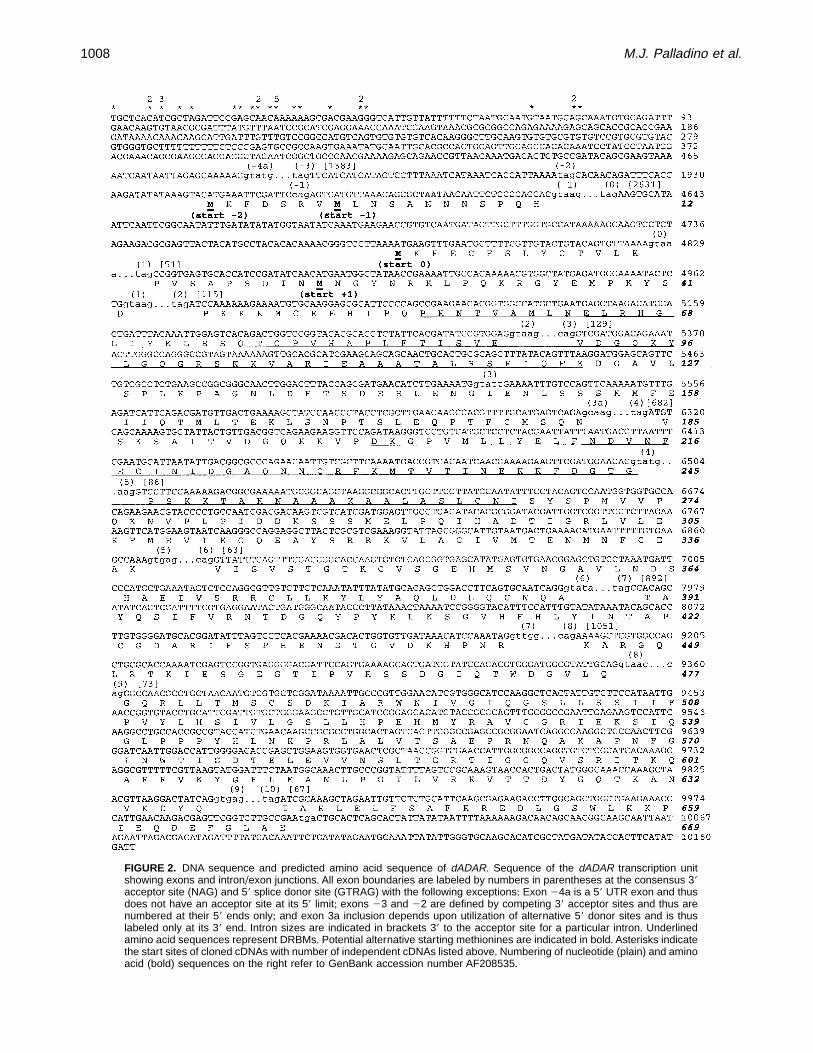
FIGURE 2. DNA sequence and predicted amino acid sequence of dADAR. Sequence of the dADAR transcription unitshowing exons and intron/exon junctions+ All exon boundaries are labeled by numbers in parentheses at the consensus 39acceptor site (NAG) and 59 splice donor site (GTRAG) with the following exceptions: Exon 24a is a 59 UTR exon and thusdoes not have an acceptor site at its 59 limit; exons 23 and 22 are defined by competing 39 acceptor sites and thus arenumbered at their 59 ends only; and exon 3a inclusion depends upon utilization of alternative 59 donor sites and is thuslabeled only at its 39 end+ Intron sizes are indicated in brackets 39 to the acceptor site for a particular intron+ Underlinedamino acid sequences represent DRBMs+ Potential alternative starting methionines are indicated in bold+ Asterisks indicatethe start sites of cloned cDNAs with number of independent cDNAs listed above+ Numbering of nucleotide (plain) and aminoacid (bold) sequences on the right refer to GenBank accession number AF208535+
1008 M.J. Palladino et al.

FIGURE 3. Amino acid sequence alignment of dADAR and known ADARs+ dADAR protein sequence alignment of the exon22-, 21-, and 3a-containing (longest) alternative splice form+ Bold lines indicate the positions of DRBMs (1–2) anddeaminase domain motifs (I–III) and the thin line indicates the position of alternative exon 3a+ Arrow indicates the positionof the S/G editing site+ The sequences are rat ADAR2 (rADAR2), human ADAR2 (hADAR2), the ADAR-like homolog ratRED2, and dADAR+ Black boxes represent identity across all homologs and gray boxes represent identity (upper case) orsimilarity (lower case) to dADAR in at least 75% of the homologs+ The consensus line symbols are as follows: !: identityacross all sequences, *: high degree of identity or similarity to dADAR, +: lack of conservation with dADAR+
Drosophila ADAR 1009
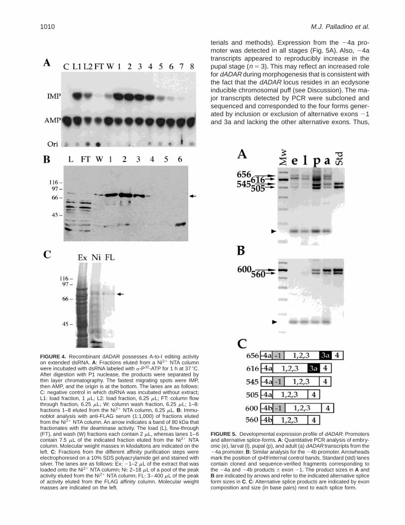
terials and methods)+ Expression from the 24a pro-moter was detected in all stages (Fig+ 5A)+ Also, 24atranscripts appeared to reproducibly increase in thepupal stage (n 5 3)+ This may reflect an increased rolefor dADAR during morphogenesis that is consistent withthe fact that the dADAR locus resides in an ecdysoneinducible chromosomal puff (see Discussion)+ The ma-jor transcripts detected by PCR were subcloned andsequenced and corresponded to the four forms gener-ated by inclusion or exclusion of alternative exons 21and 3a and lacking the other alternative exons+ Thus,
FIGURE 4. Recombinant dADAR possesses A-to-I editing activityon extended dsRNA+ A: Fractions eluted from a Ni21 NTA columnwere incubated with dsRNA labeled with a-P32-ATP for 1 h at 37 8C+After digestion with P1 nuclease, the products were separated bythin layer chromatography+ The fastest migrating spots were IMP,then AMP, and the origin is at the bottom+ The lanes are as follows:C: negative control in which dsRNA was incubated without extract;L1: load fraction, 1 mL; L2: load fraction, 6+25 mL; FT: column flowthrough fraction, 6+25 mL; W: column wash fraction, 6+25 mL; 1–8:fractions 1–8 eluted from the Ni21 NTA column, 6+25 mL+ B: Immu-noblot analysis with anti-FLAG serum (1:1,000) of fractions elutedfrom the Ni21 NTA column+ An arrow indicates a band of 80 kDa thatfractionates with the deaminase activity+ The load (L), flow-through(FT), and wash (W) fractions each contain 2 mL, whereas lanes 1–6contain 7+5 mL of the indicated fraction eluted from the Ni21 NTAcolumn+ Molecular weight masses in kilodaltons are indicated on theleft+ C: Fractions from the different affinity purification steps wereelectrophoresed on a 10% SDS polyacrylamide gel and stained withsilver+ The lanes are as follows: Ex: 21–2 mL of the extract that wasloaded onto the Ni21 NTA column; Ni: 2–18 mL of a pool of the peakactivity eluted from the Ni21 NTA column; FL: 3–400 mL of the peakof activity eluted from the FLAG affinity column+ Molecular weightmasses are indicated on the left+
FIGURE 5. Developmental expression profile of dADAR: Promotersand alternative splice-forms+ A: Quantitative PCR analysis of embry-onic (e), larval (l), pupal (p), and adult (a) dADAR transcripts from the24a promoter+ B: Similar analysis for the 24b promoter+Arrowheadsmark the position of rp49 internal control bands+ Standard (std) lanescontain cloned and sequence-verified fragments corresponding tothe 24a and 24b products 6 exon 21+ The product sizes in A andB are indicated by arrows and refer to the indicated alternative spliceform sizes in C+ C: Alternative splice products are indicated by exoncomposition and size (in base pairs) next to each splice form+
1010 M.J. Palladino et al.

four protein isoforms could be generated starting at theexon 21 or exon 1 ATGs and either including or ex-cluding exon 3a, which alters the spacing betweenDRBMs+ We did not determine the frequencies of in-clusion of alternative exons 23,22, or 0 as they occurat lower levels+
Expression of transcripts including exon 24b wasfound to be more highly regulated than that of tran-scripts containing exon 24a (Fig+ 5B)+ Two major exon24b-containing transcripts were detectable in pupaeand adults+ Cloning and sequence analysis identifiedthese two products as either including or excluding the21 exon and lacking the other alternative exons+ Un-like 24a transcripts, all 24b transcripts lacked alterna-tive exon 3a+
The dADAR expression pattern was determined byin situ hybridization to whole Drosophila embryos ofvarious developmental stages+ Although a low level ofnonspecific staining was seen in early stage 13 em-bryos (end of germ-band retraction; data not shown),specific staining was seen by late stage 13 after con-densation of the ventral nerve cord occurs+ Stainingthen proceeded to intensify within the central nervoussystem, reaching high levels in the embryonic ventralnerve cord and brain by stage 16 (Fig+ 6)+
dADAR itself undergoes RNA editing
We discovered a nucleotide position in the dADAR tran-script in exon 7 (Fig+ 1) that gave a reproducible mixedA/G signal in automated sequencing of bulk dADARRT-PCR product (Fig+ 7A)+ Cloning and sequencing ofdADAR transcripts showed that cDNAs also containedeither A or G at the same position+ Because inosinebase pairs preferentially with cytosine, an A-to-G tran-sition occurs in the course of synthesis of double-stranded cDNA when A-to-I editing occurs in pre-mRNA+Sequencing of PCR products generated from genomicDNA gave exclusively an A signal at the same position+Also, P1 bacteriophage genomic clones containing thedADAR locus had A at this position+ Similar analyseswere performed on genomic DNAs and RT-PCR prod-ucts of related Drosophila species (Fig+ 7B)+ D. simu-lans is estimated to be 2+5 million years diverged fromD. melanogaster (Powell, 1997)+ In each case, RT-PCRproducts revealed a mixed A/G signal at the same lo-cation in the related species whereas sequence of thecorresponding PCR products generated from genomicDNA gave a pure A signal+ Thus, the A-to-G changesseen in cDNAs of dADAR, at variance with the genomi-cally encoded A at this position, bear all the hallmarksof ADAR mediated A-to-I RNA editing seen in othernatural mRNA substrates+ The A that is edited occurs inthe first position of a serine (S) codon and would con-vert the coding potential to that of glycine (G) (AGT toGGT)+ This amino acid change occurs in the deami-nase domain of dADAR, which is a highly conserved
portion of the dADAR protein+ The S/G site is six aminoacids carboxy-terminal to the first cysteine in motif II ofthe deaminase domain that is thought to chelate a zincion at the active site (Betts et al+, 1994; Kim et al+, 1994;Lai et al+, 1995) (Fig+ 7C)+
The editing of the pre-mRNA encoding dADAR wasfound to occur in a highly developmentally regulatedfashion+ cDNAs were isolated from several develop-mental stages and the extent of RNA editing was de-termined (see Materials and methods, Fig+ 8)+ Editingat the S/G site of dADAR was low in embryonic andpupal cDNAs (1+0% and 3+8%, respectively) and in-creased dramatically upon eclosion+ Editing increasedmore than 40-fold from embryo to adult+ Although theanalysis of editing from adult flies revealed that editing
FIGURE 6. Embryonic expression pattern of dADAR by in situ hy-bridization+ Drosophila wild-type stage 16 embryos were hybridizedwith a dADAR probe and whole mounted (see Materials and meth-ods)+ Ventral (A), lateral (B), and dorsal (C) views are shown+ Stron-gest expression is seen in the ventral nerve cord (A) and brain (B)+Lower levels of expression can be seen in the peripheral nervoussystem in each segment (C)+ Scale bar for A--C indicates 25 mm+
Drosophila ADAR 1011

occurred in 41 6 6% of all transcripts, we found that the3a-containing transcripts are essentially unedited (no3a-containing cDNAs were edited out of 100 ana-lyzed)+ So, editing of dADAR pre-mRNA occurs in atemporal- and transcript-specific manner+
DISCUSSION
Adenosine deaminases that act on RNA have beenfound throughout the animal kingdom (Bass, 1997)+Onerole for these enzymes has been proposed in viral de-fense, through promiscuous modification of dsRNA in-termediates and subsequent mutation of viral genomes(Bass & Weintraub, 1988)+ Another role has been thatof antisense regulation of gene expression through thenuclear retention of highly modified mRNAs (Kumar &Carmichael, 1997)+ADARs can also perform highly spe-cific editing of select positions within pre-mRNAs+ De-spite the paucity of examples of specific RNA editing byADARs, it is clear that mRNAs from many tissues con-tain inosine at detectable levels (Paul & Bass, 1998)and that enzymes that perform specific A-to-I editingare also ubiquitous+
FIGURE 7. RNA editing of dADAR. A: PCR products were amplified from D. melanogaster (D.m) RNA (RT-PCR), genomicDNA (gDNA), and cloned cDNA (cDNA) and subjected to automated sequencing+ The region around the edited adenosineis shown and a mixed sequence signal is indicated by N+ B: Similar analyses to A were performed on D. simulans (D.s) andD. takahashi (D.t ) RNA (RT-PCR) and D. takahashi gDNA (gDNA)+ C: Sequence alignment of the ADAR1 (hADAR1,rADAR1, xADAR),ADAR2 (dADAR, hADAR2), and ADAT (scTAD1p, dADAT1) classes of editase in the region of the dADARS/G editing site (indicated by arrow)+ rRED2 and ceT20H4+4 are ADAR-like homologs that have not been shown to possessADAR activity+ Motif II in the catalytic domain is indicated by a black bar+
FIGURE 8. Developmental profile of dADAR editing+ Quantitation ofRNA editing was performed by cloning and restriction analysis ofpartial dADAR cDNAs (see Materials and methods)+ Clones wereobtained from embryos, pupae, and adults+ Total clones assayedwere: embryo: n 5 98; pupae: n 5 133; and adult: n 5 140+ `:significantly different from adult, p , 0+001 by Student’s t-test+
1012 M.J. Palladino et al.

The Drosophila dADAR locus
We report here the cloning and analysis of a Drosoph-ila homolog of the ADAR class of enzymes, nameddADAR. We have shown that the recombinant dADARprotein can convert A-to-I on synthetic dsRNA sub-strates+We have mapped the dADAR locus cytogenet-ically, and cloned and sequenced the entire transcriptionunit+ dADAR is subject to an array of transcriptionaland posttranscriptional regulatory mechanisms+ dADARexpression is driven by two promoters in temporallydistinct patterns+ Likewise, alternative promoters arealso utilized by mammalian ADAR1; one promoter isconstitutive and the other is interferon (IFN) inducible(George & Samuel, 1999a, 1999b; Patterson & Sam-uel, 1995)+ The IFN inducible ADAR1 is larger and lo-calized to both the nucleus and cytoplasm, whereasconstitutive ADAR1 is nuclear (Patterson et al+, 1995)+
Additional diversity is generated by alternative splic-ing of dADAR transcripts+ Alternative exons that con-tain different translation start sites are utilized in twoabundant splice forms (exon 21 and exon 1) that prob-ably generate proteins with alternative amino termini+Another alternative splice choice of dADAR (exon 3a)alters the spacing between DRBMs+ By comparison,both mammalian ADAR1 and ADAR2 also undergo al-ternative splicing to generate ADAR isoforms with dif-ferential activities on specific substrates (Gerber et al+,1997; Lai et al+, 1997; Liu & Samuel, 1999)+
Developmental expression of dADAR
The expression pattern of dADAR was determined byin situ hybridization to Drosophila embryos and shownto be largely nervous system specific+ In fact, the tem-poral and spatial expression patterns of dADAR over-lap substantially with that reported for transcripts of thepara locus (Hong & Ganetzky, 1994)+ The para locusencodes the a-subunit of the major voltage-gated Na1
channel of the Drosophila nervous system and paratranscripts have been shown to possess four A-to-I RNAediting sites (Hanrahan et al+, 1999;Reenan et al+, 2000)+In addition, the pre-mRNA encoding the Drosophila cal-cium channel Dmca1A has been reported to undergoRNA editing (Smith et al+, 1996) and these reports havebeen substantially confirmed (M+A+ O’Connell and M+J+Palladino, unpubl+ results)+ Dmca1 is also expressed inthe nervous system of late stage embryos (Smith et al+,1996)+
Transcriptional analysis of dADAR revealed that thetwo promoters are differentially regulated+ The pro-moter driving 24a expression produces transcripts thatare detectable in all developmental stages tested+ Thefour most abundant alternative splice forms made fromthis promoter are the combinations 6 exon 21 and 6exon 3a (Fig+ 5A)+ A reproducible increase is seen indADAR transcript levels during the pupal stage+ This is
consistent with the fact that dADAR is in an ecdysoneinducible puff region near the BrC, which encodes tran-scription factors that are primary response genes tothe hormone ecdysone+ dADAR may be directly ec-dysone regulated by virtue of its location or it may bea secondary response gene that is regulated by genessuch as the BrC+ Transcription from the 24b pro-moter is also highly regulated, being detected exclu-sively in the pupal and adult stages+ Two forms ofdADAR were detected from this promoter, the 6 exon21 forms; the 24b promoter produces transcripts thatnever include alternative exon 3a+ As mentioned ear-lier, the 59 donor site that is utilized to include exon 3ais a nonconsensus splice signal that may be subopti-mal+ Many regulated splicing events are associatedwith weak splice consensus signals that are activatedin a specific manner by splicing enhancer elementsand the proteins that bind them (Hertel et al+, 1997)+Recent results suggest that specific splicing enhanc-ers may be modulated dramatically by the choice ofpromoter suggesting that transcription machinery isinvolved in recruiting splicing factors (Cramer et al+,1997, 1999)+ Thus, the association of the presence ofdADAR alternative exon 3a with transcription from aspecific promoter may represent such a case of tran-scription/splicing coupling+
dADAR is edited and encodes adsRNA-dependent adenosine deaminase
We discovered in the course of sequence analysis thata region of the dADAR transcript has either A or G at aparticular nucleotide position in cDNAs+ Because mostspecific A-to-I editing sites have been discovered asthis type of sequence discrepancy and there are a num-ber of trivial nonediting explanations for this result, weproceeded along several lines of inquiry to gather fur-ther evidence for RNA editing+ Automated sequenceanalysis of bulk RT-PCR product was shown to pro-duce a mixed A/G signal, whereas sequence of PCRproduct from genomic DNA of the same flies gives onlyan A signal (Fig+ 7A)+ In addition, we were curious todetermine whether this phenomenon was evolution-arily conserved+ In two relatives of D. melanogaster weobserved the same process in adult animals (Fig+ 7B)+Also, the developmental regulation and transcript spec-ificity of dADAR editing rule out any polymerase-basedartifact or polymorphism (see below)+
The editing of this A in pre-mRNA of dADAR is pre-dicted to result in a serine (S) to glycine (G) substitutionbecause of a change in coding potential (AGT to GGT)+This S/G change occurs in a highly conserved portionof the dADAR catalytic domain between the two cys-teines in motifs II and III of the deaminase domain thatare thought to chelate a zinc ion at the active site (Mianet al+, 1998)+ It is interesting that this region betweenmotif II and III is where most diversity is seen between
Drosophila ADAR 1013

the ADARs and between ADARs and ADATs (Gerberet al+, 1998; Gerber & Keller, 1999; Keller et al+, 1999;Maas et al+, 1999)+ In mammalian ADAR2, alternativesplicing introduces 40 amino acids in this region due toinclusion of an expressed Alu cassette (Gerber et al+,1997)+ This change does not dramatically affect ADAR2specificity but does affect catalytic activity of the iso-forms on specific substrates in vitro+ By analogy, dADARS/G change would not be predicted to abrogate cata-lytic function, but rather to change some aspect of en-zymatic function such as rate of catalysis or substratespecificity+ Interestingly, rADAR2 is capable of editingits own transcript within an intron thereby generating asplice acceptor site+ The change causes alternativesplicing to take place and has been shown to causedown-regulation of rADAR2 expression that could servean autoregulatory function or prevent ADAR hyperactiv-ity (Rueter et al+, 1999)+
The developmental and transcript specificity of dADARediting is very intriguing+ dADAR probably edits its owntranscript+ Despite extensive attempts and searches todiscover other ADAR homologs in Drosophila, no otherADAR homolog has been identified (R+A+ Reenan andM+J+ Palladino, unpubl+ observation)+ The editing ofdADAR was almost exclusively adult specific+ Similarly,dADAR transcripts arising from the 24b promoter arepredominantly adult specific+ One simple explanationfor the developmental regulation of dADAR editing isthat in certain tissues, dADAR modifies its own tran-script when the enzyme concentration, and hence theediting activity, reaches a critical level+ Thus, the spe-cific role of the 24b adult-specific promoter could be toincrease dADAR enzymatic activity in certain tissues,whereupon dADAR transcript self-editing takes place+Autoregulation then could serve to further enhance,reduce, or otherwise modify the activity of dADAR+ Asimilar mechanism could lead to the activation of othernon-dADAR regulated adult-specific editing sites in Dro-sophila+ It is worth noting that two editing sites of thepara locus are edited in a developmental pattern that issimilar to the dADAR S/G site, predominantly in adult-hood (Hanrahan et al+, 2000)+ In support of such amodel is the observation that overexpression of mam-malian ADAR2 causes modification of sites not nor-mally modified in vivo (Rueter et al+, 1999)+
ADAR substrate specificity has been shown to befaithfully reproduced in vitro without the necessity forcofactors (Rueter et al+, 1995; Yang et al+, 1995; Dabiriet al+, 1996; Maas et al+, 1996; Melcher et al+, 1996b;Polson et al+, 1996;Gerber et al+, 1997;O’Connell et al+,1997; Yang et al+, 1997)+ Moreover, enzymes from dif-ferent species have been shown to reproduce specificin vivo editing patterns on defined substrates, reducingthe likelihood that species-specific cofactors are nec-essary for specific editing (Casey & Gerin, 1995; Hurstet al+, 1995)+ Therefore, models that modulate RNAediting levels by either changes in enzyme concentra-
tion or generation of substrate-specific isoforms areattractive+
One intriguing result is the lack of editing seen inexon 3a containing dADAR transcripts+ The simplestexplanation of this result is that the exon 3a-containingform may be expressed tissue specifically as are mostalternatively spliced transcripts (Chabot, 1996) and tothe exclusion of all exon 3a-lacking forms+ As men-tioned earlier, the exon 3a-containing form alters theDRBM spacing+ Thus, the exon 3a-containing form ofdADAR may be incapable of editing the dADAR tran-script or may never reach high enough levels such thatself-editing rarely or never takes place+ We are pursu-ing in vivo overexpression of the various dADAR iso-forms as well as in vitro experiments with recombinantdADAR and specific substrates to test these hypotheses+
If editing of dADAR occurs through a double-strandintermediate, like that shown for mammalian ADAR sub-strates, then the dADAR editing site may possess anECS in noncoding regions+ No obvious ECS was foundin D. melanogaster in the introns flanking the S/G siteexon+ Moreover, evolutionary comparisons of D. mel-anogaster and D. takahashi genomic sequences alsofailed to identify an ECS, even though both of the spe-cies perform the S/G edit at comparable frequencies+Such an approach has identified putative ECSs for paraediting sites (Hanarahan et al+, 2000)+We are pursuingfurther evolutionary comparisons as well as searchingfor putative ECSs in more distant regions of the dADARtranscription unit+
A hallmark of ADARs is that they have enzymaticactivity on extended dsRNA (Bass et al+, 1997)+ Eachbiochemically characterized ADAR to date possesses,in addition to promiscuous editing activity on extendeddsRNA substrates, a characteristic specific activity onknown pre-mRNA substrates+ Because dADAR has abona fide A-to-I deaminase activity like mammalianADARs, we hope to determine whether dADAR in anyof its various isoforms edits defined pre-mRNA sub-strates in Drosophila+
MATERIALS AND METHODS
Fly stocks
The wild-type strain used in this study is Canton S+ Drosoph-ila simulans (stock #14021-0251+102) and Drosophila taka-hashi (stock #14022-0311+0) were obtained from the NationalDrosophila species resource center at Bowling Green StateUniversity+
Primers
Sequence in upper case corresponds to the dADAR se-quence (GenBank accession number AF208535)+ Sequencechanges or additions to add or generate restriction enzyme
1014 M.J. Palladino et al.

sites for cloning are indicated in lower case+ Location of the59 nucleotide of the primer in relation to the reported dADARgenomic DNA is given in parentheses+ Where degenerateprimers amplified a product from the dADAR locus, the loca-tion of the 59 nucleotide of the primer is given+ When se-quences have been added at the 59 end, the number refers tothe first 59 dADAR nucleotide+
3PRIME: GGTATATCATAGCGATGTGCTTGC (10,151);AP1: CCATCCTAATACGACTCACTATAGGGC (Clontech);DIRT: ATTAATTGCTGGCCGTTGCTGTTG (10,067);DREAM-2: cggatCCRCANGGNGCNGT (8,079);DREAM-3: cggatccACXGGDATXGTXCCYTCXCC (9,246);DREND: gccgagctcTTCGGCAAGACCGAACTCG (10,005);DRIP1: ccggatccACAGCTGGACCTTCAGTGCAATC (7,055);DROP: ccggatccGTCTCCAGGCGTTGTCTTCTC (7,044);DROWN-2: CGTGAGGAATACTGATGGGCAA (8,016);DRUP-6: cggatccTTCTTCTGACCGTCAACAGTAA (6,356);DRUTR: ccgagctcGCAGCAAATGTGCAGATTTGAAC (75);MAERD-1: ccgagctcAAYGAYTGYCAYGCXGARAT (6,999);MINUS1L: ccccggatccTTAAACAGCGCTAATAACAATTCTC
CCC (1,971);MEMO-3/4(sac): gggagctcGAACATCTTGAAAATGATGTCA
GC (5508);RED-1: ccgagctcGGXCCXGTXCAYGCXCC (5,187);RED-2: ccgagctcGGXAARAAYCCXGTXATG;RP49us: GCGGGTGCGCTTGTTCGATCC;RP49ds: CCAAGGACTTCATCCGCCACC;RSP1: GACACACTTGGTGCCCGTC (6,962);RSP2: CCAACCCTACCTCGCTTG (5,590);RTEXON-5: GCAAGTGCCGCCTTAGCTGCCGC (6,634);RTRP49: CTTGAGACGCAGGCGACCGTTGG;SLIP: ccgagctCCGTTAACAAATGACACTCTGCCG (427);SHL3: ccgagctcGTGTGTAGTGCACTTTTGGCCAGCC (1,
250);SAC3a: agggagctcGTATTGAAAATTTGTCCAGTTCA(5,524);
Cloning
dADAR was cloned by degenerate PCR using a Marathon(Clontech) ds-cDNA library as template+ The library was con-structed according to the manufacturer’s protocol using adultCanton-S total RNA prepared using a modified LiCl/urea pro-cedure (Auffray & Rougeon, 1980)+ The degenerate primersMAERD-1, DREAM-2, and DREAM-3 were designed to theconserved catalytic domain amino acid sequences NDCHAEI,TAPCG, and GEGTIPV, respectively (see Fig+ 1)+ First-roundPCR was performed using the primer combination MAERD-1and DREAM-3 and Taq polymerase (Promega)+ Reactionswere performed in a Robocycler (Stratagene) using the fol-lowing parameters: denaturation: 94 8C for 45 s; annealing:53 8C for 45 s; extension: 72 8C for 30 s; for 40 cycles+ Afterone round of PCR, no amplification product was detectable+Second-round amplification conditions were a modificationof touchdown PCR (Don et al+, 1991)+ Second-round PCRconditions were as follows using primers MAERD-1 andDREAM-2: denaturation: 94 8C for 45 s; annealing for 45 s(cycles 1–10, starting at 63 8C and decreasing 1 8C per roundto 54 8C; then continuing for 40 cycles at 54 8C); extension:72 8C for 30 s; for 50 cycles total+A product was obtained thatwas subcloned into pBluescript (Stratagene) and sequenced+Based upon sequence analysis, the specific primer PROD-I
was designed and PCR was performed using the primer com-binations PROD-I and either degenerate primer RED-1 (de-signed to ADAR2-specific amino acid sequence GPVHAP) orprimer RED-2 (designed to ADAR1 specific amino acid se-quence GNKPVM) with the Marathon library as template+PROD-1 and RED-1 gave a robust PCR product that wascloned and sequenced+ A similar approach was used to ob-tain full-length 59 and 39 ends using gene specific primersand the AP1 primer, which recognizes the adapters used togenerate the cDNA library (Clontech, Marathon cDNA Ampli-fication Kit User Manual, PT1115-1)+
Initially, 30 59 clones generated by the DRUP-6 and AP1primers were analyzed+ Alternative splicing of these 59 endswas as follows: (1) Exon 24a-containing clones: 15 wereexon 24a spliced directly to exon 1; 10 included exon 21and then spliced to exon 1; 2 included the exon-2,21 unit andthen spliced to exon 1; 1 included the exon 23,22,21 unitand then spliced to exon 1; and 1 included exon 1 and exon0 spliced to exon 1+ (2) Exon 24b-containing clone; one clonehad a novel 59 end (24b), included exon 21, and was splicedto exon 1+ Further transcripts containing this 59 end wereobtained by RACE and because exon 24b is downstream ofexon 24a and is never included in exon 24a-containing tran-scripts, it is presumed to arise from an alternative transcrip-tional start site+
A full-length cDNA clone was generated by RT-PCR usingthe DRUTR and DREND primers+ This cDNA was used as aprobe for in situ hybridization to Drosophila polytene chro-mosomes using a standard procedure (Ashburner, 1989)+ Asingle band was seen on the X chromosome in the 2B6–7region+ P1 bacteriophage clones that were mapped to thisregion were obtained (Berkeley Drosophila Genome Project)+DNA from this series of P1 clones was obtained by boiling-lysis of single Escherichia coli colonies and PCR was per-formed using the RSP1 and RSP2 primer pair+ P1 clonesDS04654 and DS07495 gave strong signals of the appropri-ate size and were also shown to hybridize to a full-lengthdADAR cDNA by Southern analysis (data not shown)+ P1DS04654 was chosen for sequence analysis and .30 kb ofsequence were generated from this clone by direct auto-mated sequencing (University of Connecticut Health Centermolecular core facility)+ Genomic sequence spanning all ofthe cDNAs described in this study and including all intron–exon junctions has been entered into GenBank (accessionnumber AF208535)+ In the course of completing this manu-script, two sequences that overlap with our sequence werereported, EDGP cosmid clone DMC4F1 and BAC cloneDMBN35H14+ DMBN35H14 predicts the dADAR open read-ing frame and generates a protein that agrees completelywith our sequence with the following exceptions+ TheDMBN35H14 protein begins with exon 21 and splices toexon 1+ The DMBN35H14 protein does not predict alternativeexon 3a+ Lastly, the DMBN35H14 protein predicts translationthrough a 63-bp intron to include the sequence SPQPAKHCETNYNAKPILDQV+ This intron extends from nucleotide po-sitions 6,867 to 6,929 of our sequence+ We have never seeninclusion of this intron in cDNAs (n . 350)+
Quantitative RT-PCR
Whole RNA was isolated from unstaged embryos and pupae,larvae at L3 stage, and adult (mixed age and gender) D.
Drosophila ADAR 1015

melanogaster (Canton S) by a modification of the LiCl/ureaprocedure (Auffray & Rougeon, 1980)+ Ribosomal protein49 amplification products were used as an internal RT-PCR control (O’Connell & Rosbash, 1984; Winick et al+,1993) for total amount of RNA and efficiency of amplifica-tion+ Amplification of 24a- and 24b-specific transcripts usedthe SLIP and DRUP6 and SHL3 and DRUP6 primer com-binations, respectively+ These primer combinations span allknown sites of alternative splicing and specifically detecttranscripts arising from the two promoters+ Standard re-verse transcription (RT) reactions were performed with anequimolar mix of RT-RP49 (rp49-specific) and RT-EXON5(dADAR-specific) primers+ Standard PCR reactions were per-formed (annealing at 59 8C and extension at 72 8C for 2+25minutes); however, RP49US and RP49DS were added atthe denaturation step of cycle 16 and aliquots were takenat 22, 25, 28, 30, 32, 34, and 36 cycles+ dADAR and RP49products amplified exponentially through cycles 36 and 32,respectively (data not shown)+ dADAR products were satu-rating at cycle 34+ RP49 products did not show saturationunder these conditions+ Subsaturation PCR products (cycle32) were quantified by densitometry of ethidium stainedagarose gels (Fig+ 5)+ This procedure was performed threetimes with similar results+
In situ hybridization
Digoxigenin-labeled double-strand DNA probes were gen-erated by PCR according to the manufacturer’s instructions(Boehringer Mannheim)+ Dig-11-dUTP:dTTP was adjustedto 1:1+7+ The probe corresponds to the entire predicted openreading frame and was generated by PCR with primersMINUS1 and DREND using a full-length cloned dADARcDNA as template+ Probes were digested with CfoI, Sau3a,HaeIII, and Sau96I restriction enzymes (Promega)+ Em-bryos were collected on apple juice-agar plates for 12 hand were prepared as described (Lehmann & Tautz, 1994)+Embryos were hybridized at 44 8C for 16 h+ Preabsorbedanti-digoxigenin antibody (1:2,000) and NBT/BCIP were usedfor probe detection (Boehringer Mannheim) (Lehmann &Tautz, 1994)+ Embryos were dehydrated with ethanol, clearedwith xylene, and mounted in Poly-mount (Polysciences)+
Analysis of in vivo RNA editing
Direct automated sequencing of RT-PCR products was per-formed to assess RNA editing+ The same primers were usedto sequence cloned DNAs or genomic PCR products andwere nested with respect to those used to generate theproducts+ Editing of dADAR at the S/G site introduces anAvaII restriction enzyme site (AGTCC to GGTCC)+ For quan-titating editing frequencies (Fig+ 7), cDNAs were cloned byRT-PCR using the indicated RNA as template and the primercombination DROP and DREND+ RT-PCR products wereeither subjected to direct sequencing on an ABI model 371automated sequencer (UCHC molecular core facility) or sub-cloned into pBlueScript (Stratagene)+ For each develop-mental stage, three independent RT-PCR reactions wereperformed+ Error bars in Figure 8 are standard deviation+
Expression of epitope-tagged recombinantdADAR protein in P. pastoris andprotein purification
PCR amplification was performed on the coding sequence ofdADAR missing the first methionine and the stop codon (637amino acids) from the cDNA clone pSR1–9 with primers con-taining NheI restriction sites at their 59 termini+ The dADARisoform used was full length beginning in exon 1, includingexon 3a, and unedited at the S/G site+ The PCR product wasfirst subcloned in the T/A cloning vector pGEM-Teasy (Pro-mega) and a NheI fragment was subsequently subclonedinto the SpeI site in pSK-FLIS6 to express a recombinantprotein with the FLAG epitope tag (Sigma) at the N-terminusand a histidine hexamer at the C-terminus+ A Not I digestionof this subclone was performed and used for gene replace-ment of the AOX1 locus in the P. pastoris strain GS115 (In-vitrogen Pichia expression manual)+
Small-scale liquid nitrogen extracts for expression moni-toring were made from His1 transformants+ Immunoblot analy-sis with anti-FLAG M2 monoclonal antibody (1:5,000) (Sigma)and the conversion of adenosine to inosine in dsRNA wereused to monitor the expression of recombinant dADAR+ Thelarge-scale protein preparation was performed as previouslydescribed (O’Connell et al+, 1998)+ Extract was applied toNi21-nitrolotriacetic acid agarose (Ni21-NTA;Qiagen) and thecolumn was washed with 10 mM imidazole and eluted with250 mM imidazole+ Fractions were analyzed for their ability toconvert A to I in dsRNA (Fig+ 4A)+ Immunoblot analysis of thecolumn fractions with an anti-FLAG M2 monoclonal antibodyrevealed a band of 80 kDa that comigrated with dsRNA aden-osine deaminase activity (Fig+ 4B)+ Fractions containing re-combinant dADAR from the Ni21-NTA column were pooledand further purified by chromatography on a FLAG M2 anti-body matrix (Sigma) and eluted with FLAG peptide (Sigma)as previously described (Keegan et al+, 1999)+ Fractions wereagain analyzed for their ability to convert A to I in dsRNA+Four hundred microliters of the fractions containing peak ac-tivity were pooled, precipitated with trichloroacetic acid to afinal concentration of 10%, electrophoresed on an 12% SDS-polyacrylamide gel, and proteins were visualized by silverstaining (Fig+ 4C)+
dsRNA adenosine deaminase assay
The dsRNA substrate was prepared by in vitro transcriptionas previously described (O’Connell & Keller, 1994)+ dsRNAcontaining 200 fmol of labeled adenosine was used per as-say+ The assay was performed at 37 8C for 1 h with eitherpure enzyme or partially purified fractions as previously de-scribed (O’Connell & Keller, 1994)+ The specific activity of theenzyme could not be measured due to the low level of ex-pression of dADAR in P. pastoris+
ACKNOWLEDGMENTS
We would like to thank Stephen Helfand and Barry Hoopen-gardner for helpful discussions and comments on the manu-script+ This work was supported by National ScienceFoundation grant 9728737 and a Donaghue Medical Re-search Foundation New Investigator Award to RR+
1016 M.J. Palladino et al.

Received February 2, 2000; returned for revision March 7,2000; revised manuscript received April 21, 2000
REFERENCES
Ashburner M+ 1989+ Drosophila: A laboratory manual+ Cold SpringHarbor, New York: Cold Spring Harbor Laboratory Press+
Auffray C, Rougeon F+ 1980+ Purification of mouse immunoglobulinheavy-chain messenger RNAs from total myeloma tumor RNA+Eur J Biochem 107:303–314+
Basilio C,Wahba AJ, Lengyel P, Speyer JF,Ochoa S+ 1962+ Syntheticpolynucleotides and the amino acid code, V+ Proc Natl Acad SciUSA 48:613–616+
Bass BL+ 1993+ RNA editing: New uses for old players in the RNAworld+ In: Gesteland RF, Atkins JF, eds+ The RNA world+ ColdSpring Harbor, New York: Cold Spring Harbor Laboratory Press+pp 383–418+
Bass BL+ 1997+ RNA editing and hypermutation by adenosine deam-ination+ Trends Biochem Sci 22:157–162+
Bass BL, Nishikura K, Keller W, Seeburg PH, Emeson RB, O’ConnellMA, Samuel CE, Herbert A+ 1997+ A standardized nomenclaturefor adenosine deaminases that act on RNA+ RNA 3:947–949+
Bass BL, Weintraub H+ 1988+ An unwinding activity that covalentlymodifies its double-stranded RNA substrate+ Cell 55:1089–1098+
Betts B, Xiang S, Short SA,Wolfenden R, Carter CW+ 1994+ Cytidinedeaminase+ The 2+3 Å crystal structure of an enzyme: Transition-state analog complex+ J Mol Biol 235:635–656+
Brusa R, Zimmermann F, Koh DS, Feldmeyer D, Gass P, SeeburgPH, Sprengel R+ 1995+ Early-onset epilepsy and postnatal lethal-ity associated with an editing-deficient GluR-B allele in mice+ Sci-ence 270:1677–1680+
Burns CM, Chu H, Rueter SM, Hutchinson LK, Canton H, Sanders-Bush E, Emeson RB+ 1997+ Regulation of serotonin-2C receptorG-protein coupling by RNA editing+ Nature 387:303–308+
Casey JL, Gerin JL+ 1995+ Hepatitis D virus RNA editing: Specificmodification of adenosine in the antigenomic RNA+ J Virol 69:7593–7600+
Cattaneo R, Schmid A, Eschle D, Baczko K, ter Meulen V, BilleterMA+ 1988+ Biased hypermutation and other genetic changes indefective measles viruses in human brain infections+Cell 55:255–265+
Cavener DR+ 1987+ Comparison of the consensus sequence flankingtranslational start sites in Drosophila and vertebrates+ NucleicAcids Res 15:1353–1361+
Chabot B+ 1996+ Directing alternative splicing: Cast and scenarios+Trends Genet 12:472–478+
Cramer P, Caceres JF, Cazalla D, Kadener S, Muro AF, Baralle FE,Kornblihtt AR+ 1999+Coupling of transcription with alternative splic-ing: RNA pol II promoters modulate SF2/ASF and 9G8 effects onan exonic splicing enhancer+ Mol Cell 4:251–258+
Cramer P, Pesce CG, Baralle FE, Kornblihtt AR+ 1997+ Functionalassociation between promoter structure and transcript alternativesplicing+ Proc Natl Acad Sci USA 94:11456–11460+
Dabiri GA, Lai F,Drakas RA,Nishikura K+ 1996+ Editing of the GLuR-Bion channel RNA in vitro by recombinant double-stranded RNAadenosine deaminase+ EMBO J 15:34–45+
Don RH, Cox PT,Wainwright BJ, Baker K, Mattick JS+ 1991+ ‘Touch-down’ PCR to circumvent spurious priming during gene amplifi-cation+ Nucleic Acids Res 19:4008+
Egebjerg J, Kukekov V, Heinemann SF+ 1994+ Intron sequence di-rects RNA editing of the glutamate receptor subunit GluR2 codingsequence+ Proc Natl Acad Sci USA 91:10270–10274+
Feldmeyer D, Kask K, Brusa R, Kornau HC, Kolhekar R, Rozov A,Burnashev N, Jensen V, Hvalby O, Sprengel R, Seeburg PH+1999+ Neurological dysfunctions in mice expressing different lev-els of the Q/R site-unedited AMPAR subunit GluR-B+ Nat Neuro-sci 2:57–64+
George CX, Samuel CE+ 1999a+ Characterization of the 59-flankingregion of the human RNA-specific adenosine deaminase ADAR1gene and identification of an interferon-inducible ADAR1 pro-moter+ Gene 229:203–213+
George CX, Samuel CE+ 1999b+ Human RNA-specific adenosinedeaminase ADAR1 transcripts possess alternative exon 1 struc-tures that initiate from different promoters, one constitutively ac-
tive and the other interferon inducible+ Proc Natl Acad Sci USA96:4621–4626+
Gerber A, Grosjean H, Melcher T, Keller W+ 1998+ Tad1p, a yeasttRNA-specific adenosine deaminase, is related to the mamma-lian pre-mRNA editing enzymes ADAR1 and ADAR2+ EMBO J17:4780–4789+
Gerber A, Keller W+ 1999+ An adenosine deaminase that generatesinosine at the wobble position of tRNAs+ Science 286:1146–1149+
Gerber A,O’Connell MA, Keller W+ 1997+ Two forms of human double-stranded RNA-specific editase 1 (hRED1) generated by the in-sertion of an Alu cassette+ RNA 3:453–463+
Hanrahan CJ, Palladino MJ, Bonneau LJ, Reenan RA+ 1999+ RNAediting of a Drosophila sodium channel gene+ Ann NY Acad Sci868:51–66+
Hanrahan CJ, Palladino MJ Ganetzky B, Reenan RA+ 2000+ RNAediting of the Drosophila para Na1 channel transcript: Evolution-ary conservation and developmental regulation+Genetics+ In press+
Herb A, Higuchi M, Sprengel R, Seeburg PH+ 1996+ Q/R site editingin kainate receptor GluR5 and GluR6 pre-mRNAs requires dis-tant intronic sequences. Proc Natl Acad Sci USA 93:1875–1880+
Hertel KJ, Lynch KW, Maniatis T+ 1997+ Common themes in the func-tion of transcription and splicing enhancers+ Curr Opin Cell Biol9:350–357+
Higuchi M, Single FN, Kohler M, Sommer B, Sprengel R, SeeburgPH+ 1993+ RNA editing of AMPA receptor subunit GluR-B:A base-paired intron-exon structure determines position and efficiency+Cell 75:1361–1370+
Hong CS, Ganetzky B+ 1994+ Spatial and temporal expression pat-terns of two sodium channel genes in Drosophila+ J Neurosci14:5160–5169+
Hough RF, Lingam AT, Bass BL+ 1999+ Caenorhabditis elegans mR-NAs that encode a protein similar to ADARs derive from an op-eron containing six genes+ Nucleic Acids Res 27:3424–3432+
Hurst SR, Hough RF,Aruscavage PJ, Bass BL+ 1995+ Deamination ofmammalian glutamate receptor RNA by Xenopus dsRNA aden-osine deaminase: Similarities to in vivo RNA editing+RNA 1:1051–1060+
Keegan LP, Gerber AP, Brindle J, Leemans R, Gallo A, Keller W,O’Connell MA+ 2000+ The properties of a tRNA-specific adeno-sine deaminase from Drosophila melanogaster support an evo-lutionary link between pre-mRNA editing and tRNA modification+Mol Cell Biol 20:825–833+
Keller W,Wolf J, Gerber A+ 1999+ Editing of messenger RNA precur-sors and of tRNAs by adenosine to inosine conversion+ FEBSLett 452:71–76+
Kim U, Wang Y, Sanford T, Zeng Y, Nishikura K+ 1994+ Molecularcloning of cDNA for double-stranded RNA adenosine deaminase,a candidate enzyme for nuclear RNA editing+ Proc Natl Acad SciUSA 91:11457–11461+
Kohler M, Burnashev N, Sakmann B, Seeburg PH+ 1993+ Determi-nants of Ca21 permeability in both TM1 and TM2 of high affinitykainate receptor channels: Diversity by RNA editing+ Neuron10:491–500+
Kumar M, Carmichael GG+ 1997+ Nuclear antisense RNA inducesextensive adenosine modifications and nuclear retention of targettranscripts+ Proc Natl Acad Sci USA 94:3542–3547+
Lai F, Chen CX, Carter KC, Nishikura K+ 1997+ Editing of glutamatereceptor B subunit ion channel RNAs by four alternatively splicedDRADA2 double-stranded RNA adenosine deaminases+ Mol CellBiol 17:2413–2424+
Lai F, Drakas R, Nishikura K+ 1995+ Mutagenic analysis of double-stranded RNA adenosine deaminase, a candidate enzyme forRNA editing of glutamate-gated ion channel transcripts+ J BiolChem 270:17098–17105+
Lehmann KA, Bass BL+ 1999+ The importance of internal loops withinRNA substrates of ADAR1+ J Mol Biol 291:1–13+
Lehmann R, Tautz D+ 1994+ In situ hybridization to RNA+ MethodsCell Biol 44:575–598+
Liu Y, Samuel CE+ 1999+ Editing of glutamate receptor subunit Bpre-mRNA by splice-site variants of interferon-inducible double-stranded RNA-specific adenosine deaminase ADAR1+ J Biol Chem274:5070–5077+
Lo K, Smale ST+ 1996+ Generality of a functional initiator consensussequence+ Gene 182:13–22+
Maas S, Gerber AP, Rich A+ 1999+ Identification and characterization
Drosophila ADAR 1017

of a human tRNA-specific adenosine deaminase related to theADAR family of pre-mRNA editing enzymes+ Proc Natl Acad SciUSA 96:8895–8900+
Maas S,Melcher T, Herb A, Seeburg PH, Keller W, Krause S, HiguchiM, O’Connell MA+ 1996+ Structural requirements for RNA editingin glutamate receptor pre-mRNAs by recombinant double-strandedRNA adenosine deaminase+ J Biol Chem 271:12221–12226+
Melcher T, Maas S, Herb A, Sprengel R, Higuchi M, Seeburg PH+1996a+ RED2, a brain-specific member of the RNA-specific aden-osine deaminase family+ J Biol Chem 271:31795–31798+
Melcher T, Maas S, Herb A, Sprengel R, Seeburg PH, Higuchi M+1996b+A mammalian RNA editing enzyme+ Nature 379:460–464+
Mian IS, Moser MJ, Holley WR, Chatterjee A+ 1998+ Statistical mod-elling and phylogenetic analysis of a deaminase domain+ J Com-put Biol 5:57–72+
Morse DP, Bass BL+ 1999+ Long RNA hairpins that contain inosineare present in Caenorhabditis elegans poly(A)1 RNA+ Proc NatlAcad Sci USA 96:6048–6053+
O’Connell MA, Gerber A, Keegan LP+ 1998+ Purification of native andrecombinant double-stranded RNA-specific adenosine deami-nases+ Methods 15:51–62+
O’Connell MA,Gerber A, Keller W+ 1997+ Purification of human double-stranded RNA-specific editase 1 (hRED1) involved in editing ofbrain glutamate receptor B pre-mRNA+ J Biol Chem 272:473–478+
O’Connell MA, Keller W+ 1994+ Purification and properties of double-stranded RNA-specific adenosine deaminase from calf thymus+Proc Natl Acad Sci USA 91:10596–10600+
O’Connell PO, Rosbash M+ 1984+ Sequence, structure, and codonpreference of the Drosophila ribosomal protein 49 gene+ NucleicAcids Res 12:5495–5513+
Patterson JB, Samuel CE+ 1995+ Expression and regulation by inter-feron of a double-stranded-RNA-specific adenosine deaminasefrom human cells: Evidence for two forms of the deaminase+ MolCell Biol 15:5376–5388+
Patterson JB, Thomis DC, Hans SL, Samuel CE+ 1995+ Mechanismof interferon action:Double-stranded RNA-specific adenosine de-aminase from human cells is inducible by alpha and gamma in-terferons+ Virology 210:508–511+
Patton DE, Silva T, Bezanilla F+ 1997+ RNA editing generates a di-verse array of transcripts encoding squid Kv2 K1 channels withaltered functional properties+ Neuron 19:711–722+
Paul MS, Bass BL+ 1998+ Inosine exists in mRNA at tissue-specificlevels and is most abundant in brain mRNA+ EMBO J 17:1120–1127+
Polson AG, Bass BL, Casey JL+ 1996+ RNA editing of hepatitis deltavirus antigenome by dsRNA-adenosine+ Nature 380:454–456+
Powell JR+ 1997+ Progress and prospects in evolutionary biology:The Drosophila model+ New York: Oxford University Press+
Reenan RA, Hanrahan CJ, Ganetzky B+ 2000+ The mlenapts RNAhelicase mutation in Drosophila results in a splicing catastropheof the para Na1 channel transcript in a region of RNA editing+Neuron 25:139–149+
Rueter SM, Burns CM, Coode SA,Mookherjee P, Emeson RB+ 1995+Glutamate receptor RNA editing in vitro by enzymatic conversionof adenosine to inosine+ Science 267:1491–1494+
Rueter SM, Dawson TR, Emeson RB+ 1999+ Regulation of alternativesplicing by RNA editing+ Nature 399:75–80+
Rueter SM, Emeson RB+ 1998+ Adenosine to inosine conversion inmRNA+ In: Grosjean H, Benne R, eds+ Modification and editing ofRNA: The alteration of RNA structure and function+ East Norwalk,Connecticut: ASM Press+ pp 343–361+
Saccomanno L, Bass BL+ 1999+ A minor fraction of basic fibroblastgrowth factor mRNA is deaminated in Xenopus stage VI andmatured oocytes+ RNA 5:39–48+
Seeburg PH, Higuchi M, Sprengel R+ 1998+ RNA editing of brainglutamate receptor channels: Mechanism and physiology+ BrainRes Brain Res Rev 26:217–229+
Skeiky YA, Iatrou K+ 1991+ Developmental regulation of covalentmodification of double-stranded RNA during silkmoth oogenesis+J Mol Biol 218:517–527+
Smith LA,Wang X, Peixoto AA, Neumann EK, Hall LM, Hall JC+ 1996+A Drosophila calcium channel alpha1 subunit gene maps to agenetic locus associated with behavioral and visual defects+ JNeurosci 16:7868–7879+
Wagner RW, Yoo C,Wrabetz L, Kamholz J, Buchhalter J, Hassan NF,Khalili K, Kim SU, Perussia B, McMorris FA, Nishikura K+ 1990+Double-stranded RNA unwinding and modifying activity is de-tected ubiquitously in primary tissues and cell lines+ Mol Cell Biol10:5586–5590+
Winick J, Abel T, Leonard MW, Michelson AM, Chardon-Loriaux I,Holmgren RA, Maniatis T, Engel JD+ 1993+ A GATA family tran-scription factor is expressed along the embryonic dorsoventralaxis in Drosophila melanogaster+ Development 119:1055–1065+
Yang JH, Sklar P, Axel R, Maniatis T+ 1995+ Editing of glutamatereceptor subunit B pre-mRNA in vitro by site-specific deaminationof adenosine+ Nature 374:77–81+
Yang JH, Sklar P, Axel R, Maniatis T+ 1997+ Purification and charac-terization of a human RNA adenosine deaminase for glutamatereceptor B pre-mRNA editing+ Proc Natl Acad Sci USA 94:4354–4359+
1018 M.J. Palladino et al.