D in ESCAPE - JASN 2015.pdf
10
See discussions, stats, and author profiles for this publication at: https://www.researchgate.net/publication/278039458 Normal 25-Hydroxyvitamin D Levels Are Associated with Less Proteinuria and Attenuate Renal Failure Progression in Children with CKD Article in Journal of the American Society of Nephrology · June 2015 Impact Factor: 9.34 · DOI: 10.1681/ASN.2014090947 · Source: PubMed CITATION 1 READS 308 15 authors, including: Fatih Ozaltin Hacettepe University 150 PUBLICATIONS 2,643 CITATIONS SEE PROFILE Aleksandra M Zurowska Medical University of Gdansk 106 PUBLICATIONS 1,330 CITATIONS SEE PROFILE Giovanni Montini Fondazione IRCCS Ca' Granda - Ospedale … 152 PUBLICATIONS 3,591 CITATIONS SEE PROFILE Franz Schaefer Universität Heidelberg 752 PUBLICATIONS 13,254 CITATIONS SEE PROFILE Available from: Rukshana Shroff Retrieved on: 20 April 2016
-
Upload
arya-suryadiraja -
Category
Documents
-
view
212 -
download
0
Transcript of D in ESCAPE - JASN 2015.pdf

Seediscussions,stats,andauthorprofilesforthispublicationat:https://www.researchgate.net/publication/278039458
Normal25-HydroxyvitaminDLevelsAreAssociatedwithLessProteinuriaandAttenuateRenalFailureProgressioninChildrenwithCKD
ArticleinJournaloftheAmericanSocietyofNephrology·June2015
ImpactFactor:9.34·DOI:10.1681/ASN.2014090947·Source:PubMed
CITATION
1
READS
308
15authors,including:
FatihOzaltin
HacettepeUniversity
150PUBLICATIONS2,643CITATIONS
SEEPROFILE
AleksandraMZurowska
MedicalUniversityofGdansk
106PUBLICATIONS1,330CITATIONS
SEEPROFILE
GiovanniMontini
FondazioneIRCCSCa'Granda-Ospedale…
152PUBLICATIONS3,591CITATIONS
SEEPROFILE
FranzSchaefer
UniversitätHeidelberg
752PUBLICATIONS13,254CITATIONS
SEEPROFILE
Availablefrom:RukshanaShroff
Retrievedon:20April2016

CLINICAL RESEARCH www.jasn.org
Normal 25-Hydroxyvitamin D Levels Are Associatedwith Less Proteinuria and Attenuate Renal FailureProgression in Children with CKD
Rukshana Shroff,* Helen Aitkenhead,* Nikola Costa,* Antonella Trivelli,† Mieczyslaw Litwin,‡
Stefano Picca,§ Ali Anarat,| Peter Sallay,¶ Fatih Ozaltin,** Aleksandra Zurowska,††
Augustina Jankauskiene,‡‡ Giovanni Montini,§§ Marina Charbit,|| Franz Schaefer,¶¶ andElke Wühl,¶¶ for the ESCAPE Trial Group
*Great Ormond Street Hospital for Children NHS Foundation Trust, London, United Kingdom; †G. Gaslini Institute, Genova,Italy; ‡The Children’s Memorial Health Institute, Warsaw, Poland; §Ospedale Pediatrico Bambino Gesù, Rome, Italy; |CukurovaUniversity School of Medicine, Balcali, Adana, Turkey; ¶Semmelweis University Budapest, 1st Department of Pediatrics,Budapest, Hungary; **Department of Pediatric Nephrology, Hacettepe University Faculty of Medicine, Sihhiye, Ankara,Turkey; ††Department of Paediatric and Adolescent Nephrology and Hypertension, Medical University of Gdansk, Gdansk,Poland; ‡‡Vilnius University Paediatric Center, Vilnius, Lithuania; §§Unit of Pediatric Nephrology and Dialysis, Bologna, Italy;||Hopital Necker, Paris, France; and ¶¶Center for Pediatric & Adolescent Medicine, University of Heidelberg, Germany
ABSTRACTAngiotensin-converting enzyme inhibitors (ACEi) for renin-angiotensin-aldosterone system (RAAS) blockade areroutinely used to slow CKD progression. However, vitamin D may also promote renoprotection by suppressingrenin transcription through cross-talk between RAAS and vitamin D-fibroblast growth factor-23 (FGF-23)-Klothopathways. To determine whether vitamin D levels influence proteinuria and CKD progression in children, weperformed a post hoc analysis of the Effect of Strict Blood Pressure Control and ACE Inhibition on Progression ofCKD in Pediatric Patients (ESCAPE) cohort. In 167 children (median eGFR 51 ml/min per 1.73 m2), serum25-hydroxyvitaminD (25(OH)D), FGF-23, andKlotho levelsweremeasuredatbaselineandafter amedian8monthsonACEi.Childrenwith lower 25(OH)D levels hadhigher urinaryprotein/creatinine ratios at baseline (P=0.03) and atfollow-up (P=0.006). Levels of 25(OH)D and serum vitamin D-binding protein were not associated, but 25(OH)D#50nmol/LassociatedwithhigherdiastolicBP (P=0.004).ACEi therapyalsoassociatedwith increasedKlotho levels(P,0.001). The annualized loss of eGFR was inversely associated with baseline 25(OH)D level (P,0.001, r=0.32).Five-year renal survivalwas75% inpatientswithbaseline25(OH)D$50nmol/L and50% in thosewith lower25(OH)D levels (P,0.001). This renoprotective effect remained significant but attenuated with ACEi therapy (P=0.05).Renal survival increased 8.2% per 10 nmol/L increase in 25(OH)D (P=0.03), independent of eGFR; proteinuria, BP,and FGF-23 levels; and underlying renal diagnosis. In childrenwithCKD, 25(OH)D$50 nmol/Lwas associatedwithgreater preservation of renal function. This effect was present but attenuated with concomitant ACEi therapy.
J Am Soc Nephrol 27: ccc–ccc, 2015. doi: 10.1681/ASN.2014090947
Proteinuria and hypertension are major determi-nants of CKD progression and contribute to glomer-ulosclerosis, interstitial inflammationandprogressiverenal scarring, which aremediated, in part, throughactivation of the renin-angiotensin-aldosteronesystem (RAAS).1 Decreasing proteinuria, regard-less of its cause, is beneficial in slowing progressiveloss of renal function.2,3 Clinical trials of proteinuricchronic nephropathies indicate that RAAS inhibitionwith angiotensin-converting enzyme inhibitors
(ACEi) and angiotensin II receptor blockers(ARB) can attenuate CKD progression,2–4 yet there
Received September 29, 2014. Accepted March 30, 2015.
Published online ahead of print. Publication date available atwww.jasn.org.
Correspondence: Dr. Rukshana Shroff, Consultant PaediatricNephrologist, Great Ormond Street Hospital for Children NHS Founda-tion Trust, London WC1N 3JH, UK. Email: [email protected]
Copyright © 2015 by the American Society of Nephrology
J Am Soc Nephrol 27: ccc–ccc, 2015 ISSN : 1046-6673/2701-ccc 1

are patients who only partially benefit from ACEi/ARB treat-ment.4,5 The combination of an ACEi and ARB,6 increaseddoses of each,7 or the addition of renin blockade with aliskiren8
have had little effect on renal preservation. Recent studies havesuggested that vitamin D can suppress renin gene transcrip-tion,9 and that angiotensin II decreases renal Klotho expres-sion.10 This “cross-talk” between the RAAS and the vitaminD-fibroblast growth factor 23 (FGF23)-Klotho pathways sug-gests thatmodulation of one system can have positive effects onthe other.
Low vitamin D levels have been associated with pro-teinuria in animalmodels andpatientswith proteinuric renalfailure. In preclinical models, paricalcitol, a selective acti-vator of the vitamin D receptor, reduced albuminuria andslowed the progression of kidney injury.11 Knockout of thevitamin D receptor in diabetic mice was associated with se-vere albuminuria and glomerulosclerosis.12 In models ofdiabetic nephropathy, combined treatment with paricalcitoland an ARB blocked the development of albuminuria, re-duced renal expression of renin, maintained the structure ofthe glomerular filtration barrier, and reduced glomerulo-sclerosis.13 In a randomized controlled trial the additionof paricalcitol to ACEi or ARB therapy safely reduced resid-ual albuminuria in patients with diabetic nephropathy.14 Inaddition, vitamin D may have a blood pressure loweringeffect.15
There is a high prevalence of vitamin D deficiency inchildren, starting from early stages of CKD.16 Thismay, in part,account for proteinuria that is seen with advancing renal fail-ure and possibly explain reduced the response to ACEi/ARBtreatment. Previous studies have not looked at an associationbetween vitamin D levels and proteinuria in patients without aprimary proteinuric renal disease. Also, it is not known atwhat level, if any, vitamin D is renoprotective.
We hypothesize that normal 25(OH)D levels are associ-ated with reduced proteinuria and attenuate CKD progres-sion in children. We performed a post-hoc analysis of theESCAPE trial (Effect of Strict Blood Pressure Control andACE Inhibition on the Progression of Renal Failure inPediatric Patients) to examine an association between 25(OH)Dlevels and proteinuria, hypertension, and renal survivaland to study a mechanism for 25(OH)D effects on RAASblockade.
RESULTS
One hundred sixty-seven children from the original ESCAPEtrial were included in this post-hoc analysis. At baseline, themedian age of the study cohort was 11.4 (8.0–13.8) years andmedian eGFR 50.9 (35–63)ml/min per 1.73m2. There were 98boys (59%). Underlying diagnoses were congenital anomaliesof the kidneys and urinary tract (CAKUT) in 129 (77.3%),glomerulopathies in 15 (9%), and other congenital or hered-itary nephropathies in 23 (13.7%) patients. The median
urinary PCR at baseline was 0.74 (0.23–1.84) mg/mg (=83.6[26–208] mg/mmol). None of the patients had nephrotic syn-drome.
Clinical details of the study population are described inTable 1. The number of patients on vitamin D supplements(cholecalciferol) and active vitamin D analogs (all on calcitriol)were comparable at baseline and follow-up (Table 1). There wasno difference in 25(OH)D levels between patients who receivedcholecalciferol supplementation versus those not on any vitaminD therapy at any time point (P=0.09).
Lower 25-Hydroxyvitamin D Levels Are Associatedwith Greater ProteinuriaAt baseline, children with the lowest 25(OH)D levels had thehighest levels of proteinuria (P=0.03, r=–0.17; Figure 1A).After ACEi treatment for a median of 8 months, childrenwith higher 25(OH)D levels continued to have lower levelsof proteinuria (P,0.01, r=–0.21; Figure 1B).
Serum 25(OH)D levels were not influenced by the un-derlying renal diagnosis; children with glomerulopathies hadcomparable levels to those with CAKUT and other nephrop-athies (P=0.48; Supplemental Figure 1A). Patients with glo-merulopathies and other hereditary nephropathies did nothave more proteinuria than those with CAKUT (P=0.14; Sup-plemental Figure 1B). Serum 25(OH)D was not associatedwith vitamin D binding protein (VDBP) levels (Table 1;P=0.81 and 0.7 at baseline and follow-up, respectively),nor with serum albumin (P=0.21 and 0.82 at baseline andfollow-up, respectively). Only 10 (5.9%) and 12 (7.1%) pa-tients received cholecalciferol at baseline and follow-up;there was no difference in their 25(OH)D levels comparedwith patients who did not receive cholecalciferol, and therewas no difference in their eGFR or urinary PCR comparedwith the rest of the cohort. Patients who were on calcitriol(n=72 at baseline and 75 at follow-up) did not have anydifference in proteinuria or eGFR levels compared with thosewho did not receive an active vitamin D analog (P=0.33 atbaseline and P=0.85 at follow-up). As expected, a seasonalvariation in 25(OH)D levels was seen (Supplemental Figure2). None of the patients developed hypercalcemia from vita-min D treatment.
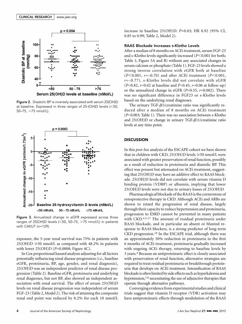
Lower 25-Hydroxyvitamin D Levels Are Associatedwith Higher Diastolic BPThere was an inverse association between the diastolic BPbefore the start of ACEi and baseline serum 25(OH)D levels(P=0.014, r=–0.19 and P=0.038, r=–0.16 for diastolic BP inmmHg and diastolic BP standard deviation score, respec-tively). Patients with 25(OH)D levels,50 nmol/L had higherdiastolic BP than those with levels $50 nmol/L (ANOVAP=0.004; Figure 2). The association between diastolic BPand 25(OH)D persisted even on ACEi treatment (P,0.004,r=–0.22). The systolic BP and 24-hour mean arterial BP atbaseline and follow-up did not show any association with25(OH)D levels at any time point.
2 Journal of the American Society of Nephrology J Am Soc Nephrol 27: ccc–ccc, 2015
CLINICAL RESEARCH www.jasn.org

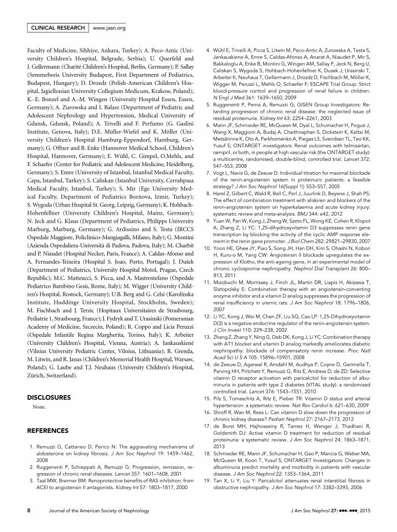
Lower 25-Hydroxyvitamin D Levels Are PositivelyAssociated with Loss of eGFR25(OH)D levels showed an association with eGFR at baseline(P=0.002; r=0.24) but not on ACEi treatment (P=0.16; Sup-plemental Figure 3, A and B). The median annualized loss ofeGFR was –1.74 (–4.1 to –0.25) ml/min per 1.73 m2 across thestudy cohort; children with glomerulopathies and other he-reditary nephropathies had a greater annualized decline ineGFR compared with those with CAKUT (P,0.06, ANOVA).
The annualized loss of eGFR was greater inpatients with baseline 25(OH)D levels,50 nmol/L compared with those with25(OH)D levels $50 nmol/L (P=0.0003,r=0.32; Figure 3). The association betweeneGFR loss and baseline 25(OH)D levels wasseen across all diagnostic groups, includingthose with CAKUT (P=0.0009, ANOVA).
The effect of 25(OH)D levels on a pre-determined composite end point of renalsurvival (defined as an annualized loss ofeGFR$50% or progression to ESRD(eGFR,10 ml/min per 1.73m2) or needfor renal replacement therapy) was exam-ined. Overall, 44 patients reached the com-
posite renal end point. The 5-year renal survival was 75% inpatients with baseline 25(OH)D $50 nmol/L compared with50% in patients with lower 25(OH)D (P,0.001; Figure 4A).After starting ACEi the beneficial effect of 25(OH)D on renaldisease progression was attenuated, but remained significant:the 5-year renal survival was 73% in patients with 25(OH)D$50 nmol/L as compared with 57% in patients with lower25(OH)D (P=0.046; Figure 4B). Using the mean 25(OH)D levelbetween baseline and follow-up as a measure of 25(OH)D
Figure 1. Correlation between 24-hour urinary PCR and serum 25(OH)D levels. (A)Baseline. (B) Follow-up. To convert mg/mg to mg/mmol, multiply by 113.
Table 1. Clinical and biochemical characteristics of the study population
Characteristics Baseline (n=167) Follow-up (n=167) P Valuea Median Change (IQR)b
Clinical featuresSystolic BPmmHg 115 (105–125) 105 (100–118) ,0.001 27 (–20–5)SDS 0.46 (–0.18–1.2) 20.23 (–0.86–0.47) ,0.001 20.67 (–1.64–0.18)Diastolic BPmmHg 70 (64–86) 64 (57–73) ,0.001 27 (–18–3)SDS 1.13 (0.44–1.86) 0.22 (–0.38–1.13) ,0.001 20.8 (–1.7–0.18)24-hour mean arterial pressuremmHg 87 (82–93) 80 (75.9–85) ,0.001 26 (–16–1)SDS 1.03 (0.29–2.1) 20.15 (–0.85–0.57) ,0.001 21.26 (–2.16 to –0.37)
Biochemical measuresEstimated GFR (ml/min per 1.73 m2) 50.9 (35.1–63.3) 45.8 (28.8–58.9) 0.08 21.74 (–4.1 to –0.25)Urine PCR (mg/mg) 0.74 (0.23–1.84) 0.39 (0.15–1.11) 0.003 20.77 (–1.9 to –0.2)25(OH)D (nmol/L) 72.3 (46–95.3) 69 (50.8–99.0) 0.98 2.4 (–24.7–23.3)Calcium (mmol/L) 2.4 (2.3–2.5) 2.4 (2.3–2.5) 0.91 0.0 (–0.01–0.15)Phosphate (mmol/L) 1.5 (1.3–1.6) 1.5 (1.3–1.7) 0.59 0 (-0.3–0.13)Parathyroid hormone (pmol/L) 5.2 (3.4–8.3) 8.0 (4.2–16.1) 0.04 2.2 (–1.4–4.9)FGF-23 (RU/ml) 182 (118–285) 308 (184–445) ,0.001 94 (–96–256)Soluble-Klotho (pg/mL) 379 (337–445) 537 (449–569) ,0.001 148 (55–243)VDBP (mmol/L) 6.8 (3.9–8.2) 7.2 (4.2–6.9) 0.75 3.9 (–1.7–4.6)
(n=107) (n=91)Urinary TGF-b1/creatinine ratio (ng/g) 25.8 (14.4–42.8) 13.5 (10.1–41.8) 0.003 27.3 (–4.7–11.1)
(n=142) (n=138)MedicationsCholecalciferol (n [%]) 10 [5.9] 12 [7.1] 0.98 —
Calcitriol (n [%]) 72 [43] 75 [44.9] 0.96 —
All values are described as median and interquartile range.aP values describe baseline versus follow-up levels using paired t test.bIQR, interquartile range.
J Am Soc Nephrol 27: ccc–ccc, 2015 Vitamin D and Childhood CKD Progression 3
www.jasn.org CLINICAL RESEARCH
exposure, the 5-year renal survival was 75% in patients with25(OH)D $50 nmol/L as compared with 48.2% in patientswith lower 25(OH)D (P=0.0008; Figure 4C).
In Cox proportional hazard analysis adjusting for all factorspotentially influencing renal disease progression (i.e., baselineeGFR, proteinuria, BP, age, gender, and renal diagnosis),25(OH)D was an independent predictor of renal disease pro-gression (Table 2). Baseline eGFR, proteinuria and underlyingrenal diagnoses, but not BP, also showed an independent as-sociation with renal survival. The effect of serum 25(OH)Dlevels on renal disease progression was independent of serumFGF-23 (Table 2,Model 2). The risk of attaining the compositerenal end point was reduced by 8.2% for each 10 nmol/L
increase in baseline 25(OH)D: P=0.03; HR 0.92 (95% CI,0.85 to 0.99; Table 2, Model 2).
RAAS Blockade Increases s-Klotho LevelsAfter amedian of 8months on ACEi treatment, serum FGF-23and s-Klotho levels significantly increased (P,0.001 for both;Table 1; Figure 5A and B) without any associated changes inserum calcium or phosphate (Table 1). FGF-23 levels showed astrong inverse correlation with eGFR both at baseline(P,0.001, r=–0.70) and after ACEi treatment (P,0.001,r=–0.77). s-Klotho levels did not correlate with eGFR(P=0.82, r=0.02 at baseline and P=0.45, r=0.06 at follow-up)or the annualized change in eGFR (P=0.55, r=0.002). Therewas no significant difference in FGF23 or s-Klotho levelsbased on the underlying renal diagnoses.
The urinary TGF-b1/creatinine ratio was significantly re-duced after a median of 8 months on ACEi treatment(P=0.003; Table 1). There was no association between s-Klothoand 25(OH)D or change in urinary TGF-b)1/creatinine ratiolevels at any time point.
DISCUSSION
In this post-hoc analysis of the ESCAPE cohort we have shownthat in children with CKD, 25(OH)D levels$50 nmol/L wereassociated with greater preservation of renal function, possiblyas a result of reduction in proteinuria and diastolic BP. Thiseffect was present but attenuated on ACEi treatment, suggest-ing that 25(OH)Dmay have an additive effect to RAAS block-ade. 25(OH)D levels did not correlate with serum vitamin Dbinding protein (VDBP) or albumin, implying that lower25(OH)D levels were not due to urinary losses of 25(OH)D.
Pharmacological blockade of theRAAS is the cornerstone ofrenoprotective therapy in CKD. Although ACEi and ARBs areshown to retard the progression of renal disease, largelythrough their capacity to reduce hypertension andproteinuria,progression to ESRD cannot be prevented in many patientswith CKD.4,5,17 The amount of residual proteinuria underRAAS blockade, and in particular an absent or blunted re-sponse to RAAS blockers, is a strong predictor of long-termCKD progression.18 In the ESCAPE trial, although there wasan approximately 50% reduction in proteinuria in the first6 months of ACEi treatment, proteinuria gradually increasedwith ongoing ACEi therapy, returning to baseline levels by3 years.4 Because an antiproteinuric effect is closely associatedwith preservation of renal function, alternative strategies arerequired to treat residual proteinuria or breakthrough protein-uria that develops on ACEi treatment. Intensification of RAASblockade is often limited by side effects such as hyperkalemia andhypotension,7,8 necessitating the use of adjunctive therapies thatoperate through alternative pathways.
Converging evidence fromexperimental studies and clinicaltrials suggest that vitamin D receptor (VDR) activation mayhave antiproteinuric effects through modulation of the RAAS
Figure 2. Diastolic BP is inversely associated with serum 25(OH)Dat baseline. Expressed in three ranges of 25-(OH)D levels (,50,50–75, .75 nmol/L).
Figure 3. Annualized change in eGFR expressed across threeranges of 25(OH)D levels (,50, 50–75, .75 nmol/L) in patientswith CAKUT (n=129).
4 Journal of the American Society of Nephrology J Am Soc Nephrol 27: ccc–ccc, 2015
CLINICAL RESEARCH www.jasn.org

system.11,13,19 Activation of the VDR can suppress the reningene by interaction with a major transcription factor bindingsite: vitamin D analogs bind to the VDR and blocks formationof the cyclic adenosine monophosphate response element-cAMP response element-binding protein complexes in thepromoter region of the renin gene,9 thereby reducing reninexpression. VDR null mice have increased renin gene expres-sion in their kidneys, accompanied by increased plasma an-giotensin II levels, hypertension, and cardiac hypertrophy.12
Conversely, when wild-type mice are treated with calcitriol,renal renin production was decreased.20
Clinical trials in adults with CKD have shown that vitaminD may augment RAAS blockade.14,21–25 In a meta-analysis ofsix studies using active vitamin D analogs, a significant re-duction in proteinuria was achieved in patients on active
vitamin D therapy (paricalcitol in fourstudies14,21,23,25 and calcitriol22,24 in two).This was an additive effect to ongoingRAAS blockade as 84% of patients receivedan ACEi or ARB for the duration of theirstudy. Both the number of patients whoachieved proteinuria reduction (odds ratio2.72, P,0.001) as well as the level of pro-teinuria reduction (mean difference –16%versus +6%; P,0.001) were greater withvitamin D analogs compared with con-trols.17 Importantly, a dose-dependent ef-fect of vitamin D on albuminuria was notconsistently observed in these trials,whereas the retrospective nature of ourstudy allowed us to determine a thresholdeffect of vitamin D treatment on renal sur-vival.
Nutritional vitaminDsupplements suchas cholecalciferol have a wide therapeuticwindow, and have been studied in onerandomized study: in 100 adults in pre-dialysis CKD who were followed up for 6
months, cholecalciferol treatment achieved mean 25(OH)Dlevels$60 nmol/L and reduced the urinary protein excretionby 53%.26 In our study, normal levels of 25(OH)D providedsimilar renoprotective benefits, and patients who receivedcalcitriol did not have any further reduction in their proteinuria,BP or change in eGFR compared with those who were not oncalcitriol. We do not have data on the dose of calcitriol pre-scribed, and due to a limited availability of serum, we wereunable to check 1,25(OH)2D levels. The absence of any effectof calcitriol treatment on proteinuria or eGFR may be due tovariable levels achieved, and possibly also the short half-life ofcalcitriol compared with cholecalciferol. Importantly, all ofthese studies have been conducted in adults, with 50–100%of study participants having diabetes mellitus.14,21–26 Thereare no studies in children, who are usually free of diabetes
Figure 4. 25(OH)D levels predict 5-year renal survival. (A) Baseline. (B) Follow-up. (C)Mean 25(OH)D levels predict renal survival.
Table 2. Cox proportional hazard analysis for renal survival (adjusting for all factors potentially influencing renal diseaseprogression)
Model 1 Model 2
ParameterParameterEstimate
HazardRatio
95% Hazard RatioConfidence Limits
Pr-ChiSq
ParameterEstimate
HazardRatio
95% Hazard RatioConfidence Limits
Pr-ChiSq
eGFR 20.06796 0.934 0.914 0.955 ,0.0001 20.05600 0.946 0.921 0.971 ,0.0001Urine PCR 0.55429 1.741 1.289 2.352 0.0003 0.62668 1.871 1.337 2.619 0.0003Mean arterial BP SDS 20.01346 0.987 0.817 1.192 0.8888 0.05218 1.054 0.862 1.287 0.609825(OH)D (per 10 nmol/L) 20.07940 0.924 0.862 0.990 0.0254 20.08562 0.918 0.849 0.993 0.0324Renal diagnosis (non-CAKUT) 21.46553 0.231 0.123 0.433 ,0.0001 21.59705 0.202 0.100 0.409 ,0.0001Male gender 20.59751 0.550 0.300 1.008 0.0530 20.58872 0.555 0.286 1.077 0.0817Age (year) 0.06019 1.062 0.986 1.143 0.1101 0.08872 1.093 1.009 1.184 0.0296FGF-23 (10 RU/ml) 0.0304 1.031 1.005 1.057 0.0185
Pr-ChiSq, probability by chi-squared test.
J Am Soc Nephrol 27: ccc–ccc, 2015 Vitamin D and Childhood CKD Progression 5
www.jasn.org CLINICAL RESEARCH

and in whom the underlying renal disease is rarely proteinuricrenal failure. Thus, clinical trials with cholecalciferol, a safeand effective vitamin D supplement with minimal need formonitoring, are recommended in children with CKD. How-ever, care must be taken in using a safe and effective vitamin Ddosing schedule because a recent study has suggested that ahigh loading dose of ergocalciferol can lead to high FGF-23levels.27 Also, long-term studies are needed to determine ifthere is a breakthrough from the antiproteinuric effect of vi-tamin D, as seen with RAAS blockade.4
We found an association between mean diastolic BP andserum 25(OH)D levels at baseline, but no correlation withsystolic BP. Recent studies have shown an association betweenBP circulating levels of 25(OH)D28 as well as with geneticvariations in CYP1A1 and CYP1B1.29 While both studieshave shown an association of 25(OH)D with systolic and di-astolic BP, a stronger correlation was seen between 25(OH)Dand diastolic BP. These findings need to be further exploredin a prospective longitudinal study.
We founda significant increase in s-Klotho levels afterRAASblockade.CKD is known to be a state ofKlothodeficiency, evenin children with CKD.30 In animal models of kidney disease,angiotensin II decreases renal Klotho expression and thisdownregulation is prevented by RAAS blockade.10,31,32
S-klotho is an anti-aging phosphaturic protein that is shownto confer cardiorenal protection in different experimentalmodels of metabolic and kidney diseases by enhancing anti-oxidant, antisenescence, and antiapoptotic mechanisms.33,34
In a recent study, serum s-Klotho levels were inversely associ-ated with proteinuria in adults with CKD stages 1–2,35
suggesting a possible association between proteinuria-inducedinterstitial inflammation and downregulation of Klotho syn-thesis. There are few clinical studies, but in adults with diabetickidney disease treatment with valsartanwas associated with anincrease in s-Klotho, although this did not associate with areduction in albuminuria.36 Cross-talk between the vitaminD and RAAS pathways may also confer additional anti-inflammatory effects of vitamin D therapy to RAAS block-ade.16 In animal models, VDR activation is associated with
inhibition of TGF-b11–13,19 and reduced expression of IL-6and IL-8 in podocytes and tubular cells, suggesting reducedintrarenal inflammation and fibrosis.37,38 However, in thisretrospective study we were not able to find an associationbetween urinary TGF-b1 expression and Klotho levels at base-line or after RAAS blockade.
The retrospective nature of our study is a clear limitation;however, this novel association between 25(OH)D levels andproteinuria as well as preservation of eGFR in childhood CKDgenerates hypotheses for future randomized controlled studiesof the renoprotective effects of vitamin D supplementation.While serum 25(OH)D levels are lower in patients with greaterproteinuria due to urinary loss of VDBP in patients withnephrotic range proteinuria, we39 and others40 have shownthat urinary VDBP loss is not associated with plasma VDBPor 25(OH)D levels in children and adults with chronic kidneydisease, where urinary loss of VDBP is not sufficient to affectvitamin D status. Although we were not able to comment onthe antiproteinuric and renoprotective effect of different vita-min D analogs, patients whowere on calcitriol (approximately45%) did not have less proteinuria or higher eGFRs as com-pared with those who did not receive any vitamin D analog.This suggests a possible threshold effect of VDR stimulation,but requires further prospective studies comparing the effectsof colecalciferol and active vitamin D analogs on renal pres-ervation. The soluble Klotho assay used in our study measuresthe larger 130 kD cleaved protein. A smaller fragment of 68–70kD, as a result of alternative mRNA splicing and currently ofunknown significance, may be present in the circulation, but isnot detected by this assay.
In conclusion, in children with CKD, 25(OH)D levels.50 nmol/L were associated with better preservation of re-nal function, even in the presence of concomitant ACEi ther-apy. Vitamin D is an effective, easily available, safe, andcheap nutritional supplement that may be a useful adjunc-tive treatment to RAAS blockade to retard progressive renalfunction decline. Randomized controlled studies on the re-noprotective effects of vitamin D in childhood CKD arerequired.
Figure 5. (A) Serum FGF-23 levels at baseline and after ACEi treatment. (B) Soluble-Klotho levels at baseline and after ACEi treatment.
6 Journal of the American Society of Nephrology J Am Soc Nephrol 27: ccc–ccc, 2015
CLINICAL RESEARCH www.jasn.org

CONCISE METHODS
Study PopulationThis study is a post-hoc analysis of the ESCAPE trial, a randomized
controlled study showing that strict BP control with a fixed dose of
ACE inhibition slows the progression of renal disease. Briefly, the
ESCAPE trial included 468 children from 33 European centers of
age 3–18 years with an eGFR of 15–80 ml/min per 1.73 m2 with
hypertensionwho received a fixed dose of the ACEi ramipril (6 mg/m2 per day) and were randomly assigned to either a conventional BP
target (50th to 90th percentile of 24-hour mean arterial BP) or an
intensified BP target (below the 50th percentile). Children were in-
cluded in this study based on the availability of paired blood samples
at baseline and after a follow-up period of at least 6 months. All
measures were taken at baseline (prior to ACEi treatment or after a
wash-out phase of 4 (2–4)months in those whowere previously on an
ACEi) and after a median follow-up of 8 (8–10) months on ACEi
therapy.
Outcome MeasuresThe effect of 25(OH)D levels on change in 24-hour urinary protein
excretion, BP, eGFR and renal survival (defined as a predetermined
composite endpoint of annualized loss of eGFR. 50%orprogression
to ESRD (eGFR,10 ml/min per 1.73 m2) or need for renal replace-
ment therapy) were studied. Because an acute decrease in eGFR
(,25% decrease) is expected after the start of ACEi therapy, the
eGFR recorded 2 months after the initiation of ramipril was used
as a baseline for the analysis of the reduction in eGFR over time.
In order to examine a potential mechanism of the 25(OH)D effect
on reduction of proteinuria, FGF23, s-Klotho and TGF-b1 were
measured in a subgroup of patients (based on availability of serum
samplea) at baseline and follow-up. To exclude a confounding effect
of VDBP loss on serum 25(OH)D levels, VDBP levels were measured
in a subgroup of children, based on availability of serum samples. The
effect of serum 25(OH)D levels on mineral dysregulation (serum
calcium, phosphate, parathyroid hormone, FGF23 and s-Klotho)
was examined.
Blood Pressure MonitoringAmbulatory BP monitoring was performed using Spacelabs 90207
oscillometric devices (Spacelabs Healthcare, Snoqualmie, WA) at
baseline and every 6 months throughout the study period. All BP
readings were normalized to standard deviation scores using Euro-
pean reference data sets.41
Laboratory AssessmentsAll biochemical measurements were made in a central laboratory at
baseline and final follow-up. Measurements of serum and urine
creatinine levels and urine protein concentration were performed as
part of the ESCAPE trial as previously described.4 25(OH)D was
analyzed by isotope-dilution liquid chromatography-tandem mass
spectrometry [expressed as a sum of 25(OH)D2 and 25(OH)D3].
The interassay coefficient of variation was 2.8%. Plasma FGF-23 con-
centrations were determined using a second-generation human FGF-23
(C-terminal) ELISA (Immutopics International, San Clemente, CA).
The intra- and interassay coefficients of variation were 3.8% and
6.3%, respectively. s-Klotho concentrations were measured by a solid-
phase sandwich ELISA (Immuno-Biologic Laboratories Co. Ltd.,
Gunma, Japan). The intra- and interassay coefficients of variation
were 2.4% and 6.2%, respectively. VDBP assay was performed using a
noncompetitive (sandwich) ELISA (K2314, Immun Diagnostik, Ger-
many). The intra- and interassay coefficients of variability were 4.4%
and 6.0%, respectively. Urinary excretion of TGF-b1 was assayed using
ELISA (DRG Instruments GmbH, Marburg, Germany) as previously
described.42
Statistical AnalysisResults are expressed as median and interquartile range (IQR) unless
otherwise stated. Univariate comparisons of continuous variables
between the groups were performed using an unpaired t test for nor-
mally distributed data, or the nonparametric Mann–Whitney U or
Kruskal–Wallis test for non-normally distributed variables. Compar-
isons of continuous variables between baseline and final follow-up
were performed using a paired t test or the non-parametric Wilcoxon
test as appropriate. For multiple comparisons of several groups, re-
peated measures ANOVA with Bonferroni correction or Kruskal–
Wallis test was performed as appropriate. Spearman correlation tests
were used for correlation analyses. The time to development of the
composite end point was determined by Kaplan–Meier analysis, with
the use of log-rank statistics to test for differences in the rates of the
end points and by Cox proportional-hazard modeling to assess the
effects of potential risk factors.
In 159 children in whom full data were available, Cox hazard
analysis was performed to include variables that are well known to
predict renal disease progression or influence 25(OH)D levels pro-
vided that they were significant on univariate analysis at P,0.15. BMI
Standard deviation core did not show any correlation with 25(OH)D
levels (r=0.02, P=0.79) nor annualized renal disease progression
(r=–0.04, P=0.57) on univariate analysis and was not included in
the Cox model. Although, as expected, the season of blood sampling
influenced 25(OH)D levels (Supplemental Figure 3), the influence
of a constantly changing process such as season per se on a long-term
outcome of renal progression is difficult to justify and season was ex-
cluded from the model. In a second model we also included FGF-23
(Table 2, Model 2), but complete data were available for only 139 pa-
tients. Klotho, calcium andphosphatewere not significant onunivariate
analysis and were excluded from the Cox regression analysis in order to
limit the number of variables and avoid potential overadjustment.
All statistical analyses were performed using SAS 9.3 (SAS Institute
Inc., Cary, NC). For all analyses, P,0.05 was considered to be statis-
tically significant.
ACKNOWLEDGMENTS
The following are members of the ESCAPE Study Group – Local
investigators (in alphabetical order of center):
A. Anarat (Cukurova University School of Medicine, Balcali,
Adana, Turkey); A. Bakkaloglu and F. Ozaltin (Hacettepe University
J Am Soc Nephrol 27: ccc–ccc, 2015 Vitamin D and Childhood CKD Progression 7
www.jasn.org CLINICAL RESEARCH
Faculty of Medicine, Sihhiye, Ankara, Turkey); A. Peco-Antic (Uni-
versity Children’s Hospital, Belgrade, Serbia); U. Querfeld and
J. Gellermann (Charité Children’s Hospital, Berlin, Germany); P. Sallay
(Semmelweis University Budapest, First Department of Pediatrics,
Budapest, Hungary); D. Drozdz (Polish-American Children’s Hos-
pital, Jagiellonian University Collegium Medicum, Krakow, Poland);
K.-E. Bonzel and A.-M. Wingen (University Hospital Essen, Essen,
Germany); A. Zurowska and I. Balasz (Department of Pediatric and
Adolescent Nephrology and Hypertension, Medical University of
Gdansk, Gdansk, Poland); A. Trivelli and F. Perfumo (G. Gaslini
Institute, Genova, Italy); D.E. Müller-Wiefel and K. Möller (Uni-
versity Children’s Hospital Hamburg-Eppendorf, Hamburg, Ger-
many); G. Offner and B. Enke (Hannover Medical School, Children’s
Hospital, Hannover, Germany); E. Wühl, C. Gimpel, O.Mehls, and
F. Schaefer (Center for Pediatric and Adolescent Medicine, Heidelberg,
Germany); S. Emre (University of Istanbul, Istanbul Medical Faculty,
Capa, Istanbul, Turkey); S. Caliskan (Istanbul University, Cerrahpasa
Medical Faculty, Istanbul, Turkey); S. Mir (Ege University Med-
ical Faculty, Department of Pediatrics Bornova, Izmir, Turkey);
S.Wygoda (UrbanHospital St. Georg, Leipzig, Germany); K. Hohbach-
Hohenfellner (University Children’s Hospital, Mainz, Germany);
N. Jeck and G. Klaus (Department of Pediatrics, Philipps University
Marburg, Marburg, Germany); G. Ardissino and S. Testa (IRCCS
Ospedale Maggiore, Policlinico-Mangiagalli, Milano, Italy); G. Montini
(Azienda Ospedaliera-Università di Padova, Padova, Italy); M. Charbit
and P. Niaudet (Hospital Necker, Paris, France); A. Caldas-Afonso and
A. Fernandes-Teixeira (Hospital S. Joao, Porto, Portugal); J. Dušek
(Department of Pediatrics, University Hospital Motol, Prague, Czech
Republic); M.C. Matteucci, S. Picca, and A. Mastrostefano (Ospedale
Pediatrico Bambino Gesù, Rome, Italy); M. Wigger (University Child-
ren’s Hospital, Rostock, Germany); U.B. Berg and G. Celsi (Karolinska
Institute, Huddinge University Hospital, Stockholm, Sweden);
M. Fischbach and J. Terzic (Hopitaux Universitaires de Strasbourg,
Pediatrie 1, Strasbourg, France); J. Fydryk andT.Urasinski (Pomeranian
Academy of Medicine, Szczecin, Poland); R. Coppo and Licia Peruzzi
(Ospedale Infantile Regina Margherita, Torino, Italy); K. Arbeiter
(University Children’s Hospital, Vienna, Austria); A. Jankauskiené
(Vilnius University Pediatric Center, Vilnius, Lithuania); R. Grenda,
M. Litwin, and R. Janas (Children’sMemorial Health Hospital, Warsaw,
Poland); G. Laube and T.J. Neuhaus (University Children’s Hospital,
Zürich, Switzerland).
DISCLOSURESNone.
REFERENCES
1. Remuzzi G, Cattaneo D, Perico N: The aggravating mechanisms ofaldosterone on kidney fibrosis. J Am Soc Nephrol 19: 1459–1462,2008
2. Ruggenenti P, Schieppati A, Remuzzi G: Progression, remission, re-gression of chronic renal diseases. Lancet 357: 1601–1608, 2001
3. Taal MW, Brenner BM: Renoprotective benefits of RAS inhibition: fromACEI to angiotensin II antagonists. Kidney Int 57: 1803–1817, 2000
4. Wühl E, Trivelli A, Picca S, Litwin M, Peco-Antic A, Zurowska A, Testa S,Jankauskiene A, Emre S, Caldas-Afonso A, Anarat A, Niaudet P, Mir S,Bakkaloglu A, Enke B, Montini G, Wingen AM, Sallay P, Jeck N, Berg U,Caliskan S, Wygoda S, Hohbach-Hohenfellner K, Dusek J, Urasinski T,Arbeiter K, Neuhaus T, Gellermann J, Drozdz D, FischbachM,Möller K,Wigger M, Peruzzi L, Mehls O, Schaefer F; ESCAPE Trial Group: Strictblood-pressure control and progression of renal failure in children.N Engl J Med 361: 1639–1650, 2009
5. Ruggenenti P, Perna A, Remuzzi G; GISEN Group Investigators: Re-tarding progression of chronic renal disease: the neglected issue ofresidual proteinuria. Kidney Int 63: 2254–2261, 2003
6. Mann JF, Schmieder RE, McQueen M, Dyal L, Schumacher H, Pogue J,Wang X, Maggioni A, Budaj A, Chaithiraphan S, Dickstein K, Keltai M,Metsärinne K, Oto A, Parkhomenko A, Piegas LS, Svendsen TL, Teo KK,Yusuf S; ONTARGET investigators: Renal outcomes with telmisartan,ramipril, or both, in people at high vascular risk (theONTARGET study):a multicentre, randomised, double-blind, controlled trial. Lancet 372:547–553, 2008
7. Vogt L, Navis G, de Zeeuw D: Individual titration for maximal blockadeof the renin-angiotensin system in proteinuric patients: a feasiblestrategy? J Am Soc Nephrol 16[Suppl 1]: S53–S57, 2005
8. Harel Z, Gilbert C, Wald R, Bell C, Perl J, Juurlink D, Beyene J, Shah PS:The effect of combination treatment with aliskiren and blockers of therenin-angiotensin system on hyperkalaemia and acute kidney injury:systematic review and meta-analysis. BMJ 344: e42, 2012
9. YuanW, PanW, Kong J, ZhengW, Szeto FL,Wong KE, Cohen R, KlopotA, Zhang Z, Li YC: 1,25-dihydroxyvitamin D3 suppresses renin genetranscription by blocking the activity of the cyclic AMP response ele-ment in the renin gene promoter. J Biol Chem 282: 29821–29830, 2007
10. Yoon HE, Ghee JY, Piao S, Song JH, Han DH, Kim S, Ohashi N, KoboriH, Kuro-o M, Yang CW: Angiotensin II blockade upregulates the ex-pression of Klotho, the anti-ageing gene, in an experimental model ofchronic cyclosporine nephropathy. Nephrol Dial Transplant 26: 800–813, 2011
11. Mizobuchi M, Morrissey J, Finch JL, Martin DR, Liapis H, Akizawa T,Slatopolsky E: Combination therapy with an angiotensin-convertingenzyme inhibitor and a vitamin D analog suppresses the progression ofrenal insufficiency in uremic rats. J Am Soc Nephrol 18: 1796–1806,2007
12. Li YC, Kong J, Wei M, Chen ZF, Liu SQ, Cao LP: 1,25-DihydroxyvitaminD(3) is a negative endocrine regulator of the renin-angiotensin system.J Clin Invest 110: 229–238, 2002
13. Zhang Z, ZhangY, NingG,DebDK, Kong J, Li YC: Combination therapywith AT1 blocker and vitamin D analog markedly ameliorates diabeticnephropathy: blockade of compensatory renin increase. Proc NatlAcad Sci U S A 105: 15896–15901, 2008
14. de Zeeuw D, Agarwal R, Amdahl M, Audhya P, Coyne D, Garimella T,Parving HH, Pritchett Y, Remuzzi G, Ritz E, Andress D; de ZD: Selectivevitamin D receptor activation with paricalcitol for reduction of albu-minuria in patients with type 2 diabetes (VITAL study): a randomisedcontrolled trial. Lancet 376: 1543–1551, 2010
15. Pilz S, Tomaschitz A, Ritz E, Pieber TR: Vitamin D status and arterialhypertension: a systematic review. Nat Rev Cardiol 6: 621–630, 2009
16. Shroff R, Wan M, Rees L: Can vitamin D slow down the progression ofchronic kidney disease? Pediatr Nephrol 27: 2167–2173, 2012
17. de Borst MH, Hajhosseiny R, Tamez H, Wenger J, Thadhani R,Goldsmith DJ: Active vitamin D treatment for reduction of residualproteinuria: a systematic review. J Am Soc Nephrol 24: 1863–1871,2013
18. Schmieder RE, Mann JF, Schumacher H, Gao P, Mancia G, Weber MA,McQueen M, Koon T, Yusuf S; ONTARGET Investigators: Changes inalbuminuria predict mortality and morbidity in patients with vasculardisease. J Am Soc Nephrol 22: 1353–1364, 2011
19. Tan X, Li Y, Liu Y: Paricalcitol attenuates renal interstitial fibrosis inobstructive nephropathy. J Am Soc Nephrol 17: 3382–3393, 2006
8 Journal of the American Society of Nephrology J Am Soc Nephrol 27: ccc–ccc, 2015
CLINICAL RESEARCH www.jasn.org

20. Kong J, Qiao G, Zhang Z, Liu SQ, Li YC: Targeted vitamin D receptor ex-pression in juxtaglomerular cells suppresses renin expression independentof parathyroid hormone and calcium. Kidney Int 74: 1577–1581, 2008
21. Agarwal R, Acharya M, Tian J, Hippensteel RL, Melnick JZ, Qiu P,Williams L, Batlle D: Antiproteinuric effect of oral paricalcitol in chronickidney disease. Kidney Int 68: 2823–2828, 2005
22. Krairittichai U, Mahannopkul R, Bunnag S: An open label, randomizedcontrolled study of oral calcitriol for the treatment of proteinuria inpatients with diabetic kidney disease. J Med Assoc Thai 95[Suppl 3]:S41–S47, 2012
23. Fishbane S, Chittineni H, Packman M, Dutka P, Ali N, Durie N: Oralparicalcitol in the treatment of patients with CKD and proteinuria:a randomized trial. Am J Kidney Dis 54: 647–652, 2009
24. Liu LJ, Lv JC, Shi SF, Chen YQ, Zhang H, Wang HY: Oral calcitriol forreduction of proteinuria in patients with IgA nephropathy: a random-ized controlled trial. Am J Kidney Dis 59: 67–74, 2012
25. Thadhani R, Appelbaum E, Pritchett Y, Chang Y, Wenger J, Tamez H,Bhan I, Agarwal R, Zoccali C, Wanner C, Lloyd-Jones D, Cannata J,Thompson BT, Andress D, Zhang W, Packham D, Singh B, Zehnder D,Shah A, Pachika A, Manning WJ, Solomon SD: Vitamin D therapy andcardiac structure and function in patients with chronic kidney disease:the PRIMO randomized controlled trial. JAMA 307: 674–684, 2012
26. Molina P, Górriz JL,MolinaMD, Peris A, Beltrán S, Kanter J, Escudero V,Romero R, Pallardó LM: The effect of cholecalciferol for lowering al-buminuria in chronic kidney disease: a prospective controlled study.Nephrol Dial Transplant 29: 97–109, 2014
27. Turner C, Dalton N, Inaoui R, Fogelman I, Fraser WD, Hampson G: Effectof a 300 000-IU loading dose of ergocalciferol (Vitamin D2) on circulating1,25(OH)2-vitaminDandfibroblast growth factor-23 (FGF-23) in vitaminDinsufficiency. J Clin Endocrinol Metab 98: 550–556, 2013
28. Vimaleswaran KS, CavadinoA, Berry DJ, Jorde R,DieffenbachAK, LuC,Alves AC, Heerspink HJ, Tikkanen E, Eriksson J, Wong A, Mangino M,Jablonski KA, Nolte IM, Houston DK, Ahluwalia TS, van der Most PJ,Pasko D, Zgaga L, Thiering E, Vitart V, Fraser RM, Huffman JE, de BoerRA, Schöttker B, SaumKU,McCarthyMI, Dupuis J, Herzig KH, Sebert S,Pouta A, Laitinen J, Kleber ME, Navis G, Lorentzon M, Jameson K,Arden N, Cooper JA, Acharya J, Hardy R, Raitakari O, Ripatti S, BillingsLK, Lahti J, Osmond C, Penninx BW, Rejnmark L, Lohman KK,Paternoster L, Stolk RP, Hernandez DG, Byberg L, Hagström E, MelhusH, Ingelsson E, Mellström D, Ljunggren O, Tzoulaki I, McLachlan S,Theodoratou E, Tiesler CM, Jula A, Navarro P, Wright AF, Polasek O,Wilson JF, Rudan I, Salomaa V, Heinrich J, Campbell H, Price JF,Karlsson M, Lind L, Michaëlsson K, Bandinelli S, Frayling TM, HartmanCA, Sørensen TI, Kritchevsky SB, Langdahl BL, Eriksson JG, Florez JC,Spector TD, Lehtimäki T, Kuh D, Humphries SE, Cooper C, Ohlsson C,März W, de Borst MH, Kumari M, Kivimaki M, Wang TJ, Power C,Brenner H, Grimnes G, van der Harst P, Snieder H, Hingorani AD, Pilz S,Whittaker JC, Järvelin MR, Hyppönen E; LifeLines Cohort Study in-vestigatorsInternational Consortium for Blood Pressure (ICBP)Cohortsfor Heart and Aging Research in Genomic Epidemiology (CHARGE)consortiumGlobal Blood Pressure Genetics (Global BPGen) con-sortiumCaroline Hayward: Association of vitamin D status with arterialblood pressure and hypertension risk: a mendelian randomisationstudy. Lancet Diabetes Endocrinol 2: 719–729, 2014
29. Park HY, Kim JH, Bae S, Choi YY, Park JY, Hong YC: Interaction effect ofserum 25-hydroxyvitamin D levels and CYP1A1, CYP1B1 polymorphisms
on blood pressure in an elderly population. J Hypertens 33: 69–76,2015
30. Wan M, Smith C, Shah V, Gullet A, Wells D, Rees L, Shroff R: Fibroblastgrowth factor 23 and soluble klotho in children with chronic kidneydisease. Nephrol Dial Transplant 28: 153–161, 2013
31. Mitani H, Ishizaka N, Aizawa T, OhnoM, Usui S, Suzuki T, Amaki T, MoriI, Nakamura Y, Sato M, Nangaku M, Hirata Y, Nagai R: In vivo klothogene transfer ameliorates angiotensin II-induced renal damage. Hy-pertension 39: 838–843, 2002
32. Zhou Q, Lin S, Tang R, Veeraragoo P, PengW, Wu R: Role of Fosinopriland Valsartan onKlothoGeneExpression InducedbyAngiotensin II in RatRenal Tubular Epithelial Cells.Kidney Blood Press Res 33: 186–192, 2010
33. Kuro-o M: Klotho in health and disease. Curr Opin Nephrol Hypertens21: 362–368, 2012
34. Shroff R, Shanahan CM: Klotho: an elixir of youth for the vasculature?J Am Soc Nephrol 22: 5–7, 2011
35. Hage V, Pelletier S, Dubourg L, Drai J, Cuerq C, Lemoine S, Hadj-AissaA, Laville M, FouqueD: In Chronic Kidney Disease, Serum alpha-Klothois Related to Serum Bicarbonate and Proteinuria, J Ren Nutr, 24: 390–394, 2014
36. Karalliedde J, Maltese G, Hill B, Viberti G, Gnudi L: Effect of renin-an-giotensin system blockade on soluble Klotho in patients with type 2diabetes, systolic hypertension, and albuminuria. Clin J Am SocNephrol 8: 1899–1905, 2013
37. Sanchez-Niño MD, Bozic M, Córdoba-Lanús E, Valcheva P, Gracia O,Ibarz M, Fernandez E, Navarro-Gonzalez JF, Ortiz A, Valdivielso JM:Beyond proteinuria: VDR activation reduces renal inflammation in ex-perimental diabetic nephropathy. Am J Physiol Renal Physiol 302:F647–F657, 2012
38. Isakova T, Gutiérrez OM, Patel NM, Andress DL, Wolf M, Levin A: Vi-tamin D deficiency, inflammation, and albuminuria in chronic kidneydisease: complex interactions. J Ren Nutr 21: 295–302, 2011
39. Prytuła A, Wells D, McLean T, Balona F, Gullett A, Knott C, Cantwell M,Hassen K, Ledermann S, Rees L, Shroff R: Urinary and dialysate losses ofvitamin D-binding protein in children on chronic peritoneal dialysis.Pediatr Nephrol 27: 643–649, 2012
40. Doorenbos CR, de Cuba MM, Vogt L, Kema IP, van den Born J, GansRO, Navis G, de Borst MH: Antiproteinuric treatment reduces urinaryloss of vitamin D-binding protein but does not affect vitamin D status inpatients with chronic kidney disease. J Steroid Biochem Mol Biol 128:56–61, 2012
41. Wühl E, Witte K, Soergel M, Mehls O, Schaefer F; German WorkingGroup on Pediatric Hypertension: Distribution of 24-h ambulatoryblood pressure in children: normalized reference values and role ofbody dimensions. J Hypertens 20: 1995–2007, 2002
42. Grenda R, Wühl E, Litwin M, Janas R, Sladowska J, Arbeiter K, Berg U,Caldas-Afonso A, Fischbach M, Mehls O, Sallay P, Schaefer F; ESCAPETrial group: Urinary excretion of endothelin-1 (ET-1), transforminggrowth factor-beta1 (TGF-beta1) and vascular endothelial growth fac-tor (VEGF165) in paediatric chronic kidney diseases: results of theESCAPE trial. Nephrol Dial Transplant 22: 3487–3494, 2007
This article contains supplemental material online at http://jasn.asnjournals.org/lookup/suppl/doi:10.1681/ASN.2014090947/-/DCSupplemental.
J Am Soc Nephrol 27: ccc–ccc, 2015 Vitamin D and Childhood CKD Progression 9
www.jasn.org CLINICAL RESEARCH