D imaging - Digital Dental Lateral-PA TMJ program captures lateral and PA views on the same...
15
2 D imaging ENGLISH
Transcript of D imaging - Digital Dental Lateral-PA TMJ program captures lateral and PA views on the same...

2D imaging
ENGLISH

WelcomeAn introduction from our President
“It’s my great pleasure to introduce you to our pioneering 2D X-ray units. Our comprehensive range of digital units meets all your daily imaging needs – working perfectly with our highly advanced Planmeca Romexis® software for the most detailed extraoral and intraoral examinations possible.
I’m extremely proud of our product innovations, and for over 40 years we’ve worked closely with dental professionals to set new standards in our field. What makes us a bit different is that all core product development and manufacturing takes place in Finland – ensuring exceptional quality and unmatched attention to detail at every stage of the process.
And we also have a dedicated team of R&D professionals behind the scenes, developing breakthrough innovations that make a real difference. Our robotic SCARA technology, for example, offers flexible, precise and complex movements needed for extraoral maxillofacial imaging. Our Planmeca ProMax® 2D X-ray units are all 3D-ready, which means you can easily upgrade at a later point. Therefore I’m thrilled to invite you to discover our world of 2D imaging.”
Heikki Kyöstilä, President Planmeca Group
Industry-leading 2D X-ray units ....................................................................... 4
A new benchmark for extraoral imaging ....................................................... 6
Planmeca ProMax® 2D ................................................................................... 8Perfect panoramic images – every time ...................................10 Effortless and comfortable ........................................................... 12Robotic arm technology ................................................................. 14All the imaging programs you need ..........................................16Extraoral bitewings ......................................................................... 18New opportunities for tomography .........................................20Quality cephalometry for orthodontics ...................................22Easy upgrade from 2D to 3D ........................................................ 24
Planmeca ProOne® ........................................................................................ 26Optimal imaging programs ......................................................... 28
Intraoral imaging .............................................................................................. 30
Planmeca ProX™ ..............................................................................................32
Planmeca ProSensor® ................................................................................... 34
Planmeca Romexis® software for all images ............................................... 36
High-performance 2D imaging ................................................................. 38
Your mobile world of imaging ...................................................................40
Share images and expertise online .......................................................... 42
Technical specifications ................................................................................... 44
2 3

09
09
Planmeca ProMax®Planmeca ProSensor®
Planmeca ProOne®
Planmeca ProX™
Industry-leading 2D X-ray units Mac OS and Windows
compatibleIntroducing our world-class range of 2D X-ray units – offering the most advanced and versatile devices and software to meet all your 2D extraoral and intraoral imaging needs.
Planmeca ProMax®Planmeca ProSensor®
Planmeca ProOne®
Planmeca ProX™
4 5

Planmeca extraoral units offer two alternative solutions to maxillofacial imaging. Planmeca ProMax® – the complete imaging centre – sets a new benchmark in panoramic and cephalometric imaging. Planmeca ProOne® is designed with simplicity in mind. It is a compact and easy-to-use panoramic X-ray unit that’s both cost-effective and flexible.
A new benchmark for extraoral imaging
6 7

Extraoral imaging
Planmeca ProMax® 2DPlanmeca ProMax® is a complete maxillofacial imaging system. The design and operation principles are based on the latest scientific research, technological innovations and the most demanding needs of modern-day radiology.
Key features:Advanced technology• Autofocus positions the focal layer automatically for perfect panoramic images
• Dynamic Exposure Control (DEC) measures the patient’s radiation transparency and automatically adjusts exposure values
• Patented SCARA (Selectively Compliant Articulated Robot Arm) technology guarantees an anatomically correct imaging geometry for clear, error-free images
• Easy upgrades – add cephalostat or 3D imaging capability at any time
Effortless use• Full-view patient positioning with triple-laser patient positioning lights
• Side entry for comfortable access
• Easy-to-use graphical interface
• Versatile Planmeca Romexis® 2D imaging software
• TWAIN support and full DICOM compliance
98
11
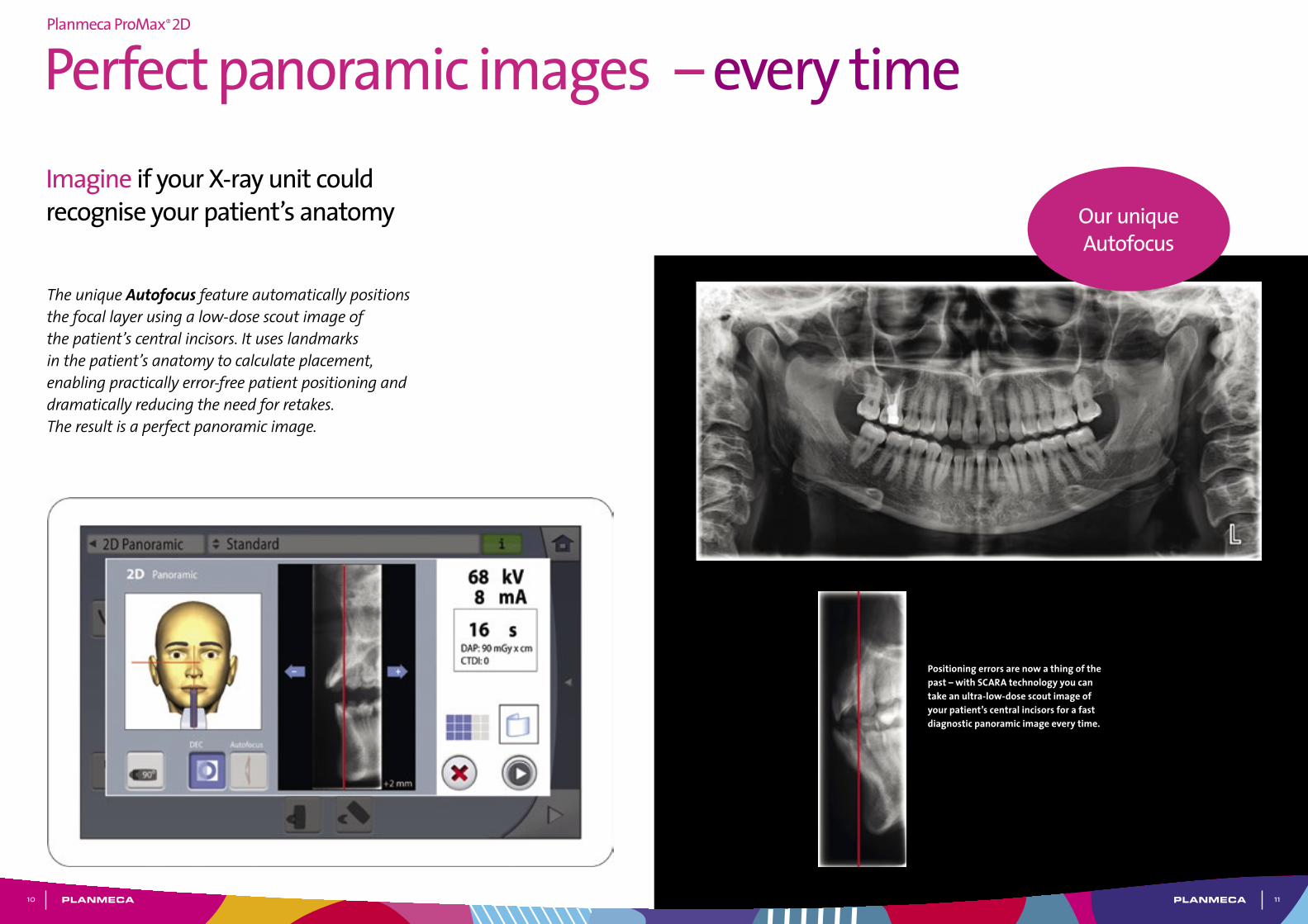
The unique Autofocus feature automatically positions the focal layer using a low-dose scout image of the patient’s central incisors. It uses landmarks in the patient’s anatomy to calculate placement, enabling practically error-free patient positioning and dramatically reducing the need for retakes. The result is a perfect panoramic image.
Positioning errors are now a thing of the past – with SCARA technology you can take an ultra-low-dose scout image of your patient’s central incisors for a fast diagnostic panoramic image every time.
Imagine if your X-ray unit could recognise your patient’s anatomy Our unique
Autofocus
Perfect panoramic images – every timePlanmeca ProMax® 2D
10

13
Laser-assisted patient alignment• A triple laser beam system accurately indicates the correct
anatomical alignment points for patient positioning
• The midsagittal plane positioning beam indicates the correct sideways alignment
• The Frankfort horizontal plane positioning beam shows the correct forward tilt of your patient’s head
• The focal layer positioning beam indicates the focal layer position and ensures images are sharp and clear
• Fine adjustments can be made using the joystick
Improved image quality with Dynamic Exposure Control (DEC)The unique digital Dynamic Exposure Control (DEC) automatically adjusts the exposure values for each individual patient based on their anatomic structure and bone density. DEC improves the quality of both panoramic and cephalometric imaging with more consistent brightness and contrast.
Adjustable focal layerDeveloped based on scientific research, the imaging geometry matches the shape of the focal layer with the patient’s anatomy, resulting in clear panoramic radiographs. Simply select the shape of the focal layer on the graphical user interface, according to the size and shape of the patient’s jaw.
Effortless and comfortableOur industry-leading Planmeca ProMax® unit is known across the world for incredible ease of use and exceptional patient comfort. A relaxed patient means a smooth imaging workflow and the best possible image quality.
Open patient positioning• Position patients effortlessly thanks
to open-face architecture
• Correct patient positioning either with Autofocus or manually
• Make fine adjustments using positioning lasers and joystick
• Work with an unrestricted view of your patient
• Avoid claustrophobic feelings in patients
• Accommodate wheelchairs easily with side-entry access
User-friendly control panel• Clear and straightforward graphical user
interface guides you smoothly through your work
• Pre-programmed sites and exposure values for different image types and targets save you time and allow you to focus on your patients
Planmeca ProMax® 2D
12

15
Planmeca ProMax® features highly advanced and exclusive robotic SCARA (Selectively Compliant Articulated Robot Arm) technology – providing flexible, precise and complex movements required for rotational maxillofacial imaging.
Imaging programsPlanmeca ProMax 2D S3 Planmeca ProMax 2D S2
Basic panoramic programs Standard panoramic
Lateral TMJ (closed & open)
PA TMJ (closed & open)
PA sinus
Standard panoramic
Lateral TMJ (closed & open)
PA TMJ (closed & open)
PA sinus
Horizontal and vertical segmenting for panoramic program
Horizontal and vertical segmenting for panoramic program
True Bitewing Bitewing
Advanced panoramic programs
Interproximal panoramic
Orthogonal (perio) panoramic
Lateral-PA TMJ
Lateral multiangle TMJ
PA multiangle TMJ
PA non rotational sinus
Lateral non rotational sinus
Tomography programs Digital linear tomography and Transtomography in digital unit
True linear tomography or Linear tomography in film unit
Child (Paediatric) mode for each program to reduce the dose
Unlimited movement rangeOur revolutionary SCARA technology combines an electro-mechanical construction with real-time computation of dynamic rotation patterns. This enables optimised radiography for each individual patient, meeting virtually any diagnostic requirement for maxillofacial dentistry.
User benefits for SCARAThe precise free-flowing arm movements allow for a wider variety of imaging programs not possible with other X-ray units with fixed rotations. SCARA offers superior imaging capabilities for both existing and future technologies.
Robotic arm technology
Different models for different needsPlanmeca ProMax® 2D S3The three-joint model (SCARA3) Planmeca ProMax® 2D S3 has been designed for all imaging needs: panoramic, true extraoral bitewing, TMJ, sinus and 2D tomography.
Planmeca ProMax® 2D S2The two-joint model (SCARA2) Planmeca ProMax® 2D S2 includes basic programs for panoramic, extraoral bitewing, TMJ and sinus imaging.
Both models can be easily upgraded to 3D imaging.
Planmeca ProMax® 2D
14

17
All the imaging programs you need
Standard Panoramic PA TMJ (closed & open)Horizontal and vertical segmenting Lateral-PA TMJ
Horizontal and vertical segmenting Lateral non-rotational sinus and PA non-rotational sinusTrue Bitewing Lateral TMJ (closed & open)
Our Planmeca ProMax® X-ray unit offers the widest variety of imaging programs available – easily meeting all your clinical needs.
Panoramic imagingIn addition to the Standard panoramic program, the following programs are offered:
• Interproximal panoramic program: generates an image, where interproximal teeth contacts are open. Primarily used for caries detection.
• Orthogonal panoramic program: produces an image with clearly visible alveolar crest for improved diagnostics. Ideal for periodontal imaging and implant planning.
Extraoral bitewingsThe Bitewing program uses improved interproximal angulation geometry. The result is a bitewing image pair with low patient dose and excellent diagnostic quality.
Horizontal and vertical segmenting for panoramic programWith the Horizontal and vertical segmenting program, exposure can be strictly limited to the diagnostic region of interest. Patient dosage is reduced by up to 90% compared to full panoramic exposure.
TMJ imagingThe TMJ imaging programs produce lateral or posteroanterior views of open or closed temporomandibular joints. The imaging angle and position can be adjusted to correspond to the anatomy of each individual patient.
The Lateral-PA TMJ program captures lateral and PA views on the same radiograph. The multi-angle TMJ programs produce radiographs with images from three different angles, from either the lateral or PA view.
Child mode for reduced doseChild mode reduces the patient dose remarkably for all programs by reducing the imaging area and exposure values. In the panoramic program the focal layer can also be narrowed.
Sinus imaging The Sinus programs provide a clear view of the maxillary sinuses.
Planmeca ProMax® 2D
16

19
Planmeca ProMax® extraoral bitewings are ideal for periodontics, elderly and child patients, claustrophobic patients, patients with a strong gag reflex, and patients in pain. Extraoral bitewings enhance clinical efficiency and take less time and effort than conventional intraoral bitewing imaging.
Extraoral bitewings
What are the advantages of extraoral bitewings?
• Ideal for all patients – no sensor positioning required
• Consistently opens interproximal contacts, giving better diagnostic value
• Larger diagnostic area than in intraoral modalities
• More clinical data: canine to third molar
• Enhanced clinical efficiency – takes less time and effort than conventional intraoral bitewings
• Enhanced patient experience and comfort – eliminates gagging
True bitewings only possible
with our SCARA3 technology
Better diagnostic value with extraoral bitewings
What if you could do all your routine diagnostic imaging extraorally?
True Bitewing program, adult
True Bitewing program, 5-year-old child
True Bitewing program, 8-year-old child
Standard panoramic image of the same patient as the bitewing above
Planmeca ProMax® 2D
18

25
Easy upgrade from 2D to 3DPlanmeca ProMax® – future proof and a great investment
Planmeca ProMax® 2D is designed with upgradeability in mind. The unit’s modular structure allows easy conversion to different imaging modalities, while the software-driven SCARA is extremely flexible, allowing you to benefit from new imaging projections.
Whether you’re upgrading your 2D unit to 3D, or adding a cephalometric arm, Planmeca has the right solution for you.
Individual options can be installed before delivery or added later, making Planmeca ProMax the most versatile all-in-one X-ray unit available.
2D unitPlanmeca ProMax 2D S2
2D unitPlanmeca ProMax 2D S3
3D unitPlanmeca ProMax 3D s
3D unitPlanmeca ProMax 3D Classic
2D unitPlanmeca ProMax 2D S3
3D unitPlanmeca ProMax 3D s
3D unitPlanmeca ProMax 3D Classic
Planmeca ProMax® 2D
24
Extraoral imaging
Planmeca ProOne®
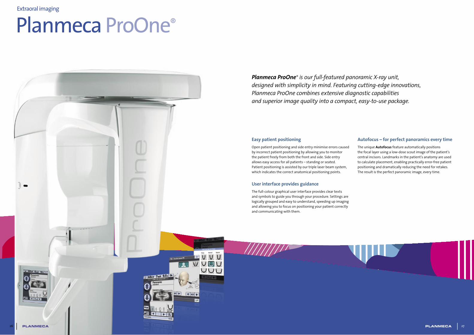
Planmeca ProOne® is our full-featured panoramic X-ray unit, designed with simplicity in mind. Featuring cutting-edge innovations, Planmeca ProOne combines extensive diagnostic capabilities and superior image quality into a compact, easy-to-use package.
Easy patient positioningOpen patient positioning and side entry minimise errors caused by incorrect patient positioning by allowing you to monitor the patient freely from both the front and side. Side entry allows easy access for all patients – standing or seated. Patient positioning is assisted by our triple laser beam system, which indicates the correct anatomical positioning points.
User interface provides guidanceThe full-colour graphical user interface provides clear texts and symbols to guide you through your procedure. Settings are logically grouped and easy to understand, speeding up imaging and allowing you to focus on positioning your patient correctly and communicating with them.
Autofocus – for perfect panoramics every timeThe unique Autofocus feature automatically positions the focal layer using a low-dose scout image of the patient’s central incisors. Landmarks in the patient’s anatomy are used to calculate placement, enabling practically error-free patient positioning and dramatically reducing the need for retakes. The result is the perfect panoramic image, every time.
2726

29
Planmeca ProOne®
Imaging programsBasic panoramic programs Standard panoramic
Lateral TMJ
PA TMJ
PA Sinus
Horizontal and vertical segmenting for panoramic program
Bitewing
Advanced panoramic programs
Interproximal panoramic
Orthogonal (perio) panoramic
Lateral-PA TMJ
Lateral multiangle TMJ
Lateral non rotational sinus
Cross-sections
Child (Paediatric) mode for each program to reduce the dose
Planmeca ProOne® offers you a wide variety of imaging programs for different radiographic needs. You can also select the correct exposure formats to minimise the radiation dose for all types of patients and diagnostic purposes.
Optimal imaging programs
Standard panoramic PA TMJBitewing PA Sinus and Lateral non rotational sinus
Lateral TMJ Cross-sectionsHorizontal and vertical segmenting for panoramic program Lateral-PA TMJ
Child mode for optimal paediatric imaging In child mode, the imaging area and exposure values are reduced in all programs and also the focal layer can be narrowed in the panoramic program. The patient dose is reduced remarkably.
28

Planmeca Romexis® is an advanced, easy-to-use software suite providing a rich set of tools to meet the imaging requirements set by any dental facility – from a small clinic to a large hospital. It supports the most versatile range of 2D and 3D imaging modalities.
Planmeca Romexis®
software for all images Mac OS and Windows
compatible
World leading imaging software
36 37

39
High-performance 2D imagingOur advanced Planmeca Romexis® software suite offers the most versatile tools for 2D imaging. Diagnose images using our full range of enhancement tools – or view them wherever you are with our mobile apps. This flexible dental imaging suite adapts to your needs and will grow into the third dimension together with your practice.
Easy and powerfulPlanmeca Romexis® is the software of choice for viewing and processing 2D images from Planmeca X-ray units. Powerful enhancement and analysis tools guarantee that accurate diagnosis is available to users in all specialties, while the intuitive interface guarantees confident, comfortable use from day one.
Sharing the resultsCases can be seamlessly transferred to mobile devices or partner clinics that use Planmeca Romexis or the free Planmeca Romexis® Viewer. Our integration with other systems allows you to freely utilise third-party products at your clinic. TWAIN support and DICOM standard compliance ensure that the software can be used together with most systems.
Integrated document managementThe printing module with multi-page support is ideal for creating professional, high-quality printouts and radiology reports to be sent to referring dentists.
Documents of any type can be attached to patient files, providing a convenient storage for cephalometric tracing reports, referral letters and other information.
Planmeca Romexis®
Free Planmeca Romexis® Viewer
applicationFull-featured viewer application
No installation required Mac OS and Windows support
Distribute to specialists or patients
Radiology interpretation moduleThe Planmeca Romexis® Radiological Findings module is the most advanced findings-recording tool on the market. Developed in cooperation with clinicians, its findings list is hierarchically categorised and can be freely edited. The module is especially designed for educational and radiology centres where uniformity of recordings is essential.
Advanced implant planning Planmeca Romexis provides powerful tools for implant planning, including realistic implant models from over 30 manufacturers.
38