Candidiasis – clinical manifestations and lab diagnosis of oral candidiasis

LUND UNIVERSITY
PO Box 117221 00 Lund+46 46-222 00 00
D-arabinitol in the diagnosis of invasive candidiasis
Sigmundsdottir, Gudrun
2010
Link to publication
Citation for published version (APA):Sigmundsdottir, G. (2010). D-arabinitol in the diagnosis of invasive candidiasis. Department of Clinical Sciences,Lund University.
General rightsCopyright and moral rights for the publications made accessible in the public portal are retained by the authorsand/or other copyright owners and it is a condition of accessing publications that users recognise and abide by thelegal requirements associated with these rights.
• Users may download and print one copy of any publication from the public portal for the purpose of private studyor research. • You may not further distribute the material or use it for any profit-making activity or commercial gain • You may freely distribute the URL identifying the publication in the public portalTake down policyIf you believe that this document breaches copyright please contact us providing details, and we will removeaccess to the work immediately and investigate your claim.
1
From the Department of Clinical Sciences and Department of Laboratory Medicine,
Medical Faculty, Lund University, Sweden 2010
D-ARABINITOL IN THE DIAGNOSIS OF INVASIVE CANDIDIASIS
av
Guðrún Sigmundsdóttir
AKADEMISK AVHANDLING
i ämnet infektionssjukdomar som med vederbörligt tillstånd av Medicinska Fakulteten vid Lunds universitet för avläggande av doktorsexamen i medicinsk vetenskap kommer att offentligen försvaras i Segerfalkssalen, Wallenberg Neurocentrum,
Sölvegatan 17, Lund, torsdagen den 18 november 2010 kl 13:00
Fakultetsopponent: Professor Christine Wennerås,
Sahlgrenska sjukhuset, Göteborg
3
Lund University, Faculty of Medicine
Doctoral Dissertation Series, no 105
D-arabinitol in the diagnosis of invasive candidiasis
by
Guðrún Sigmundsdóttir
Department of Clinical Sciences
and
Department of Laboratory Medicine
Faculty of Medicine
Lund University
Lund 2010
4
Ár skal rísa
sá er á yrkjendur fá
og ganga síns verka á vit.
Margt um dvelur
þann er um morgin sefur
hálfur er auður und hvötum.
Hávamál
© Guðrún Sigmundsdóttir
ISSN 1652-8220
ISBN 978-91-86671-21-1
Printed by Media Tryck, Lund, Sweden
5
TABLE OF CONTENTS
ORIGINAL PUBLICATIONS…………………………………………………. 6 ABBREVIATIONS…………………………………………………………....... 7 INTRODUCTION………………………………………………………………. 9
Candida the pathogen and the species…………………………………. 9 Transmission and epidemiology…........................................................... 10 Risk factors……………………………………………………………... 11 Impact of candidemia…………………………………………………... 12 Species identification…………………………………………………… 12 Laboratory diagnosis……………………………………………………. 14 Clinical manifestations………………………………………………….. 17 Arabinitol………………………………………………………………... 19 AIMS OF THE THESIS…………………………………………………………. 33 MATERIAL AND METHODS………………………………………………….. 35 Subjects…………………………………………………………………. 35 Collection of clinical and microbiological data………………………… 39 Urine samples…………………………………………………………… 40 GC-MS analysis………………………………………………………… 40
Candida and DA production……………………………………………. 41 Microbiology……………………………………………………………. 42 Statistical methods……………………………………………………… 43 RESULTS………………………………………………………………………… 45 Paper I…………………………………………………………………… 45 Paper II………………………………………………………………….. 50 Paper III..................................................................................................... 51 Paper IV…………………………………………………………………. 54 DISCUSSION……………………………………………………………………. 59 Paper I and Paper III - Urine DA/LA ratio in neonates, ……………….. 59 clinical experience in neonates and children with cancer Paper IV – DA production by C. glabrata……………………………… 62 Paper II and Paper IV - DA production and patients …………………… 64 with haematological malignancy Paper II – DA in HIV patients.................................................................. 67 Pitfalls using DA/LA ratio in urine……………………………………... 67 in the diagnosis of invasive candidiasis CONCLUSIONS..................................................................................................... 71 POPULÄR VETENSKAPLIG SAMMANFATTNING PÅ SVENSKA.............. 72 ACKNOWLEDGEMENTS………………………………………………………. 75 REFERENCES…………………………………………………………………… 77 ORIGINAL PAPERS……………………………………………………………. 91

6
ORIGINAL PUBLICATIONS
The thesis is based on the following papers, which will be referred to by their Roman numerals.
I. Guðrún Sigmundsdóttir, Bertil Christensson, Lars J. Björklund, Kristina Håkansson, Christina Pehrson, Lennart Larsson. Urine D-arabinitol/L-arabinitol ratio in diagnosis of invasive candidiasis in newborn infants. Journal of Clinical Microbiology. 2000;38:3039-3042
II. Damon P. Eisen, Paul B. Bartley, William Hope, Gudrun Sigmundsdottir, Christina Pehrson, Lennart Larsson, Bertil Christensson. Urine D-arabinitol/L-arabinitol ratio in diagnosing Candida infection in patients with haematological malignancy and HIV infection. Diagnostic Microbiology and Infectious Disease. 2002;42:39–42
III. Gudrun Sigmundsdottir, Lennart Larsson, Thomas Wiebe, Lars J. Björklund, Bertil Christensson. Clinical experience of urine D-arabinitol/L-arabinitol ratio in the early diagnosis of invasive candidiasis in paediatric high risk populations. Scandinavian Journal of Infectious Diseases. 2007;39:146 – 151
IV.Guðrún Sigmundsdóttir, Christina Pehrson, Helga Erlendsdóttir, Lennart Larsson, Ingibjörg Hilmarsdóttir, Erja Chryssanthou, Bertil Christensson. In vitro production of D-arabinitol in different Candida species. Manuscript

7
ABBREVIATIONS
DA D-arabinitol
DADH D-arabinitol dehydrogenase
LA L-arabinitol
DA/LA D-arabinitol/L-arabinitol
DA/cr D-arabinitol/creatinine
IC Invasive candidiasis
CVC Central venous catheter
PCR Polymerase chain reaction
GC Gas chromatography
MS Mass spectrometry
GC-MS Gas chromatography – mass spectrometry
PPP Pentose phosphate pathway
CSF Cerebrospinal fluid
CNS Central nervous system
CFU Colony forming units
NICU Neonatal Intensive Care Unit
POU Pediatric Oncology Unit

8

9
INTRODUCTION
Invasive candidiasis (IC) is a serious condition that affects mainly immunocompromised individuals, preterm infants and hospitalized patients with serious underlying diseases. The incidence of IC has increased dramatically since the early 1980s accompanied by increase in mortality and morbidity. There are no ideal diagnostic methods for the diagnosis of IC in humans, blood and tissue cultures are still considered the “gold standard”, although 50% of blood cultures can be false negative. A number of non-culture methods have been developed, including antibody and antigen tests, DNA amplification tests, and detection of Candida metabolites. D-arabinitol (DA) is a metabolite of most Candida species pathogenic to humans and can be measured in serum and urine. D-arabinitol levels in human body fluids are affected by kidney function and D-arabinitol/L-arabinitol (DA/LA) or D-arabinitol/creatinine (DA/cr) ratios are measured to correct for kidney dysfunction. This method has gained considerable attention, however, is not widely spread in clinical routine partly due to expensive equipment needed and/or lack of commercially available tests to measure DA levels. Timely diagnosis and initiation of appropriate antifungal therapy is imperative for improving outcomes and further studies on the diagnosis of invasive Candida infections are encouraged.
Candida – the pathogen and species
The genus Candida belongs to the family Candidaceae (1). Approximately 200 Candida species are assigned to the genus, but reassignment of known species and discovery of new species following technological advances, have led to consequent changes the number of species belonging to the genus (1; 2; 3). Candida is unicellular and ovoid, between 4-6 µm in diameter and multiplies principally by producing blastoconidia from budding. When blastoconidia are formed without separating from one another, pseudohyphae are formed and some species can form true hyphae under certain conditions. The yeast can thus vary in shape and initial clinical isolates can be pleomorphic (1). Candida species grow aerobically at 25 – 37°C and form creamy, white, smooth and flat colonies on blood agar plates (1). Growth can be detected on agar plates after 24 hours, although the colonies mostly become visible after 48 – 72 hours incubation.
Candida is a normal constituent of human flora and is commonly found in the gastrointestinal tract, the female genital tract, the respiratory system and on the skin of
10
healthy people (2). Candida albicans has been found in hospital environments, food, soil, and in animals (1; 2). Only 17 Candida species are known as human pathogens, the predominant pathogen being C. albicans. Other common species are C. glabrata, C. parapsilosis and C. tropicalis (3; 4; 5; 6). Other well known but less common pathogens are C. dubliniensis, C. guilliermondi, C. krusei, C. kefyr and C. lusitaniae.
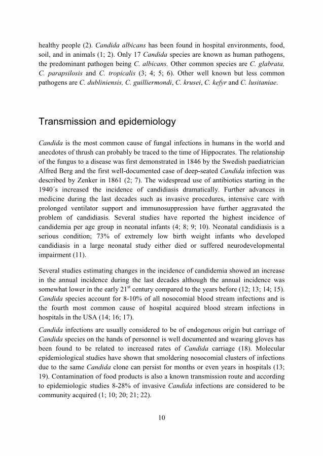
Transmission and epidemiology
Candida is the most common cause of fungal infections in humans in the world and anecdotes of thrush can probably be traced to the time of Hippocrates. The relationship of the fungus to a disease was first demonstrated in 1846 by the Swedish paediatrician Alfred Berg and the first well-documented case of deep-seated Candida infection was described by Zenker in 1861 (2; 7). The widespread use of antibiotics starting in the 1940´s increased the incidence of candidiasis dramatically. Further advances in medicine during the last decades such as invasive procedures, intensive care with prolonged ventilator support and immunosuppression have further aggravated the problem of candidiasis. Several studies have reported the highest incidence of candidemia per age group in neonatal infants (4; 8; 9; 10). Neonatal candidiasis is a serious condition; 73% of extremely low birth weight infants who developed candidiasis in a large neonatal study either died or suffered neurodevelopmental impairment (11).
Several studies estimating changes in the incidence of candidemia showed an increase in the annual incidence during the last decades although the annual incidence was somewhat lower in the early 21st century compared to the years before (12; 13; 14; 15). Candida species account for 8-10% of all nosocomial blood stream infections and is the fourth most common cause of hospital acquired blood stream infections in hospitals in the USA (14; 16; 17).
Candida infections are usually considered to be of endogenous origin but carriage of Candida species on the hands of personnel is well documented and wearing gloves has been found to be related to increased rates of Candida carriage (18). Molecular epidemiological studies have shown that smoldering nosocomial clusters of infections due to the same Candida clone can persist for months or even years in hospitals (13; 19). Contamination of food products is also a known transmission route and according to epidemiologic studies 8-28% of invasive Candida infections are considered to be community acquired (1; 10; 20; 21; 22).

11
C. albicans is the most common Candida species causing IC worldwide; 66% of IC are caused by C. albicans (23). Other Candida species causing candidemias in adults are mainly C. tropicalis, C. glabrata, C. parapsilosis, and C. krusei, which together with C. albicans cause over 90% of all IC (3; 4; 12; 24). C. parapsilosis is more common in infants compared to other age groups, and accounts for approximately 30% of all Candida bloodstream isolates compared with only 10% - 15% of Candidabloodstream isolates in adults (24; 25; 26).
There has been a shift in Candida species causing IC during the last two decades with an increase in the proportion of invasive Candida infections caused by non-albicansCandida species. Increased risk of candidemia due to non-albicans Candida species was in one study associated to fluconazole treatment, central venous catheter (CVC) exposure and mean number of antibiotics per day (27). Several other studies have found positive correlation between the usage of fluconazole and increase of C. kruseiand C. glabrata infections (12; 28; 29). Geographical variation has been observed in the frequency of isolation of emerging Candida species (23). C. glabrata has emerged as an important opportunistic fungal pathogen in the United States, but is in most other countries a much less common cause of candidemia despite an increase in the use of fluconazole. The most common non-albicans Candida species causing candidemia in other countries are C. parapsilosis and C. tropicalis (23).
Risk factors
Independent risk factors for IC for all age-groups include exposure to broad-spectrum antimicrobial agents, cancer chemotherapy, mucosal colonization by Candida species, indwelling CVC, total parenteral nutrition (TPN), neutropenia, prior surgery (especially gastrointestinal), and renal failure or hemodialysis (23).
Several risk factors have been identified in neonatal infants; in extremely low birth weight neonates (< 750 g) candidiasis was much more frequent compared with infants between 751 to 1000 g (11). Gestational age below 32 weeks, use of intralipid or parenteral nutrition, CVC, 5-min Apgar score <5; shock, intubation or length of stay in NICU more than seven days before candidemia and use of third-generation cephalosporin have all been associated with IC (11; 25).

12
Impact of candidemia
Several studies estimating the impact of candidemia have been performed and treatment of IC has been associated with longer hospital admissions and increased cost of care (30; 31). Hospital-acquired infections in the NICU are associated with increased morbidity and mortality, prolonged hospitalization, and increased hospital costs (32; 33). In a large population based study, candidemia was associated with a 10.1% increase in mortality, a mean of 21.1 days increase in length of stay and a mean increase of $92,266, in total per-patient hospital charges, in a pediatric patient population (34). For the adult patients in the same study, candidemia was associated with a 14.5% increase in mortality, a mean 10.1-day increase in length of stay, and a mean increase $39,331 in hospital charges. Other smaller studies have estimated attributable mortality between 4.4% - 49% (30; 31; 35; 36). This variation may reflect differences between hospitals, with large population based studies giving a better overall estimation of the situation.
Species identification
Candida species grow on routine blood agar and on the selective Sabouraud medium. Both C. albicans and C. dublininesis form germ tubes (hyphae) after growth in serum for 2 – 4 hours at 37°C, and the germ tube test is a simple and rapid method for identification of C. albicans, but false positive germ tube tests can occur (37). Other species are known to form pseudohyphae that can easily be distinguished from true hyphae. Candida albicans can also be identified by chlamydospore formation after 24 – 72 hours incubation at 25 – 30°C on corn meal agar. Incubation is done at 30 – 37°C and most Candida species which infect humans grow at 37°C.
The chromogenic medium CHROMagarTM (Candida France) is a culture medium used for isolation and identification of Candida. The medium is marketed for presumptive identification of C. albicans, C. tropicalis, and C. krusei based on pigments observed. It has been found unreliable in distinguishing C. dubliniensis from C. albicans but useful to detect mixed infections (38; 39; 40; 41). Other commercially available chromogenic media such as Candida ID (bioMerieux, Marcy l’E´toile, France) and CandiSelect (Bio-rad, Marnes La Coquette, France), show similar performance as CHROMagar (1).
13
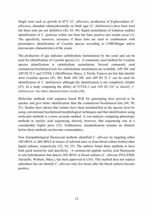
Single tests such as growth at 45°C (C. albicans), production of �-glucosidase (C. albicans), abundant chlamydoconidia on Staib agar (C. dubliniensis) have been tried but these tests are not definitive (42; 43; 44). Rapid assimilation of trehalose enables identification of C. glabrata within one hour but false positive test results occur (1). The specificity, however, increases if these tests are used in combination with presumptive identification of Candida species according to CHROMagar and/or microscopic characteristics of the yeasts.
The production of gas indicates carbohydrate fermentation by the yeast and can be used for identification of Candida species (1). A commonly used method for Candidaspecies identification is carbohydrate assimilation. Several commonly used commercial biochemical kits for carbohydrate assimilation are available, API 20C and API ID 32 C and VITEK 2 (BioMérieux Marcy, L’Etoile, France) are kits that identify most Candida species (45; 46). Both API 20C and API ID 32 C can be used for identification of C. dubliniensis although the identification is not completely reliable (47). In a study comparing the ability of VITEK-2 and API ID 32C to identify C. dubliniensis, the latter obtained better results (48).
Molecular methods with sequence based PCR for genotyping have proved to be quicker and give better identification than the commercial biochemical kits (49; 50; 51). Studies have shown that isolates have been misidentified at the species level by using conventional biochemical/morphological techniques and that identification using molecular methods is a more accurate method. A cost analysis comparing phenotypic methods to nucleic acid sequencing showed, however, that sequencing was at a considerably higher price (52). Furthermore, standardization remains an obstacle before these methods can become commonplace.
New histopathological fluorescent methods identified C. albicans by targeting either 18S rRNA or 26S rRNA in tissues of infected mice or from blood culture bottles/other liquid cultures, respectively (53; 54; 55). The authors found these methods to have both good sensitivity and specificity. A commercial peptide nucleic acid fluorescent in situ hybridization that detects 26S rRNA in blood cultures (C. albicans PNA-FISH, AdvanDx, Woburn, Mass.), has been approved in USA. This method does not replace subculture but can identify C. albicans only few hours after the blood cultures become positive.

14
Laboratory diagnosis
Direct microscopy Direct microscopy can be performed on patient samples or on cultures from different culture media. Direct microscopy can reveal the presence of yeasts and whether hyphae are present or not.
Culture Candida species grow on blood agar and Sabourauds medium. Growth can be detected on agar plates after 24 hours, although the colonies mostly become visible after 48 – 72 hours incubation (1). Most pathogenic Candida species grow at 25 – 37°C, whereas saprophytes usually fail to grow at 37°C (1). Tissue cultures and cultures from normally sterile sites are reliable diagnostic methods to detect Candida infections. This method is, however, limited by difficulties in obtaining tissue biopsies from thrombocytopenic immunosuppressed patients.
Blood cultures still serve as the golden standard for IC even if as many as 50% of patients with autopsy-proven disease had negative blood cultures ante mortem (56). Several methods have been developed to improve the sensitivity. Lysis centrifugation is one of these methods, the yield of fungi is increased by releasing fungi trapped within host phagocytic cells; this method is, however, both labour-intensive and expensive and has not gained popularity (57). Another widely used improvement is automated monitoring of blood culture bottles with either colorimetric detection or fluorescent methods. Comparison of two commercially available systems, BacT/ALERT 3D (Organon Teknika Corp, Durham, NC) and BACTEC 9240 (Becton Dickinson, USA), was done by inoculating aerobic, anaerobic, and mycology bottles from both systems with Candida (58). The aerobic BacT/ALERT media performed better than the aerobic BACTEC media with 100% and 90% detection rate respectively, but the largest difference was found between the anaerobic media with detection rate 70% and 10% in BacT/ALERT and BACTEC, respectively (58). The mycology media from BacT/ALERT and BACTEC, however, gave similar yields and had higher sensitivity compared to the aerobic and anaerobic media (58; 59).
Histopathology Histopathology examination is one of the most reliable methods to diagnose invasive Candida infections with tissue affection. Candida can be diagnosed in histochemically stained tissue, but the ease of the method is dependent on the abundance of the fungi

15
and the distinctiveness of its appearance (60). The usefulness is however limited in thrombocytopenic patients with suspected tissue affection due to risk for complications when obtaining tissue biopsies.
1�3-�-D-glucan 1�3-�-D-glucan is a component of the fungal cell wall of Candida, Aspergillus,Fusarium, and Pneumocystis jirevicii, but not in Cryptococcus neoformans and zygomycetes (61; 62). 1�3-�-D-glucan can be detected in blood samples from patients with invasive fungal infections caused by the fungi known to have (1�3)-�-D-glucan (63; 64; 65). False positive 1�3-�-D-glucan levels can occur in patients undergoing hemodialysis and patients receiving parenteral infusions of plasma components or erythrocyte and thrombocyte filtered blood products (66; 67; 68).
Several commercialized assays are available including chromogenic assays such as Fungitell (Associates of Cape Cod Inc., East Falmouth, Massachusetts, USA; previously Glucatell), the colorimetric assay FungiTec G (Seikagaku Kogyo Corporation, Tokyo, Japan) and turbidimetric assays Wako-WB003 (Wako Pure Chemical industries, Osaka, Japan). Two studies using the Glucatell and the Fungitell tests for serum 1�3-�-D-glucan detection found the assay to be highly sensitive and specific when used as a diagnostic adjunct for IFI in patients with hematologic malignancies (63; 64). Another study using Fungitec for their analysis found the test useful in monitoring surgical patients for invasive fungal infections, and concluded that it could be useful to guide for the initiation of empiric therapy (69). A multiregression analysis in the same study additionally showed that subjects who had a positive 1�3-�-D-glucan test were almost 13-fold more likely to respond to empiric therapy than those who were negative for 1�3-�-D-glucan.
Although the sensitivity of the 1�3-�-D-glucan test is overall good and test results can be quickly available, there is a definitive risk for false positive results and the test does not differentiate between various fungal species.
Molecular methods for detection of candemia Several studies evaluating molecular methods have been performed in attempts to facilitate the earlier diagnosis of fungemia and have found them to be highly sensitive (70; 71; 72; 73; 74). However, the multiplexing capacities of most real-time PCR instruments is limited, a large proportion of assays target a small number of pathogens such as C. albicans, C. tropicalis C. glabrata, C. parapsilosis, and C. krusei. The clinical utility is limited by lack of standardization and high setup costs (75).

16
Development of standards and establishment of quality control is needed to achieve the best consensus.
Both false negative and false positive test results are known to occur. The main source of false-positive test results is contamination with previously amplified products or the presence of fungal spores or DNA in the environment and reagents used (75). Precautions must be taken to minimize the risk for contamination and false positive results whenever PCR assays are used in the diagnosis of IC (76). A slight difference in the expected yield can give false negative results, which can be minimized by using real-time quantitative PCR to control the amplification yield for each clinical sample (75).
Antigen Numerous antigens have been tested as possible targets for detection of IC. The commercially available Cand-Tec (Ramco Laboratories, Houston, Texas) is based on detection of a structurally uncharacterized 56°C labile antigen. Most studies suggest that the Cand-Tec test does not provide adequate predictive value for a reliable diagnosis of IC (60; 77).
Two studies evaluating secreted aspartyl proteinase (Sap) suggested that Sap could be useful in the diagnosis of invasive C. albicans infections but further studies were recommended (78; 79). No commercial tests for Sap detection are currently available.
Mannan is cleared rapidly from the circulation and multiple sampling is required for optimal results although the sensitivity has been found to be low (60; 80). A recent study on a commercially available Platelia Candida antigen test (Bio-Rad, Segrate, Italy) which is based on detection of �-linked oligomannan residues was shown to have good specificity, but frequent sampling was necessary. However when this test was used in combination with detection of ß linked oligomannose, a sensitivity of 85% was reached (81). Interestingly, a higher sensitivity was observed when the Platelia Candida antigen test was assessed in neonatal infants, with sensitivity and specificity for cases of proven and probable candidosis, 94.4% and 94.2%, respectively (82).
Early studies examining a 47 kilodalton antigen which later proved to be enolase, showed 77% sensitivity in neutropenic patients with IC (83). Later Walsh et al found multiple sampling to improve the detection of enolase; antigenemia was detected in 11 of 13 proven cases of deep tissue infection (85%) and in 7 of 11 proven cases of fungemia (64 percent), specificity was 96% (84). The authors concluded that the enolase test was a useful indicator of deep infection in patients with cancer and
17
neutropenia. This test was commercially available but has been withdrawn from the market.
Antibodies and combined antigen/antibody tests Measuring antibodies to diagnose IC has been found of limited clinical value due to low sensitivity and specificity (60). However, by combining detection of antigens and antibodies, more satisfactory results have been achieved and by combining the CandTec test together with antibody measurements against Candida 100% sensitivity and 83.3% specificity was reached (85). Three studies have evaluated the use of combined mannan antigen and anti-mannan antibody detection using Platelia Candida Ag and Platelia Candida AB/AC/AK (Bio-Rad, Marnes La Coquette, France) (77; 86; 87). The overall sensitivity in these studies was from 78% - 100%, however, difference between Candida species causing the infection was observed with highest sensitivity for C. albicans, C. tropicalis and C. glabrata but with lower sensitivity (40% - 50%) for C. parapsilosis, C. krusei and C. kefyr (86; 87). Interestingly, the patients were generally either positive for antigens or antibodies and not for both simultaneously. Furthermore, the test sensitivity in one of these studies was dependent on the ward; i.e. hematology patients were more likely to have positive antigen tests and surgical patients were more likely to have positive antibody tests (87). These findings are, however, not surprising since antibody production in immunocompromised patients can be variable and even non-existing with subsequent difficulties in antibody detection in this group of patients. An overrepresentation of antigens can consequently be present in immunocompromised patients facilitating the detection of antigens.
Clinical manifestations
Candida has a broad disease spectrum varying from trivial superficial mucosal or skin infections in healthy people to life threatening IC in immunesuppressed patients. Skin infections are commonly seen in moist occluded body sites like armpits, in the groins and other skin folds (2). Of mucosal infection Candida vulvovaginitis is the second most frequent genital complaint in healthy females and thrushes are commonly seen on the perineal skin and in the oral mucosa in healthy neonates (2). Oral thrush in adults is either associated with cancer or AIDS or a complication to treatment with inhaled steroids (2).
Candida esophagitis is commonly associated with treatment of malignancy of the hematopoietic or lymphatic systems or AIDS although a small number occurs in

18
patients with no known underlying illness (88). Symptoms of Candida esophagitis include substernal chest pain painful swallowing and a feeling of obstruction on swallowing (2). The diagnosis is confirmed by identifying histopathological changes in biopsies obtained by endoscopy. However, a clinical picture of esophagitis together with white patches resembling thrush that by direct microscopy show masses of hyphae and pseudohyphae, is also considered sufficient to establish the diagnosis of esophagitis.
Symptoms of candidemia can vary from low grade fever not responding to antimicrobial treatment, to severe sepsis with shock and multiorgan failure. Candidacan disseminate in the body and spread to several organs simultaneously; the organs most commonly involved are the liver, kidneys, heart, brain and the eyes. Hematogenously spread cutaneous lesions are a manifestation of disseminated disease. Pathological changes observed in disseminated candidasis include diffuse microabscesses and small macroabscesses, however, macroabscesses more than a centimeter in diameter may also form. Single organs can be affected but involvement of two or more organs is commonly seen.
Hepatosplenic candidiasis is an important clinical manifestation in severely immunocompromised hosts; the kidneys are frequently also affected in these patients (2). Candida endocarditits was a rare disorder but is now more common, often related to underlying disorders such as valvular heart diseases, prolonged use of iv catheters, iv drug addiction, immunosuppressive drug treatment, prosthetic heart valves and pre-existing bacterial endocarditis (2). Candida peritonitis is a known complication of peritoneal dialysis, gastrointestinal surgery and perforation of an abdominal organ. Respiratory candidiasis can be hematogenously spread to the lungs or more rarely through endobronchial inoculation. Central nervous system (CNS) infections are usually hematogenously spread, although they can occur as a complication of ventricular shunt, after trauma or lumbar puncture. Half of patients with CNS infections have infections in other organs (2).
Detection of Candida is common in urine and usually does not indicate urinary tract infections. Candida cystitis is most often associated with bladder catheterization and diabetes mellitus. Infection in the upper urinary tract are commonly hematogenously spread although ascending infections due to underlying disorders as diabetes mellitus, urinary obstructions are known to occur (2).
Fungemia in neonatal infants can present as unspecific mild symptoms but is also associated with sepsis and shock indistinguishable from bacterial sepsis (89). Central nervous system involvement is more commonly observed in infants and can affect the

19
meninges, ventricles, or cerebral cortex with abscess formation but clinical manifestations may be unspecific. Endophthalmitis was previously detected in 45% of neonatal IC cases, but is now less than 5% due to earlier antifungal treatment and retinal examinations in neonates with systemic candidiasis are of importance (89).
Arabinitol
Arabinitol is five carbon sugar alcohol with two stereoisomers, D-arabinitol (DA) and L-arabinitol (LA). Stereoisomers are molecules that are made of the same atoms and connected by the same sequence of bonds but have different three dimensional structures. Enantiomers are the two mirror images of two stereoisomeres and arabinitol is assigned to the Dextro- (D) and Levo (L) rotatory forms according to the configuration at the centre of chirality.
CH2OH CH2OH
OH – CH HC – OH
HC – OH OH – CH
HC – OH OH – CH
CH2OH CH2OH
D-arabinitol L-arabinitol
Methods for measuring arabinitol The gaschromatography-mass spectrometry (GC/MS) technique
The first method using gaschromatography (GC) to determine DA was done by identification of trimethylsilyl derivative of arabinitol in packed GC columns (90). This method could not separate DA from other pentitols and peaks detected represented the sum of pentitols including DA, LA, xylitol and ribitol. The first combined gas-chromatography - mass spectrometry (GC-MS) used identical GC techniques as described above. This method did still not enable separation of the pentitols, required several hours of elaboration and did not gain popularity (91). In 1984 Roboz et al used capillary columns that enabled separation of the pentitols but the DA and LA enantiomers could still not be differentiated (92).
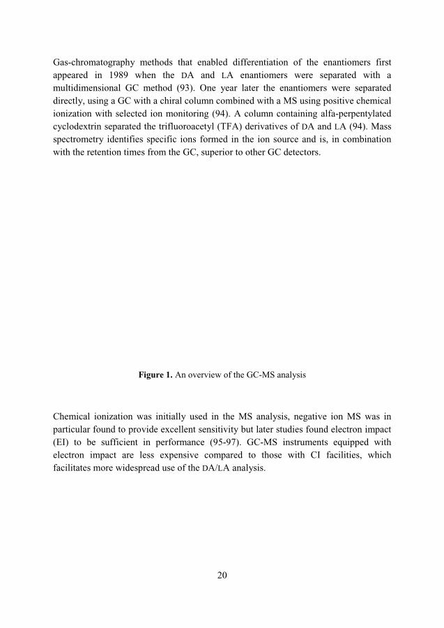
Gas-chromatography methoappeared in 1989 when multidimensional GC methdirectly, using a GC with a cionization with selected ioncyclodextrin separated the tspectrometry identifies specwith the retention times from
Figure
Chemical ionization was inparticular found to provide (EI) to be sufficient in pelectron impact are less efacilitates more widespread
20
ods that enabled differentiation of the the DA and LA enantiomers were s
hod (93). One year later the enantiomerchiral column combined with a MS using n monitoring (94). A column containing trifluoroacetyl (TFA) derivatives of DA acific ions formed in the ion source and im the GC, superior to other GC detectors.
e 1. An overview of the GC-MS analysis
nitially used in the MS analysis, negativexcellent sensitivity but later studies founerformance (95-97). GC-MS instrumenexpensive compared to those with CI use of the DA/LA analysis.
enantiomers first separated with a rs were separated positive chemical
alfa-perpentylated and LA (94). Mass is, in combination
ve ion MS was in nd electron impact nts equipped with
facilities, which

21
Enzymatic methods
The GC-MS technique requires expensive equipment, which is not generally available and enzymatic methods have been developed for DA measurements in serum. The enzymatic method is based on the NAD dependent reaction:
D-arabinitol + NAD+ � D-ribulose + NADH,
which is catalyzed by the enzyme DADH. The first experiments based on the function of this enzyme were reported by Bernard et al by incubating the patient sample with DADH producing C. tropicalis and GC was used to determine the difference in DA concentration between the treated and the untreated samples (98). The C. tropicalisstrain was affected by antifungal agents in the patient samples during the 24 hours long incubation needed for this method and other methods using purified DADH were therefore preferable. Hence the enzyme from Klebsiella pneumoniae was purified, this
�����������������
���
� ��� ��� ��� ��� ���
����
����
���
���
�
�������������
�����������
������������
�����������������
���
� ��� ��� ��� ��� ���
����
����
���
���
�
�������������
�����������
������������
Figure 2a.
Gaschromatograph showing normal DA/LA ratio
Figure 2b.
Gaschromatograph showing elevated DA /LA ratio

22
enzyme however showed 20% cross-reactivity with D-mannitol, which may be present in human serum with high risk for false positive test results (99). Others used DADH derived from Enterobacter aerogenes coupled to spectrophotofluorimetric methods but this enzyme also showed cross reactivity with D-mannitol (100).
The next step to improve the enzymatic method was purification of a highly substrate specific DADH from C. tropicalis, which was applied to a semi-automated spectrophotometric assay, the results correlated well with DA concentrations determined by GC (101). A large clinical study was performed using this method and found the test to be rapid and reliable (102). A method for overproduction of recombinant DADH from C. tropicalis, in Escherichia coli was later developed but did not come in general use in automated DA assays (103). The most recently developed method was a rapid enzymatic fluorimetric assay (Cobas Fara II centrifugal autoanalyzer, Roche) with a recombinant DADH from C. albicans also produced in Escherichia coli (104). The enzyme in this study was highly specific for DA and cross-reacted only with xylitol (4.9%). A large population based clinical study on all Candida fungemia cases in Connecticut applied this method and found it useful for diagnosing IC (105). However, there is currently no enzymatic method commercially available, limiting the use of this method to hospitals connected to research groups developing these methods.
In vitro D-arabinitol production by Candida and other fungi Several medically important Candida species produce DA in vitro and several researchers have shown that serum arabinitol and DA concentrations, serum and urine DA/LA and serum DA/cr ratios, have been found to be higher in animals and humans with invasive Candida infections compared to colonized or uninfected controls (102; 105-109).
A search of the literature for published reports on the in vitro production of DA by different Candida species reveals a limited number of articles with only few strains tested. Production of DA has been reported for 26 C. albicans strains, 25 C. tropicalisstrains, 15 C. parapsilosis strains, three C. pseudotropicalis strains, two C. lusitaniaestrains, two C. guillermondii strains and one strain of C. kefyr; absence of DA production has not been reported for any of these species (90; 95; 110; 111). Only one study has been published on the production rate by different Candida species; the nine tested C. albicans strains differed over hundred fold in production rate and there appeared to be some variation in the production rate between Candida species (110). No production of DA by C. glabrata has been detected in vitro; however, in published literature only four strains of C. glabrata have been tested altogether (110; 111).

23
Previous reports on DA production by C. krusei in vitro have been conflicting. Two in vitro studies reported either no production of DA or trace amounts in altogether five C. krusei strains that were tested (95; 110). However, in a more recent study, DA production was detected in all of seven C. krusei strains studied (111).
A new Candida species, pathogenic to humans, was identified in 1995 (44). This species previously classified as “atypical C. albicans” was named C. dubliniensis. Nothing has been published on DA production in C. dubliniensis.
Environmental fungi such as Trichoderma reesei and Zygosaccharomyces rouxi, are known to metabolise DA and have been extensively studied in attempts to transform biomass into biofuels (112). Production of DA by environmental fungi is, however, unlikely to have any effect on arabinitol levels in humans. But the possibility of DAand LA metabolism by bacteria and fungi in the gut affecting the arabinitol levels in human body fluid cannot be excluded.
D-arabinitol metabolism in bacteria D-arabinitol-phosphate dehydrogenase has been purified from Enterococcus avium; the gene has been cloned and is probably widespread among Gram positive bacteria;the strongest matches for the gene were found in Listeria monocytogenes, Listeria inocua, Staphylococcus aureus and Bacillus halodurans (113). Lactobacillus casei is also known to have an enzyme which can both synthesize DA and utilize it for growth as a single energy source (114). Enterobacter aerogenes grows on media with DA or LA as a single substrate and more than 90% of strains of Klebsiella pneumoniae and Klebsiella oxytoca and about 5% of E. coli strains carry the genes for DA catabolism and can catabolise DA (115; 116). Some bacteria are thus known to metabolise DA, but the overall knowledge on DA metabolism is otherwise limited. Whether DA detected in humans has its origin in DA synthesis by bacteria colonizing the gastrointestinal tract is not clear.
D-arabinitol metabolism in bacteria and fungi L-arabinitol has only been detected in environmental bacteria but the knowledge on LA metabolism in bacteria appears otherwise to be limited (117). L-arabinitol metabolism is more commonly found in fungi, Aspergillus niger and several environmental fungi are known to carry the genes for and to produce LA, and the enzyme L-Arabinitol-4-dehydrogenase has been extensively studied in the production of biofuels from lignocellulos biomass (118). It is unknown whether bacteria or fungi in the gastrointestinal tract can affect the LA levels in human body fluids by LA synthesis or catabolism.
24
D-arabinitol and L-arabinitol in normal human body fluids D-arabinitol
D-arabinitol is present in human body fluids, the normal level of DA in serum and urine in adults is 0.20 ± 0.053 µg/ml and 18.45 ± 7.22 µg/ml, respectively (93; 94). Urinary excretion of DA and other polyols in newborn infants have shown age dependency with highest concentrations postnatally independent of the grade of maturity (119).
Christensson and Roboz found approximately 10-fold higher levels of DA in cerebrospinal fluid (CSF) compared to other body fluids (120). These high DA levels in the CSF suggest DA synthesis in the brain or the spinal cord but exactly where it has its origin or which metabolic pathways are used, is unknown. Temporary increased permeability of the blood brain barrier, which takes place in acute meningitis, does not appear to give higher DA/LA ratios in other body fluids according to urinary DA/LA ratios obtained from patients with acute meningitis in Lund and analyzed at our laboratory (unpublished data).
The pentose phosphate pathway (PPP) is an alternative pathway for glucose oxidation in humans and DA is believed to be one of the end products with no possibilities of a reverse reaction (121). Congenital defects in two separate enzymes of the PPP have been discovered; the patients were found to have high concentrations of DA, ribitol and erythritol in urine and plasma and in the CNS (121). These studies show that metabolic pathways for DA exist in humans but the role of DA in the human body remains unknown.
Experimental research on rats has shown that DA is readily absorbed from the gut and in the same study 85% of a 1g oral dose of DA was detected in urine within 24 hours in a healthy human volunteer (122). But very little is known about DA containment in food products and whether consumption of food products containing DA results in higher DA levels in serum or urine.
The origin of DA detected in human body fluids is unclear, whether it comes from our own endogenous production of DA or if it is absorbed from the gut either from dietary DA or from DA produced by bacteria in the gut remains to be answered.
L-arabinitol
L-arabinitol can be detected in body fluids in humans; the concentrations in serum and urine are 0.11 ± 0.040 µg/ml and 12.1 ± 4.02 µg/ml, respectively (93; 94). The LA

25
concentration in CSF is 0.13 ± 0.05 µg/ml (range: 0.09-0.2 µg/ml), which is virtually identical to that in serum (120).
High LA levels in urine, plasma, and cerebrospinal fluid, have been detected in a child with multiple congenital abnormalities, the authors presumed that the LA was an end product in the brain where it accumulated (123). Metabolic pathways for LA therefore appear to be present in the human body but knowledge is otherwise very limited.
In analogy with DA, LA is probably also readily absorbed from the gut. Little is known on the containment of LA in food products, but absorption of LA from food products cannot be excluded. It is unknown whether the LA detected in human body fluids is bacterial or dietary LA absorbed in the gastrointestinal tract, or whether it originates from our own endogenous synthesis.
DA/LA ratios, DA/cr ratios and kidney function D-arabinitol is excreted in the kidneys via glomerular filtration and renal dysfunction results in elevated serum DA levels (124). An experimental study done in the early eighties showed that arabinitol was cleared at the same rate as creatinine and that the urinary arabinitol excretion rate was directly proportional to the concentration ratio of arabinitol to creatinine in serum or urine (124). Arabinitol/cr ratios were therefore applied and both experimental and clinical studies have shown that adjusting serum DA to serum creatinine is a reliable method (124-126). However, in rapidly or acutely evolving renal impairment, serum creatinine may not initially reflect decline in glomerular filtration rate and can thus give false positive DA/cr ratios (102).
DA and LA enantiomers are excreted at the same rate through the kidneys and kidney dysfunction consequently affects both enantiomers equally. By determining DA/LAratio the influence of renal dysfunction can be avoided. The GC-MS enables determination of DA/LA ratios in serum and in urine, which makes it unnecessary to determine the absolute concentrations of each enantiomer and studies have shown that serum and urine DA/LA ratios are virtually identical (94; 97). Although little is known about the normal variation of LA levels in humans, we have noticed both in our clinical everyday work and in our research projects that DA/LA ratios in urine are fairly constant also over time in healthy and hospitalized individuals without Candida infection (95; 106). Urine samples for DA/LA ratios can be delivered to the analytical laboratory as dried spots on a filter paper, which greatly facilitates handling and sending the samples (127). However, when false urine DA/LA test results are suspected serum samples with absolute levels of both DA and LA are recommended.
26
Non-Candida related changes in DA levels Elevated DA/cr ratios have been associated with Behcet’s disease, although the number of patients included in the investigation was small and this finding has never been confirmed (128). Some early studies also claimed that patients with sarcoidosis and patients treated with corticosteroids exhibited increased arabinitol levels. These results could not be verified in subsequent studies on sarcoidosis patients, who were or were not receiving steroid treatment or in an animal model, where cortisone given intra-muscularly was not found to interfere with serum or urine arabinitol levels (122; 129).
Clinical and experimental studies with DA as a diagnostic marker The first study using GC as a diagnostic method is from 1974 when major peaks were detected in seven serum samples from six patients with septicaemia caused by C. albicans (130). Five years later the compound was identified as DA and found to correlate with serum creatinine levels; four of eight patients with IC and normal kidney function had high arabinitol levels (90). Three clinical studies, published between 1980 – 1986, found arabinitol of value as a diagnostic method for IC; one study also included eight patients with infected intravenous lines and cannulas and clinical evidence of invasive infection. Seven of eight patients in this study had elevated levels (91; 108; 126). Frequent sampling was practiced in one of these studies and found to be important to optimize the method (108).
Four studies during the eighties reported low sensitivity when using the DA in the diagnosis of IC (131-134). One study included patients clinically suspected to have IC who were mostly without elevated DA levels (131). Arabinitol was found to have low sensitivity and to provide little additional information as compared to assays such as the serum mannan test (132; 133). One author did not find the test to reliably distinguish between patients with IC and with superficial Candida infection (134).
Several factors in these early clinical studies undermine the possibilities to examine the value of DA in the diagnosis of IC. The technique at that time did not allow discrimination between the pentitols, i.e. arabinitol, xylitol and ribitol. The arabinitol levels shown in these reports represent the total pentitol levels instead of arabinitol levels, and separation of the total pentitols few years later revealed that 12% of all samples were false positive due to increased ribitol and /or xylitol (135). Another major problem not yet solved at that time was how to correct for elevated DA levels caused by kidney dysfunction, patients with normal kidney function were often analysed separately. The early studies often lacked information on the patient’s risk

27
factors for IC and studies presenting these data did not analyse subgroups. Data on Candida species causing the invasive infections was in most studies incomplete, without discrimination between DA producing and non-DA producing Candidaspecies. Additionally patients with fungemia and deep organ candidiasis were enrolled and these subgroups were not analysed separately.
Methods that enabled separation of both pentitols and DA and LA enantiomers appeared in the late eighties, and all published studies from 1988 and onwards present data with DA/cr or DA/LA ratios. The first published study that measured exact DA levels was published in 1988 and all four patients with IC had markedly elevated DA/cr ratios compared to the healthy adults (99). Two studies in the early nineties included altogether 28 patients that were either autopsy proven of verified with positive blood cultures; 25 of these patients had elevated DA/LA ratios (94; 97).
Sensitivity in clinical studies in neutropenic patients:
Three large studies have been published in neutropenic patients with cancer: by Walsh et al who evaluated DA/cr ratio and included 42 fungemic patients, Lehtonen et al who evaluated DA/LA ratio and included 17 patients with fungemia or deep tissue candidiasis and Yeo who evaluated DA/cr ratio and included 15 fungemic patients (96; 102; 105). The sensitivity in these studies was 74%, 88% and 73%, respectively and the authors all concluded that DA/cr or DA/LA was useful in the diagnosis of IC. The studies by Walsh and Lehtonen were prospective, serial sampling was practiced and found necessary to achieve satisfactory sensitivity. The study by Yeo was, however, retrospective which did not enable serial sampling. Even higher sensitivity was found in patients with persistent fungemia, the sensitivity increased in two of these studies from 74% (DA/cr) or 73% (DA/LA), respectively, to 83 % in both studies for patients with persistent fungemia (102; 105). All three authors concluded that DA was useful for the initial diagnosis of IC.
The DA/LA ratio has been evaluated in neutropenic children with cancer in a study by Christensson et al who included ten patients with IC, proven with positive blood cultures and in some cases also deep tissue biopsies (106). All ten patients had elevated DA/LA ratios. The authors considered the test to be a very promising sensitive method for diagnosing IC in immunocompromised children with cancer. They also emphasized the importance of monitoring risk patients by serial sampling for early detection of infections and to monitor treatment (106).
Two studies published in 2001 and 2010 that both evaluated DA/LA ratio included four and five neutropenic IC patients, respectively; three of four and one of five patients had positive DA/LA ratios, respectively (77; 136). These studies are, however, too

28
small to assess the sensitivity of the test in confirmed invasive Candida infections. Hui et al included seven patients with candidemia, four were neutropenic (111). Urine and serum samples were obtained on the same day as the blood cultures turned positive with high DA/LA ratios in all patients. These findings were in accordance with the above presented studies. The method was considered to be reproducible and easy to use, possibilities for future applications should be explored further.
Early detection in neutropenic patients
Several authors have reported early detection of IC by serial sampling for DA/LA or DA/cr in neutropenic patients; Christensson et al found elevated DA/LA 3–21days (median 8 days) before first positive blood cultures were drawn, Lehtonen et al. diagnosed disseminated infections on average 21.7 days after the first elevation of the DA/LA ratio and Walsh et al detected DA/cr elevations in 14 (54%) before, 10 (38%) after and 2 (8%) patients simultaneously with the first positive microbiological report (96; 102; 106). The retrospective study by Yeo et al included altogether 83 candiemia cases with different risk factors (105). High DA/cr ratios were detected before the first blood culture was drawn for 30 (36%) patients, on the same day for 22 (27%) patients and after for 11 (13%) patients.
The usage of DA/LA or DA/cr in clinical routine enables early institution of antifungal therapy in IC by considering early rises in DA ratios as an indicator of increasing fungal load in invasive fungal infections not yet verified by conventional methods.
Clinical studies in non-neutropenic patients
Lehtonen et al evaluated arabinitol/creatinine in 18 postoperative patients with IC where 13 patients had elevated ratios, corresponding to 72.2% sensitivity (107). No separate analysis for different risk groups was made in Lehtonen’s study, i.e. fungemic patients, patients diagnosed at autopsy or with deep tissue biopsies, were analysed together. Elevated DA/cr ratio was detected on average 5.5 days after antifungal treatment was initiated implying that DA/cr was not suitable for early diagnosis in non-neutropenic patients. The large study by Yeo et al, cited above, also evaluated 25 fungemic postoperative patients after abdominal surgery, of whom 21 had elevated DA/cr ratios, giving 84% sensitivity; the data on elevated DA/cr ratios with regard to microbiological confirmation is cited in the section above (105). The results from these two studies are therefore conflicting regarding the value of the method in the possible early diagnosis of IC although the subgroups with regard to risk factors were not analysed in Yeo’s study. The sensitivity of the method could be higher in fungemic
29
postoperative patients compared to postoperative patients with deep tissue candidiasis but data to support this is unavailable.
A recent study by Arendrup included 18 non-hematological patients (gastrointestinal surgery n=11, solid tumor n=4, liver cirrhosis/pancreatitis n=4) with IC proven by positive blood cultures or blood cultures from normally sterile sites (77). Only seven of 18 had positive DA/LA ratios with a sensitivity of 38.9%. However, 28.9% of the patients included in this study had received antifungal treatment before the first study samples were obtained, which could affect the test results.
The common view has been that the sensitivity of DA as a diagnostic method is lower in postoperative patients compared to neutropenic patients. But when reviewing the studies evaluating patients with postoperative IC, it is clear that the studies are few and have their limitations, which makes assessment of the value of DA in IC in postoperative patients difficult. Studies evaluating fungemic patients and deep tissue candidiasis separately with screening of risk groups by serial sampling comparable to studies in neutropenic patients are needed.
Control patients and colonization
All published studies present similar data on healthy controls with overall stable low DA levels that are below the cut-off (102; 106; 137). Normal DA levels have been reported in non-fungemic hospitalised controls without defining the risk factors for IC (105). Other studies have shown that high-risk neutropenic patients colonized with Candida express higher DA levels, compared to non-neutropenic patients or healthy controls (96; 102; 106). Furthermore, high-risk neutropenic cancer patients with bacteraemia have shown significantly higher DA/cr and DA/LA ratios as compared to non-bacteraemic controls (102; 106). It is known that bacteraemia is a risk factor for fungal infection in patients with haematological disorders (138). It is reasonable thatan increased fungal load is reflected by increase in DA levels, and high-risk patients,colonized with Candida or bacteraemic patients may in fact be very close to developing IC. By contrast, Wong et al. did not find elevated DA levels in non-neutropenic rats with gastrointestinal colonization (122).
Elevated DA in empirically treated patients and it’s specificity
Several authors using either DA/LA or DA/cr reported data on empirically treated neutropenic patients. Walsh found elevated levels in 28 of 206 patients without IC; 19 of 66 patients belonged to a high risk subgroup, the nine remaining patients were probably false positive due to rapidly deteriorating kidney function which is a known

30
source for error when analysing DA/cr ratios (102). Fourteen of 33 empirically treated patients had elevated DA levels in a study by Chryssanthou et al (73). In a study by Christensson et al, 12 of 23 empirically treated neutropenic children with cancer had elevated ratios compared to only four of 67 patients not receiving antifungal therapy (106). Salonen et al found elevated DA/LA ratios in 16 of 99 neutropenic or immunosuppressed patients empirically treated after bone marrow transplantation, however, 12 of these 16 patients had surveillance cultures positive for Candida (136).
Patients receiving empirical treatment are likely to have invasive Candida infections, which conventional diagnostic culture methods may fail to diagnose, and the DA analysis probably identifies true invasive cases that the conventional methods do not pick up. This is, however, a problem that will not be solved until an analytical “gold standard “ with far greater sensitivity than that of blood cultures has been discovered. If no other explanations are available, an elevated DA level in a high-risk patient will most likely represent undetected IC, and antifungal therapy should be initiated.
The specificity in the studies by Walsh, Lehtonen and Christensson was 86%, 91% and 94%, respectively (96; 102; 106). However, if empirically treated patients with high DA levels were considered to be true IC cases the specificity increased from 86 to 94% in the study with the lowest specificity (102).
Tissue fungal load and monitoring of treatment
An experimental study from 1982 in rats with candidiasis showed high correlation of total arabinitol appearance and the arabinitol/creatinine ratios with renal colony counts of C. albicans (125). Several years later Walsh et al. found in an experimental study on rabbits with induced invasive C. albicans infection, that the mean time to elevated DA/Cr ratios after inoculation of C. albicans was 4.9 ± 0.3 days (range 1-13 days); 53 (85%) of 66 rabbits had elevated ratios on day five (109). These animals received antifungal therapy, and tissue-proven response to the therapy correlated with a reduction in the DA/Cr ratios while failure to respond was associated with persistently elevated DA/Cr levels. These findings were confirmed in a clinical study by Walsh et al; the trends of serial DA/Cr values correlated with therapeutic response in 29 of 34 patients, by increasing or decreasing in accordance to treatment effect (102). In the same investigation, DA/Cr values decreased in eight of nine patients with clearance of fungemia and increased in 21 out of 25 patients with persistent fungemia. Arendrup et al reported nine patients with non-proven IC who initially presented with elevated DA/LA ratios that normalised during antifungal treatment (77). Several researchers have reported similar results for single cases (93; 95; 101; 106; 139).
31
These studies imply that DA levels can be used to monitor treatment effect. The declining DA levels reflect the reduction in tissue fungal load due to successful antifungal treatment.
Sensitivity in deep mucocutaneous and deep tissue organ candidiasis
Only four (40%) of ten patients with hepatosplenic candidiasis (n= 5), or localized abscesses (n= 5) had elevated DA/cr ratios in a study by Walsh et al (102). Lehtonen et al found patients with fungemia to have higher DA/LA ratios compared to those with deep tissue infection without fungemia although the difference was not statistically significant (96). Walsh et al published data on 16 patients with deep mucosal candidiasis (esophagitis), only seven (44%) had elevated DA/cr ratios (102). These studies imply that DA has less value in patients with hepatosplenic candidiasis and esophagitis, although the studies are small and do not specify whether the Candidaspecies causing the infection produced DA. It is, however, possible, that patients with localised infections have less fungal load compared to fungemic patients but these findings need to be verified in larger studies.
Mortality and DA
Several research groups have found correlation between increased mortality and elevated DA levels. One study showed that 71 % of patients with cultures negative for Candida but with elevated arabinitol died within eight days compared to 13% in the group of patients with normal arabinitol levels (135). Other researchers found that empirically treated patients who died had significantly higher DA/LA ratios than patients who survived, the mortality in patients with elevated DA/LA was higher compared to those with normal ratios and the DA/LA ratios remained elevated in the last urine sample collected before death in seven of eight patients who died (136). Higher mortality has been described in patients with DA/cr ratio elevated three or more days after the onset of fungemia (105). Finally, higher mortality rate was reported in fungemic patients with persistently elevated DA/cr ratios (71%) compared to patients with normal or resolving DA/cr ratios (18%) (102). DA analysis is a useful prognostic tool not only in patients with confirmed IC but also in those given empirical antifungal treatment for probable IC.
Correlation between Candida species in vitro production and human DA levels
Several clinical studies have published data on DA levels and the Candida species causing the infections. One study reported surprisingly, equally elevated levels of DA/cr in infections caused by C. glabrata as for C. albicans, C. tropicalis, C.
32
parapsilosis but levels in three patients infected with C. krusei, C. lusitaniae and C. guillermondii, respectively, were, however, normal (102). One large study with 83 IC patients enrolled found the highest DA/cr ratio in patients with C. albicans infections and the ratio was significantly higher in patients infected with C. albicans, C. parapsilosis and C. tropicalis compared to C. glabrata (105). A study by Christensson et al on neutropenic children with cancer included ten patients with IC caused by C. albicans, C. parapsilosis, C. tropicalis and one with both C. glabrata and C. albicans, all had elevated DA/LA levels in urine (106).
Production of DA by C. albicans, C. tropicalis and C. parapsilosis is sufficient to increase DA in human body fluids to levels that enable detection of IC; however negative test results in patients with verified IC are also well known. Several factors such as fungal load have been used to explain false negative test results. It is however possible that differences observed in the DA production rate in vitro, could be reflected in the DA levels detected in the human body fluids resulting in lower sensitivity of the test in patients infected with strains with low DA production rate although this has never been tested (110).
In our clinical work we have observed repeatedly elevated DA/LA ratios in patients with verified invasive C. glabrata infections (unpublished data). In addition, increased serum arabinitol, serum or urine DA/LA ratios and serum DA/cr concentrations have been reported in neutropenic patients with C. glabrata fungemia (102; 135). Considering the low sensitivity of fungal blood cultures, unidentified co-infections with DA producing Candida species are commonly used to explain unexpected high DA/LA levels in patients with verified invasive C. glabrata infections (56). Similar reports have been published on invasive infections caused by C. krusei; in three clinical studies with altogether four patients with invasive C. krusei infections included, two had elevated DA/LA ratios and two patients had normal DA levels (102; 111; 136).
Further research on DA production by C. glabrata and C. krusei by testing more strains and by studying patients with invasive C. glabrata and C. krusei infections would be of great interest.

33
AIMS OF THE THESIS
• To examine the value of DA/LA in urine for the diagnosis of invasive candidiasis in newborn infants
• To assess the value of DA/LA ratio in urine in HIV patients without symptoms of candidiasis, with oral candidiasis or candida esophagitis
• To assess the value of serial monitoring with DA/LA ratio in urine in adult patients with hematological malignancies in the diagnosis of invasive candidiasis
• To retrospectively study how the urine DA/LA ratio was used in the clinical setting in a paediatric oncology unit (POU) and a neonatal intensive care unit (NICU).
• To investigate in vitro DA production in C. dubliniensis, C. glabrata and C. krusei
• To estimate the in vitro DA production rate in several Candida species and to examine possible differences in DA production both within and between Candidaspecies
• To examine possible pitfalls using the DA/LA ratio in urine as a diagnostic method for invasive candidiasis

34

35
MATERIAL AND METHODS
Subjects
Paper I A total of 117 newborns (66 males and 51 females) treated at the neonatal intensive care unit (NICU) at Lund University Hospital were enrolled. Ninety-seven infants were premature (gestational age <38 weeks) and 20 were full-term. Urine samples were prospectively collected from 114 infants between October 1997 and December 1998. During the first three months (October – December 1997), urine was collected from all infants admitted to the NICU, but during 1998, only children requiring long-term care with central venous catheters (CVCs) and broad-spectrum antibiotics were included. Additionally, three infants with IC confirmed immediately before or after the sampling period were included. Altogether, 411 urine samples were collected. Infants with one or more blood cultures and/or urine culture obtained by suprapubic aspiration positive for Candida were considered to have IC.
The infants were divided into four groups, for gestational age and birth weight see Table 1:
Group A, control group: There was no clinical suspicion of mucocutaneous candidiasis and no antifungal treatment was given. No surveillance cultures for colonization were collected.
Group B, mucocutaneous candidiasis: These infants were clinically diagnosed with mucocutanous candidiasis but were not considered to have IC and received only local antifungal treatment. In some cases superficial fungal cultures were positive.
Group C, empirically treated infants: These infants received fluconazole empirically although all fungal cultures were negative.
Group D, confirmed invasive candidiasis: Infants in this group had at least one blood culture positive for Candida or a positive urine culture obtained by suprapubic aspiration.

36
Table 1. Gestational age and birth weight for infants at the NICU
No. of patients
Median gestational agein weeks (range)
Median birth weightin grams (range)
Paper I, total 117 30 (24 – 42) 1810 (575-4430)
Paper I, group A 81 33 (25 - 42) 1950 (695 – 4430)
Paper I, group B 22 26 (24 – 40) 870 (575 – 3740)
Paper I, group C 8 24.5 (24 – 39) 850 (640 – 3520)
Paper I, group D 6 24.5 (24 – 27) 825 (575 – 1030)
Paper III, total 61 26 (24-40) 835 (480-4065)
Healthy newborn children and their mothers, control group:
Single urine samples collected from 40 healthy full-term infants within four days from birth were used for control. On the same day, a urine sample was collected from 16 of the mothers.
Paper II Patients with hematological malignancy
Two hundred and four adult patients with hematologic malignancy were prospectively enrolled from November 1996 through November 1999 at the Royal Brisbane Hospital. Sixty patients were excluded from further analysis due to lack of both febrile illness and neutropenia and/or if only one sample was collected. Among the 144 evaluable patients the commonest underlying diagnoses were leukemia (n=88), lymphoma (n=41), and myeloma (n=12).
The patients were divided into the three groups (A – C), see Table 2:
Group A, febrile neutropenic controls (n= 49): Patients in this group had fever > 38.3°C for more than two days (n=47) and/or neutropenia (blood neutrophil counts < 0.5 x 10⁹/liter) (n=46) but no microbiological or clinical evidence of IC. No patients received antifungal treatment or prophylaxis within two weeks of urine sampling.
Group B, empiric or prophylactic antifungal therapy (n= 81): In 81 febrile and neutropenic patients with negative blood cultures for Candida, either antifungal
37
prophylaxis or empiric treatment was given. Urine samples were collected during or within two weeks of antifungal treatment periods. Two patients received treatment for invasive Aspergillus infections.
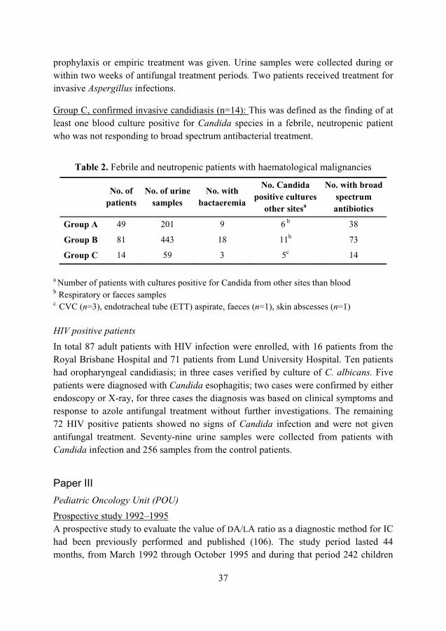
Group C, confirmed invasive candidiasis (n=14): This was defined as the finding of at least one blood culture positive for Candida species in a febrile, neutropenic patient who was not responding to broad spectrum antibacterial treatment.
Table 2. Febrile and neutropenic patients with haematological malignancies
No. of patients
No. of urinesamples
No. with bactaeremia
No. Candida positive cultures
other sitesa
No. with broadspectrum antibiotics
Group A 49 201 9 6 b 38
Group B 81 443 18 11b 73
Group C 14 59 3 5c 14
a Number of patients with cultures positive for Candida from other sites than blood b Respiratory or faeces samples c CVC (n=3), endotracheal tube (ETT) aspirate, faeces (n=1), skin abscesses (n=1)
HIV positive patients
In total 87 adult patients with HIV infection were enrolled, with 16 patients from the Royal Brisbane Hospital and 71 patients from Lund University Hospital. Ten patients had oropharyngeal candidiasis; in three cases verified by culture of C. albicans. Five patients were diagnosed with Candida esophagitis; two cases were confirmed by either endoscopy or X-ray, for three cases the diagnosis was based on clinical symptoms and response to azole antifungal treatment without further investigations. The remaining 72 HIV positive patients showed no signs of Candida infection and were not given antifungal treatment. Seventy-nine urine samples were collected from patients with Candida infection and 256 samples from the control patients.
Paper III Pediatric Oncology Unit (POU)
Prospective study 1992–1995A prospective study to evaluate the value of DA/LA ratio as a diagnostic method for IC had been previously performed and published (106). The study period lasted 44 months, from March 1992 through October 1995 and during that period 242 children

38
were admitted to the POU. Altogether 100 patients were included in the study, and 1076 urine samples were collected. Mean age of the children was nine years with an age range of 1-17 years. The upper cut-off level for DA/LA ratio in urine, for children with cancer was set at 4.6.
Table 3. Malignant diagnosis in patients at the POU
No. with acuteleukemia (%)
No. with lymphoma (%)
No. with Wilm´stumor (%)
No. with other malign. diagn. (%)
Prospective study
47 (47) 13 (13) 14 (14) 26 (26)
Retrospectivestudy
47 (56) 11 (13) 6(7) 20 (24)
Recommendations for the usage of DA/LA ratio in clinical practiceBased on the results of the prospective study, the physicians in the POU were recommended to use the DA/LA test as follows: i) Patients in the unit who were given cytotoxic chemotherapy should be monitored with urine DA/LA ratio at least twice weekly when the number of granulocytes in peripheral blood samples declined. ii) Elevated urine DA/LA ratio should not be left unattended, and based on the degree of granulocytopenia and fever not responding to broad-spectrum antibiotic treatment, elevated DA/LA ratios should lead to initiation of antifungal treatment or new DA/LA ratios should be obtained pending evaluation of the patient’s condition.
The region referring patients and the criteria for admitting patients to the unit remained unchanged between the prospective and retrospective study periods. There was no change in the policy on the use of prophylactic antifungal treatment, where oral nystatin was recommended and systemic prophylaxis with fluconazole, itraconazole or amphotericin B was to be avoided.
Retrospective study 1996–1999The retrospective study period of 44 months lasted from January 1996 through August 1999 and during that period 255 patients were admitted to the POU. Altogether 675 urine samples from 84 patients were sent for DA/LA analysis, which were all included in the study. A new method of collecting urine with a filter paper placed in the diaper enabled enrolment of younger children (127). Mean age of the children was seven years, and the age range was 0 – 18 years.
All study patients had malignant diseases and central venous catheters and received cytotoxic chemotherapy, see Table 3.
39
Neonatal Intensive Care Unit – NICU
Prospective study 1997–1999The prospective study described in Paper I lasted 15 months during which time 833 patients were admitted to the NICU; three patients from this study period had confirmed IC. Three other patients with IC who were diagnosed within a few months before or after the original study period were also included and the period of surveillance for IC was therefore extended to 20 months with 1118 admissions. Patients were included and divided into four groups as described in Paper I and gestational age and birth weight is shown in Table 1.
Recommendations for the usage of DA/LA ratio in clinical practiceAfter the prospective study at the NICU the physicians were recommended to use the urine DA/LA test as a complementary assay for diagnosing IC, but based on the results from the previous study at the NICU no specific recommendations concerning regular monitoring of newborns at risk were given.
The region referring patients and the criteria for admitting patients to the unit remained unchanged between the study periods, and there was no change in policy regarding prophylactic antifungal treatment.
Retrospective study 1999–2000During the study period of 20 months from May 1999 through December 2000, 1024 patients were admitted to the unit. DA/LA ratio in urine was analyzed in 172 samples from 60 infants. In addition, one infant with blood culture verified IC was included, although no urine samples for DA/LA ratio were obtained. Birth weight and gestational age of the infants is shown in Table 1.
Collection of clinical and microbiological data In Paper I and III the following was recorded from the study patient records at the NICU: disease diagnosis, gestational age, birth weight, skin and oral lesions likely to be caused by Candida, number of days with umbilical vein and percutaneous CVCs, antimicrobial treatment, and local and systemic antifungal treatment, outcome, date of death, autopsy results and cause of death.
In Paper III the following was recorded for both study periods from the study patients records at the POU: malignant disease diagnosis, periods with fever, periods with neutropenia (< 0.5 x 10⁹/L), treatment with broad-spectrum antibiotics and antifungal agents, outcome, date of death, autopsy results and cause of death.
40
Information on microbiological samples obtained, date of sampling, culture results and urine DA/LA ratios in Paper I and IV were collected from the database at the Division of Medical Microbiology.
Urine samples
Similar methods for collection of urine samples were used in Paper I and Paper II. The aim was to collect urine samples twice weekly. Most urine samples were collected on filter paper and dried on filter paper in both studies, which simplified the shipping of urine samples from Australia. The urine samples at the NICU were collected by placing a piece of filter paper in the diaper which was removed and dried after the infant had urinated. A few urine samples were collected in culture vials. Urine samples in culture vials were stored at -20°C and filter paper samples were stored at room temperature, pending DA/LA analysis by GC-MS. Urine culture was performed on urine samples arriving in culture vials.
GC-MS analysis
Sample preparation for analysis by GC-MS Ten µl of the sample to be analyzed (urine sample or broth) were dried under a stream of nitrogen. Hexane (200 µl) and trifluoroacetic anhydride (40 – 200 µl) were added, and the samples were heated at 80°C for 10 min in metal heating blocks, cooled to room temperature, and then dried again under a gentle nitrogen flow. Finally, 200 µl of a hexane-dichloromethane solution (1:1 [vol/vol]) was added; the sample was ready for analysis and 1 – 3 µl aliquot was used for the GC-MS analysis.
When urine samples on filter paper were analyzed, a piece of paper containing urine approximately two cm in diameter was cut from the filter paper and extracted in approximately three ml of methanol for 30 min. Of the solution, 300 to 600 µl aliquots were transferred to one ml vials and evaporated to dryness under a flow of nitrogen. Quantification of DA in Paper IV was done by using an in-house internal standard with xylitol (100 ng/�l).
Peak Urine DA/LA: The peak urine DA/LA ratio in Paper I and II was defined as the mean of the two highest values obtained within one week.
41
GC-MS A Trio-1S GC-MS system (VG, Manchester, United Kingdom) was used. The GC was Hewlett-Packard (Avondale, Pa.) model 5890 equipped with a splitless injector and a chiral column, which was 30 m by 0.25 mm (inner diameter) coated with a 0.25 mm-thick layer of cyclodextrin (Beta Dex-120; Supelco Inc., Bellafonte, Pa.). The column temperature was programmed to rise from 70 to 170°C at 7°C/min. The ion source temperature was 200°C. Helium was used as a carrier gas. Analyses were performed in the electron impact mode by using selected ion monitoring with an m/z of 519.
Candida and DA production
Growth curves and DA production Three strains of C. dubliniensis and two strains from each of the following species: C. albicans, C. tropicalis, C. krusei, C. glabrata and C. parapsilosis, were used for DA
measurements in different growth phases. The strains were collected from patients with IC. Growth curves were constructed over 48 hours to collect information on the timings of the lag phase, the exponential phase and the stationary phase, and DA was measured in all phases. The strains were cultured on Sabouraud agar for two days and suspended in 5 ml sterile isotonic NaCl. The yeast cells in the suspension were counted in a hemacytometer and the volume needed to reach a final concentration of 1500 cells/ml was calculated and added to 10 ml of broth. The culture medium used was RPMI 1640 broth (Sigma-Aldrich, Company, St. Louis, Mo.) supplemented with glucose 20 g/l and L-glutamine 0.3 g/l and buffered to pH 7.0 with MOPS (Sigma-Aldrich, Company, St. Louis, Mo.). The culture was incubated at 37°C shaking at 200 rounds per minute. RPMI 1640 broth was chosen for all experiments since it yielded the highest recovery of DA when compared to Difco Sabouraud Dextrose broth and Heart Infusion broth (both from BD Diagnostics, Sparks, USA),
To establish growth curves and determine DA production one-ml samples were collected from each broth after 4, 8, 12, 20, 24, 36 and 48 hours of incubation. The number of colony forming units (CFU) was determined at the sampling occasions by serially diluting 0.1 ml of a broth to 1:10 in sterile isotonic NaCl, plating 10-µl aliquots from the serial dilutions in duplicate on Sabouraud agar plates, and counting the number of colonies after culture for 48 hours. The mean number of colonies was used to express the number of CFU per ml. The remaining volume (0.9 ml) was

42
centrifuged at gravitational force 2571 for five minutes and the supernatant was removed and frozen at -20°C pending DA determination.
The study was performed at the Department of Microbiology at Landspitali University Hospital except for the DA analysis which was done at the Division of Medical Microbiology at Lund University Hospital.
DA production in different Candida species Candida strains from patients with invasive Candida infections in Iceland are stored at – 70°C at Landspitali University Hospital and strains were randomly selected from those collected during the period of January 1991 through December 2006. Altogether 21 C. tropicalis strains, 21 C. glabrata strains, 17 C. albicans strains, 10 C. dubliniensis strains, five C. parapsilosis strains, two strains of C. lusitaniae and one strain of each of the following species were included: C. guillermondii, C. famata and C. krusei. Additionally, 13 strains of C. krusei were obtained from the Swedish Institute for Infectious Disease Control, and the Karolinska University Hospital in Stockholm.
The strains were cultured on Sabouraud agar and incubated in supplemented RPMI 1640 broth using the same method as described for the growth curves. One-ml samples, collected from the broth after 8 and 24 hours and prepared as described above, were used to determine the number of CFU per ml and levels of DA.
Microbiology
Urine samples for DA/LA analysis arriving in culture vials were cultured on blood agar and Sabouraud agar at 36°C for 48 hours.
Paper I and III Different blood culture methods were used. From 1992 to 1996 a biphasic blood culture system (Septi-Check; Roche products, Skärholmen, Sweden) was used to culture bacteria and fungi. For detection of fungi, blood was cultured aerobically for five days at 37°C, followed by nine days at 30°C. From 1996 to 1997, BACTEC (Becton Dickinson & Co., Boul, Sweden) was used for blood cultures and from 1998 and onwards the BacT/Alert method (BioMerioux Products, Sweden) was used. Tissue samples were cultured on Sabouraud agar and on agar with 4% horse erythrocytes for

43
seven days at 30°C and 37°C. Species identification was done by testing for chlamydospores on rice agar and fermentation of glucose, galactose, saccharose, maltose, lactose and trelose.
Paper II The BacTAlert system (Organon Teknika, Durham, NC) was used at the Royal Brisbane hospital. Identification of C. albicans was performed by testing for germ tube formation. Candida species other than C. albicans were identified using the Vitek TBC identification system (bioMerieux Vitek, Hazelwood, MI) and if confirmation of identity was required, by the Api 20 C AUX system (BioMerieux sa, Marcy-l’Etoile, France).
Paper IV Species identification on all the Icelandic Candida strains in Paper IV was previously done using conventional methods with germ tube formation, CHROMagar and API 20C AUX (BioMérieux). Further identification on the Icelandic strains had also been done with PCR fingerprinting in a study by Asmundsdottir et al (13).
Species identification on the strains from Sweden was done with Bichro-Latex Krusei agglutination test (Fumouze Diagnostics, Levallois-Perret, France) or sequencing of the rDNA ITS1-5.8S-ITS2 region (140).
Statistical methods
Descriptive statistics are presented as the mean ± standard deviation (SD) for normally distributed data; the median and range were used otherwise. Differences between means were assessed by 2- tailed independent-samples (Student’s t test) or the Mann-Whitney test, when appropriate. P values of < 0.05 were regarded as statistically significant.
Comparisons between proportions were assessed by cross-tabulation (�2 or Fisher’s exact test, as appropriate). P values </0.05 were regarded as statistically significant.

44

45
RESULTS
Paper I
Healthy newborn infants
The urine DA/LA ratio for the 40 healthy full-term newborn infants was 2.5 ± 0.6 (mean ± SD) (range, 1.6 to 4.1). For the 16 mothers, it was 1.8 (range, 0.9 to 2.9), which was significantly lower than for their respective infants (P < 0.001).
NICU patients Eighteen infants included in the study had blood cultures positive for coagulase-negative staphylococci (n=15), Proteus mirabilis (n=1), Bacillus sp. (n=1), and Staphylococcus aureus (n=1). Malassezia furfur was isolated from a CVC tip in one patient. Urine culture on urine samples collected in culture vials showed no growth of bacteria or fungi. One hundred three infants were treated with antibiotics, and 96 had a CVC. Ten infants died.
Group A (n= 81)
These infants had no symptoms of Candida infection. The mean value of the DA/LA ratios for the whole group was based on calculating mean values for each patient during the course of the study. The mean urine DA/LA ratio was 2.7 ± 0.7 (mean ± SD), which was not significantly different from the mean ratio for the healthy full-term newborn infants. The upper cut-off level for NICU infants was set at 4.8 (group mean plus 3 SD). There was no correlation between gestational age or birth weight and urine DA/LA ratio. Three infants in this group died, all of causes other than IC.
Because of the heterogeneity of the infants with regard to gestational age and disease severity, a subgroup of very low-birth weight infants (< 1250 g, n=18) was selected to serve as a control group for infants with elevated urine DA/LA ratios in groups B and C when comparing treatment times with antibiotics and time with indwelling CVCs. These control infants’ gestational ages were 25 to 29 weeks (median, 26.5 weeks), their birth weights were 695 to 1175 g (median, 910 g), and they had a total treatment time with broad-spectrum antibiotics and indwelling CVCs of 11 days (median). The mean and median urine DA/LA ratio in this subgroup was 2.3 (range, 1.4 to 3.2).

46
Group B (n=22)
These infants had clinical signs of mucocutaneous candidiasis. For four infants, cultures from various locations were positive for C. albicans, but no systemic antifungal treatment was given. Two infants had elevated urine DA/LA ratios, and one of them had positive fungal cultures of urine obtained via an indwelling catheter. For both of these infants, urine DA/LA ratios fell to normal values after their CVCs had been removed or exchanged. These two infants had total times with indwelling CVCs of 41 and 12 days and total treatment times with broad-spectrum antibiotics of 41 and 21 days, respectively. Two infants in this group died, both of causes other than IC.
Group C (n=8)
Eight patients received empirical treatment with fluconazole (Table 4a and 4b). Five infants (no. 2, 4, 6, 7, and 8) had DA/LA ratios above the cut-off level. Three of these infants (no. 2, 4, and 6) had several elevated DA/LA ratios, which returned to normal during antifungal treatment, as shown by patient no. 2 in Fig. 1. The two other infants (no. 7 and 8) received empirical antifungal treatment only after urine sampling was discontinued. Three patients (no. 5, 6, and 7) had major congenital malformations. Five infants died, all from causes other than IC. The infants with elevated DA/LA ratios in groups B and C had significantly longer times with indwelling CVCs and longer treatment times with broad-spectrum antibiotics than the very low birth weight (<1250 g) infants in group A (P<0.01 and P<0.001, respectively).
Group D (n=6)
Six infants had IC based on clinical symptoms and C. albicans isolated from blood (n=5) or urine obtained by suprapubic aspiration (n=1). All six infants had at least one elevated DA/LA ratio (Table 5a and 5b). In four cases (no. 9, 10, 12, and 13), urine DA/LA ratios normalized during treatment with fluconazole (Fig. 2, patient no. 13), while the clinical conditions also improved. One patient (no. 14) also had a declining DA/LA ratio, but died from other complications of prematurity. One patient (no. 11) improved during treatment, but follow-up DA/LA ratios were not obtained. The infants in group D had significantly longer total treatment times with CVCs and broad-spectrum antibiotics than the control infants in group A (P<0.01 and P<0.01, respectively).
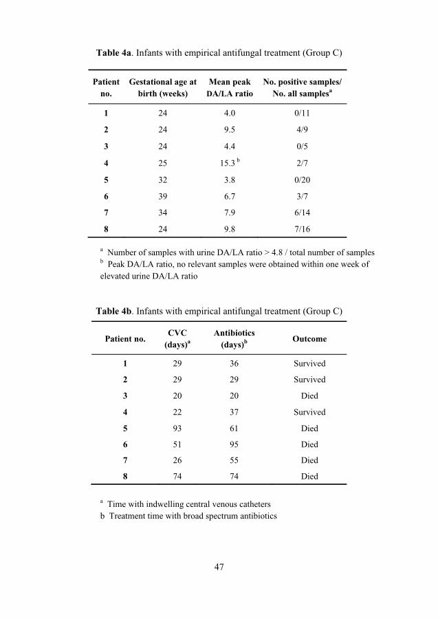
47
Table 4a. Infants with empirical antifungal treatment (Group C)
a Number of samples with urine DA/LA ratio > 4.8 / total number of samplesb Peak DA/LA ratio, no relevant samples were obtained within one week of elevated urine DA/LA ratio
Table 4b. Infants with empirical antifungal treatment (Group C)
a Time with indwelling central venous catheters b Treatment time with broad spectrum antibiotics
Patientno.
Gestational age at birth (weeks)
Mean peak DA/LA ratio
No. positive samples/ No. all samplesa
1 24 4.0 0/11
2 24 9.5 4/9
3 24 4.4 0/5
4 25 15.3 b 2/7
5 32 3.8 0/20
6 39 6.7 3/7
7 34 7.9 6/14
8 24 9.8 7/16
Patient no. CVC
(days)a Antibiotics
(days)b Outcome
1 29 36 Survived
2 29 29 Survived
3 20 20 Died
4 22 37 Survived
5 93 61 Died
6 51 95 Died
7 26 55 Died
8 74 74 Died

48
Table 5a. Infants with confirmed invasive candidiasis (group D)
PatientNo.
Gestational age atbirth (weeks)
Mean peak DA/LA ratio
No. pos. samples/ no. of all samplesa
9 25 5.4 2/6
10 24 7.9 4/12
11 24 5.8 b 1/7
12 27 9.3 3/20
13 27 9.1 3/7
14 24 30.0 3/3
a Number of samples with urine DA/LA ratio > 4.8 / total number of samplesb Peak DA/LA ratio, no relevant samples were obtained within one week of elevated urine DA/LA ratio
Table 5b. Infants with confirmed invasive candidiasis (group D)
Patient No.
Basis of diagnosis
CVCa
daysAntibiotics
Daysb Outcome
cause of death
9 3 blood cultures 23 23 Survived
10 1 urine culturec 24 24 Survived
11 1 blood culture 46 41 Survived
12 2 blood cultures 35 87 Died, Candida
endocarditis
13 3 blood cultures 24 24 Survived
14 2 blood cultures 19 19 Died, complications
of prematurity
a Time with indwelling central venous catheters b Treatment time with broad spectrum antibiotics c Sample was obtained by suprapubic aspiration

49
Figure 3 Urine DA/LA ratios for a patient no. 2 born after 24 weeks gestation. Empirical treatment with fluconazole was given from day 29 because of persistent fever and no response to broad spectrum antibiotic treatment. The vertical arrows indicate blood cultures taken on day 16 and 28 negative for Candida.
Figure 4. Urine DA/LA ratios for patient no. 13 with a gestational age of 27 weeks. He was reintubated at two weeks of age and had fever, severe thrombocytopenia, and elevated C- reactive protein. Vertical arrows indicate blood cultures positive for C. albicans. Blood cultures became negative on day 27 after removal of his CVC on day 24. Figure 3 and 4. The broken line at the value of 4.8 (mean urine DA/LA ratio for group A plus 3 SD) corresponds to the upper cut-off limit. The horizontal arrow on the y-axis, indicates the mean urine DA/LA ratio for group A at 2.7.
�
�
�
�
��
��
� � �� �� �� �� � �� �� ��
����
���
���
�����
����� ���
������������� ���������� �
!�"#���$����
�
�
�
�
��
��
�� �� �� �� � � �� �� �� �� �� �� �� �� � �
����
���
���
�����
����� ���
!�"#���$���
������������� ���������� �

50
Paper II
Haematological malignancy patients Febrile, neutropenic controls
Urine DA/LA ratios in 201 samples from 49 patients without antifungal treatment or prophylaxis was 2.0 ± 1.0 (mean ± standard deviation), and therefore, values >5.0 (mean + 3 SD) were defined as positive for this study. Two patients in this group had one single positive sample each, with DA/LA of 5.7 and 7.2, respectively, being regarded as false positives.
Empiric antifungal therapy or prophylaxis
Single positive determinations of 6.8, and 7.0 were found in two patients treated with fluconazole, and repeatedly positive DA/LA ratios were found in two patients on amphotericin B and fluconazole prophylaxis, respectively, where DA/LA peak values reached 13.6 and > 30, respectively.
Confirmed invasive candidiasis
Of the 14 patients with IC, 10 had urine samples collected either before or during the first three days of antifungal therapy. Candidiasis in these patients was due to C. albicans (n=3), C. krusei (n=4), C. tropicalis (n=2), and C. kefyr (n=1). DA/LA ratios were positive in five patients (range 5.5 – 32.0) with C. albicans (n=3), C. krusei (n=1), and C. tropicalis (n=1) fungemia. Normal DA/LA ratios were found in the other five patients with C. krusei (n=3), C. tropicalis (n=1) and C. kefyr (n=1) fungemia. The C. kefyr fungemia patient with normal DA/LA ratios was shown to have hepatosplenic (chronic) candidiasis by abdominal ultrasound that revealed multiple abscesses. In the remaining four patients with C. albicans, C. krusei, C parapsilosis and C. tropicalis fungemia, respectively, urine samples were all negative (range 1.6 – 2.8). However, in these four patients urine samples were not available until 4 – 12 days after blood cultures were positive and 4 – 10 days after antifungal therapy was instituted. Therefore, these four patients were considered as not evaluable, as DA/LA ratios are known to normalize rapidly during antifungal treatment (102; 106).
HIV positive patients The DA/LA ratio of the 72 HIV positive patients with no signs of Candida infection and no antifungal treatment was 2.0 ± 0.7 (mean ± standard deviation) which was not significantly different from the DA/LA results of the control cancer patients (see

51
above). Four patients with oral candidiasis were given antifungal treatment 3 – 7 days before urine sampling, and all showed negative DA/LA results (range 1.4 – 2.8). Only one patient with oral candidiasis had a positive peak DA/LA value of 7.1, the remaining five patients showed a mean DA/LA ratio of 3.3 (range 2.6 – 4.5). Two patients with Candida esophagitis and normal DA/LA ratios of 2.4 and 3.3 were also already on antifungal treatment when urine was sampled. The other three patients with esophageal candidiasis had not had antifungal therapy instituted when sampled, and they also showed normal values of 2.5, 2.7 and 3.9, respectively.
Paper III
Paediatric Oncology Unit (POU) During the prospective study period 33/242 (14%) patients were given antifungal treatment compared to 43/255 (17%) patients in the retrospective study (p>0.05). Empiric antifungal treatment was given to 23 patients (9.5%) in the prospective study and to 41 patient (16%) in the retrospective study (p<0.05). Two patients received prophylactic treatment with oral fluconazole and itraconazole, respectively, during the retrospective study period, while no such prophylaxis was given during the prospective study.
Invasive candidiasis was confirmed by blood culture in 10/242 (4.1%) patients admitted during the prospective study period, but only in 2/255 (0.8%) patients admitted during the retrospective study period – a difference which is statistically significant (p<0.05). In the prospective study five patients were diagnosed with C. albicans, three with C. parapsilosis, one with C. tropicalis and one patient with both C. glabrata and C. albicans. Elevated urine DA/LA ratios were detected 3 – 21 (median 8) days before the first positive blood culture was drawn (106). The two patients in the retrospective study were diagnosed with C. tropicalis and C. parapsilosis fungemia, and elevated DA/LA ratios were found 14 and 3 days, respectively, before the first positive blood culture was drawn. When an elevated urine DA/LA ratio was detected in the patient with C. tropicalis infection, treatment with liposomal amphotericin B was initiated the following day. Growth of C. tropicalis was thus detected after 13 days of antifungal treatment and the amphotericin B dose was increased when results from positive blood cultures were obtained. The patient with invasive C. parapsilosis infection received antifungal treatment on the same day as the first positive blood culture was collected.
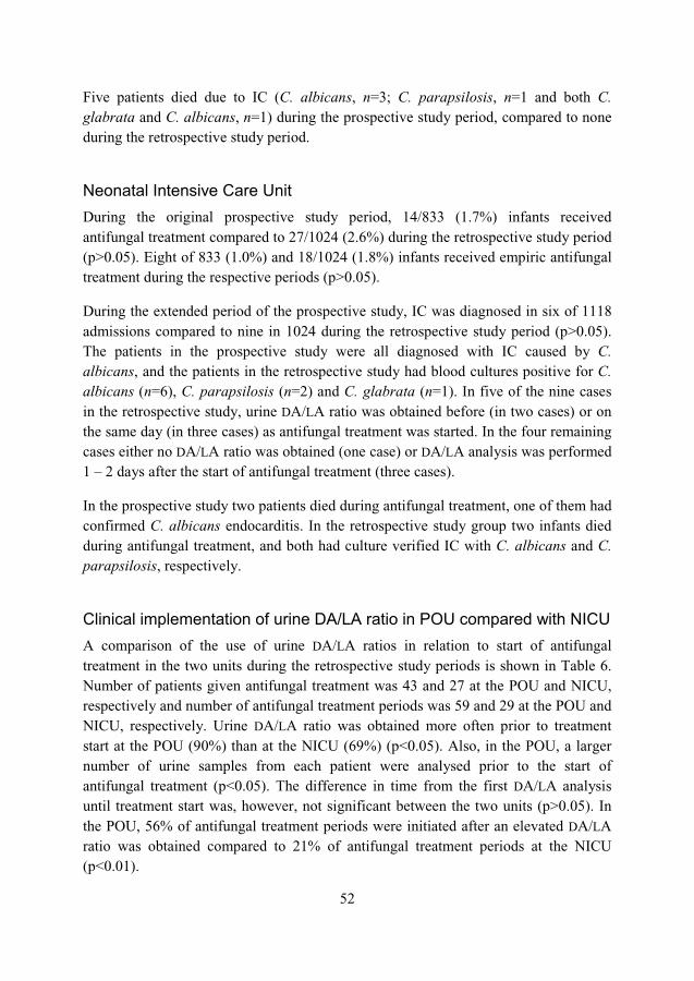
52
Five patients died due to IC (C. albicans, n=3; C. parapsilosis, n=1 and both C. glabrata and C. albicans, n=1) during the prospective study period, compared to none during the retrospective study period.
Neonatal Intensive Care Unit During the original prospective study period, 14/833 (1.7%) infants received antifungal treatment compared to 27/1024 (2.6%) during the retrospective study period (p>0.05). Eight of 833 (1.0%) and 18/1024 (1.8%) infants received empiric antifungal treatment during the respective periods (p>0.05).
During the extended period of the prospective study, IC was diagnosed in six of 1118 admissions compared to nine in 1024 during the retrospective study period (p>0.05). The patients in the prospective study were all diagnosed with IC caused by C. albicans, and the patients in the retrospective study had blood cultures positive for C. albicans (n=6), C. parapsilosis (n=2) and C. glabrata (n=1). In five of the nine cases in the retrospective study, urine DA/LA ratio was obtained before (in two cases) or on the same day (in three cases) as antifungal treatment was started. In the four remaining cases either no DA/LA ratio was obtained (one case) or DA/LA analysis was performed 1 – 2 days after the start of antifungal treatment (three cases).
In the prospective study two patients died during antifungal treatment, one of them had confirmed C. albicans endocarditis. In the retrospective study group two infants died during antifungal treatment, and both had culture verified IC with C. albicans and C. parapsilosis, respectively.
Clinical implementation of urine DA/LA ratio in POU compared with NICU A comparison of the use of urine DA/LA ratios in relation to start of antifungal treatment in the two units during the retrospective study periods is shown in Table 6. Number of patients given antifungal treatment was 43 and 27 at the POU and NICU, respectively and number of antifungal treatment periods was 59 and 29 at the POU and NICU, respectively. Urine DA/LA ratio was obtained more often prior to treatment start at the POU (90%) than at the NICU (69%) (p<0.05). Also, in the POU, a larger number of urine samples from each patient were analysed prior to the start of antifungal treatment (p<0.05). The difference in time from the first DA/LA analysis until treatment start was, however, not significant between the two units (p>0.05). In the POU, 56% of antifungal treatment periods were initiated after an elevated DA/LA ratio was obtained compared to 21% of antifungal treatment periods at the NICU (p<0.01).
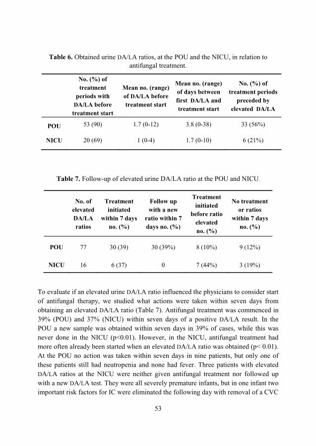
53
Table 6. Obtained urine DA/LA ratios, at the POU and the NICU, in relation to antifungal treatment.
Table 7. Follow-up of elevated urine DA/LA ratio at the POU and NICU.
To evaluate if an elevated urine DA/LA ratio influenced the physicians to consider start of antifungal therapy, we studied what actions were taken within seven days from obtaining an elevated DA/LA ratio (Table 7). Antifungal treatment was commenced in 39% (POU) and 37% (NICU) within seven days of a positive DA/LA result. In the POU a new sample was obtained within seven days in 39% of cases, while this was never done in the NICU (p<0.01). However, in the NICU, antifungal treatment had more often already been started when an elevated DA/LA ratio was obtained (p</0.01). At the POU no action was taken within seven days in nine patients, but only one of these patients still had neutropenia and none had fever. Three patients with elevated DA/LA ratios at the NICU were neither given antifungal treatment nor followed up with a new DA/LA test. They were all severely premature infants, but in one infant two important risk factors for IC were eliminated the following day with removal of a CVC
No. (%) of treatment
periods with DA/LA before
treatment start
Mean no. (range)of DA/LA before treatment start
Mean no. (range)of days between first DA/LA and treatment start
No. (%) of treatment periods
preceded by elevated DA/LA
POU 53 (90) 1.7 (0-12) 3.8 (0-38) 33 (56%)
NICU 20 (69) 1 (0-4) 1.7 (0-10) 6 (21%)
No. of elevated DA/LA ratios
Treatment initiated
within 7 days no. (%)
Follow up with a new
ratio within 7 days no. (%)
Treatment initiated
before ratio elevated no. (%)
No treatment or ratios
within 7 days no. (%)
POU 77 30 (39) 30 (39%) 8 (10%) 9 (12%)
NICU 16 6 (37) 0 7 (44%) 3 (19%)

54
and discontinuation of broad-spectrum antibiotic chemotherapy. Two patients had, however, indwelling CVC and broad-spectrum antibiotic treatment for 18 and 85 days, respectively, after an elevated DA/LA ratio was obtained.
Paper IV
Candida growth curves and DA production The lag phase lasted for 4 hours for all species except C. albicans, which had a lag phase of 8 hours followed by an exponential phase (Figure 5 and 6). The stationary phase took over from the exponential phase after approximately 20 hours. Only traces of DA were detected after 8 hours of incubation, but after 12 hours the levels of DA were high enough to be measured (Figure 7). The DA production rate (the amount of DA per CFU) decreased for most strains during the exponential phase but increased during the stationary phase, except for C. glabrata which did not produce any detectable amounts of DA.
Figure 5. 48 hours growth curves for C. albicans, C. tropicalis and C. parapsilosis
����%&��
����%&��
����%&��
����%&��
����%&��
����%&��
����%&��
����%&�
����%&�
� � �� �� �� �� � �� �� �� �� �
���
���
���
���
��
!����
' �����#��(��
' �����#��(��
' ������#���(��
' ������#���(��
' ������(���(�(��
' ������(���(�(��

55
Figure 6. 48 hours growth curves for C. dubliniensis, C. krusei and C. glabrata
Figure 7. D-arabinitol production per colony forming unit during 48 hours by five Candidaspecies.
����%&��
����%&��
����%&��
����%&��
����%&��
����%&��
����%&��
����%&�
����%&�
� � �� �� �� �� �
���
���
���
���
��
!����
' �)"�������(�(��
' �)"�������(�(��
' �*�"(����
' �*�"(����
' �*�"(����
' �+���������
' �+���������
�
���
���
���
���
���
� �� �� �� �� �
,���
+���
(��-�
����
��'!�
.�"�(
' �����#��(��
' �����#��(��
' �)"�������(�(��
' �)"�������(�(��
' �*�"(����
' �*�"(����
' �*�"(����
' ������(���(�(��
' ������(���(�(��
' ������#���(��
' ������#���(��
56
D-arabinitol production in different Candida species Sampling from the broth for DA analysis was done at the beginning of the exponential phase (eigth hours) and the stationary phase (24 hours). Only traces of DA were detected after eight hours of incubation. None of the C. glabrata strains produced any detectable amounts of DA, but DA production was seen in all other species examined(Table 8, Figure 8). The DA per CFU ratios differed considerably both between strains and species (Table 8, Figure 8). Significant differences were found between C. kruseiand C. albicans, between C. dubliniensis and C. albicans and between C. tropicalisand C. albicans with p< 0.05 on all three occasions.
Table 8. Production of D-arabinitol in nanograms per colony forming unit in nine Candida species after culture for 24 hours in RPMI broth
Candida species No. of strains
Median DA ng†/CFU†† Range DA ng /CFU
C. albicans 17 7.39 * 10-2 1.45 * 10-2 � 3.11 * 10-1
C. dubliniensis 10 1.38 * 10-2 6.44 * 10-3 � 6.38 *10-2
C. famata 1 3.30 * 10-3†††
C. glabrata 21 0
C. guillermondii 1 6.48 * 10-3†††
C. krusei 14 1.67 * 10-3 5.58 * 10-4 � 3.87 * 10-3
C. lusitaniae 2 8.57 * 10-3 7.36 * 10-3 � 9.78 * 10-3
C. parapsilosis 5 7.04 * 10-2 1.09 * 10-2 � 1.52 * 10-1
C. tropicalis 21 3.70 * 10-2 8.98 * 10-3 � 1.21 * 10-1
† ng: nanogram †† CFU: colony forming unit ††† Absolute value, mean not available

57
Figure 8. D-arabinitol production per colony forming unit by five Candida species after 24 hours inoculation
����%&��
����%���
����%���
����%���
����%���
����%���
����%���
����%���"
����
���
����
���
' �����#��( ' ������#���( ' ������(���(�( ' �)"�������(�( ' �*�"(��

58

59
DISCUSSION
Paper I and Paper III – Urine DA/LA ratio in neonates, clinical experience in neonates and children with cancer Compared to adults the healthy newborn infants and non-Candida-infected premature infants in NICU had significantly higher DA/LA ratios. At least one urine DA/LA ratio was positive in all newborn infants with confirmed IC. Follow up urine samples were available for five newborn infants who received either empiric treatment or treatment for confirmed infections, the DA/LA ratios declined during antifungal treatment, suggesting that urine DA/LA ratio can be used to monitor treatment effect. It was noted that CVC-associated fungal colonization could still be present although urine DA/LA ratios declined, probably due to decreased Candida tissue burden, as was observed for patient 13 in Figure 3. Therefore, DA/LA determinations should not replace but complement fungal blood cultures.
Seven out of 111 newborn infants (6.3%) without microbiologically proven IC had DA/LA ratios above the cut-off. These newborn infants were all either premature with a birth weight below 1250 g or had severe congenital malformations and thus belonged to risk groups for IC (141-143). Five of these newborn infants received empiric antifungal treatment and urine DA/LA ratios normalized during empiric treatment in three newborn infants in group C (no. 2, 4, and 6), which supports the clinical suspicion of IC. The remaining two newborn infants had mucocutaneous candidiasis and received only local antifungal treatment, but for these two newborn infants urineDA/LA ratios normalized after removal or exchange of CVC, indicating a possible microbiologically undiagnosed CVC-associated Candida infection. One of these two newborn infants had growth of Candida in urine obtained via an indwelling catheter, concurrent with the elevated DA/LA ratio. These seven newborn infants had in addition a significantly longer total treatment time with broad-spectrum antibiotics and indwelling CVCs than the very low-birth-weight controls.
Earlier studies have estimated a sensitivity of blood cultures for Candida of approximately 50% but information on the sensitivity of blood cultures in infants is sparse (56). If the five patients with a positive DA/LA ratio in the empirically treated newborn infants were assumed to have had invasive Candida infections, the sensitivity of blood culture would still be only 46%.

60
The study described in Paper I had several limitations. No urine samples for DA/LA ratio were obtained before the infections were microbiologically proven for three of the six confirmed cases. The first samples for DA/LA analysis for two of these three patients were obtained 1 – 2 days after the positive blood cultures but were, however, elevated in both cases. No DA/LA ratios were obtained in connection with positive blood cultures from one patient with C. albicans endocarditis but elevated DA/LA ratios were later detected and used to monitor treatment effect. For two cases the first DA/LA ratios were obtained before the infection was microbiologically proven and were elevated in both cases. Additionally, one patient with a single positive blood culture from a CVC had several negative DA/LA samples before the positive blood culture, but the last DA/LA sample before the infection was microbiologically confirmed was obtained six days before the positive blood culture. The next urine sample with a DA/LA ratio of 5.8, was taken eight days later and two days after the blood culture. The data collected in this study was therefore not adequate to evaluate DA/LA ratio in urine as a screening method for early detection of IC in neonatal infants.
Our conclusion is that urine DA/LA ratio could be used as a diagnostic test for infants at high risk or as a part of a “sepsis work-up” in suspected cases and could possibly result in earlier diagnosis of Candida infection.
In Paper III, we retrospectively studied how the test was clinically implemented at the NICU and the POU after the prospective studies were finished. Based on the results from the prospective studies, the recommendations for the everyday use of the test differed between the two units. At the POU, DA/LA analysis was suggested to play an important role in the early diagnosis of IC, as it was previously shown that urine DA/LA ratios increased several days before the first positive blood culture (106). This observation was interpreted as an indication of a successively increasing fungal load during a long period of immunosuppression mainly due to repeated courses of cytotoxic cancer chemotherapy. Thus, at the POU, regular monitoring of patients at high risk was recommended, and elevated ratios should prompt the institution of antifungal treatment or, at least, re-evaluation with new DA/LA analyses.
The sensitivity in diagnosing IC with urine DA/LA analysis in the NICU was as high as in the POU, but data collected at the NICU did not qualify to evaluate whether regular DA/LA monitoring would lead to an earlier diagnosis of IC. We also assumed that the sub-clinical phase between colonization, mucosal overgrowth, and the microorganism entering the bloodstream was probably short at the NICU, with limited role for screening and early detection of infection. Therefore, urine DA/LA analysis

61
was not recommended for regular monitoring of premature infants, but was encouraged to be used as a complementary diagnostic tool of IC.
We found that, the number of culture verified IC infections fell significantly between the two periods (10 vs 2 patients) in the POU, while empirical antifungal treatment was given significantly more often (23 vs 43 patients). An elevated DA/LA ratio was often followed by either institution of antifungal therapy (39%) or by a new DA/LA analysis (39%) within seven days. As patient characteristics remained unchanged between the two periods and systemic antifungal prophylaxis was rarely given, we concluded that early institution of antifungal therapy based on results from the regular urine DA/LA monitoring was the most likely reason for the decreased incidence of blood culture verified IC. The reduction in mortality from verified invasive Candidainfections during the second study period was encouraging, but needs to be confirmed over a longer observation period.
At the NICU, the incidence of confirmed IC and the use of antifungal treatment remained unchanged between the two study periods. During the latter period, urine samples were analyzed for DA/LA ratios more rarely and at a lesser number before antifungal treatment was commenced, compared to data from the POU, and it seemed that the DA/LA analysis was mostly used as a complementary diagnostic assay. This was in accordance with the recommendations given, but probably led to an under-utilization of the test. It is possible that had a policy been implemented at the NICU similar to that adopted at the POU for monitoring urine DA/LA ratio, the incidence of IC could have declined also in this clinical setting, but this remains to be shown. While 14-17% of children with cancer received antifungal therapy, the neonatal risk group is considerably smaller, and needs to be carefully targeted if a policy of regular monitoring is to be implemented.
Our study has some obvious limitations. Further evaluation was not planned when the test was put into clinical use which led to the problems with retrospective analysis. It was impossible to assess all decisions taken by the physicians on when to institute antifungal therapy, as patient records rarely contained such information. It is possible that earlier initiation of antifungal treatment using the DA/LA ratio in clinical routine at the POU, masked candidemias that had otherwise been verified and the overall mortality remained unchanged between the two periods. This is not supported when reviewing the causes of death in the empirically treated group; death in this group during the period with DA/LA ratio in clinical use was by causes other than IC and in those few cases with DA/LA ratios obtained close to the date of death, the ratios were normal.
62
Nevertheless, we concluded that the results of the previous prospective study of the new diagnostic assay was successfully implemented in clinical practice and even led to decreased morbidity in a well-defined group of high-risk patients, where the recommendations for the use of the test and the implications of a positive test were clearly defined. We also concluded that urine DA/LA ratio could be used in the diagnosis of IC in newborn infants. The method is more sensitive than culture and is probably highly specific. Empiric antifungal treatment should, however, never be postponed in cases with a negative urine DA/LA ratio, when IC is strongly suspected. An elevated urine DA/LA ratio should lead to new fungal cultures and repeated urine samples for DA/LA ratio. Risk factors such as CVCs or antibiotic treatment should be removed when possible and immediate initiation of systemic antifungal treatment considered.
Paper IV – DA production by C. glabrata
None of our C. glabrata strains in Paper IV produced detectable amounts of DA in vitro which is in agreement with results from previous in vitro studies (110; 111). Discrepancies between DA production by C. glabrata in vivo and in vitro is a hypothesis sometimes used in attempt to explain elevated DA/LA ratios in invasive C. glabrata infections, as has been previously discussed in the Introduction of this thesis. We therefore performed an experimental study to examine DA production by C. glabrata in vivo (unpublished data). Three New Zealand rabbits used in this study were immunosuppressed using intravenous cytarabine injections followed by intravenous injection of selected Candida strains, six to seven days after the immunosuppressive treatment was initiated. Rabbit A was infected with a C. albicansstrain from a patient with IC confirmed by positive blood cultures and high DA/LA ratios. Rabbits B and C were infected with a C. glabrata strain from a patient with C. glabrata fungemia who was later shown to have a co-infection with C. albicans, verified by postmortem cultures with growth of C. glabrata and C. albicans (106).
Rabbit A had DA/LA ratio above 30 on the 10th day and died in severe C. albicansinfection on the 11th day with an extremely high DA/LA ratio in urine, see Figure 9. The DA/LA ratios for rabbit B and C did not increase after infection with the C. glabrata strain was established. Rabbit B and C were not as severely affected by the infection as rabbit A and were euthanized after 15 and 20 days respectively.

63
Autopsy was performed on all three rabbits. Rabbit A had multiple small white spots in the internal organs and growth of C. albicans from kidneys, lungs, liver, pancreas and central venous catheters. Rabbits B and C had growth of C. glabrata from the same sites but no changes were observed macroscopically.
In vivo production of DA by C. glabrata was not detected in this small experimental study supporting in vitro findings on the absence of DA production by C. glabrata(110; 111). This suggests that elevated DA/LA levels in patients with verified invasive C. glabrata infections probably is caused by simultaneous infections with other DA producing Candida species not detected or identified in samples sent for culture.
Figure 9. DA/LA ratio in three immunosuppressed rabbits infected with C. albicansand C. glabrata at day six to seven
It is of interest that the DA/LA ratio declined on the seventh day for all three rabbits. A possible explanation is changes in dietary intake, but as 50 µg of DA in 1 g have been found in conventional rat chow (122). The rabbit feed used in our study could have contained DA and decrease in appetite as a side effect from the cytarabine treatment could therefore explain lower DA/LA ratios.
�
�
��
��
��
��
��
��
��
� � � � �� �� �� �� � �� ��
����
���
�����
����
�
"�����# ���#���#���������������$������
/��������0��1�' �����#��(
/������2�0��1�' �+�������
/������'�0��1�' �+�������

64
Paper II and IV – DA production and patients with haematological malignancy
In Paper IV we studied the DA production in several Candida species together with variation in DA production rate between Candida species and between strains within the same species. This was the first study to evaluate and confirm the DA production in C. dubliniensis but the production rate was significantly lower than that of C. albicans.No clinical studies on C. dubliniensis and DA/LA ratio in urine have been published and we therefore concluded that clinical studies were needed to assess the diagnostic performance of urine DA/LA ratio for invasive C. dubliniensis infections.
Surprisingly DA production rate (DA/CFU ratio) was reduced during the exponential phase compared to the stationary phase. A possible explanation could be increased DA consumption during the exponential phase compared to other phases. Candida is known to consume DA and the DA measured in the broth was the net amount of production and consumption (144). Another possibility is less viability or increased clumping of the cells during the stationary phase resulting in fewer colonies on culture, but with sustained DA production. The DA production continued during stationary phase, which contradicts previous negative DA/cr results in a clinical study by Walsh in patients with chronic hepatosplenic candidiasis (102).
There were differences in DA production rate in vitro both between Candida species and between strains within the same Candida species, the greatest variation was observed within C. albicans. The value of this observation can however not be predicted in vivo, and it is unknown whether these findings affect the sensitivity of the DA/LA ratio in the diagnosis of IC. This could imply that patients with IC caused by low producing Candida strains could have low or even normal urinary DA/LA ratio even with high fungal load. Clinical studies are needed to evaluate the benefit of measuring the DA/LA ratio in patients infected with different Candida species and simultaneously testing the DA production rate by the Candida strains causing the infection. Unfortunately many previous clinical studies did not emphasize the importance of the Candida species causing the infections. More research is therefore needed to examine the value of the test in different Candida species and to test DA production rate in the strains causing invasive Candida infections.
C. krusei was found to produce DA in vitro in Paper IV, but at a much lower rate than C. albicans, which could explain the discrepancies between the findings in previous studies on in vitro DA production by C. krusei and in clinical studies as previously

65
presented in the Introduction of this thesis. Four patients in Paper II had IC caused by C. krusei, of whom three patients had normal DA/LA ratios, but one had elevated DA/LA ratios. Considering the low DA production rate observed by C. krusei in Paper IV it is possible that patients with normal DA/LA ratios better reflect the expected results in patients with IC caused by C. krusei. The elevated DA/LA ratios observed in patients with C. krusei infections could have the same explanation as suggested for C. glabrata, i.e. an undetected co-infection with another DA producing Candida strain. But clinical data on C. krusei and DA/LA ratio in urine is limited and further research on DA/LA ratio in patients with invasive C. krusei infection is needed to evaluate the sensitivity of the DA/LA ratio in urine for diagnostic purposes.
Five of ten evaluable patients with IC in Paper II had elevated DA/LA ratios, of whom four were infected with Candida species previously known to produce DA/LA (three C. albicans and one C. tropicalis). We concluded that determination of urine DA/LA ratios in immunosuppressed adult patients with cancer could aid in the diagnosing of IC, provided that the infecting Candida sp. is producing DA. Normal urine DA/LA ratios were found in two cancer patients with invasive C. tropicalis and C. kefyr infection, respectively. The C. tropicalis strains included in Paper IV did, however, produce considerable amounts of DA, although the DA production rate was significantly lower compared to the C. albicans strains. The DA production rate for C. kefyr was not tested in our study but Bernard et al verified DA production by C. kefyralthough only three strains were tested (110). The patient with C. kefyr infection was diagnosed with chronic hepatosplenic candidiasis, a condition where DA/LA ratio is not considered to be as sensitive compared to patients with positive blood cultures (102). Considering the variation in DA production rate within Candida species observed in Paper IV it would have been interesting to test the in vitro DA production rate by these two strains but they were unfortunately not available.
The overall sensitivity in diagnosing IC was lower in Paper II than in previous prospective studies of children and adults with cancer (106; 102). One likely reason may be the high prevalence of C. krusei infections in our study hospital. An epidemiological study on candidemia at the Royal Brisbane Hospital for the period 1992–1999 showed a progressive increase in the proportion non-albicans Candida during this time (29). There was no change in the overall incidence of candidemia but a steady decline in the rate of C. albicans was observed with a corresponding rise in the incidence of non-C. albicans species; the increase in C. krusei infections was highly associated with fluconazole exposure (�2= 20.78, p<0.001). No evidence for spread of C. krusei using random amplification of polymorphic DNA was found, suggesting that the appearance of this organism was due to the selection pressure exerted by fluconazole. Other authors have described similar correlations between

66
widespread fluconazole use and increase in invasive infections caused by C. krusei(145; 146).
The increased prevalence of C. krusei infection at the Royal Brisbane Hospital could also partly explain the relatively low percentage of patients with positive DA/LA ratiosamong those given empiric antifungal chemotherapy or prophylaxis. Only four of 81 patients in this group had one or more positive DA/LA ratios, as compared to 12 of 23 patients on empiric therapy in our previous study of children with cancer at a department where no fluconazole prophylaxis was given (106).
There are some limitations to our study on the in vitro DA production rate. It was an in vitro study and did therefore not predict accurately the DA production in vivo. The production rate of Candida was, however, impossible to study in vivo because the fungal load in clinical cases could not be measured. Secondly, our method for measurement of the DA production rate relied on manually performed dilutions followed by culture on agar and counting of colonies, which is a cumbersome process and subject to human errors, which cannot be totally prevented. The reproducibility of the method was not tested, but our analysis of the data does not indicate errors that would affect our results. This is to our knowledge, the best method to determine the production rate.
There was a variation in the DA production rate within species with the greatest variation seen in C. albicans. Although a possible inaccuracy of our method cannot be excluded, a study by Bernard et al. showed that DA production rates varied over a wide range within species, and the largest variation was seen for C. albicans, which is in accordance with our findings (110). Intra-species differences are well known for other metabolic profiles. A study using the p-nitrophenol (pNP) as an indicator of enzyme activity to distinguish between Candida species, showed wide variation in the pNP concentration within C. albicans (147). Likewise, differences in metabolic profiles within species are often observed in tests for species identification that are based on the ability of the yeast to utilize carbohydrates by assimilation or fermentation.
Further studies on DA production rate and possible strain variation using genotyping for identification of strains would be valuable. Molecular epidemiological studies have shown that nosocomial clusters of infections due to the same Candida strain can persist for months or even years in hospitals (13; 19). Local nosocomial clusters caused by strains with low DA production rate could lead to disappointing results with frequent false negative DA/LA ratios in patients with IC.

67
Paper II – DA in HIV patients
There are no previous studies on DA in the diagnosis of IC in patients with HIV infection and a small study was conducted to explore whether DA/LA ratio would aid in the diagnosis of patients with Candida esophagitis. However, due to the introduction of HAART (highly active anti-retroviral treatment) at the same time as this study was commenced, the number of HIV positive patients with Candida esophagitis and also with oral candidiasis decreased dramatically. Only three patients with esophagitis and six patients with oral candidiasis were included, and all but one patient with oral candidiasis were DA/LA negative. HIV positive patients without clinical signs of Candida infection have DA/LA ratios similar to healthy controls and non-infected immunosuppressed cancer patients.
The main limitation of this study is the lack of culture verification and identification of the Candida species causing the infections. C. glabrata and C. krusei were possible pathogens in these patients resulting most probably in negative DA/LA ratios; verification of DA producing Candida species causing the infections would have increased the value of this study. However, these results indicate that DA/LA ratios are neither increased in mucocutaneous infections nor in localized non-disseminating infections such as esophagitis.
Pitfalls using DA/LA ratio in urine in the diagnosis of invasive candidiasis
We have considered possible pitfalls using DA/LA ratio in urine as a diagnostic method for IC. We wondered whether factors such as conditions for storage of urine samples pending analysis, dietary intake of DA, or diseases like severe kidney failure or diabetes mellitus could affect the DA/LA ratio resulting in false negative or false positive test results. We performed several studies to examine whether any of these factors were of concern. The results, not published previously, are summarized in the following section.
Storage of samples pending analysis Klebsiella species are well known pathogens in urinary tract infections and may also contaminate the urine at the time of sampling (148). Klebsiella are more often isolated from urine cultures from inpatients compared to outpatients (148). More than 90% of

68
strains of K. pneumoniae and K. oxytoca carry the genes for DA catabolism and produce the enzyme D-arabinitol dehydrogenase (DADH); in comparison, only 5% of E. coli strains carry these genes (149). It is possible that catabolism of DA in urine samples with Klebsiella can result in normal or low DA/LA ratios in urine from patients with invasive candididasis and elevated DA/LA ratios in serum.
Table 9. Urine samples with growth of Klebsiella pneumoniae
Cell count/ml DA/LA
on arrival DA/LA after 24 hours in room temperature
> 100,000 4.2 0.03
> 100,000 2.6 0.12
> 100,000 1.8 0.05
> 100,000 0.6 0.01
> 100,000 2.6 0.04
> 100,000 0.17 0.02
10,000 – 20,000 2.4 2.4
Urine samples with growth of K. pneumoniae or E. coli were collected from samples sent in clinical routine to the Department of Microbiology at Landspitali University Hospital in Iceland. Samples were obtained on filter paper for DA/LA analysis. Separate aliquots of the same samples were stored at room temperature for 24 hours and then applied on filter papers for DA/LA analysis. Median DA/LA ratio was 2.2 (range 0.17 – 4.2) in six urine samples with growth of K. pneumoniae >100,000 cells/ml but declined to median 0.035 (range 0.01 - 0.12) after 24 hours storage in room temperature, see Table 9. One urine sample, obtained approximately 20 hours before receipt in the laboratory, had DA/LA ratio 0.17 on arrival in the laboratory. There was, however, no change in DA/LA ratio in one urine sample with lower cell counts of K. pneumoniae, see Table 9.
Nine urine samples with growth of E. coli >100,000/ml, collected as control samples,showed no change in DA/LA ratio after 24 hours storage at room temperature with median DA/LA ratios 1.8 and 1.9, before and after storage, respectively.
Our conclusion is that storage of urine samples with growth of Klebsiella at room temperature may result in false negative urine DA/LA ratios in patients with IC.
69
DA/LA ratio in patients receiving ambulatory hemodialysis Urine samples were collected from 24 patients receiving ambulatory hemodialysis to examine if the hemodialysis affected the DA/LA ratio in these patients. The mean DA/LA ratio was 3.0 ± 1.0 (mean ± standard deviation), range 1.8 – 5.9. The mean DA/LA ratio for hospitalized children with cancer was 2.5 ± 0.7, the difference between mean DA/LA ratio for these groups was significant, (p<0.05) (106). The reason for this difference remains however, obscure. These results suggest setting the upper cut-off DA/LA limit slightly higher for patients receiving hemodialysis than in patients with normal kidney function.
Patients with diabetes mellitus – Candida and glucose in urine samples Diabetes mellitus is a predisposing factor for candiduria (150). Previous research has shown that urine colonized with Candida does not result in elevated DA/LA ratios (95). However, Wong et al found that C. albicans converts glucose to DA (144). Patients with diabetes mellitus are at higher risk for simultaneous candiduria and glycosuria with theoretically increased risk for false positive DA/LA ratios.
Urine samples were collected from 35 patients with diabetes mellitus at the outpatient clinic for patients with diabetes mellitus at the University Hospital in Lund, of whom seven patients had growth of Candida in urine. The mean urinary DA/LA ratio was 2.5 ± 1.2 (mean ± standard deviation) for all 35 patients, which is very similar to the mean urinary DA/LA ratio 2.5 ± 0.7 in 95 hospitalized children with cancer but significantly higher than mean the urinary DA/LA ratio of 2.0 ± 0.6 in 56 healthy children (p<0.01) (106).
In conclusion, care should be exercised when interpreting DA/LA results from individuals with diabetes mellitus since the presence of Candida and glucose simultaneously in urine samples can result in elevated DA/LA ratios.
Consumption of food products containing D-arabinitol D-arabinitol (DA) is readily absorbed from the gastrointestinal tract as was demonstrated in an report by Wong et al in 1990 (122). The authors showed that rats fed on chow containing DA had higher levels of DA in serum and urine compared to rats on DA free diet. They also reported results from a human pilot study with one volunteer who consumed 1 g of DA and excreted 85% of the DA within 24 hours.
There is little information on the presence of DA in food products. We have demonstrated that fermented liquid products such as soy sauce, red and white wine,

70
and beer contain large amounts of DA. Lower levels of DA were found in dairy cow milk. Nothing has been published on the effect dietary DA might have on DA/LA ratio in urine in humans.
Two adult volunteers (male and female) were asked to consume 750 ml of red wine over a two-hour period. The male had urine DA/LA ratio of 1.3 immediately before consumption; samples that were obtained 5 and 15 hours after the consumption stopped were 7.8 and 5.0, respectively. The other individual (female) had DA/LA ratios of 1.4, 7.7, and 1.8 at approximately the same time points.
A third volunteer consumed soy sauce (8 mg) on four occasions within 12 hours. Urine DA/LA ratio was 2.3 before the experiment started. DA/LA ratios were 4.8, 5.7, 4.9 and 4.8, after each consumption, and was back to 2.3, 10 hours after the last soy sauce consumption.
Our results show that consumption of food products containing DA can elevate the urine DA/LA ratio and should be considered when elevated DA/LA ratios are detected unexpectedly.

71
CONCLUSIONS
• DA/LA ratio is a reliable diagnostic method of invasive candidiasis in neonatal infants and can be used to monitor treatment in this patient group.
• DA/LA ratio does not replace other diagnostic methods but is a complement to blood cultures and tissue cultures.
• Screening neutropenic children with cancer with DA/LA ratio decreases the number of verified invasive Candida infections.
• HIV positive patients seem to have DA/LA ratios similar to healthy controls and non-infected immunosuppressed cancer patients.
• The changing epidemiology of candidemia with consequent increase in the proportion of fungemia due to C. krusei could result in lower sensitivity of the DA/LA ratio locally.
• DA/LA ratios are probably not elevated in mucocutaneous infections or in localized non-disseminating infections such as esophagitis.
• C. dubliniensis and C. krusei produce DA in vitro, although the production rate by C. krusei is low which could result in lower sensitivity of the urinary DA/LA ratio in patients with IC caused by C. krusei.
• C. glabrata does not produce any detectable amounts of DA in vivo or in vitro. • There is an intra- and inter species variation of DA production, which warrants
further studies on the DA metabolism in clinically relevant Candida species. • Storage of urine samples with growth of Klebsiella at room temperature may result
in false negative urine DA/LA ratios in patients with invasive candidiasis. • Consumption of food products containing DA can elevate the urine DA/LA ratio
and should be considered when elevated DA/LA ratios are detected unexpectedly
72
POPULÄR VETENSKAPLIG SAMMANFATTNING PÅ SVENSKAInvasiv candidiasis är en allvarlig jästsvampinfektion hos redan svårt sjuka patienter och denna infektion har ökat under de senaste årtiondena. Blododling, som är referensdiagnostisk metod för denna infektion, har relativt låg känslighet liksom detektion av Candidaspecifika antikroppar och antigener. Utvecklingen av PCR-baserade diagnostik går långsamt och det saknas standardiserade metoder.
D-arabinitol (DA) är en sockeralkohol som produceras in vitro och in vivo av de flesta humanpatogena Candida arter. DA och L-arabinitol (LA) förekommer i samtliga humana kroppsvätskor och utsöndras i urinen. Vid njursvikt stiger DA och LA koncentrationerna i serum, och för att korrigera för nedsatt njurfunktion mäts antingen DA/LA- eller DA/kreatinin kvot. DA/LA kvoten kan mätas i både urin och serum medan DA/kreatinin kvoten endast mäts i serum. DA/LA-kvoten som analyseras med gaskromatografi-mass spektrometri är normalt 2,0 ± 0,6 hos vuxna och något högre hos barn. DA-koncentrationen kan även mätas med en enzymatisk metod, där man utnyttjar enzymet D-arabinitoldehydrogenas och relaterar DA till kreatinin nivåer.
Flera kliniska studier har visat hög känslighet (83–94 procent) med DA-metoden vid diagnostik av invasiva Candida infektioner. Man har observerat korrelation mellan behandlingseffekt och sjunkande DA/kreatininkvoter, liksom mellan mortalitet och kvarstående förhöjda DA/kreatinin kvoter. Förhöjda DA nivåer har även detekterats flera dagar till veckor innan man med konventionella odlingsmetoder har kunnat påvisa invasiv candidiasis.
Prematura barn på intensivvårdsavdelningar har ökad risk för invasiv candidiasis, varför vi genomförde en klinisk prospektiv studie av den diagnostiska nyttan av DA/LA kvot i urin på den neonatala intensivvårdsavdelningen i Lund. Urinprov togs enkelt genom att lägga en bit filtrerpapper i blöjan. Filtrerpapperet avlägsnades när barnet hade kissat och lufttorkades innan det skickades för analys till laboratoriet. Vi kunde påvisa en 100-procentig känslighet i diagnostiken av invasiv candidiasis, och mer än hälften av de barn som fick empirisk svampbehandling hade också förhöjda värden. Dessutom normaliserades DA/LA-kvoten i urin vid framgångsrik svampbehandling både hos barn med odlingsverifierade infektioner och hos empiriskt behandlade barn.
En liknande prospektiv studie utfördes på vuxna cancerpatienter, främst med leukemi och lymfom, i Brisbane, Australien samt på HIV-positiva patienter i Lund och Brisbane. Våra resultat visade att DA/LA metoden hade en lägre känslighet hos patienterna i Brisbane jämfört med hos de nyfödda barnen i Lund. Detta förklaras

73
sannolikt av den lokala epidemiologiska situationen på sjukhuset i Australien, där C. krusei har blivit en allt vanligare orsak till invasiva Candida infektioner. Enligt resultat från vår senaste studie var nämligen bildandet av DA i provrör mycket låg hos C. krusei jämfört med andra patogena Candida arter. HIV patienter hade normala DA/LA kvoter och patienter med mukokutan candidiasis, exempelvis HIV-positiva patienter med oral candidiasis eller Candida esofagit, hade normala DA/LA -kvoter.
Efter att den prospektiva studien på den neonatala intensivvårdsavdelningen hade avslutats infördes användning av DA/LA kvot i urin i kliniskt rutinbruk. En liknande prospektiv studie hade tidigare genomförts på barnonkologiska avdelningen i Lund där DA/LA kvot också infördes i kliniskt rutinbruk efter studieperioden. I vår tredje studie granskade vi hur DA/LA kvoten verkligen användes i praktiken på dessa två avdelningar. Vi studerade även incidensen av och mortaliteten i invasiv candidiasis vid den barnonkologiska- respektive den neonatala intensivvårdsavdelningen i Lund direkt efter att de prospektiva studierna avslutats. På barnonkologen rekommenderades regelbunden provtagning två gånger i veckan på alla riskbarn, positiva DA/LA-kvoter skulle användas som incitament för tidigt insättande av svampbehandling oavsett förekomst av positiva blododlingar. På neonatalavdelningen, där de prematura barnen ofta insjuknar redan under de första levnadsveckorna, blev regelbunden DA/LA-monitorering på alla riskbarn inte rekommenderad, utan analysen skulle användas mer som ett komplement till övrig diagnostik. Den prospektiva barnonkologstudien pågick under 3,5 år, och under den följande 3,5-årsperioden efter studien, sjönk incidensen av invasiv candidiasis signifikant. Även mortaliteten i invasiv candidiasis minskade från 50 procent till 0, men för små numeriska värden medgav ingen rimlig statistisk analys. På neonatala intensivvårdsavdelningen var emellertid incidensen och mortaliteten i invasiv candidiasis oförändrad under uppföljningsperioden. Patientklientelet och alla övriga medicinska förhållanden förblev oförändrade mellan perioderna på båda avdelningarna. Dessa resultat stärker vår övertygelse att man genom att använda resultaten från regelbundna DA/LA-analyser av riskpatienter i det vardagliga kliniska arbetet kan minska såväl morbiditet som mortalitet vid invasiv candidiasis.
Vid granskning av litteraturen avseende DA produktionsförmåga hos olika Candidaarter upptäcker man att endast få stammar av olika arter har testats för DA produktion. Vi genomförde därför en studie där vi testade ett större antal stammar och verifierade avsaknad av DA produktion in vitro hos C. glabrata. Vi verifierade också DA produktion hos C. dubliniensis, och fann att C. krusei producerar låga mängder DA. Vi upptäckte även skillnader i DA produktionen inom och mellan olika Candida arter men betydelsen av detta fynd avseende DA/LA-metodens känslighet in vivo är osäker. Sänkt känslighet vid infektion med lågproducerande Candida stammar kan dock inte uteslutas och vidare studier där DA produktionsförmågan hos kliniska stammar jämförs med patienternas DA/LA kvoter efterlyses.

74
Förhöjda kvoter har rapporterats hos patienter med djupa C. glabrata infektioner även om man inte har påvisat någon DA produktion hos C. glabrata in vitro. Följande hypoteser har använts som tänkbara förklaringar: samtidigt pågående infektion med två eller fler Candida arter som inte upptäcks pga låg känslighet i blododlingar, eller att C. glabrata producerar DA in vivo trots avsaknad av DA produktion in vitro. För att undersöka tänkbar DA produktion hos C. glabrata in vivo genomförde vi en experimentell studie på tre immunsupprimerade kaniner, varav en infekterades med C. albicans och utvecklade mycket höga DA/LA kvoter medan de två andra kaninerna som blev infekterade med C. glabrata hade fortsatt låga kvoter. Någon DA produktion hos C. glabrata kunde således inte påvisas in vivo.
Slutligen så genomförde vi två mindre försök om tänkbara felkällor i DA/LA diagnostiken. I det ena försöket konstaterade vi att förvaring av urinprov, med växt av Klebsiella, i rumstemperatur kan leda till falskt låga DA/LA kvoter som förklaras av Klebsiellabakteriens förmåga att bryta ner DA. Dietärt DA resorberas i mag–tarmkanalen och i den andra studien om felkällor visade vi att vid intag av fermenterade livsmedel (kinesisk soja) eller dryck (rödvin) stiger DA/LA-kvoten, om än snabbt övergående. Vi tror dock inte att detta ska störa diagnostiken av de definierade riskgrupperna, men det är viktigt att inte använda DA/LA-analysen okritiskt.

75
ACKNOWLEDGEMENTS
I wish to express my sincere gratitude to those who have contributed to this work. To all my collaborators, colleagues and friends in Lund and Reykjavík, but especially:
My supervisor, Bertil Christensson, for your support and patience to guide me through this work. For allowing me to do things my way and not letting the geographical distance get in the way.
Lennart Larsson, my co-supervisor, for all your advice and your important contribution to the methodological part of the work.
Christina Pehrson, my co-author and collaborator at the Division of Medical Microbiology in Lund for your positive and patient guidance, for always being flexible, and for our good moments together.
Einar Larsson, my collaborator, for your help with finding important data at the Division of Medical Microbiology in Lund.
My co-authors Lars Björklund and Tomas Wiebe for your important participation in the studies at the Department of Neonatology and Pediatric Oncology in Lund, and your valuable scientific contribution to the work.
Kristina Håkansson, my co-author from the Department of Neonatology in Lund, for your enthusiasm in recruiting newborns to the study and help with managing the samples.
Damon P. Eisen, Paul B. Bartley and William Hope my co-authors from Brisbane Australia, and Erja Chryssanthou at the Department of Clinical Microbiology, Karolinska University Hospital in Stockholm for their fruitful long distance co-operation.
Helga Erlendsdóttir, my co-author and collaborator at the Department of Microbiology in Reykjavík. Thank you for helping me through the last and perhaps the most difficult part of this work. Thank you for your patience and your encouraging, warm and positive guidance, it made all the difference
Ingibjörg Hilmarsdóttir, my co-author and colleague at the Department of Microbiology in Reykjavík for your encouragement, your valuable scientific contribution, and for being accurate and thorough when I needed it most.
Haraldur Briem, my chief at the Centre for Health Security and Communicable Disease Control, for your patience, faith in me and never failing support.
Karl G. Kristinsson, my chief at the Department of Microbiology in Reykjavík, for allowing me to use the facilities at the laboratory, and thereby making the last part of this work possible.

76
Erla Sigvaldadóttir, my collaborator at the Department of Microbiology at Landspitali University Hospital for always having all the solutions to the practical problems I encountered at the laboratory.
To my co-workers Guðrún Svanborg, Hjördís, Hörður, Ólafur and Sigfús at the laboratory, and Ása, Júlíana and Þórólfur at the chief epidemiologists office and all the others for being there and making every-day work interesting and fun.
My friends, for your encouragement and for being my friends.
Sif Ormarsdóttir my friend and colleague, who although very busy took the time to proofread this work, highly appreciated and needed.
My mother Guðlaug and my father Sigmundur, my brother Sigurgeir and his wife Hildur and to my sister Sirrý and her husband Hermann and their families for always being there when needed.
My children, Gulla, Fríða and Magnús Atli, my son in law Hlynur Daði, my grandson Egill Arnar and last but not the least my husband Gylfi for all your patience and support in every possible way. I dedicate this work to you although I know what makes you really happy is that it has come to an end.
This work was supported by Bergþóra Magnúsdóttir and Jakob J. Bjarnason memorial fund, Region Skåne and the Medical Faculty at Lund University.

77
REFERENCES
1. Hazen, K C and Howell, S A. Candida, Cryptococcus, and other yeasts of medical importance. Murray PR, Baron EJ, Jorgensen JH, Landry ML, Pfaller MA. Manual of Clinical Microbiology. 9th ed. Washington DC:ASM Press c2007, Vol. 2, chapter 119, 1762-1788.
2. Edwards JE. Candida species. Bennett JE, Dolin R, Mandell GL. Principles and Practice of Infectious Diseases. 6th ed. Philadelfia : Churchill Livingstone c2005, chapter 257, 2938-2951.
3. Pfaller MA, Diekema DJ. Rare and emerging opportunistic fungal pathogens: concern for resistance beyond Candida albicans and Aspergillus fumigatus. J Clin Microbiol 2004: 42;4419-4431.
4. Hajjeh RA, Sofair AN, Harrison LH, Arthington-Skaggs BA, Mirza SA, Phelan M, et al. Incidence of bloodstream infections due to Candida species and in vitro susceptibilities of isolates collected from 1998 to 2000 in a population-based active surveillance program. J Clin Microbiol 2004; 42:1519-1527.
5. Ostrosky-Zeichner L, Larsen RA, Rex JH, Pappas PG, Hamill RJ, Horowitz HW et al. Antifungal susceptibility survey of 2,000 bloodstream Candidaisolates. Antimicrob Agents Chemotherap 2003; 47: 3149-3154.
6. Calderone RA. Taxonomy and Biology of Candida. Candida and Candidiasis. 1st ed. Washington DC : ASM Press c2002, chapter 2, 15-27.
7. Berg, Alfred. Om torsk hos barn. L.J. Hjerta 1846.
8. Chen S, Slavin M, Nguyen Q, Marriott D, Playford EG, Ellis D, et al. Active surveillance for candidemia, Australia. Emerg Infect Dis 2006; 12:1477-1630.
9. Sandven P, Bevanger L, Digranes A, Haukland HH, Mannsåker T, Gaustad P and the Norwegian Yeast Study Group al. Candidemia in Norway (1991 to 2003): Results from a Nationwide Study. J Clin Microbiol 2006; 44:1977-1981.
10. Kao AS, Brandt ME, Pruitt WR, Conn LA, Perkins BA; Stephens DS, et al. The epidemiology of candidemia in two United States cities: results of a population-based active surveillance. Clin Infect Dis 1999; 29:1164-1170.
11. Benjamin DK, Stoll BJ, Fanaroff AA, McDonald SA, Oh W, Higgins RD, et al. Neonatal candidiasis among extremely low birth weight infants: risk factors, mortality rates, and neurodevelopmental outcomes at 18 to 22 months. Pediatrics 2006; 117:84-92.

78
12. Eggiman P, Garbino J, Pittet D. Epidemiology of Candida species infections in critically ill non-immunosuppressed patients. Lancet Infect Dis 2003; 3:685-702.
13. Asmundardottir LR, Erlendsdottir H, Gottfredsson M. Increasing incidence of candidemia: Results from a 20-year nationwide study in Iceland. J Clin Microbiol 2002; 40:3489-3492.
14. Wisplinghoff H, Bischoff T, Tallent SM, Seifert H, Wenzel RP, Edmond MB. Nosocomial blood stream infections in US hospitals: analysis of 24,179 cases from a prospective nationwide surrveillance study. Clin Infect Dis 2004; 39:309-317.
15. Martin GS, Mannino DM, Eaton S, Moss M. The epidemiology of sepsis in the United States. N Engl J Med 2003; 348:1546-1554.
16. Blumberg HM, Jarvis WR, Soucie JM, Edwards JE, Patterson JE, Pfaller MA et al. Risk factors for candidal bloodstream infections in surgical intensive care unit patients: The NEMIS prospective multicenter study. Clin Infect Dis 2001; 33:177-186.
17. Pfaller M, Jones RN, Messer SA, Edmond MB, Wenzel RP and the SCOPE Participant group. National surveillance of nosocomial blood stream inf. due to species of Candida other than Candida albicans: frequency of occurrence and antifungal susceptibility in the SCOPE program. Diagn Microbiol Infect Dis 1998; 30:121-129.
18. Yildrim M, Sahin I, Kucukbayrak A, Ozdemir D, Yavus MT, Sukru O, et al. Hand carriage of Candida species and risk factors in hospital personnel. Mycoses 2008; 50:189-192.
19. Viviani MA, Cogliati M, Esposto MC, Prigitano A, Tortorano AM. Four year persistence of a single Candida albicans genotype causing bloodstream infections in a surgical ward proven by multilocus sequence typing. J Clin Microbiol 2006; 44: 218-221.
20. Kung, H-C, Wang J-L, Chang S-C, Wang J-T, Sun H-Y, Hsueh P-R, et al. Community-onset candidemia at a university hospital, 1995-2005. J Microbiol Immunol Infect 2007; 40:355-363.
21. Macphail GL, Taylor GD, Buchanan-Chell M, Ross C, Wilson S, Kureishi A. Epidemiology, treatment and outcome of candidemia: a five-year review at three Canadian hospitals. Mycoses 2002;45:141-145.
22. Pfaller MA, Diekema DJ, Jones RN, Sader HS, Fluit AC, Hollis RJ, et al. International surveillance of bloodstream infections due to Candida species: frequency of occurrence and in vitro susceptibility to fluconazole, ravuconazole, and voriconazole of isolates collected from 1997 through 1999 in
79
the SENTRY antimicrobial surveillance program. J Clin Microbiol 2001; 39:3254-3259.
23. Pfaller MA, Diekema DJ. Epidemiology of invasive candidiasis: a persistent public health problem. Clin Microbiol Rev 2007; 20:133-163.
24. Pappas PG, Rex JH, Lee J, Hamill RJ, Larsen RA, Powderly W, et al. A prospective observational study of candidemia: Epidemiology, therapy and influences on mortality in hospitalized adult and pediatric patients. Clin Infect Dis 2003; 37:634–43.
25. Saiman L, Ludington E, Pfaller M, Rangel-Frausto S, Wiblin T, Dawson, et al. Risk factors for candidemia in neonatal intensive care unit patients. Ped Infect Dis J 2000; 19:319-324.
26. Stoll BJ, Hansen N, Fanaroff AA. Late-onset sepsis in very low birth weight neonates: the experience of the NICHD Neonatal Research Network. Pediatrics 2002; 110:285-291.
27. Chow JK, Golan Y, Ruthazer R, Karchmer AW, Carmeli Y, Lichtenberg D, et al. Factors associated with candidemia caused by non-albicans Candida species versus Candida albicans in the intensive care unit. Clin Infect Dis 2008; 46:1206–1213.
28. Fridkin, SK. The changing face of fungal infections in the health care settnings. Clin Inf Dis 2005; 41:1455-1460.
29. Hope W, Morton A, Eisen DP. Increase in prevalence of nosocomial non-Candida albicans candidaemia and the association of Candida krusei with fluconazole use. J Hosp Inf 2002; 50:56-65.
30. Gudlaugsson O, Gillespie S, Lee K, Berg JV, Hu J, Messer S, et al. Attributable mortality of nosocomial candidemia, revisited. Clin Infect Dis 2003; 37:1172-1177.
31. Morgan J, Meltzer MI, Plikaytis BD, Sofair AN, Huie-White S, Wilcox S, et al. Excess mortality, hospital stay, and cost due to candidemia: a case control study using data from population based candidemia surveillance. Infect Control Hosp Epidemiol 2005; 26:540-547.
32. Payne NR, Carpenter JH, Badger GJ, Horbar JD, Rogowski J. Marginal increase in cost and excess length of stay associated with nosocomial bloodstream infections in surviving very low birth weight infants. Pediatrics 2004;114:348-355.
33. Stoll BJ, Hansen NI, Adams-Chapman I, Fanaroff AA, Hintz SR, Vohr B, et al. Neurodevelopmental and growth impairment among extremely low-birth-weight infants with neonatal infection. JAMA 2004; 292:2357-2365.

80
34. Zaoutis T, Argon J, Chu J, Berlin J, Walsh T, Feudtner C. The epidemiology and attributable outcomes of candidemia in adults and children hospitalized in the United States: a propensity analysis. Clin Infect Dis 2005; 41:1232-1239.
35. Wey SB, Mori M, Pfaller MA, Woolson RF, Wenzel RP. Hospital acquired candidemia: attributable mortality and excess length of stay. Arch Intern Med 1988; 148:2642-2645.
36. Mora-Duarte J, Betts R, Rotstein C, Betts R, Rotstein C, Colombo AL, Thompson-Moya L, Smietana J, et al. Comparison of caspofungin and amphotericin B for invasive candidiasis. N Engl J Med 2002; 347:2020-2029.
37. Terlecka JA, du Cros PA, Morrissey CO, Spelman D. Rapid differentiation of Candida albicans from non-albicans species by germ tube test directly from BacTAlert blood culture bottles. Mycoses 2006; 50:48-51.
38. Odds FC, Bernaerts R. CHROMagar Candida, a new differential isolation medium for presumptive identification of clinically important candida species. J Clin Microbiol 1994; 32:1923–1929.
39. Murray CK, Beckius ML, Green JA, Hospenthal DR. Use of chromogenic medium in the isolation of yeasts from clinical specimens. J Med Microbiol 2005; 54:981–985.
40. Schoofs A, Odds FC, Colebunders R, Leven M, Goosens H. Use of specialized isolation media for recognition and identification of Candida dubliniensisisolates from HIV-infected patients. Eur J Clin Microbiol Infect Dis 1997; 16:296-300.
41. Pasligh J, Radecke C, Fleishhacker M, Ruhnke M. Comparison of phenotypic methods for the identification of Candida dubliniensis. J Microbiol Immunol Infect 2010; 43:147-154.
42. Al Mosaid A, Sullivan IE, Salkin IF, Shanley D, Coleman DC. Differentiation of Candida dubliniensis from Candida albicans on Staib agar and caffeic acid ferric citrate agar. J Clin Microbiol 2001; 39:323-327.
43. Pinjon E, Sullivan D, Salkin I. Simple, inexpensive, reliable method for differentiation of Candida dubliniensis from Candida albicans. J Clin Microbiol 1998; 36:2093-2095.
44. Sullivan DJ, Westerneng TJ, Haynes KA, Bennett DE, Coleman DC. Candida dubliniensis sp. nov.: phenotypic and molecular characterization of a novel species associated with oral candidosis in HIV-infected individuals. Microbiology 1995; 141:1507-1521.
45. Ramani R, Gromadzki S, Pincus DH, Salkin IF, Chaturvedi V. Efficacy of API 20C and ID 32C systems for identification of common and rare clinical yeast isolates. J Clin Microbiol 1998; 36:3396–3398.

81
46. Graf B, Adam T, Zill E, Göbel UB. Evaluation of the VITEK 2 system for rapid identification of yeasts and yeast-like organisms. J Clin Microbiol 2000; 38:1782-1785.
47. Pincus DH, Coleman DC, Pruitt WR, Padhye AA, Salkin IF, Geimer M, et al. Rapid identification of Candida dubliniensis with commercial yeast identification system. J Clin Microbiol 1999; 37:3533-3539.
48. Cardenes-Perea C-D, Torres-Lana A, Alonso-Vargas R, Moragues-Tosantas M-D, Emeterio J P-S, Quindos-Andres G, et al. Evaluation of API ID 32C® and VITEK-2® to identify Candida dubliniensis. Diagn Microbiol Infect Dis 2004; 50:219-221.
49. Elie CM, Lott TJ, Reiss E, Morrison C J. Rapid identification of Candidaspecies with species-specific DNA probes. J Clin Microbiol 1998; 36:3260-3265.
50. Xu J, Millar BC, Moore JE, McClurg R, Walker MJ, Evans J, et al. Comparison of API20C with molecular identification of Candida spp isolated from bloodstream infections. J Clin Pathol 2002; 55:774-777.
51. Wise MG, Healy M, Reece K. Smith R, Walton D, Dutch W, et al. Species identification and strain differentiation for clinical Candida isolates using the DiversiLab system of automated repetitive sequence-based PCR. J Med Microbiol 2007; 56:778-787.
52. Hall L, Wohlfiel S Roberts GD. Experience with the MicroSeq D2 large-subunit ribosomal DAN sequencing kit for identificaton of commonly encountered, clinically important yeast species. J Clin Microbiol 2003; 41:5099-5102.
53. Rigby S, Procop GW, Haase G, Wilson D, Hall G, Kurtzman C, Oliveira K, et al. Fluorescence in situ hybridizationwith peptide nucleic acid probes for rapid identification of Candida albicans directly from blood culture bottles. J Clin Microbiol 2002; 40:2182-2186.
54. Oliveira K, Haase G, Kurtzman C, Hyldig-Nielsen JJ, Stender H. Differentiation of Candida albicans and Candida dubliniensis by fluorescent in situ hybridization with peptide nucleic acid probes. J Clin Microbiol 2001; 39:4138-4141.
55. Lischewsky A, Kretschman M, Hof H, Amann R, Hacker J, Morschhauser J. Detection and identification of Candida species in experimentally infected tissue and human blood by rRNA-specific fluorescent in situ hybridization. J Clin Microbiol 1997; 35:2943-2948.
56. Berenguer J, Buck M, Witebsky F, Stock F, Pizzo PA, Walsh TJ. Lysis-centrifugation blood cultures in the detection of proven invasive candidiasis:

82
disseminated versus single organ infection. Diagn Microbiol Infect Dis 1993; 17:103–109.
57. Archibald LK, McDonald LC, Addison RM, McKnight C, Byrne T, Dobbie H, et al. Comparison of BACTEC MYCO/F Lytic and WAMPOLE ISOLATOR 10 (lysis centrifugation) systems for detection of bacteremia, mycobacteremia, and fungemia in a developing country. J Clin Microbiol 2000; 38:2994-2997.
58. Horwath LL, George BJ, Murrey CK, Harrison LS, Hospenthal DR. Direct comparison of the BACTEC 9240 and Bac/ALERT 3D automated blood culture systems for Candida growth detection. J Clin Microbiol 2004; 42:115-118.
59. McDonald LC, Weinstein MP, Fune J, Mirrett S, Reimer LG, Reller LB. Controlled comparison of BacT/ALERT FAN aerobic medium and BACTEC fungal blood culture medium for detection of fungemia. J Clin Microbiol 2001; 39:622-624.
60. Ellipola AN, Morrison CJ. Laboratory diagnosis of invasive candidiasis. J Microbiol 2005; 43:65-84.
61. Miyazaki T, Kohno S, Mitsutake K, Maesaki S, Tanaka K-I, Ishikawa N, et al. Plasma (1-3)-beta-D-glucan and fungal antigenemia in patients with candidemia, aspergillosis, and cryptococcosis. J Clin Microbiol 1995; 33:3115-3118.
62. Pickering JW, Sant HW, Bowles CA, Roberts WL, Woods GL. Evaluation of a (1-3)-beta-D-glucan assay for diagnosis of invasive fungal infections. J Clin Microbiol 2005; 42:5957-5962.
63. Odabasi Z, Mattiuzzi G, Estey E, Kantarjian H, Saeki F, Ridge RJ, et al. Beta-D-glucan as a diagnostic adjunct for invasive fungal infections: validation, cutoff, development, and performance in patients with acute myelogenous leukemia and myelodysplastic syndrome. Clin Infect Dis 2004; 39:199-205.
64. Ostrosky-Zeichner L, Alexander B, Kett DH, Vasquez J, Pappas PG, Saeki F, et al. Multicenter clinical evaluation of the (1- 3) beta-D-glucan assay as an aid to diagnosis of fungal infections in humans. Clin Inf Dis 2005; 41:654-659.
65. Senn L, Robinson JO, Schmidt S, Knaup M, Asahi N, Satomura S, et al. 1,3-beta-D-glucan antigenemia for early diagnosis of invasive fungal infections in neutropenic patients with acute leukemia. Clin Inf Dis 2008; 46:878-885.
66. Kato A, Takita T, Furuhashi M, Takahashi T, Maruyama Y, Hishida A. Elevation of blood (1�3)-beta-D-glucan concentrations in hemodialysis patients. Nephron 2001; 89:15-19.
67. Obayashi T, Yoshida M, Tamura H, Aketagawa J, Tanaka S, Kawai T. Determination of plasma (1�3)-�-D-glucan: a new diagnostic aid to deep mycosis. J Med Vet Mycol 1992; 30:275-280.

83
68. Kocmanova RZ, Lengerova M, Weinbergerova B, Buresova L, Toskova M, Winterova, et al. Difficulties in using 1,3-beta-D glucan as the screening test for the early diagnosis of invasive fungal diseases in patients with hematological malignancies - high frequency of false positive results and their analysis. J Med Microbiol 2010; 59:1016-1022.
69. Takesue Y, Kakehashi M, Ohge H, Imamura Y, Murakami Y, Sasaki M, et al. Combined assessment of beta-D-glucan and degree of Candida colonization before starting empiric therapy for candidiasis in surgical patients. World J Surg 2004; 28:625-630.
70. McMullan R, Metwally L, Coyle PV, Hedderwick S, McCloskey B, O´Neill HJ, et al. A prospective clinical trial of a real-time polymerase chain reaction assay for the diagnosis of candidemia in nonneutropenic, critically ill adults. Clin Infect Dis 2008; 46;890–896.
71. Spiess B, Seifarth W, Hummel M, Frank O, Fabarius A, Zheng C, et al. DNA microarray-based detection and identification of fungal pathogens in clinical samples from neutropenic patients. J Clin Microbiol 2007: 45:3743-3753.
72. Westh H, Lisby G, Breysse F, Boddinghaus B, Chomarat M, Gant V, et al. Multiplex real-time PCR and blood culture for identification of bloodstream pathogens in patients with suspected sepsis. Clin Microbiol Infect 2009; 15:544–551.
73. Chryssanthou E, Klingspor L, Tollemar J, Petrini B, Larsson L, Christensson B, et al. PCR and other non-culture methods for diagnosis of invasive Candidainfections in allogeneic bone marrow and solid organ transplant recipients. Mycoses 1999; 42:239-247.
74. Klingspor L, Jalal S. Molecular detection and identification of Candida and Aspergillus spp. from clinical samples using real-time PCR. Clin Microbiol Infect 2006; 12:745-754.
75. Bretagne S. Advances and prospects for molecular diagnostics of fungal infections. Curr Infect Dis Rep 2010; 12:147–154.
76. Kwok S, Higuchi R. Avoiding false positives with PCR. Nature 1989; 339:237-238.
77. Arendrup MC, Bergmann OJ, Larsson L, Nielsen HV, Jarlöv JO, Christensson B. Detection of candidaemia in patients with and without underlying haematological disease. Clin Microbiol Infect 2010; 16:855-862.
78. Na B-K, Song, C-Y. Use of monoclonal antibody in diagnosis of candidiasis caused by Candida albicans: Detection of circulating aspartyl proteinase antigen. Clin Diagn Lab Immunol 1999; 6:924-929.
84
79. Morrison CJ, Hurst SF, Reiss E. Competitive binding inhibition enzyme-linked immunosorbent assay that uses the secreted aspartyl proteinase of Candida albicans as an antigenic marker for diagnosis of disseminated candidiasis. Clin Diagn Lab Immunol 2003; 10:835-848.
80. Reiss E, Morrison CJ. Nonculture methods for diagnosis of disseminated candidiasis. Clin Microbiol Rev 1993; 6:311-323.
81. Sendid B, Jouault T, Coudriau R, Camus D, Odds F, Tabouret M, et al. Increased sensitivity of mannanemia detection tests by joint detection of �- and ß- linked oligomannosides during experimental and human systemic candidiasis. J Clin Microbiol 2004; 42:164-171.
82. Oliveri S, Trovato L, Betta P, Romeo MG, Nicoletti G. Experience with the Platelia Candida ELISA for the diagnosis of invasive candidosis in neonatal patients. Clin Microbiol Infect 2008; 14:391-393.
83. Matthews R, Burnie J. Diagnosis of systemic candidiasis by an enzyme-linked dot immunobinding assay for a circulating immunodominant 47-kilodalton antigen. J Clin Microbiol 1988; 26:459-463.
84. Walsh TJ, Hathorn JW, Sobel JD, Merz WG, Sanchez V, Maret SM, et al. Detection of circulating Candida enolase by immunoassay in patients with cancer and invasive candidiasis. N Engl J Med 1991; 324:1026-1031.
85. Bär W, Hecker H. Diagnosis of systemic Candida infections in patients of the intensive care unit. Significance of serum antigens and antibodies. Mycosis 2002; 45:22-28.
86. Sendid, B, Poirot JL, Tabouret M, Caillot AB, Camus D, Poulain D. Combined detection of mannanaemia and antimannan antibodies as a strategy for the diagnosis of systemic infection caused by pathogenic Candida species. J Med Microbiol 2002; 51: 433-442.
87. Yera H, Sendid B, Francois N, Camus D, Poulain D. Contribution of serological sests and blood culture to the early diagnosis of systemic candidiasis. Eur J Clin Microbiol Infect Dis 2001; 20:864-870.
88. Mimidis K, Papadopoulos V, Margaritis V, Thomopoulos K, Gatopoulou A, Nikolopoulou V, et al. Predisposing factors and clinical symptoms in HIV-negative patients with Candida oesophagitis: are they always present? Int J Clin Pract 2005; 59:210-213.
89. Weisse ME, Aronoff S. Candida. Kliegman RM, Behrman RE, Jenson HB, Stanto, BF. Nelson. Textbook of Pediatrics. 18th ed. Saunders Elsevier c2007. Vol. 1, Chapter 231:1307-1310.
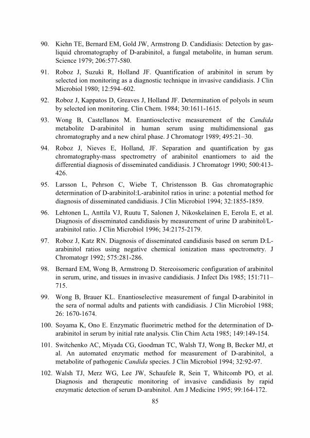
85
90. Kiehn TE, Bernard EM, Gold JW, Armstrong D. Candidiasis: Detection by gas-liquid chromatography of D-arabinitol, a fungal metabolite, in human serum. Science 1979; 206:577-580.
91. Roboz J, Suzuki R, Holland JF. Quantification of arabinitol in serum by selected ion monitoring as a diagnostic technique in invasive candidiasis. J Clin Microbiol 1980; 12:594–602.
92. Roboz J, Kappatos D, Greaves J, Holland JF. Determination of polyols in seum by selected ion monitoring. Clin Chem. 1984; 30:1611-1615.
93. Wong B, Castellanos M. Enantioselective measurement of the Candidametabolite D-arabinitol in human serum using multidimensional gas chromatography and a new chiral phase. J Chromatogr 1989; 495:21–30.
94. Roboz J, Nieves E, Holland, JF. Separation and quantification by gas chromatography-mass spectrometry of arabinitol enantiomers to aid the differential diagnosis of disseminated candidiasis. J Chromatogr 1990; 500:413-426.
95. Larsson L, Pehrson C, Wiebe T, Christensson B. Gas chromatographic determination of D-arabinitol:L-arabinitol ratios in urine: a potential method for diagnosis of disseminated candidiasis. J Clin Microbiol 1994; 32:1855-1859.
96. Lehtonen L, Anttila VJ, Ruutu T, Salonen J, Nikoskelainen E, Eerola E, et al. Diagnosis of disseminated candidiasis by measurement of urine D arabinitol/L-arabinitol ratio. J Clin Microbiol 1996; 34:2175-2179.
97. Roboz J, Katz RN. Diagnosis of disseminated candidiasis based on serum D:L-arabinitol ratios using negative chemical ionization mass spectrometry. J Chromatogr 1992; 575:281-286.
98. Bernard EM, Wong B, Armstrong D. Stereoisomeric configuration of arabinitol in serum, urine, and tissues in invasive candidiasis. J Infect Dis 1985; 151:711–715.
99. Wong B, Brauer KL. Enantioselective measurement of fungal D-arabinitol in the sera of normal adults and patients with candidiasis. J Clin Microbiol 1988; 26: 1670-1674.
100. Soyama K, Ono E. Enzymatic fluorimetric method for the determination of D-arabinitol in serum by initial rate analysis. Clin Chim Acta 1985; 149:149-154.
101. Switchenko AC, Miyada CG, Goodman TC, Walsh TJ, Wong B, Becker MJ, et al. An automated enzymatic method for measurement of D-arabinitol, a metabolite of pathogenic Candida species. J Clin Microbiol 1994; 32:92-97.
102. Walsh TJ, Merz WG, Lee JW, Schaufele R, Sein T, Whitcomb PO, et al. Diagnosis and therapeutic monitoring of invasive candidiasis by rapid enzymatic detection of serum D-arabinitol. Am J Medicine 1995; 99:164-172.

86
103. Murray JS, Wong ML, Miyada CG, Switchenko AC, Goodman TC, Wong B. Isolation, characterization and expression of the gene that encodes D-arabinitol dehydrogenase in Candida tropicalis. Gene 1995; 155:123-128.
104. Yeo SF, Zhang Y, Schafer D, Campell S, Wong B. A rapid, automated enzymatic fluorometric assay for determination of D-arabinitol in serum. J Clin Microbiol 2000; 38:1439-1443.
105. Yeo SF, Huie S, Sofair AN, Campell S, Durante A, Wong B. Measurement of serum D-arabinitol/creatinine ratios for initial diagnosis and for predicting outcome in an unselected population-based sample of patients with Candidafungemia. J Clin Microbiol 2006; 44:3894-3899.
106. Christensson B, Wiebe T, Pehrson C, Larsson L. Diagnosis of invasive candidiasis in neutropenic children with cancer by determination of D-arabinitol/L-arabinitol ratios in urine. J Clin Microbiol 1997; 35:636–64.
107. Lehtonen L, Rantala A, Oksman P, Eerola E, Lehtonen OP. Determination of serum arabinitol levels by mass spectrometry in patients with postoperative candidiasis. Eur J Clin Microbiol Infect Dis 1993; 12:330-335.
108. Deacon, AG. Estimations of serum arabinitol for diagnosis invasive candidosis. J Clin Pathol 1986: 39:842-850.
109. Walsh TJ, Lee JW, Sien T, Schaufele R, Bacher J, Switchenko AC, et al. Serum D-arbinitol measured by automated quantitative enzymatic assay for detection and therapeutic monitoring of experimental disseminated candidiasis: correlation with concentrations of Candida albicans. J Med Vet Mycol 1994; 32:205-215.
110. Bernard EM, Christiansen KJ, Tsang SF, Kiehn TE, Armstrong D. Rate of arabinitol production by pathogenic yeast species. J Clin Microbiol 1981; 14:189-194.
111. Hui M, Cheung SW, Chu KC, Chan EC-Y, Cheng AF-B. Development and application of a rapid diagnostic method for invasive candidiasis by the detection of D-/L-arabinitol using gas chromatography/mass spectrometry. Diagn Microbiol Infect Dis 2004:49:117-123.
112. Saha BC, Sakakibara Y, Cotta MA. Production of D-arabitol by a newly isolated Zygosaccharomyces rouxi. J Ind Microbiol Biotechnol 2007; 34:519-523.
113. Povelainen M, Eneyskaya EV, Kulminskaya AA, Ivanen DR, Kalkkinen N, Neustroev KN, et al. Biochemical and genetic characterization of a novel enzyme of pentitol metabolism : D-arabitol-phosphate dehydrogenase. Biochem J 2003; 371:191-197.

87
114. Hausman SZ, London J. Purification and characterization of ribitol-5-phosphate and xylitol-5-phosphate dehydrogenases from strains of Lactobacillus casei. J Bacteriol 1987; 169:1651-1655.
115. Mortlock RP, Wood WA. Metabolism of pentoses and pentitols by Aerobacter aerogens. J Bacteriol 1964; 88:838-844.
116. Shakeri-Garakani, A, et al. The genes and enzymes for the catabolism of galactitol, D-tagatose, and related carbohydrates in Klebsiella oxytoca M5a1 and other enteric bacteria display convergent evolution. Mol Gen Genomics 2004; 271:717-728.
117. van den Broek LA, van Boxtel EL, Kievit RP, Verhoef R, Beldman G, Voragen AG. Physico-chemical and transglucosylation properties of recombinant sucrose phosphorylase from Bifidobacterium adolescentis DSM20083. Appl Microbiol Biotechnol 2004; 65:219-227.
118. Kim B, Sullivan RP, Zhao H. Cloning, characterization, and engineering of fungal L-arabinitol dehydrogenases. Appl Microbiol Biotechnol 2010; 87:1407-1414.
119. Koy A, Waldhaus A., Hammen H-W, Wendel U, Mayatepek E, Schadewaldt P. Urinary excretion of pentose phosphate pathway-associated polyols in early postnatal life. Neonatology 2009: 95: 256-261.
120. Christensson B, Roboz, J. Arabinitol enantiomers in cerebrospinal fluid. J Neurol Sci 1991;105:234-239.
121. Wamelink MM, Struys EA, Jakobs C. The biochemistry, metabolism and inherited defects of the pentose phosphate pathway: A review. J Inherit Metab Dis 2008; 31:703-717.
122. Wong B, Brauer KL, Clemens JR, Beggs S. Effects of gastrointestinal candidiasis, antibiotics, dietary arabinitol, and cortisone acetate on levels of the Candida metabolite D-arabinitol in rat serum and urine. Infect Immun 1990; 58:283-288.
123. Onkenhout W, Groener JE, Verhoeven NM, Yin C, Laan LAEM. L-arabinosuria: a new defect in human pentose metabolism. Mol Gen Metabolism 2002; 77:80-85.
124. Wong B, Bernard EM, Gold JW, Fong D, Armstrong D. The arabinitol appearance rate in laboratory animals and humans: estimation from the arabinitol:creatinine ratio and relevance to the diagnosis of candidiasis. J Infect Dis 1982; 146:353-359.
125. Wong B, Bernard EM, Gold JW, Fong D, Silber A, Armstrong D. Increased arabinitol levels in experimental candidiasis in rats: Arabinitol appearance rates,

88
arabinitol/creatinine ratios, and severity of infection. J Infect Dis 1982; 146:346-352.
126. Gold JW, Wong B, Bernard EM, Kiehn TE, Armstrong D. Serum arabinitol concentrations and arabinitol/creatinine ratios in invasive candidiasis. J Infect Dis 1983; 147:504-514.
127. Roboz J, Yu Q, Holland JF. Filter paper sampling of whole blood and urine for the determination of D/L arabinitol ratios by mass spectrometry. J Microbiol Methods 1992: 15:207-214.
128. Hayasaka S, Noda S, Setogawa T. Increased D-arabinitol:creatinine ratio in sera of patients with Behcet’s disease during an active phase. British J Ophtalmol 1993; 77:39-40.
129. Wong B, Baughman RP, Brauer KL. Levels of the Candida metabolite D-arabinitol in sera of steroid treated and untreated patients with sarcoidosis. J Clin Microbiol 1989; 27:1859-1862.
130. Miller GG, Witwer MW, Braude AI, Davis CE. Rapid identification of Candidaalbicans septicemia in man by gas-liquid chromatography. J Clin Invest 1974; 54:1235-1240.
131. Eng RH, Chmel H, Buse M. Serum levels of arabinitol in the detection of invasive candidiasis in animals and humans. J Infect Dis 198; 143:677-683.
132. Wells CL, Sirany MS, Blazevic DJ. Evaluation of serum arabinitol as a diagnostic test for candidiasis. J Clin Microbiol 1983; 18:353-357.
133. De Repentigny L, Marr LD, Keller JW, Carter AW, Kuykendall RJ, Kaufman L, et al. Comparison of enzyme immunoassay and gas-liquid chromatography for the rapid diagnosis of invasive candidiasis in cancer patients. J Clin Microbiol 1985; 21:972-979.
134. Holak EJ, Wu J, Spruance SL. Value of serum arabinitol for the management of Candida infections in clinical practice. Mycopathologia 1986; 93:99-104.
135. Roboz J, Kappatos DC, Holland JF. Role of individual serum pentitol concentration in the diagnosis of disseminated visceral candidiasis. Eur J Clin Microbiol 1987; 6:708-714.
136. Salonen JH, Rimpiläinen M, Lehtonen L, Lehtonen O-P, Nikoskelainen J. Measurement of the D-arabinitol/L-arabinitol ratio in urine of neutropenic patients treated empirically with amphotericin B. Eur J Clin Microbiol Infect Dis 2001; 20:179-184.
137. Stradomska TJ, Mielniczuk Z. Gas chromatographic determination of D-/L-arabinitol ratio in healthy Polish children. J Chromatogr 2002; 773:175-181.

89
138. Guiot HL, Fibbe WE, van´t Wout JW. Risk factors for fungal infection in patients with malignant hematological disorders: implications for empirical therapy and prophylaxis. Clin Infect Dis 1994; 18:525-532.
139. Roboz J. Diagnosis and monitoring of disseminated candidiasis based on serum:urine D:L-arabinitol ratios. Chirality 1994; 6:51-57.
140. Chryssanthou E, Fernandez V, Petrini B. Performance of commercial latex agglutination tests for the differentiation of Candida dubliniensis and Candida albicans in routine diagnostics. APMIS 2007; 115:1281-1284.
141. Kossoff EH, Buescher ES, Karlowicz MG. Candidemia in a neonatal intensive care unit: trends during fifteen years and clinical features of 111 cases. Ped Infect Dis 1997; 17:504-508.
142. Baley JE, Kliegman RM, Fanaroff AA. Disseminated fungal infections in very low-birth weight infants: clinical manifestations and epidemiology. Pediatrics 1984; 73:144-152.
143. McDonald L, Baker C, Chenoweth C. Risk factors for candidemia in a children's hospital. Clin Infect Dis 1998; 26:642-645.
144. Wong B, Murray JS, Castellanos M, Croen KD. D-Arabitol metabolism in Candida albicans: studies of the biosynthetic pathway and the gene that encodes NAD-dependent D-arabitol dehydrogenase. J Bacteriol 1993; 175:6314–6320.
145. Wingard JR, Merz WG, Rinaldi MG, Johnson TR, Karp JE, Saral R. Increase in Candida krusei infection among patients with bone marrow transplantation and neutropenia treated prophylactically with fluconazole. N Engl J Med 1991; 325:1274-1277.
146. Abi-Said D, Anaisse E, Uzun O, Issam R, Pinzcowski H, Vartivarian S. The epidemiology of hematogenous candidiasis caused by different Candidaspecies. Clin Infect Dis 1997; 24:1122-1128.
147. Niimi K, Sheperd MG, Cannon RD. Distinguishing Candida Species by b-N-Acetylhexosaminidase Activity. J Clin Microbiol 2001; 39:2089-2097.
148. Sobel, J D and Kaye, D. Epidemiology of urinary tract infections. Bennet & Dolin Mandell. Principle and Practice of Infectious Diseases. 6th ed. Churchill Livingstone, 2009, Vol. 1, chapter 69, 964-966.
149. Reiner AM. Genes for ribitol and D-arabitol catabolism in Escherichia coli : their loci in Escherichia coli C and absence in K-12 and B strains. J Bacteriol 1975; 123:530-536.

90
150. Kauffman CA, Nazques JA, Sobel JD, Gallis HA, McKinsy DS, Karchmer AW, et al. Prospective multicenter surveillance study of funguria in hospitalized patients. Clin Infect Dis 2000; 30:14–18.

Paper I


JOURNAL OF CLINICAL MICROBIOLOGY,0095-1137/00/$04.00�0
Aug. 2000, p. 3039–3042 Vol. 38, No. 8
Copyright © 2000, American Society for Microbiology. All Rights Reserved.
Urine D-Arabinitol/L-Arabinitol Ratio in Diagnosis of InvasiveCandidiasis in Newborn Infants
GUDRUN SIGMUNDSDOTTIR,1 BERTIL CHRISTENSSON,2* LARS J. BJORKLUND,3
KRISTINA HÅKANSSON,3 CHRISTINA PEHRSON,1 AND LENNART LARSSON1
Department of Infectious Diseases and Medical Microbiology, Section of Medical Microbiology,1
Section of Infectious Diseases,2 and the Department of Pediatrics,3
University Hospital, SE-221 85 Lund, Sweden
Infants treated in neonatal intensive care units suffer an increased risk for invasive candidiasis, but the diag-nosis is sometimes difficult. D-arabinitol is a metabolite of most pathogenic Candida species. An elevated urineD-arabinitol/L-arabinitol (DA/LA) ratio is a sensitive sign of invasive candidiasis in children with cancer, butthe method has not been previously evaluated for newborn infants. We therefore enrolled 117 infants in a neo-natal intensive care unit, and 411 urine samples were obtained on filter paper. The DA/LA ratio was measuredby gas chromatography-mass spectrometry. For 81 infants with no suspicion of superficial or invasive candi-diasis, the urine DA/LA ratio was 2.7 � 0.7 (mean � standard deviation [SD]). The upper cutoff level was setat 4.8 (mean plus 3 SD). Of 22 infants with mucocutaneous candidiasis and not given systemic antifungal treat-ment, two had elevated DA/LA ratios, which normalized after removal of intravascular catheters. Eight otherinfants were given empiric antifungal treatment but had negative cultures; five of these had repeatedly elevatedDA/LA ratios. Six infants with culture-positive invasive candidiasis all had one or more samples with elevatedratios. For seven infants, three with suspected and four with confirmed invasive candidiasis (for whichfollow-up samples were available), ratios normalized during antifungal treatment. In conclusion, urine DA/LAratio determination is a rapid test and can be used for newborns. It is possibly more sensitive than fungal bloodcultures in the diagnosis of invasive candidiasis and can also be used for monitoring the effect of antifungaltreatment.
Invasive candidiasis has become an important infection inpreterm infants (1, 8). In one survey, the rate of candidemiahad increased 11-fold between 1981 and 1995 (4). In a recentlypublished retrospective study, the frequency of invasive can-didiasis in low-birth-weight (�1,250 g) infants was 3.5% (7).Predisposing factors are indwelling vascular catheters, totalparenteral nutrition, and long-term treatment with broad-spec-trum antibiotics (7). By contrast, invasive candidiasis rarelyaffects full-term newborns with normal birth weight. An inci-dence of 0.6% was reported for infants with birth weights of�2,500 g, and 76% of the infected infants had major congenitalmalformations (9).
Clinical signs of invasive candidiasis can be unspecific, anddiagnosis is still mostly based on blood cultures; however,blood cultures have been assumed to be positive for Candida inonly 24 to 60% of cases (9, 12). D-arabinitol, a sugar alcohol, isa major metabolite of most pathogenic Candida species (2).Both D-arabinitol (DA) and L-arabinitol (LA) are normallypresent in serum and urine, and the DA/LA ratio in urine canbe determined with gas chromatography-mass spectrometry(GC-MS) (6). We previously determined the diagnostic valueof urine DA/LA ratio for invasive candidiasis in children withcancer (3).
In this study, we report the results of a prospective studydesigned to evaluate the usefulness of assessing changes inurine DA/LA ratios in the diagnosis of invasive candidiasis inboth premature and full-term infants.
MATERIALS AND METHODS
The study was approved by the ethical research committee of the hospital, andinformed consent was obtained from all parents.
Healthy newborn infants. Urine samples were collected from 40 healthy full-term infants. One sample was collected from each child in one of the first 4 daysof life. On the same day, a urine sample was collected from 16 of the mothers.
Patients. A total of 117 newborns (66 males and 51 females) treated at theneonatal intensive care unit (NICU) at Lund University Hospital were enrolled.Gestational age was 24 to 42 weeks (median, 30 weeks), and 97 infants werepremature (gestational age, �38 weeks) and 20 were full-term. Urine sampleswere prospectively collected from 114 infants between October 1997 and De-cember 1998. During the first 3 months, urine was collected from all infantsadmitted to the NICU, but during 1998, only children requiring long-term carewith central venous catheters (CVCs) and broad-spectrum antibiotics were in-cluded. Additionally, three infants with invasive candidiasis confirmed immedi-ately before or after the sampling period were included. The following wererecorded: gestational age, birth weight, medical history, skin and oral lesionslikely to be caused by Candida, microbiological cultures, number of days withumbilical vein and percutaneous CVCs, antimicrobial treatment, and local andsystemic antifungal treatment. Altogether, 411 urine samples were collected.
Infants with one or more blood cultures and/or urine culture obtained bysuprapubic aspiration positive for Candida were considered to have invasivecandidiasis.
Test groups. The infants were divided into four groups. (i) group A, (ii) groupB, (iii) group C, and (iv) group D.
(i) Group A, control group (n � 81). In this group, there was no clinicalsuspicion of mucocutaneous or invasive infection with Candida organisms, andno antifungal treatment was given. Surveillance cultures for colonization werenot done. Gestational age was 25 to 42 weeks (median, 33 weeks), and birthweight was 695 to 4,430 g (median, 1,950 g).
(ii) Group B, mucocutaneous candidiasis (n � 22). Newborns included in thisgroup were clinically diagnosed with mucocutaneous candidiasis but were notconsidered to have invasive candidiasis and received only local antifungal treat-ment. In some cases, fungal cultures were positive. Gestational age was 24 to 40weeks (median, 26 weeks), and birth weight was 575 to 3,740 g (median, 870 g).
(iii) Group C, empirically treated infants (n � 8). Fluconazole was givenempirically to these infants although all cultures were negative for Candida.Gestational age was 24 to 39 weeks (median, 24.5 weeks), and birth weight was640 to 3,520 g (median, 850 g).
(iv) Group D, confirmed invasive candidiasis (n � 6). Infants in this group hadat least one positive blood culture or a positive urine culture obtained by supra-pubic aspiration. All children were treated with fluconazole or liposomal am-
* Corresponding author. Mailing address: Department of InfectiousDiseases and Medical Microbiology, Section of Infectious Diseases,University Hospital, SE-221 85 Lund, Sweden. Phone: 46-46-171130.Fax: 46-46-137414. E-mail: [email protected].
3039

photericin B (AmBisome). Gestational age was 24 to 27 weeks (median, 24.5weeks), and birth weight was 575 to 1,030 g (median, 825 g).
Urine samples. The aim was to collect urine samples twice weekly from theinfants admitted to the NICU. Most samples were collected by placing a piece offilter paper approximately 3 by 4 cm in the diaper (11). The filter paper wasremoved and dried after the infant had urinated. A few urine samples frominfants with indwelling urinary catheters were collected in culture vials. Samplesin culture vials were stored at �20°C, and filter paper samples were stored atroom temperature pending analysis by GC-MS. Urine culture was done on urinesamples arriving in culture vials.
Sample preparation for analysis by GC-MS. Filter paper spots approximately2 cm in diameter and containing urine were cut from the filter paper andextracted in approximately 3 ml of methanol for 30 min. Of the solution, 300- to600-�l aliquots were transferred to 1-ml vials and evaporated to dryness under aflow of nitrogen. Hexane (200 �l) and trifluoroacetic anhydride (40 �l) wereadded, and the samples were heated at 80°C for 10 min in metal heating blocks,cooled to room temperature, and then dried again under a gentle nitrogen flow.Finally, 200 �l of a hexane-dichloromethane solution (1:1 [vol/vol]) was added,and the sample was ready for analysis by GC-MS.
GC-MS. A Trio-1S GC-MS system (VG, Manchester, United Kingdom) wasused. The GC was Hewlett-Packard (Avondale, Pa.) model 5890 equipped witha splitless injector and a chiral column (30 m by 0.25 mm [inner diameter] coatedwith a 0.25-�m-thick layer of cyclodextrin [Beta Dex-120; Supelco Inc., Bel-lafonte, Pa.]). The column temperature was programmed to rise from 70 to170°C at 7°C/min. The ion source temperature was 200°C. Helium was used as acarrier gas. Analyses were performed in the electron impact mode by usingselected ion monitoring with an m/z of 519. The peak urine DA/LA ratio wasdefined as the mean of the two highest values obtained within 1 week.
Microbiology. The BacT/Alert method was used for blood cultures, and Can-dida was cultured on Sabouraud agar (10). Identification of Candida was done bytesting the yeast’s ability to ferment glucose, galactose, saccharose, maltose,lactose, and trehalose and by testing for chlamydospores (5). Urine samplesarriving in culture vials were cultured on blood agar and Sabouraud agar at 36°Cfor 48 h.
Statistical analysis. Descriptive statistics are presented as the mean � stan-dard deviation (SD) for normally distributed data; the median and range wereused otherwise. Student’s t test or the Mann-Whitney test were therefore usedwhen appropriate. P values of �0.05 were regarded as statistically significant.
RESULTS
Healthy newborn infants. The urine DA/LA ratio for the 40healthy full-term newborn infants was 2.5 � 0.6 (mean � SD)(range, 1.6 to 4.1). For the 16 mothers, it was 1.8 (range, 0.9 to2.9), which was significantly lower than for their respectiveinfants (P � 0.001).
NICU patients. Eighteen infants included in the study hadblood cultures positive for coagulase-negative staphylococci(n � 15), Proteus mirabilis (n � 1), Bacillus sp. (n � 1), andStaphylococcus aureus (n � 1). Malassezia furfur was isolatedfrom a CVC tip in one patient. Urine culture on urine samplescollected in culture vials showed no growth of bacteria or fungi.One hundred three infants were treated with antibiotics, and96 had a CVC. Ten infants died.
Group A (n � 81). These infants had no symptoms of Can-dida infection. The mean value of the DA/LA ratios for the
whole group was based on calculating mean values for eachpatient during the course of the study. The mean urine DA/LAratio was 2.7 � 0.7 (mean � SD), which was not significantlydifferent from the mean ratio for the healthy full-term new-born infants. The upper cutoff level for NICU infants was setat 4.8 (group mean plus 3 SD). There was no correlationbetween gestational age or birth weight and urine DA/LAratio. Three infants in this group died, all of causes other thaninvasive candidiasis.
Because of the heterogeneity of the infants with regards togestational age and disease severity, we selected a subgroup ofvery low-birth-weight infants (�1,250 g, n � 18) to serve as acontrol group for infants with elevated urine DA/LA ratios ingroups B and C when comparing treatment times with antibi-otics and time with indwelling CVCs. These control infants’gestational ages were 25 to 29 weeks (median, 26.5 weeks),their birth weights were 695 to 1,175 g (median, 910 g), andthey had a total treatment time with broad-spectrum antibiot-ics and indwelling CVCs of 11 days (median). The mean andmedian urine DA/LA ratio in this subgroup was 2.3 (range, 1.4to 3.2).
Group B (n � 22). These infants had clinical signs of mu-cocutaneous candidiasis. For four infants, cultures from vari-ous locations were positive for Candida albicans, but no sys-temic antifungal treatment was given. Two infants had elevatedurine DA/LA ratios, and one of them had positive fungalcultures of urine obtained via an indwelling catheter. For bothof these infants, urine DA/LA ratios fell to normal values aftertheir CVCs had been removed or exchanged. These two infantshad total times with indwelling CVCs of 41 and 12 days andtotal treatment times with broad-spectrum antibiotics of 41and 21 days, respectively. Two infants in this group died, bothof causes other than invasive candidiasis.
Group C (n � 8). Eight patients received empiric treatmentwith fluconazole (Table 1). Five infants (no. 2, 4, 6, 7, and 8)had DA/LA ratios above the cutoff level. Three of these infants(no. 2, 4, and 6) had several elevated DA/LA ratios whichreturned to normal during antifungal treatment, as shownby patient no. 2 in Fig. 1. The two other infants (no. 7 and 8)received empirical antifungal treatment only after urine sam-pling was discontinued. Three patients (no. 5, 6, and 7) hadmajor congenital malformations. Five infants died, all fromcauses other than invasive candidiasis.
The infants with elevated DA/LA ratios in groups B and Chad significantly longer times with indwelling CVCs and longertreatment times with broad-spectrum antibiotics than the verylow-birth-weight (�1,250 g) infants in group A (P � 0.01 andP � 0.001, respectively).
TABLE 1. Infants with empiric antifungal treatment (group C)
Patientno.
Gestational age atbirth (weeks)
Mean peakDA/LA ratio
No. of positive samples/no. of all samplesa
CVC(days)b
Antibiotics(days)c Outcome
1 24 4.0 0/11 29 36 Survived2 24 9.5 4/9 29 29 Survived3 24 4.4 0/5 20 20 Died4 25 15.3d 2/7 22 37 Survived5 32 3.8 0/20 93 61 Died6 39 6.7 3/7 51 95 Died7 34 7.9 6/14 26 55 Died8 24 9.8 7/16 74 74 Died
a Number of samples with a urine DA/LA ratio of �4.8/total number of samples.b Time with indwelling CVC.c Treatment time with broad-spectrum antibiotics.d Peak DA/LA ratio; no relevant samples were obtained within 1 week of elevated urine DA/LA ratio.
3040 SIGMUNDSDOTTIR ET AL. J. CLIN. MICROBIOL.

Group D (n � 6). Six infants had invasive candidiasis basedon clinical symptoms and C. albicans isolated from blood (n �5) or urine obtained by suprapubic aspiration (n � 1). All sixinfants had at least one elevated DA/LA ratio (Table 2). Infour cases (no. 9, 10, 12, and 13), urine DA/LA ratios normal-ized during treatment with fluconazole (Fig. 2, patient no. 13)while the clinical conditions also improved. One patient (no.14) also had a declining DA/LA ratio, but died from othercomplications of prematurity. One patient (no. 11) improvedduring treatment, but follow-up DA/LA ratios were not ob-tained. The infants in group D had significantly longer totaltreatment times with CVCs and broad-spectrum antibioticsthan the control infants in group A (P � 0.01 and P � 0.01,respectively).
DISCUSSION
All six infants with confirmed invasive candidiasis had atleast one positive urine DA/LA ratio. This finding is in accor-dance with our previous results for children with cancer (3).For all of the five premature infants in the present study forwhich appropriate samples were available, the DA/LA ratiosdeclined during antifungal treatment, suggesting that the urineDA/LA ratio can be used to monitor treatment effect. This isin accordance with previous animal studies in which the tissue
burden of Candida was found to correlate to serum DA/cre-atinine ratios (13). In a study of adult patients with cancer,serum DA/creatinine ratios correlated with the therapeuticresponse to antifungal treatment (14). However, it should benoted that, as shown for patient 13 in Fig. 2, CVC-associatedfungal colonization may still be present although urine DA/LAratios decline due to decreased Candida tissue burden. There-fore, DA/LA determinations should not replace but comple-ment fungal blood cultures.
In the present study, 7 out of 111 infants (6.3%) withoutmicrobiologically proven invasive candidiasis had a DA/LAratio above the cutoff, five in group C and two in group B.However, all infants with elevated DA/LA ratios in groups Band C were either premature with a birth weight below 1,250 gor had severe congenital malformations and thus belonged torisk groups for invasive candidiasis (1, 4, 8). In addition, theseinfants had a significantly longer total treatment time withbroad-spectrum antibiotics and indwelling CVCs than the verylow-birth-weight controls. DA/LA ratios normalized after re-moval or exchange of CVC in the two patients in group B withelevated DA/LA ratios, indicating a possible microbiologicallyundiagnosed CVC-associated Candida infection. One of thesepatients also had growth of C. albicans in urine, althoughobtained by indwelling catheter. Furthermore, urine DA/LAratios normalized during empiric treatment in three infants in
FIG. 1. Urine DA/LA ratios for patient no. 2 (Table 1), a girl born after 24 weeks of gestation. Empiric treatment with fluconazole was given from day 29 becauseof persistent fever and no response to broad-spectrum antibiotic treatment. Blood cultures taken on days 16 and 28 were negative for Candida. The broken line at 4.8(mean urine DA/LA ratio for group A plus 3 SD) corresponds to the upper cutoff limit. The horizontal arrow on the y axis indicates the mean urine DA/LA ratio forgroup A at 2.7. The horizontal black bar represents the treatment time with fluconazole.
TABLE 2. Infants with confirmed invasive candidiasis (group D)
Patientno.
Gestational age atbirth (weeks)
Basis ofdiagnosis
Mean peakDA/LA ratio
No. of positive samples/no. of all samplesa
CVC(days)b
Antibiotics(days)c Outcome (cause of death)
9 25 3 blood cultures 5.4 2/6 23 23 Survived10 24 1 urine cultured 7.9 4/12 24 24 Survived11 24 1 blood culture 5.8e 1/7 46 41 Survived12 27 2 blood cultures 9.3 3/20 35 87 Died, candida endocarditis13 27 3 blood cultures 9.1 3/7 24 24 Survived14 24 2 blood cultures 30.0 3/3 19 19 Died, complications of prematurity
a Number of samples with a urine DA/LA ratio of �4.8/total number of samples.b Time with indwelling CVC.c Treatment time with broad-spectrum antibiotics.d Sample was obtained by suprapubic aspiration.e Peak DA/LA ratio; no relevant samples were obtained within 1 week of elevated urine DA/LA ratio.
VOL. 38, 2000 D-ARABINITOL/L-ARABINITOL IN NEONATAL CANDIDIASIS 3041
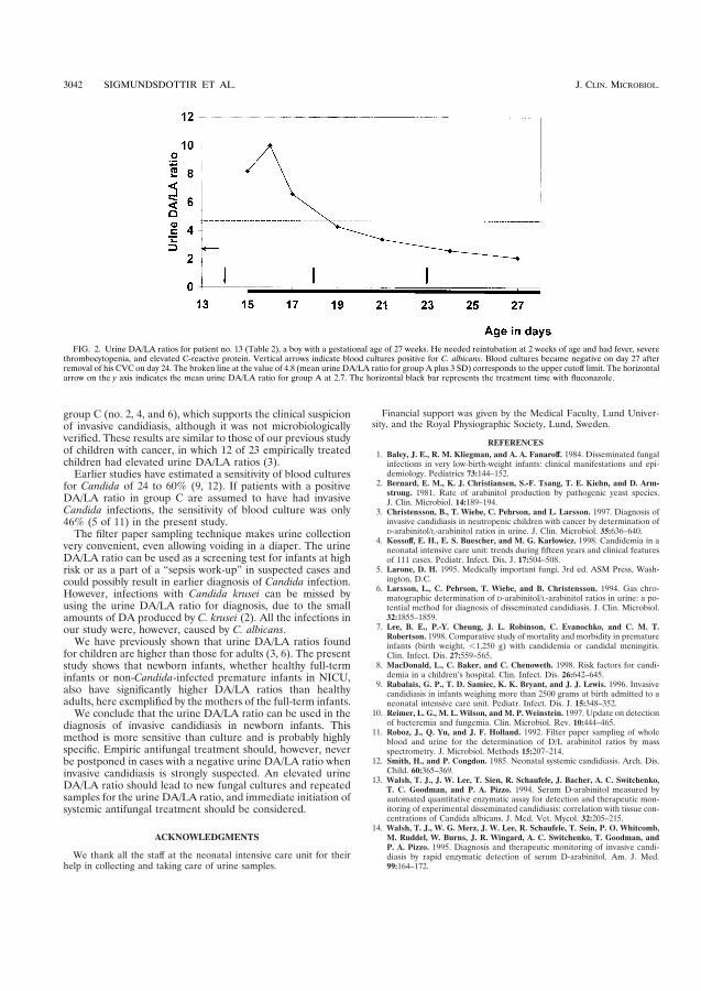
group C (no. 2, 4, and 6), which supports the clinical suspicionof invasive candidiasis, although it was not microbiologicallyverified. These results are similar to those of our previous studyof children with cancer, in which 12 of 23 empirically treatedchildren had elevated urine DA/LA ratios (3).
Earlier studies have estimated a sensitivity of blood culturesfor Candida of 24 to 60% (9, 12). If patients with a positiveDA/LA ratio in group C are assumed to have had invasiveCandida infections, the sensitivity of blood culture was only46% (5 of 11) in the present study.
The filter paper sampling technique makes urine collectionvery convenient, even allowing voiding in a diaper. The urineDA/LA ratio can be used as a screening test for infants at highrisk or as a part of a “sepsis work-up” in suspected cases andcould possibly result in earlier diagnosis of Candida infection.However, infections with Candida krusei can be missed byusing the urine DA/LA ratio for diagnosis, due to the smallamounts of DA produced by C. krusei (2). All the infections inour study were, however, caused by C. albicans.
We have previously shown that urine DA/LA ratios foundfor children are higher than those for adults (3, 6). The presentstudy shows that newborn infants, whether healthy full-terminfants or non-Candida-infected premature infants in NICU,also have significantly higher DA/LA ratios than healthyadults, here exemplified by the mothers of the full-term infants.
We conclude that the urine DA/LA ratio can be used in thediagnosis of invasive candidiasis in newborn infants. Thismethod is more sensitive than culture and is probably highlyspecific. Empiric antifungal treatment should, however, neverbe postponed in cases with a negative urine DA/LA ratio wheninvasive candidiasis is strongly suspected. An elevated urineDA/LA ratio should lead to new fungal cultures and repeatedsamples for the urine DA/LA ratio, and immediate initiation ofsystemic antifungal treatment should be considered.
ACKNOWLEDGMENTS
We thank all the staff at the neonatal intensive care unit for theirhelp in collecting and taking care of urine samples.
Financial support was given by the Medical Faculty, Lund Univer-sity, and the Royal Physiographic Society, Lund, Sweden.
REFERENCES1. Baley, J. E., R. M. Kliegman, and A. A. Fanaroff. 1984. Disseminated fungal
infections in very low-birth-weight infants: clinical manifestations and epi-demiology. Pediatrics 73:144–152.
2. Bernard, E. M., K. J. Christiansen, S.-F. Tsang, T. E. Kiehn, and D. Arm-strong. 1981. Rate of arabinitol production by pathogenic yeast species.J. Clin. Microbiol. 14:189–194.
3. Christensson, B., T. Wiebe, C. Pehrson, and L. Larsson. 1997. Diagnosis ofinvasive candidiasis in neutropenic children with cancer by determination ofD-arabinitol/L-arabinitol ratios in urine. J. Clin. Microbiol. 35:636–640.
4. Kossoff, E. H., E. S. Buescher, and M. G. Karlowicz. 1998. Candidemia in aneonatal intensive care unit: trends during fifteen years and clinical featuresof 111 cases. Pediatr. Infect. Dis. J. 17:504–508.
5. Larone, D. H. 1995. Medically important fungi, 3rd ed. ASM Press, Wash-ington, D.C.
6. Larsson, L., C. Pehrson, T. Wiebe, and B. Christensson. 1994. Gas chro-matographic determination of D-arabinitol/L-arabinitol ratios in urine: a po-tential method for diagnosis of disseminated candidiasis. J. Clin. Microbiol.32:1855–1859.
7. Lee, B. E., P.-Y. Cheung, J. L. Robinson, C. Evanochko, and C. M. T.Robertson. 1998. Comparative study of mortality and morbidity in prematureinfants (birth weight, �1,250 g) with candidemia or candidal meningitis.Clin. Infect. Dis. 27:559–565.
8. MacDonald, L., C. Baker, and C. Chenoweth. 1998. Risk factors for candi-demia in a children’s hospital. Clin. Infect. Dis. 26:642–645.
9. Rabalais, G. P., T. D. Samiec, K. K. Bryant, and J. J. Lewis. 1996. Invasivecandidiasis in infants weighing more than 2500 grams at birth admitted to aneonatal intensive care unit. Pediatr. Infect. Dis. J. 15:348–352.
10. Reimer, L. G., M. L. Wilson, and M. P. Weinstein. 1997. Update on detectionof bacteremia and fungemia. Clin. Microbiol. Rev. 10:444–465.
11. Roboz, J., Q. Yu, and J. F. Holland. 1992. Filter paper sampling of wholeblood and urine for the determination of D/L arabinitol ratios by massspectrometry. J. Microbiol. Methods 15:207–214.
12. Smith, H., and P. Congdon. 1985. Neonatal systemic candidiasis. Arch. Dis.Child. 60:365–369.
13. Walsh, T. J., J. W. Lee, T. Sien, R. Schaufele, J. Bacher, A. C. Switchenko,T. C. Goodman, and P. A. Pizzo. 1994. Serum D-arabinitol measured byautomated quantitative enzymatic assay for detection and therapeutic mon-itoring of experimental disseminated candidiasis: correlation with tissue con-centrations of Candida albicans. J. Med. Vet. Mycol. 32:205–215.
14. Walsh, T. J., W. G. Merz, J. W. Lee, R. Schaufele, T. Sein, P. O. Whitcomb,M. Ruddel, W. Burns, J. R. Wingard, A. C. Switchenko, T. Goodman, andP. A. Pizzo. 1995. Diagnosis and therapeutic monitoring of invasive candi-diasis by rapid enzymatic detection of serum D-arabinitol. Am. J. Med.99:164–172.
FIG. 2. Urine DA/LA ratios for patient no. 13 (Table 2), a boy with a gestational age of 27 weeks. He needed reintubation at 2 weeks of age and had fever, severethrombocytopenia, and elevated C-reactive protein. Vertical arrows indicate blood cultures positive for C. albicans. Blood cultures became negative on day 27 afterremoval of his CVC on day 24. The broken line at the value of 4.8 (mean urine DA/LA ratio for group A plus 3 SD) corresponds to the upper cutoff limit. The horizontalarrow on the y axis indicates the mean urine DA/LA ratio for group A at 2.7. The horizontal black bar represents the treatment time with fluconazole.
3042 SIGMUNDSDOTTIR ET AL. J. CLIN. MICROBIOL.

Paper II


Virology
Urine D-arabinitol/L-arabinitol ratio in diagnosing Candida infection inpatients with haematological malignancy and HIV infection
Damon P. Eisena,*, Paul B. Bartleya, William Hopea, Gudrun Sigmundsdottirb,Christina Pehrsonb, Lennart Larssonb, Bertil Christenssonc
aInfectious Diseases Unit, Royal Brisbane Hospital, Herston Road, Herston, Queensland, 4029, AustraliabDepartment of Medical Microbiology, Dermatology and Infection, Section of Bacteriology, Lund University Hospital, SE-221 85 Lund, Sweden
cSection of Infectious Diseases, Lund University Hospital, SE-221 85 Lund, Sweden
Received 20 March 2001; accepted 28 September 2001
Abstract
Adult patients with hematologic malignancies along with HIV infected patients were prospectively studied to determine the performanceof urine D-arabinitol/L-arabinitol (DA/LA) ratio in diagnosing invasive candidiasis. Ten evaluable febrile neutropenic patients had proveninvasive candidiasis and elevated DA/LA ratios were found in 5. Invasive candidiasis with normal DA/LA ratios was most frequently dueto Candida krusei infection. This Candida species is a non-producer of arabinitol. Only 4 of 81 febrile neutropenic patients given eitherantifungal prophylaxis or empiric antifungal treatment had elevated DA/LA ratios. Only 1 of 15 HIV positive patients with eitheroropharyngeal or esophageal candidiasis had elevated DA/LA ratios. Widespread use of fluconazole prophylaxis in bone marrowtransplantation patients at the study hospital has led to an increased prevalence of C. krusei infection. This is the likely reason for the lowsensitivity of the test in proven and suspected invasive Candida infections reported here. © 2002 Elsevier Science Inc. All rights reserved.
1. Introduction
Candida infection is a major pathogen of immunosup-pressed patients. In cancer patients, chemotherapy-inducedneutropenia is a risk factor for invasive candidiasis (Anais-sie et al., 1998), those with hematologic malignancies are athighest risk of infection. Oropharyngeal candidiasis is by farthe most common manifestation of Candida infection inHIV patients and the more severe Candida esophagitis isboth less common and indicative of severe immunocompro-mise. Conventional methods of diagnosis of systemic Can-dida infection, such as blood culture, suffer from poorsensitivity (Berenguer et al., 1993). The high attributablemortality of systemic candidiasis (Wey et al., 1988) man-dates development of improved diagnostic techniques.
D-arabinitol, a chemical marker for Candida infection,has previously been evaluated in prospective studies in adultpatients (Walsh et al., 1995), and in children (Christenssonet al., 1997; Sigmundsdottir et al., 2000). To date there areno reports of D-arabinitol analysis in HIV patients. We
conducted a three-year prospective study of two patientgroups. Patients with hematologic malignancies and HIVinfected patients were studied with serial monitoring ofD-arabinitol/L-arabinitol (DA/LA) ratio in urine.
2. Materials and methods
2.1. Patients
Two hundred and four adult patients with hematologicmalignancy were prospectively enrolled from November1996 through November 1999 at the Royal Brisbane Hos-pital. Sixty patients were excluded from further analysis dueto lack of both febrile illness and neutropenia and/or if onlyone sample was collected. Among the 144 evaluable pa-tients the commonest underlying diagnoses were leukemia(n � 88), lymphoma (n � 41), and myeloma (n � 12).Adult patients with HIV infection were enrolled at both theRoyal Brisbane Hospital and Lund University Hospital,totalling 16 and 71 patients respectively. The Lund andBrisbane Research Ethics Committees approved the re-search protocol.
* Corresponding author. Tel.: �61-7-3636-8761; fax: �61-7-3636-1388.
E-mail address: [email protected] (D.P. Eisen).
www.elsevier.com/locate/diagmicrobioDiagnostic Microbiology and Infectious Disease
42 (2002) 39–42
0732-8893/02/$ – see front matter © 2002 Elsevier Science Inc. All rights reserved.PII: S0732-8893-(01)00310-8

2.1.1. Febrile neutropenic controlsThere were 201 urine samples from 49 control patients
with fever � 38.3°C for more than 2 days (n � 47) and/orneutropenia (blood neutrophil counts �0.5 � 109/liter) (n �46) but no microbiologic or clinical evidence of invasivecandidiasis. No patients had received antifungal treatmentor prophylaxis within 2 weeks of urine sampling. Six pa-tients in the control group had growth of Candida in cul-tures obtained from the respiratory tract or faeces. Ninepatients had bacteraemia and 38 patients were treated withbroad-spectrum antibiotics. These urine samples acted asnegative controls for the study.
2.1.2. Empiric antifungal therapy or prophylaxisIn 81 febrile and neutropenic patients with negative
blood cultures for Candida, either antifungal prophylaxis orempiric treatment was given. A total of 443 urine sampleswere collected during or within 2 weeks of these treatmentperiods. Respiratory tract and/or faeces samples from elevenpatients were culture positive for Candida. Eighteen pa-tients had bacteraemia and 73 received treatment withbroad-spectrum antibiotics. Two patients received treatmentfor invasive Aspergillus infections.
2.1.3. Confirmed invasive candidiasisInvasive candidiasis was diagnosed in 14 patients. This
was defined as the finding of at least one blood culturepositive for Candida species in a febrile, neutropenic patientwho was not responding to broad-spectrum antibacterialtreatment. Fifty-nine urine samples were collected fromthese patients. Five patients had growth of Candida fromsites other than blood (central venous catheter (n � 3), ETTaspirate and faeces (n � 1), skin abscesses (n � 1)). Threepatients in this group had bacteraemia and all 14 patientsreceived broad-spectrum antibacterials.
2.1.4. HIV positive patientsA total of 87 HIV infected patients were studied. Ten
patients had oropharyngeal candidiasis; in 3 cases verifiedby culture of C. albicans. Five patients were diagnosed withCandida esophagitis; confirmed by endoscopy and X-rayrespectively in two cases. In the other 3 cases the diagnosisof Candida esophagitis was presumptive, relying on clinicalfeatures of a typical history of odynophagia and response toazole antifungal treatment without further investigations.The remaining 72 HIV positive patients showed no signs ofCandida infection and were not given antifungal treatment.There were 79 urine samples from those with Candidainfection and 256 samples from the control patients.
2.2. DA/LA analysis
Urine samples from Brisbane were collected on filterpaper as previously described (Sigmundsdottir et al., 2000),dried and shipped to Lund by airmail. Other urine sampleswere stored at �20°C until analyzed. DA/LA ratios were
analyzed by gas chromatography mass spectrometry (Chris-tensson et al., 1997; Sigmundsdottir et al., 2000).
2.3. Microbiology
Blood cultures were incubated in the automated BacT-Alert system (Organon Teknika, Durham, NC). Positiveculture bottles found to contain yeasts on Gram stainingwere subcultured on Sabouraud agar. Identification of Can-dida albicans was by testing for germ tube formation. Non-albicans Candida sp. were speciated using the Vitek TBCidentification system (bioMerieux Vitek, Hazelwood, MI)and if confirmation of identity was required, by the api 20 CAUX system (bioMerieux sa, Marcy-l’Etoile, France).
3. Results
3.1. Hematologic malignancy patients
3.1.1. Febrile, neutropenic controlsUrine DA/LA ratios in 201 samples from 49 patients
without antifungal treatment or prophylaxis was 2.0 � 1.0(mean �/� standard deviation), and therefore, values � 5.0(mean � 3 SD) were defined as positive for this study. Twopatients in this group had one single positive sample eachwith DA/LA of 5.7 and 7.2, respectively, being regarded asfalse positives.
3.1.2. Empiric antifungal therapy or prophylaxisSingle positive determinations of 6.8, and 7.0 were found
in two patients treated with fluconazole, and repeatedlypositive DA/LA ratios were found in two patients on am-photericin B and fluconazole prophylaxis, respectively,where DA/LA peak values reached 13.6 and � 30, respec-tively.
3.1.3. Confirmed invasive candidiasisOf the 14 patients with invasive candidiasis, 10 had urine
samples collected either before or during the first three daysof antifungal therapy. Candidiasis was due to C. albicans(n � 3), C. krusei (4), C. tropicalis (2), and C. kefyr (1) inthese patients. DA/LA ratios were positive in five patients(range 5.5–32.0) with C. albicans (3), C. krusei (1), and C.tropicalis (1) fungaemia. Normal DA/LA ratios were foundin the other 5 patients with C. krusei (n � 3), C. tropicalis(n � 1) and C. kefyr (n � 1) fungaemia. The C. kefyrfungaemia patient with normal DA/LA ratios was shown tohave hepatosplenic (chronic) candidiasis by abdominal ul-trasound that revealed multiple abscesses. In the remainingfour patients with C. albicans, C. krusei, C parapsilosis andC. tropicalis fungaemia, respectively, urine samples wereall negative (range 1.6–2.8). However, in these four patientsurine samples were not available until 4–12 days after bloodculture was positive and 4–10 days after antifungal therapywas instituted. Therefore, these four patients were consid-
40 D.P. Eisen et al. / Diagnostic Microbiology and Infectious Disease 42 (2002) 39–42

ered as not evaluable, as DA/LA ratios are known to nor-malize rapidly during antifungal treatment (Walsh et al.,1995; Christensson et al., 1997; Sigmundsdottir et al.,2000).
3.2. HIV positive patients
The DA/LA ratio of the 72 HIV positive patients with nosigns of Candida infection and no antifungal treatment was2.0 � 0.7, which was not significantly different (p � 0.05,Student’s two-tailed t test) from the DA/LA results of thecontrol cancer patients (see above).
Four patients with oral candidiasis were given antifungaltreatment 3–7 days before urine sampling, and all showednegative DA/LA results (range 1.4–2.8). Only one patientwith oral candidiasis had a positive peak DA/LA value of7.1, the remaining 5 patients showed a mean DA/LA ratio of3.3 (range 2.6–4.5). Two patients with Candida esophagitisand normal DA/LA ratios of 2.4 and 3.3 were also alreadyon antifungal treatment when urine was sampled. The otherthree patients with esophageal candidiasis had not had an-tifungal therapy instituted when sampled, and they alsoshowed normal values of 2.5, 2.7 and 3.9, respectively.
4. Discussion
The results of this prospective study show that the de-termination of urine DA/LA ratios in adult immunosup-pressed patients with cancer can aid in the diagnosing ofinvasive candidiasis, provided that the infecting Candida sp.is producing DA. However, if C. krusei, C. glabrata, andpossibly C. kefyr fungaemia are common in the patientpopulation studied the negative predictive value of theDA/LA result is decreased. D-arabinitol is produced in vitroby C. albicans, C. tropicalis, C. parapsilosis, (Bernard etal., 1981; Larsson et al., 1994) C. kefyr (Bernard et al.,1981), C. lusitaniae and C. guilliermondii (Larsson et al.,1994) but not by C. krusei and C. glabrata (Bernard et al.,1981; Larsson et al., 1994). However, in the study byBernard et al. (Bernard et al. 1981) the rate of in vitro DAproduction sometimes varied more than 100-fold betweendifferent strains of the same Candida sp. Such strain vari-ation in DA production could be one explanation for thelack of positive urine DA/LA findings in our two cancerpatients with invasive C. tropicalis and C. kefyr infection.Unfortunately the patient strains were not available foranalysis of in vitro DA production, and thus this hypothesiscould not be confirmed. Furthermore, the patient with C.kefyr infection with normal DA/LA ratios was diagnosedwith chronic (hepatosplenic) candidiasis, which is a diag-nosis where DA results are less frequently positive (Walshet al., 1995).
Interestingly, one of four patients with C. krusei fungae-mia showed several positive DA/LA values which declinedand normalized during antifungal treatment. Unexpected
positive urine DA/LA ratios (Christensson et al., 1997;Lehtonen et al., 1996), serum DA/creatinine ratios (Walsh etal., 1995), and serum DA � LA concentrations (Roboz etal., 1987) were previously found in several patients with C.glabrata fungaemia. A possible explanation for this is thatsuch patients are coinfected with clinically undetected DA-producing Candida sp. as has been verified by postmortemcultures (Christensson et al., 1997). Given the low sensitiv-ity of fungal blood cultures (Berenguer et al., 1993) this isa plausible explanation.
In all other patients with appropriately timed urine sam-ples, DA/LA analysis confirmed the blood culture baseddiagnosis of invasive candidiasis. Still, the overall sensitiv-ity in diagnosing invasive candidiasis was lower than inprevious prospective studies of children (Christensson et al.,1997) and adults (Walsh et al., 1995) with cancer. Onelikely reason may be the high prevalence of C. kruseiinfections in the study hospital. This seems to be selectedfor by the widespread use of fluconazole prophylaxis forbone marrow transplant patients as in other institutions(Wingard et al., 1991; Casasnovas et al., 1992). Study of theepidemiology of candidaemia at the Royal Brisbane Hospi-tal for the period 1992–1999 shows a progressive increasein the proportion non-albicans Candida during this time.There was a significant increase in the proportion of fun-gaemia due to C. krusei in the study period described here(November 1996–November 1999) when compared withpre-study (8/21 vs 2/33; p � 0.01 Fisher exact). A casecontrolled study has shown that fluconazole use is not as-sociated with the presence of candidaemia but that it ap-pears to be significantly associated with the changing epi-demiology of candidaemia. In the absence of fluconazoleuse, candidaemia is much more likely to be due to C.albicans (�2 15.5 p � 0.001) and conversely if patients havebeen exposed to fluconazole they were more likely to haveC. krusei fungaemia (�2 20.78 p � 0.001). Fluconazoleusage did not impact on the risk of C. parapsilosis, C.tropicalis or C. glabrata sepsis in this study (D. Eisen,unpublished data).
The increased prevalence of C. krusei infection at theRoyal Brisbane Hospital could also partly explain the rela-tively low percentage of patients with positive DA/LA ra-tios among those given empiric antifungal chemotherapy orprophylaxis. Only 4 of 81 patients in this group had one ormore positive DA/LA ratios, as compared to 12 of 23patients on empiric therapy in our previous study of childrenwith cancer at a department where no fluconazole prophy-laxis was given (Christensson et al., 1997).
There are no previous studies on DA detection in patientswith HIV infection. Our hypothesis was that perhapsDA/LA analysis would aid in the diagnosis of patients withCandida esophagitis. However, due to the introduction ofHAART (highly active anti-retroviral treatment) at the sametime as this study was commenced, the number of HIVpositive patients with Candida esophagitis and also withoral candidiasis decreased dramatically. Only 3 patients
41D.P. Eisen et al. / Diagnostic Microbiology and Infectious Disease 42 (2002) 39–42

with esophagitis and 6 patients with oral candidiasis wereevaluable, and all but one patient with oral candidiasis wereDA/LA negative. However, HIV positive patients withoutclinical signs of Candida infection seem to have DA/LAratios similar to healthy controls and non-infected immuno-suppressed cancer patients. Although the number of patientswas small, our results indicate that DA/LA ratios are notincreased in mucocutaneous infections or in localized non-disseminating infections such as esophagitis.
Acknowledgments
We would like to thank the nursing staff of Wards 9Dand Wattlebrae Infectious Diseases Unit for their collectionof patient samples. Drs S Khatri and G Javorski are to bethanked for their efforts in processing samples and record-ing patient clinical data.
References
Anaissie, E. J., Rex, J. H., Uzun, O., & Vartivarian, S. (1998). Predictorsof adverse outcome in cancer patients with candidemia. AmericanJournal of Medicine, 104, 238–245.
Berenguer, J., Buck, M., Witebsky, F., Stock, F., Pizzo, P. A., & Walsh,T. J. (1993). Lysis-centrifugation blood cultures in the detection oftissue-proven invasive candidiasis. Disseminated versus single-organinfection. Diagn Microbiol Infectious Disease, 17, 103–109.
Wey, S. B., Mori, M., Pfaller, M. A., Woolson, R. F., & Wenzel, R. P.(1988). Hospital-acquired candidemia. The attributable mortality andexcess length of stay. Arch Internal Medicine, 148, 2642–2645.
Walsh, T. J., Merz, W. G., Lee, J. W., Schaufele, R., Sein, T., Whitcomb,P. O., Ruddel, M., Burns, W., Wingard, J. R., & Switchenko, A. C., etal. (1995). Diagnosis and therapeutic monitoring of invasive candidi-asis by rapid enzymatic detection of serum D-arabinitol. AmericanJournal of Medicine, 99, 164–172.
Christensson, B., Wiebe, T., Pehrson, C., & Larsson, L. (1997). Diagnosisof invasive candidiasis in neutropenic children with cancer by deter-mination of D-arabinitol/L-arabinitol ratios in urine. Journal of Clini-cal Microbiology, 35, 636–640.
Sigmundsdottir, G., Christensson, B., Bjorklund, L. J., Hakansson, K.,Pehrson, C., & Larsson, L. (2000). Urine D-arabinitol/L-arabinitol ratioin diagnosis of invasive candidiasis in newborn infants. Journal ofClinical Microbiology, 38, 3039–3042.
Bernard, E. M., Christiansen, K. J., Tsang, S. F., Kiehn, T. E., & Arm-strong, D. (1981). Rate of arabinitol production by pathogenic yeastspecies. Journal of Clinical Microbiology, 14, 189–194.
Larsson, L., Pehrson, C., Wiebe, T., & Christensson, B. (1994). Gaschromatographic determination of D-arabinitol/L-arabinitol ratios inurine: a potential method for diagnosis of disseminated candidiasis.Journal of Clinical Microbiology, 32, 1855–1859.
Lehtonen, L., Anttila, V. J., Ruutu, T., Salonen, J., Nikoskelainen, J.,Eerola, E., & Ruutu, P. (1996). Diagnosis of disseminated candidiasisby measurement of urine D-arabinitol/L-arabinitol ratio. Journal ofClinical Microbiology, 34, 2175–2179.
Roboz, J., Kappatos, D. C., & Holland, J. F. (1987). Role of individualserum pentitol concentrations in the diagnosis of disseminated visceralcandidiasis. European Journal of Clinical Microbiology, 6, 708–714.
Wingard, J. R., Merz, W. G., Rinaldi, M. G., Johnson, T. R., Karp, J. E.,& Saral, R. (1991). Increase in Candida krusei infection among patientswith bone marrow transplantation and neutropenia treated prophylac-tically with fluconazole. New England Journal of Medicine, 325,1274–1277.
Casasnovas, R. O., Caillot, D., Solary, E., Bonotte, B., Chavanet, P., Bonin,A., Camerlynck, P., & Guy, H. (1992). Prophylactic fluconazole andCandida krusei infection. New England Journal of Medicine, 326,891–892.
42 D.P. Eisen et al. / Diagnostic Microbiology and Infectious Disease 42 (2002) 39–42

Paper III


ORIGINAL ARTICLE
Clinical experience of urine D-arabinitol/L-arabinitol ratio in the earlydiagnosis of invasive candidiasis in paediatric high risk populations
GUDRUN SIGMUNDSDOTTIR1,4, LENNART LARSSON2, THOMAS WIEBE3, LARS J
BJORKLUND3 & BERTIL CHRISTENSSON4
From the 1Centre for Infectious Disease Control, Directorate of Health and Department of Clinical Microbiology, Landspitali
University Hospital, Iceland, 2Department of Laboratory Medicine, Division of Medical Microbiology, 3Department of
Clinical Sciences, Division of Paediatrics, and 4Clinical and Experimental Infection Medicine, University Hospital, Lund,
Sweden
AbstractIn 2 prospective studies, we previously reported on the early and accurate diagnosis of invasive candidiasis by determiningthe D-arabinitol/L-arabinitol (DA/LA) ratio in urine in neutropenic children with cancer at the paediatric oncology unit(POU) and in premature infants at the neonatal intensive care unit (NICU) at our hospital. In this retrospective study at thesame units, we report how the DA/LA assay was implemented in clinical practice immediately after the prospective studyperiods. We found that, in the POU, the recommendation of regularly monitoring urine DA/LA ratios in patients at risk andconsidering antifungal therapy in the case of elevated ratios had been followed. A significant decrease in the incidence ofculture positive invasive candidiasis may have been attributed to the introduction of the DA/LA assay. At the NICU, wherethe DA/LA assay was recommended only as an adjunct to other diagnostic tools, morbidity in invasive candidiasis remainedunchanged. While regular monitoring of the urine DA/LA ratio probably facilitates the early detection of invasive candidiasisin paediatric oncology, it remains to be determined if the test can be used in a similar way in neonatal intensive care.
Introduction
D-arabinitol (DA) is known as a major metabolite of
most pathogenic Candida species, except C. krusei
and C. glabrata. D-arabinitol/L-arabinitol (DA/LA)
ratio can be determined with gas chromatography-
mass spectrometry [1,2]. Increased serum DA levels
or increased DA/LA urine or serum ratios have been
used for diagnosis and therapeutic monitoring of
invasive candidiasis [3�6].We have previously studied the value of urine DA/
LA ratios in diagnosing invasive candidiasis in 2
prospective studies of neutropenic children with
cancer and newborn infants [5,6]. We found that,
by regular monitoring of urine DA/LA ratios in
neutropenic children, all cases of confirmed invasive
candidiasis could be diagnosed before (median 8 d)
the first positive blood culture was drawn [5].
Likewise, all newborn infants with confirmed in-
vasive candidiasis showed elevated DA/LA ratios,
and in some cases we could demonstrate that
normalization of DA/LA ratios correlated with
therapeutic response to antifungal treatment [6].
Moreover, many patients with unconfirmed Candida
infection who responded to empirical antifungal
therapy showed increased DA/LA ratios that de-
clined during successful treatment [5,6]. The en-
couraging results of our first study on neutropenic
children [5] prompted our clinical microbiological
laboratory to include the urine DA/LA test among
the routinely available laboratory methods.
It is our impression that, in prospective controlled
clinical trials of developing new treatments, drugs or
laboratory tests, most circumstances are ideal and
the clinical monitoring is excellent. However, the
performance of the new treatment or method is more
rarely re-evaluated later, when it has become part of
the clinical routine. We therefore decided to retro-
spectively study how the urine DA/LA ratio assay
was used in the everyday clinical setting in the
Correspondence: G. Sigmundsdottir, Centre for Infectious Disease Control, Directorate of Health and Department of Clinical Microbiology, Landspitali
University Hospital, Iceland. Tel: �/354 510 1900. Fax: �/354 510 1020. E-mail: [email protected]
Scandinavian Journal of Infectious Diseases, 2007; 39: 146�151
(Received 5 June 2006; accepted 13 July 2006)
ISSN 0036-5548 print/ISSN 1651-1980 online # 2007 Taylor & Francis
DOI: 10.1080/00365540600951267
Scan
d J
Infe
ct D
is D
ownl
oade
d fr
om in
form
ahea
lthca
re.c
om b
y L
ands
pita
li U
nive
rsity
Hos
pita
l on
10/1
1/10
For
pers
onal
use
onl
y.

paediatric oncology unit (POU) and the neonatal
intensive care unit (NICU) at our hospital after the
results of the prospective studies were available.
Specifically, we studied the incidence of and mor-
tality in invasive candidiasis, and if any other changes
in routines in preventing and diagnosing invasive
candidiasis were made between the 2 periods.
Patients and methods
The prospective studies were approved by the ethical
research committee of the hospital. The retrospec-
tive studies were part of a quality control programme
at the paediatric clinic and were approved by the
head of the paediatric clinic.
Paediatric oncology unit (POU)
Prospective study 1992�1995. The study period lasted
for 44 months, from March 1992 to October 1995
and during that period 242 children were admitted
to the POU. Altogether 100 patients were included
in the study, and 1076 urine samples were collected
[5]. Mean age of the children was 9 y with an age
range of 1�17 y. The upper cut-off level for DA/LA
ratio in urine, for children with cancer was set at
4.6; values above 4.6 were considered positive.
Retrospective study 1996�1999. The retrospective
study period of 44 months lasted from January
1996 to August 1999; during that period 255
patients were admitted to the POU. Altogether 675
urine samples from 84 patients were sent for DA/LA
analysis, which were all included in the study. A new
method of collecting urine with a filter paper placed
in the diaper enabled enrolment of younger children
[6,7]. Mean age of the children was 7 y, and the age
range was 0�18 y.
Based on the results of the prospective study, the
physicians in the POU were recommended to use the
DA/LA test as follows: 1) Patients in the unit
who were given cytotoxic chemotherapy should be
monitored with urine DA/LA ratio at least twice
weekly when the number of granulocytes in periph-
eral blood samples declined. 2) Elevated urine DA/
LA ratio should not be left unattended, and based on
the degree of granulocytopenia and fever not re-
sponding to broad-spectrum antibiotic treatment,
elevated DA/LA ratios should lead to initiation of
antifungal treatment or new DA/LA ratios should
be obtained pending evaluation of the patient’s
condition.
The region referring patients and the criteria
for admitting patients to the unit remained un-
changed between the prospective and retrospective
study periods. There was no change in policy on
the use of prophylactic antifungal treatment where
oral nystatin was recommended and systemic pro-
phylaxis with fluconazole, itraconazole or amphoter-
icin B was to be avoided. All study patients had
malignant diseases (Table I) and central venous
catheters and were treated with cytotoxic che-
motherapy.
Clinical and laboratory data obtained from the
patient records and the database at the Division of
Medical Microbiology for both study periods in-
cluded: malignant disease diagnosis, periods with
fever, periods with neutropenia (B/0.5�/109/l),
treatment with broad-spectrum antibiotics and anti-
fungal agents, outcome, date of death, autopsy
results when available, microbiological culture re-
sults, and results of urine DA/LA analysis. Invasive
candidiasis was defined as 1 or more blood cultures
positive for Candida spp.
Neonatal intensive care unit � NICU
Prospective study 1997�1999. The original prospec-
tive study lasted 15 months from October 1997 to
December 1998 during which time 833 patients
were admitted to the NICU; 3 of these had
confirmed invasive candidiasis. In our previous study
[6], data on 3 more patients with invasive candidiasis
were also reported. These were diagnosed within a
few months before or after the original study period,
thus extending the period of surveillance for invasive
candidiasis to 20 months and 1118 admissions. We
collected 411 urine samples from 117 newborn
infants. The patients were divided in 4 groups:
group A (control group), group B (mucocutaneous
candidiasis), group C (empirical antifungal treat-
ment) and group D (confirmed invasive candidiasis).
The upper cut-off level for DA/LA ratio in urine, for
infants was set at 4.8; values above 4.8 were
considered positive. Birth weight and gestational
age of the different groups are shown in Table II.
Table I. Malignant diagnosis in patients at the POU.
Acute leukaemia number
(%)
Lymphoma number
(%)
Wilm’s tumour number
(%)
Other malignant diagnoses number
(%)
Prospective study 47 (47) 13 (13) 14 (14) 26 (26)
Retrospective study 47 (56) 11 (13) 6 (7) 20 (24)
D-arabinitol in invasive candidiasis 147
Scan
d J
Infe
ct D
is D
ownl
oade
d fr
om in
form
ahea
lthca
re.c
om b
y L
ands
pita
li U
nive
rsity
Hos
pita
l on
10/1
1/10
For
pers
onal
use
onl
y.

Retrospective study 1999�2000. During the study
period of 20 months from May 1999 to December
2000, 1024 patients were admitted to the unit. DA/
LA ratio in urine was analysed in 172 samples from
60 infants. In addition, 1 infant with blood culture
verified invasive candidiasis is reported, although no
urine sample was obtained. Birth weight and gesta-
tional age of the infants are shown in Table II.
The physicians at the NICU were recommended
to use the urine DA/LA test as a complementary
assay for diagnosing invasive candidiasis, but based
on the results from the previous prospective study
[6] no specific recommendations on the regular
monitoring of newborns at risk were given.
The region referring patients and the criteria for
admitting patients to the unit remained unchanged
between the study periods, and there was no change
in policy regarding prophylactic antifungal treat-
ment.
During the 2 study periods, lasting 15 and 20
months, respectively, the following were retrieved
from the patient records and the database at the
Division of Medical Microbiology: disease diagnosis,
gestational age, birth weight, antimicrobial treatment
and local and systemic antifungal treatment, d with
central venous catheters (umbilical vein and periph-
erally inserted catheters), skin and oral lesions likely
to be caused by Candida, outcome, date of death,
autopsy, cause of death, microbiological culture
results and urine DA/LA ratios. Infants with 1 or
more blood cultures or urine culture obtained by
suprapubic aspiration positive for Candida were
diagnosed as invasive candidiasis.
Microbiology
Different blood culture methods were used during
the studies. From 1992 to 1996 a biphasic blood
culture system (Septi-Check; Roche products, Skar-
holmen, Sweden) was used to culture bacteria and
fungi. For detection of fungi, blood was cultured
aerobically for 5 d at 378C, followed by 9 d at 308C;
the cultures were inspected daily for turbidity and
were also shaken twice daily. From 1996 to 1998,
Bactec (Becton Dickinson & Co., Boul, Sweden)
was used for blood cultures, and from 1998 and
onwards a BacT/Alert method (BioMerioux Pro-
ducts, Sweden) was used. Tissue samples were
cultured on Sabouraud agar and on agar with 4%
horse erythrocytes for 7 d at 308C and 378C. Typing
of the Candida organism to the species level was
performed by testing for chlamydospores on rice agar
and fermentation of glucose, galactose, saccharose,
maltose, lactose and trelose.
Urine DA/LA analyses were carried out as pre-
viously reported [5,6].
Statistical analysis
Differences between means were assessed by 2-tailed
independent-samples t-tests. Comparisons between
proportions were assessed by cross-tabulation (x2 orFisher’s exact test, as appropriate). p valuesB/0.05
were regarded as statistically significant.
Results
Paediatric oncology unit (POU)
During the prospective study period 33/242 (14%)
patients were given antifungal treatment compared
to 43/255 (17%) patients in the retrospective study
(p�/0.05). Empirical antifungal treatment was given
to 23 patients (9.5%) in the prospective study and
to 41 patients (16%) in the retrospective study
(pB/0.05). Two patients received prophylactic treat-
ment with oral fluconazole and itraconazole, respec-
tively, during the retrospective study period, while
no such prophylaxis was given during the prospective
study.
Invasive candidiasis was confirmed by blood cul-
ture in 10/242 (4.1%) patients admitted during the
prospective study period, but only in 2/255 (0.8%)
patients admitted during the retrospective study
period � a difference which is statistically significant
(pB/0.05). In the prospective study 5 patients were
diagnosed with C. albicans, 3 with C. parapsilosis, 1
with C. tropicalis and 1 patient with both C. glabrata
and C. albicans. Elevated urine DA/LA ratios were
detected 3�21 (median 8) d before the first positive
Table II. Birth weight and gestational age for study groups at the NICU.
Number of
patients
Median gestational
age (weeks)
Range gestational
age (weeks)
Median birth
weight (g)
Range birth
weight (g)
Prospective study total 117
group A 81 33 25�42 1950 695�4430group B 22 26 24�40 870 575�3740group C 8 24.5 24�39 850 640�3520group D 6 24.5 24�27 825 575�1030
Retrospective study 61 26 24�40 835 480�4065
148 G. Sigmundsdottir et al.
Scan
d J
Infe
ct D
is D
ownl
oade
d fr
om in
form
ahea
lthca
re.c
om b
y L
ands
pita
li U
nive
rsity
Hos
pita
l on
10/1
1/10
For
pers
onal
use
onl
y.

blood culture was drawn [5]. The 2 patients in the
retrospective study were diagnosed with C. tropicalis
and C. parapsilosis fungaemia, and elevated DA/LA
ratios were found 14 and 3 d, respectively, before the
first positive blood culture was drawn. When an
elevated urine DA/LA ratio was detected in the
patient with C. tropicalis infection, treatment with
liposomal amphotericin B was initiated the follo-
wing d. Growth of C. tropicalis was thus detected
after 13 d of antifungal treatment and the amphoter-
icin B dose was increased when results from positive
blood cultures were obtained. The patient with
invasive C. parapsilosis infection received antifungal
treatment on the same d as the first positive blood
culture was collected.
Five patients died due to invasive candidiasis
(C. albicans, n�/3; C. parapsilosis, n�/1 and both
C. glabrata and C. albicans, n�/1) during the
prospective study period, compared to none during
the retrospective study period.
Neonatal intensive care unit
During the original prospective study period, 14/833
(1.7%) infants received antifungal treatment com-
pared to 27/1024 (2.6%) during the retrospective
study period (p�/0.05). Eight of 833 (1.0%) and 18/
1024 (1.8%) infants received empirical antifungal
treatment during the respective periods (p�/0.05).
During the extended period of the prospective
study, invasive candidiasis was diagnosed in 6 of
1118 admissions compared to 9 in 1024 during
the retrospective study period (p�/0.05). The pa-
tients in the prospective study were all diagnosed
with invasive candidiasis caused by C. albicans, and
the patients in the retrospective study had blood
cultures positive for C. albicans (n�/6), C. para-
psilosis (n�/2) and C. glabrata (n�/1). In 5 of the
9 cases in the retrospective study, urine DA/LA ratio
was obtained before (in 2 cases) or on the same d (in
3 cases) as antifungal treatment was started. In the 4
remaining cases either no DA/LA ratio was obtained
(1 case) or DA/LA analysis was performed 1�2 d
after the start of antifungal treatment (3 cases).
In the prospective study 2 patients died dur-
ing antifungal treatment, 1 of them with confirmed
C. albicans endocarditis. In the retrospective study
group 2 infants died during antifungal treatment,
and both had culture verified invasive candidiasis
with C. albicans and C. parapsilosis, respectively.
Clinical implementation of urine DA/LA ratio in the
POU compared with the NICU
A comparison of the use of urine DA/LA ratios in
relation to start of antifungal treatment in the 2 units
during the retrospective study periods is shown in
Table III. Urine DA/LA ratio was obtained more
often prior to treatment start at the POU (90%) than
at the NICU (69%) (pB/0.05). Also, in the POU, a
larger number of urine samples from each patient
were analysed prior to the start of antifungal treat-
ment (pB/0.05). The difference in time from the first
DA/LA analysis until treatment start was, however,
not significant between the 2 units (p�/0.05). In the
POU, 56% of antifungal treatment periods were
initiated after an elevated DA/LA ratio was obtained
compared to 21% of antifungal treatment periods at
the NICU (pB/0.01).
To evaluate if an elevated urine DA/LA ratio
influenced the physicians to consider start of anti-
fungal therapy, we studied what actions were taken
within 7 d from obtaining an elevated DA/LA ratio
(Table IV). Antifungal treatment was commenced
in 39% (POU) and 37% (NICU) within 7 d of a
positive DA/LA result. In the POU a new sample
was obtained within 7 d in 39% of cases, while this
was never done in the NICU (pB/0.01). However, in
the NICU, antifungal treatment had more often
already been started when an elevated DA/LA ratio
was obtained (pB/0.01). At the POU no action was
taken within 7 d in 9 patients, but only 1 of these
patients still had neutropenia and none had fever.
Three patients with elevated DA/LA ratios at the
NICU were neither given antifungal treatment nor
followed up with a new DA/LA test. They were all
severely premature infants, but in 1 infant 2 im-
portant risk factors for invasive candidiasis were
eliminated the following d with removal of a central
venous catheter and discontinuation of broad-spec-
trum antibiotic chemotherapy. Two patients had,
however, indwelling central venous catheters and
Table III. Obtained urine DA/LA ratios, at the POU and the NICU, in relation to antifungal treatment start.
Number of patients
given antifungal
treatment
Number of
treatment periods
Number (%) of
treatment periods
where DA/LA ratios
were obtained before
treatment start
Mean number
(range) of DA/
LA ratios
obtained before
treatment start
Mean number
(range) of days
between first DA/
LA ratio and
treatment start
Number (%) of
treatment periods
preceded by an
elevated DA/
LA ratio
POU 43 59 53 (90) 1.7 (0�12) 3.8 (0�38) 33 (56%)
NICU 27 29 20 (69) 1 (0�4) 1.7 (0�10) 6 (21%)
D-arabinitol in invasive candidiasis 149
Scan
d J
Infe
ct D
is D
ownl
oade
d fr
om in
form
ahea
lthca
re.c
om b
y L
ands
pita
li U
nive
rsity
Hos
pita
l on
10/1
1/10
For
pers
onal
use
onl
y.
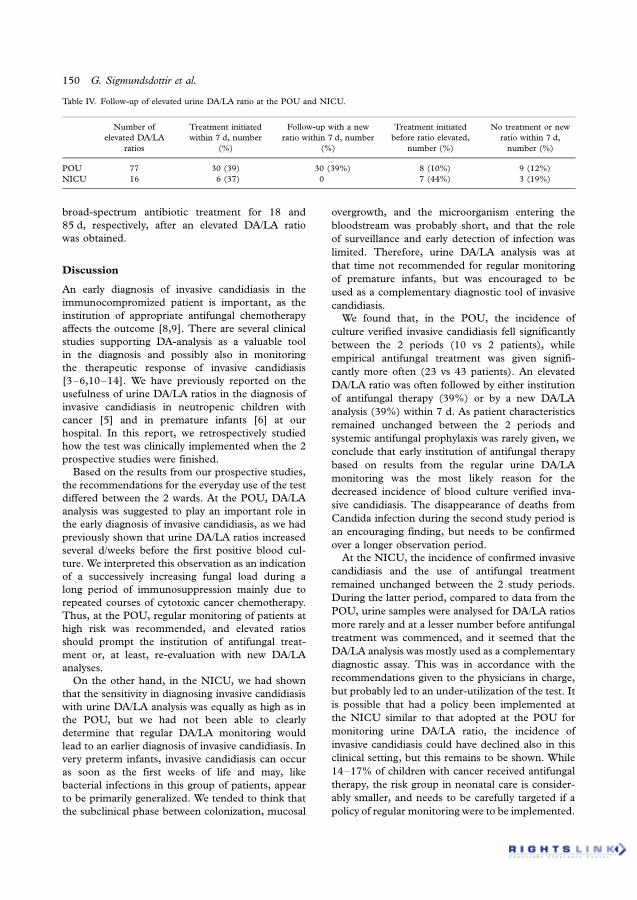
broad-spectrum antibiotic treatment for 18 and
85 d, respectively, after an elevated DA/LA ratio
was obtained.
Discussion
An early diagnosis of invasive candidiasis in the
immunocompromized patient is important, as the
institution of appropriate antifungal chemotherapy
affects the outcome [8,9]. There are several clinical
studies supporting DA-analysis as a valuable tool
in the diagnosis and possibly also in monitoring
the therapeutic response of invasive candidiasis
[3�6,10�14]. We have previously reported on the
usefulness of urine DA/LA ratios in the diagnosis of
invasive candidiasis in neutropenic children with
cancer [5] and in premature infants [6] at our
hospital. In this report, we retrospectively studied
how the test was clinically implemented when the 2
prospective studies were finished.
Based on the results from our prospective studies,
the recommendations for the everyday use of the test
differed between the 2 wards. At the POU, DA/LA
analysis was suggested to play an important role in
the early diagnosis of invasive candidiasis, as we had
previously shown that urine DA/LA ratios increased
several d/weeks before the first positive blood cul-
ture. We interpreted this observation as an indication
of a successively increasing fungal load during a
long period of immunosuppression mainly due to
repeated courses of cytotoxic cancer chemotherapy.
Thus, at the POU, regular monitoring of patients at
high risk was recommended, and elevated ratios
should prompt the institution of antifungal treat-
ment or, at least, re-evaluation with new DA/LA
analyses.
On the other hand, in the NICU, we had shown
that the sensitivity in diagnosing invasive candidiasis
with urine DA/LA analysis was equally as high as in
the POU, but we had not been able to clearly
determine that regular DA/LA monitoring would
lead to an earlier diagnosis of invasive candidiasis. In
very preterm infants, invasive candidiasis can occur
as soon as the first weeks of life and may, like
bacterial infections in this group of patients, appear
to be primarily generalized. We tended to think that
the subclinical phase between colonization, mucosal
overgrowth, and the microorganism entering the
bloodstream was probably short, and that the role
of surveillance and early detection of infection was
limited. Therefore, urine DA/LA analysis was at
that time not recommended for regular monitoring
of premature infants, but was encouraged to be
used as a complementary diagnostic tool of invasive
candidiasis.
We found that, in the POU, the incidence of
culture verified invasive candidiasis fell significantly
between the 2 periods (10 vs 2 patients), while
empirical antifungal treatment was given signifi-
cantly more often (23 vs 43 patients). An elevated
DA/LA ratio was often followed by either institution
of antifungal therapy (39%) or by a new DA/LA
analysis (39%) within 7 d. As patient characteristics
remained unchanged between the 2 periods and
systemic antifungal prophylaxis was rarely given, we
conclude that early institution of antifungal therapy
based on results from the regular urine DA/LA
monitoring was the most likely reason for the
decreased incidence of blood culture verified inva-
sive candidiasis. The disappearance of deaths from
Candida infection during the second study period is
an encouraging finding, but needs to be confirmed
over a longer observation period.
At the NICU, the incidence of confirmed invasive
candidiasis and the use of antifungal treatment
remained unchanged between the 2 study periods.
During the latter period, compared to data from the
POU, urine samples were analysed for DA/LA ratios
more rarely and at a lesser number before antifungal
treatment was commenced, and it seemed that the
DA/LA analysis was mostly used as a complementary
diagnostic assay. This was in accordance with the
recommendations given to the physicians in charge,
but probably led to an under-utilization of the test. It
is possible that had a policy been implemented at
the NICU similar to that adopted at the POU for
monitoring urine DA/LA ratio, the incidence of
invasive candidiasis could have declined also in this
clinical setting, but this remains to be shown. While
14�17% of children with cancer received antifungal
therapy, the risk group in neonatal care is consider-
ably smaller, and needs to be carefully targeted if a
policy of regular monitoring were to be implemented.
Table IV. Follow-up of elevated urine DA/LA ratio at the POU and NICU.
Number of
elevated DA/LA
ratios
Treatment initiated
within 7 d, number
(%)
Follow-up with a new
ratio within 7 d, number
(%)
Treatment initiated
before ratio elevated,
number (%)
No treatment or new
ratio within 7 d,
number (%)
POU 77 30 (39) 30 (39%) 8 (10%) 9 (12%)
NICU 16 6 (37) 0 7 (44%) 3 (19%)
150 G. Sigmundsdottir et al.
Scan
d J
Infe
ct D
is D
ownl
oade
d fr
om in
form
ahea
lthca
re.c
om b
y L
ands
pita
li U
nive
rsity
Hos
pita
l on
10/1
1/10
For
pers
onal
use
onl
y.

Our study has obvious limitations. Most impor-
tantly, we had not planned to perform a further
evaluation when the test was put into clinical use.
This led to the typical problems of a retrospective
analysis, e.g. it was not possible to determine all
considerations taken by the physicians on when to
institute antifungal therapy, as patient records rarely
contained such information. Also, the test was
introduced in very different ways in the 2 units
studied.
Nevertheless, we can conclude that the results of
our previous prospective study of the new diagnostic
assay could successfully be implemented in clinical
practice and even lead to decreased morbidity in a
well-defined group of high-risk patients, where the
recommendations for the use of the test and the
implications of a positive test were clearly defined.
Acknowledgement
We express thanks to Christina Pehrson and Einar
Larsson at the Department of Laboratory Medicine,
Eva Tyden at the Department of Paediatrics and
Jonas Cronqvist at Malmo University Hospital.
References
[1] Bernard E, Christianssen KJ, Tang SF, Kiehn TE,
Armstrong D. Rate of arabinitol production by pathogenic
yeast species. J Clin Microbiol 1981;/14:/189�94.[2] Larsson L, Pehrson C, Wiebe T, Christensson B. Gas
chromatographic determination of D-arabinitol/L-arabinitol
ratios in urine: a potential method for diagnosis of dissemi-
nated candidiasis. J Clin Microbiol 1994;/32:/1855�9.[3] Roboz J, Katz RN. Diagnosis of disseminated candidiasis
based on serum D/L-arabinitol ratio using negative chemical
ionization mass spectrometry. J Chromatogr 1992;/575:/
281�6.
[4] Walsh TJ, Merz WG, Lee JW, Schaufele R, Sein T,
Withcomb PO, et al. Diagnosis and therapeutic monitoring
of invasive candidiasis by rapid enzymatic detection of serum
D-arabinitol. Am J Med 1995;/99:/164�72.[5] Christensson B, Wiebe T, Pehrson C, Larsson L. Diagnosis
of invasive candidiasis in neutropenic children with cancer by
determination of D-arabinitol/L-arabinitol ratios in urine. J
Clin Microbiol 1997;/35:/636�40.[6] Sigmundsdottir G, Christensson B, Bjorklund LJ, Hakans-
son K, Pehrson C, Larsson L. Urine D-arabinitol/L-arabi-
nitol ratio in diagnosis of invasive candidiasis in newborn
infants. J Clin Microbiol 2000;/38:/3039�42.[7] Roboz J, Yu Q, Holland JF. Filter paper sampling of whole
blood and urine for the determination of D/L- arabinitol
ratios by mass spectrometry. J Microbiol Methods 1992;/15:/
207�14.[8] Eggimann P, Garbino J, Pittet D. Management of Candida
species infections in critically ill patients. Lancet Infect Dis
2003;/3:/772�85.[9] Blot SI, Vandewoude KH, Hoste EA, Colardyn FA. Effects
of nosocomial candidaemia on outcomes of critically ill
patients. Am J Med 2002;/113:/480�4.[10] Yeo SF, Zhang Y, Schafer D, Campell S, Wong B. A rapid,
automated enzymatic fluorometric assay for determination of
D-arabinitol in serum. J Clin Microbiol 2000;/38:/1439�43.[11] Hui M, Cheung S, Chin M, Chu K, Chan RC, Cheng AF.
Development and application of rapid diagnostic method for
invasive candidiasis by detection of D/L-arabinitol using gas
chromatography/mass spectrometry. Diagn Microbiol Infect
Dis 2004;/49:/117�23.[12] Lehtonen L, Anttila VJ, Ruutu T, Salonen J, Nikoskelainen
J, Eerola E, Ruutu P. Diagnosis of disseminated candidiasis
by measurement of urine D-arabinitol/L-arabinitol ratio. J
Clin Microbiol 1996;/34:/2175�9.[13] Eisen DP, MacGinley R, Christensson B, Larsson L, Woods
ML. Candida tropicalis vertebral osteomyelitis complicating
epidural catheterization with disease paralelled by elevated
D-arabinitol/L-arabinitol ratios. Eur J Clin Microbiol Infect
Dis 2000;/19:/61�3.[14] Stradomska TJ, Mielniczuk Z. Gas chromatographic deter-
mination of D-/L-arabinitol ratio in healthy Polish children. J
Chromatogr 2002;/773:/175�8 1.
D-arabinitol in invasive candidiasis 151
Scan
d J
Infe
ct D
is D
ownl
oade
d fr
om in
form
ahea
lthca
re.c
om b
y L
ands
pita
li U
nive
rsity
Hos
pita
l on
10/1
1/10
For
pers
onal
use
onl
y.