cytotoxic and genotoxic effects of methanol and aqueous ...
15
TITLE PAGE Article title: In vitro cytotoxic and genotoxic effects of methanol and aqueous extracts of Gymnosporia montana plant. Authors: Nishat Ansari and Divya Chandel Affiliations: Department of Zoology, BMT and Human Genetics, School of Sciences, Gujarat University, Ahmedabad, India Corresponding author: Dr. Divya Chandel. Department of Zoology, BMT and Human Genetics, School of Sciences, Gujarat University, Navrangpura – 380009, Ahmedabad, India. E-mail id: [email protected] Conflict of Interest: None
Transcript of cytotoxic and genotoxic effects of methanol and aqueous ...
TITLE PAGE
Article title: In vitro cytotoxic and genotoxic effects of methanol and aqueous extracts ofGymnosporia montana plant.
Authors: Nishat Ansari and Divya Chandel
Affiliations: Department of Zoology, BMT and Human Genetics,
School of Sciences, Gujarat University,
Ahmedabad, India
Corresponding author: Dr. Divya Chandel.
Department of Zoology, BMT and Human Genetics,
School of Sciences, Gujarat University,
Navrangpura – 380009, Ahmedabad, India.
E-mail id: [email protected]
Conflict of Interest: None
IN VITRO CYTOTOXIC AND GENOTOXIC EFFECTS OF METHANOL ANDAQUEOUS EXTRACTS OF GYMNOSPORIA MONTANA PLANT
NISHAT ANSARI AND DIVYA CHANDELDepartment of Zoology, BMT and Human Genetics, School of Sciences, Gujarat University,
Navrangpura – 380009, Ahmedabad, India.E-mail: [email protected]
ABSTRACT
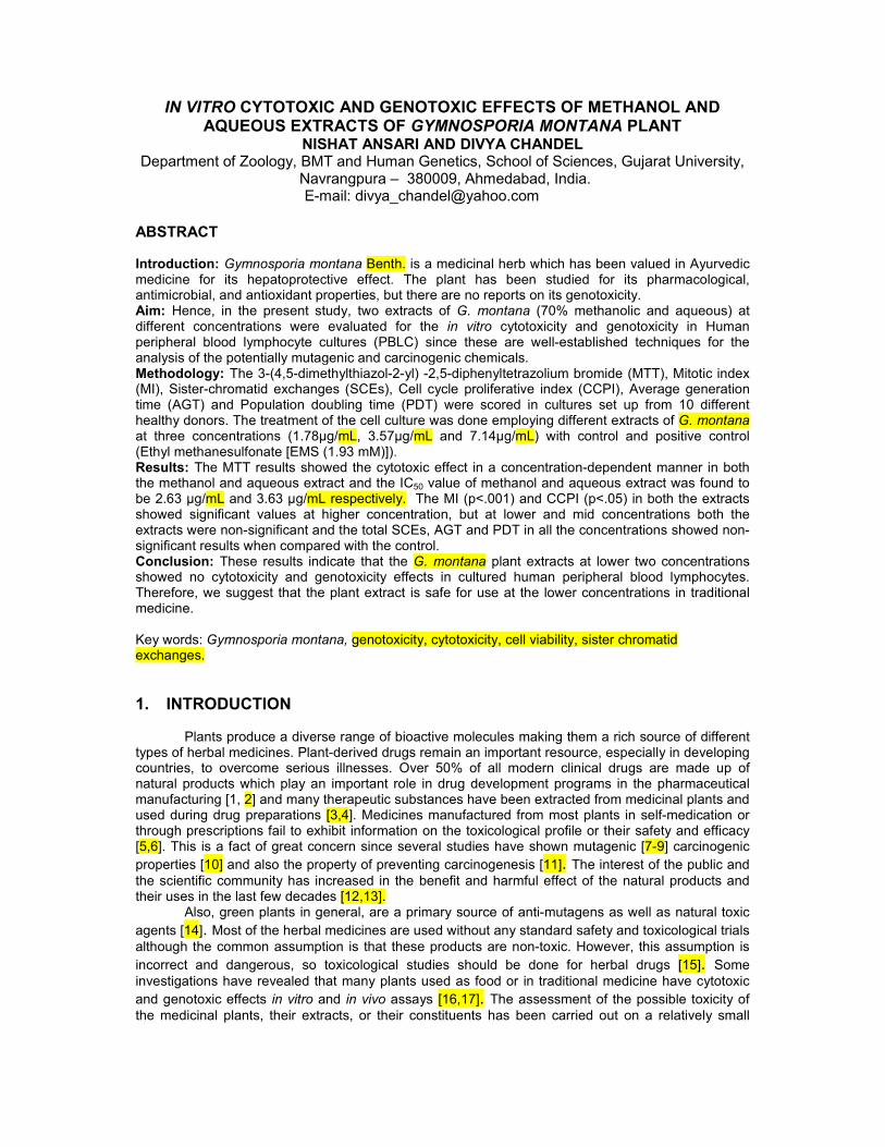
Introduction: Gymnosporia montana Benth. is a medicinal herb which has been valued in Ayurvedicmedicine for its hepatoprotective effect. The plant has been studied for its pharmacological,antimicrobial, and antioxidant properties, but there are no reports on its genotoxicity.Aim: Hence, in the present study, two extracts of G. montana (70% methanolic and aqueous) atdifferent concentrations were evaluated for the in vitro cytotoxicity and genotoxicity in Humanperipheral blood lymphocyte cultures (PBLC) since these are well-established techniques for theanalysis of the potentially mutagenic and carcinogenic chemicals.Methodology: The 3-(4,5-dimethylthiazol-2-yl) -2,5-diphenyltetrazolium bromide (MTT), Mitotic index(MI), Sister-chromatid exchanges (SCEs), Cell cycle proliferative index (CCPI), Average generationtime (AGT) and Population doubling time (PDT) were scored in cultures set up from 10 differenthealthy donors. The treatment of the cell culture was done employing different extracts of G. montanaat three concentrations (1.78µg/mL, 3.57µg/mL and 7.14µg/mL) with control and positive control(Ethyl methanesulfonate [EMS (1.93 mM)]).Results: The MTT results showed the cytotoxic effect in a concentration-dependent manner in boththe methanol and aqueous extract and the IC50 value of methanol and aqueous extract was found tobe 2.63 µg/mL and 3.63 µg/mL respectively. The MI (p<.001) and CCPI (p<.05) in both the extractsshowed significant values at higher concentration, but at lower and mid concentrations both theextracts were non-significant and the total SCEs, AGT and PDT in all the concentrations showed non-significant results when compared with the control.Conclusion: These results indicate that the G. montana plant extracts at lower two concentrationsshowed no cytotoxicity and genotoxicity effects in cultured human peripheral blood lymphocytes.Therefore, we suggest that the plant extract is safe for use at the lower concentrations in traditionalmedicine.
Key words: Gymnosporia montana, genotoxicity, cytotoxicity, cell viability, sister chromatidexchanges.
1. INTRODUCTION
Plants produce a diverse range of bioactive molecules making them a rich source of differenttypes of herbal medicines. Plant-derived drugs remain an important resource, especially in developingcountries, to overcome serious illnesses. Over 50% of all modern clinical drugs are made up ofnatural products which play an important role in drug development programs in the pharmaceuticalmanufacturing [1, 2] and many therapeutic substances have been extracted from medicinal plants andused during drug preparations [3,4]. Medicines manufactured from most plants in self-medication orthrough prescriptions fail to exhibit information on the toxicological profile or their safety and efficacy[5,6]. This is a fact of great concern since several studies have shown mutagenic [7-9] carcinogenicproperties [10] and also the property of preventing carcinogenesis [11]. The interest of the public andthe scientific community has increased in the benefit and harmful effect of the natural products andtheir uses in the last few decades [12,13].
Also, green plants in general, are a primary source of anti-mutagens as well as natural toxicagents [14]. Most of the herbal medicines are used without any standard safety and toxicological trialsalthough the common assumption is that these products are non-toxic. However, this assumption isincorrect and dangerous, so toxicological studies should be done for herbal drugs [15]. Someinvestigations have revealed that many plants used as food or in traditional medicine have cytotoxicand genotoxic effects in vitro and in vivo assays [16,17]. The assessment of the possible toxicity ofthe medicinal plants, their extracts, or their constituents has been carried out on a relatively small
number of plant species [18]. The claim that natural plant products are safe should be accepted onlyafter the plant product passes through toxicity testing using modern scientific methods [19].
G. montana belonging to the family Celastraceae is found in South Africa, Nigeria, TropicalAfrica, Mediterranean, Malaya, Australia, Arabia, Afghanistan, and India. It is a shrub or tree growingwild in dry areas and is commonly known as Vikalo in Gujarat, India, where it has been traditionallyused in treating the ulcer, jaundice, gastrointestinal disorders, toothache, dysentery, andantispasmodic effect [20]. The previous study by Patel, et al. [21] indicates that the extract of G.montana possesses significant hepatoprotective activity. A literature survey also showed that plantextracts can be mutagenic as well as antimutagenic depending on the test system used whichindicates that a battery of assays are needed before any conclusion can be reached. Since the G.montana plant has not been investigated yet for cytotoxicity and genotoxicity, this study wasundertaken to evaluate the plant by using its two different extracts i.e. 70% methanolic extract and anaqueous extract. Since the short-term tests for genotoxicity are typically used to identify potentialmutagens and carcinogens, the leaf extracts of G. montana were studied for toxicity in culturedhuman peripheral blood lymphocytes using MTT, mitotic index (MI), sister chromatid exchanges(SCEs), cell cycle proliferative index (CCPI), average generation time (AGT) and population doublingtime (PDT), as Peripheral Blood Lymphocyte cultures (PBLC) are a well established method toexamine the genotoxic damage.
2. MATERIALS AND METHODS
2.1 Collection of plant materials and extract preparation
The leaves of G. montana were collected from a nursery in sector-30 of Gandhinagar. The taxonomicidentity was authenticated and herbarium sheet with reference No. PH/14/0010 was deposited inDepartment of Pharmacognosy, K.B. Institute of Pharmaceutical Education and Research,Gandhinagar, India. The compounds in the leaves of G. montana was extracted according to ourprevious study [22] and after performing MTT and MI, the three different concentrations: Low dose(1.78 µg/mL), Mid dose (3.57 µg/mL) and High dose (7.142 µg/mL) were finalized for further studies.Both the methanolic and aqueous extracts were analyzed at these concentrations for their cytotoxicand genotoxic effect using different parameters as mentioned below.
2.2 Chemicals and reagents
All chemicals utilized in the following parameters were procured from Merck, Germany (AR Grade),while media and culture reagents were procured from HiMedia and Sigma Aldrich, USA (CultureGrade).
2.3 Protocols
The present study was approved by the Institutional Ethical Committee (IEC) of GujaratUniversity, Ahmedabad, India and 10 healthy individuals free from any infection, addiction (includingtobacco, drug or alcohol abuse and non-smoking) between 20-30 years of age were recruited. Bloodcollection was carried out according to ethical guidelines and with prior consent of the subjects.Venous blood was collected from each donor in heparinized vacutainer and mixed gently to avoidblood clotting.
2.4 Cell Viability Assay (MTT Assay)
A quantitative colorimetric assay for mammalian cell survival and cell proliferation wasassayed by the method of Mosmann [23] with slight modifications. An equal amount of Hicep andblood was taken in Tarson tubes and the WBC layer was separated for further use. 1.5 mL media, 20ul PHA, and varying doses ranging from 0.50µg/mL to 10 µg/mL of the plant extract were added to

100ul of the sample and EMS (1.93mM) was used as a positive control. The cultures were incubatedfor 72 hours and harvesting was done on the 3rd day. After addition of MTT [3-(4,5-dimethylthiazol-2-yl)-2,5-diphenyltetrazolium bromide] (30ul) the cultures were further incubated for 4 hours at 37oC.DMSO was added to the pellet to observe a color change. The samples were centrifuged and theoptical density was measured at 570 nm on Epoch Biotek.
2.5 Mitotic index (MI)
Initially, the cultures were set up as per the standard protocol [24] at different concentrations and MIwas done to finalize the three doses used in this study. By quantifying aspects of a dividing cellpopulation, one can examine how cells differ in their capability to divide under different experimentalconditions. Mitotic index is defined as the ratio between the number of cells in mitosis and the totalnumber of cells. A total of 1000 cells were scored for each individual according to the followingformula.
--------------------------------- x 100
2.6 Sister chromatid exchanges (SCEs)
Peripheral blood lymphocyte cultures were prepared according to the standard procedure [24]with slight modifications and for SCEs, 80µl of Bromodeoxyuridine (1mg/mL, BrdU) was added at 0th
hour [25] and cultures were incubated in dark at 37°C in an incubator and were harvested at 72 hoursafter colchicine treatment at the 69th hour and slides were prepared. Cleaned slides were prepared byadding 3 to 4 drops of the cell suspension uniformly on a chilled slide and were flame dried. For SisterChromatid Differentiation, the slides were stained with Hoechst 33258 for 20 min in the dark andlayered with 2X saline sodium citrate buffer. The slides were then kept in UV exposure for 45 min in amoist chamber, washed in water and stained in 2% Giemsa for 5 min and were analyzed for SCEs,CCPI, AGT and PDT.
2.7 Analysis of sister chromatid exchanges (SCEs)
Metaphases in the second cycle of cell division (M2) were selected for scoring on the basis ofthe spreading of chromosomes and differentiation of chromatids. Total exchanges were counted in 30M2 cells to calculate SCE/Plate and SCE/Chromosome to evaluate the level of Genotoxicity [26] usingthe following formula:
SCEs/Plate = Total SCEs scored-------------------------------Total M2 plates scored
SCEs/Chromosome = SCEs/Plate-----------------------------------------Total no. of chromosomes (i.e.46)
2.8 Analysis of Cell Cycle Proliferative Index (CCPI)
Differentially stained slides were scored for the proliferative kinetics based on the stainingpattern of chromosomes. Total 100 metaphases were analyzed for each individual, classifying themon the first (M1), second (M2), third (M3) generation cells. At least 100 metaphases from each culturewere analyzed for CCPI [26].
The cell cycle proliferative index (CCPI) for each individual was calculated according to the followingformula:

CCPI = 1 (M1 plate) + 2 (M2 plate) + 3 (M3 plate)---------------------------------------------------
100 (Total plates)
2.9 Analysis of average generation time and population doubling time
The cell cycle time AGT is also studied as the ratio of the BrdU time and proliferative index(CCPI)
AGT = 72 hours (BrdU time)------------------
CCPI
And, the PDT is the time in which the cell divides, i.e. 24 hours
PDT = 24 hours------------CCPI
2.10 Statistical Analysis
Values of all the data are expressed as MeanStandard Error of the 10 replicates. Thestatistical analysis was done in GraphPad Prism 6.0 software. A t-test was done to compare twogroups while One-way ANOVA was done by Tukey’s multiple comparison tests to compare more thantwo groups and significance was accepted when p < .05.
3. RESULTS
In the present study, the cells showed growth inhibition in a dose-dependent manner with asignificant increase (p<.001) in the percent cell inhibition when treated with two different extracts of G.montana at concentrations ranging from 0.50µg/mL to 10 µg/mL (Table 1.A and 1.B). The percentageof dead cells in the methanol extract for each concentration was found to be 45.6, 48.77, 53.1, 57.5,67.2, 68.4, 76.6, 77.8, 79.47, 83.9, 88.3 and in aqueous was found to be 42.2, 47.6, 48.7, 51.7, 51.8,56.5, 63.5, 71.4, 75.5, 78.262, 78.7. The IC50 value of G. montana was found to be 2.63 µg/mL inmethanol and 3.63 µg/mL in aqueous extract.
From the prepared slides, dividing and non-dividing cells were counted to determine themitotic index for the assessment of the cytotoxicity in the control, positive control and plant extracts atdifferent concentrations (Low Dose (LD): 1.78µg/mL, Mid Dose (MD): 3.57µg/mL and High Dose (HD):7.14µg/mL). Table 2 shows the mean and standard error of the MI rates of control (3.22±0.167983)and the positive control (1.52±0.099849). The MI of the three concentrations of methanol extract was2.59±0.114051, 2.53±0.140774 and 1.65±0.126802 and aqueous extract was found to be2.56±0.161499, 2.48±0.11243 and 1.6±0.142307. For MI, the extracts (both methanol and aqueous)showed a non-significant decrease in LD and MD whereas, highly significant (p<.001) decrease wasseen in the HD when it was compared with control values. Compared to the MI value of the positivecontrol, a highly significant increase (p<.001) in LD and MD and a non-significant increase in HD inmethanol extract were seen. On the other hand, the aqueous extract showed a significant increase(p<.05) in LD and MD, but a non-significant increase in the HD.
Our analysis showed that the total SCEs were non-significantly increased in all theconcentrations when both the extracts (methanol and aqueous) were compared with the control group(Table 3). Also, on comparison with the positive control, both the extract showed highly significant(p<.001) decrease in LD, but significant (p<.01) decrease in MD and HD. (Table 3). As shown in Table4, Cell cycle proliferative index (CCPI) showed a non-significant decrease in LD and MD butsignificant (p<.05) decrease in HD when compared with control in both the extracts. Whereas, incomparison with the positive control, significant (p<.05) increase in LD and MD and non-significant

(p<.05) increase in HD was seen. The AGT and PDT were non-significantly elongated in all theconcentrations when they were compared with control (Table 4).
TABLE 1. A: Cytotoxicity of G. montana in methanol and aqueous extract.
CONCENTRATION % CELL INHIBITION % CELL INHIBITION(µg / mL) (METHANOL EXTRACT) (AQUEOUS EXTRACT)
0.50 45.6 42.2
1 48.77 47.6
2 53.1 48.7
3 57.5 51.7
4 67.2 51.8
5 68.4 56.5
6 76.6 63.5
7 77.8 71.4
8 79.47 75.5
9 83.9 78.262
10 88.3 78.7
IC50 VALUE 2.63 3.63
Each value is represented as Mean±SE, n=10.
Aqueous Extract vs Methanol Extract: ****=p< .0001 (Highly significant)
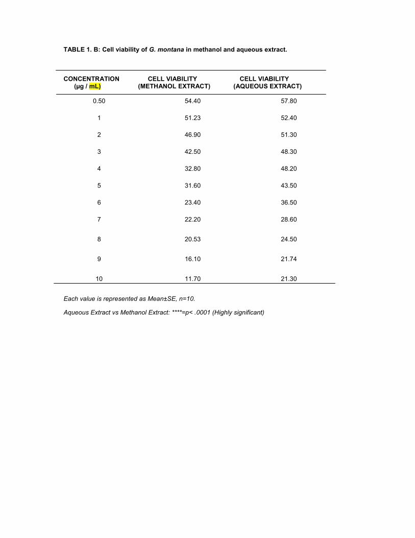
TABLE 1. B: Cell viability of G. montana in methanol and aqueous extract.
CONCENTRATION CELL VIABILITY CELL VIABILITY(µg / mL) (METHANOL EXTRACT) (AQUEOUS EXTRACT)
0.50 54.40 57.80
1 51.23 52.40
2 46.90 51.30
3 42.50 48.30
4 32.80 48.20
5 31.60 43.50
6 23.40 36.50
7 22.20 28.60
8 20.53 24.50
9 16.10 21.74
10 11.70 21.30
Each value is represented as Mean±SE, n=10.
Aqueous Extract vs Methanol Extract: ****=p< .0001 (Highly significant)
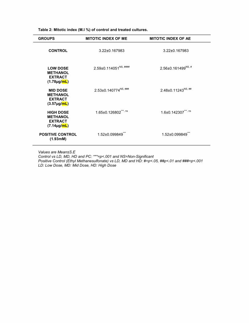
Table 2: Mitotic index (M.I %) of control and treated cultures.
GROUPS MITOTIC INDEX OF ME MITOTIC INDEX OF AE
Values are Mean±S.EControl vs LD, MD, HD and PC: ***=p<.001 and NS=Non-SignificantPositive Control (Ethyl Methanesulfonate) vs LD, MD and HD: #=p<.05, ##p<.01 and ###=p<.001LD: Low Dose, MD: Mid Dose, HD: High Dose
CONTROL 3.22±0.167983 3.22±0.167983
LOW DOSEMETHANOLEXTRACT
(1.78µg/mL)
2.59±0.114051NS, #### 2.56±0.161499NS, #
MID DOSEMETHANOLEXTRACT
(3.57µg/mL)
2.53±0.140774NS, ### 2.48±0.11243NS, ##
HIGH DOSEMETHANOLEXTRACT
(7.14µg/mL)
1.65±0.126802***, ns 1.6±0.142307***, ns
POSITIVE CONTROL(1.93mM)
1.52±0.099849*** 1.52±0.099849***

Table 3: Showing Total SCEs, SCEs/plate and SCEs/chromosome in control and treatedcultures.
EXTRACTS GROUPS TOTAL SCES SCES/PLATE SCES/CHROMOSOME
CONTROL 90.1±3.03332 2.999763±0.102211 0.065212±0.002222
METHANOLEXTRACT
LOW DOSE(1.78µg/mL)
91.2±3.761551NS, ###
3.042233±0.1242NS, ###
0.066136±0.0027NS, ###
MID DOSE(3.57µg/mL)
94.7±4.011352NS, ##
3.153752±0.124359NS, ##
0.06856±0.00288NS, ##
HIGH DOSE(7.14µg/mL)
107.1±4.062072NS, ##
3.599726±0.145142NS, ##
0.078255±0.003155NS, ##
AQUEOUSEXTRACT
LOW DOSE(1.78µg/mL)
91.8±5.603242081NS, ###
3.062538±0.004093296NS, ##
0.066577±0.00418228NS, ##
MID DOSE(3.57µg/mL)
97.4±3.739001389NS, ##
3.245319±0.162222711NS, ##
0.07055±0.002703465NS, ##
HIGH DOSE(7.14µg/mL)
108.1±3.303726066NS, ##
3.603992±0.110085075NS, ##
0.078348±0.002393154NS, ##
Values are Mean±S.EControl vs LD, MD, HD and PC: ***=p<.001 and NS=Non-SignificantPositive Control (Ethyl Methanesulfonate) vs LD, MD and HD: ##p<.01 and ###=p<.001LD: Low Dose, MD: Mid Dose, HD: High Dose

Table 4: Showing CCPI, AGT, and PDT in control and treated cultures
EXTRACTS GROUPS CCPI AGT PDT
CONTROL 2.145±0.0151
33.5815625±0.2382
11.19385417±0.07941
METHANOLEXTRACT
LOW DOSE(1.78µg/mL)
2.13±0.01334NS, ## 33.8147±0.2111 NS,
##11.2715±0.0703NS, ##
MID DOSE(3.57µg/mL)
2.063±0.03325NS, ## 34.9882±0.6056NS, # 11.6627±0.2018NS, #
HIGH DOSE(7.14µg/mL)
1.901±0.0621*, ns 38.2796±1.3895NS, ns 12.7598±0.4631NS, ns
AQUEOUSEXTRACT
LOW DOSE(1.78µg/mL)
2.084±0.0363NS, # 34.6549±0.6783NS, # 11.5516±0.2261 NS, #
MID DOSE(3.57µg/mL)
2.075±0.0251NS, # 34.7453±0.4282 NS, # 11.5817±0.1427 NS, #
HIGH DOSE(7.14µg/mL)
1.895±0.06102*, ns 38.3889±1.3733 NS,
ns12.7963±0.4577 NS, ns
POSITIVECONTROL(1.93mM)
1.718±0.0693** 42.5220±1.7090** 14.1740±0.5696**
Values are Mean±S.EControl vs LD, MD, HD and PC: *=p<.05, **=p<.01and NS=Non-SignificantPositive Control (Ethyl Methanesulfonate) vs LD, MD and HD: #=p<.05, ##p<.01 and ns=Non-SignificantLD: Low Dose, MD: Mid Dose, HD: High Dose
4. DISCUSSION
Special attention must be paid to the evaluation of the safety, efficacy, and quality of naturalproducts and their components and it is important to determine the toxic effect of plant extracts usedas herbal therapy on humans. In our study, we took a step towards exploring the cytotoxic andgenotoxic effects of a herbal plant G. montana, since it is being used as an alternative medicinelocally to cure jaundice and has a hepatoprotective effect [20,21]. Previously by Bhavita andcoworkers [20], the leaf of G. montana was evaluated for pharmacognostical parameters andphytochemical screening, which indicated that the leaf of G. montana was rich in phenol. In ourprevious study, we have tested the plant for its antioxidant activities in the two different extracts usedhere, which indicated that the G. montana plant is a good antioxidant [22]. The cytotoxic effect ofthese two leaf extracts of the plant at different concentrations was checked by doing the MTT assay,which is an appropriate method for screening new substances within a short period of time. The MTT
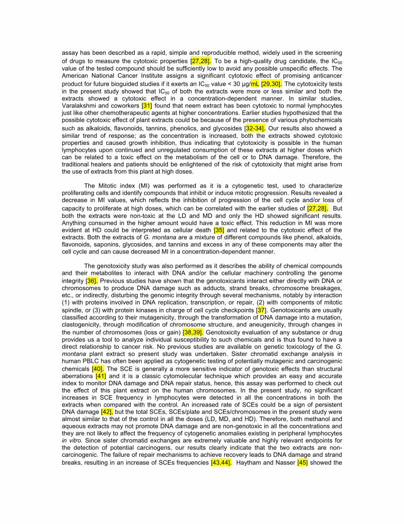
assay has been described as a rapid, simple and reproducible method, widely used in the screeningof drugs to measure the cytotoxic properties [27,28]. To be a high-quality drug candidate, the IC50value of the tested compound should be sufficiently low to avoid any possible unspecific effects. TheAmerican National Cancer Institute assigns a significant cytotoxic effect of promising anticancerproduct for future bioguided studies if it exerts an IC50 value < 30 μg/mL [29,30]. The cytotoxicity testsin the present study showed that IC50 of both the extracts were more or less similar and both theextracts showed a cytotoxic effect in a concentration-dependent manner. In similar studies,Varalakshmi and coworkers [31] found that neem extract has been cytotoxic to normal lymphocytesjust like other chemotherapeutic agents at higher concentrations. Earlier studies hypothesized that thepossible cytotoxic effect of plant extracts could be because of the presence of various phytochemicalssuch as alkaloids, flavonoids, tannins, phenolics, and glycosides [32-34]. Our results also showed asimilar trend of response; as the concentration is increased, both the extracts showed cytotoxicproperties and caused growth inhibition, thus indicating that cytotoxicity is possible in the humanlymphocytes upon continued and unregulated consumption of these extracts at higher doses whichcan be related to a toxic effect on the metabolism of the cell or to DNA damage. Therefore, thetraditional healers and patients should be enlightened of the risk of cytotoxicity that might arise fromthe use of extracts from this plant at high doses.
The Mitotic index (MI) was performed as it is a cytogenetic test, used to characterizeproliferating cells and identify compounds that inhibit or induce mitotic progression. Results revealed adecrease in MI values, which reflects the inhibition of progression of the cell cycle and/or loss ofcapacity to proliferate at high doses, which can be correlated with the earlier studies of [27,28]. Butboth the extracts were non-toxic at the LD and MD and only the HD showed significant results.Anything consumed in the higher amount would have a toxic effect. This reduction in MI was moreevident at HD could be interpreted as cellular death [35] and related to the cytotoxic effect of theextracts. Both the extracts of G. montana are a mixture of different compounds like phenol, alkaloids,flavonoids, saponins, glycosides, and tannins and excess in any of these components may alter thecell cycle and can cause decreased MI in a concentration-dependent manner.
The genotoxicity study was also performed as it describes the ability of chemical compoundsand their metabolites to interact with DNA and/or the cellular machinery controlling the genomeintegrity [36]. Previous studies have shown that the genotoxicants interact either directly with DNA orchromosomes to produce DNA damage such as adducts, strand breaks, chromosome breakages,etc., or indirectly, disturbing the genomic integrity through several mechanisms, notably by interaction(1) with proteins involved in DNA replication, transcription, or repair, (2) with components of mitoticspindle, or (3) with protein kinases in charge of cell cycle checkpoints [37]. Genotoxicants are usuallyclassified according to their mutagenicity, through the transformation of DNA damage into a mutation,clastogenicity, through modification of chromosome structure, and aneugenicity, through changes inthe number of chromosomes (loss or gain) [38,39]. Genotoxicity evaluation of any substance or drugprovides us a tool to analyze individual susceptibility to such chemicals and is thus found to have adirect relationship to cancer risk. No previous studies are available on genetic toxicology of the G.montana plant extract so present study was undertaken. Sister chromatid exchange analysis inhuman PBLC has often been applied as cytogenetic testing of potentially mutagenic and carcinogenicchemicals [40]. The SCE is generally a more sensitive indicator of genotoxic effects than structuralaberrations [41] and it is a classic cytomolecular technique which provides an easy and accurateindex to monitor DNA damage and DNA repair status, hence, this assay was performed to check outthe effect of this plant extract on the human chromosomes. In the present study, no significantincreases in SCE frequency in lymphocytes were detected in all the concentrations in both theextracts when compared with the control. An increased rate of SCEs could be a sign of persistentDNA damage [42], but the total SCEs, SCEs/plate and SCEs/chromosomes in the present study werealmost similar to that of the control in all the doses (LD, MD, and HD). Therefore, both methanol andaqueous extracts may not promote DNA damage and are non-genotoxic in all the concentrations andthey are not likely to affect the frequency of cytogenetic anomalies existing in peripheral lymphocytesin vitro. Since sister chromatid exchanges are extremely valuable and highly relevant endpoints forthe detection of potential carcinogens, our results clearly indicate that the two extracts are non-carcinogenic. The failure of repair mechanisms to achieve recovery leads to DNA damage and strandbreaks, resulting in an increase of SCEs frequencies [43,44]. Haytham and Nasser [45] showed the

reduction in sister chromatid exchanges or change in the MI may be due to the direct action of thecompounds present in the extract by inactivating it enzymatically or chemically.
The cell cycle kinetics was evaluated by the cell cycle proliferative index (CCPI), a parameterthat represents the ratio of cells in the first, second and third mitosis and was gradually decreased inthe present study similar to the results of MI for both the extracts, possibly due to an arrest of mitosiswhich allows the repair of genetic material. Cytotoxic effects occur in cells with a relatively high levelof genetic damage [46]. We propose that the extract might be causing some DNA damage at HDwhich gets repaired during the G1 phase and hence no significant difference in SCEs values areobserved. The significant decrease in cell cycle progression for HD observed in the present studywould allow the DNA repair to take place. We suggest that no DNA damage is taking at the LD andMD, hence no extra time is required for DNA repair to take place in cell cycle and hence the cell cycleprogression is similar to control values at these doses. Thus, we can say that the extracts areaffecting the cells on higher consumption as MI and CCPI, both at higher doses were affecting thecells by showing antimitotic properties but both LD and MD were having no cytotoxicity, whichindicates that both the extracts did not have a significant cytotoxic effect on these parameters at theused lower two concentrations (LD and MD). It can be because medicinal herbs contain complexmixtures of thousands of compounds that can exert their antioxidant and free radical scavengingeffect either separately or in synergistic ways [47]. Also, some bioactive compounds present in theplant can interfere with drug kinetics and produce adverse effects related or unrelated to theirpharmacological actions, such as allergic reactions, mutagenic and carcinogenic effects. In this study,both the extracts affect cell proliferation by declining the CCPI and cell progression by elongation ofAGT and PDT in a concentration-dependent manner in comparison with control and positive control.The reduction in the mitotic activity and the cell-cycle delay at higher doses could be attributed due tothe formation of toxic DNA cross-links. These cross-links inhibit DNA replication and transcription,causing cell cycle arrest and induction of apoptosis [48]. The data obtained in the present studysuggest that the compounds present in the extract of G. montana are not cytotoxic and not mutagenicat lower doses (LD and MD) and when treated cultures were compared to control and positive control,the values showed mitotic depression in all cultures, however, the values were non-significant. Thus,the plant extract is not harmful at the two lower tested levels (LD and MD) and can be safely used upto these doses and both the extracts are safe and non-toxic at these tested levels.
5. CONCLUSIONS
G. montana belonging to the family Celastraceae has been traditionally used in treatingjaundice and has not been examined for cytotoxicity and genotoxicity till date. Evaluation of theendpoints described here would help to estimate the potential toxicity of this medicinal plant. The MTTfindings indicate that cytotoxicity was aggravated with the increase in the concentration of both theleaf extracts of G. montana. The present in vitro investigations showed that the MI and CCPI for boththe extracts of G. montana were almost similar to that of control at lower two concentrations (LD andMD), but at higher doses they produce cytotoxicity. The SCEs showed no genotoxic effect in all thethree doses (LD, MD, and HD) of both the extracts. These observations specify that the G. montanaplant extracts showed no cytotoxicity up to MD (3.57µg/mL) and no genotoxicity effects at all the threedoses (LD, MD, and HD) in cultured human peripheral blood lymphocytes. It is concluded that themethanol and aqueous extracts of G. montana is safe for use in humans and can be used at thesedoses in herbal medicine. Further in vitro and in vivo investigations are needed to confirm the results.
ACKNOWLEDGEMENT
This study was supported by the Maulana Azad National Fellowship (MANF) grant from the UniversityGrants Commission (UGC), New Delhi, India.
AUTHORS CONTRIBUTION
Dr. Divya Chandel designed the experiment and drafted and revised the manuscript. Ms. NishatAnsari has performed the experiments and wrote and revised the manuscript.
COMPETING INTERESTS
Authors have declared that no competing interests exist.
REFERENCES
1. Vedamurthy AB, Paarakh PM, Jogaiah S. Comparative study of antibacterial and antioxidantactivity of fractionated plant extracts of amla [phyllanthus emblica l.], tulsi [ocimum tenuifloruml.] And neem [azadirachta indica a.juss] European Journal of Biomedical AND Pharmaceuticalsciences. 2015;2(3):253-259.
2. Sevindik M, Akgul H, Pehlivan M, Selamoglu Z. Determination of therapeutic potential ofMenthalongifolia ssp. longifolia. Fresen Environ Bull. 2017; 26: 4757-4763.
3. Ouedraogo M, Baudoux T, Stevigny C, Nortier J, Colet JM, Efferth T, Qu F, Zhou J, Chan K,Shaw D, Pelkonen O, Duez P. Review of current and “omics” methods for assessing thetoxicity (genotoxicity, teratogenicity, and nephrotoxicity) of herbal medicines and mushrooms.J Ethnopharmacol. 2012;140(3):492–512.
4. Pehlivan M, Sevindik M. Antioxidant and antimicrobial activities of salvia multicaulis. TURJAF.2018; 6(5): 628-631.
5. Fernandes JBF, Vargas VMF. Mutagenic and antimutagenic potential of the medicinal plantsM. laevigata and C. xanthocarpa. Phytoth. Res. 2003;17(3):269-273.
6. Capasso R, Izzo AA, Pinto L, Bifulco T, Vitobello C, Mascolo N. Phytotherapy and quality ofherbal medicines. Fitoterapia. 2000;71(1):58- 65.
7. Cardoso CRP, De SC, Bernardi IM, Sannomiya CC, Vilegas M, Varanda WEA. Mutagenicactivity promoted by amentoflavone and methanolic extract of Byrsonima crassa Niedenzu.Toxicology. 2006;225,(1):55-63.
8. Déciga-Campos M, Rivero-Cruz I, Arriagaalba M, Castañedacorral G, Angeleslópez GE,Navarrete A, Mata R. Acute toxicity and mutagenic activity of Mexican plants used intraditional medicine. J. Ethnopharmacol. 2007;110(2):334-342.
9. Mohd-Fuat A, Kofi E. Mutagenic and cytotoxic properties of three herbal plants fromSoutheast Asia. Trop. Biomed. 2007;24(2):49-59.
10. Sá-Ferreira ICF, Vargas VMF Mutagenicity of medicinal plant extracts inSalmonella/microsome assay. Phytoth. Res.1999;13(5):397-400.
11. Sarkar D, Sharma A and Talukder G. Plant Extracts as Modulators of Genotoxic Effects.Botanical Review Vol.1996;62(4):275-300.
12. Pehlivan M, Mohammed FS, Sevindik M, Akgul H. Antioxidant and oxidant potential of Rosacanina. Eurasian J of Forest Sci. 2018;6(4): 22-25.
13. Calapai G, Caputi AP. Herbal medicines: can we do without pharmacologist? 2007;4:41–43.14. Plewa MJ and Wagner ED. “Activation of promutagens by green plants,” Annual Review of
Genetics. 1993;27: 93–113.15. Kahaliw W, Hellman B, Engidawork E. Genotoxicity study of Ethiopian medicinal plant
extracts on HepG2 cells. BMC Complementary and Alternative Medicine 2018;18:45.16. Celik TA, Aslant OS.“Cytotoxic and genotoxic ¨ effects of Lavandula stoechas aqueous
extracts,” Biologia. 2007;62(3):292–296.17. Mohammed FS, Akgul H, Sevindik M, Khaled BMT. Phenolic Content and Biological Activities
of Rhuscoriaria var. zebaria. Fresen Environ Bull. 2018; 27(8): 5694-5702.18. Jordan SA, Cunningham DG, Marles RJ. Assessment of herbal medicinal products:
challenges, and opportunities to increase the knowledge base for safety assessment. ToxicolAppl Pharmacol. 2010;243(2):198–216.
19. Jaykaran P, Bhardwaj, Kantnaria N, Madav P, Panwar A. The internet journal of Toxicology.2009;6(1):1-6.
20. Dhru B, Patel B, Lakshmi B, Zaveri M. To evaluate anti-inflammatory and analgesic activity ofleaf of gymnosporia moantana- a native of Gujarat American journal of pharmtech research.2011;1(3):227-237.
21. Patel PB, Patel TK, Shah P, Baxi SN, Sharma HO, Tripathi CB. Protective Effect of EthanolExtract of Gymnosporia montana (Roth) Bemth. in Paracetamol-induced Hepatotoxicity inRats. Indian Journal of Pharmaceutical Sciences. 2010;72(3):392-396.
22. Ansari N, Chandel D. Antioxidant studies on methanol and aqueous extracts of gymnosporiamontana plant. Int J Pharm Pharm Sci. 2019;11(2):65-70.
23. Mosmann T. Rapid colorimetric assay for cellular growth and survival: application toproliferation and cytotoxicity assays, J. Immunol. Methods 1983;65:55-63.
24. Hungerford DA. Leukocytes cultured from small inocula of whole blood and the preparation ofmetaphase chromosomes by treatment with hypotonic KCl. Stain Tech. 1965;40:333–338.
25. Perry P, Wolff S. New Giemsa method for the differential staining of sister chromatids. Nature.1974;251:156–158.
26. Veniit S, Parry J. Mutagenicity Testing: a Practical Approach. IRL Press, Oxford, WashingtonD.C. 1984;275-306.
27. Kasurka CB, Sekero ZA, Sekero LZ. Evaluation of the genotoxicity and cytotoxicity offexofenadine in cultured human peripheral blood lymphocytes. Toxicol in vitro,2011;25(7):1480-4++
28. Ligia MCB, Eliane PA, Ana BTM; Valter ADR. The viability of lymphocyte culture, at differenttimes after blood collection, for karyotype analysis. J Bras Patol Med Lab. 2014;50(2):124-130.
29. Suffness M, Pezzuto JM. Assays related to cancer drug discovery. In: Hostettmann, K. (Ed).Methods in Plant Biochemistry: Assays for Bioactivity, Academic Press, London. 1990;6:71-133.
30. Khakdan F, Khosro P. In vitro cytotoxic activity of aqueous root extract of Althea kurdicaagainst endothelial human bone marrow cells (line k562) and human lymphocytes. Bull EnvPharmacol Life Sc.i 2013;2(6):23-9.
31. Varalakshmi KN, Sangeetha CG, Samee US, Irum G, Lakshmi H, Prachi SP. In Vitro SafetyAssessment of the Effect of Five Medicinal Plants on Human Peripheral Lymphocytes.Tropical Journal of Pharmaceutical Research. 2011;10 (1):33-40.
32. Sharma S, Kumar A, Namdeo AG. Pharmocognostical and phytochemical analysis ofNothapodytes nimmoniana stem. Int J Pharm Pharm Sci 2012;4:455-9.
33. Khan N, Kumar S, Singh RP, Ghankhar N. Preliminary phytochemical screening,fingerprinting and pharmacognostic evaluation of Nothapodytes nimmoniana leaves, stemand root collected from different geographical region. Res J Pharm Biol Chem Sci.2012a;3:362-78.
34. Dixit A, Gayakwas S, Shirodkar A, Warkad S, Devlae A, Murthy S, et al. Phytochemicalcharacterization and cells based analysis of bioactive components of Nothapodytesnimmoniana (J. Graham). Int J curr Microbiol App Sci. 2015;2:18-37.
35. Rojas E, Herrera LA, Sordo M, Gonsebatt ME, Montero R, Rodriguez R, Ostrosky-WeG.montanaan P Mitotic index and cell proliferation kinetics for the identification of antineoplasticactivity. Anti- Cancer Drugs. 1993;4:637–640.
36. Butterworth BE. A classification framework and practical guidance for establishing a mode ofaction for chemical carcinogens. Regul Toxicol Pharmacol 2006;45(1):9–23.
37. Magdolenova Z, Collins A, Kumar A, Dhawan A, Stone V, Dusinska M. Mechanisms ofgenotoxicity. A review of in vitro and in vivo studies with engineered nanoparticles.Nanotoxicology. 2014;8(3):233–278
38. Muller J, Decordier I, Hoet PH, Lombaert N, Thomassen L, Huaux F et al. Clastogenic andaneugenic effects of multi-wall carbon nanotubes in epithelial cells. Carcinogenesis2008;29(2):427–433.
39. Botta A. Relations entre génotoxicité, mutagenèse et cancérogenèse. In: l’EnvironnementIPMdSd (ed) Journées Nationales de Santé au Travail dans le BTP. Service Hospitalo-universitaire de Médecine et Santé au Travail, Marseille. 2003;9–13
40. Natarajan AT. Chromosome aberrations: past, present and future. Mutat Res. 2002; 504:3-16.41. Nigroa MM, Palermob AM, Mudryc MD, Carballo MA. Cytogenetic evaluation of two
nitroimidazole derivatives. Toxicology in Vitro. 2003;17:35–40.42. Cardoso RS, Takahashi-Hyodo S, Peitl P, Ghilardi-Neto T, Sakamoto-Hojo ET. Evaluation of
chromosomal aberrations, micronuclei and sister chromatid exchange in hospital workerschronically exposed to ionizing radiation. Teratogen Carcin Mut. 2001;21:431-439.

43. Sonoda E, Sasaki MS, Morrison C, Yamaguchi-Iwai Y, Takata M, Takeda S. Sister chromatidexchanges are mediated by homologous recombination in vertebrate cells, Mol. Cell. Biol.1999;19:5166–5169.
44. Johnson RD, Jasin M. Sister chromatid gene conversion is a prominent double-strand breakrepair pathway in mammalian cells, EMBO J. 2000;19:3398–3407.
45. Haytham MD, Nasser YA. Effect of Artemisia alba L. extract against ethinylestradiol inducedgenotoxic damage in cultured human lymphocytes. African Journal of Biotechnology.2012;11(86):15246-15250.
46. Sordo M, Herrera LA, Ostrosky-*WeG. montanaan P, Rojas E. Cytotoxic and genotoxiceffects of As, MMA, and DMA on leukocytes and stimulated human lymphocytes, TeratogCarcinog Mutagen. 2001;21:249-60.
47. Romero-Jimenez M, Sanchez M, Analla JC, Munoz-Serrano A. Genotoxicity andantigenotoxicity of some traditional medicinal herbs. Mutat. Res. 2005;585:147-155.
48. Francesco Di, Ruggiero A, and Riccardi R. Cellular and molecular aspects of drugs of thefuture: oxaliplatin. Cellular and Molecular Life Sciences. 2002;59(11):1914–1927.