Cytoplasmic Control of Premature Activation of a Secreted Protease
12
JOURNAL OF BACTERIOLOGY, Mar. 2005, p. 1751–1762 Vol. 187, No. 5 0021-9193/05/$08.000 doi:10.1128/JB.187.5.1751–1762.2005 Copyright © 2005, American Society for Microbiology. All Rights Reserved. Cytoplasmic Control of Premature Activation of a Secreted Protease Zymogen: Deletion of Staphostatin B (SspC) in Staphylococcus aureus 8325-4 Yields a Profound Pleiotropic Phenotype Lindsey N. Shaw, 1 † Ewa Golonka, 2 † Grzegorz Szmyd, 2 Simon J. Foster, 3 James Travis, 1 and Jan Potempa 1,2 * Department of Biochemistry and Molecular Biology, University of Georgia, Athens, Georgia 1 ; Faculty of Biotechnology, Jagiellonian University, Cracow, Poland 2 ; and Department of Molecular Biology and Biotechnology, University of Sheffield, Sheffield, England 3 Received 4 October 2004/Accepted 29 November 2004 The cytoplasmic protein SspC of Staphylococcus aureus, referred to as staphostatin B, is a very specific, tightly binding inhibitor of the secreted protease staphopain B (SspB). SspC is hypothesized to protect intracellular proteins against proteolytic damage by prematurely folded and activated staphopain B (M. Rzychon, A. Sabat, K. Kosowska, J. Potempa, and A. Dubin, Mol. Microbiol. 49:1051–1066, 2003). Here we provide evidence that elimination of intracellular staphopain B activity is indeed the function of SspC. An isogenic sspC mutant of S. aureus 8325-4 exhibits a wide range of striking pleiotropic alterations in phenotype, which distinguish it from the parent. These changes include a defect in growth, a less structured peptidoglycan layer within the cell envelope, severely decreased autolytic activity, resistance to lysis by S. aureus phages, extensively diminished sensitivity to lysis by lysostaphin, the ability to form a biofilm, and a total lack of extracellular proteins secreted into the growth media. The same phenotype was also engineered by introduction of sspB into an 8325-4 sspBC mutant. In contrast, sspC inactivation in the SH1000 strain did not yield any significant changes in the mutant phenotype, apparently due to strongly reduced expression of sspB in the sigma B-positive background. The exact pathway by which these diverse aberrations are exerted in 8325-4 is unknown, but it is apparent that a very small amount of staphopain B (less than 20 ng per 200 g of cell proteins) is sufficient to bring about these widespread changes. It is proposed that the effects observed are modulated through the proteolytic degradation of several cytoplasmic proteins within cells lacking the inhibitor. Seemingly, some of these proteins may play a role in protein secretion; hence, their proteolytic inactivation by SspB has pleiotropic effects on the SspC- deficient mutant. Staphylococcus aureus is a highly virulent and widely success- ful pathogen that is speculated to be the most common cause of human disease (39). Currently, S. aureus is the leading agent of nosocomial infections worldwide, causing a variety of ail- ments in a plethora of ecological niches within its host (19). These ailments range from minor complaints of superficial lesions to more serious systemic and life-threatening condi- tions, such as bacteremia. With the advent of antibiotic resis- tance and the emergence of clinical isolates resistant to last- resort glycopeptide antibiotics (41, 52), novel targets are crucial in the fight against a return to the preantibiotic era. The major focus in this area has been the characterization of ex- tracellular virulence determinants produced by the organism, in the hope of determining possible targets for drug develop- ment. The overall pathogenic diversity and success of S. aureus are largely due to the vast array of virulence determinants, which include hemolysins, toxins, adhesins, exoenzymes, and other extracellular proteins, such as staphylokinase and protein A (38, 39, 47). Moreover, in response to the changing host envi- ronment, S. aureus has the capacity to activate selected genes or groups of genes encoding virulence factors to enhance its chance of survival, dissemination, and proliferation (1, 47). This switching process is precisely controlled by global regula- tory elements, which can broadly be divided into two major groups: two-component regulatory systems and the SarA pro- tein family (10, 46, 47). Altogether, 16 two-component regu- latory systems, including the widely studied agr (accessory gene regulator) locus, have been identified in S. aureus. The sensor proteins of these systems provide a means for environmental signaling, while the response regulators, in conjunction with other transcription factors (such as sigma B or any of the 12 members of the SarA protein family), function as effectors in overlapping, multifactorial feedback networks, responding to extracellular stimuli (10). Several of the loci affect the expres- sion of proteases, and the strongest effect is exerted by the agr and sarA loci. The agr locus strongly activates and SarA directly represses transcription of the four major extracellular proteases: aureo- lysin (Aur), a metalloprotease; staphopain A (ScpA) and sta- phopain B (SspB), two homologous cysteine proteases; and the V8 or SspA protease, a serine protease (9, 32, 70). It is believed that the temporal coordination of the expression of various groups of staphylococcal genes through the quorum-sensing system (agr), tuned by other regulatory loci, enables S. aureus to switch from the expression of adhesive molecules to the * Corresponding author. Mailing address: Department of Biochem- istry and Molecular Biology, University of Georgia, Life Sciences Bldg., Athens, GA 30602. Phone: (706) 542-1713. Fax: (706) 542-3719. E-mail: [email protected]. † L.N.S. and E.G. contributed equally to this work. 1751 Downloaded from https://journals.asm.org/journal/jb on 24 December 2021 by 222.229.240.235.
Transcript of Cytoplasmic Control of Premature Activation of a Secreted Protease

JOURNAL OF BACTERIOLOGY, Mar. 2005, p. 1751–1762 Vol. 187, No. 50021-9193/05/$08.00�0 doi:10.1128/JB.187.5.1751–1762.2005Copyright © 2005, American Society for Microbiology. All Rights Reserved.
Cytoplasmic Control of Premature Activation of a Secreted ProteaseZymogen: Deletion of Staphostatin B (SspC) in Staphylococcus
aureus 8325-4 Yields a Profound Pleiotropic PhenotypeLindsey N. Shaw,1† Ewa Golonka,2† Grzegorz Szmyd,2 Simon J. Foster,3
James Travis,1 and Jan Potempa1,2*Department of Biochemistry and Molecular Biology, University of Georgia, Athens, Georgia1; Faculty of Biotechnology,
Jagiellonian University, Cracow, Poland2; and Department of Molecular Biology and Biotechnology,University of Sheffield, Sheffield, England3
Received 4 October 2004/Accepted 29 November 2004
The cytoplasmic protein SspC of Staphylococcus aureus, referred to as staphostatin B, is a very specific, tightlybinding inhibitor of the secreted protease staphopain B (SspB). SspC is hypothesized to protect intracellularproteins against proteolytic damage by prematurely folded and activated staphopain B (M. Rzychon, A. Sabat,K. Kosowska, J. Potempa, and A. Dubin, Mol. Microbiol. 49:1051–1066, 2003). Here we provide evidence thatelimination of intracellular staphopain B activity is indeed the function of SspC. An isogenic sspC mutant ofS. aureus 8325-4 exhibits a wide range of striking pleiotropic alterations in phenotype, which distinguish it fromthe parent. These changes include a defect in growth, a less structured peptidoglycan layer within the cellenvelope, severely decreased autolytic activity, resistance to lysis by S. aureus phages, extensively diminishedsensitivity to lysis by lysostaphin, the ability to form a biofilm, and a total lack of extracellular proteins secretedinto the growth media. The same phenotype was also engineered by introduction of sspB into an 8325-4 sspBCmutant. In contrast, sspC inactivation in the SH1000 strain did not yield any significant changes in the mutantphenotype, apparently due to strongly reduced expression of sspB in the sigma B-positive background. Theexact pathway by which these diverse aberrations are exerted in 8325-4 is unknown, but it is apparent that avery small amount of staphopain B (less than 20 ng per 200 �g of cell proteins) is sufficient to bring about thesewidespread changes. It is proposed that the effects observed are modulated through the proteolytic degradationof several cytoplasmic proteins within cells lacking the inhibitor. Seemingly, some of these proteins may playa role in protein secretion; hence, their proteolytic inactivation by SspB has pleiotropic effects on the SspC-deficient mutant.
Staphylococcus aureus is a highly virulent and widely success-ful pathogen that is speculated to be the most common causeof human disease (39). Currently, S. aureus is the leading agentof nosocomial infections worldwide, causing a variety of ail-ments in a plethora of ecological niches within its host (19).These ailments range from minor complaints of superficiallesions to more serious systemic and life-threatening condi-tions, such as bacteremia. With the advent of antibiotic resis-tance and the emergence of clinical isolates resistant to last-resort glycopeptide antibiotics (41, 52), novel targets arecrucial in the fight against a return to the preantibiotic era. Themajor focus in this area has been the characterization of ex-tracellular virulence determinants produced by the organism,in the hope of determining possible targets for drug develop-ment.
The overall pathogenic diversity and success of S. aureus arelargely due to the vast array of virulence determinants, whichinclude hemolysins, toxins, adhesins, exoenzymes, and otherextracellular proteins, such as staphylokinase and protein A(38, 39, 47). Moreover, in response to the changing host envi-
ronment, S. aureus has the capacity to activate selected genesor groups of genes encoding virulence factors to enhance itschance of survival, dissemination, and proliferation (1, 47).This switching process is precisely controlled by global regula-tory elements, which can broadly be divided into two majorgroups: two-component regulatory systems and the SarA pro-tein family (10, 46, 47). Altogether, 16 two-component regu-latory systems, including the widely studied agr (accessory generegulator) locus, have been identified in S. aureus. The sensorproteins of these systems provide a means for environmentalsignaling, while the response regulators, in conjunction withother transcription factors (such as sigma B or any of the 12members of the SarA protein family), function as effectors inoverlapping, multifactorial feedback networks, responding toextracellular stimuli (10). Several of the loci affect the expres-sion of proteases, and the strongest effect is exerted by the agrand sarA loci.
The agr locus strongly activates and SarA directly repressestranscription of the four major extracellular proteases: aureo-lysin (Aur), a metalloprotease; staphopain A (ScpA) and sta-phopain B (SspB), two homologous cysteine proteases; and theV8 or SspA protease, a serine protease (9, 32, 70). It is believedthat the temporal coordination of the expression of variousgroups of staphylococcal genes through the quorum-sensingsystem (agr), tuned by other regulatory loci, enables S. aureusto switch from the expression of adhesive molecules to the
* Corresponding author. Mailing address: Department of Biochem-istry and Molecular Biology, University of Georgia, Life SciencesBldg., Athens, GA 30602. Phone: (706) 542-1713. Fax: (706) 542-3719.E-mail: [email protected].
† L.N.S. and E.G. contributed equally to this work.
1751
Dow
nloa
ded
from
http
s://j
ourn
als.
asm
.org
/jour
nal/j
b on
24
Dec
embe
r 20
21 b
y 22
2.22
9.24
0.23
5.

expression of more progressive virulence determinants, such asextracellular toxins and enzymes that can damage host tissuesand the immune system (38, 39). Significantly, proteases havebeen shown to modulate bacterial surface adhesive molecules,changing the S. aureus phenotype from adhesive to invasiveand possibly contributing to the dissemination of infection (33,43, 44). In addition, these enzymes have multiple activities thatmay affect the host through inactivation of serpins, elastindegradation, prothrombin activation, and cleavage of immu-noglobulins, fibronectin, fibrinogen, and high-molecular-weight kininogen (17, 42). Accordingly, it was shown that an S.aureus SspA protease-deficient mutant was severely attenuatedin virulence in mouse abscess, bacteremia, and wound infec-tion models (12). The reduced virulence of the sspA mutant isapparently due to the polar effect of the transposon insertion insspA on the expression of sspB, which encodes a cysteine pro-tease, located downstream in the same operon (55, 62). Theinference that proteases secreted by S. aureus are crucial vir-ulence factors was contradicted by a recent study which re-vealed no alteration in S. aureus virulence in a mouse model ofseptic arthritis when isogenic extracellular protease mutantswere tested (6). However, it is typical of S. aureus that differentsets of genes are important for showing a virulent phenotype indifferent models (12, 31), and thus the significance of sta-phopains for S. aureus pathogenicity is still an open question.
In addition to regulation at the transcriptional level, theproteolytic activity of S. aureus is also under posttranslationalcontrol, which occurs via an interdependent, hierarchical cas-cade of activation (14, 55, 62). The fidelity of this system ofmaturation (aureolysin 3 SspA 3 SspB) is further enhancedby the clustering of genes encoding two of the proteases in asingle operon, sspABC. Apparently, however, this is not suffi-cient to control the activity of staphopain B, since the finalgene in the operon (sspC) encodes a very specific, dedicatedinhibitor of this enzyme, referred to as staphostatin B (58). Asimilar gene arrangement was also found in the case of thesecond cysteine protease operon (scpAB), in which the geneencoding staphopain A (scpA) is followed by scpB, which en-codes a novel inhibitor homologous to staphostatin B (SspC)(15). Operon structures encoding a cysteine protease and itsinhibitor are conserved in Staphylococcus epidermidis (16) andStaphylococcus warneri (69). Outside Staphylococcus spp., how-ever, such a system is highly unusual and must be rare in theprokaryotic kingdom. Indeed, proteinaceous protease inhibi-tors have been described only in Escherichia coli (11), Pseudo-monas aeruginosa (27), and Streptomyces species (65). Yet theregulation of proteolytic activity is not uncommon; severalmechanisms are used to prevent premature activation, and themost common is the production of proenzymes. However, withthe exception of S. aureus, an extracellular cascade of zymogenactivation has been described only for P. aeruginosa (4, 35, 51).
In the case of S. aureus the physiological necessity of suchelaborate systems, including a cytoplasmic inhibitor (SspC) tocontrol the activity of an enzyme that is apparently secreted asa proteolytically inactive 40-kDa zymogen (proSspB) (21), ispuzzling. It has been suggested that the specific, designatedinhibitors of the staphopains are needed to protect the cytosolfrom the activity of prematurely activated staphopains (18, 58).In this study we generated an isogenic sspC mutant of S. aureus8325-4 and demonstrated that in the absence of the inhibitory
protein the growth and viability of the cells were impaired. Inaddition, major alterations were found in cellular physiology,including a decrease in autolytic activity, drastically reducedsensitivity to lysostaphin-mediated lysis, and elevated biofilmproduction. Furthermore, an apparent breakdown in proteinsecretion was noted, and no detectable extracellular proteinswere found in culture supernatants. All these changes weremost likely caused by proteolytic inactivation of a subset ofcytoplasmic proteins by SspB in staphostatin-deficient cells.
MATERIALS AND METHODS
Bacterial strains, plasmids, and growth conditions. The S. aureus and E. colistrains and plasmids used in this study are listed in Table 1. E. coli was grown inLuria-Bertani medium (Fluka) at 37°C. S. aureus was grown in brain heartinfusion (BHI) broth (Oxoid) (flask/medium volume ratio, 1:2.5) at 37°C (250rpm) (8) unless otherwise indicated. When required, antibiotics were added atthe following concentrations: 100 mg of ampicillin liter�1 and 12.5 mg of tetra-cycline liter�1 for E. coli and 5 mg of tetracycline liter�1, 5 mg of erythromycinliter�1, and 25 mg of lincomycin liter�1 for S. aureus.
Construction of the sspC mutant strain. Primers OL101 and OL102 were usedto PCR generate the sspC coding region along with approximately 1 kb ofupstream and downstream flanking DNA. The 2.2-kb DNA fragment was di-gested with BamHI and SphI and cloned into pAZ106 (34) to generate pLES101by using standard cloning techniques (59). A naturally occurring XbaI site ap-proximately 50 bp 3� of the sspC start codon was used as a target site for insertionof a tetracycline resistance cassette that was generated from pDG1515 (26) byusing the OL105-OL106 primer pair. The XbaI-digested cassette was cloned intopLES101 to obtain pLES102. Electrocompetent S. aureus RN4220 was trans-formed by the method of Schenk and Ladagga (61). Integrants were confirmedby Southern blotting (LES42) and were used as donors for transduction withphage �11. Transductants were selected on the basis of their resistance totetracycline (indicating the presence of the cassette) and sensitivity to erythro-mycin (indicating loss of the plasmid and the associated functional copy of sspC)and were confirmed by Southern blot analysis in order to create strain LES43(�sspC).
Construction of sspB and sspC complementation strains. The OL1136-OL1137primer pair was used to generate a 191-bp fragment containing the natural ssppromoter located upstream of sspA (62). This fragment was digested with PstI/BamHI and ligated to pMK4 (64), creating pLES103. The OL1138-OL1139 andOL1134-OL1135 primer pairs were used to generate fragments containing thecoding regions of sspB (1,261 bp) and sspC (501 bp), respectively. These frag-ments were digested with BamHI and EcoRI and ligated separately to pLES103to create complementation constructs pLES104 (sspB) and pLES105 (sspC).These constructs were then transformed into RN4220 before �11 phage trans-duction was used to transduce pLES104 into LES17 (�sspBC) to create LES46(�sspBC sspB�). As LES43 (�sspC) is phage resistant, an RN4220/pLES105lysate was generated and used to transduce 8325-4 to create strain LES47, beforeit was used as the recipient in a transduction with a LES42 lysate. The strain wasthen resolved based on its resistance to tetracycline (sspC mutation) and chlor-amphenicol (pLES105) and its sensitivity to erythromycin, creating strain LES48(�sspC sspC�). All strains were confirmed by Southern blotting.
Analysis of cellular morphology by electron microscopy. The cellular mor-phology of strains was analyzed by using scanning electron microscope (SEM)and transmission electron microscope (TEM) techniques. Strains were grownunder standard conditions until the stationary phase (approximately 15 h), andthe cells were harvested by centrifugation. The pellets were washed three timeswith phosphate-buffered saline (PBS) and fixed in 2.5% (wt/vol) glutaraldehydein 0.1 M cacodylate buffer (pH 7.2). Cells were analyzed at the Center forUltrastructural Research (University of Georgia, Athens) by using a Philips/FEITechnai 20 TEM or a LEO 982 field emission SEM.
Biofilm production assay. Strains were grown for 24 h in BHI media contain-ing 0.25% (wt/vol) glucose in the wells of a 96-well plate at 37°C. The cells werewashed twice with PBS, fixed with absolute ethanol, and stained with a 2%(wt/vol) crystal violet solution for 2 min (3). The stain was aspirated, and thewells were washed several times with PBS. One hundred microliters of absoluteethanol was added to each well and incubated for 10 min at room temperature;then 50 �l of the eluate was removed, and its absorbance at 570 nm (A570) wasdetermined by using a microplate reader (SpectraMax; Molecular Devices).
S. aureus culture fractionation. The optical densities at 600 nm (OD600) of S.aureus cultures were standardized, and the cells were separated from the culture
1752 SHAW ET AL. J. BACTERIOL.
Dow
nloa
ded
from
http
s://j
ourn
als.
asm
.org
/jour
nal/j
b on
24
Dec
embe
r 20
21 b
y 22
2.22
9.24
0.23
5.

media by centrifugation (5,000 � g, 30 min). The supernatants were filteredthrough 0.22-�m-pore-size membrane filters, while the cell pellets were washedwith PBS. For sodium dodecyl sulfate-polyacrylamide gel electrophoresis (SDS-PAGE), Western blot analysis, and covalent labeling of the active site cysteineresidue of staphopain [by using a biotinylated derivative of the cysteine proteaseinhibitor L-3-carboxy-trans-2,3-epoxypropionyl-leucylamido-(4-guanidino) butane(E-64), referred to as DCG-04 (25)], proteins in the filtered supernatants wereconcentrated 10-fold by trichloroacetic acid (TCA) precipitation or membraneultrafiltration (10-kDa cutoff; VivaSpin Devices, Viva Science, Beverly, Mass.).Cell wall fractions were obtained by the method of Rzychon et al. (58). Whole-cell protein extracts were obtained by breaking cells in a French press, followedby centrifugation (10,000 � g, 10 min, 4°C) to remove unbroken cells and largedebris.
SDS-PAGE, gelatin zymography, and Western blotting. Exoprotein samplepreparation and analysis were performed by SDS–12% PAGE (60). Gelatinzymography was performed by the method of McAleese et al. (43), based on theoriginal method of Heussen and Dowdle (28). Western immunoblotting wasperformed by the method of Towbin et al. (66). Briefly, proteins were blottedonto a polyvinylidene difluoride membrane (Bio-Rad) and were detected byusing mouse antisera raised against Atl (1:1,000 dilution) or SspB (1:500 dilu-tion). Horseradish peroxidase-conjugated goat anti-mouse secondary antibody(diluted 1:25,000) and chemiluminescent substrates (ECL plus; Amersham Bio-sciences, Little Chalfont, United Kingdom) were used for detection of proteinson the membrane. Mouse monoclonal antibodies specific for the SspB proteinwere developed at the University of Georgia Monoclonal Antibody Facility byusing a recombinant protein.
Autolysin extraction and zymography. Analysis of extracellular cell wall-asso-ciated murein hydrolases was carried out essentially as described by Qoronflehand Wilkinson (54). Autolysin extracts prepared from 1 liter of exponential-phase cultures of S. aureus were concentrated 10-fold with a VivaSpin concen-trator (Viva Science, Beverly, Mass.), and the amounts of total protein loaded
were standardized by a bicinchoninic acid assay (Sigma). Autolysin zymographywas performed as described previously (22).
Triton X-100-induced autolysis assay. Lysis induction assays were performedas described by Mani et al. (40). Overnight cultures of S. aureus were subculturedin fresh media and grown until the mid-log phase. Cells were pelleted andwashed twice with ice-cold water, before they were resuspended to an OD600 of2.0 in 10 ml of 0.05 M Tris-HCl (pH 7.6)–0.05% Triton X-100. The suspensionswere incubated at 30°C with shaking (150 rpm), and the OD600 was measuredevery 30 min.
Peptidoglycan lysis kinetics assays. Cells were harvested from stationary-phase cultures, washed with PBS, and resuspended in 20 mM Tris-HCl (pH8.0)–2 mM EDTA–1.25% Triton X-100 to obtain standardized OD600 values.Lysis was then performed in the presence of excess lysostaphin (50 �g/ml;Sigma), and OD600 values were determined at specific times by using a micro-plate reader (SpectraMax; Molecular Devices).
Protein extraction with LiCl. Cells harvested from the exponential growthphase were washed with 50 mM Tris-HCl (pH 7.5) and pretreated with 0.5 mMphenylmethylsulfonyl fluoride before incubation with 3 M LiCl for 1 h on ice(54). The supernatant was collected by centrifugation and concentrated by TCAprecipitation.
Shedding of surface proteins with V8 protease. Exponential-phase cells werewashed with PBS, resuspended in 50 mM Tris-HCl (pH 7.5)–20 mM MgCl2–30%(wt/vol) sucrose, and treated with the V8 protease at 37°C for 2 h. The proteinsin the supernatants collected were precipitated with TCA and resolved by SDS-PAGE.
Assays for adherence of bacterial cells. Assays for adherence of S. aureus toimmobilized fibrinogen, fibronectin, and collagen (all obtained from Sigma) wereperformed as described by McAleese et al. (43).
Hemolysin assays. Blood agar plates (containing 10% [vol/vol] defibrinatedrabbit or sheep blood) were used to detect hemolysin activity of single colonies.For quantitative hemolysin assays culture medium supernatants pretreated with
TABLE 1. Bacterial strains, plasmids, and primers
Strain, plasmid, or primer Genotype or description Reference or source
E. coli DH5� �80 �(lacZ)M15 �(argF-lac)U169 endA1 recA1 hsdR17 (rk� mk
�) deoR thi-1supE44 gyrA96 relA1
59
S. aureus strains8325-4 Wild-type strain (NCTC 8325 cured of prophages) Lab stockRN4220 Restriction-deficient transformation recipient Lab stockSH1000 Functional rsbU derivative of 8325-4 rsbU� 29SH108 8325-4 atl::pAZ106 atl 22LES17 8325-4 sspB::pAUL-A sspBC 62LES22 8325-4 sspA::pAZ106 sspABC 62LES42 RN4220 pAZ106::sspC::tet This studyLES43 8325-4 sspC::tet sspC This studyLES46 8325-4 sspB::pAUL-A (LES17)/pLES104 sspBC sspB� This studyLES47 8325-4/pLES105 This studyLES48 8325-4 sspC::tet (LES43)/pLES105 sspC sspC� This study
PlasmidspAZ106 Promoterless lacZ erm insertion vector 34pMK4 Shuttle vector, Cmr 64pDG1515 Shuttle vector harboring tetracycline cassette 26pLES101 pAZ106 containing a 2.2-kb OL101-OL102 sspC PCR fragment This studypLES102 pLES101 containing a tetracycline cassette within sspC This studypLES103 pMK4 containing the ssp promoter region This studypLES104 pLES103 containing the sspB coding region This studypLES105 pLES103 containing the sspC coding region This study
Primersa
OL101 ACTGGATCCCAAACTTCATCGCTAAAGOL102 AGCTAGGCATGCGGAACGCCGTCTTGTTGATGCOL105 ACTTCTAGACGGATTTTATGACCGATGATGAAGOL106 TGATCTAGATTAGAAATCCCTTTGAGAATGOL1134 ATGGGATCCGATTAAAGGCAGGTAAAACTOL1135 ATGGAATTCATAAGAATTTAAAAGGGCOL1136 ATGCTGCAGCCATTCGCTCTCAATTCCOL1137 ATGGGATCCCAAGTTAAATATAACACTOL1138 AGTGGATCCTCAGACAATCCAGATGCAGCTOL1139 AGTGAATTCCCTATCATTGAACCATACC
a Restriction sites are underlined.
VOL. 187, 2005 PLEIOTROPIC PHENOTYPE OF sspC MUTANT OF S. AUREUS 1753
Dow
nloa
ded
from
http
s://j
ourn
als.
asm
.org
/jour
nal/j
b on
24
Dec
embe
r 20
21 b
y 22
2.22
9.24
0.23
5.

0.025 mM phenylmethylsulfonyl fluoride and diluted in 145 mM NaCl–20 mMCaCl2 were mixed with defibrinated blood (ratio, 1:40 [vol/vol]) and incubatedfor 15 min at 37°C. Hemolytic activity was measured at OD412 by using amicroplate reader (SpectraMax; Molecular Devices).
Phage absorption assay and determination of MICs. Suspensions of exponen-tial-phase cells of S. aureus 8325-4 or the sspC mutant were incubated with phage�11 or �85 at 30°C for 20 min. The bacterial cells were removed by centrifuga-tion (5,000 � g, 10 min), and 100-�l portions of serial dilutions of the superna-tant were mixed with 400 �l of the 8325-4 cells in the exponential phase of growthand 50 �l of 1 M CaCl2. Following 10 min of incubation at room temperature,100-�l samples were plated, and the number of plaques (number of PFU permilliliter) was determined after overnight incubation at 37°C. Phage stocks thatwere not incubated with bacteria were used as controls.
S. aureus (105 CFU/ml) was inoculated into Mueller-Hinton broth (DifcoLaboratories, Detroit, Mich.) and dispensed (0.2 ml/well) into 96-well microtiterplates. MICs were determined in triplicate by serial twofold dilution of theantibiotics tested by following the recommendations of the National Committeefor Clinical Laboratory Standards. The MIC was defined as the concentration ofan antibiotic that completely inhibited cell growth during an 18-h incubation at37°C. Growth was assayed with a microtiter plate reader by monitoring theoptical density at 600 nm. The effects of the following antibiotics were tested:vancomycin, teicoplanin, penicillin, oxacillin, and ampicillin (Becton Dickinson,Mountain View, Calif.)
RESULTS
Insertional inactivation of sspC in S. aureus results in adefect in growth. Initial experiments to isolate the sspC mutantstrain (LES43) revealed that overnight cultures had markedlyreduced densities compared to parental strain 8325-4 cultures(Fig. 1). More detailed analysis demonstrated that LES43(�sspC) grew very differently than 8325-4 and had a curiousgrowth defect. Consistently, the mutant had a longer lag phasethat was clearly seen when the growth was plotted on an arith-metic scale (data not shown). In addition, the sspC mutant hadlower growth rates and yields during exponential growth, asreflected by statistically important (P 0.05) differences in theexponential generation times (24.7 12.6 and 33.3 10.5 minfor the mutant and the parent strain, respectively). Further-more, the growth of the mutant appeared to stop in the pos-
texponential phase (5 h), and this was followed by a period ofstasis that lasted until approximately 8 h (Fig. 1). At this pointthe OD600 declined, and the culture density of the mutant wasone-half the culture density of 8325-4 after 24 h.
These results were consistently observed, and while thetrend remained the same, the severity of the defect was morepronounced in cultures grown in tryptic soy broth than incultures grown in BHI medium and in cultures grown withincreased aeration (a culture-to-flask volume ratio of 1:10rather than 1:2.5) (data not shown). In order to confirm thatthe decline in the cellular density of LES43 (�sspC) was aresult of cell lysis and death, a viability curve was produced forthe mutant and its parent strain. A direct correlation betweenthe decrease in OD600 and cellular viability was found, and thevalues for CFU per milliliter reflected the growth trends ob-served for 8325-4 and LES43 (�sspC) (Fig. 1, inset).
Complementational analysis studies. To assess whether thegrowth defect was functionally related to the absence of SspC,complementation studies were undertaken. sspC is the third ofthree genes in the polycistronic ssp operon (55), whose tran-scription is driven by a single promoter upstream of sspA (62).Therefore, in order to achieve complementation of the sspCmutation, it was necessary to fuse the ssp promoter to the sspCgene before it was introduced in trans into LES43 (�sspC),creating LES48 (�sspC sspC�). Growth analysis of this strainrevealed that complementation indeed restored the wild-typephenotype and that the growth closely mirrored that of 8325-4(Fig. 1).
As SspC is hypothesized to act as a cytoplasmic inhibitor ofSspB (58), we investigated whether the LES43 (�sspC) growthdefect was a result of the loss of the capacity to inhibit SspB.An existing 8325-4 sspBC mutant (LES17) (62) was comple-mented with only sspB, in a manner similar to that describedabove for the sspC mutant strain. Studies with this strain,LES46 (�sspBC sspB�) (Fig. 1), revealed that while its growth
FIG. 1. Growth analysis of LES43 (�sspC) and its complemented derivatives. Strains were grown in BHI media at 37°C (250 rpm; volume/flaskratio, 1:2.5). The results are representative of at least three separate experiments.
1754 SHAW ET AL. J. BACTERIOL.
Dow
nloa
ded
from
http
s://j
ourn
als.
asm
.org
/jour
nal/j
b on
24
Dec
embe
r 20
21 b
y 22
2.22
9.24
0.23
5.
was not impaired to the same degree as the growth of LES43(�sspC), the growth was not like that of 8325-4. The initialgrowth rates and yields of this strain during exponential growthresembled those of 8325-4 (no statistical difference in the gen-eration time was observed), yet when the strain entered thepostexponential phase, the growth was retarded. A period ofstasis between 5 and 10 h of growth was then observed, fol-lowed by a decrease in cellular density. The OD600 values forLES46 (�sspBC sspB�) were found to be only one-half those of8325-4 after 24 h. These changes in the growth pattern wereapparently related to reconstitution of the sspB gene, since the8325-4 sspBC mutant (LES17) had the same phenotype as the8325-4 strain.
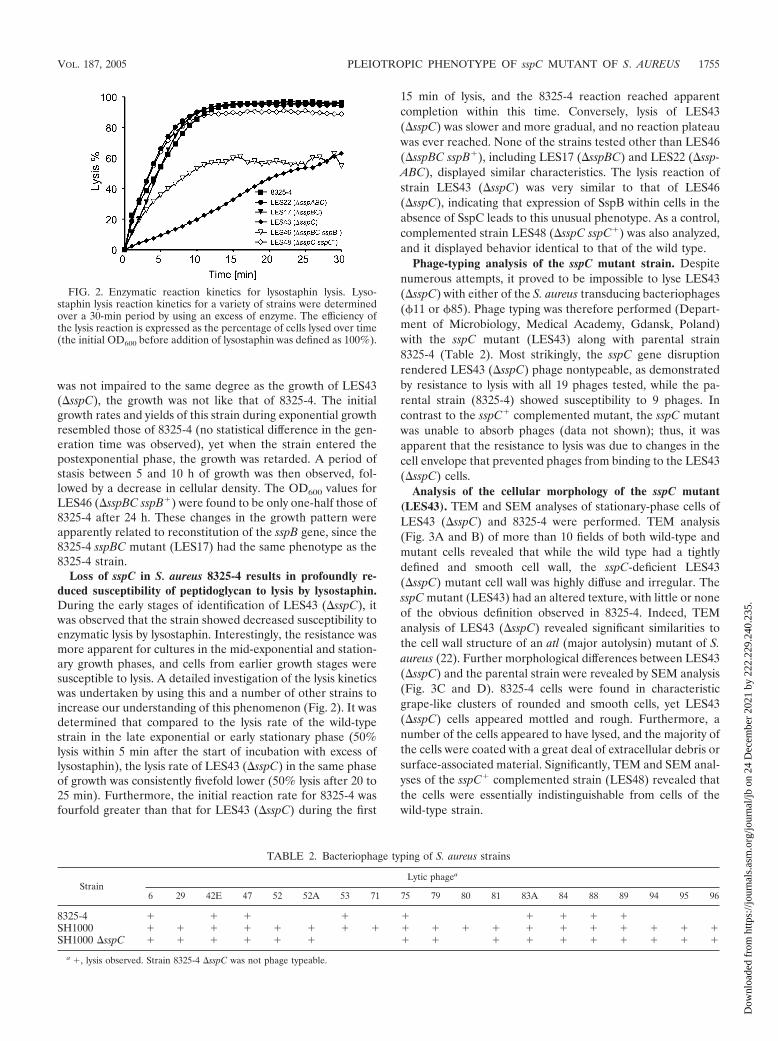
Loss of sspC in S. aureus 8325-4 results in profoundly re-duced susceptibility of peptidoglycan to lysis by lysostaphin.During the early stages of identification of LES43 (�sspC), itwas observed that the strain showed decreased susceptibility toenzymatic lysis by lysostaphin. Interestingly, the resistance wasmore apparent for cultures in the mid-exponential and station-ary growth phases, and cells from earlier growth stages weresusceptible to lysis. A detailed investigation of the lysis kineticswas undertaken by using this and a number of other strains toincrease our understanding of this phenomenon (Fig. 2). It wasdetermined that compared to the lysis rate of the wild-typestrain in the late exponential or early stationary phase (50%lysis within 5 min after the start of incubation with excess oflysostaphin), the lysis rate of LES43 (�sspC) in the same phaseof growth was consistently fivefold lower (50% lysis after 20 to25 min). Furthermore, the initial reaction rate for 8325-4 wasfourfold greater than that for LES43 (�sspC) during the first
15 min of lysis, and the 8325-4 reaction reached apparentcompletion within this time. Conversely, lysis of LES43(�sspC) was slower and more gradual, and no reaction plateauwas ever reached. None of the strains tested other than LES46(�sspBC sspB�), including LES17 (�sspBC) and LES22 (�ssp-ABC), displayed similar characteristics. The lysis reaction ofstrain LES43 (�sspC) was very similar to that of LES46(�sspC), indicating that expression of SspB within cells in theabsence of SspC leads to this unusual phenotype. As a control,complemented strain LES48 (�sspC sspC�) was also analyzed,and it displayed behavior identical to that of the wild type.
Phage-typing analysis of the sspC mutant strain. Despitenumerous attempts, it proved to be impossible to lyse LES43(�sspC) with either of the S. aureus transducing bacteriophages(�11 or �85). Phage typing was therefore performed (Depart-ment of Microbiology, Medical Academy, Gdansk, Poland)with the sspC mutant (LES43) along with parental strain8325-4 (Table 2). Most strikingly, the sspC gene disruptionrendered LES43 (�sspC) phage nontypeable, as demonstratedby resistance to lysis with all 19 phages tested, while the pa-rental strain (8325-4) showed susceptibility to 9 phages. Incontrast to the sspC� complemented mutant, the sspC mutantwas unable to absorb phages (data not shown); thus, it wasapparent that the resistance to lysis was due to changes in thecell envelope that prevented phages from binding to the LES43(�sspC) cells.
Analysis of the cellular morphology of the sspC mutant(LES43). TEM and SEM analyses of stationary-phase cells ofLES43 (�sspC) and 8325-4 were performed. TEM analysis(Fig. 3A and B) of more than 10 fields of both wild-type andmutant cells revealed that while the wild type had a tightlydefined and smooth cell wall, the sspC-deficient LES43(�sspC) mutant cell wall was highly diffuse and irregular. ThesspC mutant (LES43) had an altered texture, with little or noneof the obvious definition observed in 8325-4. Indeed, TEManalysis of LES43 (�sspC) revealed significant similarities tothe cell wall structure of an atl (major autolysin) mutant of S.aureus (22). Further morphological differences between LES43(�sspC) and the parental strain were revealed by SEM analysis(Fig. 3C and D). 8325-4 cells were found in characteristicgrape-like clusters of rounded and smooth cells, yet LES43(�sspC) cells appeared mottled and rough. Furthermore, anumber of the cells appeared to have lysed, and the majority ofthe cells were coated with a great deal of extracellular debris orsurface-associated material. Significantly, TEM and SEM anal-yses of the sspC� complemented strain (LES48) revealed thatthe cells were essentially indistinguishable from cells of thewild-type strain.
FIG. 2. Enzymatic reaction kinetics for lysostaphin lysis. Lyso-staphin lysis reaction kinetics for a variety of strains were determinedover a 30-min period by using an excess of enzyme. The efficiency ofthe lysis reaction is expressed as the percentage of cells lysed over time(the initial OD600 before addition of lysostaphin was defined as 100%).
TABLE 2. Bacteriophage typing of S. aureus strains
StrainLytic phagea
6 29 42E 47 52 52A 53 71 75 79 80 81 83A 84 88 89 94 95 96
8325-4 � � � � � � � � �SH1000 � � � � � � � � � � � � � � � � � � �SH1000 �sspC � � � � � � � � � � � � � � � �
a �, lysis observed. Strain 8325-4 �sspC was not phage typeable.
VOL. 187, 2005 PLEIOTROPIC PHENOTYPE OF sspC MUTANT OF S. AUREUS 1755
Dow
nloa
ded
from
http
s://j
ourn
als.
asm
.org
/jour
nal/j
b on
24
Dec
embe
r 20
21 b
y 22
2.22
9.24
0.23
5.

Insertional inactivation of sspC results in production of abiofilm by 8325-4. Based on increased aggregation and thealtered cell surface of the mutant, assays were conducted todetermine whether LES43 (�sspC) was capable of biofilm for-mation. 8325-4 is believed to be incapable of biofilm produc-tion; however, despite the lack of �B function in LES43(�sspC), it was found that the mutant strain produced a bio-film. In contrast, a number of 8325-4 lineage strains also lack-ing sspC, including the LES22 (�sspABC) and LES17(�sspBC) mutants, were incapable of biofilm formation.Complementation in strain LES48 (�sspC sspC�) showed re-version to the 8325-4 phenotype, whereas LES46 (�sspBCsspB�) displayed an sspC mutant-like phenotype (Fig. 4), in-dicating that the loss of SspC in the context of a functional sspBgene specifically resulted in biofilm formation.
Effect of sspC insertional inactivation on autolytic activity ofS. aureus. As similar cellular morphologies were observedwhen we compared LES43 (�sspC) with an atl (major autolysingene) mutant (22), autolysis assays in the presence of TritonX-100 were conducted (Fig. 5). sspC inactivation resulted in asignificant decrease in autolysis in LES43 (�sspC) compared tothat in 8325-4; a fivefold reduction in lysis was observed within
a 1-h period. Indeed, the lysis of the sspC strain (LES43)closely followed that of the atl mutant (22). Furthermore,LES46 (�sspBC sspB�) also exhibited decreased lysis, andthere was a threefold reduction in the rate compared to the8325-4 rate. Analysis of the sspABC (LES22) and sspBC(LES17) mutants and LES48 (�sspC sspC�) revealed no dif-ference in lysis from the parent strain.
Further analysis of peptidoglycan hydrolase activity was per-formed by using zymography with either Micrococcus luteus orS. aureus cells as substrates (Fig. 6A and B). Both the sspABC(LES22) and sspBC (LES17) mutants exhibited no apparentdifference in autolysin activity compared to 8325-4. However,LES43 (�sspC) displayed a dramatic alteration in the activityprofile. Discrete zones of activity at apparent molecular massesof 40, 35, 30, 22, and 20 kDa were replaced by a major 28-kDaband when S. aureus was used as a substrate (Fig. 6A). Fur-thermore, the 51-kDa activity against M. luteus was not ob-served with LES43 (�sspC) and was replaced by a band at
FIG. 3. Analysis of 8325-4 and LES43 (�sspC) by TEM (A and B) and SEM (C and D). Stationary-phase cultures (15 h) of 8325-4 (A and C)and LES43 (�sspC) (B and D) were harvested, washed with PBS, and resuspended in 2.5% glutaraldehyde in 0.1 M cacodylate buffer (pH 7.2).Cells were analyzed with a Philips/FEI Technai 20 TEM (magnification, �140,000) (A and B) or a LEO 982 field emission SEM (magnification,�10,000) (C and D).
FIG. 4. Analysis of biofilm formation. Strains were analyzed for theability to form a biofilm. Strain SH1000 was included as a positivecontrol. Biofilm formation was quantified by using the intensity ofstaining with crystal violet and is expressed as a percentage of thepositive control value.
FIG. 5. Triton X-100-induced lysis assay. Triton X-100 (0.05%) wasused to induce cellular lysis over a 180-min period. The efficiency ofthe lysis reaction is expressed as the percentage of cells lysed over time(the initial OD600 was defined as 100%).
1756 SHAW ET AL. J. BACTERIOL.
Dow
nloa
ded
from
http
s://j
ourn
als.
asm
.org
/jour
nal/j
b on
24
Dec
embe
r 20
21 b
y 22
2.22
9.24
0.23
5.

approximately 140 kDa (Fig. 6B). Significantly, both 51- and62-kDa activities of S. aureus were also replaced by a higher-molecular-mass activity (Fig. 6A). Since the major autolysin isproduced as a 140-kDa protein, which is proteolytically cleavedinto 62-kDa (amidase) and 51-kDa (glucosamidase) activities,the zymography data suggest that inactivation of sspC hinderspro-Atl processing. Indeed, Western blot analysis confirmedthat LES43 (�sspC) possessed only the unprocessed pro-Atlprotein (Fig. 6C). Interestingly, cleavage of the Atl zymogeninto the 62- and 51-kDa activities, as well other lower-molec-ular-mass activities, was not affected by the sspABC (LES22)and sspBC (LES17) mutations, eliminating the possibility thatSspA or SspB functions as a pro-Atl processing enzyme.
Sensitivity to antibiotics. Since the lack of phage binding,altered autolytic activity, and increased resistance to lysis bylysostaphin may be indicative of some alteration of the pepti-doglycan structure, we compared the susceptibilities of LES43(�sspC) and the parent strain to a panel of antibiotics thataffect cell wall synthesis, including vancomycin, teicoplanin,penicillin, oxacillin, and ampicillin. In this assay no differencewas observed between the strains investigated (data notshown).
Disruption of sspC causes a total loss of secreted extracel-lular proteins but only a partial loss of peptidoglycan-associ-ated proteins in S. aureus 8325-4. Further analysis was con-ducted to determine the effects of sspC insertional inactivationon the exoprotein profile of LES43 (�sspC). Interestingly, afterup to 12 h of growth not a single protein could be found in theculture medium (as determined by SDS-PAGE). This is instark contrast to the parental strain and the sspBC (LES17)mutant, which produced an array of extracellular proteins (Fig.7A). Furthermore, compared to other strains, the sspB� com-plemented sspBC mutant (LES46) showed a highly aberrant
profile of extracellular proteins. Moreover, an exoproteolyticactivity analysis performed by using gelatin zymography re-vealed no trace of active protease in LES43 (�sspC) culturemedia and only a faint band of activity for LES46 (�sspBCsspB�) (Fig. 7B). In order to confirm that the lack of extracel-lular proteolytic activity was not a result of cessation of tran-scription from the protease-encoding loci aur, ssp, and scp (62),reverse transcription-PCR was performed, which confirmedexpression from these three loci (data not shown). In accor-dance with the lack of protease secretion, the LES43 (�sspC)culture medium was also devoid of hemolysin activity. Signif-icantly, in trans restoration of sspC in LES48 (�sspC sspC�)fully restored secretion of extracellular proteins (Fig. 7A), in-cluding proteolytic activity (Fig. 7B), and reverted the hemo-lytic phenotype to that of the wild type (Fig. 7C and D).
The presence of autolysins in LES43 (�sspC) indicates thata subset of secreted proteins, specifically cell wall-associatedproteins, is still exported outside the cells in the sspC mutant.To verify this hypothesis, noncovalently associated proteinswere extracted from the surface of S. aureus by LiCl treatment(Fig. 8A), whereas proteins bound covalently to peptidoglycanwere obtained by V8 protease (SspA) treatment (Fig. 8B).Identical biomasses (wet masses of cultures) of 8325-4 andLES43 (�sspC) yielded similar amounts of solubilized protein-aceous matter (2.5 to 3.0 mg per g [wet weight]) when thepreparations were subjected to extraction with LiCl. However,the SDS-PAGE profiles revealed that LES43 (�sspC) con-tained a high-molecular-mass major protein that was absent in8325-4 and only a few proteins with electrophoretic mobilitiesequivalent to those of the parent proteins (Fig. 8A). On theother hand, preincubation of an S. aureus cell suspension withSspA resulted in release of an 80-kDa protein from the surfaceof LES43 (�sspC) but not from 8325-3 cells subjected to the
FIG. 6. Analysis of activity and processing of the major autolysin of S. aureus. (A and B) Autolysin activity zymography was performed by usingS. aureus cell walls as a substrate for amidase activity (A) and M. luteus cell walls as a substrate for glucosamidase activity (B). (C) Western blotanalysis with anti-Atl antibodies. Samples loaded on the gel were standardized to contain the same amount of protein from each strain.
VOL. 187, 2005 PLEIOTROPIC PHENOTYPE OF sspC MUTANT OF S. AUREUS 1757
Dow
nloa
ded
from
http
s://j
ourn
als.
asm
.org
/jour
nal/j
b on
24
Dec
embe
r 20
21 b
y 22
2.22
9.24
0.23
5.

same treatment (Fig. 8B). The SspA shed polypeptide mayhave represented peptidoglycan-attached staphylococcal ad-hesins belonging to the MSCRAMM family of surface pro-teins. This hypothesis was corroborated by the fact that thesspC mutant (LES43) was able to bind to immobilized fibrin-ogen and fibronectin, although the efficiency was only 20 to30% of the efficiency of 8325-4 (data not shown). Again, thesedata correspond well with the temporal difference in produc-tion of soluble extracellular and peptidoglycan-associated pro-teins. Apparently, before activation of transcription from thessp operon in mid-exponential growth MSCRAMM secretionoccurs normally; however, it is arrested in later growth phases,which seemingly accounts for the decreased level of adhesivemolecules on the mutant cells.
The lack of secreted proteins, including extracellular pro-teases and hemolysins, in the culture medium of LES43(�sspC) implies that these proteins may accumulate in thecytoplasm or at the cell membrane-wall interface. Thus, weattempted to demonstrate the presence of protease and hemo-lytic activities in crude cell extracts or the fractionated cellenvelope or cytoplasmic fractions of LES43 (�sspC). Remark-ably, despite the very high sensitivity of the hemolysin assay, no
FIG. 7. Comparison of secreted proteins and enzyme activity profiles of S. aureus strains. (A and B) Extracellular protein fractions wereobtained from the concentrated supernatants of stationary-phase cultures and were resolved by SDS-PAGE (A) or assayed for proteolytic activityby gelatin zymography (B). (C and D) Hemolytic activity was evaluated by growing S. aureus strains on rabbit (C) and sheep (D) blood agar fordetection of �- and �-hemolysin activities, respectively. WT, wild type.
FIG. 8. Analysis of surface-associated proteins. Noncovalent cellenvelope-associated proteins were extracted by LiCl treatment (A),while proteins covalently bound to the peptidoglycan were released bylimited proteolysis with SspA (V8 protease) (B). The arrowhead indi-cates the position of the V8 protease used to shed surface proteins.
1758 SHAW ET AL. J. BACTERIOL.
Dow
nloa
ded
from
http
s://j
ourn
als.
asm
.org
/jour
nal/j
b on
24
Dec
embe
r 20
21 b
y 22
2.22
9.24
0.23
5.

hemolytic activity was detected in the mutant-derived frac-tions. Also, it was not possible to detect any proteolytic activityin LES43 (�sspC) extracts, apparently due to the limited sen-sitivity of zymography and DCG-04 labeling. Nevertheless,Western blot analysis revealed an immunoreactive band at amolecular mass similar to that of mature SspB (20 kDa) in thecell extract of LES43 (�sspC). In stark contrast, SspB in theform of the unprocessed 40-kDa zymogen was detected in the8325-4 cell extract (Fig. 9A). Taking into account that thedetection limit of Western blot analysis was estimated to be 5ng (Fig. 9A), we calculated that the amount of intracellularSspB in LES43 (�sspC) was exceedingly low ( 20 ng per 200�g of cell proteins). Even so, this amount is apparently suffi-cient to degrade several proteins inside cells lacking SspC.SDS-PAGE analysis of LES43 (�sspC) cell extract proteinprofiles revealed that one major protein and a few minor pro-teins were missing when this strain was compared to the pa-rental strain and the complemented sspC mutant (LES48) (Fig.9B and C). These data suggest that uncontrolled proteolysiswithin cells lacking sspC leads to the apparently pleiotropicchange in the phenotype of LES43 (�sspC).
Insertional inactivation of the sspC gene in a sigB-positivebackground. Because expression of the sspABC operon is neg-atively regulated by the alternative sigma factor (�B), we trans-duced the sspC mutation into strains SH1000 and Newman.Surprisingly, insertional inactivation of sspC in these back-grounds had no effect on the mutant’s growth or susceptibilityto lysostaphin (data not shown). Some minor phenotypic dif-
ferences between the SH1000 sspC mutant and the parentstrain in susceptibility to lysis by specific phages were observed.The SH1000 parental strain was lysed by all of the phagestested, yet the SH1000 sspC mutant displayed susceptibility to16 of the 19 phages used (Table 2). This indicates that despitethe fact that SH1000 lacks any of the other phenotypic char-acteristics of the 8325-4 sspC mutant (LES43), inactivation ofsspC in SH1000 still has some effect.
DISCUSSION
SspC was recently described as a very specific, tightly bindinginhibitor of staphopain B (SspB) (20). Since SspB is a secretedprotein and SspC is an intracellular protein, it was hypothe-sized that SspC functions as a cytoplasmic inhibitor that isrequired to protect cytosolic proteins from degradation byprematurely folded or activated SspB (58). Here we character-ized an sspC mutant of S. aureus and obtained compellingexperimental evidence that SspC does indeed function as acytoplasmic inhibitor of the SspB protease, at least in theSigB-deficient-like background of the 8325-4 strain.
Insertional inactivation of sspC resulted in a growth defect inthe 8325-4 background, which was defined by significantlyshorter generation times during exponential growth, followedby an arrest in growth during the postexponential phase and alate-stationary-phase decline in cellular density and viability.After an approximately 5-h period of stasis, the cells began tolose viability and underwent apparent lysis. Although tran-
FIG. 9. Western blot analysis of S. aureus cell extracts for the presence of staphopain B (A) and SDS-PAGE profiling of intracellular proteinduring late exponential growth (4 h) (B) and early stationary growth (8 h) (C). Washed bacterial cells were suspended in PBS and disrupted witha French press. Debris was removed by centrifugation, and the proteins in the supernatant were analyzed by SDS-PAGE. The arrowheads in panelsB and C indicate missing protein bands in LES43 (�sspC) compared to 8325-4. To determine a detection limit for staphopain B by using Westernblotting (A), serial dilutions of purified protease were loaded and analyzed in parallel.
VOL. 187, 2005 PLEIOTROPIC PHENOTYPE OF sspC MUTANT OF S. AUREUS 1759
Dow
nloa
ded
from
http
s://j
ourn
als.
asm
.org
/jour
nal/j
b on
24
Dec
embe
r 20
21 b
y 22
2.22
9.24
0.23
5.

scriptional analysis of the ssp operon revealed that maximalexpression from this locus occurs approximately 5 h intogrowth (62), corresponding to the time at which LES43(�sspC) stops growing, SspC secretion is observed at the verybeginning of exponential growth (data not shown). Therefore,it seems certain that the detrimental impact on growth andother changes in the phenotype of the 8325-4 sspC mutant (seebelow) are a direct result of uncontrolled activity of SspB. Thispostulate was corroborated by complementational analysis ofLES46 (�sspBC sspB�), an sspBC double mutant comple-mented with only sspB, and LES48 (�sspC sspC�), an sspCmutant complemented with sspC. The sspBC mutant strain(LES17) displayed none of the phenotypic characteristics ofLES43 (�sspC) until sspB was introduced under the control ofits innate promoter (LES46), while complementation of LES43(�sspC) with sspC resulted in reversion to the phenotype of8325-4 in LES48 (�sspC sspC�).
Furthermore, for the defect in growth, LES43 (�sspC) alsodisplayed markedly altered cell wall-related properties, asdemonstrated by profound differences in the rates of autolysisand by both resistance to lysis with innate staphylococcalphages and extensively diminished susceptibility to lysis by thespecific lytic enzyme lysostaphin. All these changes becomeapparent before the culture enters the mid-exponential phaseof growth. In S. aureus resistance to lysostaphin occurs as aresult of a modification in the pentaglycine cross-linking of thecell wall; a decrease in the glycine content and an increase inthe serine content are observed, as is the case with lysostaphin-resistant Staphylococcus spp. (56, 63). Alternatively, disappear-ance of a receptor for lysostaphin on the cell wall may lead toconsiderably decreased sensitivity to lysis (45). The secondoption is a more plausible explanation for the changes in lyso-staphin sensitivity due to the sspC mutation and correlates withthe loss of phage receptors, the altered patterns of cell surface-associated proteins in LES43 (�sspC), and the unchanged sus-ceptibility to antibiotics that affect cell wall synthesis.
Autolysis was also found to be severely affected by the sspCinsertional inactivation, and LES43 (�sspC) was much lesssusceptible to autolysis than the parent strain 8325-4. Thiscorrelates well with a profoundly changed profile of cell wall-associated autolysin activities. Alterations to pro-Atl process-ing are observed, resulting in aberrantly processed forms andaccumulation of the 140-kDa zymogen. Also, the amount ofprocessed Atl associated with the LES43 (�sspC) cell envelopewas massively reduced compared to the amount in the wild-type strain. Analysis of transcription from the atl locus revealedthat although there is a basal constitutive level of expression,there is an increase as the cells enter the exponential phase (22,48). Thus, the loss of Atl function could be explained by mod-ulation of atl transcription as the mutant cells stop growing, bya protein secretion defect (see below), or by hindered Atlfolding in the presence of altered peptidoglycan and proteo-lytic degradation at the cell membrane-wall interfaces. Al-though it is unclear which mechanism is responsible for thedecrease in Atl levels, the decreased activity of this autolysinexplains the significant resistance of LES43 (�sspC) to autol-ysis.
The most fascinating phenotypic idiosyncrasy of LES43(�sspC) is the apparent lack of detectable extracellular pro-teins in culture supernatants. S. aureus secretes a plethora of
extracellular virulence determinants during the postexponen-tial and stationary phases of growth (70). Among these pro-teins are numerous proteases, hemolysins, and toxins, many ofwhich are regulated in a temporal manner by agr (2, 30, 53).The lack of such proteins in LES43 (�sspC) is unusual, indi-cating that there is a general breakdown in protein secretion inthis mutant. Such an observation is in conflict with the pres-ence of peptidoglycan-associated proteins, since both sets ofproteins use the Sec translocation system for passage throughthe cell membrane (68). This contradiction can be explained inthree mutually complementing ways. First, the cell wall pro-teins are predominantly secreted during early stages of S. au-reus growth, before expression from the sspABC operon isinduced. In this scenario intracellularly active SspB may dam-age an essential component of the Sec system and/or cytosolicfactors involved in targeting proteins to the cell secretory ma-chinery. Second, a recent study by Rosch and Caparon (57)with the gram-positive pathogen Streptococcus pyogenes re-vealed that the bulk of extracellular protein secretion in thisorganism does not occur indiscriminately throughout the cellwall but occurs at specific microdomains adapted to containSec translocons. A similar system was described in Bacillussubtilis (7) and probably functions in other gram-positive or-ganisms. Such a system in S. aureus could represent a target forthe uncontrolled SspB activity of the sspC mutant. Third, fold-ing and/or maturation of polypeptide chains newly translocatedacross the cytoplasmic membrane into the interface with thecell wall peptidoglycan is hindered in the mutant, and mis-folded proteins are degraded by quality control proteases (e.g.,HtrA) located in this compartment (49, 68). It is also plausiblethat all three pathways contribute to the absence of proteinsecretion in the sspC insertional mutant.
The attachment of �sspC cells to solid surfaces and theirsubsequent aggregation into clusters also seem to be enhanced.Although biofilm production in S. aureus 8325-4 has previouslybeen demonstrated (5, 13, 37, 67), the extent of this phenom-enon is striking in the SspC-null strain. Biofilm formation inthis strain could be a pleiotropic effect resulting from an in-crease in cellular clumping due to alterations in the outerstructures or a lack of functional autolysins, hemolysins, orproteases.
Interestingly, LES44 (SH1000 �sspC) exhibited none of thephenotypic alterations of LES43 (8325-4 �sspC). SH1000 isidentical to 8325-4 apart from the restoration of an 11-bpdeletion in rsbU (29), which is required for full activity of thealternative sigma factor, �B (23, 36, 50). The lack of a LES44growth defect is most likely explained by the observation thatSH1000 expresses very low levels of extracellular proteases,including those of the ssp operon (29, 62). Therefore, althoughLES43 (8325-4 �sspC) and LES44 (SH1000 �sspC) were de-rived from the same lineage, it can be assumed that the nearlytotal lack of ssp synthesis in LES44 protects it from the dam-aging SspB-mediated phenotype of LES43. Moreover, in thecontext of the fact that SH1000 is a direct derivative of 8325-4(29), it is interesting that the restoration of �B function rees-tablished susceptibility to lysis by phages in this strain (Table2).
In summary, the range of phenotypic events brought intoplay by a mutation in the cytoplasmic inhibitor of staphopain B(SspB) is extensive. The exact pathway by which the changes
1760 SHAW ET AL. J. BACTERIOL.
Dow
nloa
ded
from
http
s://j
ourn
als.
asm
.org
/jour
nal/j
b on
24
Dec
embe
r 20
21 b
y 22
2.22
9.24
0.23
5.

take place requires further study; however, it seems that all thechanges described here are due to the deregulation of SspBcontrol. Apparently, a minute amount of SspB can escape intothe cytoplasm from its pathway for secretion to the extracel-lular environment. This tiny amount of active protease is evi-dently enough to modulate the activity of key intracellularproteins, including possibly those involved in biofilm formationand autolysin processing and, more generally, those requiredfor efficient extracellular protein secretion. Identifying thesetargets is a matter of ongoing research in our laboratories.Here we only intend to stress as-yet-unknown functions ofSspB and put forward a question. Why does S. aureus synthe-size an enzyme which can potentially produce such widespreadchanges in the cell? In this context it is worth reiterating thatit is no coincidence that SspB is tightly regulated both at thetranscriptional level and at the posttranslational level and thatadditional control is guaranteed through the unique coexpres-sion of a protease and an inhibitor from the same locus. Sig-nificantly, all these regulation mechanisms have evolved for anenzyme which is not even an essential housekeeping proteinand has unproven importance as a virulence factor in mousemodels of staphylococcal infections (6, 55, 62). Nevertheless,SspB is conserved in all of the S. aureus clinical isolates inves-tigated to date (24). Taking all these facts into account, itseems evident that there must be selective pressure to maintainsuch a potentially harmful protein in vivo. For this reason wecannot resist speculating that SspB is an important factor forthe S. aureus commensal-pathogen dichotomy with the humanhost, and as such the enzyme itself and its endogenous inhib-itor could be attractive targets for the development of anti-staphylococcal therapies.
ACKNOWLEDGMENTS
We are indebted to Daniel Nelson (Rockefeller University) forcritical reading of the manuscript. We are grateful to Sigrun Eick(Institute of Medical Microbiology, University Hospital, Jena, Ger-many) for the MIC data.
This work was supported by grants 6P04A 011 27 and 158/E-338/SPB/5.PR UE/DZ 19/2003 awarded to E.G. and J.P. by the StateCommittee for Scientific Research (Warsaw, Poland) and by NationalInstitutes of Health funding to J.T. and J.P. Also, part of this work wascarried out with financial support from the Commission of the Euro-pean Communities specific RTD program Quality of Life and Man-agement of Living Resources (QLRT-2001-01250; Novel non-antibi-otic treatment of staphylococcal diseases). J.P. is a recipient of aSUBSYDIUM PROFESORSKIE award from the Foundation for PolishScience (Warsaw, Poland).
REFERENCES
1. Arvidson, S., and K. Tegmark. 2001. Regulation of virulence determinants inStaphylococcus aureus. Int. J. Med. Microbiol. 291:159–170.
2. Balaban, N., and R. P. Novick. 1995. Translation of RNAIII, the Staphylo-coccus aureus agr regulatory RNA molecule, can be activated by a 3�-enddeletion. FEMS Microbiol. Lett. 133:155–161.
3. Beenken, K. E., J. S. Blevins, and M. S. Smeltzer. 2003. Mutation of sarA inStaphylococcus aureus limits biofilm formation. Infect. Immun. 71:4206–4211.
4. Braun, P., A. de Groot, W. Bitter, and J. Tommassen. 1998. Secretion ofelastinolytic enzymes and their propeptides by Pseudomonas aeruginosa. J.Bacteriol. 180:3467–3469.
5. Caiazza, N. C., and G. A. O’Toole. 2003. Alpha-toxin is required for biofilmformation by Staphylococcus aureus. J. Bacteriol. 185:3214–3217.
6. Calander, A. M., I. M. Jonsson, A. Kanth, S. Arvidsson, L. Shaw, S. J. Foster,and A. Tarkowski. 2004. Impact of staphylococcal protease expression on theoutcome of infectious arthritis. Microbes Infect. 6:202–206.
7. Campo, N., H. Tjalsma, G. Buist, D. Stepniak, M. Meijer, M. Veenhuis, M.Westermann, J. P. Muller, S. Bron, J. Kok, O. P. Kuipers, and J. D. Jong-
bloed. 2004. Subcellular sites for bacterial protein export. Mol. Microbiol.53:1583–1599.
8. Chan, P., and S. J. Foster. 1998. The role of environmental factors in theregulation of virulence determinants expression in Staphylococcus aureus8325–4. Microbiology 144:2469–2479.
9. Chan, P. F., and S. J. Foster. 1998. Role of SarA in virulence determinantproduction and environmental signal transduction in Staphylococcus aureus.J. Bacteriol. 180:6232–6241.
10. Cheung, A. L., A. S. Bayer, G. Zhang, H. Gresham, and Y. Q. Xiong. 2004.Regulation of virulence determinants in vitro and in vivo in Staphylococcusaureus. FEMS Immunol. Med. Microbiol. 40:1–9.
11. Chung, C. H., H. E. Ives, S. Almeda, and A. L. Goldberg. 1983. Purificationfrom Escherichia coli of a periplasmic protein that is a potent inhibitor ofpancreatic proteases. J. Biol. Chem. 258:11032–11038.
12. Coulter, S., W. Scwan, E. Ng, M. Langhorne, H. Ritchie, S. Westbrock-Wadman, W. Hufnagle, K. Folger, A. Bayer, and C. Stover. 1998. Staphylo-coccus aureus genetic loci impacting growth and survival in multiple infectionenvironments. Mol. Microbiol. 30:393–404.
13. Cramton, S. E., C. Gerke, N. F. Schnell, W. W. Nichols, and F. Gotz. 1999.The intercellular adhesion (ica) locus is present in Staphylococcus aureus andis required for biofilm formation. Infect. Immun. 67:5427–5433.
14. Drapeau, G. R. 1978. Role of a metalloprotease in activation of the precursorof staphylococcal protease. J. Bacteriol. 136:607–613.
15. Dubin, G., M. Krajewski, G. Popowicz, J. Stec-Niemczyk, M. Bochtler, J.Potempa, A. Dubin, and T. A. Holak. 2003. A novel class of cysteine proteaseinhibitors: solution structure of staphostatin A from Staphylococcus aureus.Biochemistry 42:13449–13456.
16. Dubin, G., J. Stec-Niemczyk, T. Dylag, J. Silberring, A. Dubin, and J. Po-tempa. 2004. Characterisation of a highly specific, endogenous inhibitor ofcysteine protease from Staphylococcus epidermidis, a new member of thestaphostatin family. Biol. Chem. 385:543–546.
17. Dubin, G. 2002. Extracellular proteases of Staphylococcus spp. Biol. Chem.383:1075–1086.
18. Dubin, G. 2003. Defense against own arms: staphylococcal cysteine proteasesand their inhibitors. Acta Biochim. Pol. 50:715–724.
19. Emori, T., and R. Gaynes. 1993. An overview of nosocomial infections,including the role of the microbiology laboratory. Clin. Microbiol. Rev.6:428–442.
20. Filipek, R., M. Rzychon, A. Oleksy, M. Gruca, A. Dubin, J. Potempa, and M.Bochtler. 2003. The staphostatin-staphopain complex: a forward bindinginhibitor in complex with its target cysteine protease. J. Biol. Chem. 278:40959–40966.
21. Filipek, R., R. Szczepanowski, A. Sabat, J. Potempa, and M. Bochtler. 2004.The prostaphopain B structure: a comparison of proregion-mediated andstaphostatin-mediated protease inhibition. Biochemistry 43:14306–14315.
22. Foster, S. J. 1995. Molecular characterisation and functional analysis of themajor autolysin of Staphylococcus aureus. J. Bacteriol. 177:5723–5725.
23. Giachino, P., S. Engelmann, and M. Bischoff. 2001. �B activity depends onRsbU in Staphylococcus aureus. J. Bacteriol. 183:1843–1852.
24. Golonka, E., R. Filipek, A. Sabat, A. Sinczak, and J. Potempa. 2004. Genet-ical characterization of the staphopain genes in Staphylococcus aureus. Biol.Chem. 385:1059–1067.
25. Greenbaum, D., K. F. Medzihradszky, A. Burlingame, and M. Bogyo. 2000.Epoxide electrophiles as activity-dependent cysteine protease profiling anddiscovery tools. Chem. Biol. 7:569–581.
26. Guerout-Fleury, A. M., K. Shazand, N. Frandsen, and P. Stragier. 1995.Antibiotic-resistance cassettes for Bacillus subtilis. Gene 167:335–336.
27. Hege, T., R. E. Feltzer, R. D. Gray, and U. Baumann. 2001. Crystal structureof a complex between Pseudomonas aeruginosa alkaline protease and itscognate inhibitor: inhibition by a zinc-NH2 coordinative bond. J. Biol. Chem.276:35087–35092.
28. Heussen, C., and E. B. Dowdle. 1980. Electrophoretic analysis of plasmino-gen activators in polyacrylamide gels containing sodium dodecyl sulfate andcopolymerized substrates. Anal. Biochem. 102:196–202.
29. Horsburgh, M., J. Aish, I. White, L. Shaw, J. Lithgow, and S. Foster. 2002.�B modulates virulence determinant expression and stress resistance: char-acterization of a functional rsbU strain derived from Staphylococcus aureus8325–4. J. Bacteriol. 184:5457–5467.
30. Ji, G., R. Beavis, and R. Novick. 1995. Cell density control of staphylococcalvirulence mediated by an octopeptide pheromone. Proc. Natl. Acad. Sci.USA 92:12055–12059.
31. Karlin, S., J. Theriot, and J. Mrazek. 2004. Comparative analysis of geneexpression among low G�C gram-positive genomes. Proc. Natl. Acad. Sci.USA 101:6182–6187.
32. Karlsson, A., and S. Arvidson. 2002. Variation in extracellular proteaseproduction among clinical isolates of Staphylococcus aureus due to differentlevels of expression of the protease repressor sarA. Infect. Immun. 70:4239–4246.
33. Karlsson, A., P. Saravia-Otten, K. Tegmark, E. Morfeldt, and S. Arvidson.2001. Decreased amounts of cell wall-associated protein A and fibronectin-binding proteins in Staphylococcus aureus sarA mutants due to up-regulationof extracellular proteases. Infect. Immun. 69:4742–4748.
VOL. 187, 2005 PLEIOTROPIC PHENOTYPE OF sspC MUTANT OF S. AUREUS 1761
Dow
nloa
ded
from
http
s://j
ourn
als.
asm
.org
/jour
nal/j
b on
24
Dec
embe
r 20
21 b
y 22
2.22
9.24
0.23
5.

34. Kemp, E., R. Sammons, A. Moir, D. Sun, and P. Setlow. 1991. Analysis oftranscriptional control of the gerD spore germination gene of Bacillus subtilis168. J. Bacteriol. 173:4646–4652.
35. Kessler, E., M. Safrin, J. K. Gustin, and D. E. Ohman. 1998. Elastase andthe LasA protease of Pseudomonas aeruginosa are secreted with theirpropeptides. J. Biol. Chem. 273:30225–30231.
36. Kullik, I., P. Giachino, and T. Fuchs. 1998. Deletion of the alternative sigmafactor �B in Staphylococcus aureus reveals its function as a global regulatorof virulence genes. J. Bacteriol. 180:4814–4820.
37. Lim, Y., M. Jana, T. T. Luong, and C. Y. Lee. 2004. Control of glucose- andNaCl-induced biofilm formation by rbf in Staphylococcus aureus. J. Bacteriol.186:722–729.
38. Lindsay, J., and S. Foster. 1999. Interactive regulatory pathways controlvirulence determinant production and stability in response to the environ-ment in Staphylococcus aureus. Mol. Gen. Genet. 262:323–331.
39. Lowy, F. D. 1998. Staphylococcus aureus infections. N. Engl. J. Med. 339:520–532.
40. Mani, N., P. Tobin, and R. Jayaswal. 1993. Isolation and characterization ofautolysis-defective mutants of Staphylococcus aureus created by Tn917-lacZmutagenesis. J. Bacteriol. 175:1493–1499.
41. Maskalyk, J. 2002. Antimicrobial resistance takes another step forward. Can.Med. Assoc. J. 167:375.
42. Massimi, I., E. Park, K. Rice, W. Muller-Esterl, D. Sauder, and M. J.McGavin. 2003. Identification of a novel maturation mechanism and re-stricted substrate specificity for the SspB cysteine protease of Staphylococcusaureus. J. Biol. Chem. 277:41770–41777.
43. McAleese, F., E. Walsh, M. Sieprawska, J. Potempa, and T. Foster. 2001.Loss of clumping factor B fibrinogen binding activity by Staphylococcusaureus involves cessation of transcription, shedding and cleavage by metal-loprotease. J. Biol. Chem. 276:29969–29978.
44. McGavin, M. J., C. Zahradka, K. Rice, and J. E. Scott. 1997. Modification ofthe Staphylococcus aureus fibronectin binding phenotype by V8 protease.Infect. Immun. 65:2621–2628.
45. Navarre, W. W., and O. Schneewind. 1999. Surface proteins of gram-positivebacteria and mechanisms of their targeting to the cell wall envelope. Micro-biol. Mol. Biol. Rev. 63:174–229.
46. Novick, R. P. 2000. Pathogenicity factors and their regulation, p. 392–407. InGram-positive pathogens. ASM Press, Washington, D.C.
47. Novick, R. P. 2003. Autoinduction and signal transduction in the regulationof staphylococcal virulence. Mol. Microbiol. 48:1429–1449.
48. Oshida, T., M. Takano, M. Sugai, H. Suginaka, and T. Matsushita. 1998.Expression analysis of the autolysin gene (atl) of Staphylococcus aureus.Microbiol. Immunol. 42:655–659.
49. Pallen, M. J., and B. W. Wren. 1997. The HtrA family of serine proteases.Mol. Microbiol. 26:209–221.
50. Palma, M., and A. Cheung. 2001. �B activity in Staphylococcus aureus iscontrolled by RsbU and an additional factor(s) during bacterial growth.Infect. Immun. 69:7858–7865.
51. Park, S., and D. R. Galloway. 1998. Pseudomonas aeruginosa LasD processesthe inactive LasA precursor to the active protease form. Arch. Biochem.Biophys. 357:8–12.
52. Pearson, H. 2002. ‘Superbug’ hurdles key drug barrier. Nature 418:469.53. Projan, S., and R. Novick. 1997. The molecular basis of pathogenicity, p.
55–81. In The staphylococci in human disease. Churchill Livingstone, NewYork, N.Y.
54. Qoronfleh, M. W., and B. J. Wilkinson. 1986. Effects of growth of methicillin-resistant and -susceptible Staphylococcus aureus in the presence of beta-lactams on peptidoglycan structure and susceptibility to lytic enzymes. An-timicrob. Agents Chemother. 29:250–257.
55. Rice, K., R. Peralta, D. Bast, J. de Azavedo, and M. McGavin. 2001. De-scription of staphylococcus serine protease (ssp) operon in Staphylococcusaureus and nonpolar inactivation of sspA-encoded serine protease. Infect.Immun. 69:159–169.
56. Robinson, J. M., J. K. Hardman, and G. L. Sloan. 1979. Relationship be-tween lysostaphin endopeptidase production and cell wall composition inStaphylococcus staphylolyticus. J. Bacteriol. 137:1158–1164.
57. Rosch, J., and M. Caparon. 2004. A microdomain for protein secretion ingram-positive bacteria. Science 304:1513–1515.
58. Rzychon, M., A. Sabat, K. Kosowska, J. Potempa, and A. Dubin. 2003.Staphostatins: an expanding new group of proteinase inhibitors with aunique specificity for the regulation of staphopains, Staphylococcus spp.cysteine proteinases. Mol. Microbiol. 49:1051–1066.
59. Sambrook, J., E. F. Fritsch, and T. Maniatis. 1989. Molecular cloning: alaboratory manual, 2nd ed. Cold Spring Harbor Laboratory, Cold SpringHarbor, N.Y.
60. Schagger, H., and G. von Jagow. 1987. Tricine-sodium dodecyl sulfate-polyacrylamide gel electrophoresis for the separation of proteins in the rangefrom 1 to 100 kDa. Anal. Biochem. 166:368–379.
61. Schenk, S., and R. A. Ladagga. 1992. Improved methods for electroporationof Staphylococcus aureus. FEMS Microbiol. Lett. 94:133–138.
62. Shaw, L., E. Golonka, J. Potempa, and S. J. Foster. 2004. The role andregulation of the extracellular proteases of Staphylococcus aureus. Microbi-ology 150:217–228.
63. Sugai, M., T. Fujiwara, K. Ohta, H. Komatsuzawa, M. Ohara, and H.Suginaka. 1997. epr, which encodes glycylglycine endopeptidase resistance, ishomologous to femAB and affects serine content of peptidoglycan crossbridges in Staphylococcus capitis and Staphylococcus aureus. J. Bacteriol.179:4311–4318.
64. Sullivan, M., R. Yasbin, and F. Young. 1984. New shuttle vectors for Bacillussubtilis and Escherichia coli which allow rapid detection of inserted frag-ments. Gene 29:21–26.
65. Taguchi, S., S. Kojima, M. Terabe, Y. Kumazawa, H. Kohriyama, M. Suzuki,K. Miura, and H. Momose. 1997. Molecular phylogenetic characterization ofStreptomyces protease inhibitor family. J. Mol. Evol. 44:542–551.
66. Towbin, H., T., Staehelin, and J. Gordon. 1979. Electrophoretic transfer ofproteins from polyacrylamide gels to nitrocellulose sheets: procedure andsome applications. Proc. Natl. Acad. Sci. USA 76:4350–43504.
67. Valle, J., A. Toledo-Arana, C. Berasain, J. M. Ghigo, B. Amorena, J. R.Penades, and I. Lasa. 2003. SarA and not �B is essential for biofilm devel-opment by Staphylococcus aureus. Mol. Microbiol. 48:1075–1087.
68. van Wely, K. H., J. Swaving, R. Freudl, and A. J. Driessen. 2001. Translo-cation of proteins across the cell envelope of Gram-positive bacteria. FEMSMicrobiol. Rev. 25:437–454.
69. Yokoi, K., M. Kakikawa, H. Kimoto, K. Watanabe, H. Yasukawa, A. Ya-makawa, A. Taketo, and K. I. Kodaira. 2001. Genetic and biochemicalcharacterization of glutamyl endopeptidase of Staphylococcus warneri M.Gene 281:115–122.
70. Ziebandt, A. K., H. Weber, J. Rudolph, R. Schmid, D. Hoper, S. Engelmann,and M. Hecker. 2001. Extracellular proteins of Staphylococcus aureus and therole of SarA and sigma B. Proteomics 1:480–493.
1762 SHAW ET AL. J. BACTERIOL.
Dow
nloa
ded
from
http
s://j
ourn
als.
asm
.org
/jour
nal/j
b on
24
Dec
embe
r 20
21 b
y 22
2.22
9.24
0.23
5.