Cysts of oral regions
28
Cysts In The Oral Cavity Regions By: Naz Noori Burhan Oral surgery 5 th Grade Group E
-
Upload
naz-dizayee -
Category
Documents
-
view
824 -
download
2
Transcript of Cysts of oral regions

Cysts In The Oral Cavity
Regions
By: Naz Noori BurhanOral surgery 5th GradeGroup E
DefinitionCyst is defined as pathologic cavity having fluid,
semifluid, or gaseous contents and which is not created
by accumulation of pus.
Cysts are a reaction of the body to a condition and are
usually relatively slow growing. They can be sterile or
become infected.
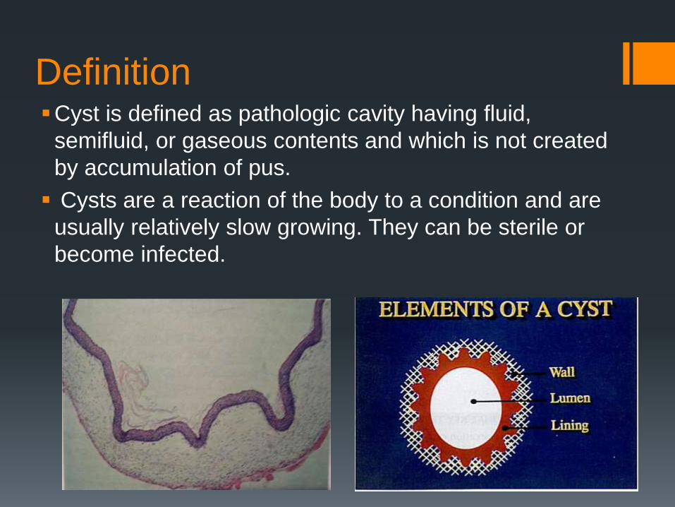
TYPES OF CYSTS TRUE CYSTS: that which is lined by epithelium e.g dentigerous
cyst, radicular cyst etc.
PSEUDO CYSTS: not lined by epithelium, e.g. Solitary bone cyst, Aneurismal bone cyst etc
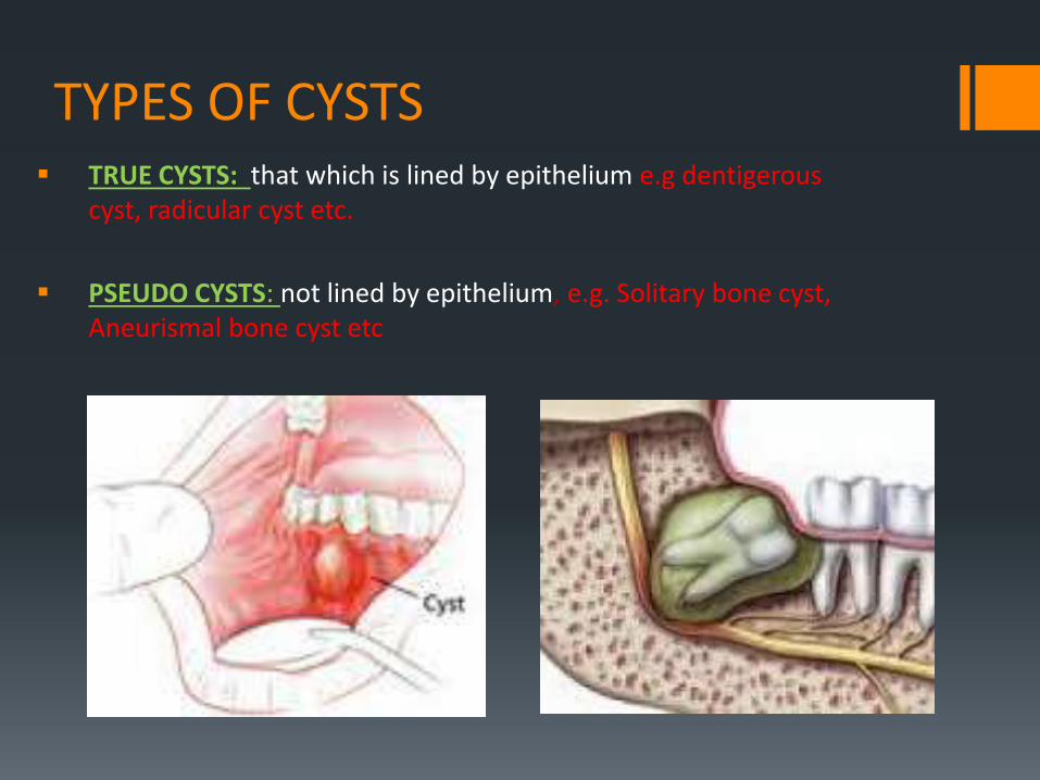
Classification
Cysts of oral region
Epithelial Lined
Odontogenic
Developmental Inflammatory
Nonodontogenic
Non Epithelial Lined
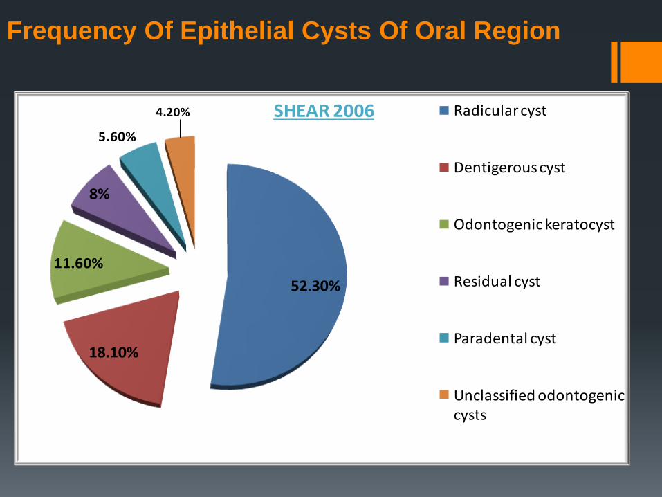
52.30%
18.10%
11.60%
8%
5.60%
4.20% SHEAR 2006 Radicular cyst
Dentigerous cyst
Odontogenic keratocyst
Residual cyst
Paradental cyst
Unclassified odontogenic cysts
Frequency Of Epithelial Cysts Of Oral Region

CLASSIFICATION OF JAW CYSTS:
A.ODONTOGENIC: DEVELOPMENTAL
Odontogenic keratocyst.
Dentigerous cyst.
Eruption cyst.
Gingival cyst of infants.
Gingival cyst of adults.
Lateral periodontal cyst.
Calcifying odontogenic cyst.
Glandular odontogenic cyst.
INFLAMMATORY
Radicular.
Residual.
Paradental.
B.NON ODONTOGENIC
Nasopalatine duct.
Nasolabial cyst.
Globulomaxillary cyst.
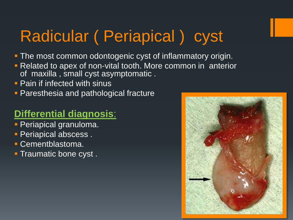
Radicular ( Periapical ) cyst The most common odontogenic cyst of inflammatory origin.
Related to apex of non-vital tooth. More common in anterior of maxilla , small cyst asymptomatic .
Pain if infected with sinus
Paresthesia and pathological fracture
Differential diagnosis: Periapical granuloma.
Periapical abscess .
Cementblastoma.
Traumatic bone cyst .
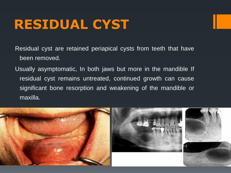
RESIDUAL CYST
Residual cyst are retained periapical cysts from teeth that have
been removed.
Usually asymptomatic, In both jaws but more in the mandible If
residual cyst remains untreated, continued growth can cause
significant bone resorption and weakening of the mandible or
maxilla.
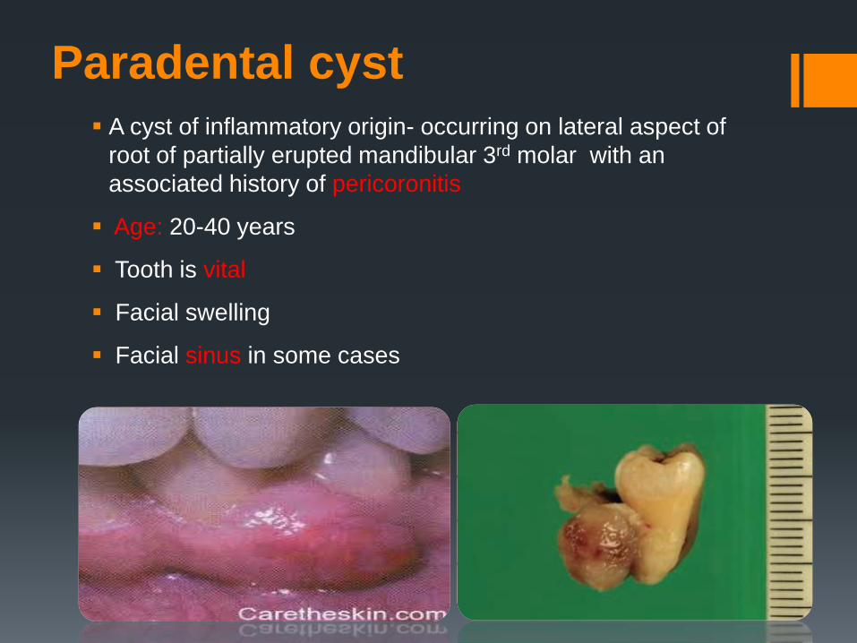
Paradental cyst
A cyst of inflammatory origin- occurring on lateral aspect of
root of partially erupted mandibular 3rd molar with an
associated history of pericoronitis
Age: 20-40 years
Tooth is vital
Facial swelling
Facial sinus in some cases

Odontogenic kerato cyst
OKC is a cyst containing keratin and lined with keratinized epithelium.
OKC’s arises from cell rests of the dental lamina.
Ocure at any age More frequently in males than in females,.
Mandible: posterior portion of body & ramus.
Maxila: 3rd molar area.
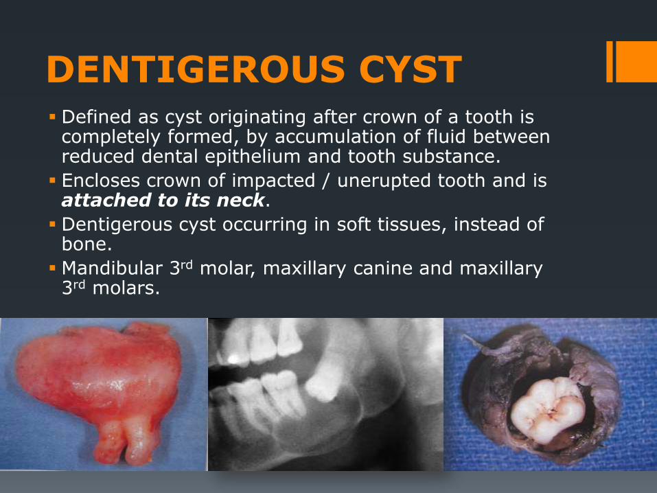
DENTIGEROUS CYST Defined as cyst originating after crown of a tooth is
completely formed, by accumulation of fluid between reduced dental epithelium and tooth substance.
Encloses crown of impacted / unerupted tooth and is attached to its neck.
Dentigerous cyst occurring in soft tissues, instead of bone.
Mandibular 3rd molar, maxillary canine and maxillary 3rd molars.
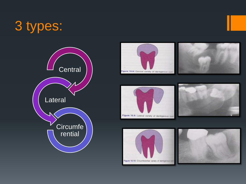
3 types:
Central
Lateral
Circumferential
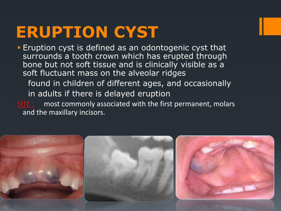
ERUPTION CYST Eruption cyst is defined as an odontogenic cyst that
surrounds a tooth crown which has erupted through bone but not soft tissue and is clinically visible as a soft fluctuant mass on the alveolar ridges
found in children of different ages, and occasionally
in adults if there is delayed eruption
SITE : most commonly associated with the first permanent, molars and the maxillary incisors.

GINGIVAL CYST A small developmental odontogenic cyst of the gingival soft
tissue derived from the rests of the dental lamina
Slowly enlarging, well circumscribed painless swelling.
on facial aspect of free / attached gingiva and gingival papilla.
Site: mandibular bicuspid/cuspid/incisor area.
DIFFERENTIAL DIAGNOSIS:
Lateral periodontal cyst.

Lateral periodontal cyst Uncommon intra-osseous odontogenic cyst similar to gingival
cyst of adult
It’s derived from rest of dental lamina
Lateral to the root surface of erupted tooth
Differential diagnosis:Middle age patient ,Both mandible and maxilla
Canine and premolar of mandible ,Near the crest of ridge
Asymptomatic, May produce bone expansion and pain
Tooth is vital .Cyst less than 1 cm.
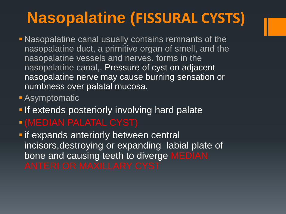
Nasopalatine (FISSURAL CYSTS)Nasopalatine canal usually contains remnants of the
nasopalatine duct, a primitive organ of smell, and the nasopalatine vessels and nerves. forms in the nasopalatine canal,, Pressure of cyst on adjacent nasopalatine nerve may cause burning sensation or numbness over palatal mucosa.
Asymptomatic
If extends posteriorly involving hard palate
(MEDIAN PALATAL CYST)
if expands anteriorly between central incisors,destroying or expanding labial plate of bone and causing teeth to diverge MEDIAN ANTERI OR MAXILLARY CYST

Nasopalatine
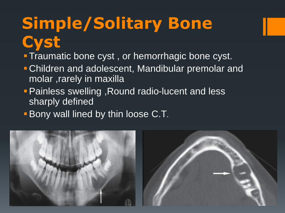
Simple/Solitary Bone CystTraumatic bone cyst , or hemorrhagic bone cyst.
Children and adolescent, Mandibular premolar and molar ,rarely in maxilla
Painless swelling ,Round radio-lucent and less sharply defined
Bony wall lined by thin loose C.T.
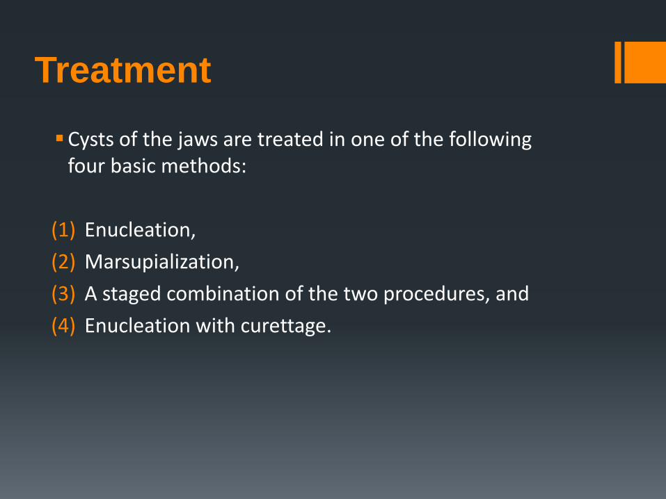
Treatment
Cysts of the jaws are treated in one of the following four basic methods:
(1) Enucleation,
(2) Marsupialization,
(3) A staged combination of the two procedures, and
(4) Enucleation with curettage.

1. Enucleation
Enucleation: This technique involves complete removal of
the cystic sac and healing of the wound by primary intention.
This is the most satisfactory method of treatment of a cyst
and is indicated in all cases where cysts are involved, whose
wall may be removed without damaging adjacent teeth and
other anatomic structures.
The surgical procedure for treatment of a cystwith enucleation includes the following steps:
1. Reflection of a mucoperiosteal flap.2. Removal of bone and exposure of part of the cyst.3. Enucleation of the cystic sac.4. Care of the wound and suturing.
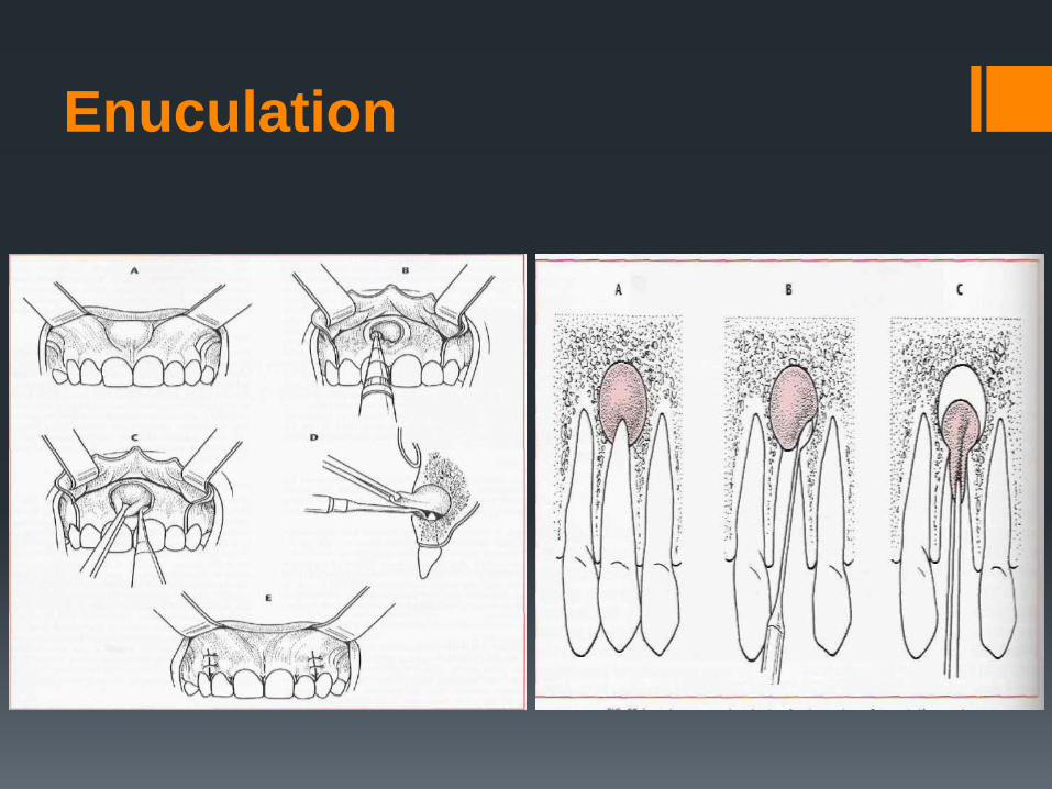
Enuculation

Surgical removal the of the cyst
2.Marsupialization
This method is usually employed for the removal of large cysts
by opening a surgical window above the lesion.
A circular incision is made, which includes the mucoperiosteum,
the underlying perforated (usually) bone, and the respective
wall of the cystic sac.
Then the contents of the cyst are evacuated, and interrupted
sutures are placed around the periphery of the cyst , suturing
the mucoperiosteum and the cystic wall together.
Afterwards, the cystic cavity is irrigated with saline solution and
packed with iodoform gauze.

Technique of Marsupiaiization
1) Anesthesia.
2) Aspiration.
3) Incision.
4) Removal of bone.
5) Removal of cystic lining specimen.
6) Visual examination of residual cystic lining.
7) Irrigation of cystic cavity.
8) Suturing Cystic lining sutured with the edge of oral mucosa.

Marsupialization method. Circular incision
includes mucosa and periosteum.
Exposure of buccal cortical plate and
removal of portion of bone with round bur
Exposure of cyst
after removal of
bone
Suturing of wound
margins with cystic
wall
Packing of cystic
cavity with iodoform
gauze
Cystic cavity after
insertion of gauze
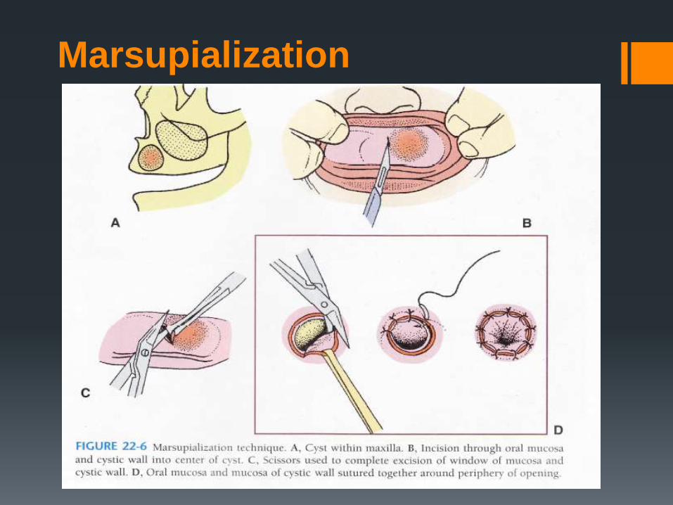
Marsupialization
Refrences
http://en.wikipedia.org/wiki/Cysts_of_the_jaws
http://www.gorlingroup.org/jupgrade/index.php/about-gorlin-
syndrome/11-jaw-cysts
http://www.slideshare.net/makkahguys/jaw-bone-
chttp://www.slideshare.net/JanmiPascual/cysts-of-oral-
region-5?related=1yst
https://www.bestdentistguide.com/oral-cysts