Cysteinyl leukotrienes overproduction and mast cell...
9
Eur Respir J. 1993, 6, 391-399 Printed in UK - all rights reserved Copyright© ERS Journals ltd 1993 Eur opean Respiratory Journal ISSN 0903 - 1936 Cysteinyl leukotrienes overproduction and mast cell activation in aspirin-provoked bronchospasm in asthma K. Sladek, A. Szczeklik Cysteinyl leukotrienes overproduction and mast cell activation in aspirin-provoked bron- chospasm in asthma. K. S/adek, A. Szczeklik. ©ERS Journals Ltd 1993. ABSTRACT: ln order to examine the hypothesis that in aspirin-induced asthma (AlA) cyclooxygenase inhibition is associated with en hanced release of leukotrienes (LTs), we measured urinary leukotriene E 4 (LTE 4 ) and 11-dehydro-thromboxane 8 2 (TXB 2 ) (as a measure of cyclooxygenase production) following challenge with oral aspirin or inhal ed methacholine, in 10 AlA patients. We also determined serum trypt ase a nd eosinophilic catonic protein (ECP) levels, in order to eval uate mast cell and eosinophil activation. Urinary L TE 4 excretion was increased sevenfold 4-6 b after aspirin challenge, whil e ll-dehydro-TXB 2 decreased gradually reaching 50% baseline levels 24 h af- ter challenge (p<O.OS). This was accompanied by a signifi cant fall in bl ood eosi- nophil count at 6 h, and a tendency to a rise in ECP. The intensity of both LTE 4 and 11-dehydro-TXB 2 responses depended on the dose of as pirin used (p<O.OOl, analysis of variance (ANOV A)). The accompanying maximum fall in forced expiratory volume in one second (FEV 1 ) was not correlated with peak LTE 4 levels. In contrast to aspirin, methacholine cha ll enge producing comparable bron- chial obstruction, did not alter eicosanoid excretion or serum tryptase or ECP levels. In a separate study, lysine-aspirin inhalat ion challenge was performed in seven AlA patients, four of whom had responded with a rise in serum tryptase to oral aspirin chaUenge. Challenge with inhaled aspirin led to similar bronchoconstriction as with oral challenge, b ut non-respi rator y symptoms such as scarlet flush or rhinorrhea were absent, and serum tryptase levels remained unchanged. This study demonstrates that, in AlA, overproduction of L Ts is accompanied by cyclooxygenase inhibition (meas ured as a faJI in TXBJ, and that the magnitude of leukotrienes' response is related to the dose of aspirin used. During the reaction to aspirin, bl ood eosinophil count fa lls, and both eosinophils and mast cells fre- quently show signs of activation. These cells may be the source of LTs as well as other mediators responsible for additional skin and nasal symptoms in aspirin- precipitated attacks of asthma. Eur Respir 1., 1993, 6, 391-399. Dept of Medicine, Copemicus University School of Medicine, Cracow, Correspondence: A. Szczcklik Dept of Medicine Copemicus Academy of Medicine 8 Skawinska Street 3 J -066 Cracow Poland Keywords: Aspirin aspirin-induced asthma eosinophil leukotrienes mast cell Received: July 27 1992 Accepted after revision November 9 1992 In about I0% of adults with asthma, but rarely in asth- matic children, aspirin and other nonsteroidal anti- inflammatory drugs (NSAID) precipitate asthma attacks. This distinct clinical syndrome is called aspirin- induced asthma (AlA) [1, 2] . Inhibition of cyclooxygenase appears to initiate a chain of reactions, leading to asthma attacks in intolerant patients [3). What follows at the biochemical level remains largely unknown. Since their discovery [4], leukotrienes have been indicated as possible mediators of aspirin-precipitated attacks, but it was onJy recently that this notion gained some clinical support [5, 6]. In allergic asthma, as opposed to AlA, inhibition of cyclooxygcnase has no effect on the course of clinical reaction, i.e. allergen- precipitated bronchoconstriction, and on all ergen- stimulated release of leukotrienes [7). In AlA, the origin of leukotrienes remains unknown. Mast cells and eosinophils, involved in asthmatic bronchoconstriction, have the capacity to synthesize substantial quantities of peptidoleukotrienes (8, 9). In the present study, we examined the hypothesis that cyclooxygenase inhibition in AlA is associated with en- hanced release of leukotrienes. We measured the appear- ance of a thromboxane A 2 (TXA 2 ) metabolite (I 1-dehydro-thromboxane B 2 (TXB 2 ), and leukotriene E 4 (LTE 4 ), in urine of AlA subjects undergoing aspirin or methacholine provocation. We also detennined serum tryptase and eosinophilic cationic protein (ECP) levels. in order to evaluate mast cell and eosinophils activation in AlA.
Transcript of Cysteinyl leukotrienes overproduction and mast cell...

Eur Respir J. 1993, 6, 391-399 Printed in UK - all rights reserved
Copyright© ERS Journals ltd 1993 European Respiratory Journal
ISSN 0903 - 1936
Cysteinyl leukotrienes overproduction and mast cell activation in aspirin-provoked bronchospasm in asthma
K. Sladek, A. Szczeklik
Cysteinyl leukotrienes overproduction and mast cell activation in aspirin-provoked bronchospasm in asthma. K. S/adek, A. Szczeklik. ©ERS Journals Ltd 1993. ABSTRACT: ln order to examine the hypothesis that in aspirin-induced asthma (AlA) cyclooxygenase inhibition is associated with enhanced release of leukotrienes (LTs), we measured urinary leukotriene E4 (LTE4) and 11-dehydro-thromboxane 82 (TXB2) (as a measure of cyclooxygenase production) following challenge with oral aspirin or inhaled methacholine, in 10 AlA patients. We also determined serum tryptase and eosinophilic catonic protein (ECP) levels, in order to evaluate mast cell and eosinophil activation.
Urinary L TE4 excretion was increased sevenfold 4-6 b after aspirin challenge, while ll-dehydro-TXB2 decreased gradually reaching 50% baseline levels 24 h after challenge (p<O.OS). This was accompanied by a significant fall in blood eosinophil count at 6 h, and a tendency to a rise in ECP. The intensity of both LTE4 and 11-dehydro-TXB2 responses depended on the dose of aspirin used (p<O.OOl, analysis of variance (ANOV A)). The accompanying maximum fall in forced expiratory volume in one second (FEV1) was not correlated with peak LTE4
levels. In contrast to aspirin, methacholine challenge producing comparable bronchial obstruction, did not alter eicosanoid excretion or serum tryptase or ECP levels.
In a separate study, lysine-aspirin inhalation challenge was performed in seven AlA patients, four of whom had responded with a rise in serum tryptase to oral aspirin chaUenge. Challenge with inhaled aspirin led to similar bronchoconstriction as with oral challenge, but non-respiratory symptoms such as scarlet flush or rhinorrhea were absent, and serum tryptase levels remained unchanged.
This study demonstrates that, in AlA, overproduction of L Ts is accompanied by cyclooxygenase inhibition (measured as a faJI in TXBJ, and that the magnitude of leukotrienes' response is related to the dose of aspirin used. During the reaction to aspirin, blood eosinophil count falls, and both eosinophils and mast cells frequently show signs of activation. These cells may be the source of LTs as well as other mediators responsible for additional skin and nasal symptoms in aspirinprecipitated attacks of asthma. Eur Respir 1., 1993, 6, 391-399.
Dept of Medicine, Copemicus University School of Medicine, Cracow,
Correspondence: A. Szczcklik Dept of Medicine Copemicus Academy of Medicine 8 Skawinska Street 3 J -066 Cracow Poland
Keywords: Aspirin aspirin-induced asthma eosinophil leukotrienes mast cell
Received: Ju ly 27 1992 Accepted after revision November 9 1992
In about I 0% of adults with asthma, but rarely in asthmatic children, aspirin and other nonsteroidal antiinflammatory drugs (NSAID) precipitate asthma attacks. This distinct clinical syndrome is called aspirininduced asthma (AlA) [1, 2] . Inhibition of cyclooxygenase appears to initiate a chain of reactions, leading to asthma attacks in intolerant patients [3). What follows at the biochemical level remains largely unknown. Since their discovery [4], leukotrienes have been indicated as possible mediators of aspirin-precipitated attacks, but it was onJy recently that this notion gained some clinical support [5, 6]. In allergic asthma, as opposed to AlA, inhibition of cyclooxygcnase has no effect on the course of clinical reaction, i.e. allergenprecipitated bronchoconstriction, and on allergen-
stimulated release of leukotrienes [7). In AlA, the origin of leukotrienes remains unknown. Mast cells and eosinophils, involved in asthmatic bronchoconstriction, have the capacity to synthesize substantial quantities of peptidoleukotrienes (8, 9).
In the present study, we examined the hypothesis that cyclooxygenase inhibition in AlA is associated with enhanced release of leukotrienes. We measured the appearance of a thromboxane A2 (TXA2) metabolite (I 1-dehydro-thromboxane B2 (TXB2), and leukotriene E4 (LTE4), in urine of AlA subjects undergoing aspirin or methacholine provocation. We also detennined serum tryptase and eosinophilic cationic protein (ECP) levels. in order to evaluate mast cell and eosinophils activation in AlA.

392 K. SLADEK, A. SZCZEKLIK
Material and methods
Subjects
Studies were performed in 10 nonsmoking asthmatic patients (three males and seven females) with AlA, age range 32-50 yrs. The duration of asthma was fTom 5- 15 yrs. on average 10 yrs. Three of the patients were atopic to common aeroallergens. All patients were in stable clinical condition (table I).
proper study. Methacholine inhalation test [11] was carried out using a DeVilbiss nebulizer model 646 (DeYilbiss Co, Somerset, PA, USA). Twenty four hours after aspirin provocation each subject underwent methacholine challenge to evaluate possible changes in airway reactivity.
Pulmonary function was assessed by measuring FEY1,
forced vital capacity (FYC) and forced mid-expiratory flow (FEF25_m;,) at baseline, and then every 30 min until
Table 1. - Characteristics of the AlA patients studied
Patient Age Sex Atopy Aspirin Threshold mg Mean chronic daily
oral dose prednisone
Oral Inhaled No I nit rs mg
I K.G. 37 F + 80 (-) 10 2 K.B. 34 F 60 ( -) 5 3 B.R. 30 F 20 (-) 5 4 s .. z. 42 F + 160 (-) 5 K.J. 37 F + 80 (-) 5 6 K.H. 48 F 20 I 7 G.H. 50 F 100 2 8 K.L. 47 M 160 2 10 9 G.J. 38 M 150 2 10 10 W.J. 33 M 150 2 11 D.J. 45 F + (-) I 5 12 S.G. 32 F (-) 2 5
Patients No. 11 and 12 underwent only lysine-aspirin inhalation cha.llenge. AlA: aspirin-induced asthma.
None had a history of respiratory tract infection, or relevant aspirin or allergen exposure for 4 weeks before the study. At the time of study, forced expiratory volume in one second (FEY1) exceeded 70% of predicted values. All patients were on chronic therapy with inbaled beclomethasone, and six were also receiving oral prednisone at a dose of 5 or lO mg per day (tables, patients l-10). Throughout the study, the steroid treatment remained unchanged. Patients stopped any inhaled or oral adrenergic agents, oral theophylline and antihistamines for at least 8, 48 and 96 h respectively, before the beginning of the study.
Atopy was diagnosed if: 1) skin prick-test was positive with at least one of the aeroallergens: mixed grass pollen, tree pollen, cat fur, dog hair, feathers, house dust, Dermatophagoides pteronyssinus (Bencard Allergy Service, Brentford, Middlesex, UK); and 2) a significant concentration of a specific immunoglobulin E (lgE) was detected in plasma (lgE FAST-Plus test, 3M Diagnostic System Inc., Santa Clara, CA, USA).
Design of study
Each subject was studied at the same time in the morning, on two separate occasions, at least 14 days apart. On each occasion, the patient was challenged with either inhaled methacholine or oral aspirin in a random order. Oral provocation test with aspirin was performed as described previously [2, 101. using a threshold dose resulting in a >20% fall in FEY1• This dose was predetermined for each subject at least 4 weeks before the
the sixth hour, and was repeated at 8 and 24 h after the challenge.
Urine for analysis of L TE4 and the enzymatic metabolite of TXA1, ll-dehydro-TXB2, was collected within 2 h before the challenge, and at 2, 4, 6, 8, 10, 14 and 24 h after aspirin or methacholine challenge. Blood for determination of eosinophil count, and tryptase and ECP plasma concentration, was taken at baseline, and then hourly for the 6 h, and at 12 and 24 h after challenges.
Additionally, inhalation aspirin challenge was performed in seven AlA patients on a separate occasion (table I) [12]. Four of these subjects, had a previously significant increase in serum tryptase levels following oral aspirin challenge. We measured serum tryptase, ECP and eosinophil count before lysine-aspirin inhalation, and then every 30 rnin for 4 h following at least 20% a fall in FEY 1 produced by aspirin.
All patients participated in the study on a voluntary basis and gave signed informed consent to a protocol approved by the Ethics Committee of the University School of Medicine in Cracow.
Methacholine challenge
Methacholine (Mch) challenge was performed using the method of ToWNLEY and HoPE [11]. Aerosol was generated by a DeYilbiss 646 nebulizer using oxygen at a flow rate of 9 l·min-1
• The dosimeter, with a manually-operated valve for 1.2 s, delivered 0 .5±0.02 ml·min·• (mean±so) of the aerosol. Measurements of FEY,, FVC

L TE4
AND MAST CELLS IN AlA 393
and FEF2s-?s% were made by pneumotachograph (Pneumascreen). When pulmonary function after placebo inhalation (solvent of methacholine solution at the same pH) was near baseline value, the subjects inhaled five breaths of doubled, increasing concentrations of methacholine aerosol at 5 min intervals. The first concentration was 0.15 mg·mJ·'. The challenge was carried out until FEY 1
fell by 20% of baseline values or until the highest concentration of methacholine (50 mg·ml·') was reached. Methacholine (Provocholine, Roche Laboratories, Nutley, NJ, USA) was dissolved freshly on the study day, in phosphate-buffered saline (PBS) to pH 7.4. Measurements of FEV1 were made at 1 and 3 min after completion of the inhalation of each concentration. The loglinear dose response curve was analysed by interpolation, to give the estimated concentration required to cause a fall in FEY1 by 20% of baseline, (P~0Mch).
Inhalation aspirin challenge
Inhalation aspirin challenge was performed as described by PHILLIPS et al. [12]. Lysine-aspirin containing 900 mg of lysine-acetylsalicylate (Aspegic, Synthelabo Pharma, Lausanne, Switzerland) with 100 mg of glycine was made up freshly on each challenge day with sterile water, to produce lysine-aspirin solution (360 mg·mJ·') at a concentration of 200 mg·ml· ' of acetylsalicylic acid. The lysine-aspirin solution was diluted in 0.9% sodium chloride to produce a range of increasing, doubled concentrations, from 11.25- 180 mg·mJ·1• A placebo solution had the same pH and concentration of lysine and glycine. Aerosol of these solutions was generated by a De Vilbiss 646 nebulizer connected to an ongen flow of 9 l·min·'. The dosimeter with a manually operated valve for 1.2 s, delivered 0.5±0.02 ml·min·1 of the aerosol. When pulmonary function after placebo inhalation was near baseline value, the subjects inhaled five breaths of increasing concentration of lysine-aspirin aerosol, beginning with a concentration of 11.25 mg·mJ·' (0.5 mg of lysine--aspirin for five breaths), until FEV1 fell by 20% from baseline values. Measurements of FEV1, FYC and FEF25_75.., were made at 15 min intervals for 60 min after inhalation of each concentration, and then hourly for 4 h after the concenlTation of lysine-aspirin inhalation which produced at least a 20% fall in FEV 1•
LTE4 assay
4-hydroxy-TEMPO (Aldrich Chemical Co., Milwaukee, WI, USA) was added to samples of urine, which were then adjusted to pH 9.0 with NaOH, and stored at -70°C until analysed [13]. LTE4 was measured in urine samples by radio-immunoassay, after extraction on Cl8 PrepSep column (Fisher Sci, Fair Lawn, NJ, USA), followed by purification and separation on high-performance liquid chromatography (HPLC) [13]. After centrifugation, freshly thawed urine supematant was acidified to pH 5.4 and then applied to the Cl8 Prep-Sep cartridge, preconditioned with Hp, MeOH and 0.1% ammonium acetate buffer at pH 5.4, containing I mM disodium ethylenediaminetetra-acetic acid (EDT A). After washing the col-
urnn with H20 to remove the polar material, Jeukotrienes were eluted with MeOH, and the sample was evaporated to dryness under nitrogen. The residue was reconstituted with 50 ~ I of mobile phase (MeOH:O.l% ammonium acetate buffer, 80:20, at pH 5.4) and injected via Rheodyne injector (Rheodyne lnc., Cotati, CA, USA) onto a reversephase C18 column (Spherisorb ODS-2, 5 ~m, 250><4 mm, Pharmacia LKB Instrument, Uppsala, Sweden), protected by guard cartridge (ODS-2, 5 11 m, 10x4 mm, Pharmacia LKB Instrument, Sweden). The flow rate generated by the HPLC gradient pump (HPLC Gradient Pump 2249, Pharmacia LKB Instrument, Uppsala, Sweden) was 1 ml ·min·' and the effluent was monitored by Variable Wavelength Monitor at a 280 nm wavelength (VWM 2141 Detector, Pharmacia LKB Instrument, Uppsala, Sweden). The fractions, having the same elution time as radiolabelled [3HJLTE4 (Amersham International plc, Aylesbury, UK) were collected at 0.5 m in intervals, evaporated under nitrogen and dissolved in radioimmunoassay buffer (0.05 M phosphate buffer pH 7.4 containing 0.14 M NaCl and 0.01% gelatin; Amersham International plc, Aylesbury, UK). Leuko-triene recovery was detem1ined using [ 14, I 5(n)-3H]LTC4 (20- 60 Ci·mmoJ·1; Amersham International plc, Aylesbury, UK) added to urine samples before extraction. lmnlllnoreactive L TE4 concentrations was measured by radioimmunoassay, using a commercial kit (Amersham International plc, Aylesbury, UK), which utilized a monoclonal leukotriene CJDJEJF4 antibody with the cross-reactivity 100%/181.8%/92.7%/121.3%, respectively.
11-dehydro-thromboxane B2 assay
The enzymatic metabolite of TXA2, ll-dehydro-TXB2,
was measured by radio-immunoassay in urine sample [14, 15]. Freshly thawed urine sample was exlTacted on an Amprep C2 column (C2 Amprep columns; Amersham International plc, Aylesbury, UK), preconditioned with MeOH and H20, then washed with H20, 10% ethanol, and hexane, and eluted with methyl formate. The sample was subsequently dried under nilTogen and exposed to 0.05 M Tris/HCl radio-immunoassay buffer in pH 7.4 (Amersham International plc, Aylesbury, UK) for 24 h at 25°C, to ensure that the ll-dehydro-TXB2 was in the open chain fonn. The concentration of 11-dehydro-TXB2
in a sample was calculated using commercial radioimmunoassay kit (Amersham International plc, Aylesbury, UK).
Pulmonary function tests
Pulmonary function tests were performed on a flowintegrating computerized pneumotachograph (Pneumoscreen, E. Jaeger, Wuerzburg, Germany).
T1yptase and ECP assay
Tryptase and ECP serum concenlTations were measured by radio-immunoassay using commercial kits (Pharmacia Diagnostic AB, Uppsala, Sweden). Venous blood

394 K. SL-ADEK, A. SZC.tEKLIK
samples (3 ml) were collected into a glass tube and allowed to clot at 37°C for I hr. Serum was then separated by centrifugation at 1,350 g for 10 min, and put into a test tube for radio-immunoassay procedure. Concentrations of tryptase and ECP were quantified against a freshly prepared standard curve.
Eosinophil cou111
Eosinophils were counted using a light microscope, after dissolving whole blood sample in Pilot's solution as described previously rt6].
Result~
At baseline, there were no differences in FEY, on any of the challenge days (2.93±0. 17 I on aspirin, and 2.90±0.15 I on methacholine) (table 2). In all subjects, both aspirin and methacholine produced a significant pulmonary reaction, as evidenced by fall in FEY, >20%. Maximal fall in FEY 1 after aspirin occurred between 2.5-3.5 h (25.3±5.0%; p<O.OOl paired t-test) (fig. 1). Following methacholine, bronchoconstriction developed within 30 min (35.9±3.2%; p<O.OOOl paired t-test) (fig.2). The dose of aspirin ranged from 20-160 mg (98.0±l7.4). Our patients had large individual differences
Table 2. - Baseline FEV ,. PC20Mch before and 24 h after oral aspirin challenge
Baseline FEY1
% pred PC20
Mch mg·ml·1
Patients ASA day Mch day Before ASA 24 h after ASA No lnit
I K.G. 72 71 1.6 1.2 2 K.B. 98 99 5.5 5.5 3 B.R. 122 Ill 10.5 9.0 4 s.z. 114 114 34.0 32.0 5 K.J. 79 82 3.5 1.5 6 K.H. 103 104 3.5 0.6 7 G.H. 78 80 6.6 5.0 8 K.L. 79 73 3.6 3.2 9 G.J. 77 80 10.0 6.0 10 W.J. 74 75 13.0 8.8
Mean 89.5 88.8 9.1 7.2 SEM 5.7 5.2 3.0 2.4
Paired t-test NS p<0.003
NS: nonsignificant; ASA: aspirin (acetylsalicylic acid); Mch: methacholine; FEY,: forced expiratory volume in one second; PC
20Mch: provocation dose
of methacholine producing a 20% fall in FEY,.
Statistical methods
The repeated-measures analysis of variance (ANOV A) of logarithmic observation with dose of aspirin (or methacholine) as a eovariable was used to analyse timedependent changes in urine and serum concentration of examined compounds. The F stat-istic was employed to test the significance. Multiple-regression analysis was performed with FEY 1, tryptase, 11-dehydro-TXB2 as the dependent variables and L TE4 as the independent variable. Comparison of baseline and maximal values of examined compounds after aspirin provocation were analysed using paired Studen's t-test, or a nonparametric Wilcoxon's signed rank test. Correlations between PC20Mch and threshold dose of aspirin, and likewise between the maximal fa ll of blood eosinophil count and P~0Mch examined at 24 h after aspirin challenge, were determined by linear regression analysis. Data are given as mean±sEM.
in bronchial responsiveness to methacholine; PC20Mch ranged from 1.6-34.0 mg·mt·• (9.1±3.0). There was no evident correlation between PCzoMch and aspirin threshold dose (r=0.46, p=0.09). Twenty four hours after aspirin ingestion, the airway hyperresponsiveness increased significantly in aU subjects (PC:wMch range 1.2-32.0 mg·mt·', 7.2 ±2.4; p<0.003 paired t-test) (table 2).
There were no differences between the baseline urinary levels of either LTE4 or 11-dehydro-TXB2 on aspirin and methacholine days (151.4±48 8 pg·mg·' creatinine (Cr) versus 161.6±46.7 pg·mg·'Cr for L TE4;
and 597.9±80.0 pg·mg·1Cr versus 557.7±47.0 pg·mg·'Cr for ll -dehydro-TXB2). The excretion of LTE4 increased after aspirin provocation, reaching a maximum between 4-6 h (1053.3±417.8; p<0.04 paired t-test) (fig. 1). The elevation of L TE4 was accompanied by a significant reduction of 11 -dehydro-TXB2 in urine (from 597 .9± 80.0 pg·mg·•cr at baseline to 279.9±43.0 pg·mg·•cr at 24 h of study; p<0.005 paired Hest ) (fig. 1).
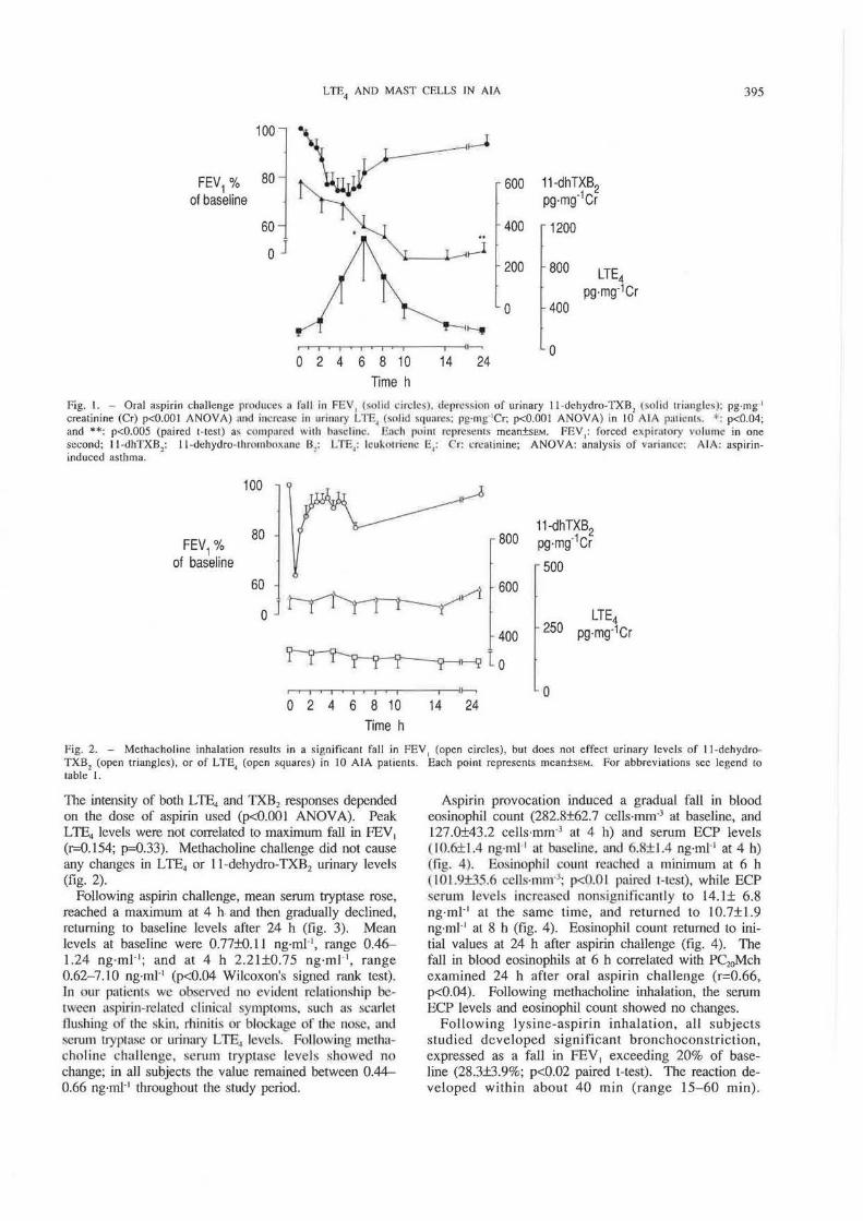
Fig. I. - Oral aspirin challenge produce~ a f'all in FEV1 t~olid drclcs), depression of urinary 11-dchydro-TXB2 (so lid triangles): pg·mg·•
creatinine (Cr) p<O.OOI ANOVA) und iut·rcasc In urinary LTE, (solid squares: pg·rng 1Cr; p<O.OOI ANOVA) in 10 AlA p:uimlls. *: p<0.04; and **: p<O.OOS (paired t-test) a.~ comparecl with ha~eliuc. Each point rcprc~cnL~ mean±SEM. FEY,: forced expinuory vo lume in one second; 11 -dhTXB,: 11 -dehydro-thrombmt;uh: Bl: LTE4: lcukutricne E,: Cr: crcminine; ANOVA: analysis of vurinncc: AlA; aspirininduced asthma.
100 ~
80 800 11-dhTXB2
FEV1% pg·mg·1cr of baseline 500
60 ~rt---.~~
600
0 LTE4
400 250 pg·mg-1Cr
~r-~tlo ~ 0
0 2 4 6 8 10 14 24 Time h
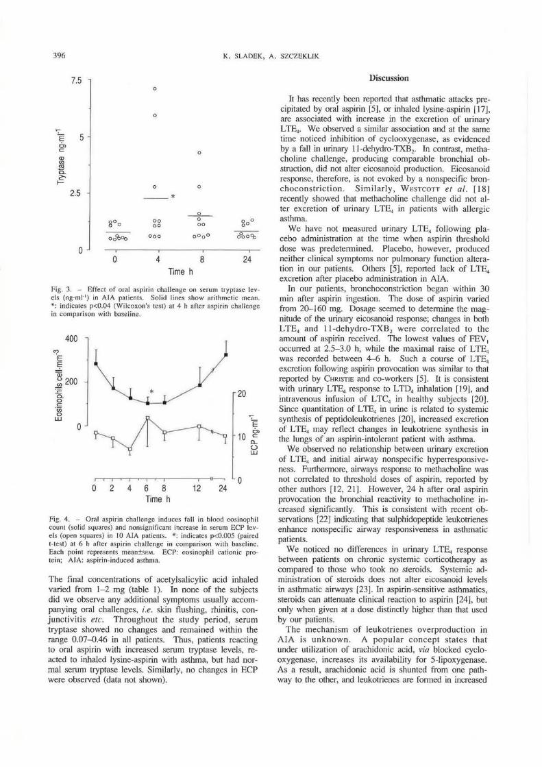
Fig. 2. - Methacholine inhalation results in a significant fall in FEY, (open circles), but does not effect urinary levels of 11-dehydroTXB2 (open triangles), or of LTE4 (open squares) in 10 AlA patients. Each point represents mean±SEM. For abbreviations see legend to table I.
TI1e intensity of both LTE4 and TXB2 responses depended on the dose of aspirin used (p<O.OOl ANOVA). Peak LTE4 levels were not correlated to maximum fall in FEV1
(r-0.154; p=0.33). Methacholine challenge did not cause any changes in LTE4 or 11-dehydro-TXB2 urinary levels (fig. 2).
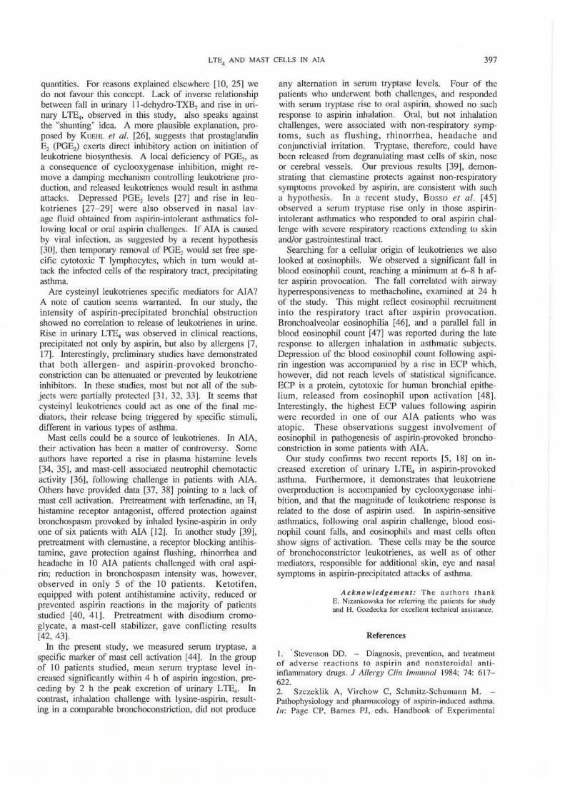
Following aspirin challenge, mean serum tryptase rose, reached a maximum at 4 h. and then gradually declined, returning to baseline levels after 24 h (fig. 3). Mean levels at baseline were 0.77±0.11 ng·ml-1, range 0.46-1.24 ng·ml-1; and at 4 h 2.21±0.75 ng·ml·' , range 0.62- 7.10 ng·mi-1 (p<0.04 Wilcoxon's signed rank test). ln our patients we observed no evident relationship between aspirin-related clinical symptoms. such as scnrlel flushing of the skin, rhinitis or blockage of tl1c nose, and serum tryptase or urinary L TE., levels. Following methacholine chall.cnge, serum tryptasc levels ~;howed no change; in all subjects the value remained between 0.44-0.66 ng·ml-1 throughout the study period.
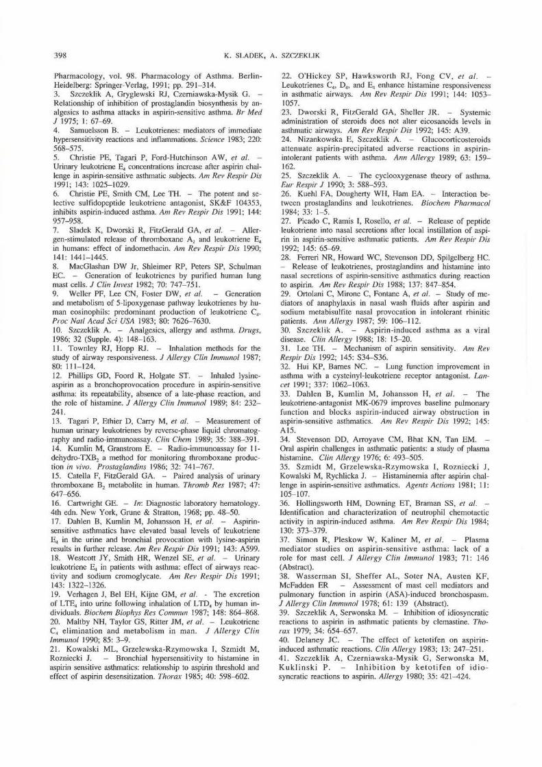
Aspirin provocation induced a gradual fall in blood eosinophil count (282.8±62.7 cells·mm-3 at baseline, and 127 .0±43.2 cells·mm·3 at 4 h) and serum ECP levels ( I 0.6± 1.4 ng·rnl 1 at buseline. and 6.8± 1.4 ng·mJ-1 at 4 h) (fig. 4). Eosinophil count reached a minimum at 6 h ( 101.9±35.6 cells·mm-3; p<O.O I paired Hest), while ECP scrum levels increased nonsignificantly to 14.1± 6.8 ng·ml-1 at the same time, and returned to 10.7±1.9 ng·ml·' at 8 h (fig. 4). Eosinophil count returned to initial values at 24 h after aspirin challenge (fig. 4). The fall in blood eosinophils at 6 h correlated with P~oMch examined 24 h after oral aspirin challenge (r=0.66, p<0.04). Following methacholine inhalation, the serum ECP levels and eosinophil count showed no changes.
Following lysine-aspirin inhalation, all subjects studied developed significant bronchoconstriction, expressed as a fall in FEV, exceeding 20% of baseline (28.3±3.9%; p<0.02 paired Hest). The reaction developed within about 40 min (range 15-60 min).
396 K. SLADEK, A. SZCZEKLIK
7.5 0
0
E 5 C'l c
0 <l> en «l a ~ t-
0 0
2.5 - --* ~
goo 00 0 goo oo 00
oo%.:0 ooo oooo o0oo<b
0 0 4 8 24
Time h
Fig. 3. - Effect of oral aspirin challenge on serum tryptase levels (ng·ml·') in AlA patients. Solid lines show arithmetic mean. •: indicales p<0.04 (Wilcoxon's test) at 4 h after asp irin challenge in comparison with baseline.
400
20
0 E
~ 10 C'l c
0.. {.) UJ
1-. 0 0 2 4 6 8 12 24
Time h
Fig. 4. - Oral aspirin challenge induces fall in blood eosinophi l counl (solid squares) and nonsignificant increase in serum ECP levels (open squares) in tO AlA patients. *: indicales p<O.OOS (paired Hesl) a1 6 h after aspirin challenge in comparison with base I in e. Each poinl represenls mean±SEM. ECP: eosinophil calionic prolein; AlA: aspirin-induced aslhma.
The final concentrations of acetylsalicylic acid inhaled varied from l-2 mg (table 1). In none of the subjects did we observe any additional symptoms usually accompanying oral challenges, i.e. skin flushing, rhinitis, conjunctivitis etc. Throughout the study period, serum tryptase showed no changes and remained within the range 0.07-0.46 in all patients. Thus, patients reacting to oral aspirin with increased serum tryptase levels, reacted to inhaled lysine-aspirin with aslhma, but had normal serum tryptase levels. Similarly, no changes in ECP were observed (data not shown).
Discussion
It has recently been reported that asthmatic attacks precipitated by oral aspirin (5], or inhaled lysine-aspirin [17], are associated with increase in the excrelion of urinary L TE4• We observed a similar association and at the same time noticed inhibition of cyclooxygenase, as evidenced by a fall in urinary 11-dehydro-TXB2• In contrast, methacholine challenge, producing comparable bronchial obstruction, did not alter eicosanoid production. Eicosanoid response, therefore, is not evoked by a nonspecific bronchoconstriction. Similarly, WESTCOTT et al. [ 18] recently showed that methacholine challenge did not alter excretion of urinary LTE4 in patients with allergic asthma.
We have not measured urinary LTE4 following placebo administration at the time when aspirin threshold dose was predetermined. Placebo, however, produced neither clinical symptoms nor pulmonary function alteration in our patients. Others [5], reported lack of LTE4
excretion after placebo administration in AlA. In our patients, bronchoconstriction began within 30
min after aspirin ingestion. The dose of aspirin varied from 20-160 mg. Dosage seemed to determine lhe magnitude of the urinary eicosanoid response; changes in both LTE4 and ll-dchydro-TXB2 were correlated to the amount of aspirin received. The lowest values of FEY 1
occurred at 2.5-3.0 h, while the maximal raise of LTE4
was recorded between 4-Q h. Such a course of LTE4
excretion following aspirin provocation was similar to that reported by CHRISTIE and eo-workers [5]. It is consistent with urinary LTE4 response to LTD4 inhalation [1 9], and intravenous infusion of LTC4 in healthy subjects [20). Since quantitation of L TE4 in urine is related to systemic synthesis of peptidoleukotrienes [20], increased excretion of L TE4 may reflect changes in leukotriene synthesis in the lungs of an aspirin-intolerant patient with asthma.
We observed no relationship between urinary excretion of L TE4 and initial airway nonspecific hyperresponsiveness. Furthermore, airways response to methacholine was nol correlated to threshold doses of aspirin, reported by other authors [12, 21]. However, 24 h after oral aspirin provocation the bronchial reactivity to methacholine increased significantly. This is consistent with recent observations [22J indicating that sulphidopeptide leukotrienes enhance nonspecific airway responsiveness in asthmatic patients.
We noticed no differences in urinary LTE4 response between patients on chronic systemic corticotherapy as compared to those who took no steroids. Systemic administration of steroids does not alter eicosanoid levels in asthmatic airways [23]. In aspirin-sensitive asthmatics, steroids can attenuate clinical reaction to aspirin [24], but only when given at a dose distinctly higher than that used by our patients.
The mechanism of leukotrienes overproduction in AIA is unknown. A popular concept states that under utilization of arachidonic acid, via blocked cyclooxygenase, increases its availability for 5-lipoxygenase. As a result, arachidonic acid is shunted from one pathway to the other, and leukotrienes are fom1ed in increased
LTE4
AND MAST CELLS fN AlA 397
quant1trcs. For reasons explained elsewhere [10, 25] we do not favour this concept. Lack of inverse relationship between fall in urinary ll-dehydro-TXB2 and rise in urinary LTE4, observed in this study, also speaks against the "shunting" idea. A more plausible explanation, proposed by KuEIIL et al. [26], suggests that prostaglandin E2 (PG~) exerts direct inhibitory action on .initiation of leukotriene biosynthesis. A local deficiency of PG~, as a consequence of cyclooxygenase inhibition, might remove a damping mechanism controlling leukotriene production, and released leukotrienes would result in asthma attacks. Depressed PGE2 levels [27] and rise in leukotrienes [27-29] were also observed in nasal lav<tgc lluid obtuinct.l from <t~pirin-into lerant asthmatics following local or oml aspirin challenges. If AlA is caused by viral infection. as suggested by il recent hypothesis [30] , then tempor:uy n:rnoval of PGEz would set free specific cytotoxic T lymphocytes, which in turn would attack the infected cells of the respiratory tract, precipitating asthma.
Are cysteinyl leukotrienes specific mediators for AlA? A note of caution seems warranted. In our study, the intensity of aspirin-precipitated bronchial obstruction showed no corTeJation to release of leukotrienes in urine. Rise in urinary L TE4 was observed in clinical reactions, precipitated not only by aspirin, but also by allergens [7, 17). Interestingly, preliminary studies have demonstrated that both allergen- and aspirin-provoked bronchoconstriction can be attenuated or prevented by leukotriene inhibitors. In these studies, most but not all of the subjeers were partially protected 131, 32. 3Jj. It seems that cysreinyl leukohicnes could act as one of the final mediators, their relem;e being triggered by specific stimuli, different in various types of asthma.
Mast cells could be a source of leukotrienes. In AlA, their activation has been a matter of controversy. Some authors have reported a rise in plasma histamine levels [34, 35], and mast-cell associated neutrophil chemotactic activity [36], following challenge in patients with AlA. Others have provided data [37, 38] pointing to a lack of mast cell activation. Pretreatment with terfenadine, an H1
histamine receptor antagonist, offered protection against bronchospasm provoked by inhaled lysine-aspirin in only one of six patients with AlA [12]. In another study [39), pretreatment with clemastine, a receptor blocking antihistamine, gave protection against flushing, rhinorrhea and headache in 10 AlA patients challenged with oral aspirin; reduction in bronchospasm intensity was, however, observed in only 5 of the 10 patients. Ketotifen, equipped with potent antihistamine activity, reduced or prevented aspirin reactions in the majority of patients studied [40, 41]. Pretreatment with disodium cromoglycate, a mast-cell stabilizer, gave conflicting results [42, 43].
In the present study, we measured serum tryptase, a specific marker of mast cell activation [44]. In the group of l 0 patients studied, mean serum tryptase level increased significantly within 4 h of aspirin ingestion, preceding by 2 h the peak excretion of urinary L TE4 • In contrast, inhalation challenge with lysine-a<>pirin, resulting in a comparable bronchoconstriction, did not produce
any alternation in serum lryptase lcvds. Four of the patients who underwent both ~hallenges, and responded with serum tryptase rise to oml aspirin. showed no such response to aspirin inhalation. Oral, but not inhalation challenges, were associated with non-respiratory symptoms, such as flushing, rhinorrhea, headache and conjunctivial irritation. Tryptase, therefore, could have been released from degranulating mast cells of skin, nose or cerebral vessels. Our previous results [39], demonstrating that clemastinc protects against non-respiratory symptoms provoked by aspirin, are consistent with such a hypothesis. In a recent study, Bosso et al. [45] observed a serum LTyptase rise only in those aspirinintolerant asthmatics who responded to oral aspirin challenge with severe respiratory reactions extending to skin and/or gastrointestinal tract.
Searching for a cellular origin of leukotrienes we also looked at eosinophils. We observed a significant fall in blood eosinophil count, reaching a minimum at 6-8 h after aspirin provocation. The fall correlated with airway hyperresponsiveness to methacholine, examined at 24 h of the study. This might reflect eosinophil reCJllitmcnt into the respiratory tract after us pirin provocation. Bronchoalveolar eosinophilia [46], and a parallel fall in blood eosinophil count [47] was reported during the late response to allergen inhalation in asthmntic subjects . Depression of the blood eosinophil count fo llowing aspirin ingestion was accompanied by a rise iJl ECP which. however, did not reach levels of statis tical signilkancc. ECP is a protein, cytotoxic for human bronchial epithelium, released from eosinophil upon activation [48]. Interestingly, the highest ECP values following aspirin were recorded in one of our AlA patients who was atopic. These observations suggest involvement of eosinophil in pathogenesis of aspirin-provoked bronchoconstriction in some patients with AlA.
Our study confirms two recent reports [5, 18] on increased excretion of urinary LTE4 in aspirin-provoked asthma. Furthermore, it demonstrates that leukotriene overproduction is accompanied by cyclooxygenase inhibition, and that the magnitude of leukotriene response is related to the dose of aspirin used. In aspirin-sensitive asthmatics, following oral aspirin challenge, blood eosinophil count falls, and eosinophils and mast cells often show signs of activation. These cells may be the source of bronchoconstrictor leukotrienes, as well as of other mediators, responsible for additional skin, eye and nasal symptoms in aspirin-precipitated attacks of asthma.
Acknowledgement: The authors thank E. Nizankowska for refeTTing 1he patients for study and H. Gozdecka for excellent technical assistance.
References
I. Stevenson DD. - Diagnosis, prevention, and treatment of adverse reactions to aspirin and nonsteroidal anriinflammatory drugs. J Allergy Clin lmmunol 1984; 74: 617-622. 2. Szczeklik A, Yirchow C, Schmitz-Schumann M. -Pathophysiology and pharmacology of aspirin-induced asthma. In: Page CP, Barnes PJ, eds. Handbook of Experimenla1
398 K. SLADEK, A. SZCZEKLIK
Pharmacology, vol. 98. Pharmacology of Asthma. BerlinHeidelberg: Springer-Verlag, 1991; pp. 291- 314. 3. Szczeklik A, Gryglewski RJ, Czemiawska-Mysik G. -Relationship of inhibition of prostaglandin biosynthesis by analgesics to asthma attacks in aspirin-sensitive asthma. Br Med J 1975; ] : 67-69. 4. Samuelsson B. - Leukotrienes: mediators of immediate hypersensitivity reactions and inflrunmations. Science 1983; 220: 568-575. 5. Christie PE, Tagari P, Ford-Hutchinson A W, et al. -Urinary leukotriene E. concentrations increase after aspirin cha.llenge in aspirin-sensitive asthmatic subjects. Am Re1' Respir Dis 1991; 143: 1025-1029. 6. Christie PE, Smith CM, Lee TH. - The potent and selective sulfidopeptide leukotriene antagonist, SK&F 104353, inhibits aspirin-induced asthma. Am Rev Respir Dis !991; 144: 957-958. 7. Sladek K, Dworski R, FitzGerald GA, et al. - Allergen-stimulated release of thromboxane A2 and leukotriene E, in humans: effect of indomethacin. Am Rev Respir Dis 1990; 141: 1441-1445. 8. MacGlashan DW Jr, Shleimer RP, Peters SP, Schulman EC. - Generation of leukotricnes by purified human lung mast cells. J C/in Invest 1982; 70: 747- 751. 9. Weller PF, Lee CN, Foster DW, et al. - Generation and metabolism of 5-lipoxygenase pathway leukotrienes by human eosinophils: predominant production of leukotriene c4. Proc Nail Acad Sci USA 1983; 80: 7626-7630. 10. Szczeklik A. - Analgesics, allergy and asthma. Drugs, 1986; 32 (Supple. 4): 148-163. 11. Townley RJ, Hopp RJ. - lnha.lation methods for the study of airway responsiveness. J Allergy Clin lmmunol 1987; 80: 111- 124. 12. Phillips GD, Foord R, Holgate ST. - Inhaled lysineaspirin as a bronchoprovocation procedure in aspirin-sensitive asthma: its repeatability, absence of a late-phase reaction, and the role of histamine. J Allergy Clin lmmunol 1989; 84: 232-241. 13. Tagari P, Ethier D, Carry M, et al. - Measurement of human urinary leukotrienes by reverse-phase liquid chromatography and radio-immunoassay. Clin Chem 1989; 35: 388-391. 14. Kumlin M, Granstrom E. - Radio-immunoassay for 11-dehydro-TXB2 a method for monitoring thromboxanc production in vivo. Prostaglandins 1986; 32: 741- 767. 15. Catella F, FitzGerald GA. - Paired analysis of urinary thromboxane B2 metabolite in human. Thromb Res 1987; 47: 647-656. 16. Cartwright GE. - In: Diagnostic laboratory hematology. 4th edn. New York, Grune & Stratton, 1968; pp. 48-50. 17. Dahlen B, Kumlin M, Johansson H, et al. - Aspirinsensitive asthmatics have elevated basal levels of leukotriene E4 in the urine and bronchial provocation with lysine-aspirin results in further release. Am Rev Respir Dis 1991; 143: A599. 18. Westcott JY, Smith HR, Wenzel SE, et al. - Urinary lcukotriene E4 in patients with asthma: effect of airways reactivity and sodium cromoglycatc. Am Rev Respir Dis 1991; 143: !322-1326. 19. Verhagen J, Bel EH, Kijne GM, er al. - The excretion of L TE4 into urine following inhalation of LTD4 by human individuals. Biochem Biophys Res Commun 1987; 148: 864-868. 20. Maltby NH, Taylor GS, Ritter JM, et al. - Leukotrienc C4 elimination and metabolism in man. J Allergy C/in lmnwnol 1990; 85: 3-9. 21. Kowalski ML, Grzelewska-Rzymowska I, Szmidt M, Rozniecki J. - Bronchial hypersensitivity to histamine in aspirin sensitive asthmatics: relationship to aspirin threshold and effect of aspirin desensitization. Thorax 1985; 40: 598-602.
22. O'Hickey SP, Hawksworth RJ, Fong CV, et al. Leukotrienes C4, D4 , and E4 enhance histamine responsiveness in asthmatic airways. Am Rev Respir Dis 1991; 144: 1053-1057. 23. Dworski R, FitzGerald GA, Sheller JR. - Systemic administration of steroids does not alter eicosanoids levels in asthmatic airways. Am Rev Respir Dis 1992; 145: A39. 24. Nizankowska E, Szezeklik A. - Glucocorticosteroids attenuate aspirin-precipitated adverse reactions in aspirinintolerant patients with asthma. Ann Allergy 1989; 63: 159-162. 25. Szczeklik A. - The cyclooxygenase theory of asthma. Eur Respir J 1990; 3: 588- 593. 26. Kuehl FA, Dougherty WH, Ham EA. - Interaction between prostaglandins and leukotrienes. Biochem Pharmacal 1984; 33: l-5. 27. Picado C, Ramis I, Rosello, et al. - Release of peptide leukotriene into nasal secretions after local instillation of aspirin in aspirin-sensitive asthmatic patients. Am Rev Respir Dis 1992; 145: 65-69. 28. Ferreri NR, Howard WC, Stevenson DD, Spilgelberg HC. - Release of leukotrienes, prostaglandins and histamine into nasal secretions of aspirin-sensitive asthmatics during reaction to aspirin. Am Rev Respir Dis 1988; 137: 847-854. 29. Ortolani C, Mirone C, Fontane A, et al. - Study of mediators of anaphylaxis in nasal wash tluids after aspirin and sodium metabisulfite nasal provocation in intolerant rhinitic patients. Ann Allergy 1987; 59: 106-112. 30. Szczeklik A. - Aspirin-induced asthma as a viral disease. Clin Allergy 1988; 18: 15-20. 31. Lee TH. - Mechanism of aspirin sensitivity. Am Rev Respir Dis 1992; 145: S34-S36. 32. Hui KP, Bames NC. - Lung function improvement in asthma with a cysteinyl-leukotriene receptor antagonist. Lancet !991; 337: 1062- 1063_ 33. Dahlen B, Kumlin M, Johansson H, et al. - The leukotriene-antagonist MK-0679 improves baseline pulmonary function and blocks aspirin-induced airway obstruction in aspirin-sensitive asthmatics. Am Rev Respir Dis 1992; 145: Al5. 34. Stevenson DD, Arroyave CM, Bhat KN, Tan EM. -Oral aspirin challenges in asthmatic patients: a study of plasma histamine. Clin Allergy 1976; 6: 493-505. 35. Szmidt M, Grzelewska-Rzymowska 1, Rozniccki J, Kowalski M, Rychlicka J. - Histaminemia after aspirin challenge in aspirin-sensitive asthmatics. Agems Actions 1981; I I: 105-107. 36. Hollingsworth HM, Downing ET, Braman SS, et al. -Identification and characteri7..ation of neutrophil chemotactic activity in aspirin-induced asthma. Am Rev Respir Dis 1984; 130: 373-379. 37. Simon R, Pleskow W, Kaliner M, et al. - Plasma mediator studies on aspirin-sensitive asthma: lack of a role for mast cell. J Allergy Clin lmmunol 1983; 71: 146 (Abstract). 38. Wasserman SI, Sheffer AL, Soter NA, Austen KF, McFadden ER - Assessment of mast cell mediators and pulmonary function in aspirin (ASA)-induced bronchospasm. J Allergy Clinlmmunol 1978; 61: 139 (Abstract). 39. Szczeklik A, Serwonska M. - Inhibition of idiosyncratic reactions to aspirin in asthmatic patients by clemastine. Thorax 1979; 34: 654-657. 40. Delaney JC. - The effect of ketotifen on aspirininduced asthmatic reactions. Clin Allergy 1983; 13: 247-251. 41. Szczeklik A, Czerniawska-Mysik G, Serwonska M, Kuklinski P. Inhibition by ketotifen of idiosyncratic reactions to aspirin. Allergy 1980; 35: 421-424.
LTE4
AND MAST CELLS IN AIA 399
42. Dahl R. - Oral and inhaled sodium cromoglycate in challenge test with food allergen or acetyl-salicylic acid. Allergy 1981; 36: 161-165. 43. Martelli NA, Usandivaras G. - Inhibition of aspirininduced bronchoconstriction by sodium cromoglycate inhalation. Thorax 1977; 32: 684-690. · 44. Schwartz LB, Metcalfe MD, Miller JS, Earl HE, Sulivan T. - Tryptase levels as an indicator of mast-cell activation in systemic anaphylaxis and mastocytosis. N Engl J Med 1987; 316: 1622-1626. 45. Bosso, N, Schwartz LB, Stevenson DD. - Tryptase and
histamine release during aspirin-induced respiratory reactions. J Allergy Clin Immunol 1991; 88: 830-837. 46. De Monchy JGR, Kauffman HF, Venge P, et al. -Bronchoalveolar eosinophilia during allergen-induced late asthmatic reactions. Am Rev Respir Dis 1985; 131: 373-376. 47. Cookson WOCM, Craddock CF, Benson MK, Durham SR.
Falls in peripheral eosinophils count parallel the late asthmatic response. Am Rev Respir Dis 1989; 139: 458-462. 48. Venge P, Hakansson L, Peterson COB. - Eosinophil activation in. allergic diseas.e. lnt Arch Allergy Appl lmmunol 1987; 39: 177- 253.