Cycloplegic Retinoscopy in Infancy
5
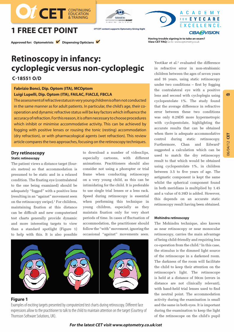
06/04/12 CET 49 CET CONTINUING EDUCATION & TRAINING 1 FREE CET POINT OT CET content supports Optometry Giving Sight Having trouble signing in to take an exam? View CET FAQ Go to www.optometry.co.uk For the latest CET visit www.optometry.co.uk/cet Retinoscopy in infancy: cycloplegic versus non-cycloplegic C-18551 O/D Fabrizio Bonci, Dip. Optom (ITA), MCOptom Luigi Lupelli, Dip. Optom (ITA), FAILAC, FIACLE, FBCLA The assessment of refractive status in very young children is often not conducted in the same manner as for adult patients. In particular, the child’s age, their co- operation and dynamic refractive status will be key factors which influence the accuracy of refraction. For this reason, it is often necessary to choose procedures which inhibit or minimise accommodative activity. This can be achieved by fogging with positive lenses or rousing the tonic (resting) accommodation (dry refraction), or with pharmacological agents (wet refraction). This review article compares the two approaches, focusing on the retinoscopy techniques. Dry retinoscopy Static retinoscopy The patient views a distance target (four- six metres) so that accommodation is presumed to be static and in a relaxed condition. The fixating eye (contralateral to the one being examined) should be adequately “fogged” with a positive lens (resulting in an “against” movement seen on the retinoscopy swipe). 1 For children, maintaining fixation at this distance can be difficult and new computerised test charts generally provide dynamic and more interesting targets to view than a standard spotlight (Figure 1) to help with this. It is also possible to download a number of videoclips, especially cartoons, with different animations. Practitioners should also consider not using a phoropter or trial frame when conducting retinoscopy on a very young child, as this can be intimidating for the child. It is preferable to use single trial lenses or a lens rack. Speed during retinoscopy is essential when performing this technique in young children, especially as they maintain fixation only for very short periods of time. In cases of fluctuation of accommodation, the practitioner should follow the “with” movement, ignoring the occasional “against” movements seen. Yeotikar et al. 2 evaluated the difference in refractive error in non-strabismic children between the ages of seven years and 16 years, using static retinoscopy under two conditions – first by fogging the contralateral eye with a positive lens and second with cycloplegia using cyclopentolate 1%. The study found that the average difference in refractive error between these two conditions was only 0.29DS more hypermetropic with cyclopentolate, highlighting the accurate results that can be obtained when there is adequate accommodative control during static retinoscopy. Furthermore, Chan and Edward 3 suggested a calculation which can be used to match the dry retinoscopy result to that which would be obtained using cyclopentolate 1%, in children between 3.5 to five years of age. The astigmatic component is kept the same whilst the spherical component found in both meridians is multiplied by 1.45 and a value of 0.39D is added. However, this depends on an accurate static retinoscopy result having been obtained. Mohindra retinoscopy The Mohindra technique, also known as near retinoscopy or near monocular retinoscopy, carries the main advantage of being child-friendly and requiring less co-operation from the child. 4 In this case, the stimulus is the dimmed light source of the retinoscope in a darkened room. The darkness of the room will facilitate the child to keep their attention on the retinoscope’s light. The retinoscope is held at a distance of 50cm (errors in distance are not clinically relevant), with hand-held trial lenses used to find the neutral point. The accommodation activity during the examination is small and the same in both eyes. It is important during the examination to keep the light of the retinoscope on the child’s pupil Figure 1 Examples of exciting targets presented by computerized test charts during retinoscopy. Different face expressions allow to the practitioner to talk to the child to maintain attention on the target (Courtesy of Thomson Software Solutions, UK). Approved for: Optometrists Dispensing Opticians 4 4
-
Upload
strauss-de-lange -
Category
Documents
-
view
86 -
download
1
Transcript of Cycloplegic Retinoscopy in Infancy
06/0
4/12
CET
49
CET CONTINUING EDUCATION & TRAINING
1 FREE CET POINT OT CET content supports Optometry Giving Sight
Having trouble signing in to take an exam? View CET FAQ Go to www.optometry.co.uk
For the latest CET visit www.optometry.co.uk/cet
Retinoscopy in infancy: cycloplegic versus non-cycloplegicC-18551 O/D
Fabrizio Bonci, Dip. Optom (ITA), MCOptomLuigi Lupelli, Dip. Optom (ITA), FAILAC, FIACLE, FBCLAThe assessment of refractive status in very young children is often not conducted in the same manner as for adult patients. In particular, the child’s age, their co-operation and dynamic refractive status will be key factors which influence the accuracy of refraction. For this reason, it is often necessary to choose procedures which inhibit or minimise accommodative activity. This can be achieved by fogging with positive lenses or rousing the tonic (resting) accommodation (dry refraction), or with pharmacological agents (wet refraction). This review article compares the two approaches, focusing on the retinoscopy techniques.
Dry retinoscopyStatic retinoscopy The patient views a distance target (four-
six metres) so that accommodation is
presumed to be static and in a relaxed
condition. The fixating eye (contralateral
to the one being examined) should be
adequately “fogged” with a positive lens
(resulting in an “against” movement seen
on the retinoscopy swipe).1 For children,
maintaining fixation at this distance
can be difficult and new computerised
test charts generally provide dynamic
and more interesting targets to view
than a standard spotlight (Figure 1)
to help with this. It is also possible
to download a number of videoclips,
especially cartoons, with different
animations. Practitioners should also
consider not using a phoropter or trial
frame when conducting retinoscopy
on a very young child, as this can be
intimidating for the child. It is preferable
to use single trial lenses or a lens rack.
Speed during retinoscopy is essential
when performing this technique in
young children, especially as they
maintain fixation only for very short
periods of time. In cases of fluctuation of
accommodation, the practitioner should
follow the “with” movement, ignoring the
occasional “against” movements seen.
Yeotikar et al.2 evaluated the difference
in refractive error in non-strabismic
children between the ages of seven years
and 16 years, using static retinoscopy
under two conditions – first by fogging
the contralateral eye with a positive
lens and second with cycloplegia using
cyclopentolate 1%. The study found
that the average difference in refractive
error between these two conditions
was only 0.29DS more hypermetropic
with cyclopentolate, highlighting the
accurate results that can be obtained
when there is adequate accommodative
control during static retinoscopy.
Furthermore, Chan and Edward3
suggested a calculation which can be
used to match the dry retinoscopy
result to that which would be obtained
using cyclopentolate 1%, in children
between 3.5 to five years of age. The
astigmatic component is kept the same
whilst the spherical component found
in both meridians is multiplied by 1.45
and a value of 0.39D is added. However,
this depends on an accurate static
retinoscopy result having been obtained.
Mohindra retinoscopy The Mohindra technique, also known
as near retinoscopy or near monocular
retinoscopy, carries the main advantage
of being child-friendly and requiring less
co-operation from the child.4 In this case,
the stimulus is the dimmed light source
of the retinoscope in a darkened room.
The darkness of the room will facilitate
the child to keep their attention on the
retinoscope’s light. The retinoscope
is held at a distance of 50cm (errors in
distance are not clinically relevant),
with hand-held trial lenses used to find
the neutral point. The accommodation
activity during the examination is small
and the same in both eyes. It is important
during the examination to keep the light
of the retinoscope on the child’s pupil
Figure 1 Examples of exciting targets presented by computerized test charts during retinoscopy. Different face expressions allow to the practitioner to talk to the child to maintain attention on the target (Courtesy of Thomson Software Solutions, UK).
Approved for: Optometrists Dispensing Opticians 4 4

06/0
4/12
CET
50
CET CONTINUING EDUCATION & TRAINING
1 FREE CET POINT OT CET content supports Optometry Giving Sight
Find out when CET points will be uploaded to Vantage at www.optometry.co.uk/cet/vantage-dates
Having trouble signing in to take an exam? View CET FAQ Go to www.optometry.co.uk
Approved for: Optometrists Dispensing Opticians 4 4
(to see the retinal reflex) for only a short
period of time so as not to stimulate
accommodation; subsequently the
optometrist’s attention should be focused
on the pupil, watching for maximum
dilation (indicating no accommodation).5
The procedure should be carried out
with one eye occluded, preferably by the
parent, while the other eye is evaluated.
However, Wesson et al.6 confirmed that
there is no substantial difference in the
result if binocular fixation is allowed
(Figure 2); indeed this can be useful if the
infant is resistant and becomes agitated
with occlusion. Several people advocate
neutralisation of the two principal
meridians of the eye separately, using
loose spherical trial lenses. However,
Saunders and Westall7 confirmed that
the accuracy of the technique can
be improved using a combination of
spherical and cylindrical lenses instead.
Once the retinoscopy result is obtained, the
refractive error was originally calculated
by adding -1.25DS to the gross finding.8,9
Saunders and Westall7 have reported
that the accuracy can be improved if
-0.75DS is added instead, for children
aged between 0-2 years, and -1.00DS
added for those children over two years
of age. They also affirmed that the result
achieved by the Mohindra procedure in
children between six months and four
years of age is similar to wet retinoscopy
(using cyclopentolate 1% – see later),
with a difference of only 0.50DS. Others
have reported similar results,10 and
certainly no differences greater than
1.00DS,11 whilst similar results were
also obtained for children with Down’s
syndrome12 and even in adults.13
The Mohindra technique is useful for
practitioners in Europe who are not
permitted to used cycloplegic agents,14
whilst there are benefits for conducting
frequent follow-up assessments without
repeated use of cycloplegic agents.15
One must remember, however, that the
accuracy of results will naturally depend
on the practitioner’s experience.16
Cycloplegic agentsControl of accommodation in children
of pre-school age is more commonly
achieved by pharmacological means,
using cycloplegic agents such as
cyclopentolate and tropicamide; atropine
can only be used by therapeutically
qualified practitioners. All of these drugs
are muscarinic receptor blockers, thus
they work by blocking the muscarinic
receptors in the ciliary body, which
in turn prevents accommodation.
A mydriatic effect is concurrently
achieved by inhibiting muscarinic
stimulation of the iris sphincter muscle.
An ideal cycloplegic would have no
ocular and systemic adverse effects.
Also, it should produce a rapid onset of
cycloplegia, blocking accommodation
completely for an adequate period
of time, before swiftly restoring
accommodative ability.17 Several
studies have reported both ocular
and systemic side effects (especially
using atropine) in those children who
have had a cycloplegic refraction,
in addition to expected mydriasis
and cycloplegia, as detailed later.18
Drug selection and instillationCycloplegia is an invasive technique
which can be uncomfortable, or even
distressing, for the child. This is notably
so because the acidic pH of the cycloplegic
agent leads to stinging on instillation.
Some practitioners advocate the use of a
local anaesthetic prior to instillation of
the cycloplegic agent; proxymetacaine
0.5% is the drug of choice as it stings less
than other topical anaesthetics. However,
this is not always recommended due to
the risks associated with an anaesthetised
cornea. To facilitate the application
of cycloplegics, cyclopentolate has
been instilled in spray form onto the
eyelashes and the closed upper lid.19
Practitioners should also be conscious
of their instillation technique, since
different degrees of cycloplegia between
the eyes can occur, especially if the
child does not keep their eyes open wide
enough and/or if there is significant post-
instillation tearing (which is very likely).
As such, practitioners can opt to instil
the higher concentration of cycloplegic
agent and/or instil further drops if
regular review (eg, periodic measurement
of the amplitude of accommodation)
reveals differing levels of cycloplegia.
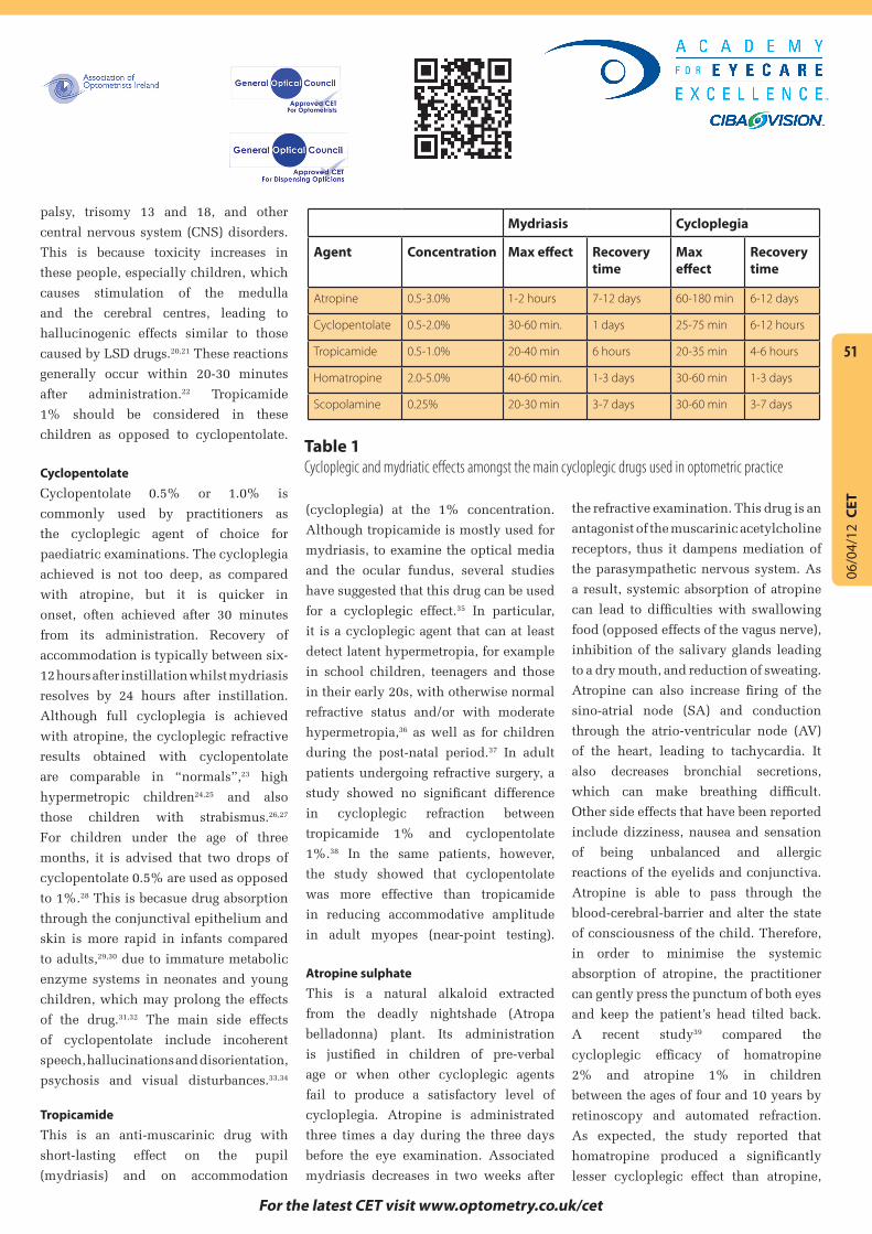
Differences in the main cycloplegic
agents are summarised in Table 1. The
optometrist should select an appropriate
agent considering factors such as the
patient’s age and whether they have
dark, or light coloured, irides. Adequate
cycloplegic effect could be achieved
with tropicamide in a teenage patient
suspected of having latent hypermetropia,
for example, whereas cyclopentolate
is likely to be required for an infant
suspected of having an accommodative
esotropia. Those with light coloured
irides may exhibit an increased response
to drugs as compared with darkly
pigmented irides, and therefore a lower
concentration/dose ought to be selected.
Overdose of cycloplegic agent has to
be avoided in children with Down’s
syndrome or those affected by cerebral
Figure 2 Mohindra retinoscopy. Hand-held trial lenses are placed in front of both eyes whilst the child fixates the retinoscope light. The procedure should be run in darkened room (the high level of room light in this image was for photographic purposes only).
06/0
4/12
CET
51
For the latest CET visit www.optometry.co.uk/cet
Having trouble signing in to take an exam? View CET FAQ Go to www.optometry.co.uk
palsy, trisomy 13 and 18, and other
central nervous system (CNS) disorders.
This is because toxicity increases in
these people, especially children, which
causes stimulation of the medulla
and the cerebral centres, leading to
hallucinogenic effects similar to those
caused by LSD drugs.20,21 These reactions
generally occur within 20-30 minutes
after administration.22 Tropicamide
1% should be considered in these
children as opposed to cyclopentolate.
Cyclopentolate Cyclopentolate 0.5% or 1.0% is
commonly used by practitioners as
the cycloplegic agent of choice for
paediatric examinations. The cycloplegia
achieved is not too deep, as compared
with atropine, but it is quicker in
onset, often achieved after 30 minutes
from its administration. Recovery of
accommodation is typically between six-
12 hours after instillation whilst mydriasis
resolves by 24 hours after instillation.
Although full cycloplegia is achieved
with atropine, the cycloplegic refractive
results obtained with cyclopentolate
are comparable in “normals”,23 high
hypermetropic children24,25 and also
those children with strabismus.26,27
For children under the age of three
months, it is advised that two drops of
cyclopentolate 0.5% are used as opposed
to 1%.28 This is becasue drug absorption
through the conjunctival epithelium and
skin is more rapid in infants compared
to adults,29,30 due to immature metabolic
enzyme systems in neonates and young
children, which may prolong the effects
of the drug.31,32 The main side effects
of cyclopentolate include incoherent
speech, hallucinations and disorientation,
psychosis and visual disturbances.33,34
Tropicamide This is an anti-muscarinic drug with
short-lasting effect on the pupil
(mydriasis) and on accommodation
(cycloplegia) at the 1% concentration.
Although tropicamide is mostly used for
mydriasis, to examine the optical media
and the ocular fundus, several studies
have suggested that this drug can be used
for a cycloplegic effect.35 In particular,
it is a cycloplegic agent that can at least
detect latent hypermetropia, for example
in school children, teenagers and those
in their early 20s, with otherwise normal
refractive status and/or with moderate
hypermetropia,36 as well as for children
during the post-natal period.37 In adult
patients undergoing refractive surgery, a
study showed no significant difference
in cycloplegic refraction between
tropicamide 1% and cyclopentolate
1%.38 In the same patients, however,
the study showed that cyclopentolate
was more effective than tropicamide
in reducing accommodative amplitude
in adult myopes (near-point testing).
Atropine sulphateThis is a natural alkaloid extracted
from the deadly nightshade (Atropa
belladonna) plant. Its administration
is justified in children of pre-verbal
age or when other cycloplegic agents
fail to produce a satisfactory level of
cycloplegia. Atropine is administrated
three times a day during the three days
before the eye examination. Associated
mydriasis decreases in two weeks after
the refractive examination. This drug is an
antagonist of the muscarinic acetylcholine
receptors, thus it dampens mediation of
the parasympathetic nervous system. As
a result, systemic absorption of atropine
can lead to difficulties with swallowing
food (opposed effects of the vagus nerve),
inhibition of the salivary glands leading
to a dry mouth, and reduction of sweating.
Atropine can also increase firing of the
sino-atrial node (SA) and conduction
through the atrio-ventricular node (AV)
of the heart, leading to tachycardia. It
also decreases bronchial secretions,
which can make breathing difficult.
Other side effects that have been reported
include dizziness, nausea and sensation
of being unbalanced and allergic
reactions of the eyelids and conjunctiva.
Atropine is able to pass through the
blood-cerebral-barrier and alter the state
of consciousness of the child. Therefore,
in order to minimise the systemic
absorption of atropine, the practitioner
can gently press the punctum of both eyes
and keep the patient’s head tilted back.
A recent study39 compared the
cycloplegic efficacy of homatropine
2% and atropine 1% in children
between the ages of four and 10 years by
retinoscopy and automated refraction.
As expected, the study reported that
homatropine produced a significantly
lesser cycloplegic effect than atropine,
Mydriasis Cycloplegia
Agent Concentration Max effect Recovery time
Max effect
Recovery time
Atropine 0.5-3.0% 1-2 hours 7-12 days 60-180 min 6-12 days
Cyclopentolate 0.5-2.0% 30-60 min. 1 days 25-75 min 6-12 hours
Tropicamide 0.5-1.0% 20-40 min 6 hours 20-35 min 4-6 hours
Homatropine 2.0-5.0% 40-60 min. 1-3 days 30-60 min 1-3 days
Scopolamine 0.25% 20-30 min 3-7 days 30-60 min 3-7 days
Table 1 Cycloplegic and mydriatic effects amongst the main cycloplegic drugs used in optometric practice

06/0
4/12
CET
52
CET CONTINUING EDUCATION & TRAINING
1 FREE CET POINT OT CET content supports Optometry Giving Sight
Find out when CET points will be uploaded to Vantage at www.optometry.co.uk/cet/vantage-dates
Having trouble signing in to take an exam? View CET FAQ Go to www.optometry.co.uk
Approved for: Optometrists Dispensing Opticians 4 4
with residual accommodation being
greater (1.80±0.40D with atropine
vs. 3.10±0.50D with homatropine;
p<0.001). Another study40 compared the
cycloplegic effect obtained at 90 minutes
after administration of two drops of
atropine 0.5% to that obtained after
three times daily instillation of atropine
0.5% for three days, in strabismic
children. It was found that although
residual accommodation was greater
at 90 minutes after instillation (1.00D)
compared with three-day atropinization
(0.50D), the former still allows a more
rapid and less toxic assessment of
refraction than the usual three-day dose.
Indications for cycloplegic refractionThere are several instances
when a cycloplegic refraction
is indicated, including:
• Hypermetropia over +5.00D
• Anisometropia more than 1.50D
• Suspect and/or manifest strabismus
(especially esotropia)
• Family history of strabismus, high
hypermetropia and amblyopia
• In the presence of unstable esophoria,
pseudomyopia and asthenopia
• Poor cooperation/fixation of the child
• When the retinal reflex during
retinoscopy changes motion
or brightness due to dynamic
accommodative status.
There are also some instances
where cycloplegic refraction
is contraindicated, including:
• Cases where administration of
cycloplegic agents will cause undue
stress for the child, resulting in a
complete lack of co-operation
• Risk of ocular and/or systemic side
effects (which are more likely with
atropine than cyclopentolate and
tropicamide)
• Risk of developing acute angle closure
glaucoma in those with a shallow
anterior chamber (especially with
cyclopentolate)
Conducting wet retinoscopy Cycloplegic retinoscopy is carried out in
a similar way to dry static retinoscopy.
Despite pharmacological control of
accommodation, some co-operation
of the child is still required to keep
fixation during the examination, so that
off-axis aberrations do not distort the
retinoscopic reflex. Zadnick et al.41 found
that the repeatability of retinoscopy
under cycloplegia is poorer than non-
cycloplegic conditions, mainly due to
the irregularity of the retinoscopic reflex
through a dilated pupil. For this reason
it is important for the practitioner to
concentrate only on the central portion
of the pupil, ignoring the movement
seen in the peripheral annulus.
Objective automated refractionWhen a cycloplegic retinoscopy result
has been obtained, it can be useful to
conduct objective automated refraction
too, for comparison of the results. A
study42 comparing cycloplegic refraction
measurements using the hand-held
autorefractor (Retinomax), a table-
mounted autorefractor (Canon FK-1)
and streak retinoscopy in a large cohort
of children between 24 and 72 months
of age found no significant difference
in the mean spherical equivalent (MSE)
refractive error between the table-
mounted autorefractor (1.03±1.64D)
and streak retinoscopy (1.09±1.58D,
p=0.66). However, the MSE using the
hand-held Retinomax (0.80±1.43D)
was significantly different (p=0.0004)
to streak retinoscopy. Astigmatism
measured using the hand-held
(-0.89±0.51D) and table-mounted
(-0.83±0.61D) autorefractors were
significantly greater than that obtained
with retinoscopy (-0.58±0.56D,
p=0.0003). Therefore, practitioners
should remember that autorefractometry
and videorefractometry, although
useful as a guide and screening tool,
are not accurate enough to base actual
prescribing decisions upon in pre-
school children.42,43 Issues of ensuring
correct fixation on the target tend to be
the primary reason for the discrepancy,
but even in co-operative older subjects,
variable readings can be obtained
from different autorefractor tools.44-45
Dry autorefraction seems particularly
inaccurate in hypermetropia46 and
astigmatism in children of pre-verbal
age,47 although hand-held autorefraction
devices can evaluate astigmatism
without administration of cycloplegic
drugs, with the same repeatability as
wet retinoscopy.48 Wet autorefraction
generally seems more accurate if
it is compared to the subjective
refraction under cycloplegia.49
Practitioners should also remember that
cycloplegic drugs might temporarily
modify the structures of anterior
chamber, inducing astigmatism or
modifying an existing astigmatic
ametropia. This could occur because
there is a change in intraocular pressure
(IOP) under cycloplegia, which alters
the position of the crystalline lens
from the normal corneal distance.
These modifications have an effect on
the refraction, but this is difficult to
quantify in terms of refractive power.50
Conclusion During the refractive assessment it is
necessary to control and often inhibit
accommodative function, especially in
paediatric cases. This can be obtained
using dry and wet techniques, both of
which are appropriate for identifying
the most appropriate correction.
Although the refractive assessment
should be carried out as naturally
as possible, when the practitioner
encounters uncooperative children or
06/0
4/12
CET
53
For the latest CET visit www.optometry.co.uk/cet
1. Which of the following statements is TRUE?a) Atropine produces cycloplegia within 2-3 hours and recovery of accommodation in 2 daysb) Cyclopentolate produces cycloplegia within 30 minutes and recovery of accommodation in 12 hoursc) Homatropine produces cycloplegia in 20-30 minutes and recovery of accommodation in 24 hoursd) All of the above
2. Which pharmacological agent should be considered in a child with Down’s Syndrome?a) Atropine b) Homatropinec) Cyclpentolate d) Tropicamide
3. When should Mohindra’s retinoscopy technique be performed? a) In all children b) Only in children aged 7-10 yearsc) Only in young children with moderate astigmatism d) In pre-verbal children
4. How should Mohindra’s retinoscopy technique be performed?a) In darkness, fixation being on the retinoscope light, using hand held lensesb) In room lighting, fixation being on a high contrast target at the retinoscope mirror c) In darkness, fixation being on a spotlight at 6 metres, using hand held lensesd) In room light, fixation being on a high contrast target at 6 metres, under cycloplegia
5. Which of the following statements regarding cycloplegic refraction is TRUE?a) It should be considered in children with high hypermetropia or strabismusb) It should be considered in every child at every sight testc) Atropine is the cycloplegic of choice for a 5-year-old childd) Objective automated refraction should be used for prescribing decisions
6. Which of the following is NOT a side effect of cyclopentolate?a) Incoherent speechb) Hallucinationsc) Mydriasisd) Conjunctival hyperaemia
PLEASE NOTE There is only one correct answer. All CET is now FREE. Enter online. Please complete online by midnight on May 4, 2012 – You will be unable to submit exams after this date. Answers to the module will be published on www.optometry.co.uk/cet/exam-archive. CET points for these exams will be uploaded to Vantage on May 14, 2012. Find out when CET points will be uploaded to Vantage at www.optometry.co.uk/cet/vantage-dates
Module questions Course code: C-18551 O/D
those with risk factors for binocular
vision anomalies the refraction should
be performed under cycloplegia.
Tropicamide seems to be as effective
as cyclopentolate for measurement of
refractive error in most non-strabismic
infants, particularly at the 1%
concentration, so it should be considered
more often in paediatric eye care in
order to reduce the possibility of adverse
reactions. Where there is suspicion of a
binocular vision anomaly, cyclopentolate
should be the agent of choice.
About the authorsFabrizio Bonci is an optometrist and
clinical research fellow at the division
of clinical neuroscience and mental
health, Imperial College, London, and
the Faculty of Medicine, Charing Cross
Hospital, London. Luigi Lupelli is an
optometrist, professor in contact lenses
at the Faculty of Science, Department
of Physics, (Optics and Optometry)
at the University of Roma Tre, Italy.
ReferencesSee www.optometry.co.uk/
clinical. Click on the article title and
then on ‘references’ to download.


![REFRACTION IN Anne Cees Houtman CHILDREN UZ …...retinoscopy -0.15 ± 1.31 Cycloplegic AR -0.12 ± 1.41 Int Ophthalmol. 2013 Oct 10. [Epub ahead of print]- abstract Comparison of](https://static.fdocuments.us/doc/165x107/5fdfa3d9d6039f6a6b08d28b/refraction-in-anne-cees-houtman-children-uz-retinoscopy-015-131-cycloplegic.jpg)