Cyclic Nucleotide driven Protein Kinase Signaling in ... · Cyclic Nucleotide-driven Protein Kinase...
37
Cyclic Nucleotide-driven Protein Kinase Signaling in Arterial Smooth Muscle (Patho)physiology Andrew W. Holt 1 , Lisandra E. de Castro Brás 1 , David A. Tulis 1 1 Introduction Cardiovascular disease (CVD) constitutes the number one killer of individuals world- wide, accounting for nearly 30% of all deaths, and is considered a true global pandemic (World Health Organization, 2011). Of the many forms of CVD, coronary artery disease (CAD) is a primary contributor to and accounts for over half of all CVD-related deaths (American Heart Association (AHA), 2014; World Health Organization, 2011). Over many decades considerable basic science and clinical investigations have been per- formed aimed at identifying, characterizing and controlling the diverse and multifacet- ed mechanisms that underlie CAD and CVD; however, despite considerable progress outcomes from these studies have not been entirely effective and the number of indi- viduals suffering and dying from these dreaded phenomena is still rising. In fact, in the United States alone it is estimated that the prevalence of CVD will increase 10% with greater than 40% of adults having some form of CVD in the next 20 years, coinciding with the economic burden of CVD that is expected to triple in that time frame (AHA, 2011). Undoubtedly, heightened efforts must be made on all fronts to combat the wide- ranging and complex mechanisms fundamental to CAD and CVD. Numerous pathologic processes have been identified that serve as central founda- tions of CAD, and of these, coronary artery dysfunction and/or uncontrolled coronary artery growth are of major significance. In response to inimical stimuli as occurs during 1 Department of Physiology, Brody School of Medicine, East Carolina University, Greenville, North Carolina, USA
Transcript of Cyclic Nucleotide driven Protein Kinase Signaling in ... · Cyclic Nucleotide-driven Protein Kinase...
Cyclic Nucleotide-driven Protein Kinase Signaling in Arterial Smooth Muscle (Patho)physiology Andrew W. Holt1, Lisandra E. de Castro Brás1, David A. Tulis1
1 Introduction
Cardiovascular disease (CVD) constitutes the number one killer of individuals world-wide, accounting for nearly 30% of all deaths, and is considered a true global pandemic (World Health Organization, 2011). Of the many forms of CVD, coronary artery disease (CAD) is a primary contributor to and accounts for over half of all CVD-related deaths (American Heart Association (AHA), 2014; World Health Organization, 2011). Over many decades considerable basic science and clinical investigations have been per-formed aimed at identifying, characterizing and controlling the diverse and multifacet-ed mechanisms that underlie CAD and CVD; however, despite considerable progress outcomes from these studies have not been entirely effective and the number of indi-viduals suffering and dying from these dreaded phenomena is still rising. In fact, in the United States alone it is estimated that the prevalence of CVD will increase 10% with greater than 40% of adults having some form of CVD in the next 20 years, coinciding with the economic burden of CVD that is expected to triple in that time frame (AHA, 2011). Undoubtedly, heightened efforts must be made on all fronts to combat the wide-ranging and complex mechanisms fundamental to CAD and CVD.
Numerous pathologic processes have been identified that serve as central founda-tions of CAD, and of these, coronary artery dysfunction and/or uncontrolled coronary artery growth are of major significance. In response to inimical stimuli as occurs during
1 Department of Physiology, Brody School of Medicine, East Carolina University, Greenville, North Carolina, USA

pathogenesis of CAD and other vascular disorders, homeostatic and contractile arterial smooth muscle (ASM) undergoes phenotypic switching to become synthetic, migratory and proliferative (Thomas et al., 1976; Ross, 1993, Davis et al., 2006). This conversion of normally quiescent ASM into a growth-promoting embryonic phenotype is manifested as loss of contractile function and induction of a reorganized architecture complete with mural remodeling and neointima formation (Ross et al., 1973; Liang et al., 2014; Tulis, 2015). While this vascular remodeling initially serves as a compensatory adaptation it can progress into an uncontrolled, pathologic and self-perpetuating cascade with severe clinical repercussions. During the pathogenesis of atherosclerosis, a primary form of CAD, this process contributes significantly to the evolution of an emerging plaque and luminal obstruction concomitant with compromised blood flow. In the coronary circula-tion this pathology is of utmost concern as it elevates local vascular resistance and, in conjunction with extravascular systolic compression, can reduce local perfusion pres-sures and limit or eliminate local blood flow, resulting in focal ischemia and hypoxia or anoxia in downstream myocardium. Clearly, the impact of pathologic ASM growth and phenotypic modulation of ASM in CAD and other vascular disorders is highly critical and of utmost importance.
Many different biochemical, molecular, and cellular signaling processes have been identified and characterized as key regulators in the phenotypic switching of ASM cells and ensuing vessel wall remodeling. The multifaceted cyclic nucleotide pathways, comprised primarily of purine-based 3´,5´-cyclic adenosine monophosphate (cyclic AMP) and 3´,5´-cyclic guanosine monophosphate (cyclic GMP) and their downstream cascade of targets including diverse protein kinases, serve ubiquitous roles in normal vessel physiology and homeostasis but also in the pathogenesis of vascular dysfunction including CAD and associated occlusive disorders. Recent work from our lab and others has identified the serine (Ser)/threonine (Thr) kinases cyclic AMP-dependent protein kinase (PKA), cyclic GMP-dependent protein kinase (PKG), the calcium-activated phos-pholipid-dependent protein kinase C (PKC) and protein kinase D (PKD) as well as AMP-activated protein kinase (AMPK) as crucial controllers of ASM pathologic growth using a variety of experimental platforms and approaches. Using rodent primary and commercial ASM cells with pharmacologic, genetic, and molecular interventions we have documented capacity of these Ser/Thr kinases to control pathologic proliferation, migration and chemotaxis, matrix balance including the influence on matrix metallopro-teinases (MMPs), apoptosis, and necrosis. We have observed the importance of Ser/Thr-specific protein phosphatases (PPs) in moderating kinase activities and in maintaining phosphorylative balance. Moreover, using whole animal models of injury-induced arte-rial growth we have verified biological ability of these pathways to operate in a whole body setting. Lastly, we have solidified many of these observations in rodent models by recapitulating them in human coronary ASM cells, thereby adding translational rele-vance to these intriguing basic science findings. Indeed, elucidation of the key influence of cyclic nucleotide-directed protein kinases on ASM anatomy and function provides important new perspectives on vessel wall biology and sheds light on potential novel targets that could be used to combat CAD and associated vascular occlusive disorders.
The purpose of this chapter is to highlight the importance of cyclic nucleotides and cyclic nucleotide-driven protein kinases in regulating ASM physiology and pathol-ogy in CAD. Discussion will cover fundamentals of arterial anatomy and physiology, an overview of CAD, and the influence of hemodynamics, fluid stresses, and matrix bal-ance including roles for matrix-degrading MMPs. Thorough discussion is included for cyclic nucleotide signaling pathways including the emerging target of cyclic nucleotide-dependent Ser/Thr protein kinases, the cytoskeletal focal adhesion protein vasodilator-stimulated serum phosphoprotein (VASP), and a new family of pharmacologic agonists which have been gaining momentum as pivotal players in ASM growth regulation dur-ing CAD and CVD. This chapter will conclude with a short synopsis including some potential future directions for investigation that have appeal from both basic science and clinical perspectives.
2 Overview of Arterial Anatomy & Physiology
Before starting discussion of disease processes, a brief overview of basic anatomy and physiology of the arterial system is warranted. Within blood vessel walls there general-ly exist three concentric layers which may differ based on vessel size, anatomical loca-tion, and primary function (reviewed in Holt & Tulis, 2015). Starting from the blood-carrying lumen, the innermost blood vessel layer is termed the tunica (Latin for coat) intima, comprised of squamous arterial endothelial cells (AECs) sitting on an internal elastic lamina and a matrix protein-rich basement membrane. The tunica intima provides an important barrier between platelet-rich luminal blood and the highly thrombogenic sub-intimal layer. Intimal AECs also communicate with underlying medial wall ASM to regulate vascular tone and function; hence, this crucial intimal layer is largely responsi-ble for controlling homeostatic blood vessel function. The tunica intima is also pivotal in the generation and liberation of autocrine, paracrine, and endocrine vasoactive factors that have capacity to modulate arterial physiology and pathobiology. The middle arte-rial layer is called the tunica media and is composed of spindle-shaped mononuclear ASM cells, sparse macrophages and fibroblasts, and an interstitial matrix. This muscular tunica media provides structural support to blood vessels and is the main functional tis-sue that controls vasoconstriction and vasodilation (and in turn, degree of blood flow) based on local tissue requirements. Adult medial ASM cells are normally fully differen-tiated and contractile, which enables their vasoreactivity and control of arterial tone and vascular resistance. These, in turn, direct distribution of blood flow in tissue-specific fashion based on local metabolic demands. Under homeostatic conditions, adult medial ASM cells are primarily quiescent, ‘resting’ in the non-proliferative G0 phase of the cell cycle. In this differentiated state, ASM cells have low turnover and basal proliferative or synthetic activities and are dedicated to their primary function of constriction and re-laxation. The outermost vascular layer is termed tunica externa or adventitia, which is separated from the medial wall by external elastic lamina and is composed of sparse ASM cells, nerve cells, fibroblasts, fat cells, and connective tissue that provide structural support as the extracellular matrix (ECM). In large conduit vessels the adventitia also

contains its own blood supply or vasa vasorum that provides nutrients and oxygen to the thick muscular wall. A schematic of normal arterial wall anatomy containing an adven-titia, medial wall, and intima is shown in Figure 1(a). In this diagram external and in-ternal elastic laminae are also depicted. Figure 1(b) shows a photomicrograph of a high magnification cross-section from a sham-operated rat carotid artery including these es-sential layers and elastic laminae. Of note, disruption of the medial elastic laminae and basement membrane along with exaggerated focal intimal growth are shown (at aster-isk) remnant from the sham surgery. Figure 1(c) shows a representative cross-section of an intact rat carotid artery with a thin intimal layer, a modest adventitia, and a com-pletely patent lumen.
Figure 1: Arterial anatomy. (a) Schematic of normal arterial wall anatomy with corresponding legend. (b) Photomicrograph of a sham-operated Verhoeff/Van Giesen-stained cross-section of a rat artery showing adventitia, the external and internal elastic laminae, the medial wall and the intima. Remnants of the sham surgery are evident (at asterisk) as fractured laminae and focal intimal growth. Scale bar in B is 80 µm. (c) Photomicrograph of Verhoeff/Van Giesen-stained rat intact carotid artery cross-section with patent lumen and faint adventitial stain-ing. Scale bar in C is 50 µm. Please refer to the online version for colored images for this figure.2
2 https://www.iconceptpress.com/book/coronary-artery-disease--causes-symptoms-and-treatments/11000164/1411001255/

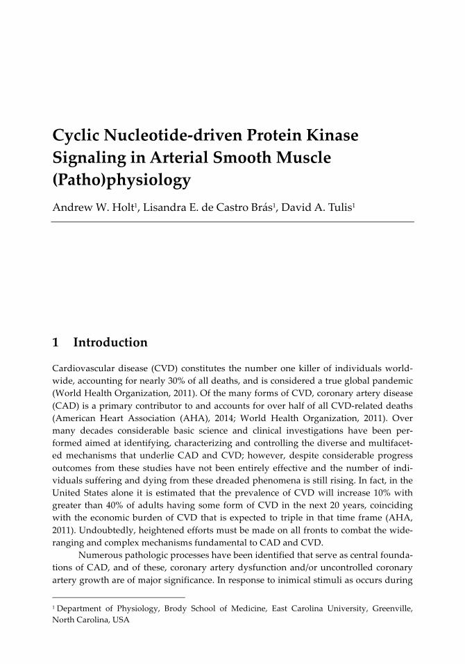
Physiologically, a major function of arteries is their ability to constrict and relax, in turn controlling vascular tone and resistance and blood flow to downstream tissues. Logically this is of critical importance in the provision of vital nutrients and oxygen to essential tissues and removal of metabolic by-products and carbon dioxide (CO2). Blood vessels also operate as routes for the distribution of circulating hormones and other vasoactive factors as well as inflammatory mediators and platelets. Another function of blood vessels is their involvement in growth adaptations following exercise, in wound healing, or after surgical intervention. These normal vessel growth responses can in-volve arteriogenesis or adaptive and constructive vascular remodeling, angiogenesis (formation of new blood vessels from existing vessels), vasculogenesis (de novo for-mation of new blood vessels), arborization or branching of existing vessels, and/or col-lateralization to provide new blood supply to an existing vascular bed. Given the vital importance of these forms of homeostatic vessel growth, context must be considered when comparing normal versus pathologic ASM growth in the setting of CAD or CVD. Enlarged photomicrographs of human confluent coronary ASM cells showing morphol-ogy (top image) as well as G-actin (red) and F-actin (green) staining (bottom image) along with listings of characteristics of normal and pathologic ASM cells are shown in Figure 2.
3 Blood Flow & Hemodynamics
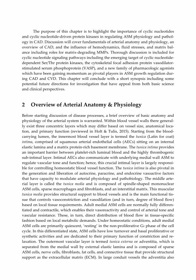
In our circulatory system there are four major types of blood flow: 1) pulsatile, 2) oscil-latory, 3) laminar, and 4) turbulent (Davies et al., 1992; Palumbo et al., 2000). Pulsatile and oscillatory blood flows share common characteristics and result from periodic fluc-tuations in pulse waves corresponding to cardiac sinus rhythm and are influenced by downstream “pulling” forces generated by tissues in demand. The rest of this discus-sion focuses on laminar and turbulent flows as they have capacities to exert significant control over normal vascular dynamics and changes in them can drastically affect vas-cular pathologies. Continuous (unidirectional) laminar blood flow is uninterrupted and occurs at or near the capillary level and is characterized by layered flow in the absence of detectable flow velocity fluctuations or turbulence. Laminar flow is generally consid-ered protective and beneficial for blood vessels and for maintenance of proper vascular tone, dilation, and tissue perfusion. Turbulent blood flow is associated with changes in the layered context of flow and is correlated with increased stresses on the vessel wall. Turbulent blood flow can be caused by branching or arborization of blood vessels (at bifurcations of arteries for example) and/or by intimal lesions or other luminal obstruc-tions which create flow disturbances and turbulence. By its nature, turbulent blood flow is correlated with alterations in downstream perfusion and stress-induced changes in vessel architecture and mural wall remodeling. A schematic of laminar and turbulent blood flow is shown in Figure 3(a). Also shown are images from vascular Doppler ultra-sound, a clinically-used non-interventional approach for estimating blood flow, vascu-lar dimensions and lumen caliber, showing use of color flow systems to detect the na-
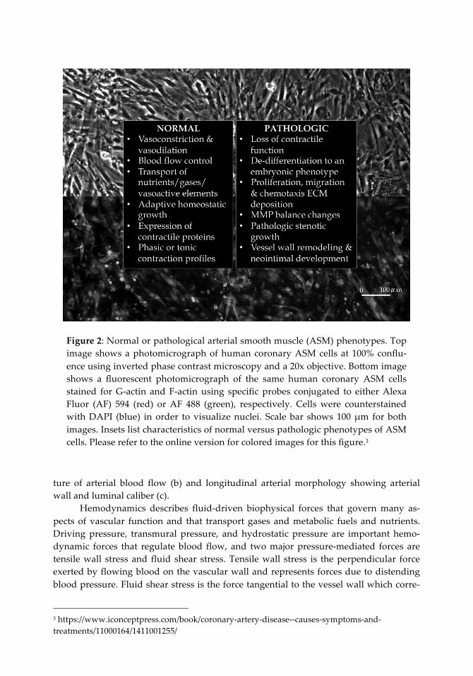
Figure 2: Normal or pathological arterial smooth muscle (ASM) phenotypes. Top image shows a photomicrograph of human coronary ASM cells at 100% conflu-ence using inverted phase contrast microscopy and a 20x objective. Bo:om image shows a fluorescent photomicrograph of the same human coronary ASM cells stained for G-actin and F-actin using specific probes conjugated to either Alexa Fluor (AF) 594 (red) or AF 488 (green), respectively. Cells were counterstained with DAPI (blue) in order to visualize nuclei. Scale bar shows 100 µm for both images. Insets list characteristics of normal versus pathologic phenotypes of ASM cells. Please refer to the online version for colored images for this figure.3
ture of arterial blood flow (b) and longitudinal arterial morphology showing arterial wall and luminal caliber (c).
Hemodynamics describes fluid-driven biophysical forces that govern many as-pects of vascular function and that transport gases and metabolic fuels and nutrients. Driving pressure, transmural pressure, and hydrostatic pressure are important hemo-dynamic forces that regulate blood flow, and two major pressure-mediated forces are tensile wall stress and fluid shear stress. Tensile wall stress is the perpendicular force exerted by flowing blood on the vascular wall and represents forces due to distending blood pressure. Fluid shear stress is the force tangential to the vessel wall which corre-
3 https://www.iconceptpress.com/book/coronary-artery-disease--causes-symptoms-and-treatments/11000164/1411001255/

Figure 3: Hemodynamics: laminar versus turbulent blood flows. (a) Schematic of an artery cut-away showing normal upstream laminar blood flow depicted as parallel lines and turbulent blood flow resulting from a stenotic plaque at an arte-rial bifurcation depicted as multidirectional and mis-directed lines. (b) Using Doppler color flow systems (VisualSonics Vevo 2100) and carotid artery ultra-sound, red color depicts blood flow towards the probe transducer (while blue color depicts blood flow away from the transducer). Through this approach one can detect laminar versus turbulent nature as well as magnitude of arterial blood flows. (c) Image represents longitudinal arterial ultrasound tracing showing ca-rotid artery wall and lumen caliber. Episodic vessel expansion coincident with systolic arterial blood flow bolus is evident. Please refer to the online version for colored images for this figure.4
sponds to the frictional force of the blood in contact with the intimal surface (Davies et al., 1992; White et al., 2007). Changes in blood flow characteristics (i.e., from homeostatic laminar flow to disrupted turbulent flow) and/or alterations in flow-directed biophysi-cal forces directly influence the vessel wall and can contribute markedly to the patho-genesis of CAD and CVD as described below.
4 https://www.iconceptpress.com/book/coronary-artery-disease--causes-symptoms-and-treatments/11000164/1411001255/
(a)
Turbulent flow
Plaque
(b) (c)

4 Fundamentals of CAD
Coronary artery disease (CAD), otherwise known as coronary heart disease, is the most common form of heart disease and is a progressive pathology that affects the coronary circulation and that ultimately leads to partial or total vessel occlusion with compro-mised blood flow and hypoxia or anoxia in vital downstream tissues. This has clear clin-ical significance as it can lead to loss of oxygen and nutrient delivery to essential myo-cardium and accumulation of toxic byproducts of cellular metabolism, such as CO2 and lactic acid. Atherosclerosis (from the Greek words athero (meaning gruel or paste) and sclerosis (hardness)) is a term used to describe the process of fatty substances, cholester-ol, cellular waste products, calcium, fibrin, and other elements building up in the inner lining of an artery and has been determined to be a primary form of CAD. Atherosclero-sis is characterized as a gradual, chronic, and cumulative disorder that involves in-flammation, occlusive growth and remodeling of the vessel wall, and build-up of a ste-notic atheroma or plaque. This multifactorial process is determined by congruent disor-ders of the immune, metabolic, circulatory, and vascular systems and includes dysfunc-tion and/or fenestration of normal intimal endothelium and basement membrane, up-regulation of vascular cell adhesion molecules, binding of low density lipoproteins to intimal proteoglycans, accumulation of lipids, cholesterol, calcium and cellular debris within the intima and sub-intimal space, macrophage and monocyte activation and formation of foam cells, activation and aggregation of localized platelets, and phenotyp-ic modulation and uncontrolled proliferation of resident ASM cells (Crowther, 2005; Falk, 2006). Accordingly, the earliest visible lesion associated with atherosclerosis is a fatty streak of accumulated fat-laden foam cells in the intimal space. This process even-tuates in a vicious and positive feedback cycle of inflammation and pathologic growth complete with luminal obstruction and development of a fibrous plaque, a hallmark of an established lesion. Unless otherwise jeopardized this plaque can remain stable for years, yet when it becomes unstable or compromised (via denudation of overlying en-dothelium and/or rupture) clinical symptoms often appear.
During the pathogenesis of atherosclerosis and other forms of stenotic CAD, ASM cells in the affected coronary circulation display a high degree of plasticity and switch from a normally quiescent and contractile phenotype to a growth-promoting, synthetic phenotype capable of robust proliferation, migration, and matrix production (Tulis, 2008; Gomez, 2012; Holt & Tulis, 2015; Tulis, 2015). During early stages of disease pro-gression this phenotypic conversion occurs in response to locally secreted growth fac-tors, mitogens and/or chemical attractants, circulating factors or hormones, or other vasoactive agents. Ensuing pathogenic processes can include stimulated cellular prolif-eration and DNA multiplication with polyploidy, aberrant cytokinesis with ensuing cellular hypertrophy, directed or ambiguous cellular migration and chemotaxis, altered matrix balance and MMP modulation via enhanced synthesis and secretion, and en-hanced cellular necrosis and apoptosis. Concomitant events during this evolution phase can also include neovascularization of the growing plaque, calcium deposition and plaque mineralization, outward expansion of the affected vessel with compensatory luminal enlargement, and sustained inflammation. Combined, these elements of pheno-
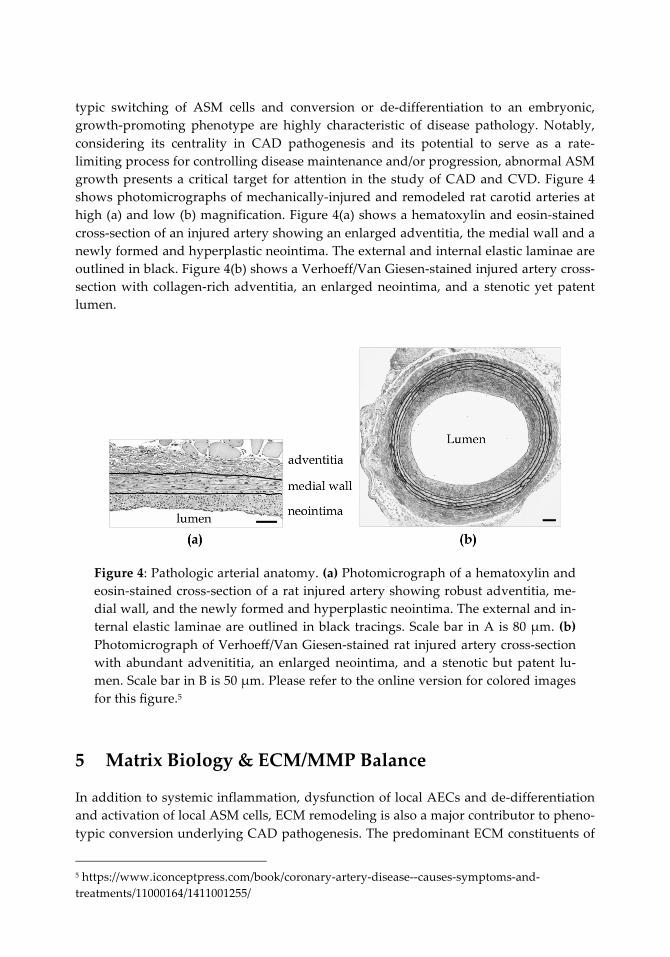
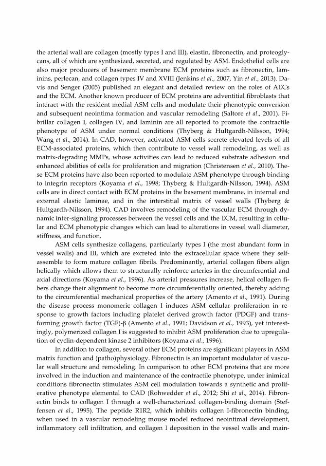
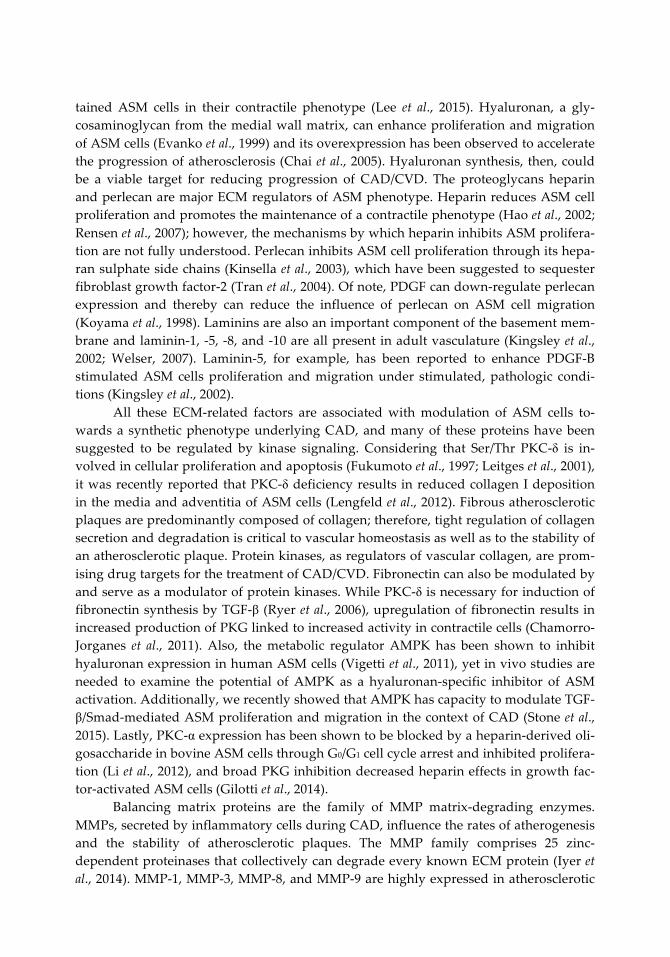
typic switching of ASM cells and conversion or de-differentiation to an embryonic, growth-promoting phenotype are highly characteristic of disease pathology. Notably, considering its centrality in CAD pathogenesis and its potential to serve as a rate-limiting process for controlling disease maintenance and/or progression, abnormal ASM growth presents a critical target for attention in the study of CAD and CVD. Figure 4 shows photomicrographs of mechanically-injured and remodeled rat carotid arteries at high (a) and low (b) magnification. Figure 4(a) shows a hematoxylin and eosin-stained cross-section of an injured artery showing an enlarged adventitia, the medial wall and a newly formed and hyperplastic neointima. The external and internal elastic laminae are outlined in black. Figure 4(b) shows a Verhoeff/Van Giesen-stained injured artery cross-section with collagen-rich adventitia, an enlarged neointima, and a stenotic yet patent lumen.
Figure 4: Pathologic arterial anatomy. (a) Photomicrograph of a hematoxylin and eosin-stained cross-section of a rat injured artery showing robust adventitia, me-dial wall, and the newly formed and hyperplastic neointima. The external and in-ternal elastic laminae are outlined in black tracings. Scale bar in A is 80 µm. (b) Photomicrograph of Verhoeff/Van Giesen-stained rat injured artery cross-section with abundant advenititia, an enlarged neointima, and a stenotic but patent lu-men. Scale bar in B is 50 µm. Please refer to the online version for colored images for this figure.5
5 Matrix Biology & ECM/MMP Balance
In addition to systemic inflammation, dysfunction of local AECs and de-differentiation and activation of local ASM cells, ECM remodeling is also a major contributor to pheno-typic conversion underlying CAD pathogenesis. The predominant ECM constituents of
5 https://www.iconceptpress.com/book/coronary-artery-disease--causes-symptoms-and-treatments/11000164/1411001255/
the arterial wall are collagen (mostly types I and III), elastin, fibronectin, and proteogly-cans, all of which are synthesized, secreted, and regulated by ASM. Endothelial cells are also major producers of basement membrane ECM proteins such as fibronectin, lam-inins, perlecan, and collagen types IV and XVIII (Jenkins et al., 2007, Yin et al., 2013). Da-vis and Senger (2005) published an elegant and detailed review on the roles of AECs and the ECM. Another known producer of ECM proteins are adventitial fibroblasts that interact with the resident medial ASM cells and modulate their phenotypic conversion and subsequent neointima formation and vascular remodeling (Saltore et al., 2001). Fi-brillar collagen I, collagen IV, and laminin are all reported to promote the contractile phenotype of ASM under normal conditions (Thyberg & Hultgardh-Nilsson, 1994; Wang et al., 2014). In CAD, however, activated ASM cells secrete elevated levels of all ECM-associated proteins, which then contribute to vessel wall remodeling, as well as matrix-degrading MMPs, whose activities can lead to reduced substrate adhesion and enhanced abilities of cells for proliferation and migration (Christensen et al., 2010). The-se ECM proteins have also been reported to modulate ASM phenotype through binding to integrin receptors (Koyama et al., 1998; Thyberg & Hultgardh-Nilsson, 1994). ASM cells are in direct contact with ECM proteins in the basement membrane, in internal and external elastic laminae, and in the interstitial matrix of vessel walls (Thyberg & Hultgardh-Nilsson, 1994). CAD involves remodeling of the vascular ECM through dy-namic inter-signaling processes between the vessel cells and the ECM, resulting in cellu-lar and ECM phenotypic changes which can lead to alterations in vessel wall diameter, stiffness, and function.
ASM cells synthesize collagens, particularly types I (the most abundant form in vessel walls) and III, which are excreted into the extracellular space where they self-assemble to form mature collagen fibrils. Predominantly, arterial collagen fibers align helically which allows them to structurally reinforce arteries in the circumferential and axial directions (Koyama et al., 1996). As arterial pressures increase, helical collagen fi-bers change their alignment to become more circumferentially oriented, thereby adding to the circumferential mechanical properties of the artery (Amento et al., 1991). During the disease process monomeric collagen I induces ASM cellular proliferation in re-sponse to growth factors including platelet derived growth factor (PDGF) and trans-forming growth factor (TGF)-β (Amento et al., 1991; Davidson et al., 1993), yet interest-ingly, polymerized collagen I is suggested to inhibit ASM proliferation due to upregula-tion of cyclin-dependent kinase 2 inhibitors (Koyama et al., 1996).
In addition to collagen, several other ECM proteins are significant players in ASM matrix function and (patho)physiology. Fibronectin is an important modulator of vascu-lar wall structure and remodeling. In comparison to other ECM proteins that are more involved in the induction and maintenance of the contractile phenotype, under inimical conditions fibronectin stimulates ASM cell modulation towards a synthetic and prolif-erative phenotype elemental to CAD (Rohwedder et al., 2012; Shi et al., 2014). Fibron-ectin binds to collagen I through a well-characterized collagen-binding domain (Stef-fensen et al., 1995). The peptide R1R2, which inhibits collagen I-fibronectin binding, when used in a vascular remodeling mouse model reduced neointimal development, inflammatory cell infiltration, and collagen I deposition in the vessel walls and main-
tained ASM cells in their contractile phenotype (Lee et al., 2015). Hyaluronan, a gly-cosaminoglycan from the medial wall matrix, can enhance proliferation and migration of ASM cells (Evanko et al., 1999) and its overexpression has been observed to accelerate the progression of atherosclerosis (Chai et al., 2005). Hyaluronan synthesis, then, could be a viable target for reducing progression of CAD/CVD. The proteoglycans heparin and perlecan are major ECM regulators of ASM phenotype. Heparin reduces ASM cell proliferation and promotes the maintenance of a contractile phenotype (Hao et al., 2002; Rensen et al., 2007); however, the mechanisms by which heparin inhibits ASM prolifera-tion are not fully understood. Perlecan inhibits ASM cell proliferation through its hepa-ran sulphate side chains (Kinsella et al., 2003), which have been suggested to sequester fibroblast growth factor-2 (Tran et al., 2004). Of note, PDGF can down-regulate perlecan expression and thereby can reduce the influence of perlecan on ASM cell migration (Koyama et al., 1998). Laminins are also an important component of the basement mem-brane and laminin-1, -5, -8, and -10 are all present in adult vasculature (Kingsley et al., 2002; Welser, 2007). Laminin-5, for example, has been reported to enhance PDGF-B stimulated ASM cells proliferation and migration under stimulated, pathologic condi-tions (Kingsley et al., 2002).
All these ECM-related factors are associated with modulation of ASM cells to-wards a synthetic phenotype underlying CAD, and many of these proteins have been suggested to be regulated by kinase signaling. Considering that Ser/Thr PKC-δ is in-volved in cellular proliferation and apoptosis (Fukumoto et al., 1997; Leitges et al., 2001), it was recently reported that PKC-δ deficiency results in reduced collagen I deposition in the media and adventitia of ASM cells (Lengfeld et al., 2012). Fibrous atherosclerotic plaques are predominantly composed of collagen; therefore, tight regulation of collagen secretion and degradation is critical to vascular homeostasis as well as to the stability of an atherosclerotic plaque. Protein kinases, as regulators of vascular collagen, are prom-ising drug targets for the treatment of CAD/CVD. Fibronectin can also be modulated by and serve as a modulator of protein kinases. While PKC-δ is necessary for induction of fibronectin synthesis by TGF-β (Ryer et al., 2006), upregulation of fibronectin results in increased production of PKG linked to increased activity in contractile cells (Chamorro-Jorganes et al., 2011). Also, the metabolic regulator AMPK has been shown to inhibit hyaluronan expression in human ASM cells (Vigetti et al., 2011), yet in vivo studies are needed to examine the potential of AMPK as a hyaluronan-specific inhibitor of ASM activation. Additionally, we recently showed that AMPK has capacity to modulate TGF-β/Smad-mediated ASM proliferation and migration in the context of CAD (Stone et al., 2015). Lastly, PKC-α expression has been shown to be blocked by a heparin-derived oli-gosaccharide in bovine ASM cells through G0/G1 cell cycle arrest and inhibited prolifera-tion (Li et al., 2012), and broad PKG inhibition decreased heparin effects in growth fac-tor-activated ASM cells (Gilotti et al., 2014).
Balancing matrix proteins are the family of MMP matrix-degrading enzymes. MMPs, secreted by inflammatory cells during CAD, influence the rates of atherogenesis and the stability of atherosclerotic plaques. The MMP family comprises 25 zinc-dependent proteinases that collectively can degrade every known ECM protein (Iyer et al., 2014). MMP-1, MMP-3, MMP-8, and MMP-9 are highly expressed in atherosclerotic
plaques and serum levels of MMP-1 and MMP-9 directly correlate with CAD severity (Newby, 2005; Tanindi et al., 2011). Even though MMPs are mostly associated with rup-ture of arterial plaques by degradation of the plaque surface ECM, MMP-2, MMP-9, and MMP-14 stimulate migration and proliferation of ASM cells from the tunica media into the intima by degrading the ECM elements of the basement membrane (Dollery & Libby, 2006; Newby, 2006). This could potentially lead to increased plaque stability via en-hanced cellular composition and thickening of the fibrous plaque. Huang and col-leagues showed that PKC-βII activity induces neointimal expansion partially through increased MMP-9 expression (Huang et al., 2010). Similarly, PKC-ε has been associated with MMP-2- and MMP-9-mediated ASM cell migration (Ding et al., 2011). PKG sup-presses MMP-2 expression and secretion in vitro (Dey & Lincoln, 2012), complementing some of our data showing that cyclic GMP/PKG signaling reduces expression and activ-ity of both MMP-2 and MMP-9 in growth retardation of ASM in cultured cells and in-tact arteries (Tulis, 2008). Indeed, these findings strongly suggest mechanistic involve-ment of ECM proteins and ECM/MMP balance and their control by cyclic nucleotide-driven protein kinases in phenotypic modulation of ASM during CAD. Figure 5 (next page) shows a schematic depicting interaction between ECM proteins in an ASM cell and the resulting functional outcomes in CAD. Included are key intracellular and extra-cellular events and important signaling factors and processes that can serve critical roles in matrix balance. These include TGF-β and PDGF (and their respective receptors), im-portant kinases such as PKC and PKG, structural matrix elements such as fibronectin, collagens, and laminin, and matrix-degrading MMPs. The figure also shows resulting functional outcomes in ASM phenotype (i.e., synthesis, proliferation, migration, con-tractility) foundational to structural changes and remodeling in the pathogenesis of CAD and CVD.
6 Cyclic Nucleotide-Driven Protein Kinase Signaling
Among the numerous molecular and cellular signaling pathways that serve to elicit con-trol over cardiovascular functions, cyclic nucleotide signaling is of critical importance. Comprised primarily of the highly characterized canonical cyclic purine nucleotides cyclic AMP and cyclic GMP, this family of second messengers operates predominantly through downstream protein kinase signaling pathways to exert control over a wide variety of cellular processes. Cyclic AMP and cyclic GMP are firmly established as criti-cal biological messengers in many mammalian tissues including ASM and this discus-sion focuses on their involvement during CAD.
It warrants mention that other cyclic purine nucleotides exist in the lesser known inosine 3´,5´-cyclic monophosphate (cyclic IMP) and xanthosine 3´,5´-cyclic monophos-phate (cyclic XMP), and in fact these have been recently theorized as potentially serving second messenger roles (Beste & Seifert, 2013; Chen et al., 2014); however, their in-volvement in ASM (patho)physiology and CAD and/or CVD has not yet been examined. An alternate family of cyclic pyrimidine nucleotides also exists and this includes cyti-dine 3´,5´-cyclic monophosphate (cyclic CMP), uridine 3´,5´-cyclic monophosphate (cy-
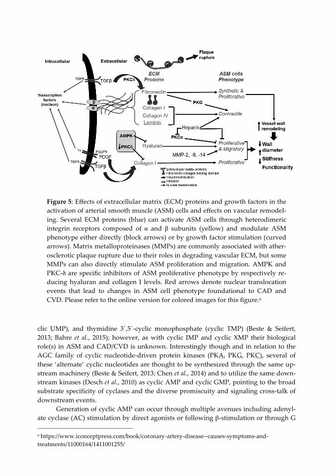
Figure 5: Effects of extracellular matrix (ECM) proteins and growth factors in the activation of arterial smooth muscle (ASM) cells and effects on vascular remodel-ing. Several ECM proteins (blue) can activate ASM cells through heterodimeric integrin receptors composed of α and β subunits (yellow) and modulate ASM phenotype either directly (block arrows) or by growth factor stimulation (curved arrows). Matrix metalloproteinases (MMPs) are commonly associated with ather-osclerotic plaque rupture due to their roles in degrading vascular ECM, but some MMPs can also directly stimulate ASM proliferation and migration. AMPK and PKC-δ are specific inhibitors of ASM proliferative phenotype by respectively re-ducing hyaluran and collagen I levels. Red arrows denote nuclear translocation events that lead to changes in ASM cell phenotype foundational to CAD and CVD. Please refer to the online version for colored images for this figure.6
clic UMP), and thymidine 3´,5´-cyclic monophosphate (cyclic TMP) (Beste & Seifert, 2013; Bahre et al., 2015); however, as with cyclic IMP and cyclic XMP their biological role(s) in ASM and CAD/CVD is unknown. Interestingly though and in relation to the AGC family of cyclic nucleotide-driven protein kinases (PKA, PKG, PKC), several of these ‘alternate’ cyclic nucleotides are thought to be synthesized through the same up-stream machinery (Beste & Seifert, 2013; Chen et al., 2014) and to utilize the same down-stream kinases (Desch et al., 2010) as cyclic AMP and cyclic GMP, pointing to the broad substrate specificity of cyclases and the diverse promiscuity and signaling cross-talk of downstream events.
Generation of cyclic AMP can occur through multiple avenues including adenyl-ate cyclase (AC) stimulation by direct agonists or following β-stimulation or through G 6 https://www.iconceptpress.com/book/coronary-artery-disease--causes-symptoms-and-treatments/11000164/1411001255/

protein-coupled receptor activation. Following AC stimulation, adenosine triphosphate (ATP) dephosphorylates to produce cyclic AMP and pyrophosphate (PPi). In similar fashion, following activation of guanylate cyclase (GC) through natriuretic peptides (which activate particulate GC) or by gaseous ligands (which activate soluble GC), gua-nosine triphosphate (GTP) is dephosphorylated to yield cyclic GMP and PPi. Detailed biomolecular mechanisms of cyclase-mediated cyclic AMP and cyclic GMP formation have been described (Tulis, 2008; Tulis, 2015). The preferred effector kinases, then, of cyclic AMP and cyclic GMP are the AGC kinases PKA and PKG, respectively (Arencibia et al., 2013). In addition to these canonical kinase pathways these cyclic nucleotides can also proceed through alternate kinase-directed pathways (Adderley et al., 2012a), direct ion channel modulation, or become degraded by specific members of the phos-phodiesterase (PDE) family (Adderley et al., 2012b). In this light, conversion of cyclic AMP or GMP into inactive 5´-AMP or 5´-GMP is accomplished through specific PDEs that cleave the phosphodiester bonds of cAMP (by PDE-4, -7, -8) or cGMP (by PDE-5, -6, -9) to yield 5´-AMP or 5´-GMP. In turn, targeted PDE inhibition is capable of indirectly maintaining elevated levels of these cyclic nucleotides and their downstream kinases. In the mid to late 1980s this rationale was investigated as an approach to maintain kinase signaling for its potential treatment of CAD, but ironically this led to discovery of the PDE-5 inhibitor Sildenafil (Viagra), the most widely-prescribed oral agent for the treat-ment of erectile dysfunction (Briganti et al. 2005; Reffelmann et al. 2003). Our research team and others have provided evidence of promiscuity among many aspects of the cyclic AMP and cyclic GMP systems including ‘cross-talk’ between the activating cyclases, interactions between cyclic AMP/PKG, cyclic GMP/PKA and PKC, and non-selective PDE-directed kinase inactivation (discussed in detail below). Nonetheless, the-se intricate signaling cascades elicit a multitude of significant biological effects in ASM and are of critical importance in vascular physiology and pathology related to CAD and CVD. Figure 6 shows a schematic of cyclic GMP synthesis including up stream NO and CO cascades and downstream kinase-specific targets including cytoskeletal VASP fun-damental to ASM (patho)physiology.
Protein kinases in general serve a wide variety of roles in a multitude of physio-logical and pathophysiological processes and represent one of the most ubiquitous, and functionally diverse families in the human genome constituting ~2% of all human genes with over 500 human protein kinases identified to date (Adderley et al., 2012a; Manning et al., 2002). Numerous kinase mutations have been identified in human diseases through genotype-phenotype analyses (Lahiry et al., 2010) and kinases have been theo-rized as instrumental therapeutic targets against CVD (Kompa & Krum, 2014; Wang et al., 2012). In fact, protein kinases already represent ~20% of all putative drug targets (Lahiry et al., 2010) and are likely the major pharmaceutical drug target of the 21st centu-ry (Cohen, 2002). More recently Dubey and colleagues (2015) showed 2-chloroadenosine (a stable adenosine analogue) increased cAMP levels and attenuated human coronary ASM cell proliferation which was reversed with PKA blockade. These recent findings highlight the relevance and timeliness of cyclic nucleotide dependent kinases currently being investigated as related to vascular growth disorders such as CAD.

Figure 6: Signaling diagram for cyclic GMP synthesis and downstream signaling. Following upstream activation by a family of nitric oxide synthase (NOS) and/or heme oxygenase (HO) enzymes, L-arginine and heme, respectively, are metabo-lized to L-citrulline (with production of nitric oxide (NO)) and carbon monoxide (CO). These diatomic gases then activate soluble guanylate cyclase (sGC) which serves to dephosphorylate guanosine triphosphate (GTP) to yield cyclic GMP and pyrophosphate (PPi). Cyclic GMP is either degraded by a family of phos-phodiesterases (PDE) or exerts downstream phosphorylative actions upon dis-tinct kinases, primarily PKG, PKA and AMPK, as well as other kinases and non-kinase targets. The kinases can then act to regulate VASP and associated cyto-skeletal/focal adhesion proteins which, in turn, helps to control aspects of arterial smooth muscle (ASM) growth and function as basic elements of CAD and/or CVD.
Several sub-families of kinases exist with a majority acting to phosphorylate ei-ther the -OH group of serine (Ser) and/or threonine (Thr) residues (the Ser/Thr kinases), which constitute about 80% of the total protein kinases (Manning et al., 2002), or tyro-sine (Tyr) residues (the Tyr kinases). For example, in regard to the Ser/Thr kinases, fol-lowing binding of two cyclic AMP molecules to the regulatory subunit (dimer) of PKA a conformational change of this tetrameric enzyme occurs which causes release of its two catalytic subunits (Terrin et al. 2012). Active PKA goes on to phosphorylate proteins that have the motif Arginine-Arginine-X-Ser exposed, thereby phosphorylating and activat-ing those targets. Like PKA, PKG as well as PKC/PKD and AMP kinase are all estab-
L-arginine
L-citrulline
NOS HO
bilirubin biliverdin BvR
Fe2+
NO
GTP cyclic GMP
5'-GMP PDE
PK-G
kinase-independent effects sGC
PK-A
AMPK, PKC/PKD, other kinases
VASP, cytoskeletal & focal adhesion proteins
PPi
control of ASM growth & function
CAD / CVD
Heme
CO
lished Ser/Thr kinases that become activated through upstream substrate binding and phosphorylate a multitude of downstream targets. Additionally, some broad kinases act on all three amino acids (termed dual-specificity kinases), while lesser-known kinases can phosphorylate unique residues such as histidine (the His kinases) (Besant et al., 2003).
PKA and PKG are select members among the more than 60 AGC kinases in the human genome and constitute the primary kinases acted upon by upstream cyclic AMP and cyclic GMP, respectively (Adderley, et al., 2012a; Arencibia et al., 2013; Pearce et al., 2010). Through the act of site-specific (Ser/Thr or Tyr) and reversible phosphorylation of target proteins via phosphotransferase activity, these molecules exert potent signal transduction mechanisms which have capacity to control countless intracellular pro-cesses including many in ASM. Despite the ubiquitous nature and diversity of kinases in humans they share a common basic structure and mechanisms of action. Thorough comprehensive reviews have been published regarding cellular, biochemical and mo-lecular mechanisms of protein kinases (Francis & Corbin, 1994; Adams, 2001; Ubersax & Ferrell, 2007; reviewed in Khalil, 2010), and herein only a succinct synopsis of key events is provided. Broadly speaking, protein kinases including members of the AGC family consist of a conserved catalytic domain of approximately 250 amino acids in length made up of one lobe of β-sheets in an N-terminus and a second lobe of α-helices in a C-terminus (Knighton et al., 1991). Once ATP binds to a cleft between these two lobes (this constitutes the active site), a set of conserved residues within the catalytic domain transfers the terminal γ-phosphate of ATP to the hydroxyl oxygen of the receiv-ing residue (Ser/Thr, Tyr) on the target (Francis & Corbin, 1994; Ubersax & Ferrell, 2007). This is followed by substrate release and removal of ADP from this active site and phosphorylation-driven activation or inactivation of the downstream target. Despite this common mechanism, kinase specificity is imparted by differences in hydrophobi-city of surface residues, the overall charge of the enzyme, characteristics of the active site including the nature and sequence of ATP/substrate binding, rate-limiting steps, presence or absence of adapter or scaffolding proteins, and sub-cellular localization of the kinase.
Modification of target proteins via post-translational phosphorylation by any particular kinase can then dictate downstream enzyme and target protein expression and/or activities and downstream responses including those fundamental to aberrant cell proliferation and migration and matrix balance (i.e., phenotypic switching) that oc-curs in CAD pathogenesis. This is highly context-specific and can involve control of cell cycle progression by cyclins, cyclin-dependent kinases (CDKs), and/or CDK inhibitors, control of cell migration by cytoskeletal and focal adhesion elements including gap junctional connexins and VASP, and alterations in matrix balance including matrix pro-teins and/or degrading MMPs (Tulis, 2008; Mendelev et al., 2009; Joshi et al., 2011; Ad-derley et al., 2012a; Adderley et al., 2012b; Joshi et al., 2012; Stone et al., 2012; Stone et al., 2013; Adderley et al., 2015; Holt & Tulis, 2015; Joshi & Tulis, 2015; Stone et al., 2015; Tu-lis, 2015). Additionally, kinase actions may be altered through modulation of de-phosphorylating protein phosphatases (PPs) which have recently been theorized to serve regulatory roles in abnormal ASM growth (Stone et al., 2012; Stone et al., 2013).

Indeed, the downstream actions of kinase-driven phosphorylation events are wide-ranging and diverse and often paradoxical, resulting in target activation or inactivation in predominantly context-specific manner (Adderley et al., 2012a; Ubersax & Ferrell, 2007).
Given the centrality of cyclic nucleotide and cyclic nucleotide-driven protein ki-nase signaling in ASM physiology and pathology, brief discussion is warranted for their roles in mediating the critical functions of vascular contraction and relaxation. As dis-cussed above, during the pathogenesis of CAD and CVD, ASM undergoes a loss of its contractile capabilities and undergoes a phenotypic reversal to an embryonic, growth-promoting, and synthetic form. Thus, a common thread of cyclic nucleotide/kinase ac-tion in ASM phenotypic modulation is the ability of these agents to elegantly regulate smooth muscle tone. Although detailed mechanisms have been previously described (reviewed in Gao et al., 2001; Webb, 2003; Khalil, 2010), generally speaking in ASM un-der normal conditions, following agonist stimulation intracellular calcium ([Ca2+i]) rises and binds calmodulin (CaM), which in turn activates the Ser/Thr myosin light chain kinase (MLCK). Activated MLCK then phosphorylates Ser19 of the 20 kD regulatory myosin light chain (MLC), which activates myosin ATPase activity and initiates actin-myosin crossbridge formation and subsequent crossbridge cycling central to ASM con-traction (Gao et al., 2001). Calcium-independent smooth muscle contraction can also oc-cur following agonist stimulation of inositol triphosphate (IP3), which in turn activates RhoA (RhoA-GTP) which then binds to and activates ROCK, leading to phosphoryla-tion and inhibition of MLC phosphatase and reduced capacity to dephosphorylate MLC (Surks, 2007). In general, inactivation of crossbridge cycling and cessation of smooth muscle contraction occurs via dephosphorylation of the MLC through actions of MLC phosphatase.
Regarding cyclic nucleotide-directed kinase control of ASM contraction, PKG stimulation promotes vascular relaxation through several mechanisms (Surks, 2007). PKG directly inhibits the mobilization of [Ca2+i], thereby preventing calcium-CaM-mediated activation of MLCK (Cornwell & Lincoln, 1989). PKG can open calcium-activated potassium channels, thereby leading to cell hyperpolarization and relaxation (Archer et al., 1994). PKG can also operate via a calcium-independent mechanism by activating MLC phosphatase and dephosphorylating MLC (Lee et al., 1997). Cyclic AMP-driven PKA shares these avenues for controlling ASM contraction through reduc-tion in [Ca2+i], direct and indirect (via enhanced MLC phosphatase) modes for inhibiting MLC phosphorylation, and stimulation of calcium-activated potassium channels and resulting hyperpolarization. Moreover, both PKA and PKG have been found to promote vasorelaxation via phosphorylation of Ser16 on the small heat shock-related protein (Hsp) 20, which appears to alter actin-myosin relations, crossbridge formation, and ac-tin-focal adhesion dynamics necessary for contraction (Somara et al., 2010; Woodrum et al., 2003). Lastly, considering kinase promiscuity among AGC kinase family members (Adderley et al., 2012a), brief discussion is warranted for PKC in vascular contraction. In smooth muscle PKC exists in the calcium-dependent forms α and β and in the calcium-independent forms ε and ζ, and both of these groups of isoforms have been implicated in PKC-mediated vascular contraction (Andrea & Walsh, 1992; Khalil, 2010). PKC can

operate to induce contraction by increasing myofilament force sensitivity to [Ca2+]i, through calcium/CaM-mediated activation of MLCK and induction of MLC phosphorylation, and by establishing actin-myosin interactions (Rasmussen et al., 1987; Khalil, 2010). PKC has also been observed to induce smooth muscle contraction in calcium-independent fashion by phosphorylating the actin-associated filament calponin, thereby reducing its affinity for F-actin and alleviating its inhibition of crossbridge cycling (Horowitz et al., 1996; Walsh et al., 1996).
Serving as an ‘off switch’ to balance kinase-driven phosphorylation is a family of enzymatic PPs. Removal of a phosphate group from kinase-targeted proteins by PPs serves to moderate cell signaling and helps to regulate many cellular processes involved in differentiation, proliferation, migration, apoptosis, and embryonic development. Sim-ilar to amino acid specificity of the kinases, there exist specific Ser/Thr PPs and specific Tyr PPs that balance the actions of Ser/Thr and Tyr kinases, respectively. Following ac-tivation of the kinases via upstream signals, they target downstream substrate proteins in a cell- and tissue-specific fashion. We recently documented ability of both global PPs and Ser/Thr PPs in conjunction with activated upstream PKA, PKG and/or AMP kinase signals to elicit control over deleterious ASM proliferation and migration (Stone et al., 2012; Stone et al., 2013).
Recent discoveries using pharmacology, over-expressing and deficient transgenic models, and site-specific modulation of targeted residues have revealed signaling prom-iscuity and lack of precision for both upstream modes of kinase activation and for downstream phosphorylated targets (Worner et al., 2007; Tulis, 2008; Mendelev et al., 2009; Desch et al., 2010; Joshi et al., 2011; Adderley et al., 2012a; Stone et al., 2012; Beste & Seifert, 2013; Chen et al., 2014; Tulis, 2015). While Ser/Thr and Tyr kinases generally act on their preferred substrates, these enzymes are also attracted to residues that flank both sides of the phosphoacceptor site (the Ser, Thr and/or Tyr); thus, the catalytic cleft of the kinase interacts not only with its preferred Ser, Thr or Tyr phosphoacceptor but also with their flanking regions, thereby binding to common recognition sequences among similar substrate family members and in turn reducing kinase specificity. This kinase crosstalk or promiscuity affords broad impact of upstream kinase signals but at the same time lends difficulty in ascertaining precise downstream signaling mecha-nisms and targets. Among the numerous bioactive targets of the AGC kinases PKA, PKG, and PKC that also serve as a target for their promiscuous signaling is the focal adhesion protein VASP.
7 VASP
Cellular migration or chemotaxis in response to pathologic cues or tissue damage relies heavily on reorganization of the actin cytoskeleton that is mediated by an array of focal adhesion adapter proteins. One of these proteins that is essential for this reorganization to occur and that predominantly acts as a substrate for many cyclic nucleotide-driven kinases is vasodilator-stimulated serum phosphoprotein or VASP. VASP, a member of the Ena/VASP Homology (EVH) family of closely-related proteins, serves critical func-

tions in cytoskeletal stability and dynamics that are involved in intracellular signaling pathways regulating integrin-ECM interactions. VASP is comprised of an N-terminal EVH1 domain (used to target focal adhesion and membrane domains), a mid-region that binds to Src-homology 3 (SH3) domains and tryptophan-rich WW domain-containing proteins that aid in Ser/Thr binding, and a C-terminal EVH2 domain that mediates tetramerization (a right-handed coiled coil with 15 residue repeats) and actin/focal adhesion binding. Originally characterized as a substrate for cyclic nucleotide-directed phosphorylation signals (Reinhard et al., 2001; Krause et al., 2002) with PKA acting preferentially on VASPSer157 and PKG acting primarily on VASPSer239 (Chen et al., 2004; Worner et al. 2007), to date at least four distinct Ser/Thr phosphorylation sites have been identified on VASP: Ser157, Ser239, Thr278, Ser322 (Butt et al., 1994; Chitaley et al., 2004; Thomson et al., 2011). Interestingly, more recent studies have observed crosstalk and lack of specificity of these and other Ser/Thr kinases to phosphorylate discrete residues on VASP. Notably, recent findings from our laboratory in commercial (Mendelev et al., 2009) and primary (Joshi et al., 2011; Adderley et al., 2012a; Adderley et al., 2012b; Adderley et al., 2015; Tulis, 2008; Tulis, 2015) ASM cells documents ability of cyclic AMP and cyclic GMP to not only communicate with their respective canonical PKA and PKG targets but to also target other kinases as well. We have observed that cyclic AMP, stimulated both directly and indirectly, induces PKG in addition to PKA and phosphorylates both the reported PKG target VASPSer239 and the accepted PKA target VASPSer157. Likewise, we have stimulated cyclic GMP, directly and indirectly, and have found stimulation of PKG and PKA as well as both VASPSer239 and VASPSer157. We have also observed both cyclic AMP and cyclic GMP have capacity to stimulate members of the diverse PKC/PKD family (Adderley et al., 2012a). Considering other kinases, we recently reported that the Ser/Thr metabolic gauge AMP kinase, tradi-tionally thought to act uniquely on its regulatory site VASPThr278 (Blume et al., 2007), also has capacity to phosphorylate VASPSer157 yet does not significantly affect VASPSer239 (Stone et al., 2012; Stone et al., 2013). Lastly, it was reported that PKC-driven PKD, an-other Ser/Thr kinase involved in extracellular receptor-mediated signal transduction, phosphorylates both its reported VASPSer322 as well as VASPSer157 (Doppler et al., 2013). A schematic of the primary structure of VASP with essential EVH1 and EVH2 domains, the SH3- and WW-domain binding region, and preferred (but not exclusive) sites of action for select Ser/Thr protein kinases (adapted from Madej et al., 2014) is shown in Figure 7.
Figure 7: Linear primary structure for VASP with EVH1 and EVH2 domains and preferred Ser/Thr sites for select kinase action.

Functionally, VASP operates as an anti-capping protein and promoter of actin polymerization by delivering monomeric globular (G) actin to the barbed end of grow-ing filamentous (F) actin (Barzik et al., 2005; Bear et al., 2002; Breitsprecher et al., 2008). Therefore, when VASP is inhibited (or phosphorylated), it can no longer drive F-actin formation and actin-mediated cell motility. Instead, phosphorylated (inactivated) VASP promotes cytostasis by keeping the G-actin pool elevated (Stone et al., 2012). If total ac-tin protein expression (β-actin) remains unchanged, then quantifying the G:F actin ratio as an estimate of the balance of depolymerized:polymerized actin serves to describe the migratory status of adherent cells since the cellular actin is either depolymerizing or polymerizing in the context of cellular movement or migration. Actin polymerization and leading edge formation are essential for directional cellular movement and migra-tion or chemotaxis, and VASP operates in support of these cytoskeletal rearrangements necessary for cell movement. Site-specific phosphorylation of VASP (at one or more of its aforementioned Ser/Thr residues) acts to inhibit its anti-capping potential and in turn serves to reduce actin polymerization and inhibit or prevent migration (Blume et al., 2007). VASP also acts as a regulator of platelet function and adhesion as well as in cyto-skeletal dynamics and processes such as cell adhesion and proliferation (Adderley et al., 2012a; Adderley et al., 2015; Cheng et al., 2014; Henes et al., 2009; Kwiatkowski et al., 2003). VASP is also suggested to have anti-tumorigenic properties (Doppler et al., 2013) and anti-inflammatory actions in diabetes (Cheng et al., 2014). In the context of CAD and CVD, we have recently examined VASP and its phosphorylated forms and their abilities to control migration of commercial, rat primary, and human coronary ASM cells. Cumulative findings show that the reported PKA target VASPSer157 and the AMPK target VASPThr278 generally act to reduce cellular proliferation while the PKG site VASPSer239 as well as VASPThr278 work to inhibit migration (Adderley et al., 2012a; Mende-lev et al., 2008; Joshi et al., 2013; Stone et al., 2012; Stone et al., 2013). In complement, other investigators reported recently that the PKC/PKD residue VASPSer322 also controls cellu-lar migration (Doppler et al., 2013). In whole, results show strong support for differen-tial VASP species in serving crucial functions to control pathologic proliferation and migration in the context of CAD. Certainly, VASP as a target of cyclic nucleotide-directed kinase signals holds great promise regarding its ability to control deleterious ASM growth that underlies significant pathologies such as CAD and CVD.
8 Pharmacologic Cyclic GMP Agonists
Despite clear significance and utility, there have been numerous concerns raised for both nitric oxide (NO)- and carbon monoxide (CO)-mediated cyclic GMP signals includ-ing reaction with molecular oxygen, thiols and transition metal ions, development of reactive species, modification of lipids, proteins and nucleic acids, and reduced efficacy via tolerance and/or tachyphylaxis following prolonged clinical treatment, and limited or restricted bioactivity (Davis et al., 2001; Andrews et al., 2002; Gori & Parker, 2002). These drawbacks, in turn, have warranted discovery of alternate routes for activating Ser/Thr kinases and their associated targets including VASP (all downstream of
NO/CO) in efforts to identify novel and potentially beneficial therapies devoid of such drawbacks. In this light, two families of synthetic sGC/cyclic GMP agonists have been developed that lack known limitations of traditional NO/CO signaling and that have capacity to serve as alternate routes for activating cyclic nucleotide signals in ASM (re-viewed in Jackson et al., 2007; Tulis, 2008; Tulis, 2015). Heme-dependent sGC stimula-tors depend on a functional (reduced) cyclase heme in order to enhance enzyme activity and cyclic GMP synthesis. Thus, under settings where the cyclase heme is removed or rendered dysfunctional these agents lose their activity and become inactive or less ac-tive. Heme-independent sGC activators, by comparison, maintain functionality under heme-deficient or -oxidized conditions and therefore are considered more therapeutical-ly advantageous for diseased or injured tissues as occurs in CAD/CVD. Table 1 shows examples of some known stimulators and activators of sGC/cyclic GMP.
sGC / cyclic GMP stimulators sGC / cyclic GMP activators A-350619 BAY 58-2667
BAY 41-2272 BAY 60-2770 BAY 41-8543 HMR-1766 BAY 51-9491 S-3448 BAY 60-4552
BAY 63-2521
YC-1
Table 1: Examples of some known stimulators and activators of sGC and cyclic GMP.
Much work from our lab over the past few years has focused on the abilities of these stimulators and activators to elicit control over ASM growth in the context of CAD/CVD using a variety of experimental platforms. YC-1 [3-(5'-hydroxymethyl-2'-furyl)-1-benzyl indazole], one of the originally characterized sGC stimulators (Ko et al., 1994; Wu et al., 1995), has been documented to elicit anti-platelet, anti-proliferative, anti-synthetic and pro-apoptotic growth-mitigating properties in ASM using in vitro, ex vivo and in vivo experimental models (Tulis et al., 2000; Tulis et al., 2002; Tulis, 2004; Keswani et al., 2009; Liu et al., 2009). More recently a next generation sGC stimulator and YC-1 mimetic BAY 41-2272 [(5-cyclo-propyl-2-[1-(2-fluorobenzyl)-1H-pyrazolo[3,4-b]pyridine- 3-yl]-pyrimidin-4-ylamine)] (Becker et al., 2001; Stasch et al., 2001) was shown to induce PKG and PKA signaling and downstream VASP phosphorylation and to control ASM growth including regulation of matrix/MMP balance (Mendelev et al., 2009; Joshi et al., 2011; Adderley et al., 2012a; Adderley et al., 2012b). Interestingly, adding to the afore-mentioned promiscuity of cyclic nucleotide-driven kinase signaling, pharmacologic blockade studies reveal that BAY 41-2272 possess divergent downstream actions on cel-lular proliferation versus cellular migration (Joshi et al., 2011; Adderley et al., 2012a).

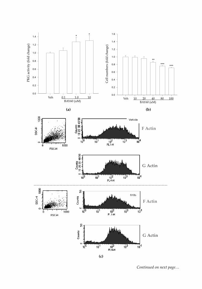
Lastly, using the heme-independent sGC activator BAY 60-2770 [4-({(4-carboxybutyl)[2-(5-fluoro-2-{[4'-(trifluoromethyl)biphenyl-4-yl]methoxy}phenyl)ethyl] amino}methyl) benzoic acid] (BAY60; a kind gift from Bayer HealthCare, Germany) in rat primary ASM cells, rat commercial (A7r5) ASM cells, and human coronary ASM cells we have ob-served it to act primarily on cyclic GMP and PKG (with minimal influence on cyclic AMP/PKA), to phosphorylate both VASPSer239 and VASPSer157, to elicit control of ASM via cytoskeletal rearrangement quantified by G:F actin, and to reduce cellular proliferation and migration as well as injury-induced neointimal formation and vessel wall remodel-ing. Figure 8 (next page) shows some of these observations in human coronary ASM cells: (a) demonstrates that BAY60 (0.1-10 µM) significantly increases PKG activity com-pared to vehicle controls after 60 minutes using an ELISA-based activity assay (Cyclex); (b) shows that BAY60 dose-dependently reduces cell proliferation after 24 hours com-pared to vehicle (Veh) controls; (c, d) show preliminary results suggesting that BAY60 (10 µM) increases G:F actin ratio compared to Veh controls after 60 minutes, indicative of a more stable cytoskeletal phenotype and reduced capacity for proliferation and mi-gration (Stone et al., 2013). Indeed, these early findings using the heme-independent sGC activator BAY60 in human coronary ASM cells (and rat primary tissues) support a role for cyclic nucleotide-directed kinases in controlling aberrant ASM growth as an underpinning of CAD.
9 Summary & Future Directions
Coronary artery disease and CVD remain the number one cause of morbidity and mor-tality in the United States and worldwide and is associated with staggering economic costs as well. Notwithstanding major advances in our knowledge of the underlying mechanisms of these disorders and significant progress in our strategies aimed at their control, all estimates suggest an increasing trend in their prevalence over the next sev-eral decades. In our never-ending struggle to understand key elements behind normal vascular biology as well as regulatory mechanisms that underlie vascular pathology, we must continue to make pivotal inroads into potential routes for controlling and/or elim-inating these dreaded disorders. It is generally thought that many, if not all, of the caus-es behind CAD/CVD are preventable and that future endeavors should focus on pre-vention strategies and early diagnoses and intervention to combat these diseases (AHA), 2011). In cumulative efforts to identify and characterize key elements behind CAD and CVD, we and others have focused our basic and clinical efforts at gaining more thorough understanding of the vital roles for ASM and the multifunctional cyclic nucleotide and cyclic nucleotide-directed kinase signals discussed herein. In our mind these represent highly promising yet incompletely understood targets capable of con-trolling pathologic vascular growth that accompanies vascular disease. Only through determined basic and clinical investigation can we hope to better understand crucial aspects of vascular biology and pathology and in that, gain insights into our seemingly unrelenting struggle against CAD and CVD.
(a) (b)
(c)
Continued on next page…
0.0
0.2
0.4
0.6
0.8
1.0
1.2
1.4
PKG
act
ivity
(fol
d ch
ange
)
Veh 0.1 1.0 10 BAY60 (uM)
* *
0.0
0.2
0.4
0.6
0.8
1.0
1.2
1.4
1.6
Cel
l num
bers
(fol
d ch
ange
)
** *** ***
Veh 10 20 40 80 100 BAY60 (uM)
F Actin
G Actin
F Actin
G Actin

… Continued from previous page
(d)
Figure 8: Kinase activity and growth retardation by the heme-independent sGC activator BAY 60-2770 (BAY60) in human coronary ASM cells. Cells were treated with vehicle (Veh) or BAY60 and assayed for PKG activity and cell proliferation. (a) After 60 min BAY60 elicited a significant, dose-dependent (~ 30%) increase in PKG activity compared to Veh. No observable changes were found in PKA activi-ty following BAY60 treatment (data not shown). (b) After 24 hours cell numbers were estimated using DNA quantification, and BAY60 elicited a significant, dose-dependent reduction in cell numbers (through 100 µM) compared to Veh con-trols. *p < 0.05, **p < 0.01, ***p < 0.001 versus Veh. Preliminary results also suggest that BAY60 increases G-actin to F-actin ratio in human coronary ASM cells. Cells were treated with Veh or BAY60 (10 µM) for 60 min, after which cells were tryp-sinized and stained for G or F actin using deoxyribonuclease I and phalloidin, re-spectively, conjugated to Alexa Fluor 594 (red for G-actin) or 488 (green for F-actin). Using the same morphology (forward/side sca$er) gate for each run, at least 10,000 cells per group were analyzed using the BD FACSVantage high-speed cell sorter. Quantification of G:F ratio was performed by dividing the mean G-actin by the mean F-actin fluorescent intensities. (c) shows representative sca!er plots for F-actin and G-actin for both the Veh (upper panels) and the BAY60 (lower panels) groups, while quantification in (d) reveals an 80% increase in G:F following BAY60 treatment compared to Veh controls. Please refer to the online version for colored images for this figure.7
7 https://www.iconceptpress.com/book/coronary-artery-disease--causes-symptoms-and-treatments/11000164/1411001255/
Veh BAY60
G. F
. (fo
ld c
hang
e)

Acknowledgements
We would like to acknowledge our many colleagues who are also engaged in this excit-ing yet often perplexing area of study as well as the many investigators who have sig-nificantly contributed to the fields of cyclic nucleotide and kinase signaling and the car-diovascular sciences but whose works were not cited in this chapter due to formatting limits. This work was supported by the Chapter 33 Post 9/11 GI Bill, award number R01HL81720 from the National Heart, Lung, and Blood Institute (NHLBI), National In-stitutes of Health (NIH), award number 14SDG1886005 from the AHA, an ECU Brody School of Medicine Seed/Bridge Grant, and a Brody Brothers Endowment Fund Award. This content is solely the responsibility of the authors and does not necessarily represent the official views of the AHA, NHLBI, NIH, ECU and/or the Brody Brothers Endow-ment Fund.
List of abbreviations
AC adenylate cyclase AECs arterial endothelial cells AGC kinases protein kinase A, protein kinase G, protein kinase C AHA American Heart Association AMPK AMP-activated protein kinase ASM arterial smooth muscle ATP adenosine triphosphate BAY 41-2272 (5-cyclopropyl-2-[1-(2-fluorobenzyl)-1H-pyrazolo[3,4-b]pyridine- 3-yl]-pyrimidin-4-ylamine BAY 60-2770 4-({(4-carboxybutyl)[2-(5-fluoro-2-{[4'-(trifluoromethyl)biphenyl- 4-yl]methoxy}phenyl) ethyl]amino}methyl)benzoic acid [Ca2+i] intracellular calcium CAD coronary artery disease CaM calmodulin CO carbon monoxide CO2 carbon dioxide CVD cardiovascular disease cyclic AMP 3´,5´-cyclic adenosine monophosphate cyclic CMP cytidine 3´,5´-cyclic monophosphate cyclic GMP 3´,5´-cyclic guanosine monophosphate cyclic IMP inosine 3´,5´-cyclic monophosphate cyclic TMP thymidine 3´,5´-cyclic monophosphate cyclic UMP uridine 3´,5´-cyclic monophosphate cyclic XMP xanthosine 3´,5´-cyclic monophosphate DNA deoxyribonucleic acid ECM extracellular matrix EVH Ena/VASP homology F-actin filamentous actin
GC guanylate cyclase G:F G-actin:F-actin ratio His histidine Hsp heat shock-related protein IP3 inositol triphosphate MLC myosin light chain MLCK myosin light chain kinase MMP matrix metalloproteinase NHLBI National Heart, Lung, and Blood Institute NIH National Institutes of Health NO nitric oxide PDE phosphodiesterase PDGF platelet derived growth factor PDGFR platelet derived growth factor receptor pGC particulate guanylate cyclase PKA cyclic AMP-dependent protein kinase (or protein kinase A) PKC protein kinase C PKD protein kinase D PKG cyclic GMP-dependent protein kinase (or protein kinase G) PPi pyrophosphate PPs protein phosphatases Ser serine sGC soluble guanylate cyclase SH3 Src-homology 3 TGF transforming growth factor TGFR transforming growth factor receptor TK tyrosine kinase Thr threonine Tyr tyrosine VASP vasodilator-stimulated serum phosphoprotein Veh vehicle (control) YC-1 3-(5'-hydroxymethyl-2'-furyl)-1-benzyl indazole
References
Adams, J.A. (2001). Kinetic and catalytic mechanisms of protein kinases. Chem. Rev. 101: 2271–2290.
Adderley, S.P., Joshi, C.N., Martin, D.N., Mooney, S., Tulis, D.A. (2012a). Multiple Kinase Involvement in the Regulation of Vascular Growth, Advances in Protein Kinases, Ch. 6, pp. 131–150, Ed. G. Da Silva Xavier, InTech Open Access Publishers, ISBN 978-953-51-0633-3.
Adderley, S.P., Joshi, C.N., Martin, D.N., Tulis, D.A. (2012b). Phosphodiesterases regulate BAY 41-2272-induced VASP phosphorylation in vascular smooth muscle cells. Front. Pharmacol. 3:10.doi: 10.3389/fphar.201200010.
Adderley, S.P., Martin, D.N., Tulis, D.A. (2015). Exchange protein activated by cAMP (EPAC) controls migration of vascular smooth muscle cells in concentration- and time-dependent manner. Arch. Physiol. 2:2.doi: 10.7243/2055-0898-2-2; http://www.hoajonline.com/ journals/pdf/2055-0898-2-2.pdf.
Amento, E.P., Ehsani, N., Palmer, H., Libby, P. (1991). Cytokines and growth factors positively and negatively regulate interstitial collagen gene expression in human vascular smooth muscle cells. Arterioscler. Thromb. Vasc. Biol. 11: 1223–1230.
American Heart Association, on behalf of the American Heart Association Advocacy Coordinating Committee, Stroke Council, Council on Cardiovascular Radiology and Intervention, Council on Clinical Cardiology, Council on Epidemiology and Prevention, Council on Arteriosclerosis, Thrombosis and Vascular Biology, Council on Cardiopulmonary, Critical Care, Perioperative and Resuscitation, Council on Cardiovascular Nursing, Council on the Kidney in Cardiovascular Disease, Council on Cardiovascular Surgery and Anesthesia, and Interdisciplinary Council on Quality of Care and Outcomes Research. (2011). Forecasting the future of cardiovascular disease in the United States. Circulation 123:933–944. doi: 10.1161/CIR.0b013e31820a55f5.
American Heart Association, on behalf of the American Heart Association Statistics Committee and Stroke Statistics Subcommittee. (2014). Heart Disease and Stroke Statistics – 2014 Update. Circulation 129: e28–e292; doi: 10.1161/01.cir. 0000441139.02102.80.
Andrea, J.E., Walsh, M.P. (1994). Protein kinase C of smooth muscle. Hypertension 20: 585–595.
Andrews, K.L., Triggle, C.R., Ellis, A. (2002). NO and the vasculature: where does it come from and what does it do? Heart Fail. Rev. 7: 423–45.
Archer, S.L., Huang, J.M.C., Hampl, V., Nelson, D.P., Shultz, P.J., Weir, E.K. (1994). Nitric oxide and cGMP cause vasorelaxation by activation of a charybdotoxin-sensitive K channel by cGMP-dependent protein kinase. Proc. Natl. Acad. Sci. USA 91: 7583–7587.
Arencibia, J.M., Pastor-Flores, D., Bauer, A.F., Schulze, J.O., Biondi, R.M. (2013). AGC protein kinases: from structural mechanisms of regulation to allosteric drug development for the treatment of human diseases. Biochim. Biophys. Acta – Proteins and Proteomics 1834: 1302–1321.
Bahre, H., Hartwig, C., Munder, A., Wolter, S., Stelzer, T., Schirmer, B., Beckert, U., Frank, D.W., Tummler, B., Kaever, V., Seifert, R. (2015). cGMP and cUMP occur in vivo. Biochem. Biophys. Res. Commun. 460: 909–914.

Barzik, M., Kotova, T.I., Higgs, H.N., Hazelwood, L., Hanein, D., Gertler, F.B., Schafer, D.A. (2005). Ena/VASP proteins enhance actin polymerization in the presence of barbed end capping proteins. J. Biol. Chem. 280: 28653–28662.
Bear, J.E., Svitkina, T.M., Krause, M., Schafer, D.A., Loureiro, J.J., Strasser, G.A., Maly, I.V., Chaga, O.Y., Cooper, J.A., Borisy, G.G., Gertler, F.B. (2002). Antagonism between Ena/VASP proteins and actin filament capping regulates fibroblast motility. Cell 109: 509–521.
Becker, E.M., Alonso-Alija, C., Apeler, H., Gerzer, R., Minuth, T., Pleib, U., Schmidt, P., Schramm, M., Schroder, H., Schroeder, W., Steinke, W., Straub, A., Stasch, J-P. (2001). NO-independent regulatory site of direct sGC stimulators like YC-1 and BAY 41-2272. BMC Pharmacol. 1: 13–24.
Besant, P.G., Tan, E., Attwood, P.V. (2003). Mammalian protein histidine kinases. Int. J. Biochem. Cell Biol. 35: 297–309; doi:10.1016/S1357-2725(02)00257-1.
Beste, K.Y., Seifert, R. (2013). cCMP, cUMP, cTMP, cIMP and cXMP as possible second messengers: development of a hypothesis based on studies with soluble guanylyl cyclase α(1)β(1). Biol. Chem. 394: 261–270.
Blume, C., Benz, P.M., Walter, U., Ha, J., Kemp, B.E., Renne, T. (2007). AMP activated protein kinase impairs endothelial actin cytoskeleton assembly by phosphorylating vasodilator-stimulated phosphoprotein. J. Biol. Chem. 282: 4601–4612.
Breitsprecher, D., Kiesewetter, A.K., Linkner, J., Urbanke, C., Resch, G.P., Small, J.V., Faix, J. (2008). Clustering of VASP actively drives processive, WH2 domain-mediated actin filament elongation. EMBO J. 27: 2943–2954.
Briganti, A., Salonia, A., Gallina, A., Saccà, A., Montorsi, P., Rigatti, P., & Montorsi, F. (2005). Drug insight: oral phosphodiesterase type 5 inhibitors for erectile dysfunction. Nature Clin. Prac. Urology 2: 239–247.
Butt, E., Abel, K., Krieger, M., Palm, D., Hoppe, V., Hoppe, J., Walter, U. (1994). cAMP- and cGMP-dependent protein kinase phosphorylation sites of the focal adhesion vasodilator-stimulated phosphoprotein (VASP) in vitro and in intact human platelets. J. Biol. Chem. 269: 14509–14517.
Chai, S., Chai, Q., Danielsen, C.C., Hjorth, P., Nyengaard, J.R., Ledet, T., Yamaguchi, Y., Rasmussen, L.M., Wogensen, L. (2005). Overexpression of hyaluronan in the tunica media promotes the development of atherosclerosis. Circ. Res. 96: 583–591.
Chamorro-Jorganes, A., Calleros, L., Griera, M., Saura, M., Luengo, A., Rodriguez-Puyol, D., Rodriguez-Puyol, M. (2011). Fibronectin upregulates cGMP-dependent protein kinase type Ibeta through C/EBP transcription factor activation in contractile cells. Am. J. Phys. Cell Phys. 300: C683–691.

Chen, L., Daum, G., Chitaley, K., Coats, S.A., Bowen-Pope, D.F., Eigenthaler, M., Thumati, N.R., Walter, U., Clowes, A.W. (2004). Vasodilator-stimulated phosphoprotein regulates proliferation and growth inhibition by nitric oxide in vascular smooth muscle cells. Arterioscler. Thromb. Vasc. Biol. 24: 1403–1408.
Chen, Z., Zhang, X., Ying, L., Dou, D., Li, Y., Bai, Y., Liu, J., Liu, L., Feng, H., Yu, X., Leung, S.W., Vanhoutte, P.M., Gao, Y. (2014). cIMP synthesized by sGC as a mediator of hypoxic contraction of coronary arteries. Am. J. Physiol. Heart Circ. Physiol. 307: H328–336.
Cheng, A.M., Rizzo-DeLeon, N., Wilson, C.L., Lee, W.J., Tateya, S., Clowes, A.W., Schwartz, M.W., Kim, F. (2014). Vasodilator-stimulated phosphoprotein protects against vascular inflammation and insulin resistance. Am. J. Physiol. Endocrinol. Metab. 307: E571–E579.
Chitaley, K., Chen, L., Galler, A., Walter, U., Daum, G., Clowes, A.W. (2004). Vasodilator-stimulated phosphoprotein is a substrate for protein kinase C. FEBS Lett. 556: 211–215.
Christensen, B., Schack, L., Klaning, E., Sorensen, E.S. (2010). Osteopontin is cleaved at multiple sites close to its integrin-binding motifs in milk and is a novel substrate for plasmin and cathepsin D. J. Biol. Chem. 285: 7929–7937.
Cohen, P. (2002). Protein kinases – the major drug target of the twenty-first century? Nat. Rev. 1: 309–315.
Cornwell, T.L., Lincoln, T.M. (1989). Regulation of intracellular Ca2+ levels in cultured vascular smooth muscle cells: reduction of Ca2+ by atriopeptin and 8-bromo-cGMP is mediated by cGMP-dependent protein kinase. J. Biol. Chem. 264:1146–1155.
Crowther, M.A. (2005). Pathogenesis of atherosclerosis. Hematology Am. Soc. Hematol. Educ. Program 2005: 436–441, doi: 10.1182/asheducation-2005.1.436.
Davidson, J.M., Zoia, O., Liu, J.M. (1993). Modulation of transforming growth factor-beta 1 stimulated elastin and collagen production and proliferation in porcine vascular smooth muscle cells and skin fibroblasts by basic fibroblast growth factor, transforming growth factor-alpha, and insulin-like growth factor-I. J. Cell Physiol. 155: 149–156.
Davies, P.F., Robotewskyj, A., Griem, M.L., Dull, R.O., Polacek, D.C. (1992). Hemodynamic forces and vascular cell communication in arteries. Arch. Path. Lab. Med. 116:1301–1306.
Davis, C.A., Haberland, M., Arnold, M.A., Sutherland, L.B., McDonald, O.G., Richardson, J.A., Childs, G., Harris, S., Owens, G.K., Olson, E.N. (2006). PRISM/PRDM6, a transcriptional repressor that promotes the proliferative gene program in smooth muscle cells. Mol. Cell Biol. 26:2626–2636.
Davis, K.L., Martin, E., Turko, I.V., Murad, F. (2001). Novel effects of nitric oxide. Annu. Rev. Pharmacol. Toxicol. 41: 203–36.
Davis, G.E., Senger, D.R. (2005) Endothelial extracellular matrix biosynthesis, remodeling, and functions during vascular morphogenesis and neovessel stabilization. Circ. Res. 97:1093–1107
Desch, M., Schinner, E., Kees, F., Hofmann, F., Seifert, R., Schlossmann, J. (2010). Cyclic cytidine 3´,5´-monosphosphate (cCMP) signals via cGMP kinase I. FEBS Lett. 584: 3979–3984.
Dey, N.B., Lincoln, T.M. (2012). Possible involvement of cyclic-GMP-dependent protein kinase on matrix metalloproteinase-2 expression in rat aortic smooth muscle cells. Mol. Cell. Biochem. 368: 27–35.
Ding, Q., Chai, H., Mahmood, N., Tsao, J., Mochly-Rosen, D., Zhou, W. (2011). Matrix metalloproteinases modulated by protein kinase Cepsilon mediate resistin-induced migration of human coronary artery smooth muscle cells. J. Vasc. Surg. 53: 1044–1051.
Dollery, C.M., Libby, P. (2006). Atherosclerosis and proteinase activation. Cardiovasc. Res. 69: 625–635.
Doppler, H.R., Bastea, L.I., Lewis-Tuffin, L.J., Anastasiadis, P.Z., Storz, P. (2013). Protein kinase D1-mediated phosphorylations regulate vasodilator-stimulated phosphoprotein (VASP) localization and cell migration. J. Biol. Chem. 288: 24382–24393.
Dubey, R.K., Fingerle, J., Gillespie, D.G., M., Z., Rosselli, M., Imthurn, B., Jackson, E.K. (2015) Adenosine attenuates human coronary artery smooth muscle cell proliferation by inhibiting multiple signaling pathways that converge on cyclin D. Hypertension; doi:10.1161/HYPERTENSIONAHA.115.05912 (in press).
Evanko, S.P., Angello, J.C., Wight, T.N. (1999). Formation of hyaluronan- and versican-rich pericellular matrix is required for proliferation and migration of vascular smooth muscle cells. Arterioscler. Thromb. Vasc. Biol. 19: 1004–1013.
Falk, E. (2006). Pathogenesis of atherosclerosis. J. Am. Coll. Cardiol. 47: C7-C12, doi:10.1016/ j.jacc.2005.09.068
Francis, S.H., Corbin, J.D. (1994). Structure and function of cyclic nucleotide-dependent protein kinases. Annu. Rev. Physiol. 56: 237–272.
Fukumoto, S., Nishizawa, Y., Hosoi, M., Koyama, H., Yamakawa, K., Ohno, S., Morii, H. (1997). Protein kinase C delta inhibits the proliferation of vascular smooth muscle cells by suppressing G1 cyclin expression. J. Biol. Chem. 272: 13816–13822.
Gao, Y., Ye, L-H., Kishi, H., Okagaki, T., Samizo, K., Nakamura, A., Kohama, K. (2001). Myosin light chain kinase as a multifunctional regulatory protein of smooth muscle contraction. IUBMB Life 51: 337–344.
Gilotti, A.C., Nimlamool, W., Pugh, R., Slee, J.B., Barthol, T.C., Miller, E.A., Lowe-Krentz, L.J. (2014). Heparin responses in vascular smooth muscle cells involve cGMP-dependent protein kinase (PKG). J. Cell Physiol. 229: 2142–2152.
Gori, T., Parker, J.D. (2002). The puzzle of nitrate tolerance: pieces smaller than we thought? Circulation 106: 2404–2408.
Gomez, D., Owens, G. K. (2012). Smooth muscle cell phenotypic switching in atherosclerosis. Cardiovasc. Res. 95: 156–164.
Hao, H., Ropraz, P., Verin, V., Camenzind, E., Geinoz, A., Pepper, M.S., Gabbiani, G., Bochaton-Piallat, M.L. (2002). Heterogeneity of smooth muscle cell populations cultured from pig coronary artery. Arterioscler. Thromb. Vasc. Biol. 22; 1093–1099.
Henes, J., Schmit, M.A., Morote-Garcia, J.C., Mirakaj, V., Kohler, D., Glover, L., Eldh, T., Walter, U., Karhausen, J., Colgan, S.P., Rosenberger, P. (2009). Inflammation-associated repression of vasodilator-stimulated phosphoprotein (VASP) reduces alveolar-capillary barrier function during acute lung injury. FASEB J. 23: 4244–4255.
Holt, A.W., Tulis, D.A. (2015). Vascular Smooth Muscle as a Therapeutic Target in Disease Pathology, Muscle Cell & Tissue, Ch. 1, pp. 3–25, Ed. K. Sakuma, InTech Open Access Publishers, ISBN 978-953-51-2156-5; http://www. intechopen.com/books/muscle-cell-and-tissue.
Horowitz, A., Clement-Chomienne, O., Walsh, M.P., Tao, T., Katsuyama, H., Morgan, K.G. (1996). Effects of calponin on force generation by single smooth muscle cells. Am. J. Physiol. 270: H1858-H1863.
Huang, C., Chang, J.S., Xu, Y., Li, Q., Zou, Y.S., Yan, S.F. (2010). Reduction of PKCbetaII activity in smooth muscle cells attenuates acute arterial injury. Atherosclerosis 212: 123–130.
Iyer, R.P., de Castro Bras, L.E., Jin, Y.F., Lindsey, M.L. (2014). Translating Koch's postulates to identify matrix metalloproteinase roles in postmyocardial infarction remodeling: cardiac metalloproteinase actions (CarMA) postulates. Circ. Res. 114: 860–871.
Jackson, Jr., E.B., Mukhopadhyay, S., Tulis, D.A. (2007). Pharmacologic modulators of soluble guanylate cyclase/cyclic guanosine monophosphate in the vascular system – from bench top to bedside. Curr. Vasc. Pharmacol. 5: 1–14.
Jenkins, C., Milsted, A., Doane, K., Meszaros, G., Too, J., Ely, D. (2007) A cell culture model using rat coronary artery adventitial fibroblasts to measure collagen production. BMC Cardiovascular Disorders 7:13.
Joshi, C.N., Martin, D.N., Fox, J.C., Mendelev, N.N., Brown, T.A., Tulis, D.A. (2011). The soluble guanylate cyclase stimulator BAY 41-2272 inhibits vascular smooth muscle growth

through the cAMP-dependent protein kinase and cGMP-dependent protein kinase pathways. J. Pharm. Exp. Ther. 339: 394–402.
Joshi, C.N., Martin, D.N., Shaver, P., Madamanchi, C., Muller-Borer, B.J, Tulis, D.A. (2012). Control of vascular smooth muscle cell growth by connexin 43. Front. Physiol. 3:220. Doi:10.3389/fphys.2012.00220.
Joshi, C.N., Tulis, D.A. (2015). Connexins and intercellular communication in arterial growth and remodeling. Arch. Physiol. 2:1; http://dx.doi.org/10.7243/2055-0898-2-1.
Keswani, A.N., Peyton, K.J., Durante, W., Schafer, A.I., Tulis, D.A. (2009). The cyclic GMP modulators YC-1 and zaprinast reduce vessel remodeling through anti-proliferative and pro-apoptotic effects. J. Cardiovasc. Pharm. Ther. 14: 116–124.
Khalil, R.A. (2010). Regulation of Vascular Smooth Muscle Function. Morgan and Claypool Life Sciences, Bookshelf ID: NBK54586, PMID: 21634065, doi:10.4199/ C00012ED1V01Y201005ISP007.
Kingsley, K., Huff, J.L., Rust, W.L., Carroll, K., Martinez, A.M., Fitchmun, M., Plopper, GE. (2002). ERK1/2 mediates PDGF-BB stimulated vascular smooth muscle cell proliferation and migration on laminin-5. Biochem. Biophys. Res. Commun. 293: 1000–1006.
Kinsella, M.G., Tran, P.K., Weiser-Evans, M.C., Reidy, M., Majack, R.A., Wight, T.N. (2003). Changes in perlecan expression during vascular injury: role in the inhibition of smooth muscle cell proliferation in the late lesion. Arterioscler. Thromb. Vasc. Biol. 23: 608–614.
Knighton, D.R., Zheng, J.H., Ten Eyck, L.F., Ashford, V.A., Xuong, N.H., Taylor, S.S., Sowadski, J.M. (1991). Crystal structure of the catalytic subunit of cyclic adenosine monophosphate-dependent protein kinase. Science 253: 407–414.
Ko, F.N., Wu, C.C., Kuo, S.C., Lee, F.Y., Teng, C.M. (1994). YC-1, a novel activator of platelet guanylate cyclase. Blood 84: 4226–4233.
Komalavilas, P., Penn, R.B., Flynn, C.R., Thresher, J., Lopes, L.B., Furnish, E.J., Guo, M., Pallero, M.A., Murphy-Ullrich, J.E., Brophy, C.M. (2008). The small heat shock-related protein, HSP20, is a cAMP-dependent protein kinase substrate that is involved in airway smooth muscle relaxation. Am. J. Physiol. Lung Cell Mol. Physiol. 294: L69–L78.
Kompa, A.R., Krum, H. (2014). Protein kinases as cardiovascular therapeutic targets. Lancet 384: 1162–1164.
Koyama, H., Raines, E.W., Bornfeldt, K.E., Roberts, J.M., Ross, R. (1996). Fibrillar collagen inhibits arterial smooth muscle proliferation through regulation of Cdk2 inhibitors. Cell 87: 1069–1078.

Koyama, N., Kinsella, M.G., Wight, T.N., Hedin, U., Clowes, A.W. (1998). Heparan sulfate proteoglycans mediate a potent inhibitory signal for migration of vascular smooth muscle cells. Circ. Res. 83: 305–313.
Krause, M., Bear, J.E., Loureiro, J.J., Gertler, F.B. (2002). The Ena/VASP enigma. J. Cell Sci. 115: 4721–4726.
Kwiatkowski, A.V., Gertler, F.B., Loureiro, J.J. (2003). Function and regulation of Ena/VASP proteins. Trends Cell Biol. 13: 386–392.
Lahiry, P., Torkamani, A., Schork, N.J., Hegele, R.A. (2010). Kinase mutations in human disease: interpreting genotype-phenotype relationships. Nature 11: 60–74.
Lee, M.R., Li, L., Kitazawa, T. (1997). Cyclic GMP causes Ca+2 desensitization in vascular smooth muscle by activating the myosin light chain phosphatase. J. Biol. Chem. 272:5063–5068.
Lee, T.H., Sottile, J., Chiang, H.Y. (2015). Collagen inhibitory peptide R1R2 mediates vascular remodeling by decreasing inflammation and smooth muscle cell activation. PLoS One 10: e0117356.
Leitges, M., Elis, W., Gimborn, K., Huber, M. (2001). Rottlerin-independent attenuation of pervanadate-induced tyrosine phosphorylation events by protein kinase C-delta in hemopoietic cells. Lab Invest. 81: 1087–1095.
Lengfeld, J., Wang, Q., Zohlman, A., Salvarezza, S., Morgan, S., Ren, J., Kato, K., Rodriguez-Boulan, E., Liu, B. (2012). Protein kinase C delta regulates the release of collagen type I from vascular smooth muscle cells via regulation of Cdc42. Mol. Biol. Cell 23: 1955–1963.
Li, L., Rui, X., Liu, T., Xu, G., He, S. (2012). Effect of heparin-derived oligosaccharide on vascular smooth muscle cell proliferation and the signal transduction mechanisms involved. Cardiovascular Drugs Ther. 26: 479–488.
Liang, M., Liang, A., Wang, Y., Jiang, J. & Cheng, J. (2014). Smooth muscle cells from the anastomosed artery are the major precursors for neointima formation in both artery and vein grafts. Basic Res. Cardiol.109: 431–449.
Liu, X.M., Peyton, K.J., Mendelev, N.N., Wang, H., Tulis, D.A., Durante, W. (2009). YC-1 stimulates the expression of gaseous monoxide-generating enzymes in vascular smooth muscle cells. Mol. Pharmacol. 75: 1–10.
Madej, T., Lanczycki, C.J., Zhang, D., Thiessen, P.A., Geer, R.C., Marchler-Bauer, A., Bryant, S.H. (2014). MMDB and VAST+: tracking structural similarities between macromolecular complexes. Nucleic Acids Res. 42: D297–D303.
Manning, G., Whyte, D.B., Martinez, R., Hunter, T., Sudarsanam, S. (2002). The protein kinase complement of the human genome. Science 298: 1912–1934; doi: 10.11.1126/science.1075762.
Mendelev, N.N., Williams, V.S., Tulis, D.A. (2009). Anti-growth properties of BAY 41-2272 in vascular smooth muscle cells. J. Cardiovasc. Pharmacol., 53: 121–131.
Newby, A.C. (2005). Dual role of matrix metalloproteinases (matrixins) in intimal thickening and atherosclerotic plaque rupture. Physiol. Rev. 85: 1–31.
Newby, A.C. (2006). Matrix metalloproteinases regulate migration, proliferation, and death of vascular smooth muscle cells by degrading matrix and non-matrix substrates. Cardiovasc. Res. 69: 614–624.
Palumbo, R., Gaetano, C., Melillo, G., Toschi, E., Remuzzi, A., Capogrossi, M.C. (2000). Shear stress downregulation of platelet-derived growth factor receptor-β and matrix metalloprotease-2 is associated with inhibition of smooth muscle cell invasion and migration. Circulation 102: 225–230.
Pearce, L.R., Komander, D., Alessi, D.R. (2010). The nuts and bolts of AGC protein kinases. Nature Rev. Mol. Cell Biol. 11: 9–22.
Rasmussen, H., Takuwa, Y., Park, S. (1987). Protein kinase C in the regulation of smooth muscle contraction. FASEB J. 1: 177–185.
Reffelmann, T., Kloner, R.A. (2003). Therapeutic potential of phosphodiesterase 5 inhibition for cardiovascular disease. Circulation 108: 239–244.
Reinhard, M., Jarchau, T., Walter, U. (2001). Actin-based motility: stop and go with Ena/VASP proteins. Trends Biochem. Sci. 26: 243–249.
Rensen, S.S., Doevendans, P.A., van Eys, G.J. (2007). Regulation and characteristics of vascular smooth muscle cell phenotypic diversity. Neth. Heart J. 15; 100–108.
Rohwedder, I., Montanez, E., Beckmann, K., Bengtsson, E., Duner, P., Nilsson, J., Soehnlein, O., Fassler, R. (2012). Plasma fibronectin deficiency impedes atherosclerosis progression and fibrous cap formation. EMBO Mol. Med. 4: 564–576.
Ross, R. (1993). The pathogenesis of atherosclerosis: a perspective for the 1990s. Nature. 362:801–809.
Ross, R. Glomset, J.A. (1973). Atherosclerosis and the arterial smooth muscle cell. Science. 180: 1332–1339.
Ryer, E.J., Hom, R.P., Sakakibara, K., Nakayama, K.I., Nakayama, K., Faries, P.L., Liu, B., Kent, K.C. (2006). PKCdelta is necessary for Smad3 expression and transforming growth factor beta-induced fibronectin synthesis in vascular smooth muscle cells. Arterioscler. Thromb. Vasc. Biol. 26: 780–786.

Saltore, S., Chiavegato, A., Faggin, E., Franch, R., Puato, M., Ausoni, S., Pauletto, P. (2001) Contribution of adventitial fibroblasts to neointima formation and vascular remodeling from innocent bystander to active participant. Circ Res. 89:1111–1121.
Shi, F., Long, X., Hendershot, A., Miano, J.M., Sottile, J. (2014). Fibronectin matrix polymerization regulates smooth muscle cell phenotype through a Rac1 dependent mechanism. PLoS One 9: e94988.
Stasch, J.P., Becker, E.M., Alonso-Alija, C., Apeler, H., Dembowsky, K., Feurer, A., Gerzer,, R., Minuth, T., Perzborn, E., Pleib, U., Schroder, H., Schroeder, W., Stahl, E., Steinke, W., Straub, A., Schramm, M. (2001). NO-independent regulatory site on soluble guanylate cyclase. Nature 410: 212–215.
Steffensen, B., Wallon, U.M., Overall, C.M. (1995). Extracellular matrix binding properties of recombinant fibronectin type II-like modules of human 72-kDa gelatinase/type IV collagenase. High affinity binding to native type I collagen but not native type IV collagen. J. Biol. Chem. 270; 11555–11566.
Stone, J.D., Holt, A.W., Vuncannon, J.R., Tulis, D.A. (2015). AMP-activated protein kinase inhibits transforming growth factor-β-mediated vascular smooth muscle cell growth: Implications for a Smad-3-dependent mechanism. Am. J. Physiol. Heart Circ. Physiol. (in press); doi:10.1152/ajpheart.00846.2014.
Stone, J.D., Narine, A., Shaver, P.R., Fox, J.C., Vuncannon, J.R., Tulis, D.A. (2013). AMP-activated protein kinase inhibits vascular smooth muscle cell proliferation and migration and vascular remodeling following injury. Am. J. Physiol. Heart Circ. Physiol. 304: H369–H381.
Stone, J.D., Narine, A., Tulis, D.A. (2012). Inhibition of vascular smooth muscle growth via signaling crosstalk between AMP-activated protein kinase and cAMP-dependent protein kinase. Front. Physiol. 3: 409. doi: 10.3389/fphys.2012. 00409.
Surks, H.K. (2007). cGMP-dependent protein kinase I and smooth muscle relaxation: a tale of two isoforms. Circ. Res. 101: 1078–1080.
Tanindi, A., Sahinarslan, A., Elbeg, S., Cemri, M. (2011). Relationship between MMP-1, MMP-9, TIMP-1, IL-6 and risk factors, clinical presentation, extent and severity of atherosclerotic coronary artery disease. Open Cardiovasc. Med. 5: 110–116.
Terrin, A., Monterisi, S., Stangherlin, A., Zoccarato, A., Koschinski, A., Surdo, N.C., Mongillo, M., Sawa, A., Jordanides, N.E., Mountford, J.C., Zaccolo, M. (2012). PKA and PDE4D3 anchoring to AKAP9 provides distinct regulation of cAMP signals at the centrosome. J. Cell Biol. 198: 607–621.
Thomas, W.A., Florentin, R.A., Reiner, J.M., Lee, W.M., Lee, K.T. (1976). Alterations in population dynamics of arterial smooth muscle cells during atherogenesis. IV. Evidence for

a polyclonal origin of hypercholesterolemic diet-induced atherosclerotic lesions in young swine. Exp. Mol. Pathol. 24:244–260.
Thomson, D.M., Ascione, M.P., Grange, J., Nelson, C., Hansen, M.D. (2011). Phosphorylation of VASP by AMPK alters actin binding and occurs at a novel site. Biochem. Biophys. Res. Commun. 414: 215–219.
Thyberg, J., Hultgardh-Nilsson, A. (1994). Fibronectin and the basement membrane components laminin and collagen type IV influence the phenotypic properties of subcultured rat aortic smooth muscle cells differently. Cell Tissue Res. 276: 263–271.
Tran, P.K., Tran-Lundmark, K., Soininen, R., Tryggvason, K., Thyberg, J., Hedin, U. (2004). Increased intimal hyperplasia and smooth muscle cell proliferation in transgenic mice with heparan sulfate-deficient perlecan. Circ. Res. 94: 550–558.
Tulis, D.A. (2015). Novel Cyclic Nucleotide Signals in the Control of Pathologic Vascular Smooth Muscle Growth, Cardiovascular Disease II, Ch. 9, pp. 175–200, iCONCEPT Press, Ltd., ISBN 978-1-922227-560; https://www.iconceptpress. com/books/042-2-1/cardiovascular-disease-ii/.
Tulis, D.A. (2008). Novel therapies for cyclic GMP control of vascular smooth muscle growth. Am. J. Ther. 15: 551–564.
Tulis, D.A. (2004). Salutary properties of YC-1 in the cardiovascular and hematological systems. Curr. Med. Chem. 2: 343–359.
Tulis, D.A., Bohl Masters, K.S., Lipke, E.A., Schiesser, R.L., Evans, A.J., Peyton, K.J., Durante, W., West, J.L., Schafer, A.I. (2002). YC-1-mediated vascular protection through inhibition of smooth muscle cell proliferation and platelet function. Biochem. Biophys. Res. Comm. 291: 1014–1021.
Tulis, D.A., Durante, W., Peyton, K.J., Chapman, G.B., Evans, A.J., Schafer, A.I. (2000). YC-1, a benzyl indazole derivative, stimulates vascular cGMP and inhibits neointima formation. Biochem. Biophys. Res. Comm. 279: 646–652.
Ubersax, J.A., Ferrell, J.E. (2007). Mechanisms of specificity in protein phosphorylation. Nature Rev. Mol. Cell Biol. 8: 530–541.
Vigetti, D., Clerici, M., Deleonibus, S., Karousou, E., Viola, M., Moretto, P., Heldin, P., Hascall, V.C., De Luca, G., Passi, A. (2011). Hyaluronan synthesis is inhibited by adenosine monophosphate-activated protein kinase through the regulation of HAS2 activity in human aortic smooth muscle cells. J. Biol. Chem. 286: 7917–7924.
Walsh, M.P., Horowitz, A., Clement-Chomienne, O., Andrea, J.E., Allen, B.G., Morgan, K.G. (1996). Protein kinase C mediation of Ca(2+)-independent contractions of vascular smooth muscle. Biochem. Cell Biol. 74: 485–502.
Wang, S., Song, P., Zou, M-H. (2012). AMP-activated protein kinase, stress response and cardiovascular disease. Clin. Sci. (London) 122: 555–573.
Wang, W.L., Yeh, Y.T., Chen, L.J., Chiu, J.J. (2014). Regulation of fibrillar collagen-mediated smooth muscle cell proliferation in response to chemical stimuli by telomere reverse transcriptase through c-Myc. Biomaterials 35: 3829–3839.
Webb, R.C. (2003). Smooth muscle contraction and relaxation. Adv. Physiol. Educ. 27: 201–206.
Welser, J.V. (2007) Regulation of Smooth Muscle Cell Phenotype by the Alpha7beta1 Integrin. University of Nevada, Reno. https://books.google.com/books?id=guXcVl8NFHYC
White, C.R., Frangos, J.A. (2007). The shear stress of it all: the cell membrane and mechanochemical transduction. Phil. Trans. Royal Soc. Lond. Series B Biol. Sci. 362: 1459–1467.
Woodrum, D., Pipkin, W., Tessier, D., Komalavilas, P., Brophy, C.M. (2003). Phosphorylation of the heat shock-related protein, HSP20, mediates cyclic nucleotide-dependent relaxation. J. Vasc. Surg. 37: 874–881.
World Health Organization; World Heart Federation; World Stroke Organization. (2011). Global atlas on cardiovascular disease prevention and control: Policies, strategies and interventions. ISBN: 978 92 4 156437 3.
Worner, R., Lukowski, R., Hofmann, F., Wegener, J.W. (2007). cGMP signals mainly through cAMP kinase in permeabilized murine aorta. Am. J. Physiol. Heart Circ. Physiol., 292: H237–H244.
Wu, C.C., Ko, F.N., Kuo, S.C., Lee, F.Y., Teng, C.M. (1995). YC-1 inhibited human platelet aggregation through NO-independent activation of soluble guanylate cyclase. Br. J. Pharmacol. 116: 1973–1978.
Yin, X., Bern, M., Xing, Q., Ho, J., Viner, R., Mayr, M. (2013) Glycoproteomic analysis of the secretome of human endothelial cells. Mol Cell Proteomics. 12: 956–978.